CASE REPORT Management of non-syndromic dens evaginatus … · Dens evaginatus is a coronal anomaly...
7
CASE REPORT Management of non-syndromic dens evaginatus affecting permanent maxillary central incisors: a systematic review Violaine Smail-Faugeron, 1,2 Julie Picou Rollin, 1 Michèle Muller Bolla, 2,3 Frederic Courson 1,2 ▸ Additional material is published online only. To view please visit the journal online (http://dx.doi.org/10.1136/bcr- 2016-216672) 1 Assistance Publique-Hôpitaux de Paris, Hôpital Bretonneau, Service d’Odontologie, Paris, France 2 Université Paris Descartes - Sorbonne Paris Cité, Faculté de Chirurgie Dentaire, Unité de Recherches Biomatériaux Innovants et Interface EA4462, Montrouge, France 3 Faculté de Chirurgie Dentaire, Université Nice - Sophia Antipolis, Pôle odontologie, CHU, Nice, France Correspondence to Dr Violaine Smail-Faugeron, [email protected] Accepted 23 September 2016 To cite: Smail-Faugeron V, Picou Rollin J, Muller Bolla M, et al. BMJ Case Rep Published online: [ please include Day Month Year] doi:10.1136/bcr-2016- 216672 SUMMARY To assess management of non-syndromic dens evaginatus affecting permanent maxillary central incisor, we performed a systematic review and also present a case report. We searched PubMed via MEDLINE and the reference lists of included reports. Eligible studies were any type of clinical studies describing the management of non-syndromic dens evaginatus affecting the crown of a permanent maxillary central incisor. We included 31 studies corresponding to 34 relevant case reports. Therapeutic options were complete reduction of the talon cusp in a single appointment (56%), periodic and gradual reduction of the cusp (26%), abstention (13%) or extraction (5%). We report an 8-year-old girl with unusual two-talon cusp, labial and lingual, on a right maxillary double central incisor. A multidisciplinary approach is key to management of permanent maxillary central incisors affected by coronary anomalies. CASE PRESENTATION An 8-year-old girl presented an abnormally shaped tooth. A labial view showed the presence of malfor- mation on the right maxillary central incisor 11 ( figure 1). The medical and familial history was unremarkable, and the patient exhibited no syn- drome or history of dental trauma. A second dental anomaly was a prominent cusp-like structure on the palatal surface of the same tooth. The tooth appeared to be x-shaped when viewed occlusally ( figure 2). The right lateral incisor (12) was rotated because of the tooth size-arch length deficiency in the maxillary segment. No periapical or peri- odontal alterations were detected radiographically ( figure 2). Panoramic radiography also showed the double tooth 11 and rotation of tooth 12 ( figure 3). However, the radiograph did not clearly define the described anomaly. To clarify the anatomy and to establish a definitive diagnosis, we referred the patient for a three-dimensional (3D) Figure 1 Labial view of double tooth 11 with labial talon cusp in an 8-year-old girl. Figure 2 Periapical radiograph of tooth 11. Figure 3 Panoramic radiography view. Smail-Faugeron V, et al. BMJ Case Rep 2016. doi:10.1136/bcr-2016-216672 1 Global health
Transcript of CASE REPORT Management of non-syndromic dens evaginatus … · Dens evaginatus is a coronal anomaly...
CASE REPORT
Management of non-syndromic dens evaginatusaffecting permanent maxillary central incisors: asystematic reviewViolaine Smail-Faugeron,1,2 Julie Picou Rollin,1 Michèle Muller Bolla,2,3
Frederic Courson1,2
▸ Additional material ispublished online only. To viewplease visit the journal online(http://dx.doi.org/10.1136/bcr-2016-216672)1Assistance Publique-Hôpitauxde Paris, Hôpital Bretonneau,Service d’Odontologie, Paris,France2Université Paris Descartes -Sorbonne Paris Cité, Faculté deChirurgie Dentaire, Unité deRecherches BiomatériauxInnovants et Interface EA4462,Montrouge, France3Faculté de Chirurgie Dentaire,Université Nice - SophiaAntipolis, Pôle odontologie,CHU, Nice, France
Correspondence toDr Violaine Smail-Faugeron,[email protected]
Accepted 23 September 2016
To cite: Smail-Faugeron V,Picou Rollin J, MullerBolla M, et al. BMJ CaseRep Published online:[please include Day MonthYear] doi:10.1136/bcr-2016-216672
SUMMARYTo assess management of non-syndromic densevaginatus affecting permanent maxillary central incisor,we performed a systematic review and also present acase report. We searched PubMed via MEDLINE and thereference lists of included reports. Eligible studies wereany type of clinical studies describing the managementof non-syndromic dens evaginatus affecting the crown ofa permanent maxillary central incisor. We included 31studies corresponding to 34 relevant case reports.Therapeutic options were complete reduction of thetalon cusp in a single appointment (56%), periodic andgradual reduction of the cusp (26%), abstention (13%)or extraction (5%). We report an 8-year-old girl withunusual two-talon cusp, labial and lingual, on a rightmaxillary double central incisor. A multidisciplinaryapproach is key to management of permanent maxillarycentral incisors affected by coronary anomalies.
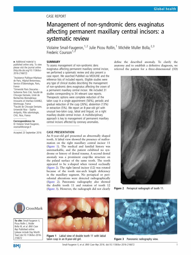
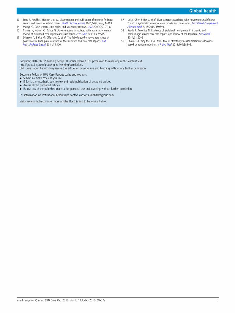
CASE PRESENTATIONAn 8-year-old girl presented an abnormally shapedtooth. A labial view showed the presence of malfor-mation on the right maxillary central incisor 11(figure 1). The medical and familial history wasunremarkable, and the patient exhibited no syn-drome or history of dental trauma. A second dentalanomaly was a prominent cusp-like structure onthe palatal surface of the same tooth. The toothappeared to be x-shaped when viewed occlusally(figure 2). The right lateral incisor (12) was rotatedbecause of the tooth size-arch length deficiencyin the maxillary segment. No periapical or peri-odontal alterations were detected radiographically(figure 2). Panoramic radiography also showedthe double tooth 11 and rotation of tooth 12(figure 3). However, the radiograph did not clearly
define the described anomaly. To clarify theanatomy and to establish a definitive diagnosis, wereferred the patient for a three-dimensional (3D)
Figure 1 Labial view of double tooth 11 with labialtalon cusp in an 8-year-old girl.
Figure 2 Periapical radiograph of tooth 11.
Figure 3 Panoramic radiography view.
Smail-Faugeron V, et al. BMJ Case Rep 2016. doi:10.1136/bcr-2016-216672 1
Global health

cone-beam CT (CBCT) of tooth 11. CBCT demonstrated thecomplex anatomy of tooth 11 and showed the radicularanatomy (figure 4A,B). The diagnosis was a double talon cusp(type 1) associated with a double tooth. The occlusion wasAngle’s Class I molar relationship on both sides.
For orthodontic purposes and complex anatomy, the treat-ment chosen was extraction of the tooth. Since the patient was
relatively young, we started periodic selective grinding of theaccessory cusp and application of fluoride varnish. When thepatient was 9 years old, the double tooth was extracted(figure 5A). After 5 days into the wound healing process, thetissue re-growth stage began (figure 5B). Seven days after extrac-tion, the sutures were removed and we took alginate impression.A transitional arc prosthesis sealed onto orthodontic bands wasplaced 15 days after extraction (figure 6A,B). Orthodontic treat-ment will begin when all primary teeth will have fallen out.
Figure 5 (A) Extracted tooth 11. (B) Frontal view 5 days afterextraction.
Figure 6 (A) External view of the temporary prosthesis. (B) Frontalview 2 weeks after extraction, with temporary prosthesis.
Figure 4 CBCT of tooth 11: (A) axial view and (B) frontal view. CBCT,cone-beam CT.
2 Smail-Faugeron V, et al. BMJ Case Rep 2016. doi:10.1136/bcr-2016-216672
Global health

GLOBAL HEALTH PROBLEM LISTDens evaginatus is a coronal anomaly referring to an accessorycusp-like structure projecting from the cingulum area or cemen-toenamel junction of an anterior tooth. The prevalence variesfrom 0.04% to 10% in permanent teeth.1 The permanent max-illary central incisor is the most affected tooth (50%).2 Hattabet al3 classified this anomaly into three types based on degree offormation and extension. Type 1 (true talon) is an additionalcusp that projects towards the palatal surface to at least half thelength between the cementoenamel junction and incisal edge(figure 7). Type 2 (semi talon) refers to an additional cusp of≥1 mm in length that extends less than half of the lengthbetween the cementoenamel junction and incisal edge (figure 8).Type 3 (trace talon) is a protruding cingulum that has a tubercle-like appearance (figure 9). Type 1 is the most frequentlydescribed (52%).4
The aetiology of dens evaginatus is unknown and could bemultifactorial, with genetic and environmental causes.5–8 Theseanomalies can induce carious lesion or pulpal necrosis, occlusalinterference, periodontal disease and poor aesthetics.9 10
Diagnosis and treatment planning are difficult for the dentist,and we lack consensus on management regardless of type.
Here, we systematically reviewed the current literaturedescribing management of permanent maxillary central incisorsaffected by non-syndromic dens evaginatus to analyse the differ-ent therapeutic options. As well, we report one case of this cor-onary anomaly.
GLOBAL HEALTH PROBLEM ANALYSISMATERIALS AND METHODSSystematic reviewA systematic review of the literature was performed.
Criteria for considering studies for this reviewEligible studies were published randomised controlled trial,comparative non-randomised study, cohort study, case series orcase report describing the management of non-syndromic densevaginatus affecting the crown of a permanent maxillary centralincisor.
Search methods for identification of studiesTo identify studies, we searched the database MEDLINE viaPubMed for articles published in English and French, with norestriction on date of publication. Search equation combinedfree text words and controlled vocabulary pertaining to the con-dition and interventions (see online supplementary file 1). Thelast search for articles was in April 2015. We checked the refer-ences of all eligible articles for relevant studies and scanned ref-erence lists from identified review articles for further studies.We searched ClinicalTrials.gov for the protocols of includedstudies and to identify ongoing trials.11
Data collectionTwo authors independently and in duplicate screened the titleand abstract of records retrieved by the search, then screenedthe selected full-text reports. Any disagreements were resolvedby discussion. Finally, we included studies after eliminatingduplicate publications.
For each randomised controlled trial, two authors independ-ently and in duplicate recorded the year of publication, inclu-sion/exclusion criteria specified, number of arms in the trial,treatments compared, detailed description of interventions,number of patients enrolled, number of treated teeth, mean ageof participants, duration of follow-up and outcome data. Weassessed the risk of bias for each trial by the CochraneCollaboration Risk of Bias tool, which includes the followingitems: selection of participants, blinding of participants and per-sonnel, blinding of outcome assessors, incomplete outcome dataand selective outcome reporting.12 For non-randomised studies,we considered the risk of selection bias as high and also assessed
Figure 7 True talon (type 1 according to Hattab’s classification) onthe labial aspect of a right maxillary central incisor.
Figure 9 Trace talon (type 3 according to Hattab’s classification) onthe lingual aspect of a left maxillary lateral incisor.
Figure 8 Semi talon (type 2 according to Hattab’s classification) onthe lingual aspect of a left maxillary lateral incisor.
Smail-Faugeron V, et al. BMJ Case Rep 2016. doi:10.1136/bcr-2016-216672 3
Global health

the risk of bias related to confounding factors. For each rando-mised or non-randomised trial, each domain was rated as low,high or unclear risk of bias. Then, each study was assigned anoverall risk of bias score: low risk (low for all key domains),high risk (high for one key or more domains) or unclear risk(unclear for one key or more domains). The two review authorscompared evaluations and resolved any disagreements by discus-sion. For each cohort study, case series or case report, twoauthors independently and in duplicate recorded the year ofpublication, gender and age of patients, teeth involved, type ofdens evaginatus according to the Hattab et al classification,pulpal anatomy for the double tooth (number of the pulpalchambers and number of the roots), presence or absence of aes-thetic, periodontal, occlusal or caries problems, presence orabsence of associated anomaly, detailed description of treatmentand requirement of endodontic treatment and orthodonticmanagement.
AnalysisWe did not perform any meta-analysis, but we described thestudy characteristics and results qualitatively with number, per-centage or mean (min–max).
RESULTSSystematic reviewLiterature searchThe search yielded 640 potentially eligible articles. We included31 articles corresponding to 34 case reports.1 13–42 The flow ofarticle selection is in figure 10. We found no published rando-mised or non-randomised trial, no published cohort study andno protocol of ongoing trials at ClinicalTrials.gov.
The median year of publication was 2005 (range 1971–2014); 9 articles were published before 2000, 15 between 2000and 2010 and 7 between 2010 and 2014.
Reported casesA total of 21 cases involved men (60%). Across the 34 casereports, the mean age of the patients was 13.6 years. The agerange varied across studies from 7 years to 47 years. Dens evagi-natus was bilateral in 5 cases (15%), so 39 incisors were affectedby dens evaginatus. Among the 34 cases, 12 featured dens evagi-natus associated with double tooth.16 20–23 26 29 31 34 39 40
According to the Hattab et al classification, the anomaly wastype 1 for 32 incisors (82%), type 2 for 2 (5%) and type 3 for 2(5%); we had insufficient information to characterise the typefor 3 incisors. Localisation of dens evaginatus was lingual for 35incisors (90%), and the condition caused occlusal interferencefor 25 (74%). Caries or pulpal necrosis and periodontal diseasewere detected in nine and four incisors, respectively (33%). Forfive teeth, the patient considered the abnormality unattractive(13%). Details and references of corresponding case reports aregiven in online supplementary file 2.
Therapeutic options for treating dens evaginatus are pre-sented in figure 11. Details and references of correspondingcase reports are given in online supplementary file 3.
For 13% of patients, the tooth with dens evaginatus withoutocclusal interference, irritation on the soft tissue, poor aesthet-ics, caries or pulpal necrosis did not need treatment. For 87%of cases, the following two options were described.
Figure 11 Therapeutic options forthe 39 permanent maxillary centralincisors reported in the 34 includedcase reports.
Figure 10 Systematic review flow chart of case reports describing themanagement of non-syndromic dens evaginatus affecting the crown ofa permanent maxillary central incisor.
4 Smail-Faugeron V, et al. BMJ Case Rep 2016. doi:10.1136/bcr-2016-216672
Global health

First, if radiographic examination showed the presence ofenamel, dentin and pulp horn into the talon cusp, there weretwo therapeutic options. For nine incisors, the treatment planwas a gradual reduction of the talon cusp on consecutive visitsand an application of a desensitising agent (fluoride) at eachvisit; for five of these incisors, the desensitising agent wasapplied without other treatment, and for one case, pulpal necro-sis appeared after application of the desensitising agent. For theother four incisors, the desensitising agent was applied, followedby direct restoration at the final visit. With the second option,five patients were scheduled for complete reduction of the taloncusp in a single appointment; three incisors underwent directpulp capping with mineral trioxide aggregate or calciumhydroxide followed by a final restoration with glass–ionomercement then resin composite; one incisor underwent pulpotomywith formocresol and calcium hydroxide followed by resin com-posite application and the final incisor underwent pulpectomy.
In the second main treatment option, if radiographic examin-ation did not show the presence of a pulp horn into the taloncusp, patients were scheduled for complete reduction of thetalon cusp in a single appointment (18 incisors) or extraction ofthe incisor (2 double incisors exhibiting type 2 dens evaginatus).Among patients scheduled for complete reduction of the cusp,direct tooth restoration was performed in cases of pulpal vitality(11 incisors) and endodontic treatment followed by direct orindirect restoration in cases of irreversible pulpitis or pulpalnecrosis (7 incisors).
Orthodontic management achieved aesthetics and occlusionin 29% of cases. In addition, 15 case reports reported the moni-toring of the patient. The duration of follow-up was 12 monthsin median (mean 13 months, min–max 3–24 months).
DISCUSSIONTo the best of our knowledge, no recent study has systematicallyreviewed the literature on the management of non-syndromicdens evaginatus affecting the permanent maxillary centralincisor. Moreover, our case report illustrated the need for earlyand correct diagnosis of a talon cusp, which requires an indivi-dualised treatment plan and a multidisciplinary approach.
Dens evaginatus is a disturbance in tooth development thatproduces a tubercle of hard tissue on the surface of the tooth.Our results are similar to those of other authors, who reportedtype 1 as the most frequent.4 The accessory cusp is most com-monly located on the lingual aspect of an anterior tooth (90%)but can also be on the labial aspect or on the lingual and labialaspects of the same tooth.
On the basis of limited evidence (only case reports), ourresults show that dentists now have a broad range of manage-ment options for similar coronary anomalies involving a centralincisor depending on a patient’s pulpal features. The manage-ment of a talon cups includes no treatment (13%); sequentialgrinding allowing for tertiary dentin deposition, which may alsobe considered to remove occlusal interferences (26%); restora-tive treatment (28%); pulp therapy (28%) and extraction of theaffected tooth (5%). We could add a preventive therapeuticoption: pit and fissure sealants (not recorded in our selectedcase reports). Besides pulpal and radicular anatomy and treat-ment options, the choice of these treatments could relate to thedegree of patient cooperation.43 In addition, only 15 casereports reported the monitoring of the patient. The duration offollow-up was 12 months in median (mean 13 months, min–max 3–24 months). The therapeutic option failed in one casewith a pulpal necrosis appearing after application of the desensi-tising agent. The other 19 included case reports did not report
the monitoring of the patient. Consequently, we do not know ifa therapeutic option failed or not.
Periapical radiography is commonly used for diagnosis andprovides important information about the root anatomy.Nevertheless, periapical radiographs are 2D dental imaging andare not accurate or conclusive.20 44 Moreover, CBCT providessubmillimetre spatial resolution with short exposure times in therange of 20 s and radiation exposure similar to a full mouthseries.45–48 Therefore, CBCT should be systematically per-formed. CBCT provides 3D dental imaging and can help deter-mine the best treatment.49 50 CBCT allows for visualising anypulpal extension into the dens evaginatus. However, we foundonly two articles, published in 2014, describing the perform-ance of CBCT.39 42 About 30% of included articles were pub-lished before 2000, which could explain the low use of CBCT.
Our clinical case illustrates these results with an accurate diag-nosis from a CBCT and follow-up. We performed extraction ofthe double tooth with dens evaginatus (then followed by anorthodontic management planned) as 5% of cases reported inour systematic review. Another therapeutic option would be dif-ficult to plan, whether for aesthetic, endodontic or orthodonticproblems. First, the type of dens evaginatus was three accordingto Hattab et al classification, and its localisation was lingual andlabial. Moreover, complex anatomy of tooth 11 revealed byCBCT was contrary to perform an orthodontic treatment.Finally, the condition caused occlusal interference, and patientneeded orthodontic management. Thus, we think that extrac-tion of the tooth was necessary in any cases to avoid aesthetic,endodontic or occlusal problems. We did not perform a histo-logical examination of the extracted double tooth, but it wouldhave been interesting to send the extracted tooth to an histo-pathological laboratory for examination of hard and soft tissues.
Our study has some limitations. First, we identified only casereports with overall low quality of evidence and no randomisedtrial. Thus, the true clinical relevance of the findings is some-what lacking. Moreover, we searched only one database and wedid not search ‘grey’ literature, and we acknowledge thatunidentified studies may exist. However, our search strategywas extensive and we consulted the largest trial registryClinicalTrials.gov run by the US National Library of Medicine atthe National Institutes of Health51 to find unpublished rando-mised trials and hence the risk of publication bias in our studyis low.52 53 In addition, in spite of the fact that systematicreviews of randomised controlled trials provide the most reliableevidence about the effects of healthcare interventions, systematicreviews of case reports can be performed in some circumstancesto highlight a mistreated specific medical area and stimulatefurther investigation.54 For example, some authors have per-formed systematic reviews of case reports for cases that are outof the ordinary or for the conditions where randomised con-trolled trials were difficult or impossible to achieve.55–58 Ourremarks could have implications for future research. Decisionsabout which treatment is best are driven by the results of rando-mised trials and systematic reviews.59 In this regard, high-qualityrandomised trials controlling for clinically relevant parametersand constraints are needed to determine what therapeuticoption is a worthwhile clinical procedure for treating non-syndromic dens evaginatus affecting permanent maxillarycentral incisors.
CONCLUSIONSA talon cusp is associated with problems such as compromisedaesthetics, occlusal interference, tooth displacement, caries andperiodontal problems. A correct and early diagnosis can prevent
Smail-Faugeron V, et al. BMJ Case Rep 2016. doi:10.1136/bcr-2016-216672 5
Global health

these complications. The presence of a talon cusp is not alwaysan indication for dental treatment, but the association of taloncusp with other dental abnormalities suggests a specific diagno-sis for adequate care and a multidisciplinary approach.
Learning points
▸ A dens evaginatus is among the most challenging problemsin dentistry.
▸ Owing to additional cusp(s) with or without pulp horn aswell as misalignment, treatment usually requires amultidisciplinary approach to address endodontic andaesthetic considerations.
▸ Since two-dimensional periapical radiographs cannot betotally conclusive for the diagnosis, CBCT should besystematically performed in all cases because it providesthree-dimensional dental imaging to help determine the besttreatment.
Acknowledgements The authors thank Laura Smales (BioMedEditing, Toronto,Canada) for editing the manuscript.
Contributors VS-F and JPC contributed to conception and design, acquisition ofdata or analysis and interpretation of data. VS-F, MMB and FC involved in draftingthe manuscript or revising it critically for important intellectual content. VS-F, JPC,MMB and FC provided final approval of the version published, are accountable forthe paper and ensure that all questions regarding the accuracy or integrity of thepaper are investigated and resolved.
Competing interests None declared.
Patient consent Obtained.
Provenance and peer review Not commissioned; externally peer reviewed.
REFERENCES1 Tulunoglu O, Cankala DU, Ozdemir RC. Talon’s cusp: report of four unusual cases.
J Indian Soc Pedod Prev Dent 2007;25:52–5.2 Sharma G, Nagpal A. Talon cusp: a prevalence study of its types in permanent
dentition and report of a rare case of its association with fusion in mandibularincisor. J Oral Dis 2014;2014.
3 Hattab FN, Yassin OM, al-Nimri KS. Talon cusp in permanent dentition associatedwith other dental anomalies: review of literature and reports of seven cases. ASDCJ Dent Child 1996;63:368–76.
4 Arfat B, Çolak H, Çelebi A, et al. The frequency and characteristics of talon cusps ina Turkish population. Eur J Gen Dent 2012;1:39–43. http://dx.doi.org/10.4103/2278-9626.101357
5 Gupta S, Tandon A, Chandra A, et al. Syndontia with talon cusp. J Oral MaxillofacPathol 2012;16:266–71.
6 Hattab FN, Yassin OM, al-Nimri KS. Talon cusp—clinical significance andmanagement: case reports. Quintessence Int 1995;26:115–20.
7 Lee CK, King NM, Lo EC, et al. Talon cusp in the primary dentition: literaturereview and report of three rare cases. J Clin Pediatr Dent 2006;30:299–305.
8 Mallineni SK, Manan NM, Lee CK, et al. Talon cusp affecting primary dentition intwo siblings: a case report. Rom J Morphol Embryol 2013;54:211–13.
9 Altug-Atac AT, Erdem D. Prevalence and distribution of dental anomalies inorthodontic patients. Am J Orthod Dentofacial Orthop 2007;131:510–14.
10 Türkaslan S, Gökce HS, Dalkız M. Esthetic rehabilitation of bilateral geminatedteeth: a case report. Eur J Dent 2007;1:188–91.
11 Chan AW, Altman DG. Identifying outcome reporting bias in randomised trials onPubMed: review of publications and survey of authors. BMJ 2005;330(7494):753.
12 Higgins JP, Altman DG, Gøtzsche PC, et al. The Cochrane Collaboration’s tool forassessing risk of bias in randomised trials. BMJ 2011;343:d5928.
13 Asadi SG, Asadi ZG. Presentation and management of talon cusp. J Pak Med Assoc1998;48:314–15.
14 Bolan M, Gerent Petry Nunes AC, de Carvalho Rocha MJ, et al. Talon cusp: reportof a case. Quintessence Int 2006;37:509–14.
15 Davis PJ, Brook AH. The presentation of talon cusp: diagnosis, clinical features,associations and possible aetiology. Br Dent J 1986;160:84–8.
16 Denyar BE. Microcomposite resin restoration of a “geminated” tooth. Ont Dent1982;59:20–1, 24.
17 Ferraz JA, de Carvalho Júnior JR, Saquy PC, et al. Dental anomaly: dens evaginatus(talon cusp). Braz Dent J 2001;12:132–4.
18 Glavina D, Skrinjarić T. Labial talon cusp on maxillary central incisors: a raredevelopmental dental anomaly. Coll Antropol 2005;29:227–31.
19 Gosselin ML, Doyle T, MacLellan J, et al. A talon cusp mistaken for a mesiodens:case report. J Can Dent Assoc 2012;78:c6.
20 Gündüz K, Açikgõz A. An unusual case of talon cusp on a geminated tooth. BrazDent J 2006;17:343–6.
21 Harris WE. Endodontic treatment of a fused mesiodens: report of case. J Am DentAssoc 1971;83:643–6.
22 Hattab FN. Double talon cusps on supernumerary tooth fused to maxillary centralincisor: review of literature and report of case. J Clin Exp Dent 2014;6:e400–7.
23 Hattab FN, Hazza’a AM. An unusual case of talon cusp on geminated tooth. J CanDent Assoc 2001;67:263–6.
24 Henderson HZ. Talon cusp: a primary or a permanent incisor anomaly. J IndianaDent Assoc 1977;56:45–6.
25 Jowharji N, Noonan RG, Tylka JA. An unusual case of dental anomaly: a “facial”talon cusp. ASDC J Dent Child 1992;59:156–8.
26 Lomçali G, Hazar S, Altinbulak H. Talon cusp: report of five cases. Quintessence Int1994;25:431–3.
27 Maroto M, Barbería E, Arenas M, et al. Displacement and pulpal involvement of amaxillary incisor associated with a talon cusp: report of a case. Dent Traumatol2006;22:160–4.
28 McKaig SJ, Shaw L. Dens evaginatus on the labial surface of a central incisor: acase report. Dent Update 2001;28:210–12.
29 Miri SS, Ghorbani H, Rashed Mohassel A. Endodontic treatment of fused teeth withtalon cusp. Case Rep Dent 2014;2014:738185.
30 Patil R, Singh S, Subba Reddy VV. Labial talon cusp on permanent central incisor: acase report. J Indian Soc Pedod Prev Dent 2004;22:30–2.
31 Peker I, Alkurt MT. Talon cusp: a case series. Gen Dent 2009;57:524–7.32 Ragno JR, Jr. Dens evaginatus of a central incisor in a black American female. Gen
Dent 1986;34:372–3.33 Rantanen AV. Talon cusp. Oral Surg Oral Med Oral Pathol 1971;32:398–400.34 Sener S, Unlu N, Basciftci FA, et al. Bilateral geminated teeth with talon cusps: a
case report. Eur J Dent 2012;6:440–4.35 Shetty P, Xavier AM. Management of a talon cusp using mineral trioxide aggregate.
Int Endod J 2011;44:1061–8.36 Shey Z, Eytel R. Clinical management of an unusual case of dens evaginatus in a
maxillary central incisor. J Am Dent Assoc 1983;106:346–8.37 Soares AB, de Araújo JJ, de Sousa SM, et al. Bilateral talon cusp: case report.
Quintessence Int 2001;32:283–6.38 Sumer AP, Zengin AZ. An unusual presentation of talon cusp: a case report. Br
Dent J 2005;199:429–30.39 Tarım Ertaş E, Yırcalı Aticı M, Arslan H, et al. Endodontic treatment and esthetic
management of a geminated central incisor bearing a talon cusp. Case Rep Dent2014;2014:123681.
40 Tomazinho FS, Baratto-Filho F, Leonardi DP, et al. Occurrence of talon cusp on ageminated maxillary central incisor: a case report. J Oral Sci 2009;51:297–300.
41 Vasudev SK, Goel BR. Endodontic management of dens evaginatus of maxillarycentral incisors: a rare case report. J Endod 2005;31:67–70.
42 Yazıcıoğlu O, Ulukapı H. Management of a facial talon cusp on a maxillarypermanent central incisor: a case report and review of the literature. J Esthet RestorDent 2014;26:374–81.
43 Olivan-Rosas G, Lopez-Jimenez J, Gimenez-Prats MJ, et al. Considerations anddifferences in the treatment of a fused tooth. Med Oral 2004;9(3):224–8.
44 Sivolella S, Bressan E, Mirabal V, et al. Extraoral endodontic treatment, odontotomyand intentional replantation of a double maxillary lateral permanent incisor: casereport and 6-year follow-up. Int Endod J 2008;41:538–46.
45 Danforth RA, Clark DE. Effective dose from radiation absorbed during a panoramicexamination with a new generation machine. Oral Surg Oral Med Oral Pathol OralRadiol Endod 2000;89:236–43.
46 Gibbs SJ. Effective dose equivalent and effective dose: comparison for commonprojections in oral and maxillofacial radiology. Oral Surg Oral Med Oral Pathol OralRadiol Endod 2000;90:538–45.
47 Jung MS, Lee SP, Kim GT, et al. Three-dimensional analysis of deciduous maxillaryanterior teeth using cone-beam computed tomography. Clin Anat 2012;25:182–8.
48 Mah JK, Danforth RA, Bumann A, et al. Radiation absorbed in maxillofacialimaging with a new dental computed tomography device. Oral Surg Oral Med OralPathol Oral Radiol Endod 2003;96:508–13.
49 Castro IO, Estrela C, Souza VR, et al. Unilateral fusion of maxillary lateral incisor:diagnosis using cone beam computed tomography. Case Rep Dent 2014;2014:934218.
50 Lucey S, Heath N, Welbury RR, et al. Case report: cone-beam CT imaging in themanagement of a double tooth. Eur Arch Paediatr Dent 2009;10(Suppl 1):49–53.
51 Zarin DA, Tse T, Ide NC. Trial Registration at ClinicalTrials.gov between May andOctober 2005. N Engl J Med 2005;353:2779–87.
52 Dwan K, Altman DG, Arnaiz JA, et al. Systematic review of the empirical evidenceof study publication bias and outcome reporting bias. PLoS One 2008;3:e3081.
6 Smail-Faugeron V, et al. BMJ Case Rep 2016. doi:10.1136/bcr-2016-216672
Global health
53 Song F, Parekh S, Hooper L, et al. Dissemination and publication of research findings:an updated review of related biases. Health Technol Assess 2010;14:iii, ix–xi, 1–193.
54 Martyn C. Case reports, case series and systematic reviews. QJM 2002;95:197–8.55 Cramer H, Krucoff C, Dobos G. Adverse events associated with yoga: a systematic
review of published case reports and case series. PLoS One 2013;8:e75515.56 Driessen A, Balke M, Offerhaus C, et al. The fabella syndrome—a rare cause of
posterolateral knee pain: a review of the literature and two case reports. BMCMusculoskelet Disord 2014;15:100.
57 Lei X, Chen J, Ren J, et al. Liver damage associated with Polygonum multiflorumThunb: a systematic review of case reports and case series. Evid Based ComplementAlternat Med 2015;2015:459749.
58 Saada F, Antonios N. Existence of ipsilateral hemiparesis in ischemic andhemorrhagic stroke: two case reports and review of the literature. Eur Neurol2014;71:25–31.
59 Chalmers I. Why the 1948 MRC trial of streptomycin used treatment allocationbased on random numbers. J R Soc Med 2011;104:383–6.
Copyright 2016 BMJ Publishing Group. All rights reserved. For permission to reuse any of this content visithttp://group.bmj.com/group/rights-licensing/permissions.BMJ Case Report Fellows may re-use this article for personal use and teaching without any further permission.
Become a Fellow of BMJ Case Reports today and you can:▸ Submit as many cases as you like▸ Enjoy fast sympathetic peer review and rapid publication of accepted articles▸ Access all the published articles▸ Re-use any of the published material for personal use and teaching without further permission
For information on Institutional Fellowships contact [email protected]
Visit casereports.bmj.com for more articles like this and to become a Fellow
Smail-Faugeron V, et al. BMJ Case Rep 2016. doi:10.1136/bcr-2016-216672 7
Global health