Case Report Fetal Rhabdomyoma of the Right Tonsil with...
4
Case Report Fetal Rhabdomyoma of the Right Tonsil with Polyp-Like Appearance Ching-Ping Wang, 1 Yi-Hao Chang, 1 and Ya-Ting Chang 2 1 Department of Otolaryngology, Taichung Veterans General Hospital, 1650 Taiwan Boulevard Section 4, Taichung 40705, Taiwan 2 Department of Stomatology, Taichung Veterans General Hospital, 1650 Taiwan Boulevard Section 4, Taichung 40705, Taiwan Correspondence should be addressed to Yi-Hao Chang; [email protected] Received 13 February 2015; Revised 15 June 2015; Accepted 16 June 2015 Academic Editor: Abr˜ ao Rapoport Copyright © 2015 Ching-Ping Wang et al. is is an open access article distributed under the Creative Commons Attribution License, which permits unrestricted use, distribution, and reproduction in any medium, provided the original work is properly cited. Skeletal muscle neoplasms, in contrast to other groups of tumors, are almost malignant. e benign variant, rhabdomyoma, is distinctly rare. Rhabdomyomas can be classified generally into two types: cardiac and extracardiac. Extracardiac rhabdomyoma can be further divided into three subtypes: adult, fetal, and genital type. Adult rhabdomyoma is the most common subtype of rhabdomyoma even though it remains relatively rare. Fetal rhabdomyomas are less common than the adult type. In this paper we report a rare case of a fetal rhabdomyoma with polyp-like appearance originating from right tonsil. Punch biopsy and then right tonsillectomy were performed for complete excision. ere was no obvious recurrence. 1. Introduction e term “rhabdomyoma” was introduced by Zenker in 1864. It was used to describe a benign tumor showing muscle cells with different degrees of differentiation and maturity. Rhabdomyomas are rare benign tumor consisting of striated muscle. ey can be classified generally into two types: cardiac and extracardiac [1]. Most rhabdomyomas arise from cardiac muscle. Cardiac rhabdomyoma occurs almost exclusively in the pediatric age group and may be associated with tuberous sclerosis, neurofibromatosis, and sebaceous adenomas [2]. Extracardiac rhabdomyoma can be further divided into three subtypes (adult, fetal, and genital) with regard to clinical and morphological differences, although some overlapping exists. Genital rhabdomyoma is seen most frequently in the vagina or vulva of young and middle-aged women. Fetal and adult rhabdomyoma are located predominantly in the head and neck region; the former occurs primarily in the subcutaneous tissue of the head and neck of male infants usually younger than 3 years, and the latter is characterized by a slowly growing mass typically seen in the head and neck of elderly patient, with a male predominance. Seventy- seven percent of all extracardiac rhabdomyomas occur in the head and neck and 14% occur in the genital region [3]. Adult rhabdomyoma is the most common subtype of rhabdomyoma even though it remains relatively rare. Fetal rhabdomyomas are less common than the adult type. In this paper we report a rare case of a fetal rhabdomyoma with polyp-like appearance originating from right tonsil. 2. Case report A 57-year-old patient presented to our otolaryngology department on August 17, 2009, with a small painless polyp which he has had over right tonsil for one month. is patient had past history of diabetes mellitus under regular oral hypoglycemic agents control for more than 5 years. He denied any other medical or surgical history. Physical examination revealed a smooth surface polyp about 1.4 × 1.0 × 0.5 cm 3 over right tonsil without bleeding or ulceration (Figure 1); there was no other oral lesion or palpable neck lymph node. Fiberoptic nasolaryngoscopy was performed and the finding was unremarkable. We thought the polyp was a benign lesion, so we did not pay much attention to it and did not take a photo for documentation. We performed punch biopsy under local anesthesia. en histopathological Hindawi Publishing Corporation Case Reports in Otolaryngology Volume 2015, Article ID 713278, 3 pages http://dx.doi.org/10.1155/2015/713278
-
Upload
duongtuyen -
Category
Documents
-
view
220 -
download
0
Transcript of Case Report Fetal Rhabdomyoma of the Right Tonsil with...

Case ReportFetal Rhabdomyoma of the Right Tonsil withPolyp-Like Appearance
Ching-Ping Wang,1 Yi-Hao Chang,1 and Ya-Ting Chang2
1Department of Otolaryngology, Taichung Veterans General Hospital, 1650 Taiwan Boulevard Section 4, Taichung 40705, Taiwan2Department of Stomatology, Taichung Veterans General Hospital, 1650 Taiwan Boulevard Section 4, Taichung 40705, Taiwan
Correspondence should be addressed to Yi-Hao Chang; [email protected]
Received 13 February 2015; Revised 15 June 2015; Accepted 16 June 2015
Academic Editor: Abrao Rapoport
Copyright © 2015 Ching-Ping Wang et al. This is an open access article distributed under the Creative Commons AttributionLicense, which permits unrestricted use, distribution, and reproduction in any medium, provided the original work is properlycited.
Skeletal muscle neoplasms, in contrast to other groups of tumors, are almost malignant. The benign variant, rhabdomyoma, isdistinctly rare. Rhabdomyomas can be classified generally into two types: cardiac and extracardiac. Extracardiac rhabdomyomacan be further divided into three subtypes: adult, fetal, and genital type. Adult rhabdomyoma is the most common subtype ofrhabdomyoma even though it remains relatively rare. Fetal rhabdomyomas are less common than the adult type. In this paper wereport a rare case of a fetal rhabdomyoma with polyp-like appearance originating from right tonsil. Punch biopsy and then righttonsillectomy were performed for complete excision. There was no obvious recurrence.
1. Introduction
The term “rhabdomyoma” was introduced by Zenker in1864. It was used to describe a benign tumor showingmuscle cells with different degrees of differentiation andmaturity. Rhabdomyomas are rare benign tumor consistingof striated muscle. They can be classified generally into twotypes: cardiac and extracardiac [1].Most rhabdomyomas arisefrom cardiac muscle. Cardiac rhabdomyoma occurs almostexclusively in the pediatric age group and may be associatedwith tuberous sclerosis, neurofibromatosis, and sebaceousadenomas [2].
Extracardiac rhabdomyoma can be further divided intothree subtypes (adult, fetal, and genital) with regard to clinicaland morphological differences, although some overlappingexists. Genital rhabdomyoma is seen most frequently in thevagina or vulva of young and middle-aged women. Fetaland adult rhabdomyoma are located predominantly in thehead and neck region; the former occurs primarily in thesubcutaneous tissue of the head and neck of male infantsusually younger than 3 years, and the latter is characterizedby a slowly growing mass typically seen in the head andneck of elderly patient, with a male predominance. Seventy-seven percent of all extracardiac rhabdomyomas occur in
the head and neck and 14% occur in the genital region[3]. Adult rhabdomyoma is the most common subtype ofrhabdomyoma even though it remains relatively rare. Fetalrhabdomyomas are less common than the adult type. In thispaper we report a rare case of a fetal rhabdomyoma withpolyp-like appearance originating from right tonsil.
2. Case report
A 57-year-old patient presented to our otolaryngologydepartment on August 17, 2009, with a small painless polypwhich he has had over right tonsil for one month. Thispatient had past history of diabetes mellitus under regularoral hypoglycemic agents control for more than 5 years.He denied any other medical or surgical history. Physicalexamination revealed a smooth surface polyp about 1.4 ×1.0 × 0.5 cm3 over right tonsil without bleeding or ulceration(Figure 1); there was no other oral lesion or palpable necklymph node. Fiberoptic nasolaryngoscopy was performedand the finding was unremarkable. We thought the polypwas a benign lesion, so we did not pay much attention to itand did not take a photo for documentation. We performedpunch biopsy under local anesthesia. Then histopathological
Hindawi Publishing CorporationCase Reports in OtolaryngologyVolume 2015, Article ID 713278, 3 pageshttp://dx.doi.org/10.1155/2015/713278
2 Case Reports in Otolaryngology
Figure 1: A polyp over right tonsil.
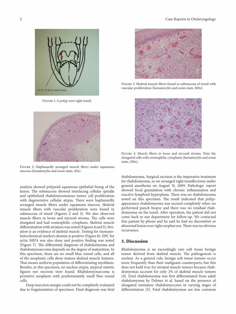
Figure 2: Haphazardly arranged muscle fibers under squamousmucosa (hematoxylin and eosin stain, 40x).
analysis showed polypoid squamous epithelial lining of thelesion. The submucosa showed interlacing cellular spindleand epithelioid rhabdomyomatous tumor cell proliferationwith degenerative cellular atypia. There were haphazardlyarranged muscle fibers under squamous mucosa. Skeletalmuscle fibers with vascular proliferation were found insubmucosa of tonsil (Figures 2 and 3). We also observedmuscle fibers in loose and myxoid stroma. The cells wereelongated and had eosinophilic cytoplasm. Skeletal muscledifferentiationwith striationwas noted (Figures 4 and 5). Stri-ation is an evidence of skeletal muscle. Testing for immuno-histochemical markers desmin is positive (Figure 6). IHC foractin M874 was also done and positive finding was noted(Figure 7). The differential diagnosis of rhabdomyoma andrhabdomyosarcoma depends on the degree of maturation. Inthis specimen, there are no small blue round cells, and allof the neoplastic cells show mature skeletal muscle features.That means uniform population of differentiating myoblasts.Besides, in this specimen, no nuclear atypia, atypical mitoticfigures nor necrosis were found. Rhabdomyosarcoma isprimitive neoplasm with predominantly small blue roundcells.
Deep resectionmargin could not be completely evaluateddue to fragmentation of specimen. Final diagnosis was fetal
Figure 3: Skeletal muscle fibers found in submucosa of tonsil withvascular proliferation (hematoxylin and eosin stain, 100x).
Figure 4: Muscle fibers in loose and myxoid stroma. Note theelongated cells with eosinophilic cytoplasm (hematoxylin and eosinstain, 200x).
rhabdomyoma. Surgical excision is the imperative treatmentfor rhabdomyoma, so we arranged right tonsillectomy undergeneral anesthesia on August 31, 2009. Pathologic reportshowed focal granulation with chronic inflammation andreactive lymphoid hyperplasia. There was no rhabdomyomanoted on this specimen. The result indicated that polyp-appearance rhabdomyoma was excised completely when weperformed punch biopsy and there was no residual rhab-domyoma on the tonsil. After operation, the patient did notcome back to our department for follow-up. We contactedthis patient by phone and he said he had no discomfort orabnormal lesion over right oropharynx.Therewas no obviousrecurrence.
3. Discussion
Rhabdomyoma is an exceedingly rare soft tissue benigntumor derived from skeletal muscle. The pathogenesis isunclear. As a general rule, benign soft tissue tumors occurmore frequently than their malignant counterparts, but thisdoes not hold true for striated muscle tumors because rhab-domyomas account for only 2% of skeletal muscle tumors[4]. Fetal rhabdomyoma was first differentiated from adultrhabdomyoma by Dehner et al. based on the presence ofelongated immature rhabdomyocytes in varying stages ofdifferentiation [5]. Fetal rhabdomyomas are less common

Case Reports in Otolaryngology 3
Figure 5: Skeletal muscle differentiation with striation is noted(hematoxylin and eosin stain, 400x).
Figure 6: IHC stain for desmin is positive (hematoxylin and eosinstain, ×400).
than the adult type. The fetal type occurs primarily in thesubcutaneous tissue of the head and neck of male infantsyounger than 3 years old. In this presented case, the onset age,location, and polyp-like appearance were all unusual. In theliterature, no other case of fetal rhabdomyoma over tonsil hasbeen reported.
Regarding therapeutic options for this tumor, the goldstandard of treatment is surgery. Rhabdomyoma mayrecur, but no cases of malignant transformation have beendescribed in the literature. Recurrence is probably due toincomplete excision or multifocality. When total excisioncannot be accomplished, reoperation or narrow follow-upis indicated to prevent advanced revision surgeries. In thispresented case, complete excision was done, and there wasno obvious recurrence.
4. Conclusion
Skeletal muscle neoplasms, in contrast to other groups oftumors, are almost always malignant. The benign variant,rhabdomyoma, is distinctly rare. Our patient was a rare caseof fetal rhabdomyoma located in right tonsil. Initially wethought it was a polyp over right tonsil and performed punchbiopsy. Pathology report showed fetal rhabdomyoma, and
Figure 7: IHC stain for actin M874 is positive (hematoxylin andeosin stain, ×400).
curative treatment was obtained by surgery. There was noobvious recurrence.
Although rhabdomyoma of tonsil is a rare disease, weshould keep inmind this differential diagnosis when wemeetpatient with polyp over tonsil or other H&N areas. Be sure totake photodocumentation routinely before punch biopsy orexcision even though it looks like benign lesion. If you meetpatient with rhabdomyoma in the future, surgical excisionis an imperative treatment. Recurrence rate is extremely lowwhen complete excision is done.
Conflict of Interests
The authors declare that there was no conflict of interestsregarding the publishing of this paper.
References
[1] B. Pichi, V.Manciocco, P.Marchesi et al., “Rhabdomyoma of theparapharyngeal space presenting with dysphagia,” Dysphagia,vol. 23, no. 2, pp. 202–204, 2008.
[2] C. O. Reid and C. J. Smith, “Rhabdomyoma of the floor ofthe mouth: a new case and review of recently reported intra-oral rhabdomyomas,”TheBritish Journal of Oral&MaxillofacialSurgery, vol. 23, no. 4, pp. 284–291, 1985.
[3] A. Amelia Souza, V. C. de Araujo, F. Passador Santos et al.,“Intraoral adult rhabdomyoma: a case report,” Case Reports inDentistry, vol. 2013, Article ID 741548, 5 pages, 2013.
[4] J. S. Jimenez, A. D. Ferrer, F. A. Granados et al., “Adultrhabdomyoma in the masticatory area. New case presentationand review of the literature,”Medicina Oral, vol. 6, no. 1, pp. 64–68, 2001.
[5] L. P. Dehner, F. M. Enzinger, and R. L. Font, “Fetal rhabdomy-oma. An analysis of nine cases,” Cancer, vol. 30, no. 1, pp. 160–166, 1972.

Submit your manuscripts athttp://www.hindawi.com
Stem CellsInternational
Hindawi Publishing Corporationhttp://www.hindawi.com Volume 2014
Hindawi Publishing Corporationhttp://www.hindawi.com Volume 2014
MEDIATORSINFLAMMATION
of
Hindawi Publishing Corporationhttp://www.hindawi.com Volume 2014
Behavioural Neurology
EndocrinologyInternational Journal of
Hindawi Publishing Corporationhttp://www.hindawi.com Volume 2014
Hindawi Publishing Corporationhttp://www.hindawi.com Volume 2014
Disease Markers
Hindawi Publishing Corporationhttp://www.hindawi.com Volume 2014
BioMed Research International
OncologyJournal of
Hindawi Publishing Corporationhttp://www.hindawi.com Volume 2014
Hindawi Publishing Corporationhttp://www.hindawi.com Volume 2014
Oxidative Medicine and Cellular Longevity
Hindawi Publishing Corporationhttp://www.hindawi.com Volume 2014
PPAR Research
The Scientific World JournalHindawi Publishing Corporation http://www.hindawi.com Volume 2014
Immunology ResearchHindawi Publishing Corporationhttp://www.hindawi.com Volume 2014
Journal of
ObesityJournal of
Hindawi Publishing Corporationhttp://www.hindawi.com Volume 2014
Hindawi Publishing Corporationhttp://www.hindawi.com Volume 2014
Computational and Mathematical Methods in Medicine
OphthalmologyJournal of
Hindawi Publishing Corporationhttp://www.hindawi.com Volume 2014
Diabetes ResearchJournal of
Hindawi Publishing Corporationhttp://www.hindawi.com Volume 2014
Hindawi Publishing Corporationhttp://www.hindawi.com Volume 2014
Research and TreatmentAIDS
Hindawi Publishing Corporationhttp://www.hindawi.com Volume 2014
Gastroenterology Research and Practice
Hindawi Publishing Corporationhttp://www.hindawi.com Volume 2014
Parkinson’s Disease
Evidence-Based Complementary and Alternative Medicine
Volume 2014Hindawi Publishing Corporationhttp://www.hindawi.com