Case Report Bimaxillary Keratocystic Odontogenic Tumour:...
6
Case Report Bimaxillary Keratocystic Odontogenic Tumour: A Case of Diagnostic and Therapeutic Difficulty Victoria Nwebuni Okoje-Adesomoju, 1 Akinyele Olumuyiwa Adisa, 2 Olalere Omoyosola Gbolahan, 1 and Mofoluwaso Abimbola Olajide 2 1 Department of Oral and Maxillofacial Surgery, University College Hospital, Ibadan 200009, Nigeria 2 Department of Oral Pathology, University College Hospital, Ibadan 200009, Nigeria Correspondence should be addressed to Olalere Omoyosola Gbolahan; [email protected] Received 15 January 2014; Accepted 24 February 2014; Published 26 March 2014 Academic Editor: Yasuhiko Sugawara Copyright © 2014 Victoria Nwebuni Okoje-Adesomoju et al. is is an open access article distributed under the Creative Commons Attribution License, which permits unrestricted use, distribution, and reproduction in any medium, provided the original work is properly cited. Keratocystic odontogenic tumour (KCOT) is a benign cystic intraosseous tumour of odontogenic origin that is usually solitary except when syndromic. It rarely occurs in the maxilla; therefore a rapidly progressive, nonsyndromic bimaxillary KCOT with locoregional extension poses significant diagnostic and management challenges. To the best of the authors’ knowledge, documentation of a nonsyndromic bimaxillary KCOT is nonexistent in the English literature. We therefore present the case of an extensive bimaxillary KCOT in a 38-year-old Nigerian male. 1. Introduction Phillipsen was the first to report the entity odontogenic ker- atocyst (OKC) in 1956 [1]. Unlike other benign odontogenic cysts, OKC tends to be clinically aggressive, demonstrating a high mitotic count and high epithelial turnover rate [2]. It also has infiltrative propensities that lead to formation of daughter cysts and a high recurrence rate [3, 4]. For these reasons, OKC was revised to keratocystic odontogenic tumor (KCOT) in the 2005 WHO classification of head and neck tumours [5] to fit its biologic behavior. KCOT typically occurs in the 2nd to 3rd decades of life with a slight male predilection [6]. KCOT growth is by increased epithelial turnover usually along the path of the least resistance and so cortical bony expansion is not common [7]. ey are oſten asymptomatic [7]and, however, can present with a swelling, pain, paraesthesia, purulent discharge, nasal obstruction, and mobility of teeth [2]. If leſt untreated, KCOT can become quite large and locally destructive with invasion of adjacent structures [2]. KCOT lacks pathognomonic clinical and radiologic fea- tures and thus can mimic cystic, inflammatory, and neoplastic lesions affecting the jaw. ese make clinical diagnosis chal- lenging. e most common location in the jaw is the posterior mandible with occasional occurrence in the maxilla [8, 9]. Treatment of maxillary KCOT and subsequent rehabilitation is challenging because by the onset of symptoms it would have progressed widely beyond the confines of the maxilla. Treatment for KCOT ranges from conservative therapy such as marsupialization or enucleation (with or without cryother- apy or Carnoy’s solution application) to marginal or radical resection [10, 11]. e aim of treatment should be eradication of the tumour, minimizing complications, and prevention of recurrence. When maxillary KCOT is extensive, a multidisci- plinary approach with detailed treatment planning to salvage compromised vital and aesthetic structures is needed. We therefore report the case of a massive bimaxillary KCOT which proved challenging in its clinical diagnosis and management. 2. Case Report A 38-year-old male presented to our clinic on account of a diffuse bilateral maxillary swelling of 3 years duration. Hindawi Publishing Corporation Case Reports in Medicine Volume 2014, Article ID 194810, 5 pages http://dx.doi.org/10.1155/2014/194810
Transcript of Case Report Bimaxillary Keratocystic Odontogenic Tumour:...
Case ReportBimaxillary Keratocystic Odontogenic Tumour: A Case ofDiagnostic and Therapeutic Difficulty
Victoria Nwebuni Okoje-Adesomoju,1 Akinyele Olumuyiwa Adisa,2
Olalere Omoyosola Gbolahan,1 and Mofoluwaso Abimbola Olajide2
1 Department of Oral and Maxillofacial Surgery, University College Hospital, Ibadan 200009, Nigeria2 Department of Oral Pathology, University College Hospital, Ibadan 200009, Nigeria
Correspondence should be addressed to Olalere Omoyosola Gbolahan; [email protected]
Received 15 January 2014; Accepted 24 February 2014; Published 26 March 2014
Academic Editor: Yasuhiko Sugawara
Copyright © 2014 Victoria Nwebuni Okoje-Adesomoju et al. This is an open access article distributed under the CreativeCommons Attribution License, which permits unrestricted use, distribution, and reproduction in any medium, provided theoriginal work is properly cited.
Keratocystic odontogenic tumour (KCOT) is a benign cystic intraosseous tumour of odontogenic origin that is usually solitaryexcept when syndromic. It rarely occurs in the maxilla; therefore a rapidly progressive, nonsyndromic bimaxillary KCOTwith locoregional extension poses significant diagnostic and management challenges. To the best of the authors’ knowledge,documentation of a nonsyndromic bimaxillary KCOT is nonexistent in the English literature. We therefore present the case ofan extensive bimaxillary KCOT in a 38-year-old Nigerian male.
1. Introduction
Phillipsen was the first to report the entity odontogenic ker-atocyst (OKC) in 1956 [1]. Unlike other benign odontogeniccysts, OKC tends to be clinically aggressive, demonstrating ahighmitotic count and high epithelial turnover rate [2]. It alsohas infiltrative propensities that lead to formation of daughtercysts and a high recurrence rate [3, 4]. For these reasons, OKCwas revised to keratocystic odontogenic tumor (KCOT) in the2005 WHO classification of head and neck tumours [5] to fitits biologic behavior.
KCOT typically occurs in the 2nd to 3rd decades oflife with a slight male predilection [6]. KCOT growth isby increased epithelial turnover usually along the path ofthe least resistance and so cortical bony expansion is notcommon [7]. They are often asymptomatic [7]and, however,can present with a swelling, pain, paraesthesia, purulentdischarge, nasal obstruction, and mobility of teeth [2]. Ifleft untreated, KCOT can become quite large and locallydestructive with invasion of adjacent structures [2].
KCOT lacks pathognomonic clinical and radiologic fea-tures and thus canmimic cystic, inflammatory, andneoplastic
lesions affecting the jaw. These make clinical diagnosis chal-lenging.Themost common location in the jaw is the posteriormandible with occasional occurrence in the maxilla [8, 9].Treatment of maxillary KCOT and subsequent rehabilitationis challenging because by the onset of symptoms it wouldhave progressed widely beyond the confines of the maxilla.Treatment for KCOT ranges from conservative therapy suchasmarsupialization or enucleation (with or without cryother-apy or Carnoy’s solution application) to marginal or radicalresection [10, 11]. The aim of treatment should be eradicationof the tumour, minimizing complications, and prevention ofrecurrence.Whenmaxillary KCOT is extensive, a multidisci-plinary approach with detailed treatment planning to salvagecompromised vital and aesthetic structures is needed.
We therefore report the case of a massive bimaxillaryKCOT which proved challenging in its clinical diagnosis andmanagement.
2. Case Report
A 38-year-old male presented to our clinic on account ofa diffuse bilateral maxillary swelling of 3 years duration.
Hindawi Publishing CorporationCase Reports in MedicineVolume 2014, Article ID 194810, 5 pageshttp://dx.doi.org/10.1155/2014/194810

2 Case Reports in Medicine
Figure 1: Clinical pictures showing a right and left maxillary swelling. The intraoral component involves both halves of the maxilla.
The swelling had progressively increased in size from apeanut-sized growth in the upper right quadrant to involvethe right infraorbital region resulting in epiphora but with-out visual disturbances. Two years after the onset of theright maxillary swelling, he noticed a painless left maxillaryswelling. The growth of both swellings resulted in mobilityof adjacent teeth and recurrent infection evidenced by pusdischarge. His medical history was noncontributory. Clinicalexamination revealed a nodular projection covered by darklypigmented skin over the rightmaxillary swelling and a centralarea of ulceration over the left maxillary swelling (Figure 1).There was bilateral circumorbital edema and proptosis ofboth eyes, but vision was intact. The nasal bridge wasflattened, the nasolabial folds were partially obliterated andboth nostrils contained intranasal masses (Figure 1). Bothswellings were firm in consistency but the entiremid-facewasmovable. Intraorally there was pus discharge from the socketof 15 and mobility of all the maxillary teeth.
Clinical impressions were a deep mycotic infection, anon-Hodgkin’s lymphoma, or an epithelial malignancy orig-inating from the maxillary sinuses. Radiology, histology, andmicrobiology investigations were conducted. Craniofacialcomputerized tomography revealed complete destruction ofthe maxillae, palate, nasal septum, and nasal bone by amass occupying the whole of the maxillae and maxillaryantrum (Figures 2 and 3). There was destruction of the floorof the orbit, involvement of the extraocular muscles, andproptosis (Figures 4, 5, and 6). There was also destruction of
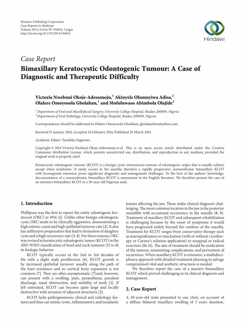
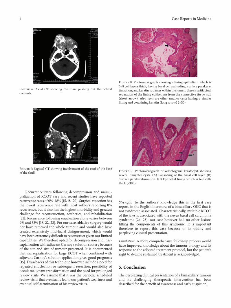
the ethmoid, sphenoid, and frontal sinuses as well as theroof of the frontal sinus (Figure 7). The chest radiograph andmicrobiology findings were unremarkable. Histology showedmultiple cysts that had lining epithelia 5–12 cells thick, havingbasal cell palisading and surface undulation with parakera-tinization. Some of the cysts had keratin deposits within them(Figures 8 and 9). A diagnosis of KCOT was made.
3. Treatment
Surgical decompression was carried out under general anaes-thesia using nasotracheal intubation. A modified Trotter-Weber Ferguson incision was made in the right maxillaryregion, while a Caldwell-Luc incision was made in the leftmaxilla to expose the lesion. The exposed cavities containedmasses of friable necrotic tissue that were curetted and sentfor histology. The cavities were well irrigated with normalsaline and packed with dry sterile gauze. After shieldingadjacent vital structures with Sofra-Tulle dressing, chemicalcautery of the tumour bed was achieved with Carnoy’ssolution.The cavities were then packed with argotone soakedgauze to allow for proper haemostasis; this was left in place for3 days.The patient had regular saline irrigation and dressing,initially every other day and after discharge on a weekly basis.Perceptible reduction in tumour size was achieved after amonth of review, but complete resolution of the mass was notachieved before the patient defaulted from review visits andwas lost to follow up.

Case Reports in Medicine 3
Figure 2: Axial computerized tomography showing a mass destroy-ing the maxilla and the nasal tissues.
Figure 3: Sagittal computerized tomography (CT) showing destruc-tion of the maxilla and nasal tissues with “floating” upper teeth.
4. Discussion
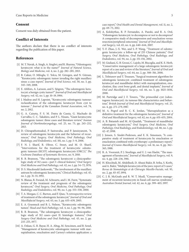
Appropriate therapy for any disease first requires correctdiagnosis, which is formedby a combination of clinical detailswith or without supporting investigations. Our patient’spattern and site of presentation for KCOTwere quite unusual,leading to a myriad of confusing clinical opinions and itwas only following histologic examination that a workingdiagnosis of KCOT was reached. Unlike other jaw cystswith unremarkable histological features, the histology of theKCOT is rather unique and even though clinical featuresmay not be clearly defined, the histology is distinctive. It istypified by a parakeratinized or orthokeratinized stratifiedsquamous epithelial lining (6–8 cell layers thick) which iscorrugated, a prominent and palisaded basal layer whichmay be cuboidal or columnar, lumen containing keratin,and a connective tissue wall without inflammation as wellas absent rete pegs [12, 13]. The parakeratinized type is saidto be commoner and tends to run a more aggressive course,while the orthokeratinized type has been suggested as asimple odontogenic keratocyst with no aggressive features[2, 13, 14]. The case reported here is a parakeratinized type.In reclassifying odontogenic keratocyst as a neoplasm, the
Figure 4: Coronal CT showing involvement of the paranasal sinusesand orbital floor.
Figure 5: Coronal CT showing maxillary and orbital involvement.
budding of the basal layer into connective tissue wall is oneof the factors considered important [3].
Although there is no universally accepted treatment yetfor KCOT, the primary aim of treatment is to achieve totaleradication utilizing an appropriate technique, taking intoconsideration site, size, location, invasion of the surroundingtissues, and previous treatments [15, 16]. Treatment optionsthat have been used for KCOT includemarsupialization, enu-cleation and curettage, and enucleationwith chemical cautery(Carnoy’s solution), thermal (cryotherapy), or mechanical(peripheral ostectomy) cautery of surrounding tissue andosseous resection with or without continuity defect. Theextensive maxillary involvement of our patient necessitateda technique that would thoroughly remove tumour withthe least compromise to function and aesthetics; hence theapproach of combined surgical decompression, curettage,and chemical cautery.
Studies have shown that decompression and marsupi-alization relieves the pressure within the cavity of KCOTleading to a reduction in size and formation of new bone [17].Following marsupialization for decompression, interleukin-1 and cytokeratin 10 which are important in cyst expansionare dissipated. Loss of these factors reduces the biologicaggressiveness of the tumour and subsequently the cysticlining is replaced by oral epithelium.
4 Case Reports in Medicine
Figure 6: Axial CT showing the mass pushing out the orbitalcontents.
Figure 7: Sagittal CT showing involvement of the roof of the baseof the skull.
Recurrence rates following decompression and marsu-pialization of KCOT vary and recent studies have reportedrecurrence rates of 0%–18% [15, 18–20]. Surgical resection hasthe lowest recurrence rate with most authors reporting 0%recurrence, but it also has the highest morbidity and greatestchallenge for reconstruction, aesthetics, and rehabilitation[21]. Recurrence following enucleation alone varies between9% and 33% [16, 22, 23]. For our case, ablative surgery wouldnot have removed the whole tumour and would also havecreated extensively mid-facial disfigurement, which wouldhave been extremely difficult to reconstruct given our limitedcapabilities. We therefore opted for decompression and mar-supializationwith adjuvant Carnoy’s solution cautery becauseof the site and size of tumour presented. It is documentedthat marsupialization for large KCOT when combined withadjuvant Carnoy’s solution application gives good prognosis[15]. Drawbacks of this technique however include a need forrepeated enucleation or subsequent resection, possibility ofoccult malignant transformation and the need for prolongedreview visits. We assume that it was the periodic scheduledreview visits that eventually led to our patient’s weariness andeventual self-termination of his review visits.
Figure 8: Photomicrograph showing a lining epithelium which is6–8 cell layers thick, having basal cell palisading, surface parakera-tinisation, and keratin squames within the lumen; there is artifactualseparation of the lining epithelium from the connective tissue wall(short arrow). Also seen are other smaller cysts having a similarlining and containing keratin (long arrow) (×50).
Figure 9: Photomicrograph of odontogenic keratocyst showingseveral daughter cysts. (A) Palisading of the basal cell layer. (B)Surface parakeratinisation. (C) Epithelial lining which is 6–8 cellsthick (×100).
Strength. To the authors’ knowledge this is the first casereport, in the English literature, of a bimaxillary OKC that isnot syndrome associated. Characteristically, multiple KCOTof the jaws is associated with the nevus basal cell carcinomasyndrome [24, 25]; our case however had no other lesionsfitting the components of this syndrome. It is importanttherefore to report this case because of its oddity andperplexing clinical presentation.
Limitation. A more comprehensive follow-up process wouldhave improved knowledge about the tumour biology and itsresponse to the adopted treatment protocol, but the patient’sright to decline sustained treatment is acknowledged.
5. Conclusion
The perplexing clinical presentation of a bimaxillary tumourand its challenging therapeutic intervention has beendescribed for the benefit of awareness and early suspicion.
Case Reports in Medicine 5
Consent
Consent was duly obtained from the patient.
Conflict of Interests
The authors declare that there is no conflict of interestsregarding the publication of this paper.
References
[1] M.T.Navak,A. Singh,A. Singhvi, andR. Sharma, “Odontogenickeratocyst: what is in the name?” Journal of Natural Science,Biology and Medicine, vol. 4, no. 2, pp. 282–285, 2013.
[2] B. Cakur, O. Miloglu, U. Yolcu, M. Goregen, and N. Gursan,“Keratocystic odontogenic tumor invading the right maxillarysinus: a case report,” Journal of Oral Science, vol. 50, no. 3, pp.345–349, 2008.
[3] E. Ahlfors, A. Larsson, and S. Sjogren, “The odontogenic kera-tocyst: a benign cystic tumor?” Journal of Oral andMaxillofacialSurgery, vol. 42, no. 1, pp. 10–19, 1984.
[4] J. Madras and H. Lapoint, “Keratocystic odontogenic tumour:reclassification of the odontogenic keratocyst from cyst totumour ,” Journal of the Canadian Dental Association, vol. 74,no. 2, 2012.
[5] G. A. Caixeta, F. S. M. Dutra de Cassia, G. Machado deCarvalho, C. C. Takahiro, and P. L. Nizam, “Giant keratocysticodontogenic tumor: three cases and literature review,” IranianJournal of Otorhinolaryngology, vol. 25, no. 73, pp. 245–252,2013.
[6] D. Chirapathomsakul, P. Sastravaha, and P. Jansisyanont, “Areview of odontogenic keratocysts and the behavior of recur-rences,” Oral Surgery, Oral Medicine, Oral Pathology, OralRadiology, and Endodontics, vol. 101, no. 1, pp. 5–9, 2006.
[7] F. N. J. Sharif, R. Oliver, C. Sweet, and M. O. Sharif,“Interventions for the treatment of keratocystic odonto-genic tumours (KCOT, odontogenic keratocysts (OKC)),” TheCochrane Database of Systematic Reviews, no. 9, 2010.
[8] R. B. Brannon, “The odontogenic keratocyst: a clinicopatho-logic study of 312 cases—part I: clinical features,” Oral SurgeryOralMedicine and Oral Pathology, vol. 42, no. 1, pp. 54–72, 1976.
[9] D. S. Macdonald-Jankowski, “The involvement of the maxillaryantrum by odontogenic keratocysts,”Clinical Radiology, vol. 45,no. 1, pp. 31–33, 1992.
[10] N. Blanas, B. Freund, M. Schwartz, and I. M. Furst, “Systematicreview of the treatment and prognosis of the odontogenickeratocyst,” Oral Surgery, Oral Medicine, Oral Pathology, OralRadiology, and Endodontics, vol. 90, no. 5, pp. 553–558, 2000.
[11] T. A.Morgan, C. C. Burton, and F. Qian, “A retrospective reviewof treatment of the odontogenic keratocyst,” Journal of Oral andMaxillofacial Surgery, vol. 63, no. 5, pp. 635–639, 2005.
[12] E. A. Grasmuck and B. L. Nelson, “Keratocystic odontogenictumor,” Head and Neck Pathology, vol. 4, no. 1, pp. 94–96, 2010.
[13] R. B. Brannon, “The odontogenic keratocyst: a clinicopatho-logic study of 312 cases—part II: histologic features,” OralSurgery Oral Medicine and Oral Pathology, vol. 43, no. 2, pp.233–255, 1977.
[14] A. Ozkan, G. R. Bayar, H. A. Altug, M. Sencimen, and B. Senel,“Management of keratocystic odontogenic tumour with mar-supialisation, enucleation and Carnoy’s solution application: a
case report,”Oral Health and Dental Management, vol. 11, no. 2,pp. 69–73, 2012.
[15] A. Kolokythas, R. P. Fernandes, A. Pazoki, and R. A. Ord,“Odontogenic keratocyst: to decompress or not to decompress?A comparative study of decompression and enucleation versusresection/peripheral ostectomy,” Journal of Oral and Maxillofa-cial Surgery, vol. 65, no. 4, pp. 640–644, 2007.
[16] Y.-F. Zhao, J.-X. Wei, and S.-P. Wang, “Treatment of odonto-genic keratocysts: a follow-up of 255 Chinese patients,” OralSurgery, Oral Medicine, Oral Pathology, Oral Radiology, andEndodontics, vol. 94, no. 2, pp. 151–156, 2002.
[17] M.Giuliani, G. B. Grossi, C. Lajolo,M. Bisceglia, andK. E.Herb,“Conservative management of a large odontogenic keratocyst:report of a case and review of the literature,” Journal of Oral andMaxillofacial Surgery, vol. 64, no. 2, pp. 308–316, 2006.
[18] L. Tolstunov and T. Treasure, “Surgical treatment algorithm forodontogenic keratocyst: combined treatment of odontogenickeratocyst and mandibular defect with marsupialization, enu-cleation, iliac crest bone graft, and dental implants,” Journal ofOral and Maxillofacial Surgery, vol. 66, no. 5, pp. 1025–1036,2008.
[19] M. Partridge and J. F. Towers, “The primordial cyst (odon-togenic keratocyst): its tumour-like characteristics and behav-iour,” British Journal of Oral and Maxillofacial Surgery, vol. 25,no. 4, pp. 271–279, 1987.
[20] M. A. Pogrel and R. C. K. Jordan, “Marsupialization as adefinitive treatment for the odontogenic keratocyst,” Journal ofOral andMaxillofacial Surgery, vol. 62, no. 6, pp. 651–655, 2004.
[21] A. B. Batanieh and M. Al Quadah, “Treatment of mandibularodontogenic keratocysts,” Oral Surgery, Oral Medicine, OralPathology, Oral Radiology, and Endodontology, vol. 86, no. 1, pp.42–47, 1998.
[22] J. Jensen, S. Sindet-Pedersen, and E. K. Simonsen, “A com-parative study of treatment of keratocysts by enucleation orenucleation combined with cryotherapy: a preliminary report,”Journal of Cranio-Maxillofacial Surgery, vol. 16, no. 8, pp. 362–365, 1988.
[23] R. A. Voorsmit, P. J. Stoelinga, and U. J. van Haelst, “The man-agement of keratocysts,” Journal of Maxillofacial Surgery, vol. 9,no. 4, pp. 228–236, 1981.
[24] H. Khochtali, M. Abdelhedi, D. Abassi Bakir, B. Sriha, S. Korbi,andA. Bakir, “Multiple keratocysts of the jaws: report of 3 cases,”Revue de Stomatologie et de Chirurgie Maxillo-Faciale, vol. 95,no. 2, pp. 83–87, 1994.
[25] C. J. R. McGrath and R. W. T. Myall, “Conservative manage-ment of recurrent keratocysts in basal-cell naevus syndrome,”Australian Dental Journal, vol. 42, no. 6, pp. 399–403, 1997.

Submit your manuscripts athttp://www.hindawi.com
Stem CellsInternational
Hindawi Publishing Corporationhttp://www.hindawi.com Volume 2014
Hindawi Publishing Corporationhttp://www.hindawi.com Volume 2014
MEDIATORSINFLAMMATION
of
Hindawi Publishing Corporationhttp://www.hindawi.com Volume 2014
Behavioural Neurology
EndocrinologyInternational Journal of
Hindawi Publishing Corporationhttp://www.hindawi.com Volume 2014
Hindawi Publishing Corporationhttp://www.hindawi.com Volume 2014
Disease Markers
Hindawi Publishing Corporationhttp://www.hindawi.com Volume 2014
BioMed Research International
OncologyJournal of
Hindawi Publishing Corporationhttp://www.hindawi.com Volume 2014
Hindawi Publishing Corporationhttp://www.hindawi.com Volume 2014
Oxidative Medicine and Cellular Longevity
Hindawi Publishing Corporationhttp://www.hindawi.com Volume 2014
PPAR Research
The Scientific World JournalHindawi Publishing Corporation http://www.hindawi.com Volume 2014
Immunology ResearchHindawi Publishing Corporationhttp://www.hindawi.com Volume 2014
Journal of
ObesityJournal of
Hindawi Publishing Corporationhttp://www.hindawi.com Volume 2014
Hindawi Publishing Corporationhttp://www.hindawi.com Volume 2014
Computational and Mathematical Methods in Medicine
OphthalmologyJournal of
Hindawi Publishing Corporationhttp://www.hindawi.com Volume 2014
Diabetes ResearchJournal of
Hindawi Publishing Corporationhttp://www.hindawi.com Volume 2014
Hindawi Publishing Corporationhttp://www.hindawi.com Volume 2014
Research and TreatmentAIDS
Hindawi Publishing Corporationhttp://www.hindawi.com Volume 2014
Gastroenterology Research and Practice
Hindawi Publishing Corporationhttp://www.hindawi.com Volume 2014
Parkinson’s Disease
Evidence-Based Complementary and Alternative Medicine
Volume 2014Hindawi Publishing Corporationhttp://www.hindawi.com