
Liver limited Metastatic Colorectal Cancer. Case Presentation
Case ReportA Case of Calcified Metastatic Colorectal AdenocarcinomaMimicking a Benign Lesion: Pitfalls in Diagnosis
Peter Michail,1 Iftah Amith,2 Sanila George,3 and Mathew K. George4
1School of Medicine and Public Health, University of Newcastle, University Drive, Callaghan, NSW 2308, Australia2University of New England, Elm Avenue, Armidale, NSW 2351, Australia3Hunter Imaging, 7 Bligh Street, Tamworth, NSW 2348, Australia4Department of Medical Oncology, North West Cancer Centre, 31 Dean Street, Tamworth, NSW 2340, Australia
Correspondence should be addressed to Peter Michail; [email protected]
Received 8 November 2014; Revised 24 January 2015; Accepted 26 January 2015
Academic Editor: Francesco A. Mauri
Copyright © 2015 Peter Michail et al. This is an open access article distributed under the Creative Commons Attribution License,which permits unrestricted use, distribution, and reproduction in any medium, provided the original work is properly cited.
The radiological finding of a calcified intracranial lesion commonly represents a slow growing benign mass. Brain metastasesoriginating from colorectal cancers are rare, occurring in approximately 2-3% of patients.Therefore the presence of a calcified brainlesion in a patient with a positive oncological history requires a high index of suspicion for brain metastases. Presented herein isa case of a frontoparietal calcified lesion initially overlooked as a benign tumour. Subsequent imaging following a neurologicalepisode revealed a significant increase in size of the lesion with surrounding tissue oedema, prompting further investigation forsuspicion of a calcified metastatic colorectal adenocarcinoma.
1. Introduction
The radiological findings of calcified intracranial lesionsgenerally represent slow growing, benign masses [1]. Mostcommon differential diagnoses include angiomas, gliomas,meningiomas, and granulomatous lesions [1]. It is rare formetastatic brain lesions to present with calcification; howeverthese represent approximately 1–3.5% of cases found oncomputer tomography (CT) [1–4].
We present a case of a frontoparietal calcified brain lesioninitially diagnosed as either a meningioma or a granuloma.Subsequently, with a delay of just under a year, the lesion wascorrectly diagnosed asmetastatic colorectal adenocarcinoma.
2. Case Presentation
The patient is a 61-year-old female who was diagnosed withcolorectal carcinoma (CRC) in 2011. The diagnosis was madeafter colonoscopic examination revealed an obstructingmass.The patient underwent an anterior resection with subsequentinvestigations revealing high grade metastatic adenocarci-noma. The patient was started on palliative chemotherapyconsisting of FOLFOX and Avastin.
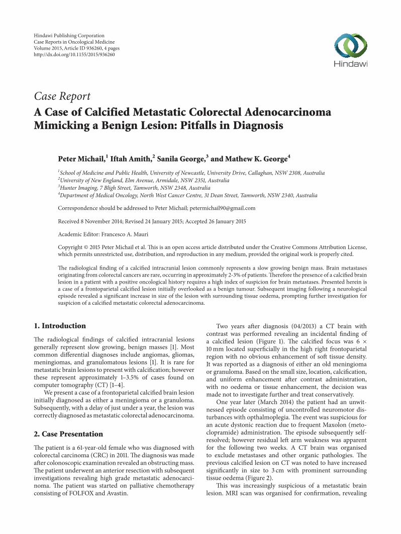
Two years after diagnosis (04/2013) a CT brain withcontrast was performed revealing an incidental finding ofa calcified lesion (Figure 1). The calcified focus was 6 ×10mm located superficially in the high right frontoparietalregion with no obvious enhancement of soft tissue density.It was reported as a diagnosis of either an old meningiomaor granuloma. Based on the small size, location, calcification,and uniform enhancement after contrast administration,with no oedema or tissue enhancement, the decision wasmade not to investigate further and treat conservatively.
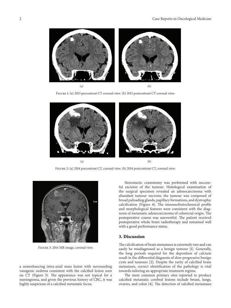
One year later (March 2014) the patient had an unwit-nessed episode consisting of uncontrolled neuromotor dis-turbances with opthalmoplegia. The event was suspicious foran acute dystonic reaction due to frequent Maxolon (meto-clopramide) administration. The episode subsequently self-resolved; however residual left arm weakness was apparentfor the following two weeks. A CT brain was organisedto exclude metastases and other organic pathologies. Theprevious calcified lesion on CT was noted to have increasedsignificantly in size to 3 cm with prominent surroundingtissue oedema (Figure 2).
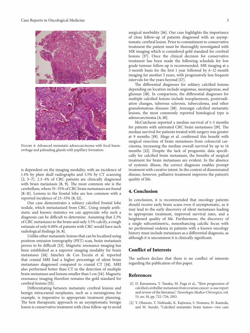
This was increasingly suspicious of a metastatic brainlesion. MRI scan was organised for confirmation, revealing
Hindawi Publishing CorporationCase Reports in Oncological MedicineVolume 2015, Article ID 936260, 4 pageshttp://dx.doi.org/10.1155/2015/936260
2 Case Reports in Oncological Medicine
(a) (b)
Figure 1: (a) 2013 precontrast CT coronal view. (b) 2013 postcontrast CT coronal view.
(a) (b)
Figure 2: (a) 2014 precontrast CT, coronal view. (b) 2014 postcontrast CT, coronal view.
Figure 3: 2014 MR image, coronal view.
a nonenhancing intra-axial mass lesion with surroundingvasogenic oedema consistent with the calcified lesion seenon CT (Figure 3). The appearance was not typical for ameningioma, and given the previous history of CRC, it washighly suspicious of a calcified metastatic focus.
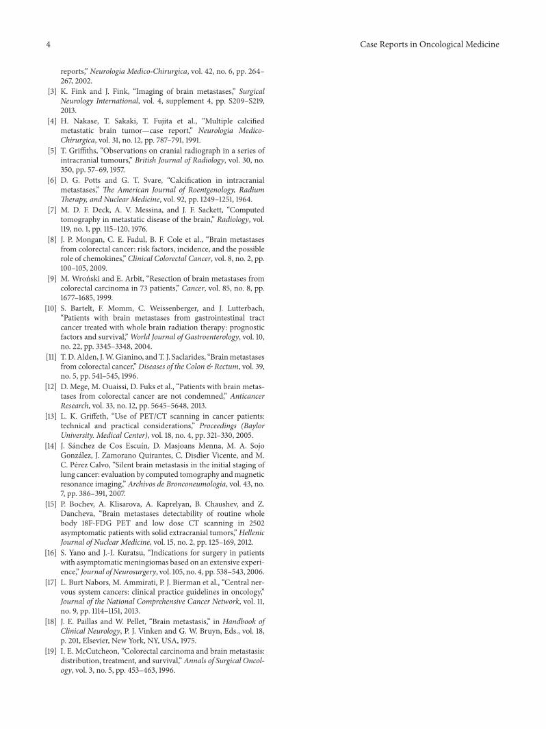
Stereotactic craniotomy was performed with success-ful excision of the tumour. Histological examination ofthe surgical specimen revealed an adenocarcinoma withabundant tumour necrosis; the tumour was composed ofbroad palisading glands, papillary formations, and dystrophiccalcification (Figure 4). The immunohistochemical profileand morphological features were consistent with the diag-nosis of metastatic adenocarcinoma of colorectal origin. Thepostoperative course was uneventful. The patient receivedpostoperative whole brain radiotherapy and remained wellwith a good performance status.
3. Discussion
Thecalcification of brainmetastases is extremely rare and caneasily be misdiagnosed as a benign tumour [1]. Generally,the long periods required for the deposition of calciumresult in the differential diagnosis of slow progressive benigncysts and tumours [2]. Despite the rarity of calcified brainmetastases, correct identification of the pathology is vitaltowards tailoring an appropriate treatment regime.
The most common primary sites reported to producecalcified metastatic cerebral lesions include breast, lungs,ovaries, and colon [4]. The detection of calcified metastases
Case Reports in Oncological Medicine 3
Figure 4: Advanced metastatic adenocarcinoma with focal haem-orrhage and palisading glands with papillary formation.
is dependent on the imaging modality, with an incidence of1.4% by plain skull radiographs and 3.5% by CT scanning[2, 5–7]. 2.3–4% of CRC patients are clinically diagnosedwith brain metastasis [8, 9]. The most common site is thecerebellum,where 35–55%ofCRCbrainmetastases are found[8–11]. Lesions in the frontal lobe are less common with areported incidence of 23–33% [8, 12].
Our case demonstrates a solitary calcified frontal lobenodule, which metastasised from CRC. Using simple arith-metic and known statistics we can appreciate why such adiagnosis can be difficult to determine. Assuming that 2.3%of CRCmetastases to the brain and only 3.5% calcify, a roughestimate of only 0.08% of patients with CRCwould have suchradiological findings [6, 8].
Unlike othermetastatic lesions that can be localised usingpositron emission tomography (PET) scan, brain metastasisproves to be difficult [13]. Magnetic resonance imaging hasbeen established as a superior imaging modality for brainmetastases [14]. Sanchez de Cos Escuın et al. reportedthat cranial MRI had a higher percentage of silent brainmetastases diagnosed compared to cranial CT [14]. MRIalso performed better than CT in the detection of multiplebrainmetastases and lesions smaller than 1 cm [14]. Magneticresonance imaging therefore remains the gold standard forcerebral lesions [15].
Differentiating between metastatic cerebral lesions andbenign intracranial neoplasms, such as a meningioma forexample, is imperative to appropriate treatment planning.The best therapeutic approach to an asymptomatic benignlesion is conservative treatment with close follow-up to avoid
surgical morbidity [16]. Our case highlights the importanceof close follow-up of patients diagnosed with an asymp-tomatic cerebral lesion. Prior to commitment to conservativetreatment the patient must be thoroughly investigated withMR imaging which is considered gold standard for cerebrallesions [17]. Once the clinical decision for conservativetreatment has been made the following schedule for lowgrade tumour follow-up is recommended. MR imaging at a3-month basis for the first 1 year followed by 6–12-monthimaging for another 5 years, with progressively less frequentintervals for the years beyond [17].
The differential diagnoses for solitary calcified lesionsdepending on location include angiomas, meningiomas, andgliomas [18]. In comparison, the differential diagnoses formultiple calcified lesions include toxoplasmosis, postirradi-ation changes, tuberous sclerosis, tuberculoma, and othergranulomatous diseases [18]. Amongst calcified metastaticlesions, the most commonly reported histological type isadenocarcinoma [4, 10].
McCutcheon reported a median survival of 1–3 monthsfor patients with untreated CRC brain metastases [19]. Themedian survival for patients treated with surgery was greaterat 9 months [19]. Mege et al. confirmed this benefit withsurgical resection of brain metastases from colorectal car-cinoma, increasing the median overall survival by up to 14months [12]. Despite the lack of prognostic data specifi-cally for calcified brain metastasis, the benefits of surgicaltreatment for brain metastases are evident. In the absenceof systemic illness, the correct diagnosis enables prompttreatment with curative intent. In the context of disseminateddisease, however, palliative treatment improves the patient’squality of life.
4. Conclusion
In conclusion, it is recommended that oncology patientsshould receive early brain scans even if asymptomatic, as itmay lead to the early discovery of silent metastases leadingto appropriate treatment, improved survival rates, and aheightened quality of life. Furthermore, the discovery ofa single subcentimetric, nonenhancing calcific focus withno perilesional oedema in patients with a known oncologyhistory must include metastases as a differential diagnosis, asalthough it is uncommon it is clinically significant.
Conflict of Interests
The authors declare that there is no conflict of interestsregarding the publication of this paper.
References
[1] D. Kawamura, T. Tanaka, M. Fuga et al., “Slow progression ofcalcified cerebellarmetastasis fromovarian cancer: a case reportand review of the literature,”NeurologiaMedico-Chirurgica, vol.53, no. 10, pp. 722–726, 2013.
[2] Y. Ohmoto, T. Nishizaki, K. Kajiwara, S. Nomura, H. Kameda,and M. Suzuki, “Calcified metastatic brain tumor—two case
4 Case Reports in Oncological Medicine
reports,” Neurologia Medico-Chirurgica, vol. 42, no. 6, pp. 264–267, 2002.
[3] K. Fink and J. Fink, “Imaging of brain metastases,” SurgicalNeurology International, vol. 4, supplement 4, pp. S209–S219,2013.
[4] H. Nakase, T. Sakaki, T. Fujita et al., “Multiple calcifiedmetastatic brain tumor—case report,” Neurologia Medico-Chirurgica, vol. 31, no. 12, pp. 787–791, 1991.
[5] T. Griffiths, “Observations on cranial radiograph in a series ofintracranial tumours,” British Journal of Radiology, vol. 30, no.350, pp. 57–69, 1957.
[6] D. G. Potts and G. T. Svare, “Calcification in intracranialmetastases,” The American Journal of Roentgenology, RadiumTherapy, and Nuclear Medicine, vol. 92, pp. 1249–1251, 1964.
[7] M. D. F. Deck, A. V. Messina, and J. F. Sackett, “Computedtomography in metastatic disease of the brain,” Radiology, vol.119, no. 1, pp. 115–120, 1976.
[8] J. P. Mongan, C. E. Fadul, B. F. Cole et al., “Brain metastasesfrom colorectal cancer: risk factors, incidence, and the possiblerole of chemokines,” Clinical Colorectal Cancer, vol. 8, no. 2, pp.100–105, 2009.
[9] M. Wronski and E. Arbit, “Resection of brain metastases fromcolorectal carcinoma in 73 patients,” Cancer, vol. 85, no. 8, pp.1677–1685, 1999.
[10] S. Bartelt, F. Momm, C. Weissenberger, and J. Lutterbach,“Patients with brain metastases from gastrointestinal tractcancer treated with whole brain radiation therapy: prognosticfactors and survival,”World Journal of Gastroenterology, vol. 10,no. 22, pp. 3345–3348, 2004.
[11] T. D. Alden, J.W.Gianino, andT. J. Saclarides, “Brainmetastasesfrom colorectal cancer,”Diseases of the Colon & Rectum, vol. 39,no. 5, pp. 541–545, 1996.
[12] D. Mege, M. Ouaissi, D. Fuks et al., “Patients with brain metas-tases from colorectal cancer are not condemned,” AnticancerResearch, vol. 33, no. 12, pp. 5645–5648, 2013.
[13] L. K. Griffeth, “Use of PET/CT scanning in cancer patients:technical and practical considerations,” Proceedings (BaylorUniversity. Medical Center), vol. 18, no. 4, pp. 321–330, 2005.
[14] J. Sanchez de Cos Escuın, D. Masjoans Menna, M. A. SojoGonzalez, J. Zamorano Quirantes, C. Disdier Vicente, and M.C. Perez Calvo, “Silent brain metastasis in the initial staging oflung cancer: evaluation by computed tomography andmagneticresonance imaging,” Archivos de Bronconeumologia, vol. 43, no.7, pp. 386–391, 2007.
[15] P. Bochev, A. Klisarova, A. Kaprelyan, B. Chaushev, and Z.Dancheva, “Brain metastases detectability of routine wholebody 18F-FDG PET and low dose CT scanning in 2502asymptomatic patients with solid extracranial tumors,”HellenicJournal of Nuclear Medicine, vol. 15, no. 2, pp. 125–169, 2012.
[16] S. Yano and J.-I. Kuratsu, “Indications for surgery in patientswith asymptomatic meningiomas based on an extensive experi-ence,” Journal of Neurosurgery, vol. 105, no. 4, pp. 538–543, 2006.
[17] L. Burt Nabors, M. Ammirati, P. J. Bierman et al., “Central ner-vous system cancers: clinical practice guidelines in oncology,”Journal of the National Comprehensive Cancer Network, vol. 11,no. 9, pp. 1114–1151, 2013.
[18] J. E. Paillas and W. Pellet, “Brain metastasis,” in Handbook ofClinical Neurology, P. J. Vinken and G. W. Bruyn, Eds., vol. 18,p. 201, Elsevier, New York, NY, USA, 1975.
[19] I. E. McCutcheon, “Colorectal carcinoma and brain metastasis:distribution, treatment, and survival,” Annals of Surgical Oncol-ogy, vol. 3, no. 5, pp. 453–463, 1996.

Submit your manuscripts athttp://www.hindawi.com
Stem CellsInternational
Hindawi Publishing Corporationhttp://www.hindawi.com Volume 2014
Hindawi Publishing Corporationhttp://www.hindawi.com Volume 2014
MEDIATORSINFLAMMATION
of
Hindawi Publishing Corporationhttp://www.hindawi.com Volume 2014
Behavioural Neurology
EndocrinologyInternational Journal of
Hindawi Publishing Corporationhttp://www.hindawi.com Volume 2014
Hindawi Publishing Corporationhttp://www.hindawi.com Volume 2014
Disease Markers
Hindawi Publishing Corporationhttp://www.hindawi.com Volume 2014
BioMed Research International
OncologyJournal of
Hindawi Publishing Corporationhttp://www.hindawi.com Volume 2014
Hindawi Publishing Corporationhttp://www.hindawi.com Volume 2014
Oxidative Medicine and Cellular Longevity
Hindawi Publishing Corporationhttp://www.hindawi.com Volume 2014
PPAR Research
The Scientific World JournalHindawi Publishing Corporation http://www.hindawi.com Volume 2014
Immunology ResearchHindawi Publishing Corporationhttp://www.hindawi.com Volume 2014
Journal of
ObesityJournal of
Hindawi Publishing Corporationhttp://www.hindawi.com Volume 2014
Hindawi Publishing Corporationhttp://www.hindawi.com Volume 2014
Computational and Mathematical Methods in Medicine
OphthalmologyJournal of
Hindawi Publishing Corporationhttp://www.hindawi.com Volume 2014
Diabetes ResearchJournal of
Hindawi Publishing Corporationhttp://www.hindawi.com Volume 2014
Hindawi Publishing Corporationhttp://www.hindawi.com Volume 2014
Research and TreatmentAIDS
Hindawi Publishing Corporationhttp://www.hindawi.com Volume 2014
Gastroenterology Research and Practice
Hindawi Publishing Corporationhttp://www.hindawi.com Volume 2014
Parkinson’s Disease
Evidence-Based Complementary and Alternative Medicine
Volume 2014Hindawi Publishing Corporationhttp://www.hindawi.com


![[D2a] Treatment for metastatic colorectal cancer in the ...](https://static.fdocuments.us/doc/165x107/62daacb8a695811f783e9cbe/d2a-treatment-for-metastatic-colorectal-cancer-in-the-.jpg)


![Liver resection for metastatic colorectal cancer - [email protected]](https://static.fdocuments.us/doc/165x107/620633768c2f7b1730055cf8/liver-resection-for-metastatic-colorectal-cancer-emailprotected.jpg)


