Case record......Primary subependymal CNS lymphoma (MRI approach)
20
CLINICAL PICTURE: A 30 years old male patient presented clinically with manifestations of increased intracranial pressure, bilateral papilledema, lower cranial nerve palsy, personality changes and hypothalamic manifestations. RADIOLOGICAL FINDINGS: Figure 1. Postcontrast CT scan study showing a densely enhanced periventricular lesions. The lesions are seen completely surrounding the 4th, lateral and third ventricles with some perilesional edema lucencies. The lesions have a minimal mass effect and do not appear to be compressing the ventricular system or inducing obstructive hydrocephalus. The periventricular lesions are ensheathing the ventricular system and appear to radiate from it centrifugally outwards forming butterfly lesions around the 4th ventricle and the frontal horns of the lateral ventricles. Figure 2. Postcontrast CT scan study showing a densely enhanced periventricular lesions. The lesions are seen completely surrounding the lateral and the third ventricles with some perilesional edema lucencies. The lesions have a minimal mass effect and do not appear to be compressing the ventricular system or inducing obstructive hydrocephalus. The periventricular lesions are ensheathing the ventricular system and appear to radiate from it centrifugally outwards forming butterfly lesions the frontal horns of the lateral ventricles. CASE OF THE WEEK PROFESSOR YASSER METWALLY CLINICAL PICTURE RADIOLOGICAL FINDINGS
-
Upload
professor-yasser-metwally -
Category
Education
-
view
1.917 -
download
2
Transcript of Case record......Primary subependymal CNS lymphoma (MRI approach)

CLINICAL PICTURE:
A 30 years old male patient presented clinically with manifestations of increased intracranial pressure, bilateral papilledema, lower cranial nerve palsy, personality changes and hypothalamic manifestations.
RADIOLOGICAL FINDINGS:
Figure 1. Postcontrast CT scan study showing a densely enhanced periventricular lesions. The lesions are seen completely surrounding the 4th, lateral and third ventricles with some perilesional edema lucencies. The lesions have a minimal mass effect and do not appear to be compressing the ventricular system or inducing obstructive hydrocephalus. The periventricular lesions are ensheathing the ventricular system and appear to radiate from it centrifugally outwards forming butterfly lesions around the 4th ventricle and the frontal horns of the lateral ventricles.
Figure 2. Postcontrast CT scan study showing a densely enhanced periventricular lesions. The lesions are seen completely surrounding the lateral and the third ventricles with some perilesional edema lucencies. The lesions have a minimal mass effect and do not appear to be compressing the ventricular system or inducing obstructive hydrocephalus. The periventricular lesions are ensheathing the ventricular system and appear to radiate from it centrifugally outwards forming butterfly lesions the frontal horns of the lateral ventricles.
CASE OF THE WEEK
PROFESSOR YASSER METWALLY
CLINICAL PICTURE
RADIOLOGICAL FINDINGS
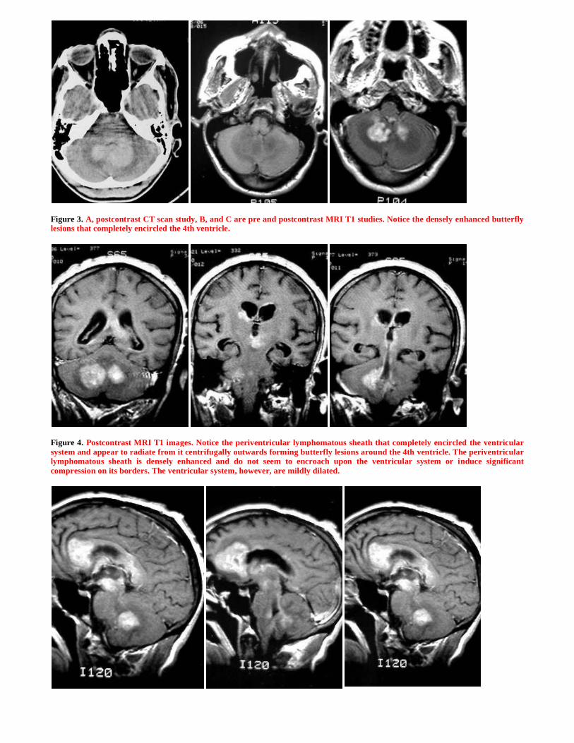
Figure 3. A, postcontrast CT scan study, B, and C are pre and postcontrast MRI T1 studies. Notice the densely enhanced butterfly lesions that completely encircled the 4th ventricle.
Figure 4. Postcontrast MRI T1 images. Notice the periventricular lymphomatous sheath that completely encircled the ventricular system and appear to radiate from it centrifugally outwards forming butterfly lesions around the 4th ventricle. The periventricular lymphomatous sheath is densely enhanced and do not seem to encroach upon the ventricular system or induce significant compression on its borders. The ventricular system, however, are mildly dilated.
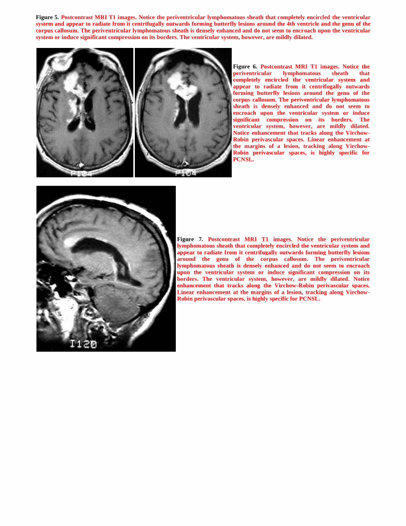
Figure 5. Postcontrast MRI T1 images. Notice the periventricular lymphomatous sheath that completely encircled the ventricular system and appear to radiate from it centrifugally outwards forming butterfly lesions around the 4th ventricle and the genu of the corpus callosum. The periventricular lymphomatous sheath is densely enhanced and do not seem to encroach upon the ventricular system or induce significant compression on its borders. The ventricular system, however, are mildly dilated.
Figure 6. Postcontrast MRI T1 images. Notice the periventricular lymphomatous sheath that completely encircled the ventricular system and appear to radiate from it centrifugally outwards forming butterfly lesions around the genu of the corpus callosum. The periventricular lymphomatous sheath is densely enhanced and do not seem to encroach upon the ventricular system or induce significant compression on its borders. The ventricular system, however, are mildly dilated. Notice enhancement that tracks along the Virchow-Robin perivascular spaces. Linear enhancement at the margins of a lesion, tracking along Virchow-Robin perivascular spaces, is highly specific for PCNSL.
Figure 7. Postcontrast MRI T1 images. Notice the periventricular lymphomatous sheath that completely encircled the ventricular system and appear to radiate from it centrifugally outwards forming butterfly lesions around the genu of the corpus callosum. The periventricular lymphomatous sheath is densely enhanced and do not seem to encroach upon the ventricular system or induce significant compression on its borders. The ventricular system, however, are mildly dilated. Notice enhancement that tracks along the Virchow-Robin perivascular spaces. Linear enhancement at the margins of a lesion, tracking along Virchow-Robin perivascular spaces, is highly specific for PCNSL.
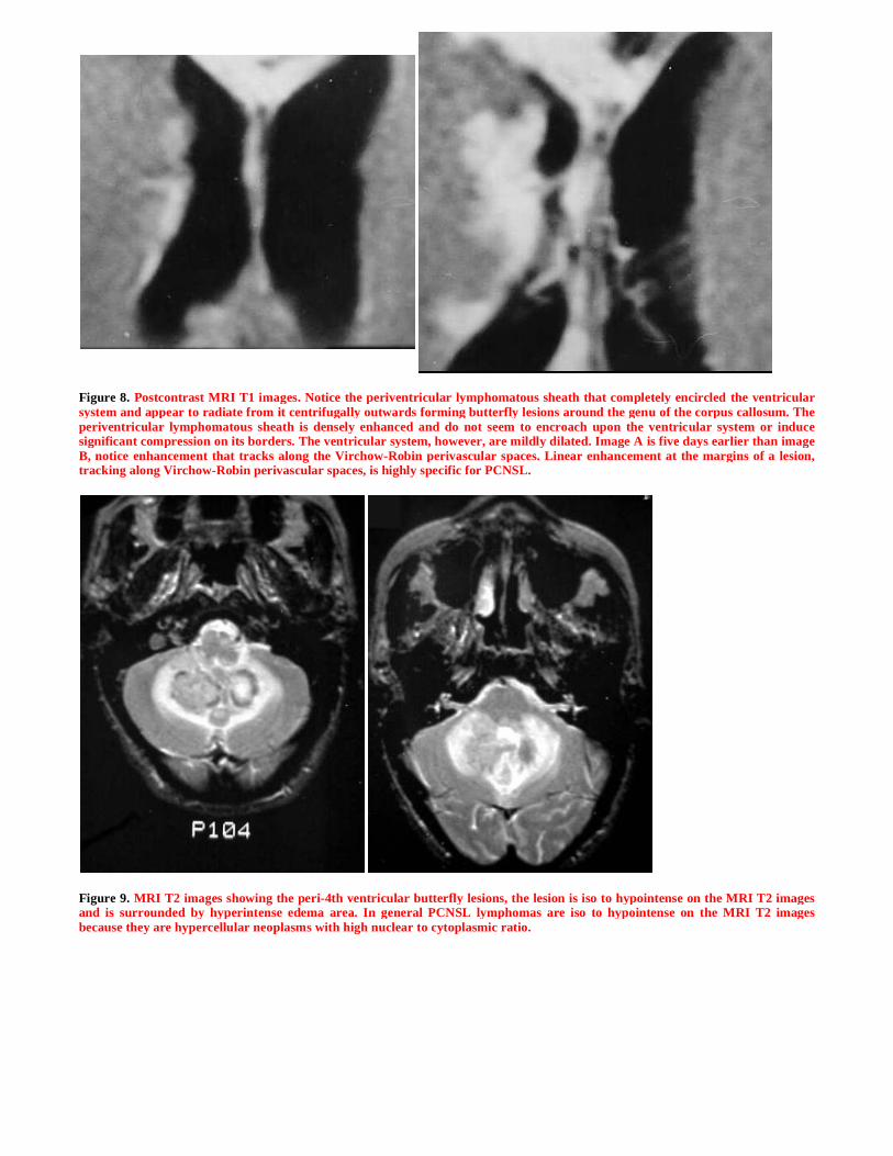
Figure 8. Postcontrast MRI T1 images. Notice the periventricular lymphomatous sheath that completely encircled the ventricular system and appear to radiate from it centrifugally outwards forming butterfly lesions around the genu of the corpus callosum. The periventricular lymphomatous sheath is densely enhanced and do not seem to encroach upon the ventricular system or induce significant compression on its borders. The ventricular system, however, are mildly dilated. Image A is five days earlier than image B, notice enhancement that tracks along the Virchow-Robin perivascular spaces. Linear enhancement at the margins of a lesion, tracking along Virchow-Robin perivascular spaces, is highly specific for PCNSL.
Figure 9. MRI T2 images showing the peri-4th ventricular butterfly lesions, the lesion is iso to hypointense on the MRI T2 images and is surrounded by hyperintense edema area. In general PCNSL lymphomas are iso to hypointense on the MRI T2 images because they are hypercellular neoplasms with high nuclear to cytoplasmic ratio.
CNS lymphomas include primary parenchymal CNS (brain or spinal) lymphomas and secondary CNS lymphomas (Epidural pachymeningeal lymphoma, orbital lymphoma, leptomeningeal lymphoma and Intravascular lymphomatosis). In primary CNS lymphoma the disease starts in the parenchyma of the brain or spinal cord and staging does not reveal extraneural disease. Extraneural involvement is the rule in secondary CNS lymphoma, although CNS manifestations might be the initial presentation of the disease. [88]
Radiological pathology of primary CNS lymphoma
Primary CNS lymphoma is an uncommon disease that historically constituted approximately I% of primary brain tumors. Sporadic disease is most common in older adults. With the advent of acquired immunodeficiency syndrome (AIDS)-associated lymphomas, there has been a marked increase in the number of cases, particularly in younger people, in whom the disease was previously rare. There has also been a significant increase in non-human immunodeficiency virus (HIV)-associated primary CNS lymphoma among older patients. A relationship between Epstein-Barr virus and HIV-associated lymphomas has been observed. The causes of sporadic cases and their increasing incidence in the nonimmunocompromised are unknown, but viral and environmental agents have been proposed as factors. Primary CNS lymphoma occurs throughout the brain, but it is characteristically periventricular. Sporadic cases tend to be limited to one or two sites, whereas AIDS-associated tumors are commonly multifocal. [88]
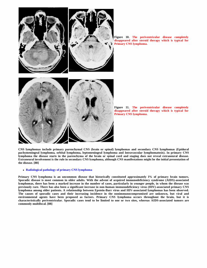
Figure 10. The periventricular disease completely disappeared after steroid therapy which is typical for Primary CNS lymphoma.
Figure 11. The periventricular disease completely disappeared after steroid therapy which is typical for Primary CNS lymphoma.
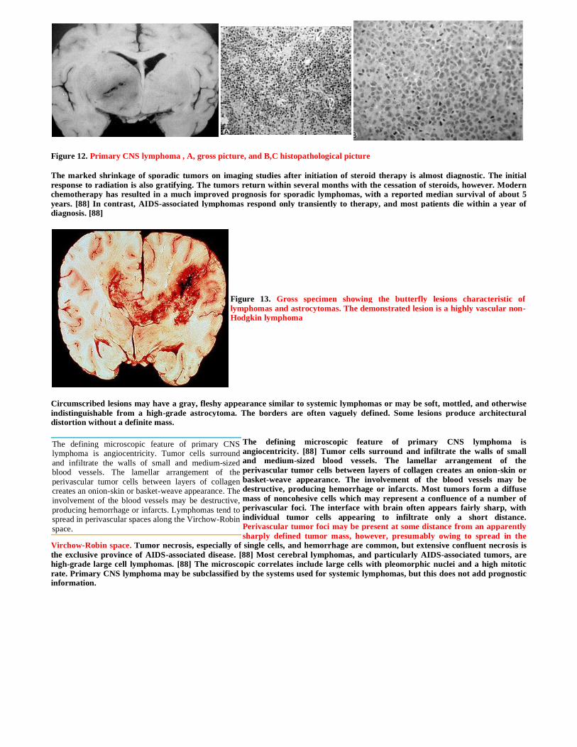
Figure 12. Primary CNS lymphoma , A, gross picture, and B,C histopathological picture
The marked shrinkage of sporadic tumors on imaging studies after initiation of steroid therapy is almost diagnostic. The initial response to radiation is also gratifying. The tumors return within several months with the cessation of steroids, however. Modern chemotherapy has resulted in a much improved prognosis for sporadic lymphomas, with a reported median survival of about 5 years. [88] In contrast, AIDS-associated lymphomas respond only transiently to therapy, and most patients die within a year of diagnosis. [88]
Circumscribed lesions may have a gray, fleshy appearance similar to systemic lymphomas or may be soft, mottled, and otherwise indistinguishable from a high-grade astrocytoma. The borders are often vaguely defined. Some lesions produce architectural distortion without a definite mass.
The defining microscopic feature of primary CNS lymphoma is angiocentricity. [88] Tumor cells surround and infiltrate the walls of small and medium-sized blood vessels. The lamellar arrangement of the perivascular tumor cells between layers of collagen creates an onion-skin or basket-weave appearance. The involvement of the blood vessels may be destructive, producing hemorrhage or infarcts. Most tumors form a diffuse mass of noncohesive cells which may represent a confluence of a number of perivascular foci. The interface with brain often appears fairly sharp, with individual tumor cells appearing to infiltrate only a short distance. Perivascular tumor foci may be present at some distance from an apparently sharply defined tumor mass, however, presumably owing to spread in the
Virchow-Robin space. Tumor necrosis, especially of single cells, and hemorrhage are common, but extensive confluent necrosis is the exclusive province of AIDS-associated disease. [88] Most cerebral lymphomas, and particularly AIDS-associated tumors, are high-grade large cell lymphomas. [88] The microscopic correlates include large cells with pleomorphic nuclei and a high mitotic rate. Primary CNS lymphoma may be subclassified by the systems used for systemic lymphomas, but this does not add prognostic information.
Figure 13. Gross specimen showing the butterfly lesions characteristic of lymphomas and astrocytomas. The demonstrated lesion is a highly vascular non-Hodgkin lymphoma
The defining microscopic feature of primary CNS lymphoma is angiocentricity. Tumor cells surround and infiltrate the walls of small and medium-sized blood vessels. The lamellar arrangement of the perivascular tumor cells between layers of collagen creates an onion-skin or basket-weave appearance. The involvement of the blood vessels may be destructive, producing hemorrhage or infarcts. Lymphomas tend to spread in perivascular spaces along the Virchow-Robin space.
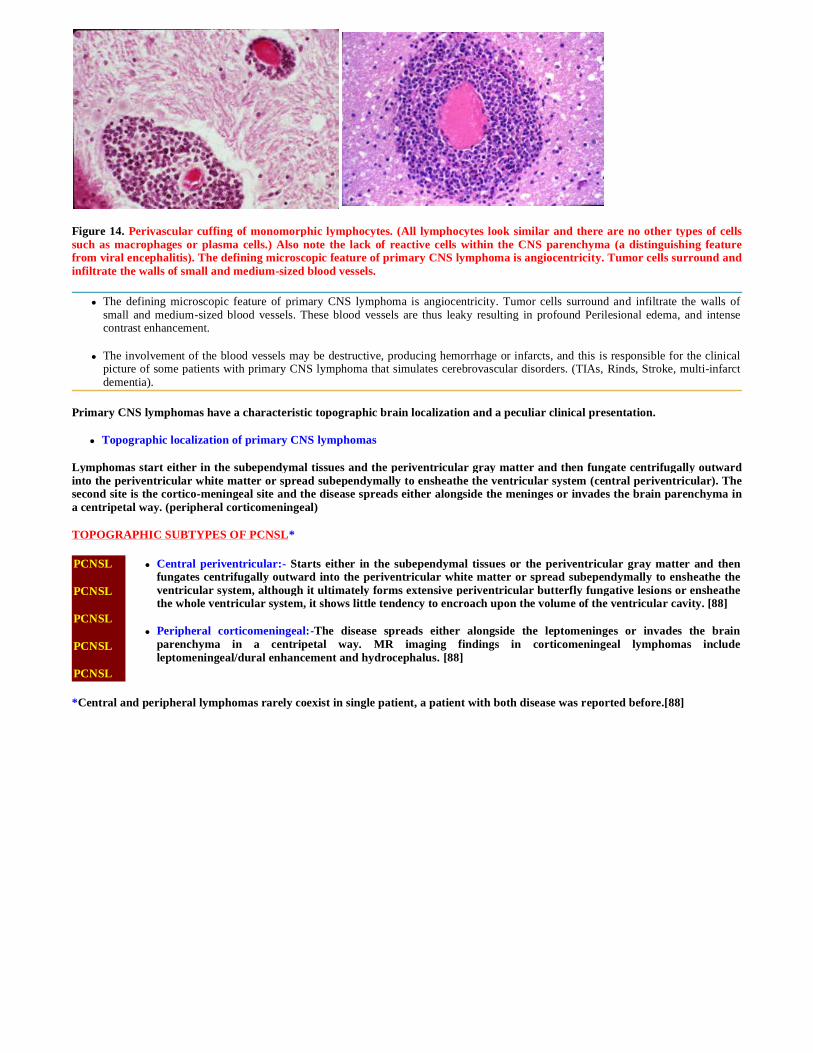
Figure 14. Perivascular cuffing of monomorphic lymphocytes. (All lymphocytes look similar and there are no other types of cells such as macrophages or plasma cells.) Also note the lack of reactive cells within the CNS parenchyma (a distinguishing feature from viral encephalitis). The defining microscopic feature of primary CNS lymphoma is angiocentricity. Tumor cells surround and infiltrate the walls of small and medium-sized blood vessels.
Primary CNS lymphomas have a characteristic topographic brain localization and a peculiar clinical presentation.
Topographic localization of primary CNS lymphomas
Lymphomas start either in the subependymal tissues and the periventricular gray matter and then fungate centrifugally outward into the periventricular white matter or spread subependymally to ensheathe the ventricular system (central periventricular). The second site is the cortico-meningeal site and the disease spreads either alongside the meninges or invades the brain parenchyma in a centripetal way. (peripheral corticomeningeal)
TOPOGRAPHIC SUBTYPES OF PCNSL*
*Central and peripheral lymphomas rarely coexist in single patient, a patient with both disease was reported before.[88]
The defining microscopic feature of primary CNS lymphoma is angiocentricity. Tumor cells surround and infiltrate the walls of small and medium-sized blood vessels. These blood vessels are thus leaky resulting in profound Perilesional edema, and intense contrast enhancement.
The involvement of the blood vessels may be destructive, producing hemorrhage or infarcts, and this is responsible for the clinical picture of some patients with primary CNS lymphoma that simulates cerebrovascular disorders. (TIAs, Rinds, Stroke, multi-infarct dementia).
PCNSL
PCNSL
PCNSL
PCNSL
PCNSL
Central periventricular:- Starts either in the subependymal tissues or the periventricular gray matter and then fungates centrifugally outward into the periventricular white matter or spread subependymally to ensheathe the ventricular system, although it ultimately forms extensive periventricular butterfly fungative lesions or ensheathe the whole ventricular system, it shows little tendency to encroach upon the volume of the ventricular cavity. [88]
Peripheral corticomeningeal:-The disease spreads either alongside the leptomeninges or invades the brain parenchyma in a centripetal way. MR imaging findings in corticomeningeal lymphomas include leptomeningeal/dural enhancement and hydrocephalus. [88]
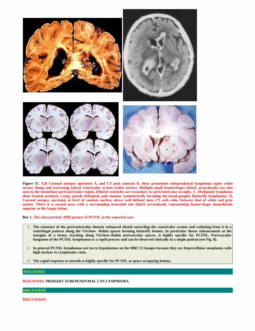
Figure 15. A,B Coronal autopsy specimen A, and CT post contrast B, show prominent subependymal lymphoma (open white arrow) lining and traversing lateral ventricular system (white arrow). Multiple small hemorrhages (black arrowheads) are also seen in the immediate periventricular region. Dilated ventricles are secondary to periventricular atrophy. C, Malignant lymphoma (four frontal sections). Large, poorly delimited, pale tumour symmetrically invading the basal ganglia (butterfly lymphoma). D, Coronal autopsy specimen at level of caudate nucleus shows well-defined mass (*) with color between that of white and gray matter. There is a second mass with a surrounding brownish rim (black arrowhead), representing hemorrhage, immediately superior to the larger lesion.
Box 1. The characteristic MRI picture of PCNSL in the reported case
DIAGNOSIS: PRIMARY SUBEPENDYMAL CNS LYMPHOMA
DISCUSSION:
1. The existence of the periventricular densely enhanced sheath encircling the ventricular system and radiating from it in a centrifugal pattern along the Virchow -Robin spaces forming butterfly lesions. In particular linear enhancement at the margins of a lesion, tracking along Virchow-Robin perivascular spaces, is highly specific for PCNSL. Perivascular fungation of the PCNSL lymphomas is a rapid process and can be observed clinically in a single patient (see Fig. 8).
2. In general PCNSL lymphomas are iso to hypointense on the MRI T2 images because they are hypercellular neoplasms with high nuclear to cytoplasmic ratio.
3. The rapid response to steroids is highly specific for PCNSL as space occupying lesions.
DIAGNOSIS:
DISCUSSION

Primary central nervous system lymphoma (PCNSL) is a rare form of non-Hodgkin lymphoma that affects the brain, spinal cord, leptomeninges, and eyes. The clinical presentation and neuroimaging appearance of PCNSL differ in immunocompetent patients and in those with acquired immunodeficiency syndrome (AIDS). A magnetic resonance (MR) image of the brain in immunocompetent patients with PCNSL typically demonstrates one or more homogeneously enhancing lesions located in the periventricular white matter, characteristically spanning the corpus callosum. In patients with AIDS, multiple ring-enhancing lesions are more common. After neuroimages raising the suspicion of PCNSL are obtained, a definitive diagnosis should be established in both immunocompetent and AIDS patients by performing pathological analysis of cerebrospinal fluid (CSF), vitreous fluid, or a biopsy specimen. Brain biopsy sampling remains the gold standard for PCNSL diagnosis in all patients, although the possibility of establishing routine, minimally invasive diagnostic procedures in which Epstein-Barr virus polymerase chain reaction (PCR) analysis of the CSF and nuclear imaging are used is currently under investigation in the population of patients with AIDS. At the time of diagnosis, the patient should undergo further evaluation, which should include a physical examination, ophthalmic evaluation with a slit-lamp examination, serum lactate dehydrogenase levels, human immunodeficiency virus testing, computed tomography scans of the chest/abdomen/pelvis, bone marrow biopsy sampling, contrast-enhanced brain MR imaging, and lumbar puncture (LP). Testicular ultrasonography studies should be considered in men. In patients who cannot undergo LP or in those with evidence of spinal cord dysfunction, contrast-enhanced MR imaging of the entire spine should be considered.
Primary central nervous system lymphoma is a rare form of extranodal non-Hodgkin lymphoma that affects the brain, spinal cord, leptomeninges, and eyes. Although this entity is rare, PCNSL has been the subject of intensive research since the late 1980s, when Eby, et al.,[28] first reported its rising incidence. A definitive clinical overview of PCNSL was written in 1993 by Fine and Mayer,[38] based on an analysis of 792 immunocompetent patients and 315 with AIDS-related PCNSL whose cases were detailed in 72 English-language articles published between 1980 and 1992. Since this review, many other large case series of PCNSL have been published, comprising more than 1000 cases.[8,14,33,36,42,44,48,49,66,70,72,73,79] This article is a summary of the clinical presentation, differential diagnosis, and "extent of disease" evaluation of PCNSL in immunocompetent and immunocompromised patients.
Clinical Presentation
Age and Sex
In immunocompetent patients, the median age at diagnosis of PCNSL is 60 years, with a male/female ratio of 1.2.[16] The highest risk group appears to be those 60 years of age or older, in whom the incidence has increased disproportionately since the mid-1990s.[55,72] Among patients with AIDS, the typical age at presentation is younger; the mean age is 31 to 36 years.[38,70] In fact, PCNSL has been diagnosed in HIV-positive children as young as 2 years old.[30] Consistent with the population at highest risk for AIDS in the past, the published male/female ratio for AIDS-associated PCNSL in adults is 7.38 to 1.[80] The incidence of PCNSL in patients with AIDS appears to have peaked in the early 1990s, with a decline since then that is reflective of the advent of highly active antiretroviral therapy.[19,86] The incidence of PCNSL also appears to be stabilizing or decreasing slightly in immunocompetent patients with these tumors.[72]
Predisposing Conditions
The most important risk factor for PCNSL is an alteration in immune system functioning ( Table 1 ). Immunocompromised conditions that predispose patients to PCNSL include the following: AIDS; iatrogenic immunosuppression for transplant procedures or for autoimmune diseases such as rheumatoid arthritis; and rare congenital immunodeficiency syndromes such as ataxia-telangiectasia, severe combined immunodeficiency, and Wiskott- Aldrich syndrome.[1,47] In patients with AIDS, advanced disease is the most important predisposing factor, demonstrated by a median CD4 cell count in AIDS-associated PCNSL of 30 to 37 cells/mm.[3,9,70]
Table 1. Risk Factors for PCNSL
Symptoms and Signs
As with all masses in the central nervous system, the location of PCNSL lesions determines the clinical presentation. The presenting symptoms and signs in one large case series of 248 immunocompetent patients with PCNSL included the following: focal neurological deficits in 70% of patients; neuropsychiatric symptoms in 43%; headache/nausea/vomiting suggestive of increased intracranial pressure in 33%; seizures in 14%; and ocular symptoms in 4%.[8] Common focal deficits include aphasia, hemiparesis, and ataxia due to discrete intracerebral lesions as well as less common cranial nerve palsies secondary to

leptomeningeal deposits. Neuropsychiatric changes such as apathy, depression, slowed thinking, and confusion have been attributed to the infiltration of white matter tracts by PCNSL lesions that involve the periventricular regions or the corpus callosum. The fact that seizures occur as the initial manifestation of PCNSL in less than 15% of immunocompetent patients who have these tumors may be partially explained by the fact that PCNSL lesions less often involve excitable cerebral cortex than do other types of brain tumors.[37]
Of the 41% of patients with PCNSL shown to have leptomeningeal involvement in one series, the leptomeningeal tumor was asymptomatic in the majority. Less than one third of patients with PCNSL who had definite leptomeningeal involvement showed any symptoms or signs characteristic of leptomeningeal tumor.[7] The incidence of cranial nerve palsies among these patients is reported to be 5 to 31%.[10,47]
For the 10 to 20% of immunocompetent patients found to have ocular involvement at the time of PCNSL diagnosis, and for those with the PIOL variant, both eyes are affected in the majority of cases. Such patients' most common visual complaints are "floaters" and blurred vision.[52] Some may experience diminished visual acuity, whereas others may have painful, red eyes, which can be misleadingly suggestive of uveitis or other nonmalignant inflammatory diseases. Twenty percent of patients with ocular lymphoma will be asymptomatic.[11]
The rare spinal cord lesions found in patients with PCNSL are primarily discrete intramedullary nodules that may be single or multiple.[57,60] The symptoms and signs of in tramedullary spinal cord lymphoma resemble those of other intramedullary spinal tumors and depend on the lesion's location within the spinal cord. Presenting symptoms may include limb paresthesias and/or numbness, limb weakness (often asymmetrical), impaired gait, and perineal numbness with bladder or bowel dysfunction.
Patients with AIDS who have PCNSL are more likely than immunocompetent patients to present with mental status changes or seizures. The presenting features often occur in a span of only days to weeks in patients with AIDS, as opposed to weeks to months in immunocompetent hosts.[20] In the review by Fine and Mayer,[38] in which the presentation of PCNSL in 315 patients with AIDS was compared with that in 792 immunocompetent patients, focal neurological deficits occurred in approximately 50% of individuals in both groups; mental status changes (including behavioral changes) were seen in 53% of patients with AIDS compared with 35% of immunocompetent patients; and seizures were reported in 27% of patients with AIDS compared with 11% of immunocompetent patients.
Time From Symptom Onset to Diagnosis
The mean period between onset of symptoms and the diagnosis of PCNSL is 3 months in immunocompetent patients and 2 months in those with AIDS.[37] Administration of corticosteroid agents can delay or confound the diagnosis due to cytolysis of lymphoma cells.[85]
Anatomical Distribution of Lesions
PCNSL can present in four distinct anatomical distributions in both immunocompetent patients and in those with AIDS: 1) discrete or diffuse intracranial mass lesions that are solitary or multiple, often in contact with ventricular or meningeal surfaces; 2) leptomeningeal disease; 3) ocular lymphoma with or without other lesions; and 4) rare spinal cord lesions.
Discrete intracranial PCNSL lesions differ in number in immunocompetent compared with AIDS patients. In immunocompetent patients with PCNSL, lesions are solitary approximately 70% of the time.[8] In contrast, AIDS-associated PCNSL lesions are just as likely to be multiple as solitary.[38] The distribution of intracranial lesions follows that of other adult primary brain tumors: approximately 85% of discrete lesions are found in supratentorial compared with 15% in infratentorial sites.[8,14] Of all intracranial lesions, more than 60% are periventricular (involving the basal ganglia, thalamus, or corpus callosum), and 12% of discrete PCNSL masses arise in the corpus callosum, a site that is particularly suggestive for this tumor type as a diagnosis. With respect to lobar location, lesions are more often located in the frontal (20%), parietal (18%), and temporal lobes (15%) than in the occipital lobes (4%).[8]
Primary leptomeningeal lymphoma, defined as PCNSL limited to the meninges without cerebral parenchymal disease or systemic lymphoma, is rare and comprises approximately 7% of all cases of PCNSL in immunocompetent patients.[59] Leptomeningeal involvement of intracranial PCNSL, on the other hand, is more common and has been reported in up to 41% of cases, either by findings obtained using clinical diagnostic modalities (positive CSF cytology, leptomeningeal enhancement on MR imaging) or by postmortem pathological findings.[7]
The PIOL variant is a rare subset of PCNSL involving the vitreous, retina, choroid, or optic nerve. In addition to those with PIOL, 10 to 20% of immunocompetent patients are found to have ocular involvement at the time of PCNSL diagnosis.[32,76] Subsequent to diagnosis and treatment, cerebral lymphoma develops in the majority of patients with PIOL, with the percentage varying according to the length of the follow-up period.[52]
The spinal cord is the least common site of involvement in patients with PCNSL. Rare cases have been documented, comprising less than 1% of all patients with PCNSL.[57,60] Spinal cord lesions in cases of multifocal PCNSL may arise from two routes of spread: direct invasion from the caudal brainstem and dissemination via the CSF. Primary intramedullary spinal lymphoma is exceedingly rare, with fewer than 20 cases published in the literature.[15,50,75,81] Fifty-two biopsy-proven cases of primary spinal epidural lymphoma have been described.[67]
Diagnosis
Neuroimaging Modalities
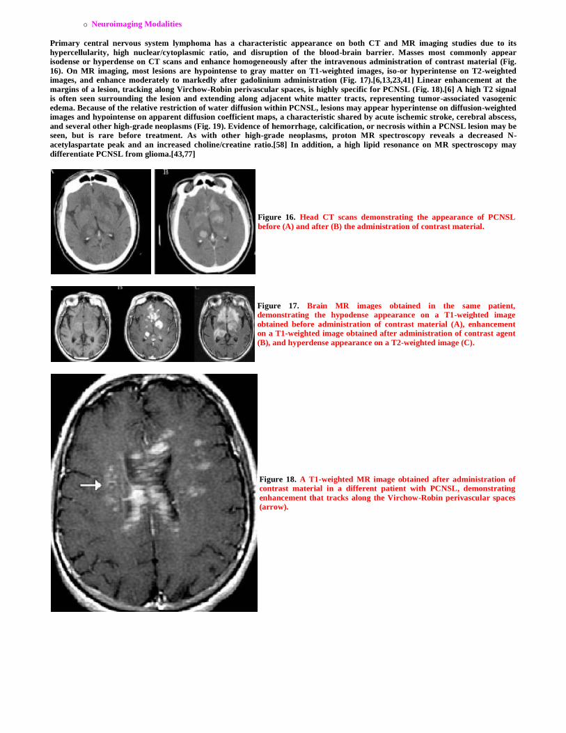
Primary central nervous system lymphoma has a characteristic appearance on both CT and MR imaging studies due to its hypercellularity, high nuclear/cytoplasmic ratio, and disruption of the blood-brain barrier. Masses most commonly appear isodense or hyperdense on CT scans and enhance homogeneously after the intravenous administration of contrast material (Fig. 16). On MR imaging, most lesions are hypointense to gray matter on T1-weighted images, iso-or hyperintense on T2-weighted images, and enhance moderately to markedly after gadolinium administration (Fig. 17).[6,13,23,41] Linear enhancement at the margins of a lesion, tracking along Virchow-Robin perivascular spaces, is highly specific for PCNSL (Fig. 18).[6] A high T2 signal is often seen surrounding the lesion and extending along adjacent white matter tracts, representing tumor-associated vasogenic edema. Because of the relative restriction of water diffusion within PCNSL, lesions may appear hyperintense on diffusion-weighted images and hypointense on apparent diffusion coefficient maps, a characteristic shared by acute ischemic stroke, cerebral abscess, and several other high-grade neoplasms (Fig. 19). Evidence of hemorrhage, calcification, or necrosis within a PCNSL lesion may be seen, but is rare before treatment. As with other high-grade neoplasms, proton MR spectroscopy reveals a decreased N-acetylaspartate peak and an increased choline/creatine ratio.[58] In addition, a high lipid resonance on MR spectroscopy may differentiate PCNSL from glioma.[43,77]
Figure 16. Head CT scans demonstrating the appearance of PCNSL before (A) and after (B) the administration of contrast material.
Figure 17. Brain MR images obtained in the same patient, demonstrating the hypodense appearance on a T1-weighted image obtained before administration of contrast material (A), enhancement on a T1-weighted image obtained after administration of contrast agent (B), and hyperdense appearance on a T2-weighted image (C).
Figure 18. A T1-weighted MR image obtained after administration of contrast material in a different patient with PCNSL, demonstrating enhancement that tracks along the Virchow-Robin perivascular spaces (arrow).
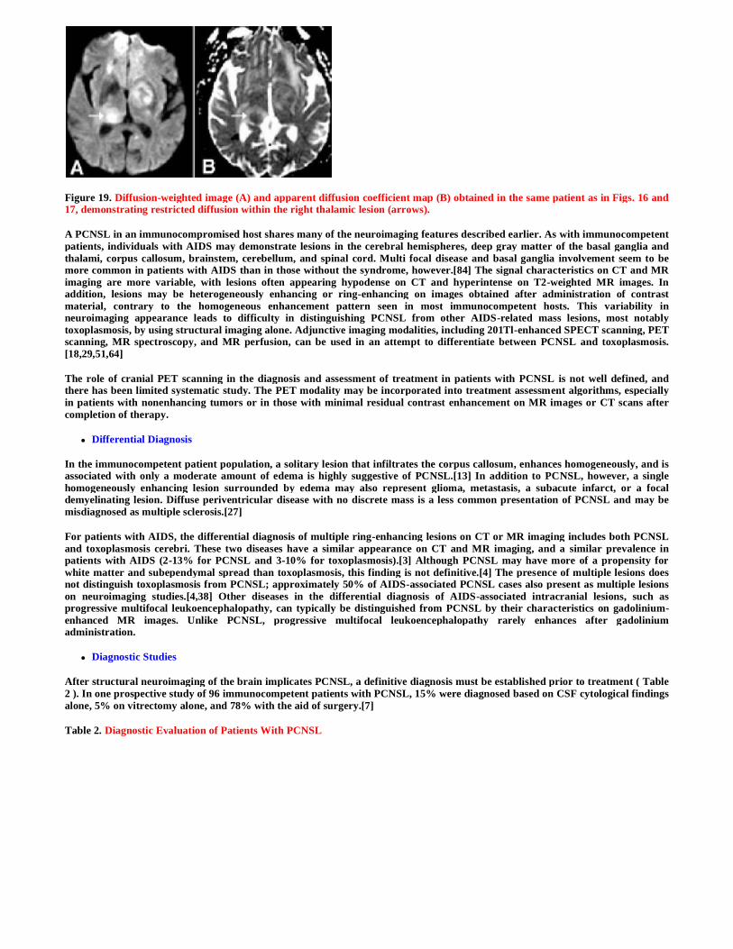
Figure 19. Diffusion-weighted image (A) and apparent diffusion coefficient map (B) obtained in the same patient as in Figs. 16 and 17, demonstrating restricted diffusion within the right thalamic lesion (arrows).
A PCNSL in an immunocompromised host shares many of the neuroimaging features described earlier. As with immunocompetent patients, individuals with AIDS may demonstrate lesions in the cerebral hemispheres, deep gray matter of the basal ganglia and thalami, corpus callosum, brainstem, cerebellum, and spinal cord. Multi focal disease and basal ganglia involvement seem to be more common in patients with AIDS than in those without the syndrome, however.[84] The signal characteristics on CT and MR imaging are more variable, with lesions often appearing hypodense on CT and hyperintense on T2-weighted MR images. In addition, lesions may be heterogeneously enhancing or ring-enhancing on images obtained after administration of contrast material, contrary to the homogeneous enhancement pattern seen in most immunocompetent hosts. This variability in neuroimaging appearance leads to difficulty in distinguishing PCNSL from other AIDS-related mass lesions, most notably toxoplasmosis, by using structural imaging alone. Adjunctive imaging modalities, including 201Tl-enhanced SPECT scanning, PET scanning, MR spectroscopy, and MR perfusion, can be used in an attempt to differentiate between PCNSL and toxoplasmosis.[18,29,51,64]
The role of cranial PET scanning in the diagnosis and assessment of treatment in patients with PCNSL is not well defined, and there has been limited systematic study. The PET modality may be incorporated into treatment assessment algorithms, especially in patients with nonenhancing tumors or in those with minimal residual contrast enhancement on MR images or CT scans after completion of therapy.
Differential Diagnosis
In the immunocompetent patient population, a solitary lesion that infiltrates the corpus callosum, enhances homogeneously, and is associated with only a moderate amount of edema is highly suggestive of PCNSL.[13] In addition to PCNSL, however, a single homogeneously enhancing lesion surrounded by edema may also represent glioma, metastasis, a subacute infarct, or a focal demyelinating lesion. Diffuse periventricular disease with no discrete mass is a less common presentation of PCNSL and may be misdiagnosed as multiple sclerosis.[27]
For patients with AIDS, the differential diagnosis of multiple ring-enhancing lesions on CT or MR imaging includes both PCNSL and toxoplasmosis cerebri. These two diseases have a similar appearance on CT and MR imaging, and a similar prevalence in patients with AIDS (2-13% for PCNSL and 3-10% for toxoplasmosis).[3] Although PCNSL may have more of a propensity for white matter and subependymal spread than toxoplasmosis, this finding is not definitive.[4] The presence of multiple lesions does not distinguish toxoplasmosis from PCNSL; approximately 50% of AIDS-associated PCNSL cases also present as multiple lesions on neuroimaging studies.[4,38] Other diseases in the differential diagnosis of AIDS-associated intracranial lesions, such as progressive multifocal leukoencephalopathy, can typically be distinguished from PCNSL by their characteristics on gadolinium-enhanced MR images. Unlike PCNSL, progressive multifocal leukoencephalopathy rarely enhances after gadolinium administration.
Diagnostic Studies
After structural neuroimaging of the brain implicates PCNSL, a definitive diagnosis must be established prior to treatment ( Table 2 ). In one prospective study of 96 immunocompetent patients with PCNSL, 15% were diagnosed based on CSF cytological findings alone, 5% on vitrectomy alone, and 78% with the aid of surgery.[7]
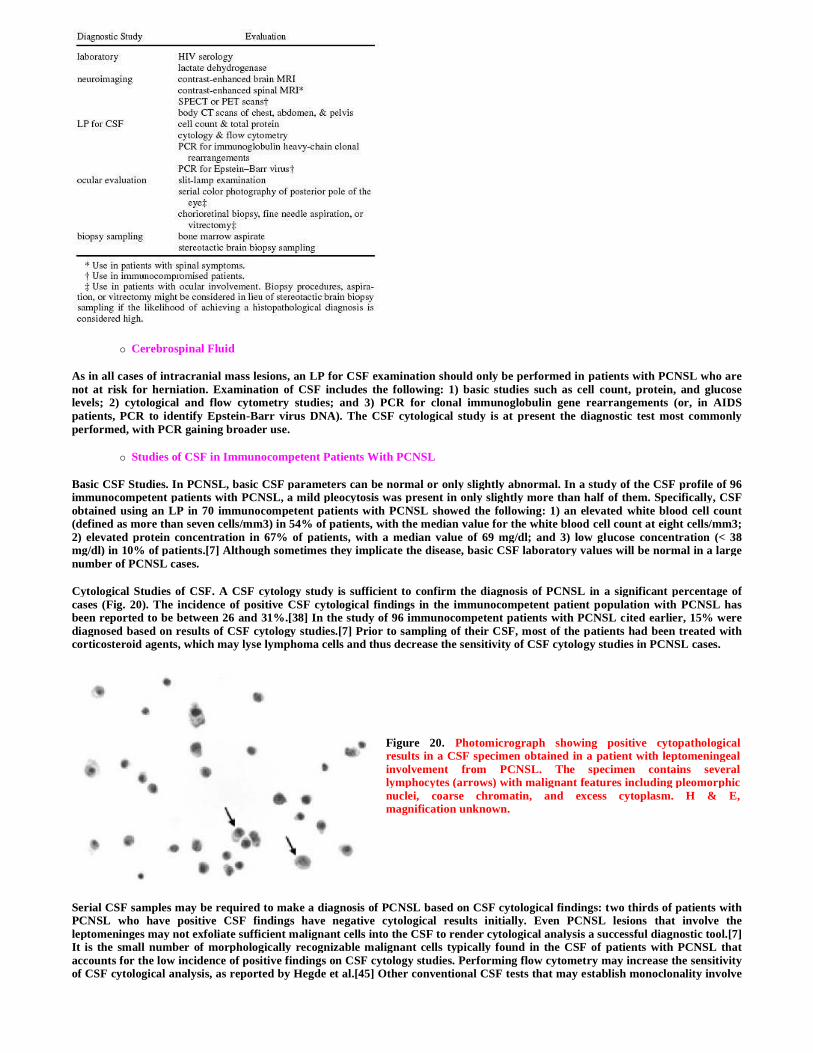
Table 2. Diagnostic Evaluation of Patients With PCNSL
Cerebrospinal Fluid
As in all cases of intracranial mass lesions, an LP for CSF examination should only be performed in patients with PCNSL who are not at risk for herniation. Examination of CSF includes the following: 1) basic studies such as cell count, protein, and glucose levels; 2) cytological and flow cytometry studies; and 3) PCR for clonal immunoglobulin gene rearrangements (or, in AIDS patients, PCR to identify Epstein-Barr virus DNA). The CSF cytological study is at present the diagnostic test most commonly performed, with PCR gaining broader use.
Studies of CSF in Immunocompetent Patients With PCNSL
Basic CSF Studies. In PCNSL, basic CSF parameters can be normal or only slightly abnormal. In a study of the CSF profile of 96 immunocompetent patients with PCNSL, a mild pleocytosis was present in only slightly more than half of them. Specifically, CSF obtained using an LP in 70 immunocompetent patients with PCNSL showed the following: 1) an elevated white blood cell count (defined as more than seven cells/mm3) in 54% of patients, with the median value for the white blood cell count at eight cells/mm3; 2) elevated protein concentration in 67% of patients, with a median value of 69 mg/dl; and 3) low glucose concentration (< 38 mg/dl) in 10% of patients.[7] Although sometimes they implicate the disease, basic CSF laboratory values will be normal in a large number of PCNSL cases.
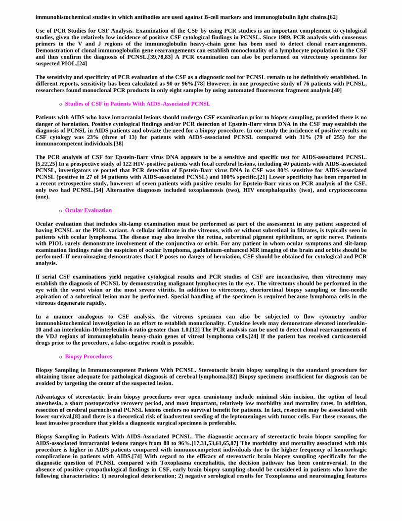
Cytological Studies of CSF. A CSF cytology study is sufficient to confirm the diagnosis of PCNSL in a significant percentage of cases (Fig. 20). The incidence of positive CSF cytological findings in the immunocompetent patient population with PCNSL has been reported to be between 26 and 31%.[38] In the study of 96 immunocompetent patients with PCNSL cited earlier, 15% were diagnosed based on results of CSF cytology studies.[7] Prior to sampling of their CSF, most of the patients had been treated with corticosteroid agents, which may lyse lymphoma cells and thus decrease the sensitivity of CSF cytology studies in PCNSL cases.
Serial CSF samples may be required to make a diagnosis of PCNSL based on CSF cytological findings: two thirds of patients with PCNSL who have positive CSF findings have negative cytological results initially. Even PCNSL lesions that involve the leptomeninges may not exfoliate sufficient malignant cells into the CSF to render cytological analysis a successful diagnostic tool.[7] It is the small number of morphologically recognizable malignant cells typically found in the CSF of patients with PCNSL that accounts for the low incidence of positive findings on CSF cytology studies. Performing flow cytometry may increase the sensitivity of CSF cytological analysis, as reported by Hegde et al.[45] Other conventional CSF tests that may establish monoclonality involve
Figure 20. Photomicrograph showing positive cytopathological results in a CSF specimen obtained in a patient with leptomeningeal involvement from PCNSL. The specimen contains several lymphocytes (arrows) with malignant features including pleomorphic nuclei, coarse chromatin, and excess cytoplasm. H & E, magnification unknown.
immunohistochemical studies in which antibodies are used against B-cell markers and immunoglobulin light chains.[62]
Use of PCR Studies for CSF Analysis. Examination of the CSF by using PCR studies is an important complement to cytological studies, given the relatively low incidence of positive CSF cytological findings in PCNSL. Since 1989, PCR analysis with consensus primers to the V and J regions of the immunoglobulin heavy-chain gene has been used to detect clonal rearrangements. Demonstration of clonal immunoglobulin gene rearrangements can establish monoclonality of a lymphocyte population in the CSF and thus confirm the diagnosis of PCNSL.[39,78,83] A PCR examination can also be performed on vitrectomy specimens for suspected PIOL.[24]
The sensitivity and specificity of PCR evaluation of the CSF as a diagnostic tool for PCNSL remain to be definitively established. In different reports, sensitivity has been calculated as 90 or 96%.[78] However, in one prospective study of 76 patients with PCNSL, researchers found monoclonal PCR products in only eight samples by using automated fluorescent fragment analysis.[40]
Studies of CSF in Patients With AIDS-Associated PCNSL
Patients with AIDS who have intracranial lesions should undergo CSF examination prior to biopsy sampling, provided there is no danger of herniation. Positive cytological findings and/or PCR detection of Epstein-Barr virus DNA in the CSF may establish the diagnosis of PCNSL in AIDS patients and obviate the need for a biopsy procedure. In one study the incidence of positive results on CSF cytology was 23% (three of 13) for patients with AIDS-associated PCNSL compared with 31% (79 of 255) for the immunocompetent individuals.[38]
The PCR analysis of CSF for Epstein-Barr virus DNA appears to be a sensitive and specific test for AIDS-associated PCNSL.[5,22,25] In a prospective study of 122 HIV-positive patients with focal cerebral lesions, including 40 patients with AIDS-associated PCNSL, investigators re ported that PCR detection of Epstein-Barr virus DNA in CSF was 80% sensitive for AIDS-associated PCNSL (positive in 27 of 34 patients with AIDS-associated PCNSL) and 100% specific.[21] Lower specificity has been reported in a recent retrospective study, however: of seven patients with positive results for Epstein-Barr virus on PCR analysis of the CSF, only two had PCNSL.[54] Alternative diagnoses included toxoplasmosis (two), HIV encephalopathy (two), and cryptococcoma (one).
Ocular Evaluation
Ocular evaluation that includes slit-lamp examination must be performed as part of the assessment in any patient suspected of having PCNSL or the PIOL variant. A cellular infiltrate in the vitreous, with or without subretinal in filtrates, is typically seen in patients with ocular lymphoma. The disease may also involve the retina, subretinal pigment epithelium, or optic nerve. Patients with PIOL rarely demonstrate involvement of the conjunctiva or orbit. For any patient in whom ocular symptoms and slit-lamp examination findings raise the suspicion of ocular lymphoma, gadolinium-enhanced MR imaging of the brain and orbits should be performed. If neuroimaging demonstrates that LP poses no danger of herniation, CSF should be obtained for cytological and PCR analysis.
If serial CSF examinations yield negative cytological results and PCR studies of CSF are inconclusive, then vitrectomy may establish the diagnosis of PCNSL by demonstrating malignant lymphocytes in the eye. The vitrectomy should be performed in the eye with the worst vision or the most severe vitritis. In addition to vitrectomy, chorioretinal biopsy sampling or fine-needle aspiration of a subretinal lesion may be performed. Special handling of the specimen is required because lymphoma cells in the vitreous degenerate rapidly.
In a manner analogous to CSF analysis, the vitreous specimen can also be subjected to flow cytometry and/or immunohistochemical investigation in an effort to establish monoclonality. Cytokine levels may demonstrate elevated interleukin-10 and an interleukin-10/interleukin-6 ratio greater than 1.0.[12] The PCR analysis can be used to detect clonal rearrangements of the VDJ regions of immunoglobulin heavy-chain genes of vitreal lymphoma cells.[24] If the patient has received corticosteroid drugs prior to the procedure, a false-negative result is possible.
Biopsy Procedures
Biopsy Sampling in Immunocompetent Patients With PCNSL. Stereotactic brain biopsy sampling is the standard procedure for obtaining tissue adequate for pathological diagnosis of cerebral lymphoma.[82] Biopsy specimens insufficient for diagnosis can be avoided by targeting the center of the suspected lesion.
Advantages of stereotactic brain biopsy procedures over open craniotomy include minimal skin incision, the option of local anesthesia, a short postoperative recovery period, and most important, relatively low morbidity and mortality rates. In addition, resection of cerebral parenchymal PCNSL lesions confers no survival benefit for patients. In fact, resection may be associated with lower survival,[8] and there is a theoretical risk of inadvertent seeding of the leptomeninges with tumor cells. For these reasons, the least invasive procedure that yields a diagnostic surgical specimen is preferable.
Biopsy Sampling in Patients With AIDS-Associated PCNSL. The diagnostic accuracy of stereotactic brain biopsy sampling for AIDS-associated intracranial lesions ranges from 88 to 96%.[17,31,53,61,65,87] The morbidity and mortality associated with this procedure is higher in AIDS patients compared with immunocompetent individuals due to the higher frequency of hemorrhagic complications in patients with AIDS.[74] With regard to the efficacy of stereotactic brain biopsy sampling specifically for the diagnostic question of PCNSL compared with Toxoplasma encephalitis, the decision pathway has been controversial. In the absence of positive cytopathological findings in CSF, early brain biopsy sampling should be considered in patients who have the following characteristics: 1) neurological deterioration; 2) negative serological results for Toxoplasma and neuroimaging features
atypical for toxoplasmosis; 3) discordant results between PCR of the CSF for Epstein-Barr virus and thallium-enhanced SPECT scans, if other infectious origins are not suspected; and 4) improvement during a brief trial of therapy for toxoplasmosis.[4,56]
Extent of Disease Evaluation
After the diagnosis of PCNSL has been established with analysis of CSF, a vitreal aspirate, or a biopsy specimen, an extent of disease evaluation should be performed in every patient. Full ocular evaluation, including a slit-lamp examination, should be done in every patient, because asymptomatic ocular involvement is not uncommon and specific treatment is indicated for ocular lymphoma.[32] In addition, in patients who are deemed eligible for LP, the CSF should be collected for cell count, chemistry, cytopathology, flow cytometry, and PCR tests. The serum lactate dehydrogenase level should be measured in every case because this parameter is an important prognostic factor in patients with PCNSL.[34] Because PCNSL occurs so commonly in patients with AIDS, HIV serological studies should be requested for every apparently immunocompetent patient in whom PCNSL is diagnosed. Evaluation of cognitive function is important both at diagnosis and in follow-up visits to gauge the benefit of therapy and to monitor for treatment-related neurocognitive decline.[2]
Given the rarity of spinal cord involvement, enhanced spinal MR imaging is indicated only in cases in which clinical suspicion of spinal cord tumor is high.[26] However, contrast-enhanced MR imaging of the entire spine should be considered in patients who cannot undergo an LP due to the presence of increased intracranial pressure. In this situation, spinal imaging may identify leptomeningeal deposits of tumor, and this could affect subsequent management.
Evidence of systemic lymphoma has been found at the time of diagnosis in up to 8% of patients initially thought to have isolated PCNSL.[35,63,69,71] For this reason, complete systemic staging, including CT scans of the chest, abdomen, and pelvis and bone marrow biopsy sampling with aspirate has been recommended by the International Primary CNS Lymphoma Collaborative Group.[2] Use of testicular ultrasonography should be considered in men to rule out occult testicular lymphoma that has metastasized to the brain. Whole-body PET or PET-CT scanning may be incorporated into routine extent of disease evaluations in the future.
Systemic dissemination of lymphoma is found over time in 7 to 10% of patients with PCNSL, generally in the terminal stages of the disease or at postmortem examination. In such cases, lymphoma may be found in the lymph nodes and visceral organs of the abdomen/retroperitoneum, mediastinal lymph nodes, lungs, bone marrow, or testes. The clinical significance of late-disease, systemic deposits of lymphoma is controversial.[46,68] During the course of disease, if a patient with PCNSL presents with new ocular or other CNS symptoms and signs, reevaluation–for example, with repeated slit-lamp examination or LP–is indicated.[11,76]
Conclusions
The clinical presentation and neuroimaging appearance of PCNSL differ in immunocompetent and AIDS patients and are nonspecific in both. Magnetic resonance imaging of the brain revealing a homogeneously enhancing, single lesion that infiltrates the corpus callosum is highly suggestive of PCNSL in immunocompetent patients, whereas multiple ring-enhancing lesions are more common in patients with AIDS. After neuroimages are obtained that raise the suspicion of PCNSL, a definitive diagnosis should be established in both immunocompetent and AIDS patients by analysis of CSF, vitreous fluid, or biopsy specimens. Brain biopsy sampling remains the gold standard for PCNSL diagnosis in all patients, although the possibility of routine, minimally invasive diagnosis by using PCR analysis of the CSF and nuclear imaging is currently under investigation. At the time of diagnosis, the patient should undergo an evaluation of the extent of disease that includes the following: a physical examination; ophthal mic evaluation with a slit-lamp examination; serum lactate dehydrogenase evaluation; HIV testing; chest/abdomen/pelvis CT scans; bone marrow biopsy sampling; contrast-enhanced brain MR imaging; and LP. In patients who cannot undergo LP or in those with evidence of spinal cord dysfunction, a contrast-enhanced MR image of the entire spine should be considered.
Abbreviation Notes
PCNSL = primary central nervous system lymphoma; PCR = polymerase chain reaction; PET = positron emission tomography; PIOL = primary intraocular lymphoma; SPECT = single-photon emission computed tomography
SUMMARY:
In general epidural lymphomas (spinal or intracranial) and lymphomatous leptomeningitis are diseases that respect the dura. Epidural lymphomas (spinal or intracranial) start in the epidural spaces and usually extensively spread up and down alongside the meninges, yet they remain confined to the epidural spaces, and infiltration or penetration of the dura never occur. In general secondary lymphomas, being confined to the extra dural spaces, are not primarily a CNS disease. The disease usually extends to the epidural spaces after more extensive extra-neural dissemination. 1
SUMMARY
On the other hand PCNSLs are primarily intraparenchymal neural lesions. Intraaxial (brain or spinal) lesions are mandatory for the diagnosis of primary CNS lymphomas. The existence parenchymal intraaxial CNS disease should obviate the need for staging as the disease is invariably primary. Although extradural disease is secondary to systemic lymphoma, however it can be the initial presentation of the disease, and subsequently staging is mandatory whenever epidural lymphoma deposits are discovered.
In general epidural (spinal or intracranial) lymphomas and lymphomatous leptomeningitis are different from the PCNSL in the following points. [88]
PCNSL is a histiocytic lymphoma and this is consistent with the microglial origin of the PCNSL (microglial cells are derived from blood monocytes) while secondary lymphomas are lymphocytic lymphomas, and this is probably due to the fact that spinal lymphomas are secondary to systemic lymphomas, lymphocytic lymphomas are the most common systemic lymphomas. [88]
Although the histiocytic subtype is more aggressive than the lymphocytic subtype, the prognosis of PCNSL is much better compared with other CNS lymphomas. [88] However, it should be noted that all patients with PCNSL are in stage one disease (disease confined to a single extra lymphocytic site), while all patients with epidural (spinal or intracranial) lymphomas are in stage IV disease. It looks like that it is the stage of the disease, rather than the histopathological subtype, that ultimately determines the prognosis. [88]
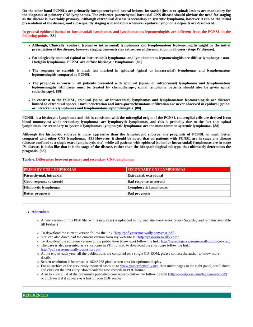
Table 6. Differences between primary and secondary CNS lymphomas
Addendum
A new version of this PDF file (with a new case) is uploaded in my web site every week (every Saturday and remains available till Friday.)
To download the current version follow the link "http://pdf.yassermetwally.com/case.pdf". You can also download the current version from my web site at "http://yassermetwally.com". To download the software version of the publication (crow.exe) follow the link: http://neurology.yassermetwally.com/crow.zip The case is also presented as a short case in PDF format, to download the short case follow the link:
http://pdf.yassermetwally.com/short.pdf At the end of each year, all the publications are compiled on a single CD-ROM, please contact the author to know more
details. Screen resolution is better set at 1024*768 pixel screen area for optimum display. For an archive of the previously reported cases go to www.yassermetwally.net, then under pages in the right panel, scroll down
and click on the text entry "downloadable case records in PDF format" Also to view a list of the previously published case records follow the following link (http://wordpress.com/tag/case-record/)
or click on it if it appears as a link in your PDF reader
Although, Clinically, epidural (spinal or intracranial) lymphomas and lymphomatous leptomeningitis might be the initial presentation of the disease, however staging demonstrates extra-neural dissemination in all cases (stage IV disease).
Pathologically epidural (spinal or intracranial) lymphomas and lymphomatous leptomeningitis are diffuse lymphocytic non-Hodgkin lymphomas. PCNSL are diffuse histiocytic lymphomas. [88]
The response to steroids is much less marked in epidural (spinal or intracranial) lymphomas and lymphomatous leptomeningitis compared to PCNSL.
The prognosis is worse in all patients presented with epidural (spinal or intracranial) lymphomas and lymphomatous leptomeningitis (All cases must be treated by chemotherapy, spinal lymphoma patients should also be given spinal radiotherapy). [88]
In contrast to the PCNSL, epidural (spinal or intracranial) lymphomas and lymphomatous leptomeningitis are diseases limited to extradural spaces. Dural penetration and intra-parenchymatous infiltration are never observed in epidural (spinal or intracranial) lymphomas and lymphomatous leptomeningitis. [88]
PRIMARY CNS LYMPHOMAS SECONDARY CNS LYMPHOMAS Parenchymal, intraaxial Extraaxial, extradural Good response to steroid Bad response to steroid Histiocytic lymphomas Lymphocytic lymphomas Better prognosis Bad prognosis
REFERENCES

References
1. Abla O, Sandlund JT, Sung L, Brock P, Corbett R, Kirov I, et al: A case series of pediatric primary central nervous system lymphoma: favorable outcome without cranial irradiation. Pediatr Blood Cancer 47: 880-885, 2005
2. Abrey LE, Batchelor TT, Ferreri AJ, Gospodarowicz M, Pulczynski EJ, Zucca E, et al: Report of an international workshop to standardize baseline evaluation and response criteria for primary CNS lymphoma. J Clin Oncol 23: 5034-5043, 2005
3. Anonymous: Evaluation and management of intracranial mass lesions in AIDS. Report of the Quality Standards Subcommittee of the American Academy of Neurology. Neurology 50: 21-26, 1998
4. Antinori A, Ammassari A, De Luca A, Cingolani A, Murri R, Scoppettuolo G, et al: Diagnosis of AIDS-related focal brain lesions: a decision-making analysis based on clinical and neuroradiologic characteristics combined with polymerase chain reaction assays in CSF. Neurology 48: 687-694, 1997
5. Arribas JR, Clifford DB, Fichtenbaum CJ, Roberts RL, Powderly WG, Storch GA: Detection of Epstein-Barr virus DNA in cerebrospinal fluid for diagnosis of AIDS-related central nervous system lymphoma. J Clin Microbiol 33: 1580-1583, 1995
6. Atlas SW: Magnetic Resonance Imaging of the Brain and Spine, ed 3. Philadelphia: Lippincott Williams & Wilkins, 2002
7. Balmaceda C, Gaynor JJ, Sun M, Gluck JT, DeAngelis LM: Leptomeningeal tumor in primary central nervous system lymphoma: recognition, significance, and implications. Ann Neurol 38: 202-209, 1995
8. Bataille B, Delwail V, Menet E, Vandermarcq P, Ingrand P, Wager M, et al: Primary intracerebral malignant lymphoma: report of 248 cases. J Neurosurg 92: 261-266, 2000
9. Besson C, Goubar A, Gabarre J, Rozenbaum W, Pialoux G, Chatelet FP, et al: Changes in AIDS-related lymphoma since the era of highly active antiretroviral therapy. Blood 98: 2339-2344, 2001
10. Braus DF, Schwechheimer K, Muller-Hermelink HK, Schwarzkopf G, Volk B, Mundinger F: Primary cerebral malignant non-Hodgkin's lymphomas: a retrospective clinical study. J Neurol 239: 117-124, 1992
11. Buggage RR, Chan CC, Nussenblatt RB: Ocular manifestations of central nervous system lymphoma. Curr Opin Oncol 13: 137-142, 2001
12. Buggage RR, Whitcup SM, Nussenblatt RB, Chan CC: Using interleukin 10 to interleukin 6 ratio to distinguish primary intra ocular lymphoma and uveitis. Invest Ophthalmol Vis Sci 40: 2462-2463, 1999
13. Buhring U, Herrlinger U, Krings T, Thiex R, Weller M, Kuker W: MRI features of primary central nervous system lymphomas at presentation. Neurology 57: 393-396, 2001
14. Camilleri-Broet S, Martin A, Moreau A, Angonin R, Henin D, Gontier MF, et al: Primary central nervous system lymphomas in 72 immunocompetent patients: pathologic findings and clinical correlations. Groupe Ouest Est d'etude des Leucenies et Autres Maladies du Sang (GOELAMS). Am J Clin Pathol 110: 607-612, 1998
15. Caruso PA, Patel MR, Joseph J, Rachlin J: Primary intra medullary lymphoma of the spinal cord mimicking cervical spondylotic myelopathy. AJR Am J Roentgenol 171: 526-527, 1998
16. CBTRUS: Primary Brain Tumors in the United States, 1998-2002. Statistical Report. Chicago: Central Brain Tumor Registry of the United States, 2005
17. Chappell ET, Guthrie BL, Orenstein J: The role of stereotactic biopsy in the management of HIV-related focal brain lesions. Neurosurgery 30: 825-829, 1992
18. Chinn RJ, Wilkinson ID, Hall-Craggs MA, Paley MN, Miller RF, Kendall BE, et al: Toxoplasmosis and primary central nervous system lymphoma in HIV infection: diagnosis with MR spectroscopy. Radiology 197: 649-654, 1995
19. Chow KU, Mitrou PS, Geduldig K, Helm EB, Hoelzer D, Brodt HR: Changing incidence and survival in patients with AIDS-related non-Hodgkin's lymphomas in the era of highly active antiretroviral therapy (HAART). Leuk Lymphoma 41: 105-116, 2001
20. Ciacci JD, Tellez C, VonRoenn J, Levy RM: Lymphoma of the central nervous system in AIDS. Semin Neurol 19: 213-221, 1999
21. Cingolani A, De Luca A, Larocca LM, Ammassari A, Scerrati M, Antinori A, et al: Minimally invasive diagnosis of acquired immunodeficiency syndrome-related primary central nervous system lymphoma. J Natl Cancer Inst 90: 364-369, 1998
22. Cinque P, Brytting M, Vago L, Castagna A, Parravicini C, Zanchetta N, et al: Epstein-Barr virus DNA in cerebrospinal fluid from patients with AIDS-related primary lymphoma of the central nervous system. Lancet 342: 398-401, 1993
23. Coulon A, Lafitte F, Hoang-Xuan K, Martin-Duverneuil N, Mokhtari K, Blustajn J, et al: Radiographic findings in 37 cases of primary CNS lymphoma in immunocompetent patients. Eur Radiol 12: 329-340, 2002
24. Coupland SE, Hummel M, Muller HH, Stein H: Molecular analysis of immunoglobulin genes in primary intraocular lymphoma. Invest Ophthalmol Vis Sci 46: 3507-3514, 2005
25. De Luca A, Antinori A, Cingolani A, Larocca LM, Linzalone A, Ammassari A, et al: Evaluation of cerebrospinal fluid EBV-DNA and IL-10 as markers for in vivo diagnosis of AIDS-related primary central nervous system lymphoma. Br J Haematol 90: 844-849, 1995
26. DeAngelis LM: Primary central nervous system lymphoma. Curr Opin Neurol 12: 687-691, 1999
27. DeAngelis LM: Primary central nervous system lymphoma imitates multiple sclerosis. J Neurooncol 9: 177-181, 1990
28. Eby NL, Grufferman S, Flannelly CM, Schold SC Jr, Vogel FS, Burger PC: Increasing incidence of primary brain lymphoma in the US. Cancer 62: 2461-2465, 1988
29. Ernst TM, Chang L, Witt MD, Aronow HA, Cornford ME, Walot I, et al: Cerebral toxoplasmosis and lymphoma in AIDS: perfusion MR imaging experience in 13 patients. Radiology 208: 663-669, 1998
30. Esptein LG, DiCarlo FJ Jr, Joshi VV, Connor EM, Oleske JM, Kay D, et al: Primary lymphoma of the central nervous system in children with acquired immunodeficiency syndrome. Pediatrics 82: 355-363, 1988
31. Feiden W, Bise K, Steude U, Pfister HW, Moller AA: The stereotactic biopsy diagnosis of focal intracerebral lesions in AIDS patients. Acta Neurol Scand 87: 228-233, 1993
32. Ferreri AJ, Blay JY, Reni M, Pasini F, Gubkin A, Tirelli U, et al: Relevance of intraocular involvement in the management of primary central nervous system lymphomas. Ann Oncol 13: 531-538, 2002
33. Ferreri AJ, Blay JY, Reni M, Pasini F, Spina M, Ambrosetti A, et al: Prognostic scoring system for primary CNS lymphomas: the International Extranodal Lymphoma Study Group experience. J Clin Oncol 21: 266-272, 2003
34. Ferreri AJ, Reni M, Pasini F, Calderoni A, Tirelli U, Pivnik A, et al: A multicenter study of treatment of primary CNS lymphoma. Neurology 58: 1513-1520, 2002
35. Ferreri AJ, Reni M, Zoldan MC, Terreni MR, Villa E: Importance of complete staging in non-Hodgkin's lymphoma presenting as a cerebral mass lesion. Cancer 77: 827-833, 1996
36. Feuerhake F, Baumer C, Cyron D, Illerhaus G, Olschewski M, Tilgner J, et al: Primary CNS lymphoma in immunocompetent patients from 1989 to 2001: a retrospective analysis of 164 cases uniformly diagnosed by stereotactic biopsy. Acta Neurochir (Wien) 148: 831-838, 2006
37. Fine HA, Loeffler JS: Primary central nervous system lymphoma, in Canellos GP, Lister TA, Sklar JL (eds): The Lymphomas. Philadelphia: W.B. Saunders, 1998, pp 481-494
38. Fine HA, Mayer RJ: Primary central nervous system lymphoma. Ann Intern Med 119: 1093-1104, 1993
39. Galoin S, Daste G, Apoil PA, Chollet F, Roda D, Blancher A, et al: Polymerase chain reaction on cerebrospinal fluid cells in the detection of leptomeningeal involvement by B-cell lymphoma and leukaemia: a novel strategy and its implications. Br J Haematol 99: 122-130, 1997
40. Gleissner B, Siehl J, Korfel A, Reinhardt R, Thiel E: CSF evaluation in primary CNS lymphoma patients by PCR of the CDR III IgH genes. Neurology 58: 390-396, 2002
41. Gliemroth J, Kehler U, Gaebel C, Arnold H, Missler U: Neuroradiological findings in primary cerebral lymphomas of non-AIDS patients. Clin Neurol Neurosurg 105: 78-86, 2003
42. Hao D, DiFrancesco LM, Brasher PM, deMetz C, Fulton DS, DeAngelis LM, et al: Is primary CNS lymphoma really becoming more common? A population-based study of incidence, clinicopathological features and outcomes in Alberta from 1975 to 1996. Ann Oncol 10: 65-70, 1999
43. Harting I, Hartmann M, Jost G, Sommer C, Ahmadi R, Heiland S, et al: Differentiating primary central nervous system lymphoma from glioma in humans using localised proton magnetic resonance spectroscopy. Neurosci Lett 342: 163-166, 2003
44. Hayabuchi N, Shibamoto Y, Onizuka Y: Primary central nervous system lymphoma in Japan: a nationwide survey. Int J Radiat Oncol Biol Phys 44: 265-272, 1999
45. Hegde U, Filie A, Little RF, Janik JE, Grant N, Steinberg SM, et al: High incidence of occult leptomeningeal disease detected by flow cytometry in newly diagnosed aggressive B-cell lymphomas at risk for central nervous system involvement: the role of flow cytometry versus cytology. Blood 105: 496-502, 2005
46. Herbst KD, Corder MP, Justice GR: Successful therapy with methotrexate of a multicentric mixed lymphoma of the central nervous system. Cancer 38: 1476-1478, 1976
47. Herrlinger U, Schabet M, Bitzer M, Petersen D, Krauseneck P: Primary central nervous system lymphoma: from clinical presentation to diagnosis. J Neurooncol 43: 219-226, 1999
48. Herrlinger U, Schabet M, Brugger W, Kortmann RD, Kanz L, Bamberg M, et al: Primary central nervous system lymphoma 1991-1997: outcome and late adverse effects after combined modality treatment. Cancer 91: 130-135, 2001
49. Herrlinger U, Schabet M, Clemens M, Kortmann RD, Petersen D, Will BE, et al: Clinical presentation and therapeutic outcome in 26 patients with primary CNS lymphoma. Acta Neurol Scand 97: 257-264, 1998
50. Herrlinger U, Weller M, Kuker W: Primary CNS lymphoma in the spinal cord: clinical manifestations may precede MRI detectability. Neuroradiology 44: 239-244, 2002
51. Hoffman JM, Waskin HA, Schifter T, Hanson MW, Gray L, Rosenfeld S, et al: FDG-PET in differentiating lymphoma from nonmalignant central nervous system lesions in patients with AIDS. J Nucl Med 34: 567-575, 1993
52. Hormigo A, Abrey L, Heinemann MH, DeAngelis LM: Ocular presentation of primary central nervous system lymphoma: diagnosis and treatment. Br J Haematol 126: 202-208, 2004
53. Iacoangeli M, Roselli R, Antinori A, Ammassari A, Murri R, Pompucci A, et al: Experience with brain biopsy in acquired immune deficiency syndrome-related focal lesions of the central nervous system. Br J Surg 81: 1508-1511, 1994
54. Ivers LC, Kim AY, Sax PE: Predictive value of polymerase chain reaction of cerebrospinal fluid for detection of Epstein-Barr virus to establish the diagnosis of HIV-related primary central nervous system lymphoma. Clin Infect Dis 38: 1629-1632, 2004
55. Kadan-Lottick NS, Skluzacek MC, Gurney JG: Decreasing incidence rates of primary central nervous system lymphoma. Cancer 95: 193-202, 2002
56. Kasamon YL, Ambinder RF: AIDS-related primary central nervous system lymphoma. Hematol Oncol Clin North Am 19: 665-687, 2005
57. Kawasaki K, Wakabayashi K, Koizumi T, Tanaka R, Takahashi H: Spinal cord involvement of primary central nervous system lymphomas: histopathological examination of 14 autopsy cases. Neuropathology 22: 13-18, 2002
58. Kuker W, Nagele T, Korfel A, Heckl S, Thiel E, Bamberg M, et al: Primary central nervous system lymphomas (PCNSL): MRI features at presentation in 100 patients. J Neurooncol 72: 169-177, 2005
59. Lachance DH, O'Neill BP, Macdonald DR, Jaeckle KA, Witzig TE, Li CY, et al: Primary leptomeningeal lymphoma: report of 9 cases, diagnosis with immunocytochemical analysis, and re view of the literature. Neurology 41: 95-100, 1991
60. Lee DK, Chung CK, Kim HJ, Kim K, Choe G, Moon CW, et al: Multifocal primary CNS T cell lymphoma of the spinal cord. Clin Neuropathol 21: 149-155, 2002
61. Levy RM, Russell E, Yungbluth M, Hidvegi DF, Brody BA, Dal Canto MC: The efficacy of image-guided stereotactic brain biopsy in neurologically symptomatic acquired immunodeficiency syndrome patients. Neurosurgery 30: 186-190, 1992
62. Li CY, Witzig TE, Phyliky RL, Ziesmer SC, Yam LT: Diagnosis of B-cell non-Hodgkin's lymphoma of the central nervous system by immunocytochemical analysis of cerebrospinal fluid lymphocytes. Cancer 57: 737-744, 1986
63. Loeffler JS, Ervin TJ, Mauch P, Skarin A, Weinstein HJ, Canellos G, et al: Primary lymphomas of the central nervous system: patterns of failure and factors that influence survival. J Clin Oncol 3: 490-494, 1985
64. Lorberboym M, Estok L, Machac J, Germano I, Sacher M, Feldman R, et al: Rapid differential diagnosis of cerebral toxoplasmosis and primary central nervous system lymphoma by thallium-201 SPECT. J Nucl Med 37: 1150-1154, 1996
65. Luzzati R, Ferrari S, Nicolato A, Piovan E, Malena M, Merighi M, et al: Stereotactic brain biopsy in human immunodeficiency virus-infected patients. Arch Intern Med 156: 565-568, 1996
66. Miller DC, Hochberg FH, Harris NL, Gruber ML, Louis DN, Cohen H: Pathology with clinical correlations of primary central nervous system non-Hodgkin's lymphoma. The Massachusetts General Hospital experience 1958-1989. Cancer 74: 1383-1397, 1994
67. Monnard V, Sun A, Epelbaum R, Poortmans P, Miller RC, Verschueren T, et al: Primary spinal epidural lymphoma: patients' profile, outcome, and prognostic factors: a multicenter Rare Cancer Network study. Int J Radiat Oncol Biol Phys 65: 817-823, 2006
68. Nasir S, DeAngelis LM: Update on the management of primary CNS lymphoma. Oncology (Williston Park) 14: 228-242, 244,

2000
69. Nelson DF, Martz KL, Bonner H, Nelson JS, Newall J, Kerman HD, et al: Non-Hodgkin's lymphoma of the brain: can high dose, large volume radiation therapy improve survival? Report on a prospective trial by the Radiation Therapy Oncology Group (RTOG): RTOG 8315. Int J Radiat Oncol Biol Phys 23: 9-17, 1992
70. Newell ME, Hoy JF, Cooper SG, DeGraaff B, Grulich AE, Bryant M, et al: Human immunodeficiency virus-related primary central nervous system lymphoma: factors influencing survival in 111 patients. Cancer 100: 2627-2636, 2004
71. O'Neill BP, Dinapoli RP, Kurtin PJ, Habermann TM: Occult systemic non-Hodgkin's lymphoma (NHL) in patients initially diagnosed as primary central nervous system lymphoma (PCNSL): how much staging is enough? J Neurooncol 25: 67-71, 1995
72. Olson JE, Janney CA, Rao RD, Cerhan JR, Kurtin PJ, Schiff D, et al: The continuing increase in the incidence of primary central nervous system non-Hodgkin lymphoma: a surveillance, epidemiology, and end results analysis. Cancer 95: 1504-1510, 2002
73. Panageas KS, Elkin EB, DeAngelis LM, Ben-Porat L, Abrey LE: Trends in survival from primary central nervous system lymphoma, 1975-1999: a population-based analysis. Cancer 104: 2466-2472, 2005
74. Pell MF, Thomas DG, Whittle IR: Stereotactic biopsy of cerebral lesions in patients with AIDS. Br J Neurosurg 5: 585-589, 1991
75. Pels H, Vogt I, Klockgether T, Schlegel U: Primary non-Hodgkin's lymphoma of the spinal cord. Spine 25: 2262-2264, 2000
76. Peterson K, Gordon KB, Heinemann MH, DeAngelis LM: The clinical spectrum of ocular lymphoma. Cancer 72: 843-849, 1993
77. Raizer JJ, Koutcher JA, Abrey LE, Panageas KS, DeAngelis LM, Lis E, et al: Proton magnetic resonance spectroscopy in immunocompetent patients with primary central nervous system lymphoma. J Neurooncol 71: 173-180, 2005
78. Rhodes CH, Glantz MJ, Glantz L, Lekos A, Sorenson GD, Honsinger C, et al: A comparison of polymerase chain reaction examination of cerebrospinal fluid and conventional cytology in the diagnosis of lymphomatous meningitis. Cancer 77: 543-548, 1996
79. Salvesen Haldorsen I, Aarseth JH, Hollender A, Larsen JL, Espeland A, Mella O: Incidence, clinical features, treatment and outcome of primary central nervous system lymphoma in Norway. Acta Oncol 43: 520-529, 2004
80. Schabet M: Epidemiology of primary CNS lymphoma. J Neurooncol 43: 199-201, 1999
81. Schild SE, Wharen RE Jr, Menke DM, Folger WN, Colon-Otero G: Primary lymphoma of the spinal cord. Mayo Clin Proc 70: 256-260, 1995
82. Sherman ME, Erozan YS, Mann RB, Kumar AA, McArthur JC, Royal W, et al: Stereotactic brain biopsy in the diagnosis of malignant lymphoma. Am J Clin Pathol 95: 878-883, 1991
83. Storch-Hagenlocher B, Haas J, Vogt-Schaden ME, Bentz M, Hoffmann LA, Biessmann A, et al: Molecular analysis of the CDR3 encoding region of the immunoglobulin heavy chain locus in cerebrospinal fluid cells as a diagnostic tool in lymphomatous meningitis. Ann Neurol 47: 211-217, 2000
84. Thurnher MM, Rieger A, Kleibl-Popov C, Settinek U, Henk C, Haberler C, et al: Primary central nervous system lymphoma in AIDS: a wider spectrum of CT and MRI findings. Neuroradiology 43: 29-35, 2001
85. Weller M: Glucocorticoid treatment of primary CNS lymphoma. J Neurooncol 43: 237-239, 1999
86. Wolf T, Brodt HR, Fichtlscherer S, Mantzsch K, Hoelzer D, Helm EB, et al: Changing incidence and prognostic factors of survival in AIDS-related non-Hodgkin's lymphoma in the era of highly active antiretroviral therapy (HAART). Leuk Lymphoma 46: 207-215, 2005
87. Zimmer C, Marzheuser S, Patt S, Rolfs A, Gottschalk J, Weigel K, et al: Stereotactic brain biopsy in AIDS. J Neurol 239: 394-400, 1992
88. Metwally, MYM: Textbook of neuroimaging, A CD-ROM publication, (Metwally, MYM editor) WEB-CD agency for electronic publication, version 10.1a January 2009