Cardiac tissue engineering: state-of-the-art methods and ...
21
REVIEW Open Access Cardiac tissue engineering: state-of-the-art methods and outlook Anh H. Nguyen 1,2† , Paul Marsh 2† , Lauren Schmiess-Heine 2 , Peter J. Burke 2,3,4 , Abraham Lee 3,5 , Juhyun Lee 6 and Hung Cao 2,3,7* Abstract The purpose of this review is to assess the state-of-the-art fabrication methods, advances in genome editing, and the use of machine learning to shape the prospective growth in cardiac tissue engineering. Those interdisciplinary emerging innovations would move forward basic research in this field and their clinical applications. The long-entrenched challenges in this field could be addressed by novel 3-dimensional (3D) scaffold substrates for cardiomyocyte (CM) growth and maturation. Stem cell-based therapy through genome editing techniques can repair gene mutation, control better maturation of CMs or even reveal its molecular clock. Finally, machine learning and precision control for improvements of the construct fabrication process and optimization in tissue-specific clonal selections with an outlook of cardiac tissue engineering are also presented. Keywords: Cardiac tissue engineering, CRISPR/Cas9 systems, 3D scaffolds, Machine learning Introduction The adult mammalian heart is among the least regenera- tive organs thus cardiomyocytes (CMs) are threatened by a multitude of factors; such as necrosis, apoptosis, and oncosis (or ischemic cell death), which may lead to heart failure [1, 2]. Necrosis, or premature cell death due to physical or chemical injury, and apoptosis, or programmed cell death, have more recently been found to be linked together during pathological states of heart disease [3]. Regarding cardiac pathogenesis, myocardial infarction results in scar tissue, regions where CMs are replaced with fibrillar collagen and/or fibroblast-like cells [4]. Oncosis, or ischemic cell death, is recognized as distinct from necrosis in that the cell swells instead of shrinks, but necrosis and oncosis both follow cell injury [5]. Heart failure, as of 2017, affected about 38 million people globally [6], and 6.5 million of those are in the U.S. alone [7]. Besides heart pathogenesis, the risk of heart disease rises steadily and sharply with age [8]. All of these factors compete with the low cell turnover rates of mature mammalian CMs, which is somewhere around 0.3–1% annually [6]. For these reasons and more, the heart is one of the most important topics for tissue en- gineering research. These researches not only would re- veal mechanism of cardiac repair and improvement of cardiac function through tissue engineering that provide new scientific insights, but also propel forward the find- ings to new therapeutic designs for clinical treatment. To date, although cardiac tissue engineering has not been absolutely ready for routine clinical applications, autologous and allogeneic adult stem cell transplants have been successfully in cardiac therapies with random- ized clinical trials (RCTs) in some reported cases [9]. Therefore, engineering innovations hold promise to shape research and treatment directions in the years to come. Together with tissue-engineered hearts for trans- plantation, current methods have been focused on stem cell transplantation in which cells are seeded onto 3D polymer scaffolds followed by electrical, mechanical or chemical stimulation (heparin and hyaluronic acid) in order to promote stem cell differentiation. Eventually, the diseased and injured heart tissues are expected to re- store [10–12]. However, the concerns of histocompatibil- ity of regenerated cardiac cells and stem cell-derived pro-arrhythmic substrates [13, 14] have limited the use of stem cell-based therapies for human heart failure. As © The Author(s). 2019 Open Access This article is distributed under the terms of the Creative Commons Attribution 4.0 International License (http://creativecommons.org/licenses/by/4.0/), which permits unrestricted use, distribution, and reproduction in any medium, provided you give appropriate credit to the original author(s) and the source, provide a link to the Creative Commons license, and indicate if changes were made. The Creative Commons Public Domain Dedication waiver (http://creativecommons.org/publicdomain/zero/1.0/) applies to the data made available in this article, unless otherwise stated. * Correspondence: [email protected] † Anh H. Nguyen and Paul Marsh contributed equally to this work. 2 Electrical Engineering and Computer Science Department, University of California Irvine, Irvine, CA, USA 3 Biomedical Engineering Department, University of California Irvine, Irvine, CA, USA Full list of author information is available at the end of the article Nguyen et al. Journal of Biological Engineering (2019) 13:57 https://doi.org/10.1186/s13036-019-0185-0
Transcript of Cardiac tissue engineering: state-of-the-art methods and ...

Nguyen et al. Journal of Biological Engineering (2019) 13:57 https://doi.org/10.1186/s13036-019-0185-0
REVIEW Open Access
Cardiac tissue engineering: state-of-the-art
methods and outlook Anh H. Nguyen1,2†, Paul Marsh2†, Lauren Schmiess-Heine2, Peter J. Burke2,3,4, Abraham Lee3,5, Juhyun Lee6 andHung Cao2,3,7*Abstract
The purpose of this review is to assess the state-of-the-art fabrication methods, advances in genome editing, and theuse of machine learning to shape the prospective growth in cardiac tissue engineering. Those interdisciplinary emerginginnovations would move forward basic research in this field and their clinical applications. The long-entrenchedchallenges in this field could be addressed by novel 3-dimensional (3D) scaffold substrates for cardiomyocyte (CM)growth and maturation. Stem cell-based therapy through genome editing techniques can repair gene mutation, controlbetter maturation of CMs or even reveal its molecular clock. Finally, machine learning and precision control forimprovements of the construct fabrication process and optimization in tissue-specific clonal selections with an outlook ofcardiac tissue engineering are also presented.
Keywords: Cardiac tissue engineering, CRISPR/Cas9 systems, 3D scaffolds, Machine learning
IntroductionThe adult mammalian heart is among the least regenera-tive organs thus cardiomyocytes (CMs) are threatenedby a multitude of factors; such as necrosis, apoptosis,and oncosis (or ischemic cell death), which may lead toheart failure [1, 2]. Necrosis, or premature cell deathdue to physical or chemical injury, and apoptosis, orprogrammed cell death, have more recently been foundto be linked together during pathological states of heartdisease [3]. Regarding cardiac pathogenesis, myocardialinfarction results in scar tissue, regions where CMs arereplaced with fibrillar collagen and/or fibroblast-likecells [4]. Oncosis, or ischemic cell death, is recognizedas distinct from necrosis in that the cell swells instead ofshrinks, but necrosis and oncosis both follow cell injury[5]. Heart failure, as of 2017, affected about 38 millionpeople globally [6], and 6.5 million of those are in theU.S. alone [7]. Besides heart pathogenesis, the risk ofheart disease rises steadily and sharply with age [8]. Allof these factors compete with the low cell turnover rates
© The Author(s). 2019 Open Access This articInternational License (http://creativecommonsreproduction in any medium, provided you gthe Creative Commons license, and indicate if(http://creativecommons.org/publicdomain/ze
* Correspondence: [email protected]†Anh H. Nguyen and Paul Marsh contributed equally to this work.2Electrical Engineering and Computer Science Department, University ofCalifornia Irvine, Irvine, CA, USA3Biomedical Engineering Department, University of California Irvine, Irvine,CA, USAFull list of author information is available at the end of the article
of mature mammalian CMs, which is somewhere around0.3–1% annually [6]. For these reasons and more, theheart is one of the most important topics for tissue en-gineering research. These researches not only would re-veal mechanism of cardiac repair and improvement ofcardiac function through tissue engineering that providenew scientific insights, but also propel forward the find-ings to new therapeutic designs for clinical treatment.To date, although cardiac tissue engineering has not
been absolutely ready for routine clinical applications,autologous and allogeneic adult stem cell transplantshave been successfully in cardiac therapies with random-ized clinical trials (RCTs) in some reported cases [9].Therefore, engineering innovations hold promise toshape research and treatment directions in the years tocome. Together with tissue-engineered hearts for trans-plantation, current methods have been focused on stemcell transplantation in which cells are seeded onto 3Dpolymer scaffolds followed by electrical, mechanical orchemical stimulation (heparin and hyaluronic acid) inorder to promote stem cell differentiation. Eventually,the diseased and injured heart tissues are expected to re-store [10–12]. However, the concerns of histocompatibil-ity of regenerated cardiac cells and stem cell-derivedpro-arrhythmic substrates [13, 14] have limited the useof stem cell-based therapies for human heart failure. As
le is distributed under the terms of the Creative Commons Attribution 4.0.org/licenses/by/4.0/), which permits unrestricted use, distribution, andive appropriate credit to the original author(s) and the source, provide a link tochanges were made. The Creative Commons Public Domain Dedication waiverro/1.0/) applies to the data made available in this article, unless otherwise stated.

Nguyen et al. Journal of Biological Engineering (2019) 13:57 Page 2 of 21
a result, immune tolerance and growth of stem cells onnovel biomaterials have recently emerged as a promisingapproach for cardiac repair [12]. Interestingly, recentfindings in molecular mechanisms during the develop-mental stages of mammalian hearts have suggested thatnew CMs may arise from existing CMs and progenitoror stem cells at early stages of embryo and newborn de-velopment [15–19]. Toward this end, stem cells, includ-ing cardiac stem cells (CSCs) [20], embryonic stem cells[21], bone marrow-derived mesenchymal stem cells [22],and cord-derived mesenchymal stem cells [23] are essen-tial materials for cell-based tissue engineering applica-tions; which have already entered the clinical settingwith some challenges [24–26]. However, the capacityand significance of adult mammalian cardiomyocytesand CSCs regeneration remain controversial [27–30].One of reasons is that specific stem cell markers that areused to identified CSCs, such as c-KIT, are necessary butnot sufficient for their identification [31–33]. Recently,Kretzschmar et al., have used single-cell mRNA sequen-cing and genetic lineage tracing to interrogate existenceof CSCs with unbiased mouse models of proliferationand they found that cycling cardiomyocytes only domin-antly presented in the early postnatal growth phase [27,32], while many noncardiac cell types mainly present indamaged adult myocardium [27, 34]. Although the geneexpression profile was shown the same in both injury-activated cardiac fibroblasts and neonatal cardiac fibro-blasts under in an autocrine fashion, there is no evidenceof a latent CSC population [32]. Although the presenceof CSC population in adult hearts is still controversial,differentiating other stem cells into mature cardiomyo-cytes is attractive in cardiac therapies.To get a high yield of mature cardiomyocytes, scaffold-
ing and its derivates of growth factor/stimulating deviceshave been deployed as a support substrate for cellgrowth and transplantation to the host tissue in regen-erative medicine [35, 36]. For instance, cell alignment isessential for cardiovascular tissues in order to maintainthe microarchitecture and biological functions; therefore,various strategies have been developed to induce cardiaccell alignment. Those methods include topographicalpatterning (e.g., micro- and nano-grooves and alignednanofibers), chemical treatment (patterns with cell-adhesive or repellent chemistries), controlled stress/strain conditions (e.g., stretching, fluid shear stress, andcompression), and a combination of them [13, 14]. In itsearly stage, tissue engineering research involving CMsrevolved around injection of differentiated stem cellswith the hope they would grow and synchronize withthe host [6]. However, it was found that these cells re-quired environmental conditions which were biomimeticto early cell growth conditions, in order to differentiateand bind into a syncytium [15]. This could be pulsatile
electrical stimulation similar to native syncytium electricfields [15], simultaneous electrical stimulation and cyclicmechanical stretching [37], or any combination of thesewith bioinspired antioxidant materials and other micro-environment cues [12, 17], which can be optimized byalgorithms based on experimental datasets.The recent rise of artificial intelligence, especially ma-
chine learning and deep learning, has paved the way fora wide range of applications, and cardiac tissue engineer-ing is not an exception. Machine learning (ML) aims todevelop algorithms that discover trends and patterns inexisting data and use this information to make predic-tions on new data. ML has proven to be of great poten-tial value in a variety of application domains, includingbiological investigations and healthcare where accurateanalysis of biomedical data benefits early prediction anddetection of diseases [38]. ML encompasses a diverse setof schemes by which a machine extracts certain features,“learns” the pattern of features associated with a certaingroup and then predicts the group based on feature pat-terns of new samples. The ML methods are particularlyeffective in situations where prediction involves largedata sets, especially datasets of terabyte or petabyte size[39]. Specifically, ML algorithms can perform efficientdata training to identify relationships of inputs and out-puts, although there are not typically intuitive interpreta-tions for how hidden layers in these algorithms operate[40]. However, in this field, it is still in the proof-of-concept phase where structures and algorithms havebeen focused in order to minimize or eliminate humanintervention in these processes. For example, ML hasbeen used for automated drug classification based oncontractility of human pluripotent stem cell-derivedengineered cardiac tissue [41], protein-ligand binding af-finity [42], and histopathological image analysis [43]. Re-garding 3D scaffold constructs, the fabrication could becontrolled and optimized with an adaptive neuro fuzzyinference system and a Pareto-based self-learning evolu-tionary algorithm [44].In addition to many strategies for precision control of
myocardial microenvironment of smart biomaterial scaf-fold for cellular adhesion, growth, and maturation [45,46], ML and evolutionary algorithms have been used toidentify stemness features associated with oncogenic de-differentiation [47], 3D scaffold design [48], local micro-environment changes, and to drive cellular differentiationpathways in CM maturation. Artificial intelligence-basedapproaches, such as machine learning and deep learning,refer to a set computer programs that deal with data train-ing and perform intelligent analysis [49–51]. Machinelearning is an integration of algorithms such as naïveBayesian [52], support vector machines (SVM) and updat-ing deep neural networks which are highly dependent onhigh-quality data. ML with the model of end-to-end (E2E)

Nguyen et al. Journal of Biological Engineering (2019) 13:57 Page 3 of 21
increases levels of accuracy of the process from big data-sets created from high-throughput screening data for drugdiscovery and development [53]. Recently, deep learningas part of machine learning methods has catalyzed interestfor drug discovery [54]. Deep neural networks approaches[55, 56] can process with all combinatorial variations usingthe single E2E black-box network or the deep classifica-tion network [57], which were deployed for biomedical re-searches in cardiac contractile dysfunction and arrhythmia[58, 59], facial phenotypes of genetic disorders [60], preci-sion phenotyping and clinical diagnostic support systems[53]. In tissue engineering field, it was reported that smartscaffolds integrated with a wireless ML-driven sensingresponded to changes of electrophysiological phenotypes,local tissue microenvironment (e.g. pH, protease activity,and biosignatures) [61], and CM phenotyping (e.g. β-Adrenergic receptor) [62, 63]. This may allow training thedata for self-repair approaches in the design of 3D scaf-folds and cardiac regeneration. Moreover, ML allows per-forming multifunction by controlling serial signals of thebiomimetic paracrine in custom design to identify cellshape phenotypes associated with microenvironment cues[64, 65]. Thus, novel ML-based scaffold designs may pro-vide not only a robust substrate for cardiac tissue culturebut also a real-time database for precision bioactive con-trol (e.g., timed release of growth factors) in the micro-environment that may be required for improvements ofCM regeneration and repair.In the next sections of this paper, molecular and bio-
material engineering approaches will be introduced anddiscussed followed by methods for nano-scaffold fabrica-tion. Updates of upcoming and ongoing ML applicationsin tissue engineering, especially as it relates to cardiactissue engineering, will be then broadly covered.
Genome editing and stem cell differentiationCRISPR/Cas systems for cardiac tissue engineeringGene mutants in human cardiac failureAccording to statistics, it was revealed that gene-relatedfactors and genetic variations are responsible for complexforms of cardiovascular disease (CVD) [7]. For example,genetic variants of missense mutations (T983I) in theKCNH2 (LQT2) gene frequently relate to and arrhythmo-genic disorders like QT syndrome [18]. Techniques usinginduced pluripotent stem cells (iPSCs) and genome editingcan intervene at molecular levels for cell adhesion, differ-entiation, and cell alignment in cardiac tissue engineering[19, 66]. Genome editing based on programmable nucle-ases is a molecular process that uses clustered regularlyinterspaced short palindromic repeats systems (CRISPR)with Caspase 9 (Cas9) guiding enzymes and has been usedto introduce the catecholaminergic polymorphic ventricu-lar tachycardia type 1 (CPVT1) associated cardiac ryano-dine receptor 2(RYR2) mutation in healthy wild iPSCs [19].
In principle, CRISPR/Cas9 systems are nucleic acid-targeting defensive tools of prokaryotes, whose operation isexploited to edit mammalian genomic materials and con-trol transcriptional regulation of endogenous genes; inturn, these genes can be used to control molecular routinesin tissue regeneration [67]. By introducing F2483I RYR2mutations to wild type human iPSCs (hiPSCs), calcium sig-naling pathology can be observed and compared betweeniPSC-derived CMs from CPVT1 patient cells and gene-edited cells. Results show that increased diastolic Ca2+ andreduced sarcoplasmic reticulum store size in gene-editedand patient-derived CMs are consistent with each other[19]. Alternatively, CRISPR/Cas9 engineered R453C-βMHC [68] and corrected PRKAG2 mutations in patients[69] allow them to recover physiological mitochondrialfunctions, as well as electrophysiological and structural ab-normalities, making this a reasonable approach to recoverCM functionality [68, 69].
Potential of CRISPR/Cas systems in cardiac tissueengineeringThe CRISPR/Cas9 system is based on two components: asynthetic, single-stranded guide RNA (sgRNA) and Cas9enzymes. The spacer part of the sgRNA can be designed tobind complementary DNA targets for Cas9 cleavage at aprotospacer adjacent motif (PAM) in the DNA targets, inorder to generate a single-strand or double-strand break.Subsequently, a new DNA is formed through one of thetwo molecular mechanisms: non-homologous end joining(NHEJ) or homology directed repair (HDR). These mecha-nisms serve to introduce random mutations and to pre-cisely edit DNA sequences, respectively [70]. However,several challenges exist with the use of this system, such asoff-target effects and the difficulty in delivery of large Cas9sequences. Off-target effects refer to nonspecific and mis-matched genetic modifications that can arise using engi-neered programmable nuclease techniques. In CRISPR/Cas9 systems, these off-target effects can be resolved by re-ducing non-specific binding of gRNA sequences. CRISPR/Cas9 systems can be introduced to cells in the form ofplasmid DNA, RNA, or proteins, which can be used forengineering cells in cardiac tissue regeneration [68, 71]. Re-cently, Doudna et al. explored CasX enzymes risen from aTnpB-type transposase, a distinct family of RNA-guidedgenome editor (CRISPR/CasX), that can be used as a thirdplatform for RNA-programmed genome editing [72]. Withthe compact size, dominant RNA content, and minimaltrans-cleavage activity, CasX is the smaller size comparedto that of the previous reported Cas9 and Cas12a. Thisprovides an increased efficiency of therapeutic delivery andovercoming the human immune systems, which may offermore advantages relative to current CRISPR/Cas systems.CRISPR/Cas systems can be also utilized to reactivate non-dividing cells and terminally differentiated mammalian
Nguyen et al. Journal of Biological Engineering (2019) 13:57 Page 4 of 21
cells, or change cell structures on-demand to address tissuearchitecture formation, both of which having been demon-strated for cardiac stem cell engineering [67–69]. More-over, due to difficulty in ex vivo culture of primary CMs, apotential alternative approach is using a CRISPR/Cas9 sys-tem to edit iPSCs-derived CMs in situ. These edited iPSCscan differentiate into readily transplantable cells: iPSC-cardiac progenitors or iPSC- derived CMs to deliver to thediseased heart though intracoronary or intramyocardialroutes. As an example, iPSC-derived CMs have beenseeded on micro-threads then transferred to cardiac tissueand contractile cardiac fibers [73]. Unfortunately, iPSC-derived CMs are immature with regards to their structureand function, and this immaturity has narrowed downtheir applications in drug screening and cell-based therap-ies [74]. One of solutions is to create the geometry of theenvironment based on extracellular matrix (ECM) for cel-lular behavior and maturation [75].Attachment of CMs or iPSC-cardiac progenitors to cul-
ture systems is highly dependent on levels of fibronectinand collagen IV in the extracellular matrix (ECM), both ofwhich feature prominently in cardiac cell fate [61]. Withthe CRISPR/Cas9 system, the expression of those matrixproteins can be increased, which improves cell homingfunctions in culture systems. In another report, this editingtool has been used to eliminate inactivated genes in matureCMs through the Adeno-associated virus 9 (AAV9)-sgRNAs system [76]; it has also been used for editing themitochondrial genome in order to control membrane po-tential disruption and cell growth inhibition, which arerelated to cancer genesis in transplanted tissues [40].Moreover, the CRISPR/Cas9 system has been applied tohuman stem cell-derived CMs for cardiovascular diseasemodeling and cardiotoxicity screening; enabling studies ofnew cardiovascular disease treatments and drug-inducedcardiotoxicity [77]. In addition, the CRISPR/Cas9 systemcan address safety concerns by reducing immunogenicityand even the risk of arrhythmia by removing the mutantryanodine receptor 2 (RYP2) from the multimeric com-plexes [78]. To minimize the risk of immunogenicity, inaddition, the suicidal thymidine kinase gene can be in-duced into the genome of stem cells for iPSCs and embry-onic stem cells (ESCs) to efficiently protect hESC-derivedallografts from immune rejection [66, 79]. Molecular activ-ities of ion channels and gap junctions determine the func-tionally proficient electromechanical coupling betweenmyocardial cells. Defects in the molecular activities respon-sible for restoring myocardial electrical conduction can bemitigated by targeted genes [80] and macrophage cell ther-apy [81]. Macrophages are innate immune cells that resideand accumulate in the healthy and injured hearts. A com-plex crosstalk between cardiomyocytes and macrophagesregulates the fate of cardiomyocytes in the injured heartand plays central roles in cardiac hypertrophy [82].
Given that the clear majority of heterogeneous CMs inpostnatal tissue is postmitotic, a new routine for homolo-gous recombination of these cells is required. This beginsby analyzing the transcriptome during the differentiationprocess of human PCSs to mature CMs in order to identifya key transcriptional roadmap for molecular intervention[35]. Interestingly, CRISPR/Cas9 systems can contribute tocell differentiations by controlling the gene profile expres-sion through Cas activity. Polstein et al. reported a light-inducible CRISPR/Cas9 system to control endogenous geneactivation and transcription [83, 84]. Alternatively, CRISPR/Cas9 systems provide direct benefits in controlling of im-mune response for CM engraftment [85]. Since matureCMs are postmitotic cells, they lack the HDR repairingmechanism and the CRISPR/Cas9 system doesn’t work inthese cells. This restriction can be overcome with iPSC-CMs from patients or endothelial cells (ECs), smoothmuscle, and cardiac progenitor cells in which genes ofinterest are edited ex vivo. Then these cells can differentiateto all cardiac lineages used for cardiac regeneration. Inaddition, together with synthetic biology, bioinformatics,and deep learning CRISPR/Cas9 systems are able to reduceoff-target consequences and create gene regulatory net-works for multicellular development [61, 86]. UsingCRISPR/Cas9 systems to reprogram fibroblasts into skeletalmyocytes with the targeted activation of the endogenousMyod1 gene locus results in elevated expression levels ofmyogenic markers, mainly because activation is comparableto a lentiviral vector-delivered MYOD1 transcription factor[87]. With such an activation, in vivo CMs and other car-diac lineages at injury sites can be converted from cardiacresident fibroblasts. This process relates to the complexmultilayered regulatory systems that induce cell differenti-ation and heart development as a system biology level [88].Gene regulatory networks play an important role in the
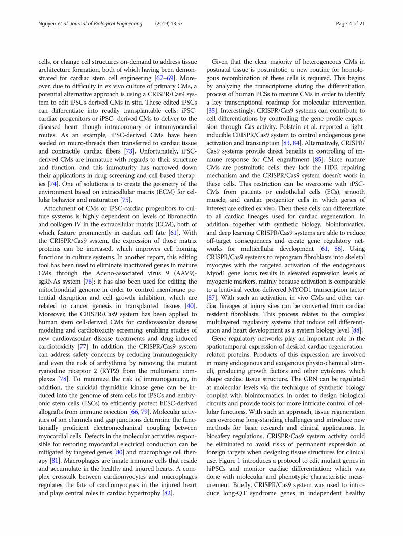
spatiotemporal expression of desired cardiac regeneration-related proteins. Products of this expression are involvedin many endogenous and exogenous physio-chemical stim-uli, producing growth factors and other cytokines whichshape cardiac tissue structure. The GRN can be regulatedat molecular levels via the technique of synthetic biologycoupled with bioinformatics, in order to design biologicalcircuits and provide tools for more intricate control of cel-lular functions. With such an approach, tissue regenerationcan overcome long-standing challenges and introduce newmethods for basic research and clinical applications. Inbiosafety regulations, CRISPR/Cas9 system activity couldbe eliminated to avoid risks of permanent expression offoreign targets when designing tissue structures for clinicaluse. Figure 1 introduces a protocol to edit mutant genes inhiPSCs and monitor cardiac differentiation; which wasdone with molecular and phenotypic characteristic meas-urement. Briefly, CRISPR/Cas9 system was used to intro-duce long-QT syndrome genes in independent healthy

Fig. 1 (1) Introduction of LQTS genes in independent healthy hPSC lines using CRISPR/Cas9. (2) Generation of disease-cardiomyocyte hiPSCs. (3) Isogenicsets of hPSC-CMs were differentiated from the edited hiPSCs lines. (4) Molecular analysis and phenotyping of hPSC-CMs (upper) molecular pathogenesis,(middle) drug screening, and (bottom) physiological functions
Nguyen et al. Journal of Biological Engineering (2019) 13:57 Page 5 of 21
hiPSC lines to generate disease-CM hiPSCs. This resultedin the formation of isogenic sets of hiPSC-CM which werecharacterized with phenotyping and molecular analysis.CRISPR/Cas9 systems for tissue-specific engineering ofstem cells not only provide new avenues for functional tis-sue engineering and regenerative medicine, but also con-trol the immunological balance in both the early andchronic stages after cardiac injury [89]. Proinflammatorycytokines present in increased levels in diseased andinjured tissues, which leads to the increase of tissue deg-radation and can prevent differentiation of hiPSCs [90].Recently, reports strongly suggested that controlling in-flammatory cytokine secretion from resident cardiomyo-cytes and cell interaction is one potential approach forcardiac angiogenesis and cellular regeneration [91, 92].Previous studies have reported that transplantation of
cells genetically engineered for constitutive overexpressionof interleukin 1 receptor antagonist (IL-1Ra) is effectivewhen creating cells-integrated scaffolds for implantation[93]. This approach also provides great promise in combat-ing inflammatory levels of interleukin 1 (IL-1), a challengefor transplanted and/or engineered tissues. To this end,RNA interference or CRISPR/Cas9 systems have been usedfor controlling the expression of inflammatory cytokines[43]. Alternatively, regulation of gene expression of growthfactors and anti-inflammatory cytokines (IL-4, IL-1Ra, andIL-10) in cell-based engineering platforms are also a
considerable approach. Compared to RNAi technology,however, the CRISPR/Cas9 systems provide permanent re-moval of inflammatory cytokines from the cell genome,this guarantee long term control of anti-inflammation incardiac tissue regeneration.Due to numerous challenges in current cardiac tissue re-
generation, the CRISPR/Cas9 system has become an effect-ive alternative which can tackle those by providingcomplex genome editing and transcription regulation, inorder to control differentiation, at genomic and molecularlevels [67, 70]. While still in its early stages, ongoing re-search on the use of CRISPR/Cas9 systems for more-complex implementation of the CM molecular clock [94]by controlling the transcription-translation feedback loopmay be a milestone in tissue engineering. In brief, CRISPR/Cas9 systems hold potentials to dramatically improve com-prehension of cellular processes and contribute signifi-cantly to cardiac tissue engineering.
Stem cell differentiationDifferentiation of stem-cell-derived CMs into the desiredlineages requires many aspects of the scaffold constructs,cell fate, and cell’s environment [36, 73, 95–98]. UsinghiPSCs to differentiate into mature CMs has been consid-ered as a potential approach towards therapeutics in car-diac tissue generation. With optimal protocols, fetalhiPSCs can be differentiated into almost 100% pure CMs.

Nguyen et al. Journal of Biological Engineering (2019) 13:57 Page 6 of 21
Although human ESC-derived CMs are a predominantsource of adult human cardiac myocyte for clinical thera-peutics, they still lack many essential features such as be-ing well-organized and distributed, and functionaltransverse tubules (T-tubules) [99]. Chong et al. reportedthat mature human ESC-derived CMs, rather than imma-ture, may become the preferred candidate to reduce therisk of arrhythmias in the transplantation therapy [100]. Inaddition, adult-like hiPSC- derived CMs can be widelyused for applications in stem cell-based disease modelingand in drug toxicity screening [95, 101]. Some strategies ofgenerating cardiac tissue from stem cell-derived CMs, inwhich their cellular morphology is similar to human adultcardiac structure and function, have been reported [74,102, 103]. Ronaldson-Bouchard et al. used different stages(day 12 and day 24 differentiation) of hiPSC-derived CMsand co-cultured them with fibroblasts in a fibrin-basedhydrogel to grow mature cardiac tissues around two flex-ible pillars [104]. These pillars were used to induce forcesin the contracting tissues, as forces are observed in nativemyocardium. After 1 week in culture, either constant elec-trical stimulation (2Hz for 3 weeks) or intensity training(2 to 6 Hz ramp over 2 weeks, then back to 2 Hz for oneweek) were applied to stimulate the differentiation andgrowth of hiPSCs to maturize CMs, which were deter-mined through the molecular, cellular, and functional levelof the differentiation [104, 105]. At the molecular level,genes associated with adult-like conduction, atrialisoform-related ventricular isoform of myosin, ATPproduction, and calcium transportation were highlyexpressed, which indicated maturation. At the cellularlevel, growth of CMs with ordered sarcomeres and a highdensity of mitochondria, were observed [104]. Vital pro-teins such as T-tubules and folding of the sarcolemmamembrane, involved in calcium transportations, werefound in the cell [106, 107]. Cell alignment in tissue con-structs, where cells were adhered to one another withmechanical strength at gap junctions, promoted electricalsignaling transmission between cells in the constructs.Well-aligned hiPSC-derived ventricular CMs on the hu-man ventricular cardiac anisotropic sheet, a cardiomimeticbiohybrid material, was reported in fully key electrophysio-logical features of native human ventricle [108]. This wasobserved only when hiPSC-CMs received an intensitytraining at an early stage [109]. After spending the inten-sity training, cardiac tissues were able to efficientlyperform action potentials through a process of excitation-contraction coupling. Electrical stimulation (excitation) in-duces mechanical response (contraction), which allowsmyocardium to contract. Wiegerinck et al. reported thatincreased beating frequency was the simultaneous resultof increased contraction force and faster relaxation [110].Various regulatory factors involved in CM maturation,hormone-driven cues [99], intensive electrical stimulation
[111, 112], cell composition and matrix/media [113, 114]have shown the most potential to achieve hiPSC-derivedCMs in scaffold environments.In cardiac tissue engineering, natural polymer scaffolds
play an important role in promoting differentiation andgrowth of hiPSC-derived CMs owing to their minimal im-munogenicity and biodegradability. Kaiser et al. used ablended fibrin and collagen scaffold to differentiatehiPSC-derived CMs into engineered myocardium [97]. Re-sults showed that expression of cardiac troponin T (cTnT)in CM populations were dependent on the scaffold com-paction. While the decreased compaction showed the low-est (24.4%) and highest (60.2%) positive expression ofcTnT+ CM purities, the highest compaction showed 40–50% cTnT+ population [97]. This study clarifies the correl-ation of hiPSC-derived CMs and scaffold interactions andprovides a basis for integrated design of customized scaf-fold constructs for cardiac tissue engineering.
Biomaterials and 3D scaffold fabricationCharacteristics of biomaterialsBiomaterials in the forms of hydrogels, carriers, and scaf-folds play a vital role in anchoring cells and helping themgenerate into functional tissues [115–117]. Although thoseforms have different specific patterns in tissue engineer-ing, all of them serve as a framework substance for prolif-eration and differentiation of the desired tissue. Forexample, carrier materials enable cells or chondrons toproduce the ECM that holds growth factors in skin woundhealing and cardiac remodeling and repair [118, 119]. Por-ous hydrogels entrap embedded cells and allow diffusionof gas and metabolites through their pore network [120,121]. Similarly, scaffolds are also porous matrices, thoughthey allow cell migration and attachment to the damagedtissue, as well as act as a substitute for lost tissue in thebody [122]. The developing highly-porous scaffold bioma-terials significantly depend on their types of materials,functionalization, and geometry.Typically, biomaterials for tissue engineering are synthe-
sized or modified from primary natural materials, thenfurther processes are conducted to form appropriatemorphology and characteristics for a desired application.They include polyglycolic acid (PGA) [123], poly(L)-lacticacid (PLA), poly(DL) glycolate (PLGA), and polyvinyl al-cohol and their derivatives [124–126]. In contrast, naturalbiomaterials include collagens, alginate, chitosan, fibrinand hyaluronic acids. Recently, advances in syntheticchemistry have contributed to novel hybrid biomaterialswith outstanding properties in terms of conductivity andstrength [127, 128]. For use in cardiac tissue engineering,it is required for biomaterials to support tissue reconstruc-tion and regeneration via active support for cell-to-tissueprocesses by promoting cell-cell adhesion, proliferationand differentiation. These biomaterials can also culture
Nguyen et al. Journal of Biological Engineering (2019) 13:57 Page 7 of 21
healthy tissues by forming three-dimensional structuresfor gas and nutrient transportation as well as formation ofvascular supportive substructures for blood vessels. Thebiomaterials used for scaffold fabrication processes canoptimize constructs used in clinical settings; allowing formaximizing cellular adhesion space, ECM secretion, re-vascularization, and paracrine processes.
Shaping biomaterials in 3D structuresScaffold materials play a key role in tissue engineeringand have been used more and more in clinical practice[129–131]. These materials form a biomimetic ECMwhich promotes cell adhesion and differentiation, as wellas 3D organotypic cultures [132]. By combining modernadvances of three major fabrication techniques, namelyelectrospinning, self-assembled monolayers, and ther-mally induced phase separation, with peptides and DNA,biomimetic 3D scaffolds have been developed for CMregeneration [133–135]. These systems support differen-tiation of various stem cells down multiple lineages andcreate relevant 3D specific tissues for clinical practice.Obviously, specific cell types could be seeded on the
biomimetic nanofibrous scaffold to regenerate desired tis-sues. Both primary and stem cells can be used, for differ-ent purposes [36, 98, 112]. Primary cells are collecteddirectly from mature tissue and cultured to obtain the de-sired cell number and form tissue constructs. However,quick phenotypic changes, limited proliferation numbers,and aging of primary cells inhibit their use once the cellsare transferred from their natural living conditions to arti-ficial ones [132, 136]. While CMs can be taken from spe-cific tissue sources for targeted applications, robustscaffolds and engineered biological tissues are needed toimprove to CM characteristics in new implanting environ-ments. Most scaffolds used for cardiac tissue engineeringare hydrogel materials and 3D nanofiber matrices, whichfeature benefits such as controlled release of growth fac-tors and good electrical conductivity [137, 138]. Resultsfrom confocal laser scanning microscopy, scanning probenano-tomography, and transmission electron microscopyshow that cardiac cells and fibroblasts actively interactwith 3D nanofibrous substrates, but in different ways[139]. While fibroblasts make contact with nanofibersthrough focal adhesion clusters, without wrapping thefiber, CMs develop a distinguished sheath structure andcovering fiber to increase contact area [139, 140]. Theseresults point to a new perspective on how cultured cellsinteract with 3D nanofibrous scaffolds. A host of previousstudies reported that matrix anisotropy and stiffness pre-dominantly influence 3D structural cell phenotypes, cellmigration, proliferation, and differentiation of culturedCMs [141]. Cardiac cells grown in 3D matrices werealways in tight contact with each other through cellularjunctions, which results in considerable mechanical
adhesion between cardiac cells and fibers. The increase inmechanical adhesion was found to be linked with the in-creased contact area between the cells and fibrous struc-tures [142]. The contact area plays a role for focaladhesion kinase in cardiac mitochondrial biogenesis in-duced by mechanical stress, which contributes to thehypertrophic growth of cardiomyocytes via control ofmitochondrial transcription cascade [143].Cellular parameters like the number of mitochondria
and endoplasmic reticulum membranes featured highercounts of cells grown in 2D constructs. Moreover, Wobmaand colleagues reported that upgraded “smart” scaffoldscan directly control biologically active molecules like hor-mones in the paracrine pathways directly through the cellmembrane, avoiding dissipation through the whole tissuesolution [144]. In such a system, bioactive molecules areefficiently used for CMs because they increase the diffu-sion of these molecules from neighboring cells throughparacrine hormones. It is also helpful if conducting mate-rials are integrated into these platforms prior to cardiaccell regeneration. Fibers are immersed in cardiac cells topromote high densities of electrical contacts, thus formingan electrical network on the outer part of the nanofibrousstructures isolated from the surrounding integrin micro-domains. With currently-available biomimetic models[129], the physical basis for this could be explained withvan der Waals forces and DLVO theory. DLVO theory isthe typical explanation of the stability of colloids in sus-pension [145]. The explanation of the cell interaction sta-bility is governed by physical and chemical interactionsbetween cellular surfaces that the balance between twoopposing forces-electrostatic repulsion and van der Waalsattraction is under DLVO theory [146, 147]. The inter-action energy is calculated by the sum of van der Waalforces and electric repulsion energy; thus zeta potential,hydrodynamic diameter, and cellular surface thermo-dynamic properties play an important role in the inter-action energy in the scaffold microenvironment for cellalignment and elongation [148].Model of generation, alignment, and stabilization of spin-
dle shaped fibroblasts and vessel under oscillatory stretchwas also reported [149]. These results reveal a new mech-anism for vessel network formation: under oscillatorystrain, 3D scaffolds can promote mural cell alignment, cellproliferation, translocation of a mechanosensitive transcrip-tional activator (YAP) into cell nuclei, and increased ex-pression levels of β-catenin. This directs ECM alignmentalong the orientation of the fibroblasts. Furthermore, ECs,which are tolerant to stretch stimulus, form aligned vesselsdirected by the fibroblast and ECM alignment. However,there is loss of fibroblast alignment and vessel alignmentdue to mechanical uncoupling of the cells after addingblebbistatin to the culture medium [149]. In addition, bothfibroblasts and vessels lose alignment when the cellular

Nguyen et al. Journal of Biological Engineering (2019) 13:57 Page 8 of 21
proliferation and signaling pathways responding to mech-anical stimulus are inhibited. Stretch stimulus promotes thestable production of growth factors, which enhances muralcell differentiation, thereby enriching stability and align-ment. These findings demonstrate how increased mechan-ical strain affects cell development, differentiation, andshape formation during the vascularization process. Cellu-lar stretching is restricted by nucleus size, which is less sen-sitive to deformation [139]. At the adhesive site, the cell isstretched by surface tension force. Absorbing fibers is notenergetically beneficial in the case of the actin cytoskeleton,hence contact is minimized with fibers by reduction of cellmembrane surface area [150]. Thus, these cells are able togenerate enough forces to overcome the resistance of theactin cortex at several filament assembly complex locations.In contrast to fibroblasts, CMs contain integrins in costa-mere structures that anchor sarcomeres to the ECM, somyocytes have much higher affinity with the substrate andserve to stabilize areas of cell-ECM interaction. Therefore,when CMs grow on suspended fibers, the myofibrils startattaching and aligning with them to increase the area ofinteraction with the substrate [139].The 3D microenvironment increases adherence and dir-
ect reprogramming of fibroblasts into CMs throughoutthe matrix via a metalloproteinase dependent mechanism[151]. The nanofibrous poly(L-lactide) (PLLA) scaffoldsadsorb serum proteins and ECM proteins like fibronectin,vitronectin, and laminin at quantities four times higherthan solid walled PLLA scaffolds [151, 152]. In nanofi-brous form, the absorption of protein is influenced bymany surface characteristics such as protein absorptionlayers, surface-to-volume ratio, surface nm-scale morph-ology, crystallinity, and orientation of the polymer in itsnanofibrous form. Finally, nanofibrous scaffolds promotecell adhesion in many cell types, giving them an advantageover solid walled scaffolds.
3D-gel of hybrid biomaterialsNatural biomaterials can be produced from self-assembledmonolayers (SAM) of different polymers through hydrogenbonds, van der Waals forces, and hydrophobic and electro-static interactions [153]. SAM fabrication is very usefuland robust, thus some recent studies have attempted tomimic collagen structures from ECM-derived binding pep-tides, which increased cell adhesion and cardiac repair bycardiac progenitor cells [154]. These systems can workwith other self-assembling materials like phage displaypeptides and genetic materials to improve adhesion, prolif-eration, and controlled differentiation; rendering many ap-plications in tissue engineering [155]. Wang et al. reporteda procedure to fabricate biomaterials for 3D scaffold for-mation based on SAMs from bacteriophage display [156].In this approach, a panel of desired peptides was displayedon M13 phages, a bacteriophage of Escherichia coli, for the
purpose of CM generation by activating ligand-linked mi-croenvironments in damaged cardiac tissues (Fig. 2) [150].As seen in Fig. 2, RGD and DLEFIFEER ligand motifs thatmediate adhesion to the cell adhesive receptors were dis-played on major coat protein pVIII and determinedthrough an interaction between nephronectin and α8β1 in-tegrin receptor [158]. Using a 3D printer, assembly of theshort peptide-coated nanoparticles into a 3D functionalstructure was driven by noncovalent interactions to form ascaffold [158]. The mechanisms of these self-assembledprocesses have led to major advances in the understandingof biological and chemical 3D folding processes for bio-mimetic supramolecular peptide assemblies in coatings,gels and electroactive materials. The specific function ofthese materials relies on their helical peptides, β strandpeptides, and surface binding monolayer-forming peptides,which electrically stabilized the phage nanofiber inside theRGD-phage scaffold. Subsequently, hiPSCs were seeded inthe RGD-phage scaffold and induced the formation of car-diomyocytes [159].The geometry of the scaffold substrate is very important
in cardiovascular tissue engineering because the cardiactissues need to be highly differentiated to perform highspecific functionality. For example, the microscopic levelof heart valve needs to be at anisotropic geometry, inorder to have particular shape of semilunar valves at themacroscopic level [160]. Microenvironment and contrac-tion properties of cardiomyocytes can be influenced bymorphology and mechanical properties by increasing themodulus in the range of 1–30 kPa of 2D substrates [161].Developing these properties in synthetic 3D scaffold mayprovide a significant means of controlling cell fate bothin vitro and in vivo. An ideal polyester biomaterial elasto-mer for cardiac tissue engineering should exhibits arelatively-low Young’s modulus, with high elongation andtensile strength [162]. Through a one-step polycondensa-tion reaction and ultraviolet reaction, poly(octamethylenemaleate (anhydride) 1,2,4-butanetricarboxylate) (124 poly-mer) is formed the prepolymer gel and a cross-linkedelastomer with highly elastic and tunable properties [162],of which they are dependent on the UV light exposure,monomer composition, and porosity of the cured elasto-mer. Interestingly, the material does not only provide itselastomeric properties falling within the range of those ofadult heart myocardium, but also is optimized for higherelasticity for cardiac cell attachment and interactionin vitro and in vivo [162]. Finally, the polymer expressedrelatively-stable degradation characteristics that supportpotential tissue implants. Recently, Shiekh et al. developedand evaluated an elastomeric antioxidant polyurethane(PUAO) for cardiomyocyte functionality [12]. A serial ana-lysis including uniaxial and cyclic tensile testing, thermalanalysis, cytotoxicity, antioxidant analysis, and degrad-ation reveals that PUAO reduces intracellular oxidative

Fig. 2 (See legend on next page.)
Nguyen et al. Journal of Biological Engineering (2019) 13:57 Page 9 of 21
(See figure on previous page.)Fig. 2 Biomaterials are based on self-assembled monolayers from bacteriophage display for 3D scaffolds formation. (Top), RGD peptide isdisplayed and fused to the solvent-exposed terminal of each copy of major coat protein (pVIII) through genetic engineering. The side wall offilamentous phage by RGD-coding gene into gene VIII to generate RGD-phage. (Bottom) The 3D scaffold of RGD-phage nanofibers (negativelycharged) self-assemblies with polycationic biomaterials and integrated into a 3D printed bio-ceramic scaffold [156], which electrically stabilizes thephage nanofiber inside the scaffold. The resulted scaffold is seeded with hiPSCs and the implanted into cardiac defect. The presence of RGD-phage in the scaffold induced the formation of cardiomyocytes [157]
Nguyen et al. Journal of Biological Engineering (2019) 13:57 Page 10 of 21
stress in H9C2 cardiomyocytes and neutralized reactiveoxygen species (ROS) promoted cell death. Moreover,PUAO film displayed synchronous beating with maturecardiomyocytes showing high expression of cardiac spe-cific α-actinin, troponin-T, and connexin-43 proteins [12].Additionally, cultured cardiomyocytes on PUAO filmexpressed the physiological intracellular calcium function-ality similar to mature cardiomyocytes [12].Shin et al. used directed SAM to selectively trap target
carbon nanotubes (CNTs) as an effort to control thegrowth of supramolecular hydrogel fibers and improvefunctionality of bioengineered cardiac tissues [117]. Sur-faces of CNTs stimulate the formation of hydrogelators inthe vicinity of the fiber constructs, which results in in-creased fiber formation, changes in network morphology,and increased mechanical properties. Subsequently, thiscan improve electrophysiological performance of cardiactissue in terms of increased beating rate and lower excita-tion threshold [117, 163]. Besides CNTs, metallic nanopar-ticles, with their size-dependent properties, have shownpromise in overcoming many of the current limits of car-diac tissue engineering. Li et al. reported a nanocompositecomposed of gold nanoparticles (AuNPs) and a collagenmatrix, which improved tissue growth via localizedstrength, thus enhancing the assembly of intercalated discsby β1-integrin-mediated signals [151]. In addition, 3Dstructures based on rigid CNTs scaffolds have been used toimprove CMs viability, proliferation, and maturation, butthey require undesirable invasive surgeries for implantation[164]. On the platform of 3D gel-based matrix, an inject-able reverse thermal gel (RTG) functionalized with CNTs(RTG-CNT) that switches their morphology from a so-lution at room temperature to a three-dimensional (3D)gel-based matrix shortly after reaching bodytemperature was developed [164]. This extends long-term CMs survival, promotes CMs alignment and pro-liferation, or improves CM physiological function. Re-cently, Mason et al. reported a highly-ordered 3Dfibrous protein scaffold derived from a self-assemblyprocesses [153]. This resulted from a balanced system oflow-entropy processes in which a set of interactionsbetween different chain residues formed amorphous ag-gregates, thus mimicking self-assembling protein sys-tems in nature. As an alternative to self-assembly,electrospinning produces nanofibers and nanofibrousstructures from a broad range of biomaterials-based
dopes in which advantages, drawbacks and potential ap-plications are discussed in next sections.
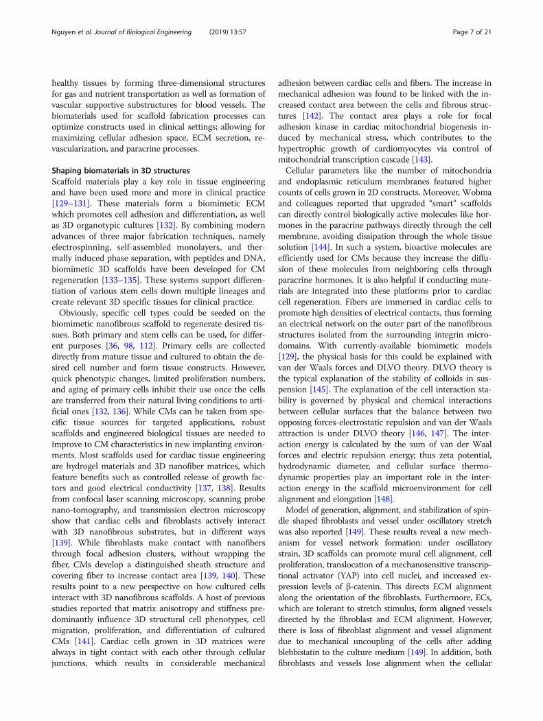
Electrospinning for 3D scaffold fabricationElectrospinning could be used to make nanofibers from avariety of polymers and it is well suited to 3D nano-scaffold constructs in cardiac tissue engineering [165]. Inessence, the electrospinning technique is based on an elec-tric field to create a charge on the surface of polymer solu-tions, thus generating a force opposing its surface tensionand allowing fibers to be drawn out [166]. Many parame-ters can be used to tune this process, including electricalcharges from the jet, solvent characteristics, length ofpolymers, flow rates, voltage levels, and collector distance;all of these considerations, and others, need to be takeninto account to get a final polymer fiber in nanofibrousarchitecture [167, 168]. The resulting products are col-lected on solid or liquid substrates, or even substrate free,to form 3D micro-fibrous and nanofibrous scaffolds.Suhaeri et al. reported a new platform based on afibroblast-derived, matrix-coupled, aligned and electro-spun nanofiber [45]. In their work, a hybrid scaffoldstructure composed of poly(l-lactide-co-caprolactone)(PLLA-PCL) and fibroblast-derived ECM (PLLA-PCL/FDM) was aligned to form an artificial cardiac microenvir-onment. The physical mechanical property of PLLA-PCLin the parallel direction shows the anisotropic nature ofthe aligned PLLA-PCL fibers. The PLLA-PCL/FDM wasproduced from the fibroblast culture on the PLLA-PCLfiber for 5–7 days and the ECM was collected from a sub-sequent decellularization. On this co-culture system, cel-lular characteristics of differentiation, phenotyping, cellviability, and maturation of H9c2 and neonatal rat CMswere significantly improved compared to those in fibro-nectin (FN)-coated electro-spun PLLA-PCL fibers (Fig. 3)[45]. On the aligned scaffold, cells spread along the direc-tional cues instead of the random growth in everydirection observed in the random scaffold. In addition,non-sulfated polysaccharides [169], biopolymers [170],and both organic and inorganic frameworks [171] havebeen integrated into PLGA to improve its biocompatibilityand mechanical properties; and this highly depends onpolymer concentration. However, due to collector plateconstructs, nanofibrous scaffolds made from electrospin-ning are generally 2D; limiting their clinical relevance. Re-cently, a rotating cylinder has been demonstrated as a

a
b
d e f
c
Fig. 3 Fabrication and characterization of PLCL/FDM. a Illustration represents the fabrication process of PLCL/FDM. b Random and aligned orientations ofPLCL fibers. Scale bar of SEM images is 10 μm. c Fibrillary ECM components in FDM were stained against FN and collagen type I. The direction of PLCLfiber alignment is shown by double headed arrows. Scale bar is 50 μm. d ATR-FTIR spectra of FDM with C=O at 1753 cm− 1 from PLCL and amide group at1645 cm− 1 from FDM. e AFM images for surface topographical features of PLCL and PLCL/FDM; color scale shows their surface roughness and differencein height. f Quantitative comparison of root mean square (RMS) roughness calculated from AFM images. Statistical significance (***p< 0.001). Thereproduced image is permitted from [45]
Nguyen et al. Journal of Biological Engineering (2019) 13:57 Page 11 of 21
replacement for the collector plate used in electrospin-ning, which was utilized to produce a tubular scaffold andallow for growth factors to be released in a controllablefashion [172, 173]. A scaffold platform with polyca-prolactone (PCL) nanofibers and vascular endothelialgrowth factor (VEGF)-encapsulated gelatin particleswas fabricated to extend half-life time and stimula-tions of VEGF to mesenchymal stem cells (MSCs)and ECs [174]. In addition, paracrine mechanismsthat are involved in MSC differentiation into cardio-myocytes are only limited to cell differentiation rates,not directly impacting to cell differentiation [175,176]. Jiang et al. reported that this construct candrive the differentiation of MSCs to ECs and keep thestability of the tubular structure [174], indicating thatgrowth factor (GF)-releasing scaffolds are potentialplatforms based on the electrospinning process forcardiac tissue engineering.
Recently, it has been shown that use of a Teas chartcould provide useful information in terms of solubilityand spin-ability for the electrospinning process [177–179].Polymers should have solubility in the target condition, asvalues outside of a specific range will result in electro-sprayed beads and aggregates [177]. Higher fidelity nano-scale topography and bio-activity integration in the 3Darchitecture on the ECM-inspired nanofibrous scaffoldsshowed outstanding advantages for engineering 3D aniso-tropic cardiac tissues [137, 180].
Thermally-induced phase separationThermally induced phase separation (TIPS) is anotherrobust method to make 3D scaffolds. It involves fivesteps: polymer preparation, phase separation and gel-ation, solvent extraction, freezing, and freeze drying[181]. Once a polymer is dissolved in a specific solvent,the solution becomes thermodynamically unstable and

Nguyen et al. Journal of Biological Engineering (2019) 13:57 Page 12 of 21
results in two material phases: one “rich” in polymer andanother phase “lean” in polymer. The resultant polymerstructure depends on the ratio of polymer to solvent andconditions of the phase separation. Once the solvent isextracted, the phase of lean polymer is removed, and thepolymer rich phase is identified as being in one of threecategories: powder, closed cell foam, and open cell foam.Open cell foam is the type used to make 3D scaffolds forhuman chondrocyte growth and ECM formation [182].ECM-derived porous foams are biologically-relevantsubstrates in advanced 3D in vitro cell culture modelsthrough controlling freezing and lyophilization proce-dures [183].Luca et al. reported the formation of surface structures
of TIPS-based scaffolds formed in water at roomtemperature [184]. The TIPS method allows for tuningof surface morphology which benefit tissue regenerationof preosteoblasts [184]. Peña et al. presented an inject-able and biomimetic RTG that was functionalized withpoly-L-lysine or laminin to promote longevity of cul-tured CMs, neonatal rat ventricular myocytes (NRVM),and adult rat ventricular myocytes (ARVM) [130]. Theirresults showed that the RTG functionalized with lysinestimulated NRVM grow and differentiated heart-likefunctional syncytia. Beating cells were recorded after 21days in both cases of RTG and Lysin-functionalizedRTG [130]. In addition, TIPS can be combined withporogen leaching to increase levels of architectural con-trol. Porogen leaching (paraffin, sugar) can promote theformation of micropores with morphologies such asspherical, tubular, and disk shaped pores within the scaf-fold [185]. These micropores play important roles in en-hanced cell penetration, proliferation, mass transport ofnutrients, and growth factors in studies of angiogenesisand tissue formation. Several research groups have de-veloped anatomically shaped molds with reverse solidfreeform fabrication (SFF) in a PLLA solution [186, 187].Architectural features were formed through three steps:ECM-mimicking materials, formation of pores for cellpenetration and mass transport, and anatomical scaffoldshaping. This last step is vital for structural tissue likebone and cartilage. TIPS can be used in concert withporogen leaching and 3D molds and with commonchemical and biological polymers to create structural tis-sue scaffolds with excellent processing flexibility.
Bioprinting for 3D scaffoldsAdvancements in 3D printing have now begun to see itsuse in tissue engineering. State-of-the-art techniques inthis field includes laser direct writing and multiphotonpolymerization, which can be used for computer-aidedscaffold design [188]. The process of designing and manu-facturing scaffolds in this way includes several steps: de-sign of functionally graded scaffolds, modeling of selective
laser sintering and fused deposition modeling (FDM) pro-cesses, development of bioreactors, and 3D bioprinting[188–190]. Laser systems such as femtosecond- andultraviolet-based sources allow for precise manufacture of3D tissue scaffolds, which are engineered entirely throughcomputer-aided design [191]. Zheng et al. reported theprocess of using computer-controlled UV laser systemsfor 3D scaffolds with many kinds of polymers such aspolyethylene glycol diacrylate (PEG-DA), ormocomp, pen-taerythritol tetra-acrylate (PETRA) [192]. More recently, aclass of micro-architected materials with high-orderedstructural connectivity and nanoscale features was printedby projection micro-stereolithography [192]. By using bio-polymers, the technique could be used to produce bio-compatible micro-lattices for soft tissue engineering,which are used as injectable scaffolds that can either in-duce endogenous cardiomyocyte repairing [193].Seeded cardiomyocytes can be grown in hexagonal 3D
fiber scaffolds made by melt electro-writing, a form of 3Dprinting. The resultant hexagonal microstructures haveoutstanding mechanical characteristics, allowing for largeanisotropic reversible deformations; this deformable struc-ture mimics microstructure of myocardial tissue [137].Moreover, the high porosity of these structures aids for-mation of aligned tissues and are effective as cardiacpatches on contracting hearts. These functional humanmyocardial patches feature properties highly desirable forclinically relevant cardiac repair [96]. As a result, iPSC-derived CMs have been successfully cultured in multi-cellular 3D bioprinting substrates for vascularized hearttissue [98]. Human umbilical vein endothelial cells(HUVECs) and iPSC-CMs have been encapsulated withinhydrogel strands, containing alginate and PEG-fibrinogen,and forced out through custom microfluidic printingheads to form spatial depositions with high fidelity andresolution. Maiullari and colleagues have reported a 3Dcardiac tissue composed of iPSC-CMs from different tai-lored geometries with a high orientation index [98]. Bloodvessel-like shapes differentiated from HUVECs can beused for in vivo grafting, which is a better integrated sup-port for engineered cardiac tissue [98]. These findings alsobring important contributions to functional heart tissuegeneration in vitro through 3D PEG-fibrinogen hydrogelsto recover their pluripotency [98]. This technique plays akey role in the design of printed micro-fibrous constructsused to assemble complex vascular networks. For ex-ample, bio-printed ECs following this can effectively de-velop vasculature in the transplanted tissues in the samemanner of native vessels [194]. The results of bio-printed3D vessel-based therapy directed to restore blood flowcan counteract cell death and promote regeneration in therevascularization of ischemic or damaged organs, whichhighly relies on microenvironment engineering for sup-plies of oxygen and nutrient.

Nguyen et al. Journal of Biological Engineering (2019) 13:57 Page 13 of 21
However, due to the lack of oxygen and nutrient diffu-sion (in the 100–200 μm scale) in porous structures, mi-gration of iPSCs tends to be in the outer zone ofhydrogels; and this produces inhomogeneous cellulardistribution in vascular networks in vivo [195, 196].These diffusion problems could be solved via an inte-grated system of porous structures and parallel fibers toform an engineered vascular network. By addition of 1%w/w PEG-DA monomer to bioprinting materials, thehomogeneous culture biosystem fully supplies nutrientsto all regions of the 3D constructs [98] . This techniquehas been used for iPSC-derived CMs culture to producemyocardial-like tissue [98] and generate 3D vascularstructure [197]. Alternatively, circulation in the 3D con-structs is supplied by a microfluidic device bearing a Y-junction (2 inlets, 1 outlet) in which the flows of twodifferent bio-inks are precisely driven by external micro-fluidic pump [98]. Interestingly, this construct showedgreat promise for artificial skeletal muscle generationonce the dimensions of channel were reduced to 500 ×500 μm2 (cross-section) to create an extremely-smalldead volume (< 2 μL); this allowed rapid tuning be-tween the two bio-inks during printing. This systemalso allows building heterogeneous structures compos-ing of iPSC-derived CM and HUVEC could poten-tially mimic native cardiac contraction in better thanthose described above.Functional contraction of the myocardium is orches-
trated by electrical stimulation propagation in the rightsequence and is driven partially by CM spatial orienta-tion; therefore, proper orientation is a critical goal fororganization of CMs [98, 159]. The organization of CMsembedded in 3D bio-printed fiber structures is impactedby the surrounding fiber matrix direction; often, growthof iPSC-derived CMs is directed along the fiber printingdirection. Contraction can be further enhanced withhigher material conductivities. Scaffolds that coupleelectrical and elastic materials have become valuablefor cardiac cell function, but current conductive ma-terials do not show tunable physiological propertiesfor cell behaviors [138, 198]. Electrospun conductivescaffolds were reported of use in cardiac tissue engin-eering for enhancement of connexin 43 expression[96, 198]. By integration of AuNPs into hydrogel scaf-folds, the polymer templated gel becomes tunablewith a Young’s modulus similar to that of myocar-dium, polyaniline, and polypyrrole. Neonatal rat CMswere cultured on the scaffold and expressed highlevel of connexin 43, with or without electrical stimu-lation. Hosoyama et al. have also reported a novelnanoengineered hybrid electro-conductive cardiacpatch for treating the infarcted myocardium [96] ofwhich classification and localization from medical im-ages are detected by machine learning [199–203].
Machine learning and precision control for 3Dscaffold fabricationMachine learning in tissue platformAs mentioned, currently the most obvious use of ma-chine learning (ML) in this field is identifying patterns intissue-related data and/or classifying specific tissue con-structs. One example of a problem of interest is that ofclassifying the phenotype of differentiated, stem cell-derived CMs. One group sought to classify CM pheno-type by matching distinct groups of shapes with distinctgroups of action potential waveforms [204]. It was doneby staining the cells of interest, optically mapping themduring contraction, converting time-varying pixel inten-sity to discrete waveforms, and then using ML algo-rithms to identify groupings of AP behavior which theycould compare to cell cluster shape data. The employedML is what’s known as spectral clustering whose algo-rithm attempts to minimize a “similarity” weight valuebetween sets of inputs, thereby grouping them [205]. Inthis case, the authors used aligned and averaged AP asthe input to the clustering algorithm, allowing the algo-rithm to minimize similarities between groups of the APwaveforms, and then mapped these groupings to cellcluster spatial distributions. These methods have beensuccessfully applied in biomedicine and cell biology withvarious stage-of-the-art machine-learning algorithms[58, 60, 206].A more-recent example of ML used in this space was
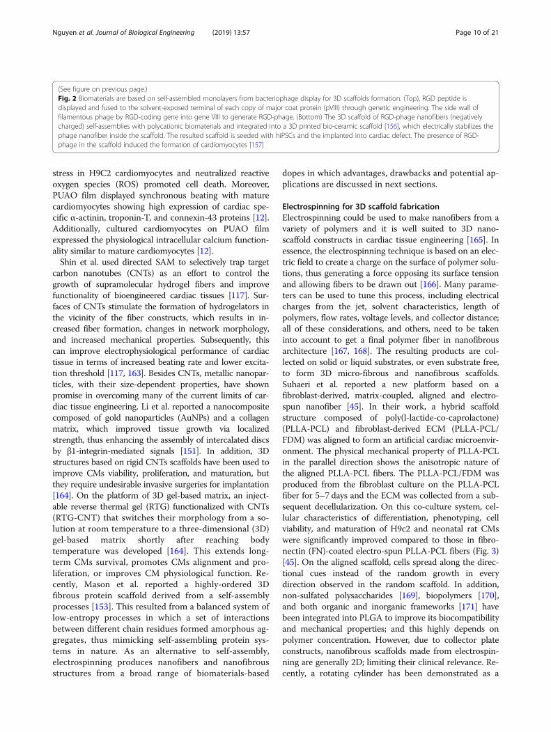
geared toward not only classification of cardiac tissuecontractile events [207] but extending this classificationset into a predictive model for preclinical screening ef-fects of drugs on cardiomyocyte function [41]. The pre-dictive models are highly dependent on machinelearning methods such as naïve Bayesian, support vectormachines (SVM), and end-to-end (E2E)-integrated MLsystem [53], of which they are leveraged by bigger data-sets generated from high-throughput screening data. Leeet al. reported a SVM to develop a drug screening assayon hiPSCs-derived cardiac tissue (Fig. 4) [41]. In this ap-proach, groups of linearly separable data were demar-cated by planes in order to classify them [208]; and theplanes themselves were statistical maximizations ofgroup separation based on feature points (i.e. supportvectors), rather than the more-computationally intensivenearest-neighbor piecewise approach [209].They first qualified models by generating force data
and derived parameters from stimulated cardiac cells,mixing the data with a control set, allowing a binarySVM to attempt to classify the data, and then quantify-ing the resulting SVM accuracy [210]. This classificationmodel accuracy then becomes a proxy for cardiac activ-ity of the drug. About 50% accuracy means that theSVM could not separate control from drug but accuracygreater than 50% indicates that the statistical model was

Fig. 4 Machine learning for drug screening on human iPSCs-derived engineered cardiac tissue. a Waveform pattern parameters are determinedbased on concentration of cardioactive compounds compared to the binary support vector machine (SVM). The collected data points would bein line with those of vehicle as if the compound does not modulate the contractile behavior of human ventricular cardiac tissue strips (hvCTSs). Ifdata of cardio active effects are more distinguishable, it shows in a higher SVM accuracy which is possible to separate two compound groups.The degree of cardio activity of a given concentration for target compound is shown in a singular quantitative index with the binary SVMapproach. b Library of compounds is built on a model for prediction of mechanistic action of screened compounds. Data from the library groupallow the machine learning defines boundaries of various drug families. Finally, the developed model can be applied for the unknowncompounds on tissue engineering. The image is reproduced with permission from [41]
Nguyen et al. Journal of Biological Engineering (2019) 13:57 Page 14 of 21
able to group the drug and control outputs into differentregions of the parameter space and, therefore, declare adifference in behavior [41, 211]. Data of cardio active ef-fects express in a higher SVM accuracy, if they are moredistinguishable from two compound groups. Based on agiven concentration, the degree of cardio activity for atarget compound is shown in a singular quantitative
index with the binary SVM approach [41, 207]. Next, alibrary of this drug screen testing data was combinedand an SVM designed for multiple classes was used todefine parameter space regions for each. The library ofcompounds was built on a multiple-category predictionmodel for mechanistic action of screened compoundsand chemogenomic databases [212, 213]. Data from the

Nguyen et al. Journal of Biological Engineering (2019) 13:57 Page 15 of 21
library group allow the machine learning defines bound-aries of various drug families and mechanism of action[214]. Finally, the developed model can be applied for theunknown compounds on tissue engineering. After doingso, a withheld data set of the same form was fed into theirpredictive model to see if the SVM could properly classifydrug interactions [215], integrating multiple omics data[216], and unknown drug compounds [217]. In their dem-onstration, they were able to classify cardiac activity of un-known compounds with an accuracy of roughly 72% andgeneralize the results to other drug families with an accur-acy above 70% [218]. Further, ML and its myriad algo-rithms can also be used on the protein and gene side oftissue engineering, as it has been demonstrated or pro-posed for histopathological image analysis [43], ligand af-finity [42], folding structure [219], gene expression andbiomarker data mining [220, 221], and in evaluation ofpre-implantation embryos [222]. Large datasets such asthe “Tissue Atlas” [223], a human proteome map catego-rized by tissue, could easily be used as a training and test-ing set for ML algorithms targeting identification ofimpaired tissue or disease onset.
Precision control in fabrication of 3D scaffoldThe ever-widening and accelerating field of roboticsboth contributes to and has the possibility of benefittingfrom tissue engineering. The contribution of robotics totissues engineering lies mostly in the manufacturingspace; as automated fabrication has hastened tissue con-struct research. Of particular popularity at the momentis the concept of robotic bio-fabrication, also known asorgan printing or bioprinting. Bioprinting was definedby members of the first international workshop on thesubject in 2004 as the “use of material transfer processesfor patterning and assembling biologically relevant mate-rials—molecules, cells, tissues, and biodegradable bio-materials—with a prescribed organization to accomplishone or more biological functions” [224]. In other words,it’s the use of automated fabrication to faster transferfrom the scaffold design and tissue culture, to clinicalsettings, especially in the field around regenerativecardiomyocytes.As discussed earlier, 2D and 3D cardiomyocyte cultures
in biomimetic conditions are crucial to the improvementof knowledge surrounding cardiac tissue development[225]. Researchers have presented methods for formingthese tissue constructs in a variety of manners— fromusing electrospinning to create scaffolds enabling cell at-tachment and growth [96] to 3D patterning of tissue-similar constructs [226], or using printer depositedspheroids to induce scaffold-less self-assembly of tissue[227, 228], although some of these technologies have sig-nificant hurdles to overcome still. Within the last decade,researchers have begun to concern themselves with the
systems design of holistic industrial bio-fabrication lines,including the design stage prior to and maturation stageafter bio-fabrication [229]. In-vivo bio-fabrication is alsogetting attention; beyond bioresorbable printed scaffolds[230], there have even been demonstrations in mice oflaser printing of photoactive resins above the calvaria toform bone-like caps [230], which was integrated with therobotic controlling.Tissue engineering is also feeding back into robotics in
two important ways—inspiring bio-mimetic robotic sys-tems [231] and becoming a potential component withinrobots themselves [232]. Most bio-similar robots up tothis point have focused on the use of soft materials togrip and move, as the field has acknowledged that thelimited conformability of robotics prior to this trend isdirectly counter to the variety of conformable structuresseen in nature [231]. Much of the interest in artificialtissue has been focused on muscle. One group demon-strated artificial muscle composed of polymer-basedcomposites which bend and flex under cation exchange[233], similar to action potential propagation in cardiactissue. Another group demonstrated this same conceptusing a collagen gel filled with rat CMs and initiatedcontractile behavior strictly chemically, using epineph-rine and nifedipine [234]. This is somewhere betweenthe former and latter contributions of tissue engineeringbut there are recent examples in which robotics systemshave been designed from the systems level to takeadvantages of engineered tissues, themselves being bio-similar robotic systems. As an example of engineeredtissue integrated robotics, researchers have demon-strated actuators which are comprised of myoblast-filled hydrogels and triggered by electrical stimulation[235], antagonistically contracting against each otherto create both contraction and extension. It is of notehere that not only are the actuators themselves engi-neered tissues, but they have been attached to theirskeletal frame by culturing methods, and even themechanical systems design mimics natural tissue. It islikely that more bio-similar, bio-integrated robotic hy-brids are on the horizon.
ConclusionsCardiac tissue engineering has benefited greatly from ad-vances in genetic engineering, material engineering, elec-trical engineering, and biochip design. Within geneticengineering, genome editing is a pioneering tool that hasbeen used in the generation of new cellular, tissue andanimal models to investigate cell-cell adhesion, differen-tiation of hiPSCs, and generation of CMs for variouscardiac disease. However, the post-mitotic nature ofCMs and various technical barriers present hurdles forbringing engineered cardiac tissue directly to therapeuticapplications. Other cells such as cardiac fibroblasts, ECs,

Nguyen et al. Journal of Biological Engineering (2019) 13:57 Page 16 of 21
and muscle cells can potentially substitute for CMs indeveloping tissues for cardiovascular diseases.One major technical advancement in this field is the abil-
ity to design a physical framework of biocompatible mate-rials and the control of mechanical characteristics, whichcan be applied clinically. Due to the nature of CMs, scaffoldsused for CM growth should be readily tunable for align-ment/organization to produce efficient contractions. Further,electrical stimulation should be integrated into the system toperform intensity training in the later stages of CM culture[111]. This enables the connection of native and differenti-ated cells, at single cell levels of cellular communications,between hiPSC and CMs. Communication between CMsand their micro-environment within the engineered tissueshould be understood in tandem with development of 3Dbiomimetic scaffolds and bioreactors in order to promotecost-effective scale-up of tissue production.There exists a variety of supporting technologies which
could be applied in the process of tissue engineering. Onepossibility is that machine learning be used involved in thedesign and processing of micro-physiological systems.High-throughput fabrication could be optimized via scaf-fold geometry, cellular paracrine factors, and cellular com-munication, in order to maximize survival rates andcompletely functionalize engineered cardiac tissue. At themolecular and cellular level, engineered cardiac tissue de-rived from the HLA-null line should be tailored towardsdeveloping immune-resistant modified hiPSC-derived CMlines; this can be done using genome editing tools focusedon solving cryopreservation general implantation issues.Confucius said, “Our greatest glory is not in never fail-
ing, but in rising every time we fail.” We believe withfocused and continued progress achieved by scientistsacross a range of multidisciplinary fields, cardiac tissueengineering will soon be viable for clinical use.
Abbreviations124 polymer: Poly(octamethylene maleate (anhydride) 1,2,4-butanetricarboxylate); AHM: Artificial heart muscle; ARVMs: Adult ratventricular myocytes; AuNPs: Gold nanoparticles; Cas9: Caspase 9;CM: Cardiomyocyte; CNTs: Carbon nanotubes; CPVT1: Catecholaminergicpolymorphic ventricular tachycardia type 1; CRISPR: Clustered regularlyinterspaced short palindromic repeats systems; CSCs: Cardiac stem cells;ECM: Extracellular matrix; ECs: Endothelial cells; FDM: Fused depositionmodeling; FN: Fibronectin; GF: Growth factor; HDR: Homology directedrepair; hiPSC-CMs: Human induced pluripotent stem cells-derived CMs;hiPSCs: Human iPSCsHuman iPSCs; hMSCs: Human mesenchymal stem cells;HUVECs: Human umbilical vein endothelial cells; IL-1: Interleukin 1; IL-1Ra: Interleukin 1 receptor antagonist; iPSC-CMs: iPSCs-derivedcardiomyocytes; iPSCs: Induced pluripotent stem cells; ML: Machine learning;MNPs: Magnetic nanoparticles; MSCs: Mesenchymal stem cells; NHEJ: Non-homologous end joining; NRVMs: Neonatal rat ventricular myocytes;PAM: Protospacer adjacent motif; PCL: Polycaprolactone; PEG-DA: Polyethylene glycol diacrylate; PETRA: Pentaerythritol tetra-acrylate;PGA: Polyglycolic acid; PLA: Poly(L)-lactic acid; PLLA: Poly(L-lactide);PUAO: Elastomeric antioxidant polyurethane; ROS: Reactive oxygen species;RTG: Reverse thermal gel; RYR2: Ryanodine receptor 2; SAM: Self-assembledmonolayers; SFF: Solid freeform fabrication; sgRNA: Single-stranded guideRNA; TIPS: Thermally induced phase separation; VEGF: Vascular endothelialgrowth factor; YAP: Mechanosensitive transcriptional activator
AcknowledgementsWe are thankful for being invited by the Editors to contribute this importantreview paper.
Authors’ contributionsAHN initiated the manuscript. AHN, PM, LSH and HC wrote and revised. PJB, JHL,AL and HC gave advice and discussion. All authors read through the manuscript.All authors read and approved the final manuscript.
FundingP.M. was funded by the National Science Foundation Award #1652818 toH.C.
Availability of data and materialsN/A.
Ethics approval and consent to participateN/A.
Consent for publicationN/A.
Competing interestsThe authors declare that they have no competing interests.
Author details1Electrical and Computer Engineering Department, University of Alberta,Edmonton, Alberta, Canada. 2Electrical Engineering and Computer ScienceDepartment, University of California Irvine, Irvine, CA, USA. 3BiomedicalEngineering Department, University of California Irvine, Irvine, CA, USA.4Chemical Engineering and Materials Science Department, University ofCalifornia Irvine, Irvine, CA, USA. 5Mechanical and Aerospace EngineeringDepartment, University of California Irvine, Irvine, CA, USA. 6BioengineeringDepartment, University of Texas at Arlington, Arlington, TX, USA. 7HenrySamueli School of Engineering, University of California, Irvine, USA.
Received: 6 February 2019 Accepted: 3 June 2019
References1. Heallen TR, Martin JF. Heart repair via cardiomyocyte-secreted vesicles. Nat
Biomed Eng. 2018;2(5):271.2. Mohamed TMA, Ang YS, Radzinsky E, Zhou P, Huang Y, Elfenbein A, Foley A,
Magnitsky S, Srivastava D. Regulation of cell cycle to stimulate adultcardiomyocyte proliferation and cardiac regeneration. Cell. 2018;173(1):104–+.
3. Zimmer A, Bagchi AK, Vinayak K, Bello-Klein A, Singal PK. Innate immuneresponse in the pathogenesis of heart failure in survivors of myocardialinfarction. Am J Phys Heart Circ Phys. 2019;316(3):H435–45.
4. Frangogiannis NG. The functional pluralism of fibroblasts in the infarctedmyocardium. Circ Res. 2016;119(10):1049–51.
5. Weerasinghe P, Buja LM. Oncosis: an important non-apoptotic mode of celldeath. Exp Mol Pathol. 2012;93(3):302–8.
6. Tzahor E, Poss KD. Cardiac regeneration strategies: staying young at heart.Science. 2017;356(6342):1035–9.
7. MEMBERS WG, Benjamin EJ, Blaha MJ, Chiuve SE, Cushman M, Das SR, Deo R, deFerranti SD, Floyd J, Fornage M. Heart disease and stroke statistics—2017 update:a report from the American Heart Association. Circulation. 2017;135(10):e146.
8. Dhingra R, Vasan RS. Age as a risk factor. Med Clin North Am. 2012;96(1):87–91.9. Barker RA, Carpenter MK, Forbes S, Goldman SA, Jamieson C, Murry CE,
Takahashi J, Weir G. The challenges of first-in-human stem cell clinical trials:what does this mean for ethics and institutional review boards? Stem CellRep. 2018;10(5):1429–31.
10. Hirt MN, Hansen A, Eschenhagen T. Cardiac tissue engineering: state of theart. Circ Res. 2014;114(2):354–67.
11. Bejoy J, Wang Z, Bijonowski B, Yang M, Ma T, Sang Q-X, Li Y. Differentialeffects of heparin and hyaluronic acid on neural patterning of humaninduced pluripotent stem cells. ACS Biomater Sci Eng. 2018;4(12):4354–66.
12. Shiekh PA, Singh A, Kumar A. Engineering bioinspired antioxidant materialspromoting cardiomyocyte functionality and maturation for tissueengineering application. ACS Appl Mater Interfaces. 2018;10(4):3260–73.

Nguyen et al. Journal of Biological Engineering (2019) 13:57 Page 17 of 21
13. Zhu C, Rodda AE, Truong VX, Shi Y, Zhou K, Haynes JM, Wang B, Cook WD,Forsythe JS. Increased cardiomyocyte alignment and intracellular calciumtransients using micropatterned and drug-releasing poly (glycerol sebacate)elastomers. ACS Biomater Sci Eng. 2018;4(7):2494–2504
14. Chen K, Vigliotti A, Bacca M, McMeeking RM, Deshpande VS, Holmes JW.Role of boundary conditions in determining cell alignment in response tostretch. Proc Natl Acad Sci. 2018;115(5):986.
15. Tandon N, Cannizzaro C, Chao P-HG, Maidhof R, Marsano A, Au HTH, RadisicM, Vunjak-Novakovic G. Electrical stimulation systems for cardiac tissueengineering. Nat Protoc. 2009;4(2):155.
16. Stoppel WL, Kaplan DL, Black LD III. Electrical and mechanical stimulation ofcardiac cells and tissue constructs. Adv Drug Deliv Rev. 2016;96:135–55.
17. Radisic M, Park H, Chen F, Salazar-Lazzaro JE, Wang Y, Dennis R, Langer R, FreedLE, Vunjak-Novakovic G. Biomimetic approach to cardiac tissue engineering:oxygen carriers and channeled scaffolds. Tissue Eng. 2006;12(8):2077–91.
18. Allegue C, Gil R, Blanco-Verea A, Santori M, Rodríguez-Calvo M,Concheiro L, Carracedo Á, Brion M. Prevalence of HCM and long QTsyndrome mutations in young sudden cardiac death-related cases. Int JLegal Med. 2011;125(4):565–72.
19. Yamaguchi N, Zhang X-H, Wei H, Morad M. Generation and characterizationof CPVT1 cardiomyocytes using human induced pluripotent stem cells andCRISPR/Cas9 gene editing. Biophys J. 2018;114(3):116a.
20. Tang J, Cui X, Caranasos TG, Hensley MT, Vandergriff AC, Hartanto Y, ShenD, Zhang H, Zhang J, Cheng K. Heart repair using Nanogel-encapsulatedhuman cardiac stem cells in mice and pigs with myocardial infarction. ACSNano. 2017;11(10):9738–49.
21. Wang H, Hao J, Hong CC. Cardiac induction of embryonic stem cells by a smallmolecule inhibitor of Wnt/β-catenin signaling. ACS Chem Biol. 2011;6(2):192–7.
22. Chen H, Zhang Y, Ding P, Zhang T, Zan Y, Ni T, Lin R, Liu M, Pei R. Bonemarrow-derived mesenchymal stem cells encapsulated in functionalizedGellan gum/collagen hydrogel for effective vascularization. ACS Appl BioMater. 2018;1(5):1408–15.
23. Chetty SS, Praneetha S, Govarthanan K, Verma RS, Vadivel Murugan A.Noninvasive tracking and regenerative capabilities of transplanted humanumbilical cord-derived mesenchymal stem cells labeled with I-III-IVsemiconducting nanocrystals in liver-injured living mice. ACS Appl MaterInterfaces. 2019;11(9):8763–78.
24. Banerjee Monisha N, Bolli R, Hare Joshua M: Clinical Studies of Cell Therapyin Cardiovascular Medicine. Circ Res. 2018;123(2):266–287.
25. Poulos J. The limited application of stem cells in medicine: a review. StemCell Res Ther. 2018;9(1):1–1.
26. Martin I, Galipeau J, Kessler C, Le Blanc K, Dazzi F. Challenges formesenchymal stromal cell therapies. Sci Transl Med. 2019;11(480):eaat2189.
27. Kretzschmar K, Post Y, Bannier-Hélaouët M, Mattiotti A, Drost J, Basak O, LiVSW, van den Born M, Gunst QD, Versteeg D, et al. Profiling proliferativecells and their progeny in damaged murine hearts. Proc Natl Acad Sci. 2018;115(52):E12245–54.
28. van Berlo JH, Molkentin JD. An emerging consensus on cardiacregeneration. Nat Med. 2014;20:1386.
29. Vicinanza C, Aquila I, Cianflone E, Scalise M, Marino F, Mancuso T, FumagalliF, Giovannone ED, Cristiano F, Iaccino E, et al. Kitcre knock-in mice fail tofate-map cardiac stem cells. Nature. 2018;555:E1.
30. Lee RT. Adult cardiac stem cell concept and the process of science.Circulation. 2018;138(25):2940–2.
31. Ellison GM, Vicinanza C, Smith AJ, Aquila I, Leone A, Waring CD, Henning BJ,Stirparo GG, Papait R, Scarfo M, et al. Adult c-kit(pos) cardiac stem cells arenecessary and sufficient for functional cardiac regeneration and repair. Cell.2013;154(4):827–42.
32. Li Y, He LJ, Huang XZ, Bhaloo SI, Zhao H, Zhang SH, Pu WJ, Tian XY, Li Y, LiuQZ, et al. Genetic lineage tracing of nonmyocyte population by dualrecombinases. Circulation. 2018;138(8):793–805.
33. Vicinanza C, Aquila I, Scalise M, Cristiano F, Marino F, Cianflone E, MancusoT, Marotta P, Sacco W, Lewis FC, et al. Adult cardiac stem cells aremultipotent and robustly myogenic: c-kit expression is necessary but notsufficient for their identification. Cell Death Differ. 2017;24:2101.
34. Pinto AR, Ilinykh A, Ivey MJ, Kuwabara JT, D'Antoni ML, Debuque R,Chandran A, Wang L, Arora K, Rosenthal NA, et al. Revisiting cardiac cellularcomposition. Circ Res. 2016;118(3):400–9.
35. Song H-HG, Rumma RT, Ozaki CK, Edelman ER, Chen CS. Vascular tissueengineering: progress, challenges, and clinical promise. Cell Stem Cell. 2018;22(3):340–54.
36. Richards DJ, Tan Y, Coyle R, Li Y, Xu R, Yeung N, Parker A, Menick DR, Tian B,Mei Y. Nanowires and electrical stimulation synergistically improve functionsof hiPSC cardiac spheroids. Nano Lett. 2016;16(7):4670–8.
37. Au - Llucià-Valldeperas A, Au - Bragós R, Au - Bayés-Genís A. Simultaneouselectrical and mechanical stimulation to enhance Cells' Cardiomyogenicpotential. JoVE. 2019;(143):e58934. https://doi.org/10.3791/58934.
38. Miotto R, Wang F, Wang S, Jiang X, Dudley JT. Deep learning for healthcare:review, opportunities and challenges. Brief Bioinform. 2017;19(6):1236–46.
39. Soni J, Ansari U, Sharma D, Soni S. Predictive data mining for medical diagnosis:An overview of heart disease prediction. Int J Comput Appl. 2011;17(8):43–8.
40. Jo A, Ham S, Lee GH, Lee Y-I, Kim S, Lee Y-S, Shin J-H, Lee Y. Efficientmitochondrial genome editing by CRISPR/Cas9. Biomed Res Int. 2015;2015:10. https://doi.org/10.1155/2015/305716.
41. Lee EK, Tran DD, Keung W, Chan P, Wong G, Chan CW, Costa KD, Li RA,Khine M. Machine learning of human pluripotent stem cell-derivedengineered cardiac tissue contractility for automated drug classification.Stem Cell Rep. 2017;9(5):1560–72.
42. Ballester PJ, Mitchell JB. A machine learning approach to predictingprotein–ligand binding affinity with applications to molecular docking.Bioinformatics. 2010;26(9):1169–75.
43. Komura D, Ishikawa S. Machine learning methods for histopathologicalimage analysis. Comput Struct Biotechnol J. 2018;16:34–42.
44. Rahmani-Monfared K, Fathi A, Mozaffari A, Rabiee SM. Application of self-learning evolutionary algorithm for optimal design of a porouspolymethylmethacrylate scaffold fabricated by laser drilling process. ProcInst Mech Eng E. 2013;227(3):211–24.
45. Suhaeri M, Subbiah R, Kim S-H, Kim C-H, Oh SJ, Kim S-H, Park K. Novel platformof cardiomyocyte culture and coculture via fibroblast-derived matrix-coupledaligned electrospun nanofiber. ACS Appl Mater Interfaces. 2016;9(1):224–35.
46. Cui X, Tang J, Hartanto Y, Zhang J, Bi J, Dai S, Qiao SZ, Cheng K, Zhang H.NIPAM-based microgel microenvironment regulates the therapeutic functionof cardiac stromal cells. ACS Appl Mater Interfaces. 2018;10(44):37783–96.
47. Malta TM, Sokolov A, Gentles AJ, Burzykowski T, Poisson L, Weinstein JN,Kaminska B, Huelsken J, Omberg L, Gevaert O, et al. Machine learningidentifies Stemness features associated with oncogenic dedifferentiation.Cell. 2018;173(2):338–+.
48. Asadi-Eydivand M, Solati-Hashjin M, Fathi A, Padashi M, Abu Osman NA.Optimal design of a 3D-printed scaffold using intelligent evolutionaryalgorithms. Appl Soft Comput. 2016;39:36–47.
49. Lee CS, Tyring AJ, Wu Y, Xiao S, Rokem AS, DeRuyter NP, Zhang Q, Tufail A,Wang RK, Lee AY. Generating retinal flow maps from structural opticalcoherence tomography with artificial intelligence. Sci Rep. 2019;9(1):5694.
50. Berry C. Artificial intelligence and the dental practitioner. BDJ In Pract. 2019;32(4):18–9.
51. Thomas PBM, Chan T, Nixon T, Muthusamy B, White A. Feasibility of simplemachine learning approaches to support detection of non-glaucomatousvisual fields in future automated glaucoma clinics. Eye. 2019;1476–5454,https://doi.org/10.1038/s41433-019-0386-2.
52. Bender A, Scheiber J, Glick M, Davies JW, Azzaoui K, Hamon J, Urban L,Whitebread S, Jenkins JL. Analysis of pharmacology data and the predictionof adverse drug reactions and off-target effects from chemical structure.Chemmedchem. 2007;2(6):861–73.
53. Ekins S, Puhl AC, Zorn KM, Lane TR, Russo DP, Klein JJ, Hickey AJ, Clark AM.Exploiting machine learning for end-to-end drug discovery anddevelopment. Nat Mater. 2019;18(5):435–41.
54. Aliper A, Plis S, Artemov A, Ulloa A, Mamoshina P, Zhavoronkov A. Deeplearning applications for predicting pharmacological properties of drugs anddrug repurposing using transcriptomic data. Mol Pharm. 2016;13(7):2524–30.
55. Gulshan V, Peng L, Coram M, Stumpe MC, Wu D, Narayanaswamy A,Venugopalan S, Widner K, Madams T, Cuadros J, et al. Development andvalidation of a deep learning algorithm for detection of diabetic retinopathy inretinal fundus photographs. J Am Med Assoc. 2016;316(22):2402–10.
56. Esteva A, Kuprel B, Novoa RA, Ko J, Swetter SM, Blau HM, Thrun S.Dermatologist-level classification of skin cancer with deep neural networks.Nature. 2017;542:115.
57. Janowczyk A, Madabhushi A. Deep learning for digital pathology image analysis:a comprehensive tutorial with selected use cases. J Pathol Informatics. 2016;7:29.
58. Attia ZI, Kapa S, Lopez-Jimenez F, McKie PM, Ladewig DJ, Satam G, PellikkaPA, Enriquez-Sarano M, Noseworthy PA, Munger TM, et al. Screening forcardiac contractile dysfunction using an artificial intelligence–enabledelectrocardiogram. Nat Med. 2019;25(1):70–4.
Nguyen et al. Journal of Biological Engineering (2019) 13:57 Page 18 of 21
59. Hannun AY, Rajpurkar P, Haghpanahi M, Tison GH, Bourn C, Turakhia MP, NgAY. Cardiologist-level arrhythmia detection and classification in ambulatoryelectrocardiograms using a deep neural network. Nat Med. 2019;25(1):65–9.
60. Gurovich Y, Hanani Y, Bar O, Nadav G, Fleischer N, Gelbman D, Basel-Salmon L,Krawitz PM, Kamphausen SB, Zenker M, et al. Identifying facial phenotypes ofgenetic disorders using deep learning. Nat Med. 2019;25(1):60–4.
61. Bychkov D, Linder N, Turkki R, Nordling S, Kovanen PE, Verrill C, WallianderM, Lundin M, Haglund C, Lundin J. Deep learning based tissue analysispredicts outcome in colorectal cancer. Sci Rep. 2018;8(1):3395. https://doi.org/10.1038/s41598-018-21758-3.
62. Rybin VO, Xu XH, Lisanti MP, Steinberg SF. Differential targeting of beta-adrenergic receptor subtypes and adenylyl cyclase to cardiomyocytecaveolae - a mechanism to functionally regulate the cAMP signalingpathway. J Biol Chem. 2000;275(52):41447–57.
63. Pasqualini FS, Sheehy SP, Agarwal A, Aratyn-Schaus Y, Parker KK. Structuralphenotyping of stem cell-derived cardiomyocytes. Stem Cell Rep. 2015;4(3):340–7.
64. Chen D, Sarkar S, Candia J, Florczyk SJ, Bodhak S, Driscoll MK, Simon CG,Dunkers JP, Losert W. Machine learning based methodology to identify cellshape phenotypes associated with microenvironmental cues. Biomaterials.2016;104:104–18.
65. Sommer C, Gerlich DW. Machine learning in cell biology - teachingcomputers to recognize phenotypes. J Cell Sci. 2013;126(24):5529–39.
66. Yagyu S, Hoyos V, Del Bufalo F, Brenner MK. An inducible caspase-9 suicidegene to improve the safety of therapy using human induced pluripotentstem cells. Mol Ther. 2015;23(9):1475–85.
67. Sander JD, Joung JK. CRISPR-Cas systems for editing, regulating andtargeting genomes. Nat Biotechnol. 2014;32(4):347.
68. Mosqueira D, Mannhardt I, Bhagwan JR, Lis-Slimak K, Katili P, Scott E, HassanM, Prondzynski M, Harmer SC, Tinker A. CRISPR/Cas9 editing in humanpluripotent stem cell-cardiomyocytes highlights arrhythmias,hypocontractility, and energy depletion as potential therapeutic targets forhypertrophic cardiomyopathy. Eur Heart J. 2018;39(43):3879–92.
69. Jehuda RB, Eisen B, Shemer Y, Mekies LN, Szantai A, Reiter I, Cui H, Guan K, Haron-Khun S, Freimark D. CRISPR correction of the PRKAG2 gene mutation in thepatient's induced pluripotent stem cell-derived cardiomyocytes eliminateselectrophysiological and structural abnormalities. Heart Rhythm. 2018;15(2):267–76.
70. Cong L, Ran FA, Cox D, Lin S, Barretto R, Habib N, Hsu PD, Wu X, Jiang W,Marraffini L. Multiplex genome engineering using CRISPR/Cas systems.Science. 2013;339(6121):819–23.
71. Motta BM, Pramstaller PP, Hicks AA, Rossini A. The impact of CRISPR/Cas9technology on cardiac research: from disease modelling to therapeuticapproaches. Stem Cells Int. 2017;2017:13. https://doi.org/10.1155/2017/8960236.
72. Liu J-J, Orlova N, Oakes BL, Ma E, Spinner HB, Baney KLM, Chuck J, Tan D,Knott GJ, Harrington LB, et al. CasX enzymes comprise a distinct family ofRNA-guided genome editors. Nature. 2019;566(7743):218–23.
73. Hansen KJ, Laflamme MA, Gaudette GR. Development of a contractilecardiac Fiber from pluripotent stem cell derived cardiomyocytes. FrontCardiovasc Med. 2018;5(52). https://doi.org/10.3389/fcvm.2018.00052.
74. Fong AH, Romero-López M, Heylman CM, Keating M, Tran D, Sobrino A, TranAQ, Pham HH, Fimbres C, Gershon PD, et al. Three-dimensional adult cardiacextracellular matrix promotes maturation of human induced pluripotent stemcell-derived cardiomyocytes. Tissue Eng A. 2016;22(15–16):1016–25.
75. Mauretti A, Spaans S, Bax NAM, Sahlgren C, Bouten CVC: Cardiac ProgenitorCells and the Interplay with Their Microenvironment. Stem cellsinternational 2017;2017:7471582. https://doi.org/10.1155/2017/7471582.
76. Carroll KJ, Makarewich CA, McAnally J, Anderson DM, Zentilin L, Liu N, GiaccaM, Bassel-Duby R, Olson EN. A mouse model for adult cardiac-specific genedeletion with CRISPR/Cas9. Proc Natl Acad Sci. 2016;113(2):338–43.
77. Christidi E, Huang HM, Brunham LR. CRISPR/Cas9-mediated genome editing inhuman stem cell-derived cardiomyocytes: applications for cardiovascular diseasemodelling and cardiotoxicity screening. Drug Discov Today Technol. 2018;28:13–21
78. Monteiro LM, Vasques-Novoa F, Ferreira L, Nascimento DS. Restoring heartfunction and electrical integrity: closing the circuit. NPJ Regen Med. 2017;2(1):9.
79. He JJ, Rong ZL, Fu XM, Xu Y. A safety checkpoint to eliminate Cancer risk ofthe immune evasive cells derived from human embryonic stem cells. StemCells. 2017;35(5):1154–61.
80. Motloch LJ, Akar FG. Gene therapy to restore electrophysiological functionin heart failure. Expert Opin Biol Ther. 2015;15(6):803–17.
81. Hulsmans M, Clauss S, Xiao L, Aguirre AD, King KR, Hanley A, Hucker WJ,Wülfers EM, Seemann G, Courties G. Macrophages facilitate electricalconduction in the heart. Cell. 2017;169(3):510–522. e520.
82. Gomez I, Duval V, Silvestre J-S. Cardiomyocytes and macrophages discourseon the method to govern cardiac repair. Front Cardiovasc Med. 2018;5:134.
83. Polstein LR, Gersbach CA. A light-inducible CRISPR-Cas9 system for controlof endogenous gene activation. Nat Chem Biol. 2015;11(3):198.
84. Zhou XX, Zou X, Chung HK, Gao Y, Liu Y, Qi LS, Lin MZ. A single-chainphotoswitchable CRISPR-Cas9 architecture for light-inducible gene editingand transcription. ACS Chem Biol. 2017;13(2):443–8.
85. Dzilic E, Lahm H, Dreßen M, Deutsch M-A, Lange R, Wu SM, Krane M, DopplerSA. Genome editing redefines precision medicine in the cardiovascular field.Stem Cells Int. 2018;2018:11. https://doi.org/10.1155/2018/4136473.
86. Lin J, Wong K-C. Off-target predictions in CRISPR-Cas9 gene editing usingdeep learning. Bioinformatics. 2018;34(17):i656–63.
87. Chakraborty S, Ji H, Kabadi AM, Gersbach CA, Christoforou N, Leong KW. ACRISPR/Cas9-based system for reprogramming cell lineage specification.Stem Cell Rep. 2014;3(6):940–7.
88. Waardenberg AJ, Ramialison M, Bouveret R, Harvey RP. Genetic networksgoverning heart development. Cold Spring Harb Perspect Med. 2014;4(11)4(11):a013839. https://doi.org/10.1101/cshperspect.a013839.
89. Bertero A, Murry CE. Hallmarks of cardiac regeneration. Nat Rev Cardiol. 2018;15(10):579–80.
90. Brunger JM, Zutshi A, Willard VP, Gersbach CA, Guilak F. CRISPR/Cas9 editingof murine induced pluripotent stem cells for engineering inflammation-resistant tissues. Arthritis Rheumatol. 2017;69(5):1111–21.
91. Aoyagi T, Matsui T. The cardiomyocyte as a source of cytokines in cardiacinjury. J Cell Sci Ther. 2011;2012(S5):003.
92. Nian M, Lee P, Khaper N, Liu P. Inflammatory cytokines and postmyocardialinfarction remodeling. Circ Res. 2004;94(12):1543–53.
93. Glass KA, Link JM, Brunger JM, Moutos FT, Gersbach CA, Guilak F. Tissue-engineered cartilage with inducible and tunable immunomodulatoryproperties. Biomaterials. 2014;35(22):5921–31.
94. Schroder EA, Lefta M, Zhang XP, Bartos D, Feng HZ, Zhao YH, Patwardhan A, JinJP, Esser KA, Delisle BP. The cardiomyocyte molecular clock, regulation of Scn5a,and arrhythmia susceptibility. Am J Phys Cell Phys. 2013;304(10):C954–65.
95. Birket MJ, Ribeiro MC, Kosmidis G, Ward D, Leitoguinho AR, van de Pol V,Dambrot C, Devalla HD, Davis RP, Mastroberardino PG. Contractile defectcaused by mutation in MYBPC3 revealed under conditions optimized forhuman PSC-cardiomyocyte function. Cell Rep. 2015;13(4):733–45.
96. Hosoyama K, Ahumada M, McTiernan CD, Davis DR, Variola F, Ruel M, LiangW, Suuronen EJ, Alarcon EI. Nanoengineered electroconductive collagen-based cardiac patch for infarcted myocardium repair. ACS Appl MaterInterfaces. 2018;10(51):44668–77.
97. Kaiser NJ, Kant RJ, Minor AJ, Coulombe KL. Optimizing blended collagen-fibrin hydrogels for cardiac tissue engineering with human iPSC-derivedcardiomyocytes. ACS Biomater Sci Eng. 2018;5(2):887–99.
98. Maiullari F, Costantini M, Milan M, Pace V, Chirivì M, Maiullari S, Rainer A,Baci D, Marei HE-S, Seliktar D. A multi-cellular 3D bioprinting approach forvascularized heart tissue engineering based on HUVECs and iPSC-derivedcardiomyocytes. Sci Rep. 2018;8(1):13532.
99. Parikh SS, Blackwell DJ, Gomez-Hurtado N, Frisk M, Wang L, Kim K, Dahl CP,Fiane A, Tønnessen T, Kryshtal DO. Thyroid and glucocorticoid hormonespromote functional T-tubule development in human-induced pluripotentstem cell–derived CardiomyocytesNovelty and significance. Circ Res. 2017;121(12):1323–30.
100. Chong JJ, Yang X, Don CW, Minami E, Liu Y-W, Weyers JJ, Mahoney WM,Van Biber B, Cook SM, Palpant NJ. Human embryonic-stem-cell-derivedcardiomyocytes regenerate non-human primate hearts. Nature. 2014;510(7504):273.
101. Kim C, Wong J, Wen J, Wang S, Wang C, Spiering S, Kan NG, Forcales S, PuriPL, Leone TC. Studying arrhythmogenic right ventricular dysplasia withpatient-specific iPSCs. Nature. 2013;494(7435):105.
102. Yang XL, Pabon L, Murry CE. Engineering adolescence maturation of humanpluripotent stem cell-derived cardiomyocytes. Circ Res. 2014;114(3):511–23.
103. Karakikes I, Ameen M, Termglinchan V, Wu JC. Human induced pluripotentstem cell-derived cardiomyocytes insights into molecular, cellular, andfunctional phenotypes. Circ Res. 2015;117(1):80–8.
104. Ronaldson-Bouchard K, Ma SP, Yeager K, Chen T, Song L, Sirabella D,Morikawa K, Teles D, Yazawa M, Vunjak-Novakovic G. Advanced maturationof human cardiac tissue grown from pluripotent stem cells. Nature. 2018;556(7700):239.
105. Maxwell JT, Xu C. Stem-cell-derived cardiomyocytes grow up: start Youngand train harder. Cell Stem Cell. 2018;22(6):790–1.

Nguyen et al. Journal of Biological Engineering (2019) 13:57 Page 19 of 21
106. Jayasinghe ID, Crossman DJ, Soeller C, Cannell MB. Comparison of theorganization of t-tubules, sarcoplasmic reticulum and ryanodine receptors inrat and human ventricular myocardium. Clin Exp Pharmacol Physiol. 2012;39(5):469–76.
107. Shannon TR, Bers DM. Integrated Ca2+ management in cardiac myocytes.In: Sideman S, Beyar R, editors. Cardiac Engineering: From Genes and Cellsto Structure and Function, vol. 1015; 2004. p. 28–38.
108. Shum AMY, Che H, Wong AOT, Zhang CZ, Wu HK, Chan CWY, Costa K,Khine M, Kong CW, Li RA. A micropatterned human pluripotent stem cell-based ventricular cardiac anisotropic sheet for visualizing drug-inducedArrhythmogenicity. Adv Mater. 2017;29(1):1602448. https://doi.org/10.1002/adma.201602448.
109. Tu C, Chao Benjamin S, Wu Joseph C. Strategies for Improving the Maturityof Human Induced Pluripotent Stem Cell-Derived Cardiomyocytes.Circulation Research. 2018;123(5):512–14.
110. Wiegerinck RF, Cojoc A, Zeidenweber CM, Ding G, Shen M, Joyner RW,Fernandez JD, Kanter KR, Kirshbom PM, Kogon BE. Force frequencyrelationship of the human ventricle increases during early postnataldevelopment. Pediatr Res. 2009;65(4):414.
111. Cao H, Kang BJ, Lee C-A, Shung KK, Hsiai TK. Electrical and mechanical strategiesto enable cardiac repair and regeneration. IEEE Rev Biomed Eng. 2015;8:114–24.
112. Nunes SS, Miklas JW, Liu J, Aschar-Sobbi R, Xiao Y, Zhang B, Jiang J, Massé S,Gagliardi M, Hsieh A. Biowire: a platform for maturation of humanpluripotent stem cell–derived cardiomyocytes. Nat Methods. 2013;10(8):781.
113. Mills RJ, Titmarsh DM, Koenig X, Parker BL, Ryall JG, Quaife-Ryan GA, VogesHK, Hodson MP, Ferguson C, Drowley L. Functional screening in humancardiac organoids reveals a metabolic mechanism for cardiomyocyte cellcycle arrest. Proc Natl Acad Sci. 2017;114(40):E8372–81.
114. Tiburcy M, Hudson JE, Balfanz P, Schlick S, Meyer T, Chang Liao M-L, LeventE, Raad F, Zeidler S, Wingender E. Defined engineered human myocardiumwith advanced maturation for applications in heart failure modeling andrepair. Circulation. 2017;135(19):1832–47.
115. Bhutani S, Nachlas ALY, Brown ME, Pete T, Johnson CT, García AJ, Davis ME.Evaluation of hydrogels presenting extracellular matrix-derived adhesionpeptides and encapsulating cardiac progenitor cells for cardiac repair. ACSBiomater Sci Eng. 2018;4(1):200–10.
116. Shevach M, Fleischer S, Shapira A, Dvir T. Gold nanoparticle-Decellularizedmatrix hybrids for cardiac tissue engineering. Nano Lett. 2014;14(10):5792–6.
117. Martinelli V, Bosi S, Peña B, Baj G, Long CS, Sbaizero O, Giacca M, Prato M,Mestroni L. 3D carbon-nanotube-based composites for cardiac tissueengineering. ACS Appl Bio Mater. 2018;1(5):1530–7.
118. Briquez PS, Hubbell JA, Martino MM. Extracellular matrix-inspired growth factordelivery Systems for Skin Wound Healing. Adv Wound Care. 2015;4(8):479–89.
119. Mewhort HEM, Svystonyuk DA, Turnbull JD, Teng G, Belke DD, Guzzardi DG, ParkDS, Kang S, Hollenberg MD, Fedak PWM. Bioactive extracellular matrix scaffoldpromotes adaptive cardiac remodeling and repair. JACC Basic Transl Sci. 2017;2(4):450–64.
120. Reis LA, Chiu LL, Feric N, Fu L, Radisic M. Biomaterials in myocardial tissueengineering. J Tissue Eng Regen Med. 2016;10(1):11–28.
121. El-Sherbiny IM, Yacoub MH. Hydrogel scaffolds for tissue engineering:Progress and challenges. Glob Cardiol Sci Pract. 2013;2013(3):316–42.
122. Wade RJ, Bassin EJ, Gramlich WM, Burdick JA. Nanofibrous hydrogels withspatially patterned biochemical signals to control cell behavior. Adv Mater.2015;27(8):1356–62.
123. Giuliani A, Moroncini F, Mazzoni S, Belicchi MLC, Villa C, Erratico S, ColomboE, Calcaterra F, Brambilla L, Torrente Y, et al. Polyglycolic acid-polylactic acidscaffold response to different progenitor cell in vitro cultures: ademonstrative and comparative X-ray synchrotron radiation phase-contrastmicrotomography study. Tissue Eng Part C Methods. 2014;20(4):308–16.
124. Gregor A, Filová E, Novák M, Kronek J, Chlup H, Buzgo M, Blahnová V, LukášováV, Bartoš M, Nečas A. Designing of PLA scaffolds for bone tissue replacementfabricated by ordinary commercial 3D printer. J Biol Eng. 2017;11(1):31.
125. Singh A, Peppas NA. Hydrogels and scaffolds for immunomodulation. AdvMater. 2014;26(38):6530–41.
126. Bertuoli PT, Ordoño J, Armelin E, Pérez-Amodio S, Baldissera AF, Ferreira CA, PuiggalíJ, Engel E, del Valle LJ, Alemán C. Electrospun conducting and biocompatibleuniaxial and Core–Shell fibers having poly(lactic acid), poly(ethylene glycol), andpolyaniline for cardiac tissue engineering. ACS Omega. 2019;4(2):3660–72.
127. Wang L, Song D, Zhang X, Ding Z, Kong X, Lu Q, Kaplan DL. Silk–graphenehybrid hydrogels with multiple cues to induce nerve cell behavior. ACSBiomater Sci Eng. 2019;5(2):613–22.
128. Li D, Liu T, Yu X, Wu D, Su Z. Fabrication of graphene–biomacromoleculehybrid materials for tissue engineering application. Polymer Chemistry.2017;8(30):4309–4321.
129. Patterson J, Martino MM, Hubbell JA. Biomimetic materials in tissueengineering. Mater Today. 2010;13(1–2):14–22.
130. Peña B, Martinelli V, Jeong M, Bosi S, Lapasin R, Taylor MR, Long CS,Shandas R, Park D, Mestroni L. Biomimetic polymers for cardiac tissueengineering. Biomacromolecules. 2016;17(5):1593–601.
131. Silvestri A, Boffito M, Sartori S, Ciardelli G. Biomimetic materials and scaffoldsfor myocardial tissue regeneration. Macromol Biosci. 2013;13(8):984–1019.
132. You J, Raghunathan VK, Son KJ, Patel D, Haque A, Murphy CJ, Revzin A.Impact of nanotopography, heparin hydrogel microstructures, andencapsulated fibroblasts on phenotype of primary hepatocytes. ACS ApplMater Interfaces. 2014;7(23):12299–308.
133. Madl CM, Mehta M, Duda GN, Heilshorn SC, Mooney DJ. Presentation of BMP-2mimicking peptides in 3D hydrogels directs cell fate commitment in osteoblastsand mesenchymal stem cells. Biomacromolecules. 2014;15(2):445–55.
134. Rexeisen EL, Fan W, Pangburn TO, Taribagil RR, Bates FS, Lodge TP, TsapatsisM, Kokkoli E. Self-assembly of fibronectin mimetic peptide-Amphiphilenanofibers. Langmuir. 2010;26(3):1953–9.
135. Ban K, Park H-J, Kim S, Andukuri A, Cho K-W, Hwang JW, Cha HJ, Kim SY,Kim W-S, Jun H-W, et al. Cell therapy with embryonic stem cell-derivedcardiomyocytes encapsulated in injectable Nanomatrix gel enhances cellengraftment and promotes cardiac repair. ACS Nano. 2014;8(10):10815–25.
136. Le Bihan M-C, Barrio-Hernandez I, Mortensen TP, Henningsen J, Jensen SS,Bigot A, Blagoev B, Butler-Browne G, Kratchmarova I. Cellular proteomedynamics during differentiation of human primary myoblasts. J ProteomeRes. 2015;14(8):3348–61.
137. Wu Y, Wang L, Guo B, Ma PX. Interwoven aligned conductive nanofiberyarn/hydrogel composite scaffolds for engineered 3D cardiac anisotropy.ACS Nano. 2017;11(6):5646–59.
138. Martins AM, Eng G, Caridade SG, Mano JF, Reis RL, Vunjak-Novakovic G.Electrically conductive chitosan/carbon scaffolds for cardiac tissueengineering. Biomacromolecules. 2014;15(2):635–43.
139. Balashov V, Efimov A, Agapova O, Pogorelov A, Agapov I, Agladze K. Highresolution 3D microscopy study of cardiomyocytes on polymer scaffoldnanofibers reveals formation of unusual sheathed structure. Acta Biomater.2018;68:214–22.
140. Min B-M, Lee G, Kim SH, Nam YS, Lee TS, Park WH. Electrospinning of silk fibroinnanofibers and its effect on the adhesion and spreading of normal humankeratinocytes and fibroblasts in vitro. Biomaterials. 2004;25(7–8):1289–97.
141. Soares CP, Midlej V, de Oliveira MEW, Benchimol M, Costa ML, MermelsteinC. 2D and 3D-organized cardiac cells shows differences in cellularmorphology, adhesion junctions, presence of myofibrils and proteinexpression. PLoS One. 2012;7(5):e38147.
142. Wang L, Wu Y, Hu T, Guo B, Ma PX. Electrospun conductive nanofibrousscaffolds for engineering cardiac tissue and 3D bioactuators. Acta Biomater.2017;59:68–81.
143. Tornatore TF, Costa AP, Clemente C, Judice C, Rocco SA, Calegari VC,Cardoso L, Cardoso AC, Goncalves A, Franchini KG. A role for focal adhesionkinase in cardiac mitochondrial biogenesis induced by mechanical stress.Am J Phys Heart Circ Phys. 2011;300(3):H902–12.
144. Wobma HM, Liu D, Vunjak-Novakovic G. Paracrine effects of mesenchymalstromal cells cultured in three-dimensional settings on tissue repair. ACSBiomater Sci Eng. 2017;4(4):1162–75.
145. Dukhin AS, Goetz PJ. Chapter 2 - Fundamentals of Interface and ColloidScience. In: Dukhin AS, Goetz PJ, editors. Characterization of Liquids,Dispersions, Emulsions, and Porous Materials Using Ultrasound. 3rd ed:Amsterdam: Elsevier; 2017. p. 19–83.
146. Verwey EJW. Theory of the stability of lyophobic colloids. J Phys ColloidChem. 1947;51(3):631–6.
147. Tashiro Y, Hasegawa Y, Shintani M, Takaki K, Ohkuma M, Kimbara K,Futamata H. Interaction of bacterial membrane vesicles with specificspecies and their potential for delivery to target cells. Front Microbiol.2017;8:571.
148. Aubin H, Nichol JW, Hutson CB, Bae H, Sieminski AL, Cropek DM, Akhyari P,Khademhosseini A. Directed 3D cell alignment and elongation inmicroengineered hydrogels. Biomaterials. 2010;31(27):6941–51.
149. Landau S, Ben-Shaul S, Levenberg S. Oscillatory strain promotes vesselstabilization and alignment through fibroblast YAP-mediatedMechanosensitivity. Adv Sci. 2018;5(9):1800506.

Nguyen et al. Journal of Biological Engineering (2019) 13:57 Page 20 of 21
150. Martins IM, Reis RL, Azevedo HS. Phage display technology in biomaterialsengineering: progress and opportunities for applications in regenerativemedicine. ACS Chem Biol. 2016;11(11):2962–80.
151. Li Y, Shi X, Tian L, Sun H, Wu Y, Li X, Li J, Wei Y, Han X, Zhang J. AuNP–collagen matrix with localized stiffness for cardiac-tissue engineering:enhancing the assembly of intercalated discs by β1-integrin-mediatedsignaling. Adv Mater. 2016;28(46):10230–5.
152. Mohan T, Niegelhell K, Nagaraj C, Reishofer D, Spirk S, Olschewski A, StanaKleinschek K, Kargl R. Interaction of tissue engineering substrates withserum proteins and its influence on human primary endothelial cells.Biomacromolecules. 2017;18(2):413–21.
153. Mason TO, Shimanovich U. Fibrous protein self-assembly in biomimeticmaterials. Adv Mater. 2018;30(41):e1706462. https://doi.org/10.1002/adma.201706462.
154. Oh H, Bradfute SB, Gallardo TD, Nakamura T, Gaussin V, Mishina Y, Pocius J,Michael LH, Behringer RR, Garry DJ et al. Cardiac progenitor cells from adultmyocardium: Homing, differentiation, and fusion after infarction.Proceedings of the National Academy of Sciences. 2003;100(21):12313–18.
155. Andrieu J, Re F, Russo L, Nicotra F. Phage-displayed peptides targetingspecific tissues and organs. J Drug Target. 2019;27(5-6):555–65.
156. Wang J, Yang M, Zhu Y, Wang L, Tomsia AP, Mao C. Phage nanofibersinduce vascularized osteogenesis in 3D printed bone scaffolds. Adv Mater.2014;26(29):4961–6.
157. Kim Y, Kwon C, Jeon H. Genetically engineered phage induced selective H9c2cardiomyocytes patterning in PDMS microgrooves. Materials. 2017;10(8).https://doi.org/10.3390/ma10080973.
158. Sánchez-Cortés J, Mrksich M. Using self-assembled monolayers tounderstand α8β1-mediated cell adhesion to RGD and FEI motifs innephronectin. ACS Chem Biol. 2011;6(10):1078–86.
159. Wang B, Wang G, To F, Butler JR, Claude A, McLaughlin RM, Williams LN, deJongh Curry AL, Liao J. Myocardial scaffold-based cardiac tissue engineering:application of coordinated mechanical and electrical stimulations. Langmuir.2013;29(35):11109–17.
160. D'Amore A, Luketich SK, Raffa GM, Olia S, Menallo G, Mazzola A, D'Accardi F,Grunberg T, Gu XZ, Pilato M, et al. Heart valve scaffold fabrication:bioinspired control of macro-scale morphology, mechanics and micro-structure. Biomaterials. 2018;150:25–37.
161. Tallawi M, Rai R, Boccaccini AR, Aifantis KE. Effect of substratemechanics on cardiomyocyte maturation and growth. Tissue Eng B Rev.2015;21(1):157–65.
162. Davenport Huyer L, Zhang B, Korolj A, Montgomery M, Drecun S, Conant G,Zhao Y, Reis L, Radisic M. Highly elastic and moldable polyester biomaterial forcardiac tissue engineering applications. ACS Biomater Sci Eng. 2016;2(5):780–8.
163. Chan V, Raman R, Cvetkovic C, Bashir R. Enabling microscale and nanoscaleapproaches for bioengineered cardiac tissue. ACS Nano. 2013;7(3):1830–7.
164. Peña B, Bosi S, Aguado BA, Borin D, Farnsworth NL, Dobrinskikh E, RowlandTJ, Martinelli V, Jeong M, Taylor MRG, et al. Injectable carbon nanotube-functionalized reverse thermal gel promotes cardiomyocytes survival andmaturation. ACS Appl Mater Interfaces. 2017;9(37):31645–56.
165. Kitsara M, Agbulut O, Kontziampasis D, Chen Y, Menasche P. Fibers forhearts: a critical review on electrospinning for cardiac tissue engineering.Acta Biomater. 2017;48:20–40.
166. Yang Y, Jia ZD, Liu JA, Li Q, Hou L, Wang LM, Guan ZC. Effect of electricfield distribution uniformity on electrospinning. J Appl Phys. 2008;103(10):104307. https://doi.org/10.1063/1.2924439.
167. Xue J, Xie J, Liu W, Xia Y. Electrospun nanofibers: new concepts, materials,and applications. Acc Chem Res. 2017;50(8):1976–87.
168. Teo W-E, Inai R, Ramakrishna S. Technological advances in electrospinningof nanofibers. Sci Technol Adv Mater. 2011;12(1):013002.
169. Azeem A, Marani L, Fuller K, Spanoudes K, Pandit A, Zeugolis D. Influence ofnonsulfated polysaccharides on the properties of electrospun poly (lactic-co-glycolic acid) fibers. ACS Biomater Sci Eng. 2016;3(7):1304–12.
170. Han J, Lazarovici P, Pomerantz C, Chen X, Wei Y, Lelkes PI. Co-electrospunblends of PLGA, gelatin, and elastin as potential nonthrombogenic scaffoldsfor vascular tissue engineering. Biomacromolecules. 2010;12(2):399–408.
171. Zhou P, Cheng X, Xia Y, Wang P, Zou K, Xu S, Du J. Organic/inorganiccomposite membranes based on poly (L-lactic-co-glycolic acid) andmesoporous silica for effective bone tissue engineering. ACS Appl MaterInterfaces. 2014;6(23):20895–903.
172. Badrossamay MR, McIlwee HA, Goss JA, Parker KK. Nanofiber assembly byrotary jet-spinning. Nano Lett. 2010;10(6):2257–61.
173. Hasan A, Memic A, Annabi N, Hossain M, Paul A, Dokmeci MR, Dehghani F,Khademhosseini A: Electrospun scaffolds for tissue engineering of vasculargrafts. Acta Biomater. 2014;10(1):11–25.
174. Jiang YC, Wang XF, Xu YY, Qiao YH, Guo X, Wang DF, Li Q, Turng LS.Polycaprolactone nanofibers containing vascular endothelial growth factor-encapsulated gelatin particles enhance mesenchymal stem cell differentiationand angiogenesis of endothelial cells. Biomacromolecules. 2018;19(9):3747–53.
175. Farzaneh M, Rahimi F, Alishahi M, Khoshnam SE. Paracrine mechanismsinvolved in mesenchymal stem cell differentiation into cardiomyocytes. CurrStem Cell Res Ther. 2019;14(1):9–13.
176. Yao Y, Huang J, Geng Y, Qian H, Wang F, Liu X, Shang M, Nie S, Liu N, Du X,et al. Paracrine action of mesenchymal stem cells revealed by single cellgene profiling in infarcted murine hearts. PLoS One. 2015;10(6):e0129164.
177. Mahalingam S, Raimi-Abraham BT, Craig DQ, Edirisinghe M. Solubility–spinnability map and model for the preparation of fibres of polyethylene(terephthalate) using gyration and pressure. Chem Eng J. 2015;280:344–53.
178. Luo CJ, Nangrejo M, Edirisinghe M. A novel method of selecting solvents forpolymer electrospinning. Polymer. 2010;51(7):1654–62.
179. Luo CJ, Stride E, Edirisinghe M. Mapping the influence of solubility anddielectric constant on electrospinning Polycaprolactone solutions.Macromolecules. 2012;45(11):4669–80.
180. Shang Y, Chen Z, Fu F, Sun L, Shao C, Jin W, Liu H, Zhao Y. Cardiomyocyte-driven structural color actuation in anisotropic inverse opals. ACS Nano.2019;13(1):796–802.
181. Nam YS, Park TG. Porous biodegradable polymeric scaffolds prepared bythermally induced phase separation. J Biomed Mater Res. 1999;47(1):8–17.
182. Conoscenti G, Schneider T, Stoelzel K, Pavia FC, Brucato V, Goegele C, LaCarrubba V, Schulze-Tanzil G. PLLA scaffolds produced by thermally inducedphase separation (TIPS) allow human chondrocyte growth and extracellularmatrix formation dependent on pore size. Mater Sci Eng C. 2017;80:449–59.
183. Au - Kornmuller A, Au - Brown CFC, Au - Yu C, Au - Flynn LE. Fabrication ofextracellular matrix-derived foams and microcarriers as tissue-specific cell cultureand delivery platforms. JoVE. 2017;(122):e55436. https://doi.org/10.3791/55436.
184. Di Luca A, de Wijn JR, van Blitterswijk CA, Camarero-Espinosa S, Moroni L.Tailorable surface morphology of 3D scaffolds by combining additivemanufacturing with thermally induced phase separation. Macromol RapidCommun. 2017;38(16):1700186.
185. Lin-Gibson S, Cooper JA, Landis FA, Cicerone MT. Systematic investigationof porogen size and content on scaffold morphometric parameters andproperties. Biomacromolecules. 2007;8(5):1511–8.
186. Hinton TJ, Hudson A, Pusch K, Lee A, Feinberg AW. 3D printing PDMSelastomer in a hydrophilic support Bath via freeform reversible embedding.ACS Biomater Sci Eng. 2016;2(10):1781–6.
187. Kim JY, Jin G-Z, Park IS, Kim J-N, Chun SY, Park EK, Kim S-Y, Yoo J, Kim S-H,Rhie J-W. Evaluation of solid free-form fabrication-based scaffolds seededwith osteoblasts and human umbilical vein endothelial cells for use in vivoosteogenesis. Tissue Eng A. 2010;16(7):2229–36.
188. Zhang B, Song J. 3D-printed biomaterials for guided tissue regeneration.Small Methods. 2018;2(9):1700306. https://doi.org/10.1002/smtd.201700306.
189. Jammalamadaka U, Tappa K. Recent advances in biomaterials for 3Dprinting and tissue engineering. J Func Biomater. 2018;9(1):22.
190. Ouyang L, Highley CB, Rodell CB, Sun W, Burdick JA. 3D printing of shear-thinning hyaluronic acid hydrogels with secondary cross-linking. ACSBiomater Sci Eng. 2016;2(10):1743–51.
191. Ho CMB, Mishra A, Hu K, An J, Kim Y-J, Yoon Y-J. Femtosecond-laser-based3D printing for tissue engineering and cell biology applications. ACSBiomater Sci Eng. 2017;3(10):2198–214.
192. Zheng X, Lee H, Weisgraber TH, Shusteff M, DeOtte J, Duoss EB, Kuntz JD,Biener MM, Ge Q, Jackson JA. Ultralight, ultrastiff mechanical metamaterials.Science. 2014;344(6190):1373–7.
193. Radisic M, Christman KL. Materials science and tissue engineering: repairingthe heart. Mayo Clin Proc. 2013;88(8):884–98.
194. Sarker M, Naghieh S, Sharma N, Chen X. 3D biofabrication of vascularnetworks for tissue regeneration: a report on recent advances. J PharmAnal. 2018;8(5):277–96.
195. Shiekh PA, Singh A, Kumar A. Oxygen-releasing antioxidant Cryogelscaffolds with sustained oxygen delivery for tissue engineering applications.ACS Appl Mater Interfaces. 2018;10(22):18458–69.
196. Grimes DR, Kannan P, Warren DR, Markelc B, Bates R, Muschel R, PartridgeM. Estimating oxygen distribution from vasculature in three-dimensionaltumour tissue. J R Soc Interface. 2016;13(116):20160070.

Nguyen et al. Journal of Biological Engineering (2019) 13:57 Page 21 of 21
197. Hasan A, Paul A, Vrana NE, Zhao X, Memic A, Hwang Y-S, Dokmeci MR,Khademhosseini A. Microfluidic techniques for development of 3Dvascularized tissue. Biomaterials. 2014;35(26):7308–25.
198. You JO, Rafat M, Ye GJC, Auguste DT. Nanoengineering the heart:conductive scaffolds enhance Connexin 43 expression. Nano Lett. 2011;11(9):3643–8.
199. Chen MQ, Fang L, Zhuang Q, Liu HF. Deep learning assessment of myocardialinfarction from MR image sequences. IEEE Access. 2019;7:5438–46.
200. Dilsizian ME, Siegel EL. Machine meets biology: a primer on artificialintelligence in cardiology and cardiac imaging. Curr Cardiol Rep. 2018;20(12).
201. Ibrahim KS, Sorayya M, Aziida N, Sazzli SK. Preliminary study on applicationof machine learning method in predicting survival versus non-survival aftermyocardial infarction in Malaysian population. Int J Cardiol. 2018;273:8.https://doi.org/10.1016/j.ijcard.2018.11.049.
202. Margulis K, Zhou ZP, Fang QZ, Sievers RE, Lee RJ, Zare RN. Combiningdesorption electrospray ionization mass spectrometry imaging and machinelearning for molecular recognition of myocardial infarction. Anal Chem.2018;90(20):12198–206.
203. Yang F, Yang XL, Kng TS, Lee G, Liang Z, San TR, Yi S. Multi-dimensionalproprio-proximus machine learning for assessment of myocardial infarction.Comput Med Imaging Graph. 2018;70:63–72.
204. Gorospe G, Zhu R, Millrod MA, Zambidis ET, Tung L, Vidal R. Automatedgrouping of action potentials of human embryonic stem cell-derivedcardiomyocytes. IEEE Trans Biomed Eng. 2014;61(9):2389–95.
205. Von Luxburg U. A tutorial on spectral clustering. Stat Comput. 2007;17(4):395–416.206. Komorowski M, Celi LA, Badawi O, Gordon AC, Faisal AA. The artificial
intelligence clinician learns optimal treatment strategies for sepsis inintensive care. Nat Med. 2018;24(11):1716–20.
207. Lee EK, Kurokawa YK, Tu R, George SC, Khine M. Machine learning plusoptical flow: a simple and sensitive method to detect cardioactive drugs. SciRep. 2015;5:11817.
208. Wang L. Support vector machines: theory and applications, vol 177, vol. 177:Berlin: Springer Science & Business Media; 2005. p. 348.
209. Davies ER. Computer and machine vision: theory, algorithms, practicalities:Oxford: Academic Press; 2012. p. 700.
210. Mathur A, Foody GM. Multiclass and binary SVM classification:implications for training and classification users. IEEE Geosci RemoteSens Lett. 2008;5(2):241–5.
211. Sundermann B, Bode J, Lueken U, Westphal D, Gerlach AL, Straube B, WittchenH-U, Ströhle A, Wittmann A, Konrad C, et al. Support vector machine analysisof functional magnetic resonance imaging of Interoception does not reliablypredict individual outcomes of cognitive behavioral therapy in panic disorderwith agoraphobia. Front Psych. 2017;8:99–99. https://doi.org/10.3389/fpsyt.2017.00099.
212. Nidhi GM, Davies JW, Jenkins JL. Prediction of biological targets forcompounds using multiple-category Bayesian models trained onChemogenomics databases. J Chem Inf Model. 2006;46(3):1124–33.
213. Schenone M, Dančík V, Wagner BK, Clemons PA. Target identification andmechanism of action in chemical biology and drug discovery. Nat ChemBiol. 2013;9(4):232–40.
214. Young DW, Bender A, Hoyt J, McWhinnie E, Chirn G-W, Tao CY, Tallarico JA,Labow M, Jenkins JL, Mitchison TJ, et al. Integrating high-content screeningand ligand-target prediction to identify mechanism of action. Nat ChemBiol. 2007;4:59.
215. Keum J, Nam H. SELF-BLM: prediction of drug-target interactions via self-training SVM. PLoS One. 2017;12(2):e0171839. https://doi.org/10.1371/journal.pone.0171839.
216. Kim S, Jhong J-H, Lee J, Koo J-Y. Meta-analytic support vector machine forintegrating multiple omics data. BioData Min. 2017;10:2–2.
217. Huang S, Cai N, Pacheco PP, Narrandes S, Wang Y, Xu W. Applications ofsupport vector machine (SVM) learning in Cancer genomics. CancerGenomics Proteomics. 2017;15(1):41–51.
218. Madhukar NS, Gayvert K, Gilvary C, Elemento O. A machine learningapproach predicts tissue-specific drug adverse events. bioRxiv. 2018:288332.https://doi.org/10.1101/288332.
219. Cheng J, Baldi P. A machine learning information retrieval approach toprotein fold recognition. Bioinformatics. 2006;22(12):1456–63.
220. Al-Thanoon NA, Qasim OS, Algamal ZY. Tuning parameter estimation inSCAD-support vector machine using firefly algorithm with application ingene selection and cancer classification. Comput Biol Med. 2018;103:262–8.
221. Moteghaed NY, Maghooli K, Garshasbi M. Improving classification of Cancerand mining biomarkers from gene expression profiles using hybridoptimization algorithms and fuzzy support vector machine. J Med SignalsSens. 2018;8(1):1.
222. Tan TC, Ritter LJ, Whitty A, Fernandez RC, Moran LJ, Robertson SA,Thompson JG, Brown HM. Gray level co-occurrence matrices (GLCM) toassess microstructural and textural changes in pre-implantation embryos.Mol Reprod Dev. 2016;83(8):701–13.
223. Uhlén M, Fagerberg L, Hallström BM, Lindskog C, Oksvold P, Mardinoglu A,Sivertsson Å, Kampf C, Sjöstedt E, Asplund A. Tissue-based map of thehuman proteome. Science. 2015;347(6220):1260419.
224. Mironov V, Reis N, Derby B. Bioprinting: a beginning. Tissue Eng. 2006;12(4):631–4.
225. Lam S, Simon M, Tran D, Alonzo L, Flohn N, Lee A, George S. Electricalstimulation of iPSC-derived cardiomyocytes in a 3D tissue matrix inside amicrofluidic device. In: Tissue engineering part A: 2014. New Rochelle: MaryAnn Liebert, Inc. 2014;20:S99–9.
226. Kolesky DB, Truby RL, Gladman AS, Busbee TA, Homan KA, Lewis JA. 3Dbioprinting of vascularized, heterogeneous cell-laden tissue constructs. AdvMater. 2014;26(19):3124–30.
227. Mironov V, Visconti RP, Kasyanov V, Forgacs G, Drake CJ, Markwald RR.Organ printing: tissue spheroids as building blocks. Biomaterials. 2009;30(12):2164–74.
228. Jia Z, Zhou W, Yan J, Xiong P, Guo H, Cheng Y, Zheng Y. Constructingmultilayer silk protein/Nanosilver biofunctionalized hierarchically structured3D printed Ti6Al4 V scaffold for repair of infective bone defects. ACSBiomater Sci Eng. 2019;5(1):244–61.
229. Mironov V, Kasyanov V, Markwald RR. Organ printing: from bioprinter toorgan biofabrication line. Curr Opin Biotechnol. 2011;22(5):667–73.
230. Keriquel V, Guillemot F, Arnault I, Guillotin B, Miraux S, Amédée J, Fricain J-C,Catros S. In vivo bioprinting for computer-and robotic-assisted medicalintervention: preliminary study in mice. Biofabrication. 2010;2(1):014101.
231. Kim S, Laschi C, Trimmer B. Soft robotics: a bioinspired evolution in robotics.Trends Biotechnol. 2013;31(5):287–94.
232. Carlsen RW, Sitti M. Bio-hybrid cell-based actuators for microsystems. Small.2014;10(19):3831–51.
233. Brochu P, Pei Q. Advances in dielectric elastomers for actuators and artificialmuscles. Macromol Rapid Commun. 2010;31(1):10–36.
234. Takemura R, Akiyama Y, Hoshino T, Morishima K: Chemical switching ofjellyfish-shaped micro robot consisting only of cardiomyocyte gel. In: Solid-state sensors, actuators and microsystems conference (TRANSDUCERS), 201116th International: 2011. IEEE: 2442-2445.
235. Morimoto Y, Onoe H, Takeuchi S. Biohybrid robot powered by anantagonistic pair of skeletal muscle tissues. Sci Robot. 2018;3(18):eaat4440.
Publisher’s NoteSpringer Nature remains neutral with regard to jurisdictional claims in publishedmaps and institutional affiliations.