Circuitry of cardiovascular system and structure-function relationship Dr. Shafali.
Upload
ashlyn-mosleyCategory
view
219download
0
Excitable tissue- cardiac muscle
Dr. Shafali Singh

Learning Objectives
• To know the structure and function of cardiac muscle( as a syncytium, intercalated disc, gap junctions) and how it differs from skeletal muscle and other smooth muscle.
• Characteristics of resting ventricular muscle cell• Action potential in a cardiac muscle cell fast
response fiber• The mechanisms underlying contraction, relaxation,
and regulation of the force of contraction of cardiac muscle cells

Cardiac muscle: syncytium of myocytes with intercalated disc and gap junctions

Characteristics of a Resting Ventricular Muscle Cell
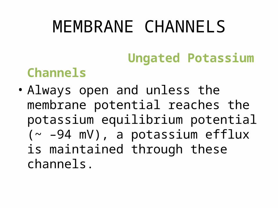
MEMBRANE CHANNELS
Ungated Potassium Channels• Always open and unless the membrane
potential reaches the potassium equilibrium potential (~ –94 mV), a potassium efflux is maintained through these channels.

Voltage-Gated Sodium Channels (fast channels)• These are closed under resting conditions.• Membrane depolarization is the signal that
causes these channels to quickly open and then close.
• These channels have the same characteristics as the voltage-gated sodium channels in the neuron axon.
• Once closed, they will not respond to a second stimulus until the cell repolarizes.

Voltage-Gated Calcium Channels (slow channels)
• Closed under resting conditions, when the membrane potential is highly negative.
• Depolarization is the signal that causes these channels to open.
• They open more slowly than the sodium channels.
• Consequently, they are sometimes called the slow channels.
• They are also called L-type, for long-acting channels.

• Because they allow sodium as well as calcium to pass, they are also called slow calcium-sodium channels.
• The calcium entering the cell through these channels will participate in contraction and will also be involved in the release of additional calcium from the sarcoplasmic reticulum.

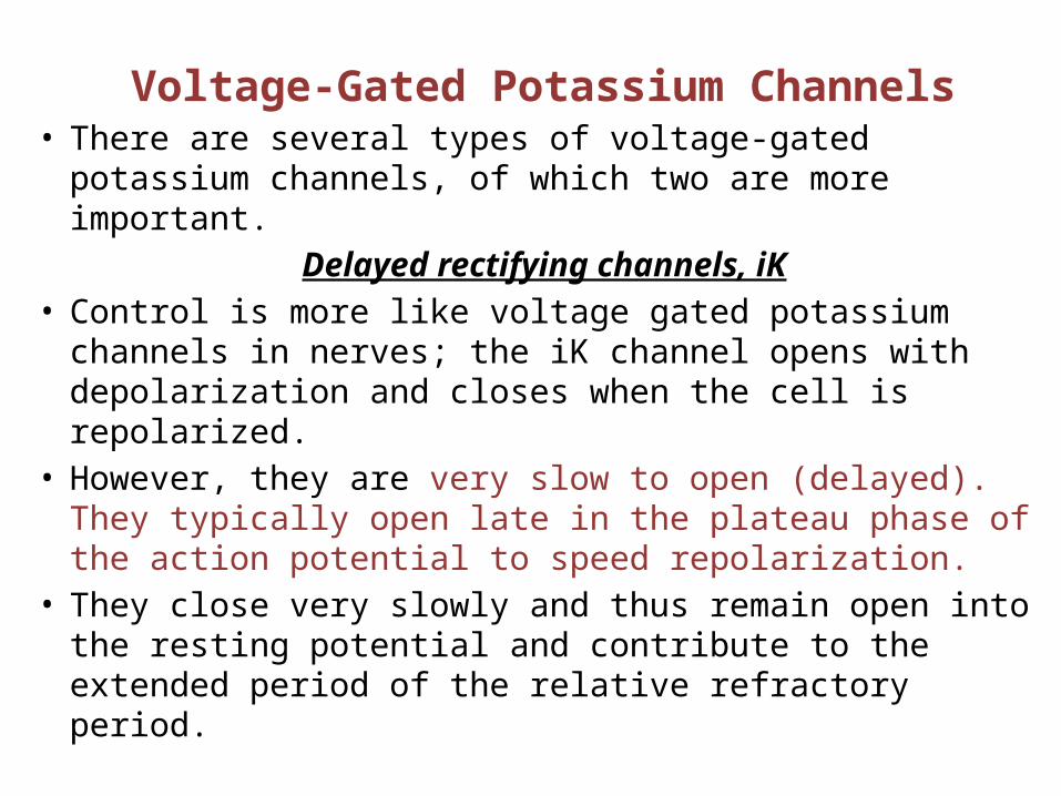
Voltage-Gated Potassium Channels• There are several types of voltage-gated potassium
channels, of which two are more important.Delayed rectifying channels, iK
• Control is more like voltage gated potassium channels in nerves; the iK channel opens with depolarization and closes when the cell is repolarized.
• However, they are very slow to open (delayed). They typically open late in the plateau phase of the action potential to speed repolarization.
• They close very slowly and thus remain open into the resting potential and contribute to the extended period of the relative refractory period.
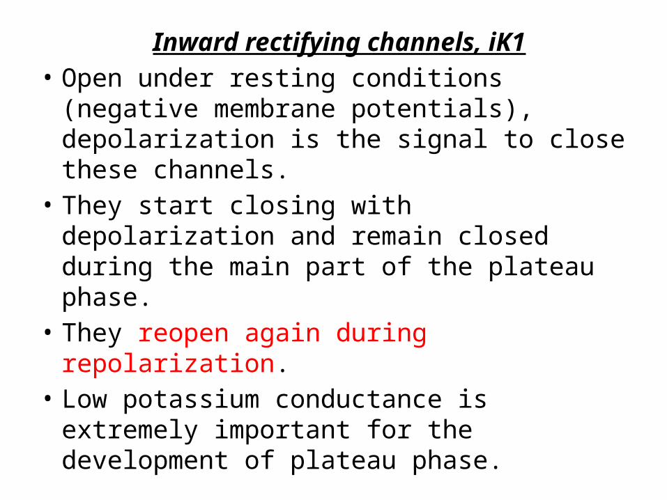
Inward rectifying channels, iK1• Open under resting conditions (negative
membrane potentials), depolarization is the signal to close these channels.
• They start closing with depolarization and remain closed during the main part of the plateau phase.
• They reopen again during repolarization. • Low potassium conductance is extremely
important for the development of plateau phase.

Q. Which of the following changes would be expected to make the membrane potential of a muscle cell more positive than normal (resting cell)?
A. Increased conductance to calciumB. Increased conductance to potassiumC. Increased conductance to chlorideD. Decreased conductance to sodium

• the fast response, occurs in normal atrial and ventricular myocytes and in the specialized conducting fibers (Purkinje fibers of the heart)
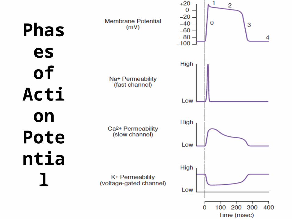
Phasesof
Action Potent
ial

Ionic Basis of the Action Potential
Phase 0, Upstroke • Fast channels open, ↑
gNa. Sodium influx causes depolarization.
• The channels open and close quickly, and they have closed by the time the main part of the plateau phase is entered.
Phase 1, Early Repolarization • This slight repolarization is
due to a net transient potassium current and the closing of the sodium channels.
• Subendocardial fibers lack phase 1.

Phase 2, Plateau• L-type Ca2+ channels are open, gCa ↑ permitting a
calcium influx.• Voltage-gated potassium channels, the iK1, are
closed; gK ↓ compared with resting membrane.• Potassium efflux continues through the ungated
potassium channels• Calcium channel antagonists shorten the plateau.
L-type channels are blocked by Ca++ channel antagonists such as verapamil, amlodipine, and diltiazem
• Potassium channel antagonists lengthen the plateau.

IN THE CLINIC- Ca++ channel antagonists
• Verapamil, Amlodipine, and Diltiazem. • Decrease gCa and thereby impede the influx of Ca++ into
myocardial cells. • Decrease the duration of the action potential plateau and
diminish the strength of the cardiac contraction • Also depress the contraction of vascular smooth muscle and
thereby induce generalized vasodilation. This diminished vascular resistance reduces the counterforce (afterload) that opposes the propulsion of blood from the ventricles into the arterial system.Hence, vasodilator drugs such as the Ca++ channel antagonists are often referred to as afterload-reducing drugs


Phase 3, Final Repolarization • L-type Ca2+ channels close, gCa ↓; this eliminates
any influx through these channels.• Voltage-gated potassium channels, the delayed
rectifier iK, then the iK1 are opening, gK ↑ leading to a large potassium efflux, and the cell quickly repolarizes.
• If the voltage-gated potassium channels did not open, the cell would still repolarize but more slowly, because of closure of calcium channels and potassium efflux through the ungated potassium channels.

Phase 4, Restoration of Ionic Concentrations • gK high; voltage-gated and ungated potassium
channels open. • The delayed rectifiers, iK, gradually close but
are responsible for the relative refractory period during early phase 4 with the cell in hyperpolarized state.

Which phase is most affected if the voltage gated Potassium channels (delayed rectifier) are blocked?

High Calcium conductance is seen in which of the following phases?

Permeability
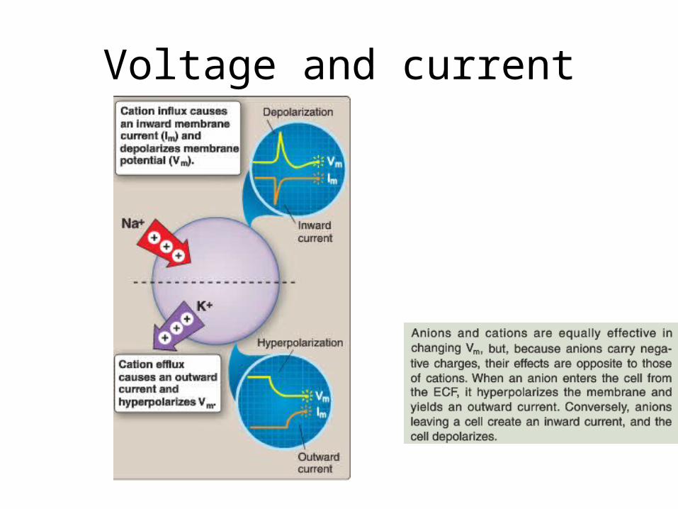
Voltage and current


EXCITATION-CONTRACTION COUPLING


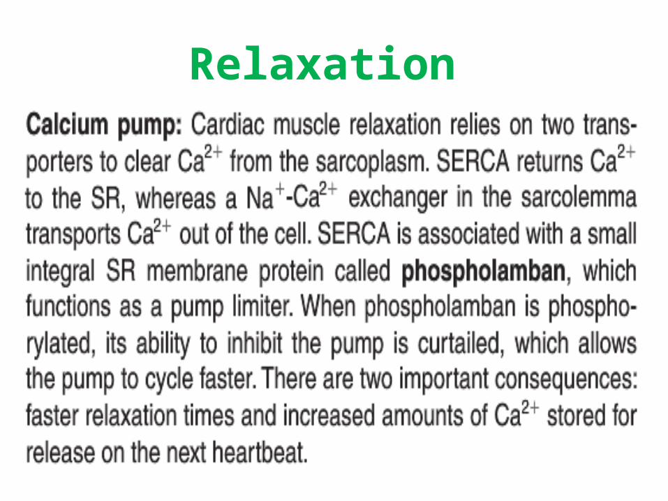
Relaxation


Force of contraction is relatively insensitive to extracellular Ca2+A. Cardiac muscleB. Skeletal muscleC. Smooth muscle

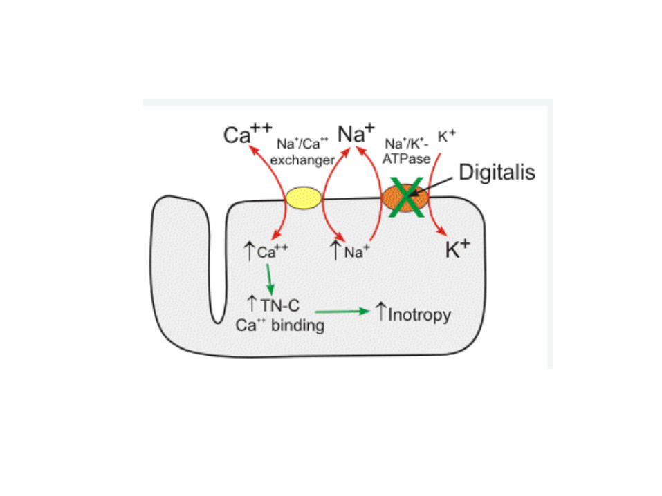
CONTRACTLITY REGULATION

I. Intracellular Calcium

II.SympatheticActivation


III. Preload Dependence

Pre load/Stretch Limits
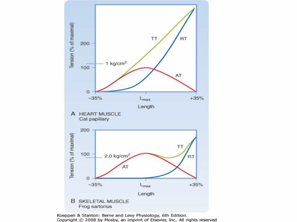

Correlation between Muscle Fiber Length & Tension

Hypertrophy of the heart • In response to exercise -beneficial, with
improved cardiac performance, increased oxygen consumption, and normal relaxation
• Chronic pressure overload, -- progress to dilated cardiac hypertrophy characterized by decreased contractile ability
• Genetic mutations --familial hypertrophic cardiomyopathy, in which a mutation in a single intracellular protein may alter contractile function and promote a hypertrophic response


V. Energy Source
• < 1 sec ,cardiac mus contraction duration.• It maintains modest ATP stores that support
short contra and then regenerate these stores using aerobic pathway when relaxed.
• Limited anaerobic capacity creates high dependence on O2.
• Prolonged O2 deprivation (min)causes irreversible hypoxic mus damage and myocardial infraction.