C:/Ahmed/isvc2007 camera ready/lg-seg+trac
10
Transcript of C:/Ahmed/isvc2007 camera ready/lg-seg+trac
4D Ventri ular Segmentation and Wall MotionEstimation Using EÆ ient Dis rete OptimizationAhmed Besbes1, Nikos Komodakis1, Ben Glo ker2, Georgios Tziritas3, andNikos Paragios11 GALEN Group, Laboratoire MAS, E ole Centrale de Parisfahmed.besbes,nikos.komodakis,nikos.paragiosg�e p.fr2 Chair for Computer Aided Medi al Pro edures (CAMP)Te hnis he Universit�at M�un henglo ker�in.tum.de3 University of Crete, Computer S ien e Departmenttziritas� sd.uo .grAbstra t. In this paper we propose a novel approa h to ventri ularmotion estimation and segmentation. Our method is based on a MRFformulation where an optimal intensity-based separation between the en-do ardium and the rest of the ardia volume is to be determined. Su h aterm is de�ned in the spatiotemporal domain, where the ventri ular wallmotion is introdu ed to a ount for orresponden es between the on-se utive segmentation maps. The estimation of the deformations is donethrough a ontinuous deformation �eld (FFD) where the displa ementsof the ontrol points are determined using dis rete labeling approa h.Prin iples from linear programming and in parti ular the Primal/DualS hema is used to re over the optimal solution in both spa es. Promis-ing experimental results obtained on 13 MR spatiotemporal data setsdemonstrate the potentials of our method.1 Introdu tionThe segmentation of the left ventri le has been a problem well addressed inmedi al imaging. Prior art either refers to model-free approa hes or model-based.Model-free methods do not make an expli it assumption on the form/geometri properties as well as the appearan e of the ventri le. MRFs [1℄, snakes [2℄, levelsets [3℄, shortest path [4℄ have been onsidered in this ontext. On the other hand,model-based methods often onsider ertain geometri priors for the ventri lewhi h ould range from simple 2D shapes [4℄ and 3D models whi h also en odelo al variations [1℄ to omplex biome hani al ardia models [5℄.Ventri ular wall motion estimation was often addressed through the use ofMR-Tagging [6℄ [7℄ te hniques that onsist of introdu ing a re tangular patternon the a quisition. Dire t 3D motion estimation in MR is a more hallengingproblem sin e it is known that the left ventri le undergoes a rather omplexdeformation within the ardia y le. In order to a ount for the ill-posednessof the problem, the use of shape models towards establishing visual orrespon-den es and tra king was often onsidered [8℄ or 4D models have been onstru ted

2 A. Besbes, N. Komodakis, B. Glo ker, G. Tziritas, N. Paragioswith spatial and temporal deformations being en oded [9℄. Voxel-based methodsoften explore the visual preservation assumption [10℄ while being onstrainedto provide a smooth deformation map. More omplex models use biome hani al onstraints to determine su h a deformation [11℄, an approa h whi h might failwhen pro essing diseased data.In most of the ases, these methods do not relate segmentation with ventri u-lar motion estimation. Furthermore, one an laim that they are sensitive to theinitial onditions either be ause of the non- onvexity of the designed ost fun -tion or due to the sub-optimal optimization approa h. In this paper, we propose anovel approa h to address both segmentation and ventri ular motion estimation.We over ome the ill-posedness of the motion estimation problem through the useof interpolation te hniques with higher order polynomials, while we introdu etemporal segmentation onsisten y through the use of deformations �eld. In or-der to eÆ iently re over the optimal solution to the problem, we re-formulate the ost fun tion in a fully dis rete domain where the latest developments of linearprogramming are onsidered to determine the lowest potential of the ost fun -tion. Very promising results and omparisons with manual segmentation fromphysi ians demonstrate the potentials of our approa h.2 Ventri ular Segmentation and Wall Motion Estimation2.1 Spatiotemporal SegmentationLet us onsider a spatiotemporal volume V(x; t) : � [0::� ℄! R, with beingthe volume domain. The task of segmenting the endo ardium an be reformu-lated using a labeling approa h, or assigning a label �(x; t) : � [0::� ℄! f0; 1g.Here, label 0 orresponds to the foreground (i.e., the ventri le), whereas label 1 orresponds to the ba kground. Without loss of generality, let us assume that ertain statisti al properties on the intensities of the left ventri le p(Vj� = 0),as well as on the intensities of the ba kground p(Vj� = 1) are available or anbe determined on the y. Let us also assume that we have a prior left ventri le losed surfa e (St)�t=0 de�ned as:�S(x; t) = 8><>:0 if x 2 St�D(x;S) if x 2 StinD(x;S) if x 2 Stout : (1)with D being the Eu lidean distan e between a given voxel and the surfa e,and(St; Stin; Stout) being the partition of de�ned by St, 8t 2 [0::� ℄. We de�nea penalization fun tion p�(�;�) : R� f0; 1g ! R, with � > 0 as a de reasing(respe tively in reasing) fun tion of � if � = 0 (respe tively � = 1), and equalto identity for � < �.In su h a ase, the optimal labeling should refer to the maximum onditionalposterior between the de isions and the data support. If spatial and temporal

Ventri ular Segmentation & Motion Estimation 3independen e are assumed between voxels, that labeling an then be re overedthrough the minimum of:Eseg;dt(�) = �Xt=0 Xx2�log [p (V(x; t)j�(x; t)) :p� (�S(x; t);�(x; t))℄= �Xt=0 Xx2 V pdt(�(x; t)) : (2)whi h is equivalent to assigning to ea h voxel the label whi h is optimally sup-ported from the observation. Su h a simplisti formulation ould produ e sub-optimal results due the presen e of noise and therefore one should introdu eadditional smoothness onstraints on the label spa e, whi h aims to enfor e reg-ularity on the de isions, or:Eseg;sp(�)= �Xt=0 Xx20� Xy2N (x) (�(x; t); �(y; t))1A= �Xt=0 Xx2y2N (x)Vsp(�(x; t); �(y; t)) :(3)with being a fun tion measuring the dissimilarity between labels of neighboringpixels and N (x) de�nes the lo al neighborhood of x in the 3D spatial domain.For more robustness, one an also onsider temporal onstraints on the labelingif the deformations from one volume to the next are not so important, whi h is,however, de�nitely not the ase for the left and right ventri ular motion. On theother hand, if we assume that this deformation is known, say, d(x; t), then one an imagine using d(x; t) towards determining the temporal derivative on thelabel spa e and introdu e a smoothness onstraint of the following form:Eseg;tm(�j d) = ��1Xt=0 Xx2 (�(x; t); �(x + d(x; t); t + 1))dx= ��1Xt=0 Xx2 Vtm(�(x; t); �(x + d(x; t); t + 1)) : (4)The interpretation of this term is straightforward, assuming known orrespon-den es one would expe t a oherent labeling between anatomi al stru tureswithin the ardia y le. Based on this fa t, we an therefore pro eed as fol-lows: we will �rst estimate the deformation d(x; t), i.e. register the 3D volumes,and then we will extra t the optimal segmentation (i.e. the optimal labeling�(x; t))) by minimizing the total energy E4D of the resulting binary 4D MarkovRandom Field, where the total energy is given by:Eseg(�j d) = Eseg;dt(�) + �Eseg;sp(�) + �Eseg;tm(�j d) : (5)Intuitively, the edges of the resulting 4D MRF will onsist of regular links, on-ne ting (in a grid-like manner) voxels belonging to the same 3D volume. On

4 A. Besbes, N. Komodakis, B. Glo ker, G. Tziritas, N. Paragiosthe other hand, they refer to irregular links in the temporal domain, onne tingvoxels between adja ent 3D volumes, being determined via the previously esti-mated deformation d(x; t). We also note that be ause our MRF is binary, theexa t global optimum an be easily extra ted [12℄.However, establishing orresponden es between volumes is an ill-posed prob-lem. Even if we assume the visual preservation assumption to be valid (not oftenthe ase for medi al image modalities), one should determine three unknownvariables from a single onstraint. To deal with this issue, in the next se tion weshow how we an regularize this motion estimation problem by reformulating itas another dis rete MRF optimization problem.2.2 Ventri ular Motion EstimationLet us thus assume that we wish to ompute the deformation d(x; t) betweentwo adja ent 3D volumes at time t. To this end, we will introdu e a sparse defor-mation grid G super-imposed on the sour e volume (no parti ular assumption ismade on the grid ex ept that it is sparser than the original volume). The entralidea of our approa h is to deform the grid (with a 3D displa ement ve tor d(p; t)for ea h ontrol point p) su h that the underlying volumes are perfe tly aligned.Without loss of generality, we an then assume that the displa ement of a voxelx an be expressed using a linear or non-linear ombination of the grid points,or: d(x; t) =Xp2G �(jx� pj) d(p; t) : (6)where �(�) is the weighting fun tion measuring the ontribution of the ontrolpoint p to the displa ement �eld d(x; t). The use of su h a model is motivated bythe fa t that the observations refer to anatomi al stru tures with a rather naturaltemporal deformation. Furthermore, su h an approa h ould help us to a ountfor the ill-posedness of the problem due to the fa t that the estimation of a single3D displa ement is now an over- onstrained problem with many observationsbeing available. For �(�), we use a three-dimensional Free Form Deformation(FFD) model based on ubi B-splines [13℄ (other interpolation models an alsobe onsidered).Therefore, based on (6), to estimate d(x; t) it suÆ es to spe ify the displa e-ments for the ontrol points. To this end, we will onsider a quantized versionof the deformation spa e, say, fd1; :::; dig - being 3D deformation ve tors - aswell as a orresponding set of dis rete labels, say, L = f1; :::; ig. A label assign-ment, say, !(p) 2 L to a grid point p is asso iated with displa ing p by the orresponding ve tor d!(p), i.e.:d(p; t) = d!(p) : (7)The visual preservation imposes the onstraint that the observation of the sameanatomi al pat h should be onsistent a ross volumes, i.e., V(x; t) � V(x +d(x; t); t+1). In our dis rete framework the deformation d(x; t) is de�ned basedon (6), (7), i.e. displa ements are asso iated with labels, one an reformulate

Ventri ular Segmentation & Motion Estimation 5ventri ular deformation estimation as a labeling problem. Consequently, the goalis to assign a set of appropriate labels f!(p)g (to the grid points) so that thevisual preservation onstraint is satis�ed as mu h as possible, or equivalently sothat the following data ost is minimized:Emot;dt(!) = Xx2 jV(x; t)� V(x+ d(x; t); t + 1)j (6);(7)� Xp2GUpdt(!(p)) : (8)Here, the singleton potential fun tions Updt(�) are not independent, thus the de-�ned data term an only be approximated. Hen e, we pre ompute the jLj � jGj(where jGj is the number of grid points) data term in a look-up table. The entryfor label !(p) and node p is determined by:Updt(!(p)) = ZZ(p) ��1(jx� pj) � ���V(x; t)� V(x+ d!(p); t+ 1)��� dx : (9)with the sum of absolute di�eren es being onsidered as measure of similarity(��1 is the inverse proje tion between x and p). The use of an interpolation te h-nique to determine the deformations of the volume will inherit natural smooth-ness to the estimates. However, one should also expe t sin e we aim to re overmeasurements for physi al obje ts deformations that the same assumption issatis�ed for the deformation of the orresponding ontrol points. Similar to thesegmentation ase, one an onsider a term whi h enfor es spatial similaritiesa ross labels, or: Emot;sm(!) = Xp2q2N (p)Usm(!(p); !(q)) : (10)where N represents the neighborhood system asso iated with the deformationgrid G. For the distan e Usm(�; �), we onsider a simple pie ewise smoothnessterm based on the Eu lidean distan e between the deformations orrespondingto the assigned labels, i.e.:Usm(!(p); !(q)) = �pq �jd!(p) � d!(q)j� : (11)with �pq being a (spatially varying) weighting to ontrol the in uen e of thesmoothness/prior term. Su h a smoothness term, together with the data term,allows to onvert the problem of volume registration into a dis rete MRF opti-mization problem with the following energy [14℄:Emot(!) = Emot;dt(!) +Emot;sm(!) : (12)2.3 4D Segmentation & Ventri ular Motion EstimationOne an now onsider an obje tive fun tion whi h re overs both the 4D segmen-tation map as well as the orresponding deformation �elds:Eseg;mot(�; !) = Eseg(�j!) + Emot(!) : (13)

6 A. Besbes, N. Komodakis, B. Glo ker, G. Tziritas, N. Paragios(a) The primal-dual prin iple (b) The primal-dual s hemaFig. 1: (a) By weak duality, the optimal ost Tx� will lie between the osts bTy and Tx of anypair (x;y) of integral-primal and dual feasible solutions. Therefore, if bTy and Tx are lose enough(e.g. their ratio r1 is � f), so are Tx� and Tx (e.g. their ratio r0 is � f as well), thus proving thatx is an f-approximation to x�. (b) A ording to the primal-dual s hema, dual and integral-primalfeasible solutions make lo al improvements to ea h other, until the �nal osts bTyt, Txt are loseenough (e.g. their ratio is � f). We an then apply the primal-dual prin iple (as in Fig. (a)) andthus on lude that xt is an f-approximation to x�whi h is a fully dis rete optimization problem. For optimizing the resulting MRF,we seek to assign a pair of labels (�(p); !(p)) to ea h node p 2 G, so that theMRF energy in (13) is minimized. To this end, a re ently proposed method, alled Fast-PD (Fast Primal Dual), will be used. This is an optimization te h-nique, whi h builds upon prin iples drawn from the duality theory of linearprogramming in order to eÆ iently derive almost optimal solutions for a verywide lass of NP-hard MRFs. For more details about the Fast-PD algorithm, thereader is referred to [12℄. Here, we will just provide a brief, high level des riptionof the basi driving for e behind that algorithm.3 Linear ProgrammingThe driving for e of the algorithm onsists of the primal-dual s hema, whi h is awell-known te hnique in the Linear Programming literature. To understand howthe primal-dual s hema works in general, we will need to onsider the followingpair of primal and dual Linear Programs (LPs):Primal: min Tx Dual: max bTys.t. Ax = b;x � 0 s.t. ATy � (14)Here A represents a oeÆ ient matrix, while b; are oeÆ ient ve tors. Also,x, y represent the ve tors of primal and dual variables respe tively. We seekan optimal solution to the primal program, but with the extra onstraint of xbeing integral. Due to this integrality requirement, this problem is in generalNP-hard and so we need to settle with estimating approximate solutions. Aprimal-dual f -approximation algorithm a hieves that by use of the followingprin iple (illustrated also in Fig. 1(a)):Primal-Dual Prin iple 1 If x and y are integral-primal and dual feasible so-lutions having a primal-dual gap less than f , i.e.: Tx � f � bTy; (15)

Ventri ular Segmentation & Motion Estimation 7then x is an f-approximation to the optimal integral solution x�, i.e. Tx� � Tx � f � Tx�.Based on the above prin iple, that lies at the heart of any primal-dual te hnique,the following iterative s hema an be used for deriving an f -approximate solution(this s hema is also illustrated graphi ally in Fig. 1(b)):Primal-Dual S hema 1 Keep generating pairs of integral-primal, dual solu-tions f(xk;yk)gtk=1, until the elements xt, yt of the last pair are both feasibleand have a primal-dual gap whi h is less than f , i.e. ondition (15) holds true.In order to apply the above s hema to MRF optimization, it suÆ es that we ast the MRF optimization problem as an equivalent integer program. TheFast-PD algorithm is then derived by applying the primal-dual s hema to thispair of primal-dual LPs, while using f=2dmaxdmin (dmax �maxa6=b d(a; b); dmin �mina6=b d(a; b)) as the approximation fa tor in (15). Fast-PD is a very generalMRF optimization method, whi h an handle a very wide lass of MRFs. Essen-tially, it only requires that the MRF pairwise potential fun tion is nonnegative.Furthermore, as already mentioned, it an guarantee that the generated solu-tion is always within a worst- ase bound from the optimum. In fa t, besides thisworst- ase bound, it an also provide per-instan e approximation bounds, whi hprove to be mu h tighter, i.e. very lose to 1, in pra ti e. It thus allows the globaloptimum to be found up to a user/appli ation bound. Finally, it provides great omputational eÆ ien y, sin e it is typi ally 3-9 times faster than any otherMRF optimization te hnique with guaranteed optimality properties [12℄.4 ValidationIn order to validate the performan e of the method we have onsidered a setof 13 MR spatiotemporal volumes of the heart, with manual segmentation fromtwo lini al experts being available for the diastole and the systole. These datasets had a spatial resolution of around 100x100x12 and a voxel size of around1.77x1.77x6 millimeters. We used as prior information two learned distributionsof endo ardium voxels and ba kground voxels expressed as mixture of Gaus-sians. These distributions were time-independent, and were used in diastole andsystole as well. We also added a shape prior onstraint (�xed shape S, initializedby the user) to a ount for the ellipti geometry of the left ventri le. In termsTable 1: Comparison of automati and experts' segmentations in diastoleComparison DSC Mean (Std) Sensitivity Spe i� ity ASD Mean (Std)Our Method vs Expert1 0.86(�0.03) 99.06% 95.76% 1.54(�0.39 )Our Method vs Expert2 0.87(�0.02) 99.11% 96.88% 1.31(�0.37 )Expert1 vs Expert2 0.89(�0.02) 99.49% 94.46% 0.87(�0.12 )Expert2 vs Expert1 0.89(�0.02) 99.53% 94.16% 1.34(�0.47 )

8 A. Besbes, N. Komodakis, B. Glo ker, G. Tziritas, N. Paragiosof segmentation performan e we ompare the experts' segmentation of the en-do ardium with the one obtained using the proposed method. We are interestedon several ommon evaluation measurements [15℄, and in parti ular the Di esimilarity oeÆ ient (DSC), the sensitivity, the spe i� ity, and the average sur-fa e distan e (ASD) from the experts segmentations. The ASD is omputed inmillimeters from an anisotropi 3D Eu lidean distan e transform of the surfa es.These measurements are omputed in both diastole and systole and are presentedin [Tab. (1)℄ and [Tab. (2)℄. We also ompare in these tables the performan esof our method to those a hieved manually by the experts.
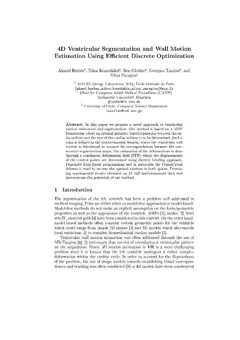
(a) entripetal motion �eld (b) entrifugal motion �eldFig. 2: Motion estimation. (a) beginning of systole (b) beginning of diastoleWe a hieve an ASD whi h is below the voxel size in both diastole and systole.The DSC whi h measures the overlap between surfa es shows that our segmen-tation is loser to the one given by Expert2 than to the one given by Expert1.Overall, our performan e is satisfa tory ompared to the one a hieved by theexperts. We get a worse sensitivity than the experts, but a better spe i� ity. Interms of ventri ular motion estimation, we present in [Fig. (2)℄ the deformation�eld of the endo ardium and its motion estimation. We see in parti ular in this�gure that the motion �eld is oherent with the left ventri le motion: the en-tripetal motion �eld at the beginning of systole is justi�ed by the ontra tion ofthe myo ardium, and the entrifugal motion �eld at the beginning of diastole isjusti�ed by its expansion. The 3D images in [Fig. (3)℄ show that we also orre tlysegment the papillary mus les.With a reasonable number of displa ement labels (the omplexity is linear tothe number of labels), the method takes about 10-20 se onds to onverge (using aDELL Duo with (3GHz,2GB)) assuming that a ventri le isolation has been doneand is able to produ e good orresponden es with a 16� 16� 16 FFD grid. The
Ventri ular Segmentation & Motion Estimation 9(a) in diastole (b) in systoleFig. 3: Papillary mus les. In ea h image : automati segmentation & experts' manual segmentation ardia y le being quantized by 20-25 time points, the whole 4D segmentationand motion estimation omputation takes about 70-80 se onds for a 4D volume.5 Dis ussionIn this paper we have proposed a novel dis rete approa h to spatiotemporal seg-mentation and ventri ular motion estimation. The strength of our approa h isthe oupling between the two problems and the use of a powerful ombinato-rial algorithm to produ e their solution. In order to demonstrate the on ept,we have onsidered a set of several heart 4D MRI exams and we have obtainedquite satisfa tory results. More hallenging perspe tives are related with the in-trodu tion of prior knowledge both in spa e and time related with the evolvinggeometry of the stru tures of interest. The prior information used in our ap-proa h remains quite simple, and is time-independent. That is why our resultsare promising and an be probably improved by the use of more omplex priorinformation whi h an better apture the anatomy and the temporal dynami sof the ardia y le. Knowledge-based segmentation using models that en odeimportant statisti al variation of training examples within dis rete optimizationis a quite promising dire tion to be onsidered.
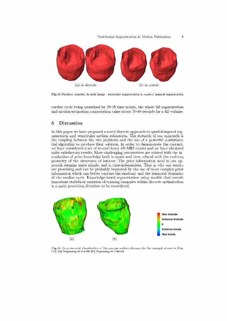
(a) (b)Fig. 4: Color-en oded visualization of the average surfa e distan e for the example shown in [Fig.(2)℄. (a) beginning of systole (b) beginning of diastole

10 A. Besbes, N. Komodakis, B. Glo ker, G. Tziritas, N. ParagiosTable 2: Comparison of automati and experts' segmentations in systoleComparison DSC Mean (Std) Sensitivity Spe i� ity ASD Mean (Std)Our Method vs Expert1 0.82(�0.03) 99.39% 93.34% 1.51(�0.39 )Our Method vs Expert2 0.85(�0.03) 99.46% 94.34% 1.28(�0.37 )Expert1 vs Expert2 0.86(�0.03) 99.69% 91.07% 0.86(�0.15 )Expert2 vs Expert1 0.86(�0.03) 99.66% 91.50% 1.06(�0.22 )Referen es1. Shi, P., Sinusas, A.J., Constable, R.T., Ritman, E., Dun an, J.S.: Point-tra kedquantitative analysis of left ventri ular surfa e motion from 3d image sequen es.IEEE Trans. Med. Imaging 19(1) (2000) 36{502. M Inerney, T., Terzopoulos, D.: Deformable models in medi al images analysis: asurvey. Medi al Image Analysis 1(2) (1996) 91{1083. Paragios, N.: A variational approa h for the segmentation of the left ventri le in ardia image analysis. Int. J. Comput. Vision 50(3) (2002) 345{3624. Jolly, M.P.: Automati segmentation of the left ventri le in ardia mr and timages. Int. J. Comput. Vision 70(2) (2006) 151{1635. Sermesant, M., Forest, C., Penne , X., Delingette, H., Aya he, N.: Deformablebiome hani al bodels: Appli ation to 4D ardia image analysis. Medi al ImageAnalysis 7(4) (2003) 475{4886. Montillo, A., Metaxas, D.N., Axel, L.: Automated model-based segmentation ofthe left and right ventri les in tagged ardia mri. In: MICCAI (1). (2003) 507{5157. Guttman, M., Prin e, J., M Veigh, E.: Tag and ontour dete tion in tagged mrimages of the left ventri le. IEEE Trans. Med. Imaging 13(1) (1994) 74{888. M Ea hen II, J.C., Dun an, J.S.: Shape-based tra king of left ventri ular wallmotion. IEEE Trans. Med. Imaging 16(3) (1997) 270{2839. Bos h, J.G., Mit hell, S.C., Lelieveldt, B.P.F., Nijland, F., Kamp, O., Sonka, M.,Reiber, J.H.C.: Automati segmentation of e ho ardiographi sequen es by a tiveappearan e motion models. IEEE Trans. Med. Imaging 21(11) (2002) 1374{138310. Horn, B.K.P., S hun k, B.G.: Determining opti al ow. Artif. Intell. 17(1-3) (1981)185{20311. Sermesant, M., Delingette, H., Aya he, N.: An ele trome hani al model of theheart for image analysis and simulation. IEEE Trans. Med. Imaging 25(5) (2006)612{62512. Komodakis, N., Tziritas, G., Paragios, N.: Fast, approximately optimal solutionsfor single and dynami mrfs. In: IEEE Conferen e on Computer Vision & PatternRe ognition (CVPR). (2007)13. Sederberg, T.W., Parry, S.R.: Free-form deformation of solid geometri models.In: SIGGRAPH '86: Pro eedings of the 13th Annual Conferen e on ComputerGraphi s and Intera tive Te hniques. (1986) 151{16014. Glo ker, B., Komodakis, N., Paragios, N., Tziritas, G., Navab, N.: Inter and intra-modal deformable registration: Continuous deformations meet eÆ ient optimal lin-ear programming. In: Information Pro essing in Medi al Imaging (IPMI). (2007)15. Gerig, G., Jomier, M., Chakos, M.: Valmet: A new validation tool for assessingand improving 3d obje t segmentation. In: MICCAI. (2001) 516{523