C h a p t e r 26 The Urinary System PowerPoint ® Lecture Slides prepared by Jason LaPres North...
33
C h a p t e r 26 The Urinary System PowerPoint ® Lecture Slides prepared by Jason LaPres North Harris College Houston, Texas Copyright © 2009 Pearson Education, Inc., publishing as Pearson Benjamin Cummings
-
Upload
kerry-owen -
Category
Documents
-
view
215 -
download
0
Transcript of C h a p t e r 26 The Urinary System PowerPoint ® Lecture Slides prepared by Jason LaPres North...

C h a p t e r
26
The Urinary System
PowerPoint® Lecture Slides prepared by Jason LaPres
North Harris CollegeHouston, Texas
Copyright © 2009 Pearson Education, Inc.,publishing as Pearson Benjamin Cummings

Introduction
The urinary system includes the kidneys, the ureters, the urinary bladder, and the urethra.
Performs vital excretory functions: Regulating plasma concentrations of ions Regulating blood volume and blood pressure by adjusting
the volume of water lost in the urine, releasing erythropoietin, and releasing renin
Contributing to the stabilization of blood pH Conserving valuable nutrients Eliminating organic waste products Synthesizing calcitriol. Assisting the liver in detoxifying poisons
Copyright © 2009 Pearson Education, Inc., publishing as Pearson Benjamin Cummings
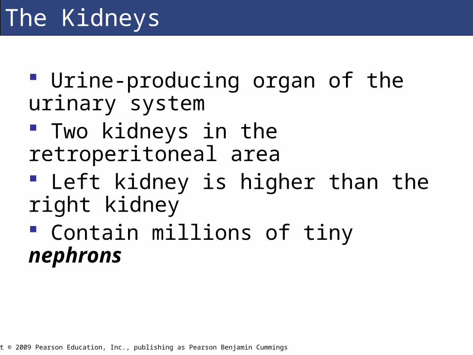
The Kidneys
Urine-producing organ of the urinary system Two kidneys in the retroperitoneal area Left kidney is higher than the right kidney Contain millions of tiny nephrons
Copyright © 2009 Pearson Education, Inc., publishing as Pearson Benjamin Cummings

The Kidneys
Figure 26.1a An Introduction to the Urinary System (a) Anterior View
Copyright © 2009 Pearson Education, Inc., publishing as Pearson Benjamin Cummings

The Kidneys
Figure 26.1b An Introduction to the Urinary System: (a) Posterior View
Copyright © 2009 Pearson Education, Inc., publishing as Pearson Benjamin Cummings

The Kidneys
Figure 26.1c An Introduction to the Urinary System: (c) Transverse Section at L1
Copyright © 2009 Pearson Education, Inc., publishing as Pearson Benjamin Cummings

The Kidneys
Copyright © 2009 Pearson Education, Inc., publishing as Pearson Benjamin Cummings
Figure 26.1d An Introduction to the Urinary System: (d) Transverse Section at T12

The Kidneys
Figure 26.2 The Urinary System in Gross Dissection
Copyright © 2009 Pearson Education, Inc., publishing as Pearson Benjamin Cummings

The Kidneys
Figure 26.3a Structure of the Kidney: (a) Frontal Section of Left Kidney, Anterior View
Copyright © 2009 Pearson Education, Inc., publishing as Pearson Benjamin Cummings

The Kidneys
Figure 26.3b, c Structure of the Kidney: (b) Calyces and Renal Pelvis; (c) Urogram
Copyright © 2009 Pearson Education, Inc., publishing as Pearson Benjamin Cummings

The Kidneys
Figure 26.4a Blood Supply to the Kidneys, (a) Frontal Section Copyright © 2009 Pearson Education, Inc., publishing as Pearson Benjamin Cummings

The Kidneys
Copyright © 2009 Pearson Education, Inc., publishing as Pearson Benjamin Cummings
Figure 26.4b Blood Supply to the Kidneys, (b) Cortical Circulation

The Kidneys
Copyright © 2009 Pearson Education, Inc., publishing as Pearson Benjamin Cummings
Figure 26.4c Blood Supply to the Kidneys, (c) Flowchart of Renal Ciriulation

The Kidneys
Figure 26.5 Renal Vessels and Blood Flow
Copyright © 2009 Pearson Education, Inc., publishing as Pearson Benjamin Cummings

The Kidneys
Figure 26.6 A Typical Nephron
Copyright © 2009 Pearson Education, Inc., publishing as Pearson Benjamin Cummings

The Kidneys
Figure 26.7a, b Histology of the Nephron, Copyright © 2009 Pearson Education, Inc., publishing as Pearson Benjamin Cummings

The Kidneys
Copyright © 2009 Pearson Education, Inc., publishing as Pearson Benjamin Cummings
Figure 26.7a, c Histology of the Nephron,

The Kidneys
Copyright © 2009 Pearson Education, Inc., publishing as Pearson Benjamin Cummings
Figure 26.7a, d Histology of the Nephron,

The Kidneys
Figure 26.7e, f Histology of the Nephron
Copyright © 2009 Pearson Education, Inc., publishing as Pearson Benjamin Cummings

The Kidneys
Figure 26.8a, c The Renal Corpuscle
Copyright © 2009 Pearson Education, Inc., publishing as Pearson Benjamin Cummings

The Kidneys
Figure 26.8b The Renal Corpuscle Copyright © 2009 Pearson Education, Inc., publishing as Pearson Benjamin Cummings

The Kidneys
Figure 26.8d The Renal Corpuscle Copyright © 2009 Pearson Education, Inc., publishing as Pearson Benjamin Cummings

The Kidneys
Figure 26.8e The Renal Corpuscle Copyright © 2009 Pearson Education, Inc., publishing as Pearson Benjamin Cummings

The Kidneys
Figure 26.9a Images of the Urinary System: (a) Color-Enhanced CT Scan
Copyright © 2009 Pearson Education, Inc., publishing as Pearson Benjamin Cummings
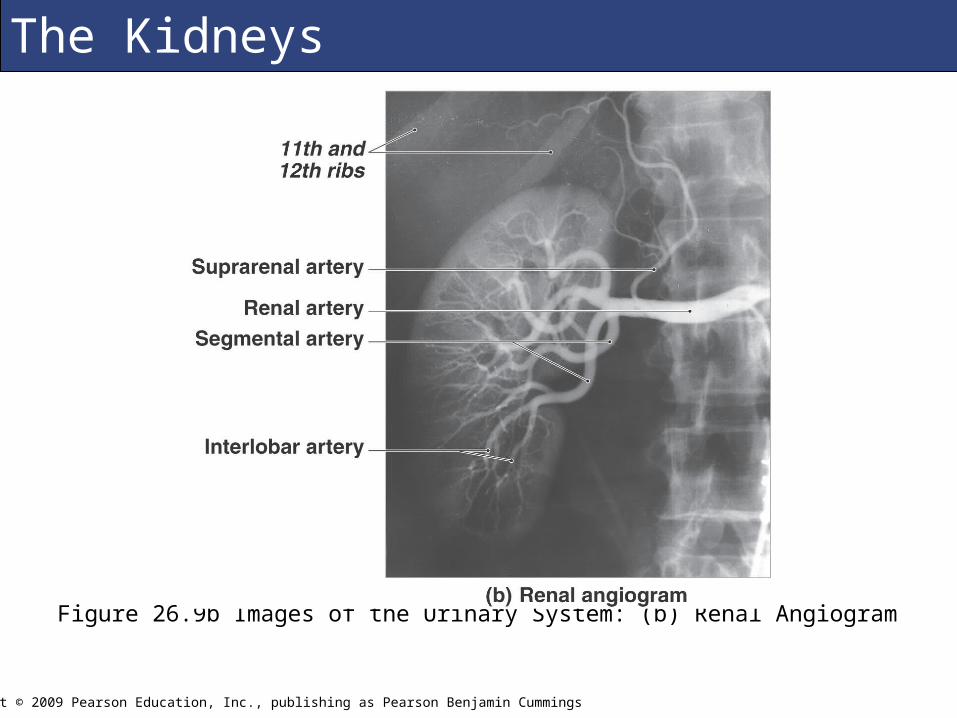
The Kidneys
Figure 26.9b Images of the Urinary System: (b) Renal Angiogram
Copyright © 2009 Pearson Education, Inc., publishing as Pearson Benjamin Cummings

The Kidneys
Figure 26.12 Histology of a Renal Glomerulus of a Patient with a Condition Similar to Danni’s.
Copyright © 2009 Pearson Education, Inc., publishing as Pearson Benjamin Cummings

Structures for Urine Transport, Storage, and Elimination
Ureters Urinary bladder Urethra
There are significant differences between the male and female urethra.
Copyright © 2009 Pearson Education, Inc., publishing as Pearson Benjamin Cummings

Figure 26.9c Images of the Urinary System (c) Normal Pyelogram
Copyright © 2009 Pearson Education, Inc., publishing as Pearson Benjamin Cummings
Structures for Urine Transport, Storage, and Elimination

M
Figure 26.10a Organs Responsible for the Conduction and Storage of Urine: (a) Male Pelvis, Sagittal Section
Copyright © 2009 Pearson Education, Inc., publishing as Pearson Benjamin Cummings
Structures for Urine Transport, Storage, and Elimination

M
Figure 26.10b Organs Responsible for the Conduction and Storage of Urine: (b) Female Pelvis, Sagittal Section
Copyright © 2009 Pearson Education, Inc., publishing as Pearson Benjamin Cummings
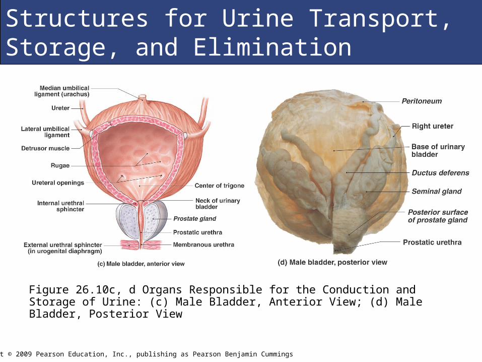
Structures for Urine Transport, Storage, and Elimination
M
Figure 26.10c, d Organs Responsible for the Conduction and Storage of Urine: (c) Male Bladder, Anterior View; (d) Male Bladder, Posterior View
Copyright © 2009 Pearson Education, Inc., publishing as Pearson Benjamin Cummings
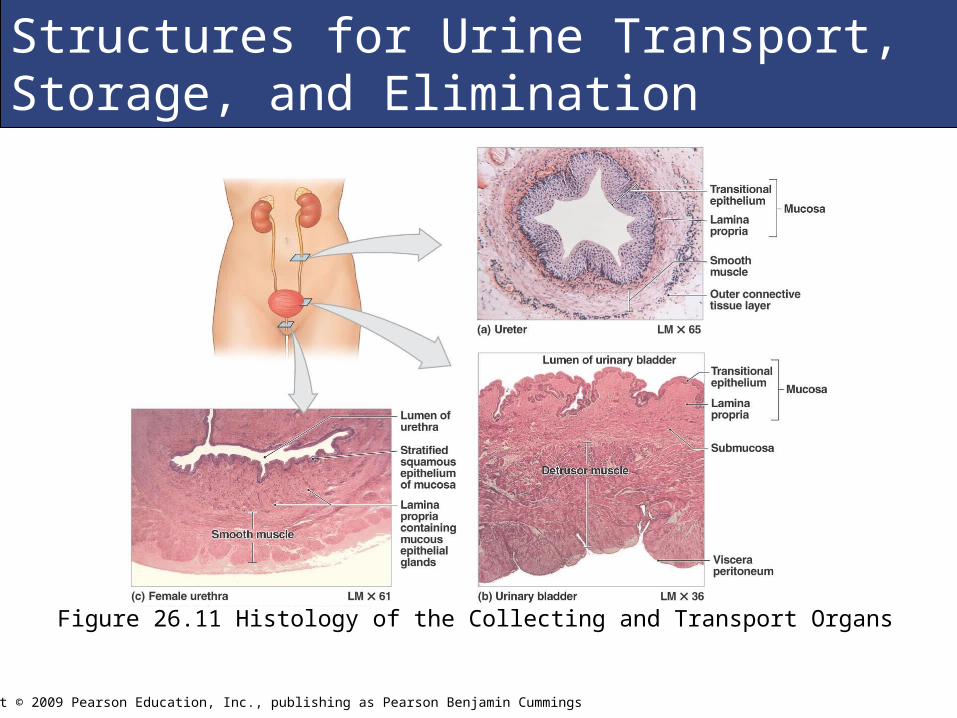
Structures for Urine Transport, Storage, and Elimination
M
Figure 26.11 Histology of the Collecting and Transport Organs
Copyright © 2009 Pearson Education, Inc., publishing as Pearson Benjamin Cummings
Structures for Urine Transport, Storage, and Elimination

Aging and the Urinary System
A decline in the number of functional nephrons A reduction in glomerular filtration Reduced sensitivity to ADH Problems with the micturition reflex related to the following factors:
Loss of tone in sphincter muscles leading to incontinence Strokes, Alzheimer’s disease, or other CNS problems
impair ability to control micturition Urinary retention may develop in men whose prostrate
glands are enlarged
Copyright © 2009 Pearson Education, Inc., publishing as Pearson Benjamin Cummings