Burns -Harrison's internal medicine
33
Burns -Binod timalsina
-
Upload
nitish-shah -
Category
Health & Medicine
-
view
279 -
download
3
Transcript of Burns -Harrison's internal medicine

Burns-Binod timalsina
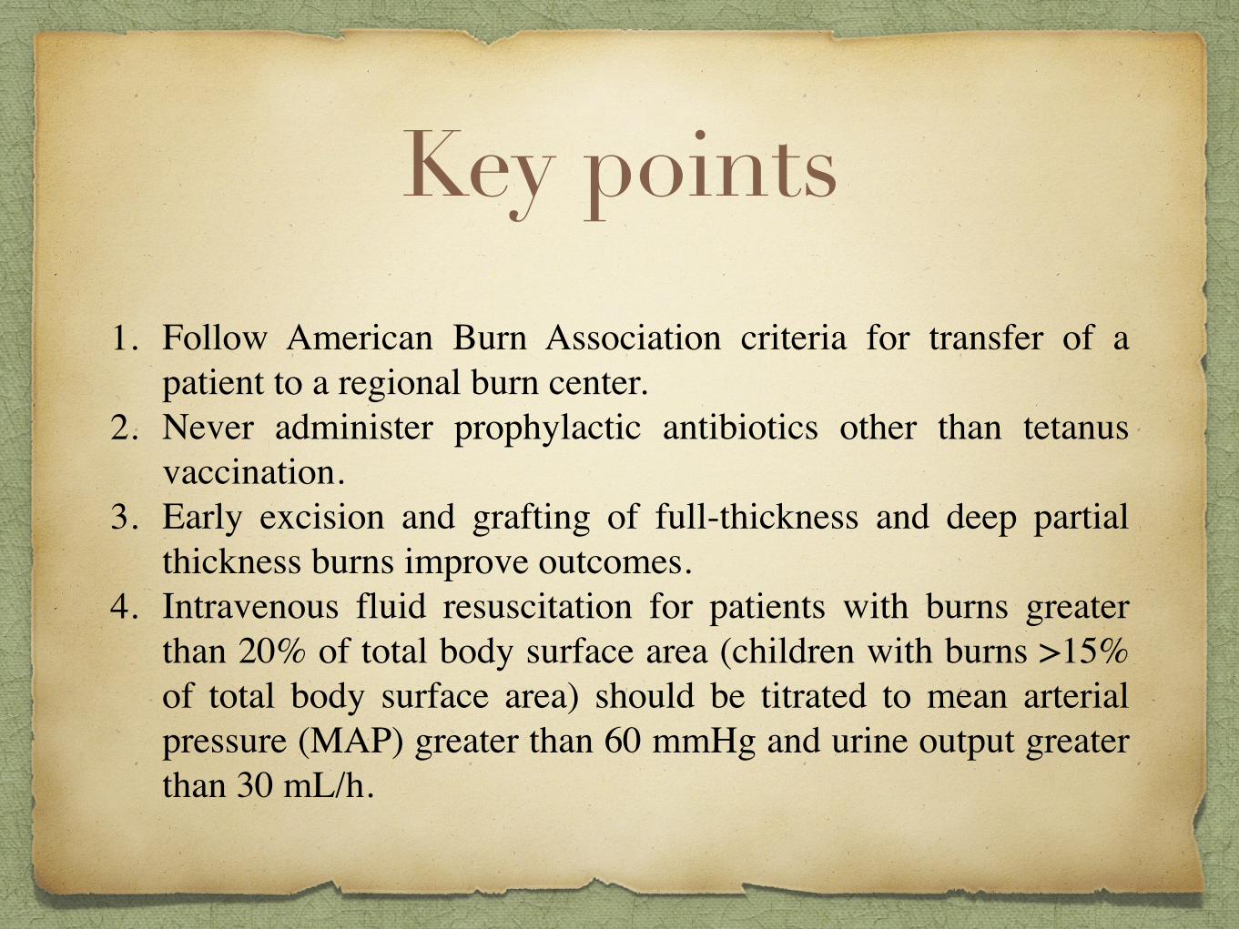
Key points
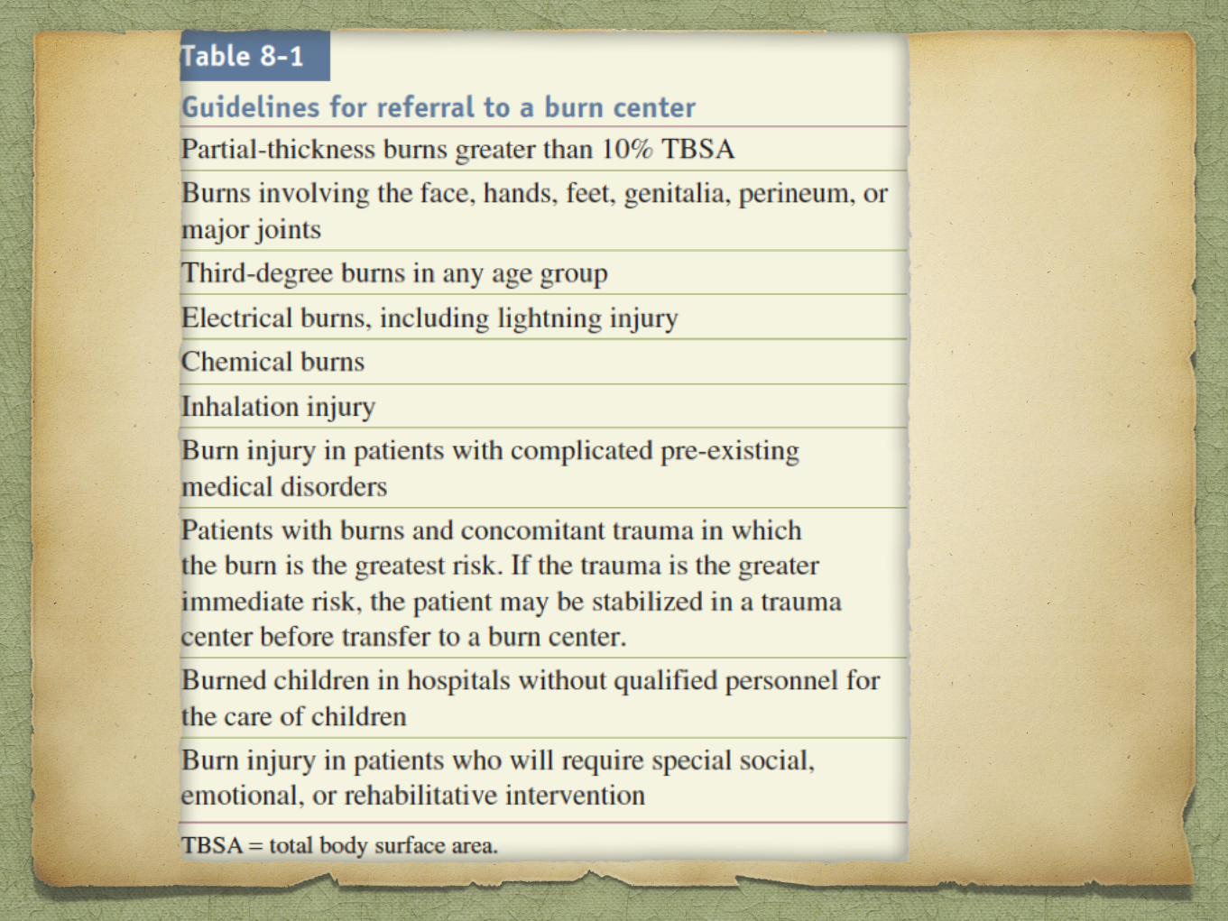
1. Follow American Burn Association criteria for transfer of a patient to a regional burn center.
2. Never administer prophylactic antibiotics other than tetanus vaccination.
3. Early excision and grafting of full-thickness and deep partial thickness burns improve outcomes.
4. Intravenous fluid resuscitation for patients with burns greater than 20% of total body surface area (children with burns >15% of total body surface area) should be titrated to mean arterial pressure (MAP) greater than 60 mmHg and urine output greater than 30 mL/h.
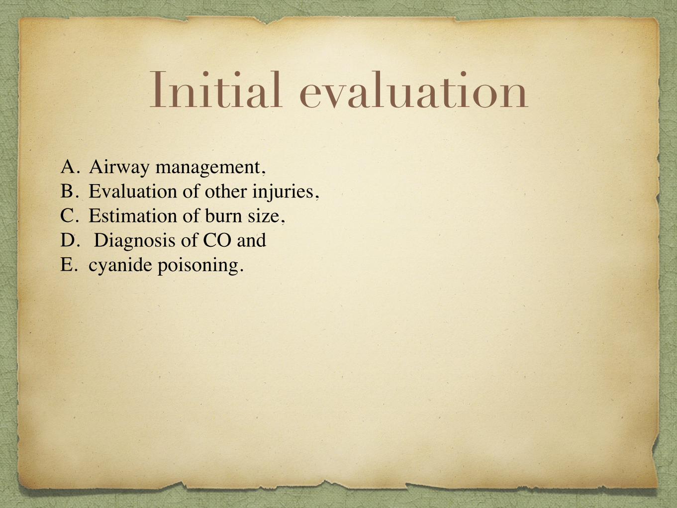
Initial evaluationA. Airway management, B. Evaluation of other injuries,C. Estimation of burn size, D. Diagnosis of CO and E. cyanide poisoning.

Classification of BurnsBurns are commonly classified as • Thermal, • Electrical, or • Chemical burns
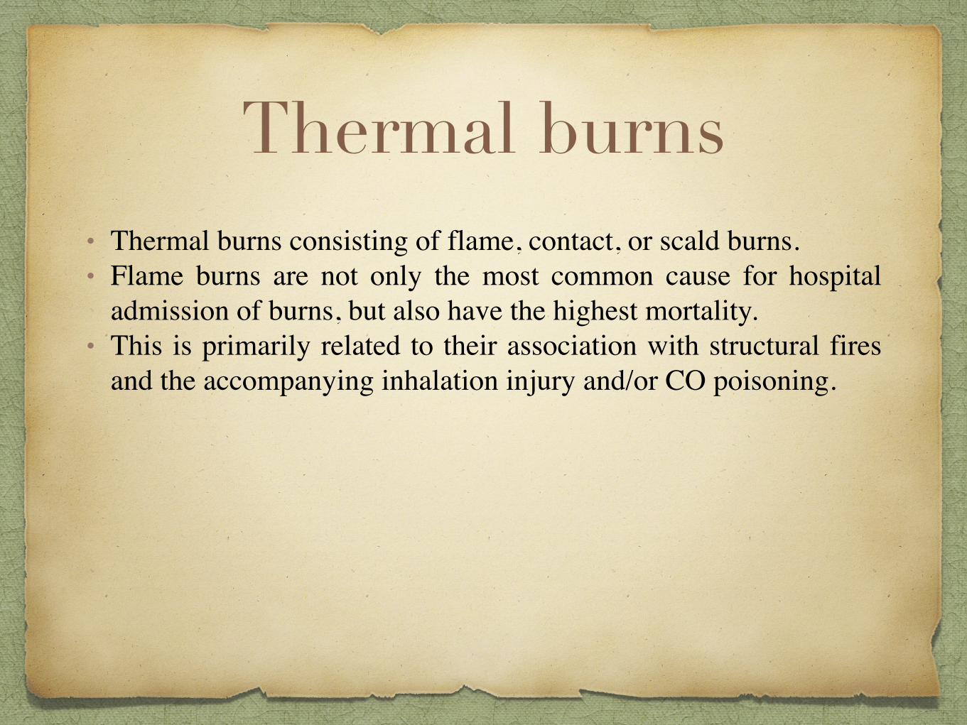
Thermal burns• Thermal burns consisting of flame, contact, or scald burns. • Flame burns are not only the most common cause for hospital
admission of burns, but also have the highest mortality.• This is primarily related to their association with structural fires
and the accompanying inhalation injury and/or CO poisoning.
Electrical Burns
• Electrical burns have special concerns including the potential for cardiac arrhythmias and compartment syndromes with concurrent rhabdomyolysis.
• A baseline ECG is recommended in all patients with an electrical injury, and a normal ECG in a low-voltage injury may preclude hospital admission.
• Long-term neurologic and visual symptoms are not uncommon with high-voltage electrical injuries, and ophthalmologic and neurologic consultation should be obtained to better define a patient’s baseline function.
Chemical burns
• Chemical burns are less common but potentially severe burns.• The most important components of initial therapy are careful
removal of the toxic substance from the patient and irrigation of the affected area with water for a minimum of 30 minutes, except in cases of concrete powder or powdered forms of lye, which should be swept from the patient to avoid activating the aluminum hydroxide with water.
• The offending agents in chemical burns can be systemically absorbed and may cause specific metabolic derangements.
• Formic acid has been known to cause hemolysis and hemoglobinuria, and hydrofluoric acid causes hypocalcemia.
Burn depth• Superficial (first-degree), • Partial-thickness (second-degree), • Full thickness (third-degree), and • Fourth-degree burns, which affect underlying soft tissue.
Burn depth• Clinically, first-degree burns are painful but do not blister,
• Second-degree burns have dermal involvement and are extremely painful with weeping and blisters, and
• Third degree burns are leathery, painless, and non blanching.
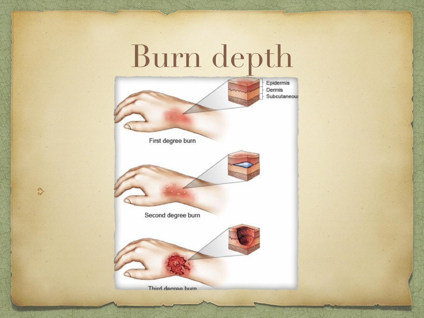
Burn depth
Management Immediate care
Assessment of burn wound
Fluid resuscitation
Treatment of burn wound
Pre hospitalEnsure rescuer safety – house fire , chemical , electrical
Stop the burning process – stop, drop & roll
Check for other injuries
Cool the burn wound Cool the burn wound – minimum 10mts to an hour
Oxygen
elevate
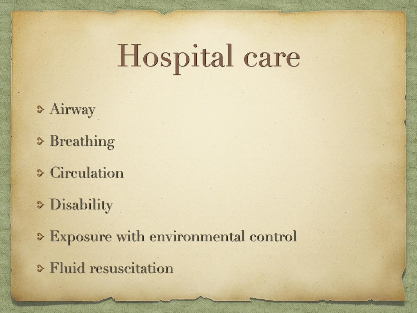
Hospital careAirway
Breathing
Circulation
Disability
Exposure with environmental control
Fluid resuscitation
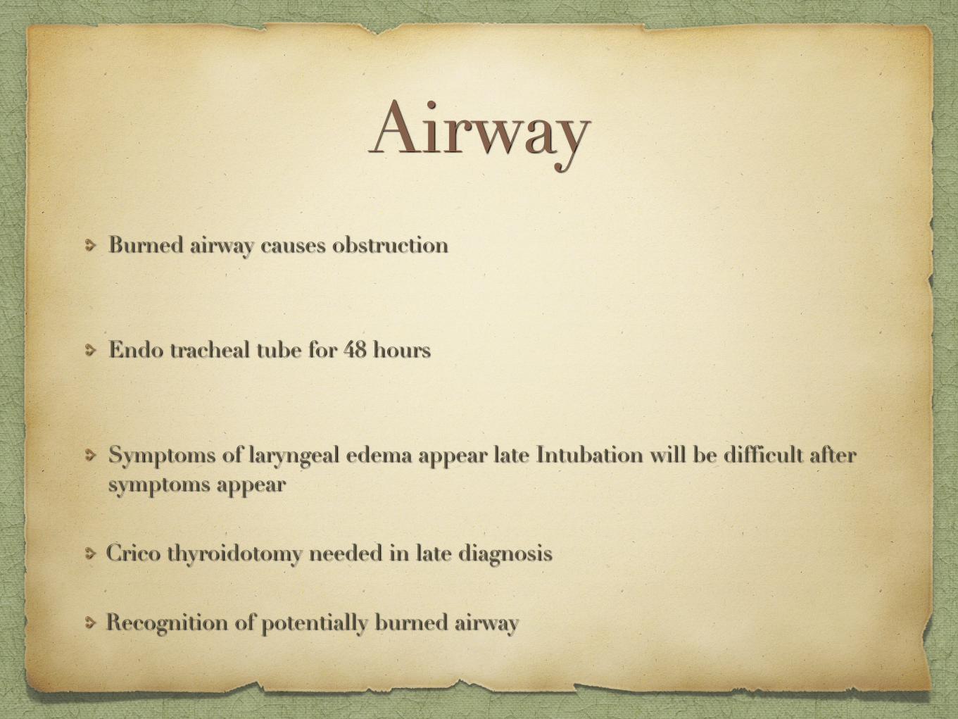
Airway Burned airway causes obstruction
Endo tracheal tube for 48 hours
Symptoms of laryngeal edema appear late Intubation will be difficult after symptoms appear
Crico thyroidotomy needed in late diagnosis
Recognition of potentially burned airway
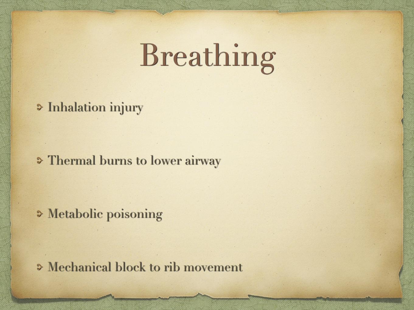
Breathing Inhalation injury
Thermal burns to lower airway
Metabolic poisoning
Mechanical block to rib movement
Inhalation injury
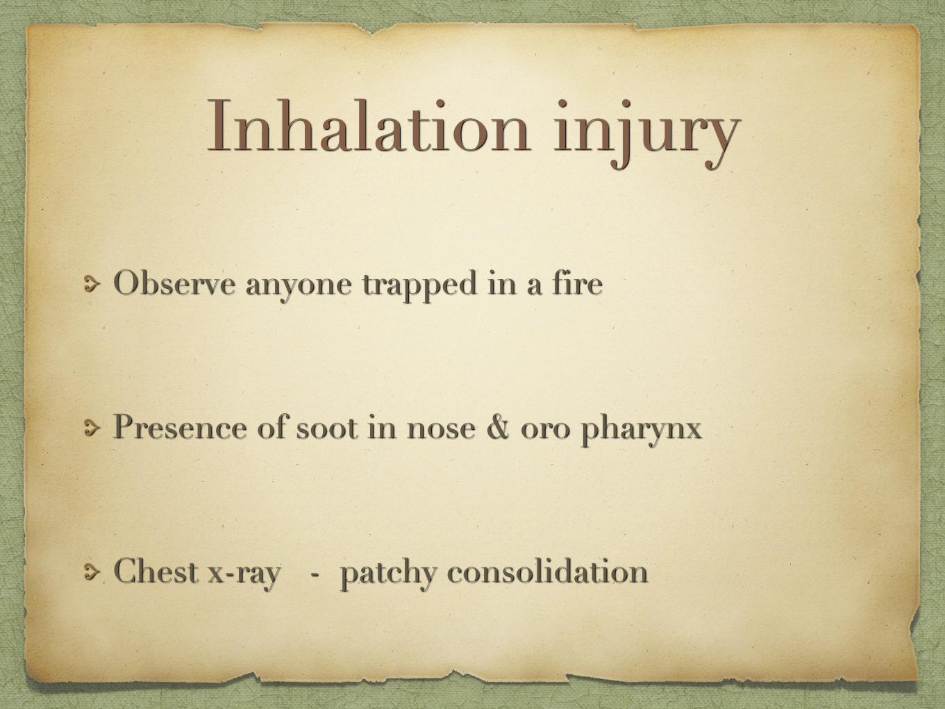
Observe anyone trapped in a fire
Presence of soot in nose & oro pharynx
Chest x-ray - patchy consolidation
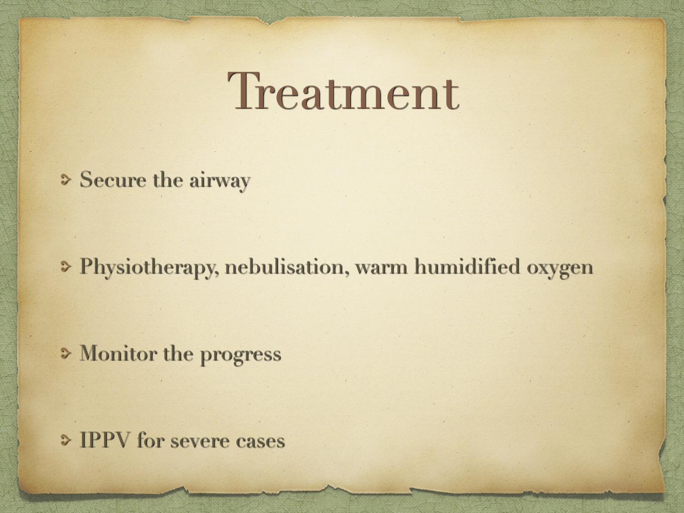
Treatment Secure the airway
Physiotherapy, nebulisation, warm humidified oxygen
Monitor the progress
IPPV for severe cases
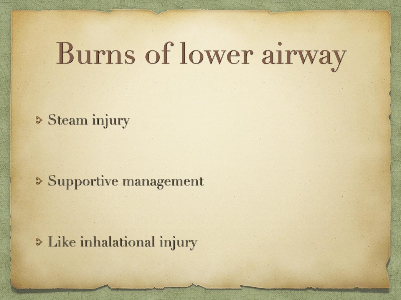
Burns of lower airway
Steam injury
Supportive management
Like inhalational injury
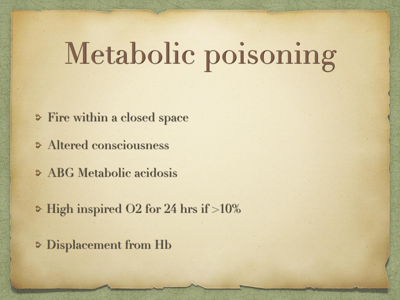
Metabolic poisoning
Fire within a closed space
Altered consciousness
ABG Metabolic acidosis
High inspired O2 for 24 hrs if >10%
Displacement from Hb
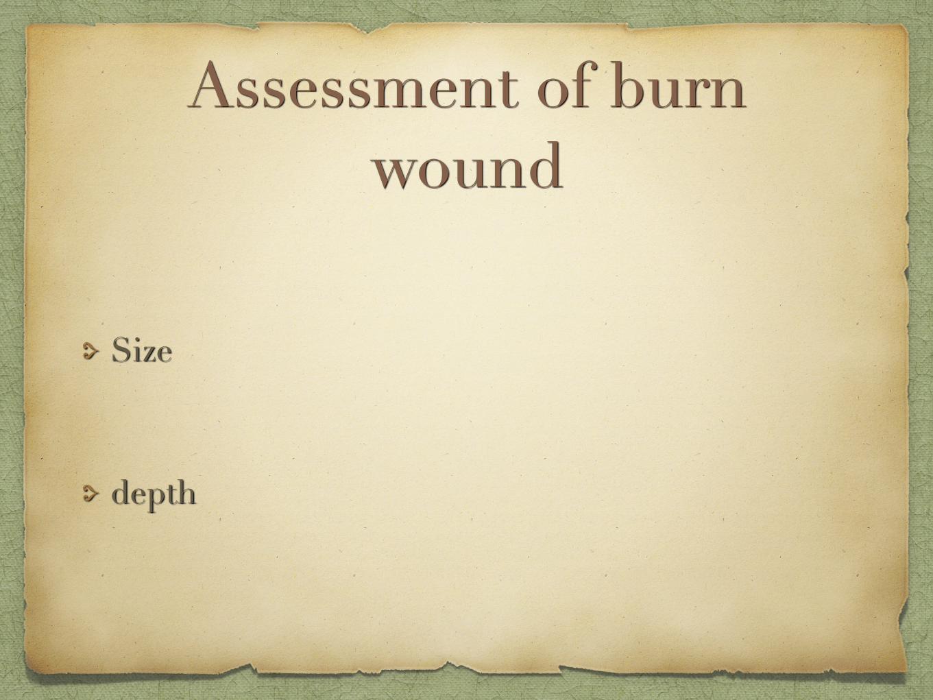
Assessment of burn wound
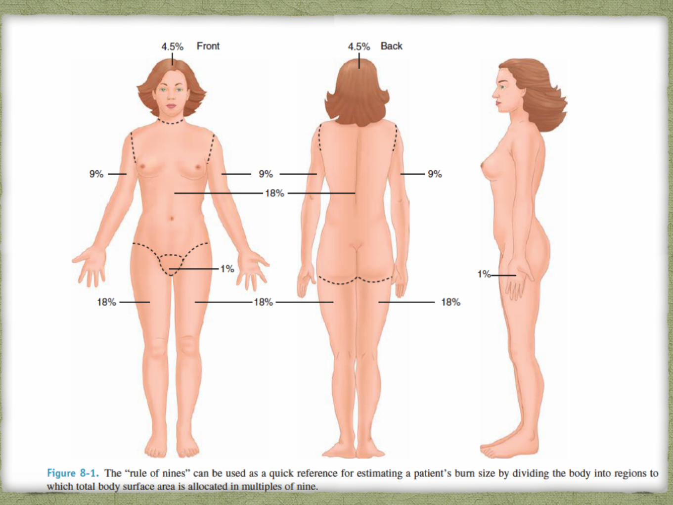
Size
depth

Size Formally assessed in a controlled environment
Allows areas to be exposed & any soot / debris to be washed off
Do not cause hypothermia Patients whole hand – 1% of TBSA
Lund & Browder chart
Wallace rule of nine - approximate

Depth
Superficial partial thickness
Deep partial thickness
Full thickness
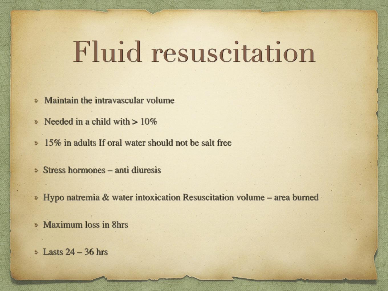
Fluid resuscitationMaintain the intravascular volume
Needed in a child with > 10%
15% in adults If oral water should not be salt free
Stress hormones – anti diuresis
Hypo natremia & water intoxication Resuscitation volume – area burned
Maximum loss in 8hrs
Lasts 24 – 36 hrs
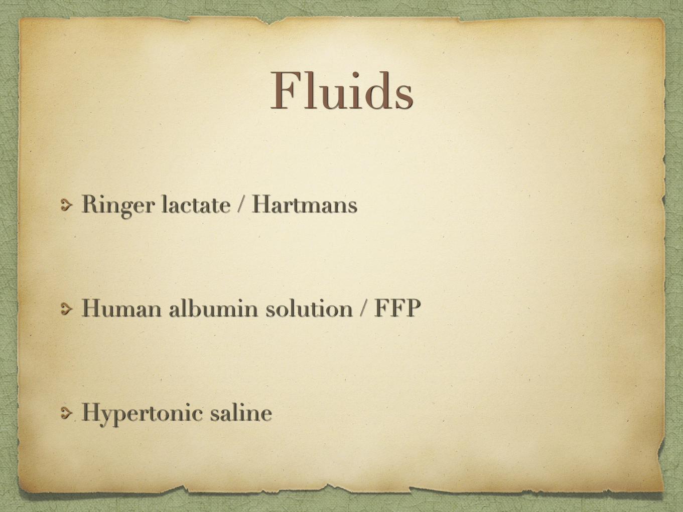
Fluids
Ringer lactate / Hartmans
Human albumin solution / FFP
Hypertonic saline
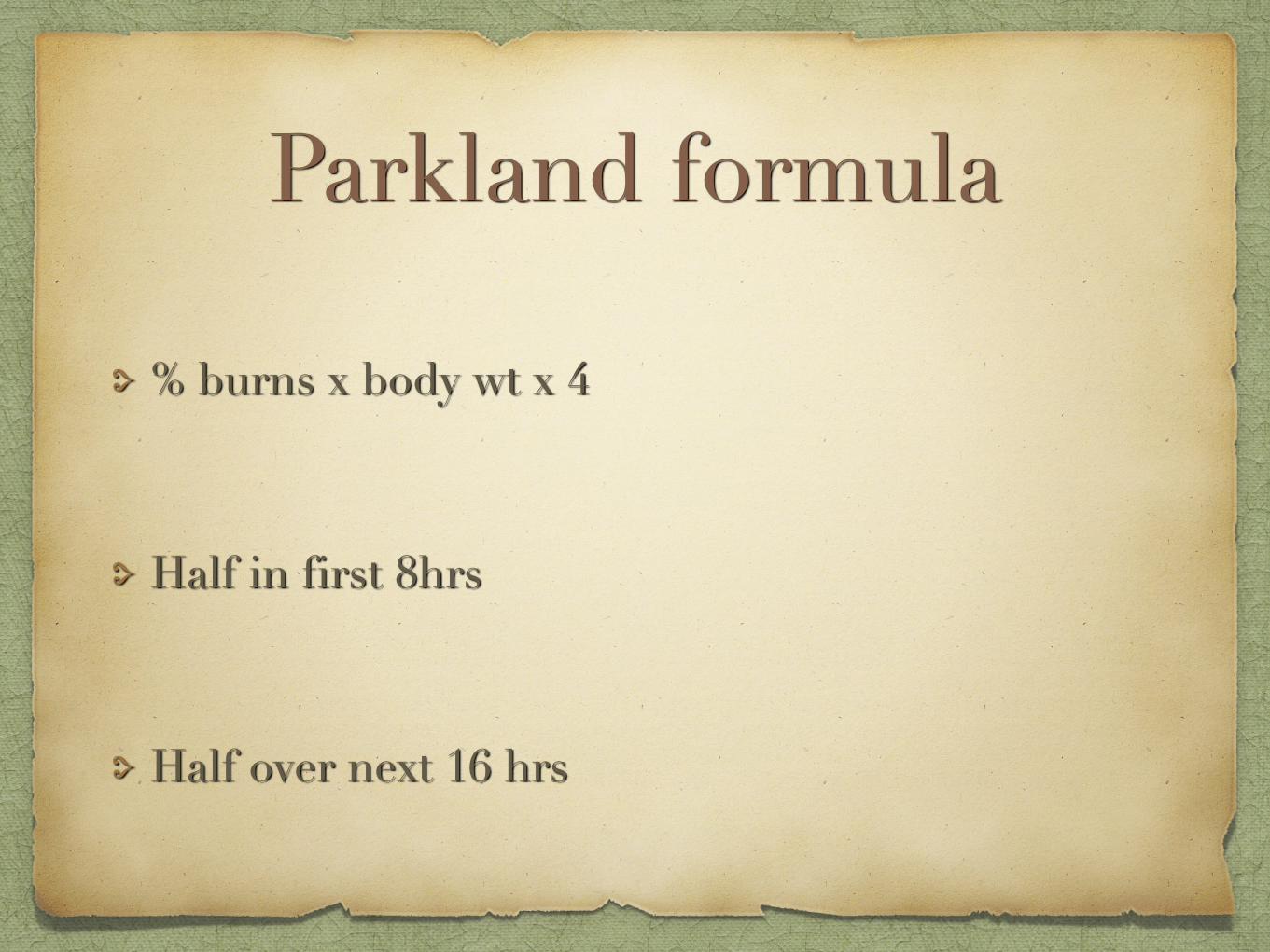
Parkland formula
% burns x body wt x 4
Half in first 8hrs
Half over next 16 hrs
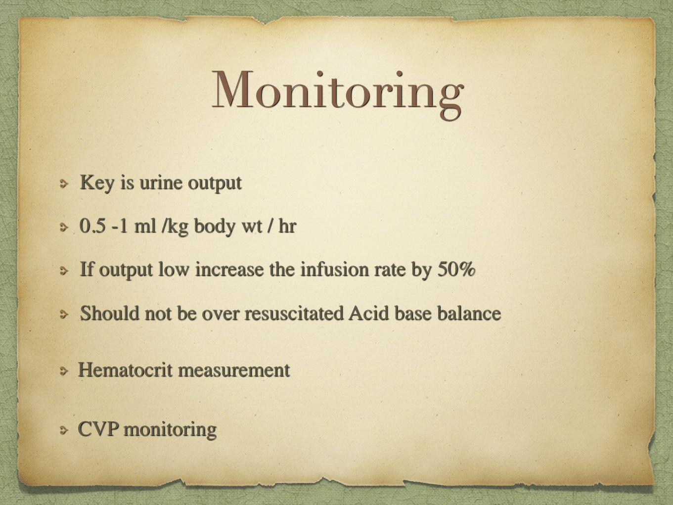
Monitoring
Key is urine output
0.5 -1 ml /kg body wt / hr
If output low increase the infusion rate by 50%
Should not be over resuscitated Acid base balance
Hematocrit measurement
CVP monitoring
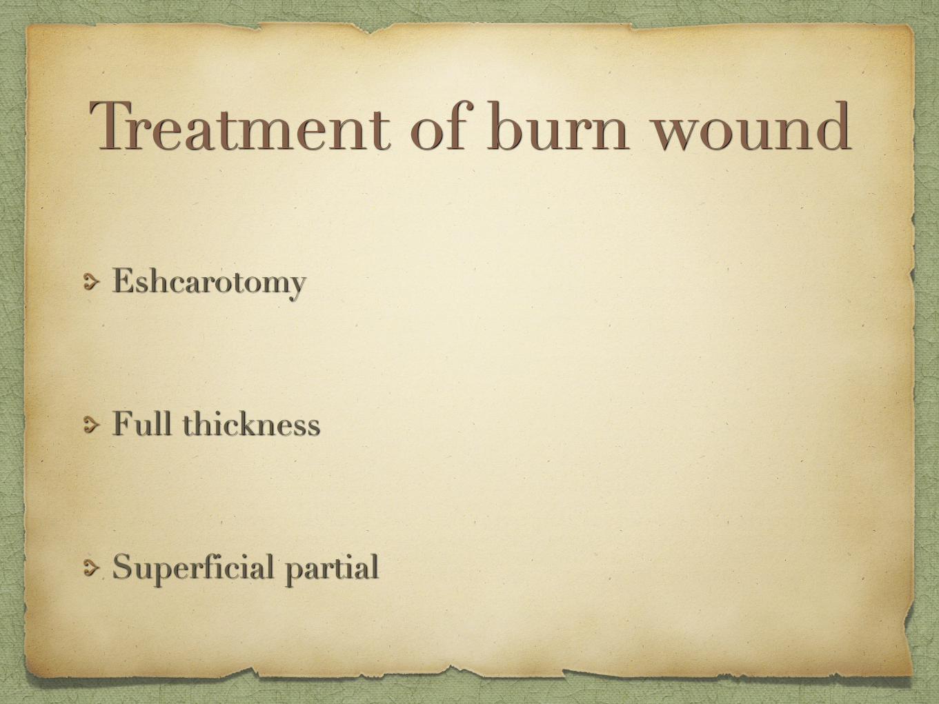
Treatment of burn wound
Eshcarotomy
Full thickness
Superficial partial

Eshcarotomy
Circumferential full thickness burns of limbs
Incised in mid axial line to avoid nerves
Management of burn wound same
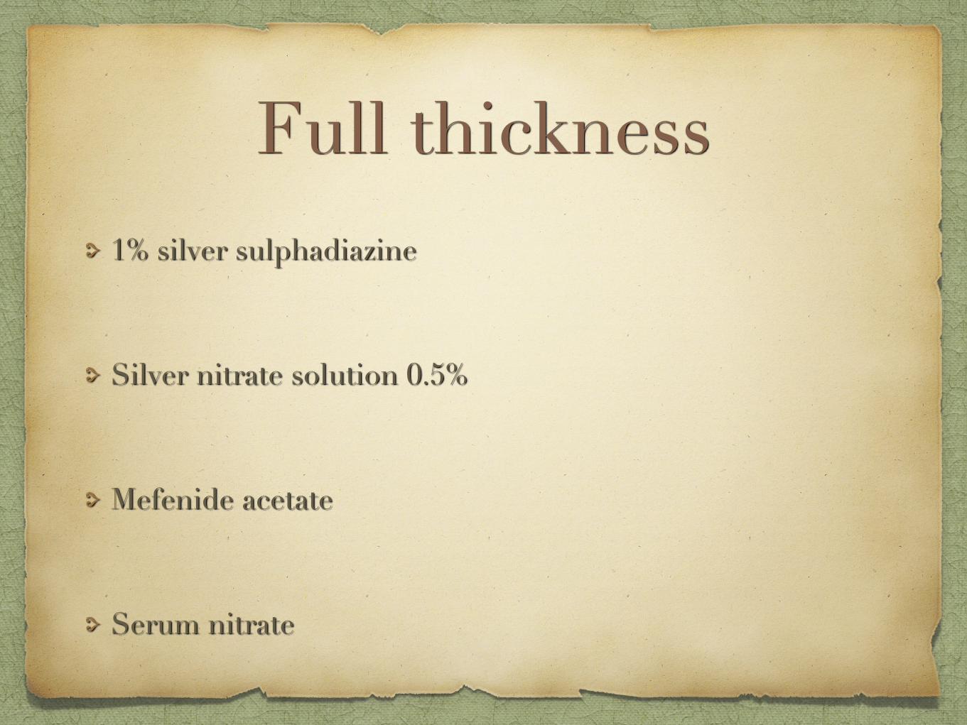
Full thickness1% silver sulphadiazine
Silver nitrate solution 0.5%
Mefenide acetate
Serum nitrate
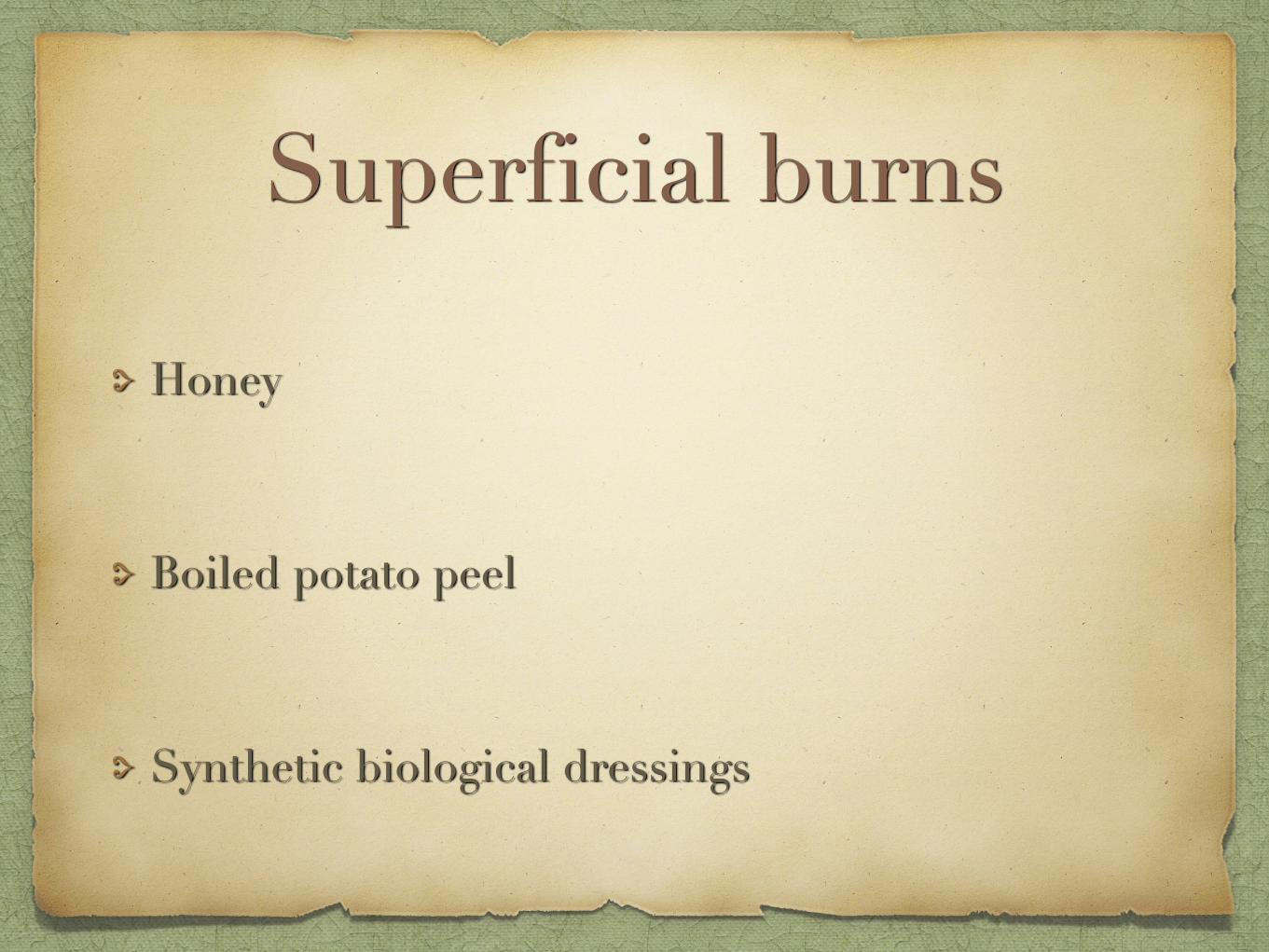
Superficial burns
Honey
Boiled potato peel
Synthetic biological dressings

Thank you !!!


![Pneumonia [Harrison's]](https://static.fdocuments.us/doc/165x107/54515befb1af9f83248b46c1/pneumonia-harrisons.jpg)