Intron Evolution: Testing Hypotheses of Intron Evolution ...
Upload
truongnhanCategory
view
213download
0
ICMR - NATIONAL INSTITUTE OF IMMUNOHAEMATOLOGY
Sept - Dec 2017Vol. 48 : No 3
IMMUNOHAEMATOLOGY BULLETIN
IMMUNOHAEMATOLOGY BULLETIN

Dr. Babu Rao Vundinti, Scientist E, received BGRC thSilver Jubilee Award for the year 2016 on 11 October
2017 from Indian Council of Medical Research, New Delhi for the research work on “Understanding the molecular basis of Hematological diseases”. The award was given for his signicant contribution to the eld of Human Genetics of Hematological diseases in the past 20 years. He carried out extensive research on diagnosis, molecular pathology of Fanconi anemia and identied several novel mutations and also established its prenatal diagnosis.
Dr. Manisha Madkaikar, was appointed as Director of National Institute of Immunohaematology from 14 Dec, 2017
Dr. Prabhakar Kedar, Scientist D, received ICMR Prize for Biomedical Research for Scientists belonging to Underprivileged Communities for the year 2013 on
th11 October 2017 from Indian Council of Medical Research, New Delhi for his research work on “Studies on hereditary hemolytic anemia due to red cell enzymopathies”. The award was given for his signicant contribution to the eld of Enzymopathies for the past 20 years. He has reported several novel mutations in the human PKLR gene in Indian patients and has developed a new test for diagnosis of red cell membrane protein defects.

Genetic modulators of fetal hemoglobinPriya Hariharan & Anita Nadkarni
1
Summary :The clinical heterogeneity of sickle cell disease
and β-thalassemia is so variable that it prompted
the researchers to identify the genetic
modulators of the disease. Though the primary
modulator of the disease are the type of β-globin
mutations that affects the degree of β-globin
chain synthesis, the co-inheritance of α-
thalassemia and the fetal hemoglobin levels also
act as a potent secondary genetic modier of
hemoglobinopathies. During the course of
development the human body sequentially
expresses three different forms of hemoglobin:
embryonic, fetal and adult hemoglobin in the
maturing erythroblasts. Since increased
production of the fetal hemoglobin ameliorates
the severity of hemoglobinopathies, the study of
genetic factors that play a role in the regulatory
network of fetal to adult hemoglobin switch will
facilitate development of new therapeutic
strategies for hemoglobinopathies. In this review
we have tried to summarise various mechanisms
of hemoglobin switching and the genetic
modulators lying within and outside the β-globin
gene cluster with their plausible role in governing
the fetal hemoglobin (HbF) levels.
Introduction: Way back in 1866, Korber et al., discovered that
the hemoglobin obtained from umbilical cord
was far more resistant to alkali denaturation 1than the normal adult hemoglobin (HbA, α β ) . 2 2
After almost 60 years of this discovery, in 1934
Brinkman et al., proved that the variation in the
alkali resistant characteristic of hemoglobin was
due to the globin portion of the molecule and
thus alkali resistant hemoglobin was termed as 2 2fetal hemoglobin (HbF, µ ¡ ) since it formed the
2major hemoglobin fraction of the growing fetus .
A string of experiments showed that the fetal
hemoglobin had an altered physio-chemical
propert ies as compared to the adult
hemoglobin, like having increased resistance to
elution from the RBCs under acid conditions,
decreased solubility in phosphate buffer,
increased resistance to heat and higher oxygen 3
afnity . In the same time period, Drescher -
Kiinzer and Huehns et al., identied that the
embryonic hemoglobin in small human embryos
(gestational age:7-12 weeks) had an alkali 4denaturation between HbA and HbF . These
series of discoveries left behind a perplexing
question; does the hemoglobin in humans during
the ontogeny undergo successive switches? and
if yes, is there a perspective for molecular cure
by reactivat ing fetal g lobin genes in
hemoglobinopathy patients ?
Priya Hariharan & Anita Nadkarni
Department of Hematogenetics
National Institute of Immunohematology (ICMR),th13 Floor, New Multistoreyed Building, KEM Hospital Campus, Parel, Mumbai 400 012.
Email: [email protected]

2
Association of elevated HbF and hemoglobinopathies :The possibility that hemoglobinopathies can be
cured by induction of fetal hemoglobin was
realized as the symptoms are seen in six months
of age after the birth when there is gradual
reduction in the HbF levels. In late 1950s, it was
observed that patients with Cooleys anemia
survived beyond the period of the fetal to adult
hemoglobin switch because of the prolonged
synthesis of fetal hemoglobin in the erythroid 5cells . In β-thalassemia, the main cause of the
disease is imbalance in the globin chain ratio,
wherein the excess unbound α globin chain
precipitates, inhibiting the maturation of the
erythroid precursors, ultimately leading to 5ineffective erythropoiesis and anemia . The
clinical pathogenicity of β-thalassemia develops
during the rst year of life when the g-globin
genes are gradually silenced with the activation
of δ and β- globin genes. Thus continued g-globin
gene expression in β-thalassemics, may lead to
reduction in the globin chain imbalance as g-
globin synthesized will combine with the excess 6
unbound α-globin chain . Similarly appreciating
the role of fetal hemoglobin (HbF;α2γ2) in sickle
cell disease started more than 60 years ago when
Janet Watson conrmed that infants with sickle
cell disease had few symptoms and that their
deoxygenated erythrocytes took longer time to
sickle and did not deform as extensively as did 7
their sickle cell trait-carrying mother's cells .
Sickle cell disease is caused by the mutation in th
codon 6 position in the β-globin gene by
substitution of glutamic acid by valine, which
affects the biophysical properties of the adult 5
hemoglobin . The pathophysiology of sickle cell
disease is dependent on the polymerization of
deoxy sickle hemoglobin under low oxygen
condition, which is retarded with increased HbF
concentration. Both HbF and its mixed hybrid S
tetramer (α β γ) cannot enter the deoxy sickle 2
hemoglobin polymer phase, thus circumventing 8
the cellular damage evoked by HbS polymers . In
both the hemoglobinopathy conditions,
increased g-globin gene expression acts as a well-
known disease modier. Hence understanding
the molecular mechanism of hemoglobin
switching and identication of molecular targets,
for reversing this switch, is a subject of intense 5research .
The hemoglobin switch :G A
During the ontogeny, globin genes (ε, γ, γ, δ, β)
in the β-globin cluster are sequentially
expressed, leading to production of different
hemoglobin molecules with distinct physiological
properties. The site for primary hemoglobin
synthesis is the embryonic yolk sac and the rst
wave of hemoglobin switch (primitive to
denitive) occurs in the fetal liver after 5 weeks
of gestation, wherein there is a switch from 2 2 9
embryonic (ζ ε ) to fetal hemoglobin (µ ¡ ) . 2 2
rdTowards the end of 3 trimester, there is a 2 2gradual change from fetal (µ ¡ ) to adult
hemoglobin (α β ). This second major hemoglobin 2 2
switch occurs in the bone marrow and lasts until 9
6 months of age after birth . Figure 1 shows a
schematic representation of different
hemoglobin progressively expressed during
distinct stages of development. With the dawn of
molecular era, models for gene switching
suggested, that this process involved complex
interaction between the cis-acting elements that
includes the locus control region of the beta
globin gene cluster and trans acting transcription
factors which co-ordinately carry out chromatin 10
remodelling activities .

3
The cis- acting elements :The β- globin gene cluster contains a 5' distal
regulatory element known as Locus Control
Region (LCR), which has ve active DNAase I
hypersensitive sites (HS) 1-5, that is required for
appropriate globin gene expression. This is Gfollowed with the series of globin genes (ε, γ,
Aγ, δ, β) arranged sequentially in their order of
expression. To the downstream of the β globin
gene is another HS site 3' HS1 [Figure 2]. The
β–globin cluster is anked with olfactory 11
receptors at both 5' and 3' ends . Wijgerde et
al., proposed a competitive model for globin
gene regulation, where in the LCR participates
in the long range looping interaction, that leads
to only one productive LCR-globin gene 11
interaction . The potent transcriptional
activation of the LCR is also due to the clustering
of various erythroid transcription factors that
bind to the hypersensitivity sites which act in
concert to facilitate long range looping activity.
The cis- acting promoter sequences in the
individual globin genes and the LCR serve as a
template for binding of various transcription 11factors . It is observed that naturally occurring
mutations in g-globin promoter region are
associated with hereditary persistence of fetal
hemoglobin phenotype (HPFH).The mechanisms
underlying the continued expression of HbF with
these mutations are thought to involve
alterations in the protein binding motifs that
ultimately ablates the binding of repressors or 12
enhancers affecting the gene expression . The
most extensively studied variation in several
population groups is the XmnI polymorphism
(C®T) residing at -158 position (HBG2 c.-211 G
C®T) in the ¡globin promoter region. The T
allele of XmnI polymorphism is linked to the
raised HbF levels and milder hemoglobinopathy 13conditions . Though T allele in its homozygous
state seems to have little effect on HbF levels in
Figure 1: Expression of globin genes during the ontogeny. The figure describes the site of hemoglobin synthesis and
2 major hemoglobin switches with respect to the gestational age of the fetus. Keys: HBG1 and HBG2 : A -globin and g
G -globin gene . HBB : β globin gene .g

4
normal individuals, it has been associated with
increase of HbF especially during the
erythropoietic stress, suggesting its role in HbF 14
induction . In clinically milder patients where
no mutation is detected in the� g-globin
promoter region, the other genetic modulators
[HBS1L-MYB intergenic region, BCL11A, KLF1]
leading to raised HbF phenotype are screened.
Role of factors involved in the
hemoglobin switch and new
therapeutic targets:
More recently recognised variability in HbF
expression both in non anemic individuals and
patients with β-hemoglobin disorders stimulated
efforts to detect the genetic factors that lead to
this deviation. Genome wide association
mapping studies have identied quantitative
trait loci in the nuclear factors that play a role in
the globin gene regulation and switching. The
role of major transcription factors that are
involved in the process of hemoglobin switching
are as follows: [Figure 3].
v B- cell CLL / lymphoma 11 A [BCL 11 A]:
B- cell CLL / lymphoma 11 A is a zinc nger
transcription protein which is encoded by
BCL11A gene located in chromosome 2p16 .
This protein expressed in 4 different iso-forms (
XS, S, L, XL) plays an important role in 15
lymphopoiesis and neurogenesis . The
pioneering role of BCL11A polymorphisms in
intron 2 in association with fetal hemoglobin
levels was rst demonstrated by Uda et al.,
2008 after conducting genome wide association
studies in the Sardanian population, where in
variants in BCL11A were found to be
signicantly associated with increase in F-cell 16
numbers in hemoglobinopathy patients .This
suggested that the 3kb second intronic region of
BCL11A had regulatory elements or functional
motifs responsible for modulating the g-globin
gene expression. Further Sankaran et al.,
proved that BCL11A may function as a potent
repressor of the g- globin gene as lower levels
of BCL11A m-RNA is expressed in variants 17
causing higher levels of HbF . This was
validated by performing gene knockdown of
BCL11A using short-hairpin RNA (shRNA)
Figure 2: Structure of -globin gene cluster showing embryonic (ε), fetal ( and adult (δ, β) globin genes β G gamma , A gamma
arranged according to the order of their expression that is controlled by locus control region ( LCR ).

5
approaches in primary adult erythroid 17
progenitors .Chromatin Immuno Precipitation
(ChIP) technology demonstrated that BCL11 A,
interacts with NuRD chromatin remodelling and
repressor complex, erythroid transcription
factors – GATA -1, FOG -1, SOX6 and Matrin-3 to
silence the gamma globin gene, suggesting the
pivotal role of BCL11A in gamma globin 17silencing .
v Kruppel like factor 1 [KLF 1]:
Erythroid Kruppel like factor (EKLF) or KLF1, a
erythroid specic transcription factor plays a 18critical role in erythropoiesis . This founding
member of Kruppel like factor family was rst
identied by Ira Miller and James Bieker in
1992 and named it as KLF1/ EKLF due to its
zinc nger homology with that of Drosophila
body pattern determining Kruppel–gap 18gene . This master erythroid gene regulator,
co-ordinates three important processes that
includes erythroid lineage commitment, the g
to β globin gene switch and activation or
suppression of certain erythroid specic
genes ( red cell metabolism and structure
proteins) by formation of chromatin 19
remodelling complex . KLF1 transcription
factor was thought to take part in β-globin
gene activation by binding to the CACCC box
of the beta-globin promoter, as ablating this
region lead to β-thalassemia intermedia
condition. This phenomenon was validated by
knockdown of KLF1 gene in mouse embryo 19
that succumbed to severe beta thalassemia .
Though Singleton et al., 2008 primarily
reported KLF1 gene mutations leading to
clinically benign In (Lu) blood group, the rst
event of raised HbF phenotype was
mentioned by Borg et al., in an extended
Maltese family where in the proposita
inheriting two KLF1 gene variants p.K288X
Figure 3: Schematic representation of hemoglobin switching in presence of various transcription factors. During the
switch process from fetal to adult hemoglobin , the - globin repressor complex occupies the globin promoter g� g-�
region, thus blocking the - promoter interaction with LCR and enhancing its interaction with the β-globin gene in g�
presence of KLF1, thus leading to increase in adult hemoglobin.

6
and p.M39L showed a HPFH phenotype with 20,21
HbF levels of 19.5% . The mechanism that
KLF1 participates in repressing the gamma
globin levels via BCL11A activation was
proved by Zhou et al by conducting gene
knock down assay, in which reduction of
KLF1 gene markedly decreases the BCL11A
levels and indirectly increasing the gamma β-22
globin mRNA levels . Thus established the
bipartite role of KLF1 gene, in indirectly
reducing the ¡ globin gene expression by
BCL11A activation and directly activating β-
globin gene by favouring looping interactions
between the LCR and the β-globin gene 22against g-globin gene promoter .
v HBS1L- MYB intergenic zone [HBS1L-
MYB]:
With the aim of studying the genetic variants
associated with the HbF regulation, genome
mapping studies identied another loci
located in chromosome 6, residing within 126-
kb intergenic region between the HBS1L and
MYB genes. This region is distributed in 3
disequilibrium blocks which are referred to as
HPIM block 1, 2, 3 amongst which block 2 that
spans 24 kb region is found to be associated
with reduced disease severity in patients with
hemoglobinopathy by increasing the HbF
levels. Though the function of HBS1L in red
blood cell development is uncharacterized,
the MYB gene (encoding the c-Myb
transcription factor [TF]) is a key regulator of 23
hematopoiesis and erythropoiesis . The
functional importance of the intergenic region
was rst observed when transgene insertion
within the murine HBS1L-MYB intergenic
region completely disrupted the binding of
core complex proteins LDB1, GATA1, TAL1,
and ETO2 and abolished MYB transcription 23which resulted in severe anemia . The most
effective of these, are the variants residing
within the core enhancer binding region in 23HMIP-2A and -2B . A three -bp deletion
(rs66650371) at HMIP-2A is suspected to
directly cause HbF variability. Recent reports
have described a transcriptional network
within erythroid cells whereby KLF1 and MYB
activate each other by formation of an Active
Chromatin Hub(ACH) structure that brings
intergenic enhancer regions in proximity with 23
the MYB promoter . This same study showed
that perturbations to the ACH structure, or to
KLF1 expression, do not affect gene
expression of HBS1L, suggesting that the
primary role of the GWAS-identied HBS1L-
MYB intergenic region is to regulate the 23expression of MYB .
v GATA 1:
The GATA family of transcription factors
(GATA 1 to GATA 6) are a group to zinc
containing transcription regulators that bind
to consensus (WGATAR) DNA motif present in
the regulatory region of various erythroid 24, 25development genes . The role of GATA 1 as
a lineage specic transcription factor and in
regulating the HbF levels was rst suggested
by Philips et al., in a patient suffering from
congenital erythropoietic porphyria with
elevated HbF levels thus suggesting its
paramount role in erythroid maturation and 26
hemoglobin switching . GATA 1 has 2 regions,
the N-terminal transactivation that stabilizes
DNA binding and a C terminal zinc nger 26
domain that binds to the GATA 1 motifs .
Bottardi et al., proved that GATA 1 works in
concert with other co-factors like friend of
GATA 1 (FOG-1), histone acetyltransferase
CBP/P300 / Mediator complex subunit MED–I
and a chromatin modeller–BRG1 to bring
about long range chromatin interactions
between β-LCR and the globin genes to

7
27regulate their gene expressions . Further
Chromatin Immuno Precipitation assays
showed that GATA 1 along with IKAROS :
transcription factor, forms a repressome
complex, that limits the recruitment of
transcription activators to the g-globin
promoters, thus by silencing the g-globin
gene and progressively increasing the 27
transcription of β-globin gene .
v SOX6 :
SOX6 a member of the SOX transcription
factor family is characterised by high mobility
group (HMG) DNA binding domain. This gene
codes for a 79 amino acid DNA binding protein
that binds to the minor groove of the DNA,
leading to conformational changes in the DNA 2 8structure . SOX 6 thus acts as an
architectural protein by dynamically
remodelling the chromatin structure and
assisting the binding of other transcription 28factors . Its role in silencing the embryonic
globin gene was rst shown in a murine
model, as SOX6 decient mice persistently
expressed the embryonic globin. This
hypothesis was further proved by Zanhua et
al., that 36–base pair region of the embryonic
globin proximal promoter is a binding site for
SOX6 mediated repression that leads to the 28silencing of the ε-globin gene . Chromosome
conformation capture (3C) assay, ChlP
suggested that SOX6 co-occupies with BCL11
A and GATA1, in the β-globin gene cluster and
conducts silencing of g-globin transcription in 29
the adult erythroid proginetors . These
results were further validated by carrying out
combined knockdown experiments of SOX6
and BCL11A in primary human erythroblast
culture which greatly enhanced the HbF
production thus suggesting that both the
transcription factors played a combined role 29
in g-globin gene regulation .
Targeted translational research for
i n c r e a s i n g t h e H b F l e v e l s :
Opportunities and challenges!The common transcription factors that govern
the developmental switches execute their action
by complex inter-dependent networks. The
mechanisms underlying the function of these
regulators are so stringent, that any deviations in
their expression or protein-protein interaction
may evoke disastrous phenotype leading to 30anemia, leukemia or lymphomas . Hence it is
pivotal to elucidate their mechanisms, with the
basic step of placing these factors in a hierarchy
in which they function. Despite of vigorous
research for the search of HbF inducers, only
Hydroxyurea (HU) – a ribonucleotide reductase
inhibitor, remains the sole FDA approved
medication for sickle cell disease. However the
HU utility is limited by its unpredictable HbF
induction that varies patient to patient and
myelo-suppression. Thus there is a need for
developing other potential strategies to elevate
HbF levels in hemoglobinopathy patients. One
such approach is to develop target based therapy
which includes knocking down the target by RNA
interference with high specicity and selectivity.
The other way it could be achieved is by
controlling small molecules by using therapeutic
drugs. However in either technique, the greatest
challenge is to selectively impair the activity of
universally expressed transcription factors
specically in the erythroid lineage without
affecting its universal function in the other 31
cells .
Indian scenario and NIIH experience:
In India, the rst study by Dadheech S. et al.,
showed that the C allele of rs 11886868 (C®T) in
BCL11A gene is associated with amelioration of

8
the severity of β-thalassemia as well as sickle cell 32
anemia (SCA) . Roy P. et al., showed that,A®G
allele on the rs4895441 in the HBS1L-MYB
intergenic region is associated with the raised 33HbF levels in the eastern population of India .
Previous study by Upadhye D et al., have tried to
correlate the SNPs in the BCL11A and HBS1L-MYB
gene with the HbF levels and clinical severity in
paediatric sickle cell anemia patients. They
identied A allele of the SNP rs4671393 in BCL
11A gene and G allele of rs9389268 in the HBS1L-
MYB gene to be associated with raised HbF levels 34and with milder clinical symptoms . In our recent
study we have identied the C allele of
rs11886868 and rs66650371 (-TAC) (3 base
deletion) in the MYB gene to be associated with
raised HbF levels in adult SCA and thalassemia
intermedia patients. Similarly, the rst event of
KLF1 variations associated with raised HbF 22
phenotype was mentioned by Borg et al ,
however no such studies have been carried out in
India to understand the prevalence and effect of
KLF1 variations in our population.We have
identied 12 different KLF1 gene mutations after
screening 325 hemoglobinopathy patients, most
of the mutations associated with mild increase in
HbF levels. We found a novel KLF1 gene variation
codon 211 A®G (c.632 A>G)to be associated with
increased HbF levels in thalassemia trait [HbA2:
4.1%, HbF: 8.6%, β-globin gene mutation: codon
15 G®A heterozygous] and speculate that co-
inheritance of this novel KLF1 variant might
have ameliorated the clinical manifestations of
her six year old untransfused β-thalassemia
homozygous child [HbA :1.6 %, HbF:97.9%, β-2
globin gene mutation: codon 15 (GàA) 35homozygous] . Also we identied novel 5 bp
Gdeletion -ATAAG (-533 to -529) in the γ globin
promoter which when co-inherited with the β-
thalassemia trait condition lead to an elevated
HbF levels [HbA : 3.6%, HbF: 31.7% and HbA :2 2
363.9%, HbF : 23.6%] .
Conclusion :
Increase in HbF levels has potential therapeutic
implications as it ameliorates the clinical severity
of β-hemoglobinopathies. With this review we
conclude that SNPs located at various
quantitative trait loci, are associated with
meagre elevation of HbF levels in the range of 3-
5%, nevertheless they can increase the HbF
output to clinically benecial levels when co-
inherited with hemoglobinopathies. Identication
of such variants are of importance as in future
they can be used in genome editing to naturally
increase the HbF levels in patients with β-
hemoglobinopathies. The study of transcription
factors and their co-enzymes that play role in
gamma globin gene silencing may help in
determining new potential interacting surfaces
and enzymatic mechanisms. Curtailing the
activity of these proteins may thus lead to the
development of novel therapies for treating
hemoglobinopathies.
References :1. Lorkin PA. Fetal and embryonic hemoglobins.
J Med Genet.1973;10:50-64.
2. Brinkman R, Wildschut A, Wittermans A, et
al. On the occurrence of two kinds of
hemoglobin in normal human blood. J.
Physiol. 1934;80:377-87.
3. Giardina B. Fetal hemoglobin: Structure and
f u n c t i o n . S c a n d J C l i n L a b
Invest.1982;42:32-37.
4. Huehns R., Dance N, Beaven H, et al. Human
embryonic hemoglobins. Nature.1964;
201:1095-97.

9
5. Philipsen S. Molecular control of hemoglobin
switching. Hematology Education: the
education programme for the annual
congress of the European Hematology
Association.2013;7:1-9.
6. Ngo DA, Steinberg MH. Genomic approaches
to identifying targets for treating β
hemoglobinopathies. BMC Med Genomics.
2015;8:1-13.
7. Watson J .The signicance of the paucity of
sickle cells in newborn Negro infants. Am J
Med Sci. 1948;215:419-23.
8. Akinsheye I, Alsultan A, Solovieff N, et al.
Fetal hemoglobin in sickle cell anemia.
Blood. 2011;118:19-27.
9. Sankaran V, Xu J and Orkin SH. Advances in
the understanding of hemoglobin switching.
Br J Haematol. 2010;149:181-94.
10. Bank A. Regulation of human fetal
hemoglobin: new players, new complexities.
Blood. 2006;107:435-43.
11. Bauer D and Orkin S.Update on fetal
h e m o g l o b i n g e n e r e g u l a t i o n i n
hemoglobinopathies. Curr Opin Pediatr.
2011;23:1-8.
12. Sykes K and Kaufman R. A naturally occurring
gamma globin gene mutation enhances SP1
binding activity. Mol. Cell. 1990;10:95-102
13. Nadkarni A, Gorakshakar A, Lu C, et al.
Molecular pathogenesis and clinical
variability of beta-thalassemia syndromes
among Indians. Am J Hematol. 2001 68:75-80.;
14. Mastropietro F, Modiano G, Cappabianca MP,
et al. Factors regulating Hb F synthesis in
thalassemic diseases. BMC Blood Disorders.
2002;2:2.
15. Liu P, Keller J, Ortiz M, et al.Bcl11a is
essential for normal lymphoid development
Nature Immunol. 2003;4:525–32.
16. Uda M, Galanello R, Sanna S, et al. Genome-
wide association study shows BCL11A
associated with persistent fetal hemoglobin
and amelioration of the phenotype of β-
thalassemia. Proc Natl Acad Sci U S A.
2008;105:1620-25.
17. Sankaran V, Xu J, OrkinS. Transcriptional
silencing of fetal hemoglobin by BCL11A. Ann
N Y Acad Sci. 2010;1202:64-8.
18. Miller I, Bieker J. A novel, erythroid cell-
specic murine transcription factor that
binds to the CACCC element and is related to
the Kruppel family of nuclear proteins. Mol
Cell Biol. 1993;13:2776-86.
19. Grech L and Borg J. Control of globin gene
expression by Kruppel-like Factors. Journal
of The Malta Chamber of Scientists.
2014;2:66-77.
20. Singleton B, Burton N, Green C,et al.
Mutations in EKLF/KLF1 form the molecular
basis of the rare blood group In(Lu)
phenotype. Blood. 2008;112:2081-88.
21. Borg J, Papadopoulos P, Georgitsi M, et al.
Haploinsufciency for the erythroid
transcription factor KLF1 causes hereditary
persistence of fetal hemoglobin. Nat Genet.
2010;42:801-05.
22. Zhou D, Liu K, Sun C,et al. KLF1 regulates
BCL11A expression and γ- to β-globin gene
switching. Nat Genet. 2010;4:742-4.
23. Stadhouders R, Aktuna S, Thongjuea S, et al.
HBS1L-MYB intergenic variants modulate
fetal hemoglobin via long-range MYB
enhancers.J Clin Invest. 2014;124:1699-10.
24. BresnickE, Lee H , Fujiwara T, et al. Gata
switches as developmental drivers. J Biol
Chem. 2010;8.285:31087-93.
10
25. Phillips J, Steensma D, Pulsipher M, et al.
Congenital erythropoietic porphyria due to a
mutation in GATA1: the rst trans-acting
mutation causative for a human porphyria.
Blood. 2007;109:2618-21.
26. Ferreira R, Ohneda K, Yamamoto M, et al.
GATA1 function, a paradigm for transcription
factors in hematopoiesis. Mol Cell Biol.
2005;25:1215-27.
27. Bottardi S, Zmiri F, Bourgoin V, et al. Ikaros
interacts with P-TEFb and cooperates with
GATA-1 to enhance transcription elongation.
Nucleic Acids Res.2011;39:3505-19.
28. Yi Z, Cohen-Barak O, Hagiwara N, et al. Sox6
Directly Silences Epsilon Globin Expression in
Denitive Erythropoiesis. PLoS Genet.
2006;2:14.
29. XuJ, Sankaran V, Ni M, et al. Transcriptional
silencing of {gamma}-globin by BCL11A
involves long-range interactions and
cooperation with SOX6. Genes Dev.
2010;24:783-98.
30. Fujiwara T, O'Geen H, Keles S, et al.
Discovering Hematopoietic Mechanisms
Through Genome-Wide Analysis of GATA
Factor Chromatin Occupancy. Mol Cell.
2009;36:667-81.
31. Degterev A ,Lugovskoy A, Cardone M, et al
.Identication of small-molecule inhibitors
of interaction between the BH3 domain and
Bcl-x Nat Cell Biol. 2001;3:173 - 82L .
32. Dadheech S, Madhulatha D, Jain S, et al.
Association of BCL11A genetic variant
(rs11886868) with severity in β-thalassaemia
major & sickle cell anaemia. Indian J Med
Res . 2016;143:449-54.
33. Roy P, Bhattacharya G, Mandal A, et al.
Inuence of BCL11A, HBS1L-MYB, HBBP1
single nucleotide polymorphisms and the
HBG2 XmnI polymorphism On Hb F levels.
Hemoglobin. 2012;36:592-9.
34. Upadhye D, Jain D, Trivedi Y, et al.
I n u e n c e o f s i n g l e n u c l e o t i d e
polymorphisms in the BCL11A and HBS1L-
MYB gene on the HbF levels and clinical
severity of sickle cell anaemia patients.
Ann Hematol.2016;95:1201-03.
35. Hariharan P, Gorivale M, Colah R, et al. Does
the Novel KLF1 Gene Mutation Lead to a
Delay in Fetal hemoglobin Switch? Ann Hum
Genet. 2017;81:125-28.
36. Hariharan P, Sawant M, Gorivale M, et al.
Synergistic effect of two β globin gene
cluster mutations leading to hereditary
persistence of fetal hemoglobin (HPFH)
phenotype. Mol Biol Rep. 2017 44:413-17.;
37. Wienert B, Funnell A, Norton L, et al.Editing
the genome to introduce a benecial naturally
occurring mutation associated with increased
fetal globin. Nat Commun. 2015;14:6.

ceefve<ee HeìJeOe&ve
meejebMe
11
meve 1866 cesW keÀesyesxì Deewj meeefLe³ees keÀes Jew%eeefvekeÀ efvejer#eCe cesW
Helee ®euee keÀer veeYeerjppet (umbilical cord) mes efjmevesJeeueer
efnceesiueesyeerve Òeewæ{ (adult) efnceesiueesyeerve mes Deueie nw~ peneB
Òeewæ{ efnceesiueesyeerve DeukeÀueer kesÀ mebHeke&À mes legjble efJeIeìerle nes
peeleer nw (alkali denaturation), JenerR veeYeer jppet jefmele
efnceesiueesyeerve keÀeHeÀer osj kesÀ yeeo Fme Òeef¬eÀ³ee cesW efnmmee uesleer
nw~ meve 1934 cesW efyeb´keÀceve Deewj meeefLe³ees ves Gpeeiej efkeÀ³ee keÀer
efnceesiueesyeerve cesb ceewpeto ‘iueesyeerve’ Òeesìerve Fme Òeef¬eÀ³ee keÀe
keÀejCe nw~ ‘iueesyeerve’ efnceesiueesyeerve jsCet (molecule) keÀe
cenlJeHetCe& efnmmee nwQ~
keÀF& DeY³eeme cesW meeceves Dee³ee keÀer Òeewæ{ efnceesiueesyeerve Deewj
efHeÀìue efnceesiueesyeerve cesW cegueYetle YeewleerkeÀ Deewj jemee³eefvekeÀ
(physical & chemical) Deblej neslee nw~
Fmeer yeer®e Jew%eeefvekeÀesW keÀer pees[er ([^sPej-efkeÀvePej Deewj nsvme)
ves Òeewæ{ efnceesiueesyeerve Deewj efHeÀìue efnceesiueesyeerve kesÀ yeer®e keÀer
keÀ[er oMee&vesJeeues Ye´tCeer³e efnceesiueesyeerve (embryonic
hemoglobin) keÀer Keespe keÀer~ Deye Jew%eeefvekeÀesW keÀes keÀF&
meJeeueesW kesÀ GÊej {gb{ves Les~ meyemes Dence meJeeue Lee, ‘ke̳ee
ceeveJeer peerJeve keÀer Deueie Deueie DeJemLee cesW efnceesiueesyeerve
yeveves keÀer Òeef¬eÀ³ee cesW yeoueeJe nesles jnles nw? Deueie Òeesìerve
yeveves mes efnceesiueesyeerve ©He yeoue uesleer nw? Ssmee ke̳ebg neslee nw?
ke̳ee Fme yeoueeJe keÀe Ghe³eesie Òeewæ{ efnceesiueesyeerve mes peg[s jesieesW
cesW efkeÀ³ee pee mekeÀlee nw?
peerJe kesÀ Ye´tCe DeJemLee cesW Mejerj kesÀ Deueie Deueie DebieesW cesW efnceesiueesyeerve mebMues<eCe (synthesis) neslee nw~ ÒeeLeefcekeÀ efnceesiueesyeerve keÀer GlHeÊeer Ye´tCe keÀer Lewueer cesW (embryonic
sac) cesW nesleer nw~ Fme oewjeve Ye´tCekeÀ efnceesiueesyeerve yevevee keÀeHeÀer no lekeÀ keÀce nes peelee nwQ~ Deewj Òeewæ{ efnceesiueesyeerve yeveves ueieleer nwQ~ efMeMegDeJemLee kesÀ 6 ceen lekeÀ efnceesiueesyeerve keÀF& DeJemLeeDeesW mes iegpejleer jnleer nw~
efnceesiueesyeerve mebyebefOele efyeceeefj³eesW cesW yeerìe Lesuesmesefce³ee / kegÀueerpe Devesefce³ee SkeÀ peìerue efyeceejer nw~ yeerìe vee nesves mes Òeewæ{ yevevesJeeueer efnceesiueesyeerve meeOeejCe ©He mes keÀece venerR keÀjleer~ efnceeiueesyeerve DeeefkeÌmepeve keÀes GlekeÀesW lekeÀ HenBg®eeves keÀe cenlJeHetCe& keÀe³e& keÀjleer nw~ Fme efyeceejer kesÀ ©iCeesb ces Jew%eeefvekeÀesW ves keÀeHeÀer DevegmebOeeve efkeÀ³ee~ Le@uemesefce³ee jesie nesves kesÀ keÀejCe Mejerj kesÀ efnceesiueesyeerve yeveeves keÀer Òeef¬eÀ³ee cesW ie[ye[er nes peeleer nwQ, efpemekesÀ keÀejCe jkeÌle#eerCelee (anaemia) kesÀ ue#eCe ÒekeÀì nesles nw~ Heeref[le keÀes yeej-yeej keÀeHeÀer cee$ee cesW Ketve ®e{evee Heæ[lee nw~ Deece leewj Hej Leeuesmesefce³ee kesÀ ©iCe Òeewæ{ Dee³eg lekeÀ efpeboieer venerR peer mekeÀles~ DevegmebOeeve ces Òe³eeme efkeÀ³es ie³es keÀer efHeÀìue efnceesiueesyeerve keÀes ÒeJeefle&le (induce) keÀjJee kesÀ Gmes efMeMegDeJemLee kesÀ yeeo Yeer yeveves keÀer Òeef¬eÀ³ee yejkeÀjej jnsW~
efmekeÀue mesue jesie SkeÀ DevegJebefMekeÀ jkeÌleeqJekeÀej nwQ, efpemecesW Mejerj Deueie ÒekeÀej keÀe efnceesiueesyeerve (HbS) yevelee nwQ~ efmekeÀue mesue cesW ueeue jkeÌle keÀesefMekeÀe ojebleer (Sickle) keÀe DeekeÀej OeejCe keÀjleer nw Deewj keÀþesj (hard) yeve peeleer nw~ efmekeÀue efnceesiueesyeerve yengueerkeÀjCe (Polymerization of
efHeÀìue efnceesiueesyeerve kesÀ DevegJebefMekeÀ keÀejkeÀ

12
deoxy sickle Hb under low oxygen) efmekeÀue mesue jesie keÀe ÒecegKe keÀejCe nw~ efHeÀìue efnceesiueesyeerve keÀer DeefOekeÀ cee$ee efmekeÀue mesue kesÀ ue#eCeesW keÀes Ieìeleer nwb~ Fme DevegmebOeeve mes efHeÀìue efnceesiueesyeerve keÀer Ghe³egkeÌlelee efmeodOe ngF&~
efHeÀìue efnceesiueesyeerve keÀer cee$ee ye{ves cesW keÀF& IeìkeÀ keÀejCe nwQ~ FvecesW ie@cee iueesyeerve Òeceesìj JesefjSMeve, HBS1L-MYB,
BCL11 A, Deeefo Hej ÒekeÀeMe [euee ie³ee nw~ ie@cee iueesyeerve Òeceesìj peerve cesW HeefjJele&ve (Mutation) keÀer Jepen mes Òeew{ Dee³eg cesW Yeer p³eeoe cee$ee cesW efHeÀìue efnceesiueesyeerve lew³eej neslee jnlee nw~
yeerìe Lesuesmesefce³ee cesW efHeÀìue efnceesiueesyeerve keÀer ye{er ngF& cee$ee keÀe Ghe³eesie
Deueie Deueie ÒekeÀej kesÀ efnceesiueesyeerve SkeÀ keÀ[er kesÀ lejn yeoueles jnles nw~ ³en SkeÀ peìerue Òeef¬eÀ³ee nw~ Fme cesW íesìe yeoueeJe Yeer ©iCe cesW Devesefce³ee ³ee u³egkesÀefce³ee keÀe ªHe OeejCe keÀj mekeÀlee nwQ~ DeyelekeÀ ngS DevegmebOeeve cesW efHeÀìue efnceesiueesyeerve keÀes uebyes mece³e lekeÀ Deewj DeefOekeÀ cee$ee cesW yeveles jnves mes yeerìe LesWuesefmeefce³ee kesÀ ue#eCeesW cesW megOeej neslee nwQ~
efJeMJe cesW meyemes Henues ‘iesuesefveuees'ves meejoeefve³ee kesÀ ueesieesW cesW efHeÀìue efnceesiueesyeerve Hej DevegmebOeeve efkeÀ³ee~ Gmeves efoKee efo³ee keÀer keÀF& Deefueue (allele) keÀer ceewpegoieer kesÀ keÀejCe Lesuesefmeceer³ee Fbìjefce[er³ee cesW Lesueefmeefce³ee cespej mes DeefOekeÀ cee$ee cesW efHeÀìue efnceesiueesyeerve Hee³eer peeleer nbw~ FmekesÀ HeM®eele Goe., yeesie& Deeefo Jew%eeefvekeÀesW ves Fme efJe<e³e Hej efJemle=le DevegmebOeeve efkeÀS~
Sve. Dee³e. Dee³e. S®e. cesW DevegmebOeeveë GHeeO³es Deewj
men³eesieerDeesW ves ‘efmekeÀue mesue Devesefce³ee’ ces Deueie Deueie peerve Deewj efHeÀìue efnceesiueesyeerve cee$ee kesÀ DevegHeeve (level) Hej DevegmebOeeve efkeÀ³ee~ Gvns BCL II A peerve Deewj Deefueue HBS1L-MYB peerve Deefueue kesÀ meeLe efHeÀìue efnceesiueesyeerve
keÀe mebyebOe ÒemLeeefHele keÀjves cesW keÀece³eeyeer efceueer~
FmekesÀ DeueeJee, neefue³ee DeO³e³eve ces nces 325 ceefjpees cesW Deye lekeÀ KLF1 peerve cesW 12 HeefjJele&ve (mutation) keÀe Helee ®euee~ KLF I peerve cesW Hee³es ieS HeefjJele&veesW ces (codon 211
AàG) keÀe mebyebOe ye{s ngS efHeÀìue efnceesiueesyeerve mes Hee³ee ie³ee~
efve<keÀ<e& ë efHeÀìue efnceesiueesyeerve keÀer ye{leer cee$ee yeerìe Lesuesefmeefce³ee ©iCeesW kesÀ ue#eCeesW cesW megOeej ueeleer nwQ~ DevegmebOeeve kesÀ peefjS peerve ‘LesjHeer’ Üeje efHeÀìue efnceesiueesyeerve cesW 3-5% FpeeHeÀe Yeer HeÀe³eoscebo Meeefyele nes mekeÀlee nwQ~ Fme Hej DevegmebOeeve keÀeefce³eeyeer keÀer keÀieej Hej nwQ~

1. Abstract awarded Harold Gunson fellowship th
at the 28 Regional conference of ISBT held at
Guangzhou, China on 25-28 November, 2017
PHENOTYPIC CHARACTERISTICS OF NOVEL RHD
VARIANTS IN INDIANS
D S Parchure, S Kulkarni, H Gogri, V
Gopalkrishnan, JM Chen, C Le Maréchal, L Raud, C
Férec, M Madkaikar, K Ghosh, Y Fichou. National
Institute of Immunohaematology ICMR, Mumbai,
India and EFS - Inserm UMR1078, BREST, France.
Background: The RhD antigen being unarguably
the most clinically signicant blood group antigen
has led to it being studied in several
comprehensive studies in Caucasians, Black
Africans and East Asians. Several phenotypic
studies have been carried out in Indians but an
extensive study was lacking. India being a
formidable heterogeneous population a study of
the phenotypic variability would denitely add to
the repertoire of RhD variants and a better
understanding of the same.
Aims: To phenotype and study novel RhD variants
in Indians.
Methods: Samples (n=223) presenting with a weak
D phenotype by serological analysis using routine
commercial anti-D reagents were genotyped for
the RHD gene by conventional molecular
approaches, i.e by melting curve analysis and
sequencing the RHD coding regions. Quantitative
multiplex PCR of short uorescent fragments
(QMPSF) assay was performed for assessment of
RHD exon copy number variations. The novel D
variant samples identied were further
characterized by using the Advanced Partial RhD
Typing Kit (Alba Bioscience, Edinburgh, UK).
Serological data of D antigen epitope prole and
antigenic sites on these D variant samples was
also evaluated.
Results: Out of the 223 RhD variants identied in
the study seven variants were found to be novel.
They are as follows: Exon 3 duplication (n = 130),
RHD(G63C) (3), RHD(A59T) (1), RHD(A237V) (1),
RHD-CE(5:E223Q,V238M,V245L) -D (1 ) ,
RHD(T201R)- CE(5)-D(I342T) (1), RHD(L214F)-
CE(7)-D (1). Majority of the exon 3 duplication
variants were detected in the IgM phase with
weaker strength and showed presence of the 'C'
antigen. They also showed a heterogeneous
pattern of agglutination with varying strengths on
testing with panel of anti-D reagents from
ALBAclone Advanced Partial RhD Typing Kit (Alba
Bioscience, Edinburgh, UK). Absence of epitopes
1.2, 8.1 and 6.3 in different combinations was
observed. Additionally, ow cytometry analysis in
a subset of samples showed a range of D antigenic
sites (range: 1276-6050 D antigens/cell). Another
novel weak D mutation RHD(A237V) predicted to
be present on the top of transmembrane domain
eight which is close to the extracellular domain
showed absence of 1.1, 1.2, 6.3, 8.1 and 9.1
epitopes and was found to be responsible for the
production of anti-D. For three samples showing
weak D mutation RHD(G63C) predicted to be
located in the second transmembrane domain the
mean antigenic density was found to be in the
Award Winning Abstracts

range of 5088-7406 D antigens/cell.
Summary / Conclusions: All the novel RhD
variants gave weaker reaction at IgM phase of
testing and did not require indirect antiglobulin
test for detection. Overall we describe a major
novel weak D allele resulting due to exon 3
duplication in Indians. The varying pattern of
agglutination, strength and the antigenic density
observed for this novel variant can be attributed
to the altered transcript biosynthesis.
Preliminary data also suggest inter individual
variation in the relative amount of transcripts
produced. The study contributes to improve Rh
blood group diagnostics signicantly in more than
one billion Indians.
2. thBest oral paper award rst prize at the 6
National Conference of Indian Society of th
Transfusion Medicine held at Lucknow, on 3-5
November, 2017
NOVEL MOLECULAR MECHANISM PRODUCING
WEAK D PHENOTYPE IN INDIANS
1 1 1Swati Kulkarni , Disha Parchure , Harita Gogri , 1 1Vidhya G , Manisha Madkaikar , Kanjaksha
1 2 1Ghosh , Yann Fichou National Institute of .
2Immunohaematology ICMR, Mumbai, India, EFS -
Inserm UMR1078, BREST, France
The Rh blood group system is the most
polymorphic system and is implicated in
hemolytic transfusion reaction and hemolytic
disease of the fetus and newborn. Molecular
genetics of the RH genes has been extensively
studied in Caucasians, Africans and East Asians
and the variant alleles giving rise to weak and
partial RhD phenotypes have been reported.
Though the variability of Rh expression has been
documented in Indian population, extensive
genetic studies on Rh antigens have not been
carried out. In the present study we aim to
characterize the molecular bases of weak D
expression in Indians.
Material & Methods: Samples (n=223) presenting
with a weak D phenotype by serological analysis
were genotyped in the RHD gene by conventional
molecular approaches. Firstly, all samples were
tested for the three common variant RHD alleles
(weak D, type 1, 2, and 3) in the Caucasian
population by melting curve analysis. Samples
were then analyzed by sequencing the RHD
coding regions for identication of base
subst i tut ions/ insert ions /de let ions.
Quantitative multiplex PCR of short uorescent
fragments (QMPSF) assay was performed for
assessment of RHD exon copy number variations.
This test aims to calculate the number of both
the RHD and the RHCE genes/exons in two
separate tests and is performed mainly to look
for partial D variants resulting by the formation
of hybrid genes by rearrangement. Data of
serological D epitope prole and antigenic site on
these D variant samples was also evaluated.
Results: Three common variant RHD alleles
(weak D, type 1, 2, and 3) were absent in study
population and sequencing analysis identied
only a limited number of variations, including
novel single nucleotide substitutions.
Conversely, QMPSF approach revealed a
common duplication of exon 3 in a signicant
proportion of weak D samples (58.3%), suggesting
a novel, predominant rare allele specic of the
Indian population.
Further functional analysis by cloning /
sequencing, minigene splicing assay showed that
this genetic variation results in the expression of
several transcripts, including a wild-type
product. These results suggest that this allele
quantitatively affects the expression of the
normal transcript, and then subsequently the
expression of the normal RhD protein, nally
resulting in a weak RhD phenotype. Genomic
characterization of the duplicated region nally
identied a ˜12 kilobase region encompassing
the very 3′-end of exon 2, full intron 3, exon 3,
and a partial sequence of intron 3. Serological
epitope prole in this novel variant samples
showed a variable pattern. Flow cytometry
analysis in a subset of duplication positive
samples showed a varying D antigen density in
range of 1276–6050 D antigens/cell. Conclusion:
Overall, we have identied a novel, predominant
RHD variant allele specic to the Indian
population. As most of the weak D variants
reported in literature are due to SNP and
formation of hybrid RHD-CE genes, our study of
genotyping of the RhD variants led us to discover
a new molecular mechanism producing weak RhD
variants in Indians. This discovery not only
extends the current knowledge of RH molecular
genetics but also extends the spectrum of
mutational mechanisms involved in the
variability of Rh expression. The study will also
help us to develop Indian specic genotyping
assay.
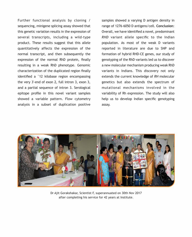
Dr Ajit Gorakshakar, Scientist F, superannuated on 30th Nov 2017
after completing his service for 42 years at Institute.

Hindi Pakhwada conducted at NIIH Seminar hall on 26-
27th Sept 2017. Essay writing and elocution competition
were conducted. Chief guest for this occasion were Mr.
Vipul Lakhnavi and Mr. Vinod Kumar Sharma.
Program on Swachh Bharat Abhiyaan was conducted in
N.P. High School, Lalbaug Mumbai- 21st Nov 2017.
Drawing & Essay writing competition was held in which
30 students participated and in elocution competition
ve students took part. The topic for all the three
competitions, were on Swachh Bharat Abhiyaan.
NIIH staff and students conducted Sadbhavna Diwas Oath
at Seminar Hall on 24th Nov 2017
Miss. Disha Parchure, PhD student from Department of Transfusion Medicine, was awarded the prestigious Harold Gunson fellowship (for transfusion medicine
t hstudents/researchers) at the 28 Regional conference of ISBT held at Guangzhou, China on 25-28 November, 2017. This fellowship is a travel grant exclusively for young researchers. She was awarded the fellowship for her abstract entitled "Phenotypic characteristics of novel RHD variants in Indians"
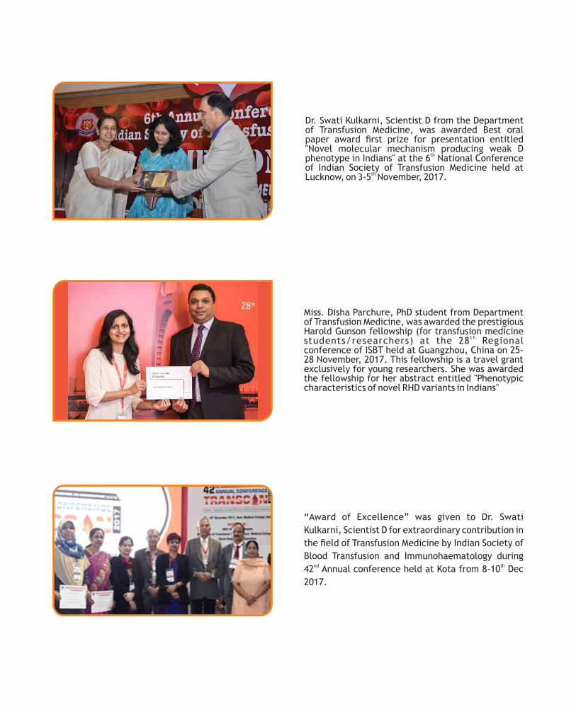
Dr. Swati Kulkarni, Scientist D from the Department of Transfusion Medicine, was awarded Best oral paper award rst prize for presentation entitled "Novel molecular mechanism producing weak D
thphenotype in Indians" at the 6 National Conference of Indian Society of Transfusion Medicine held at
thLucknow, on 3-5 November, 2017.
“Award of Excellence” was given to Dr. Swati
Kulkarni, Scientist D for extraordinary contribution in
the eld of Transfusion Medicine by Indian Society of
Blood Transfusion and Immunohaematology during nd th42 Annual conference held at Kota from 8-10 Dec
2017.

Immunohaematology Bulletin is brought out by:
ICMR-NATIONAL INSTITUTE OF IMMUNOHAEMATOLOGYth13 oor, New multi-storeyed Building, K.E.M. Hospital Campus,
Parel, Mumbai- 400012 (INDIA). Web: www.niih.org.in
Flowcytometry workshop on Diagnosis of Primary Immunodeciency Disorders (PID) was thorganized by Pediatric Immunology and leukocyte biology department, on 7-8 Sept 2017 at NIIH.