BSc., MSc., PhD Medical Microbiology
23
Staining techniques & Gram stain Dr. Shler Ghafour Raheem BSc., MSc., PhD Medical Microbiology [email protected] Lab. No. 6
Transcript of BSc., MSc., PhD Medical Microbiology

Staining techniques &
Gram stain
Dr. Shler Ghafour RaheemBSc., MSc., PhD Medical Microbiology
Lab. No. 6

1- Principle of staining.
2- Types of staining techniques.
- Simple staining
- Differential staining (Gram staining)
- Special staining
3- Smear preparation
- Labeling of smears
- Making of smears
- Fixation of smears
4- Procedures of Gram staining

Principle of staining
Bacterial cell are almost colorless and
transparent .
Their shape and size can be easily
determined under the microscope .

POSITIVE STAINING: where the actual cells are themselves
colored and appear in a clear background.
(a) Simple staining: A stain which provides color contrast but
gives same color to all bacteria and cells. Ex: Methylene blue,
Diluted carbol fuchsin.
(b) Differential Staining: A stain which imparts different
colors to different bacteria is called differential stain(which
contains more than one stain).
Ex: Gram’s stain , Acid fast staining.
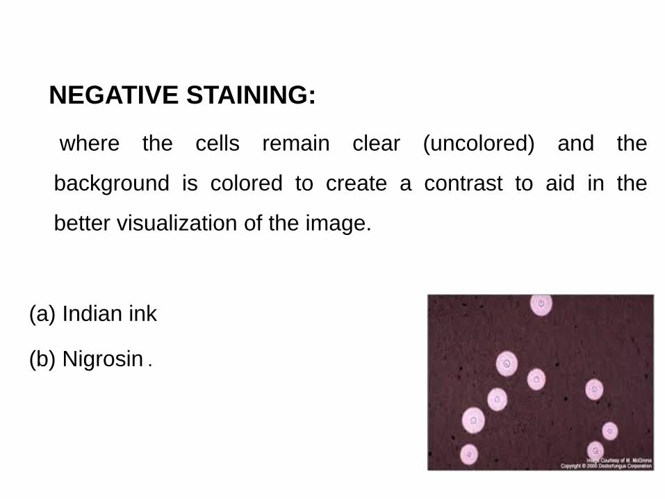
NEGATIVE STAINING:
where the cells remain clear (uncolored) and the
background is colored to create a contrast to aid in the
better visualization of the image.
(a) Indian ink
(b) Nigrosin .

TYPES OF STAINING TECHNIQUES
Simple staining(use of a single stain)
Differential staining(use of two contrasting stains
separated by a decolorizing agent)
For visualization of morphological
shape & arrangement.
Identification
Gram stain
Acid faststain

Simple staining
It is the use of single basic dye to color the
bacterial organism.
e.g. Methylene blue, crystal violet, safranin.
Purpose:-
To show the morphological shapes and
arrangement of bacterial cells.

Differential stainGram Stain: It is the most important
differential stain used in bacteriology because
it classified bacteria intotwo major groups:
a)Gram positive:
Appears violet after Gram’s stain
b) Gram negative:
Appears red after Gram’s stain
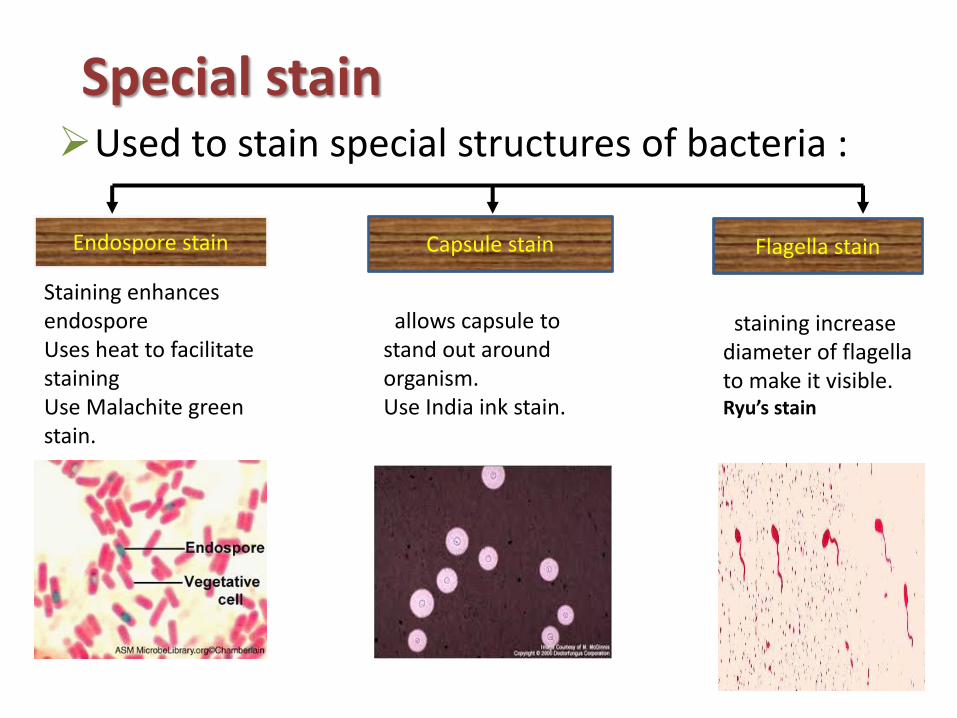
Special stainUsed to stain special structures of bacteria :
Endospore stain Capsule stain Flagella stain
staining increase diameter of flagella to make it visible.Ryu’s stain
allows capsule to stand out around organism.Use India ink stain.
Staining enhances endosporeUses heat to facilitate staining Use Malachite green stain.

Acid – Fast Staining
Negative acid – Fast BacilliPositive acid – Fast Bacilli

Spore Staining

Flagellar Staining

Capsular Staining

BASIC SHAPES OF BACTERIA
Cocci Bacilli

Arrangements
Cocci
Irregular Clusters Chains or PairsTetrads
Staphylococci Micrococci Streptococci

Typical shapes of bacteria

Other shapes of bacteria
Comma shaped
Spirochetes
Spirilla
17

Gram staining
1. CRYSTAL VIOLET
• Primary stain
• Violet colored, stains all micro-organism
2. GRAM IODINE
• Mordant
• Forms Crystal violet iodine complexes
3. DECOLORIZER
• Acetone + Methanol
• Removes Crystal violet iodine complex from thin peptidoglycan layers
• Dissolves outer layer of Gram negative org

4. GRAM SAFRANINE
• Counter stain
• Red colored
• Stains thin walled Gram
negative organism

Preparation of bacterial smear for
Gram Stain
• A Bacterial smear is a thin layer of bacteria placed on a slide for staining.
• Purpose:
To kill the microorganism, fix them to the slide to and prevent them from being washed out during the process of staining.

Steps to Gram Stain
1. Prepare a fixed smear
2. Add Crystal Violet (60 seconds)
3. Wash entirely with water
4. Rinse with Iodine (60 seconds)
5. Wash with Ethyl alcohol and dry
6. Add Safranin (60 seconds)
7. Wash with Water & dry gently
8. Use microscope of 100×


References
• Luis M. de la Maza, Marie T. Pezzlo, Cassiana E.Bittencourt, Ellena M. Peterson. 2020. Color Atlas ofMedical Bacteriology. (2020, Wiley) - libgen.lc.
• Warren E. Levinson, Peter Chin-Hong, ElizabethJoyce, Jesse Nussbaum, Brian Schwart. 2018.Review of Medical Microbiology & Immunology,15th edition. McGraw-Hill Education.
• Gary W. Procop,Deirdre L. Church , et al. 2017. Koneman'sColor Atlas and Textbook of Diagnostic Microbiology.7th Edition. Jones & Bartlett Learning