Evaluación de la cadena de valor del cacao Theobroma cacao ...

STUDIES ON SECONDARY METABOLITES ASSOCIATED WITH WITCHES’
BROOM DISEASE, FLORAL BIOLOGY, AND SEED FERMENTATION IN CACAO
by Fábio Clasen Chaves
A dissertation submitted to the
Graduate School-New Brunswick
Rutgers, The State University of New Jersey
in partial fulfillment of the requirements
for the degree of
Doctor of Philosophy
Graduate Program in Plant Biology
written under the direction of
Professor Thomas J. Gianfagna
and approved by
________________________
________________________
________________________
________________________
New Brunswick, New Jersey
May, 2008

ii
ABSTRACT OF THE DISSERTATION
STUDIES ON SECONDARY METABOLITES ASSOCIATED WITH WITCHES’
BROOM DISEASE, FLORAL BIOLOGY AND SEED FERMENTATION IN CACAO
by Fábio Clasen Chaves
Dissertation Director: Professor Thomas J. Gianfagna
Theobroma cacao L., a tree native to the Amazon, is cultivated in the tropics
throughout the world for its seeds, used primarily for chocolate production. Cacao
production is limited by several problems. Cocoa pod borer, an insect that burrows
through pods, damages seeds, allowing contamination by toxigenic fungal species. Many
fungal diseases infect cacao. Among them, Moniliophthora perniciosa, the causal agent
of witches’ broom disease, severely affects plantations throughout South America and the
Caribbean. Cacao yields are further limited by the naturally low rates of fruit set.
Moreover, disease tolerant varieties are usually self-incompatible low producers and do
not give a superior chocolate flavor compared to some disease susceptible and self-
compatible genotypes with highly valued aroma compounds.
During this project, problems associated with three main aspects of cacao were
investigated: disease, production and processing. Studies on plant-endophyte-pathogen
interactions allowed for the identification of new possible mechanisms of disease control;
studies on cacao flower physiology indicated ways to improve pollination and therefore
increase fruit set and crop yield; and investigations of the fermentation step of cacao
processing permitted discovery of a method for maintaining higher levels of compounds
valued by cacao manufacturers.

iii
Flavan-3-ol monomers and oligomers, purine alkaloids and salicylic acid, volatile
organic compounds, polyketides and other phenolic compounds were among the
determined bioactive compounds found in cacao, pathogens and endophytes, with
influence on disease, production, and processing.
iv
ACKNOWLEDGMENTS
I would like to thank: my advisor, Thomas J. Gianfagna, for his assistance,
encouragement, guidance and support. His exemplar work ethic, sober advice, and
personable demeanor have and will continue to influence my future career; the members
of my graduate committee, Bingru Huang, James White Jr., Mark Kelm, and Prakash
Hebbar, for their solicitude and keen guidance throughout my dissertation work; friends,
colleagues and collaborators, Alan Pomella, Ajay Singh, Debora Esposito, Elisabeth Ng,
Fernando Vega, Francisco Posada, Jeanne Peters, Jinpeng Xing, John Munafo, Logan
Logendra, Luciana Ambrozevicius, Luis Mejia, Madhu Aneja, Marshall Bergen, Monica
Torres, Patricia Alvarez, Patrick McCullough, Paulo Ribeiro, Richard Merrit, Rod Sharp,
Shimon Rachmilevitch, Ulisses Hernandez, Yan Xu, and others that I may have
inadvertently left out, whose friendship, partnership and shared experience positively
contributed to my stay at Rutgers; all faculty members of Rutgers University, especially
those from the Plant Biology and Pathology Department whose knowledgeable and
informative lectures and seminars provided the basis for the development of my Ph.D.
studies at Rutgers; CAPES (Coordenação de Aperfeiçoamento de Pessoal de Nível
Superior) for providing me with a graduate fellowship; Laura Cortese, my sweetheart,
who has helped me avoid the use of ‘Portuguenglish’ during the writing of this
dissertation; my family, Daniel, Morenei, Tania, and Vitor, for their constant support,
love and care.

v
You may travel the world, meet a million people and still if you do not know what you
are looking for, you will never be satisfied with what you find.
An experience may be very pleasant, but if you do not feel you are building something
you won’t be happy.
I may have heard them or may have read them; I sure did learn and believe them.

vi
TABLE OF CONTENTS
ABSTRACT OF THE DISSERTATION ................................................................................................... ii ACKNOWLEDGMENTS............................................................................................................................iv TABLE OF CONTENTS.............................................................................................................................vi LIST OF TABLES ........................................................................................................................................x LIST OF FIGURES ................................................................................................................................... xii 1. INTRODUCTION.....................................................................................................................................1 2. PLANT-ENDOPHYTE-PATHOGEN INTERACTIONS .....................................................................6 2.1. PLANT ....................................................................................................................................................6 2.1.1. INTRODUCTION...............................................................................................................................6 2.1.2. MATERIALS AND METHODS......................................................................................................12 2.1.2.1. CHEMICAL ELICITORS ............................................................................................................12 2.1.2.2. PATHOGEN INOCULATION .....................................................................................................12 2.1.2.3. LEAF SIZE EXPERIMENT .........................................................................................................13 2.1.2.4. SA AND SAG ANALYSIS.............................................................................................................15 2.1.2.5. PURINE ALKALOIDS AND PROCYANIDIN MONOMER ANALYSIS ..............................16 2.1.2.6. PROCYANIDIN OLIGOMER ANALYSIS ................................................................................17 2.1.2.7. STATISTICAL ANALYSIS ..........................................................................................................17 2.1.3. RESULTS...........................................................................................................................................18 2.1.3.1. ENDOGENOUS LEVELS OF PURINE ALKALOIDS AND PROCYANIDIN MONOMERS IN FLUSH LEAVES OF GREENHOUSE GROWN TREES.................................................................18 2.1.3.2. LEVELS OF PURINE ALKALOIDS AND PROCYANIDIN MONOMERS IN FLUSH LEAVES OF GREENHOUSE GROWN TREES AFTER APPLICATION OF CHEMICAL ELICITORS.................................................................................................................................................19 2.1.3.3. ENDOGENOUS LEVELS OF SA AND SAG IN LEAVES OF GREENHOUSE-GROWN TREES..........................................................................................................................................................21 2.1.3.4. LEVELS OF SA AND SAG IN LEAVES OF GREENHOUSE-GROWN TREES AFTER APPLICATION OF CHEMICAL ELICITORS ......................................................................................22 2.1.3.5. LEVELS OF SA AND SAG IN LEAVES OF FIELD-GROWN TREES AFTER PATHOGEN INOCULATION..........................................................................................................................................25 2.1.3.6. CHANGES IN CACAO PROCYANIDIN CONTENT THROUGHOUT LEAF DEVELOPMENT........................................................................................................................................28 2.1.3.7. EFFECT OF PATHOGEN INOCULATION ON CACAO LEAF PROCYANIDINS ............32 2.1.3.8. PCA ANALYSIS COMPARING CAFFEINE, THEOBROMINE, (+)-CATECHIN, (-)-EPICATECHIN, SALICYLIC ACID, AND SALICYLIC ACID GLYCOSIDE LEVELS OF FLUSH LEAVES TREATED WITH ELICITORS ...............................................................................................36 2.1.4. DISCUSSION ....................................................................................................................................41 2.1.4.1. EFFECT OF CHEMICAL ELICITORS ON PURINE ALKALOIDS AND PROCYANIDIN MONOMERS ..............................................................................................................................................41

vii
2.1.4.2. EFFECT OF CHEMICAL ELICITORS AND PATHOGEN INOCULATION IN FREE SA AND SAG OF CACAO LEAVES..............................................................................................................44 2.1.4.3. LEAF SIZE EXPERIMENT .........................................................................................................46 2.1.4.4. EFFECT OF PATHOGEN INOCULATION IN PROCYANIDIN LEVELS ..........................48 2.1.5. CONCLUSIONS................................................................................................................................50 2.2. ENDOPHYTES AND THEIR METABOLITES...............................................................................53 2.2.1. INTRODUCTION.............................................................................................................................53 2.2.2. MYCOTOXINS PRODUCED BY COFFEE ENDOPHYTES......................................................56 2.2.2.1. MATERIALS AND METHODS...................................................................................................59 2.2.2.1.1 FUNGAL ISOLATES ..................................................................................................................59 2.2.2.1.2. NUTRIENT MEDIA ...................................................................................................................59 2.2.2.1.3. OCHRATOXIN ANALYSIS......................................................................................................59 2.2.2.1.4. AFLATOXIN ANALYSIS..........................................................................................................60 2.2.2.2. RESULTS AND DISCUSSION.....................................................................................................60 2.2.2.3. CONCLUSIONS.............................................................................................................................67 2.2.3. ASPERGILLUS ORYZAE FROM COFFEE, A NON-TOXIGENIC ENDOPHYTE WITH THE ABILITY TO SYNTHESIZE KOJIC ACID IN CULTURE ..................................................................68 2.2.3.1. MATERIALS AND METHODS...................................................................................................69 2.2.3.1.1. FUNGAL ISOLATE ...................................................................................................................69 2.2.3.1.2. KA EXTRACTION FROM FUNGAL CULTURE..................................................................69 2.2.3.1.3. SEEDLING INOCULATION AND IN PLANTA EXTRACTION FOR KA AND CAFFEINE ANALYSIS .............................................................................................................................70 2.2.3.1.4. KA AND CAFFEINE ANALYSIS.............................................................................................71 2.2.3.2. RESULTS AND DISCUSSION.....................................................................................................71 2.2.3.3. CONCLUSIONS.............................................................................................................................73 2.2.4. MORE ENDOPHYTES FROM COFFEE WITH POTENTIAL BIOCONTROL ACTIVITY AGAINST CACAO PATHOGENS ...........................................................................................................76 2.2.4.1. MATERIALS AND METHODS...................................................................................................77 2.2.4.1.1. FUNGAL ISOLATES .................................................................................................................77 2.2.4.1.2. MEDIA EFFECT ........................................................................................................................78 2.2.4.1.3. DUAL CULTURE PLATE ASSAYS.........................................................................................78 2.2.4.1.4. CELLOPHANE COVERED MEDIA BIOASSAY ..................................................................79 2.2.4.1.5. BIOASSAY TEST FOR VOLATILE ANTIMICROBIALS ...................................................79 2.2.4.1.6. ANALYSIS OF THE VOLATILES PRODUCED BY ENDOPHYTES ................................80 2.2.4.1.7. EFFECT OF CAFFEINE ON FUNGAL GROWTH...............................................................81 2.2.4.2. RESULTS AND DISCUSSION.....................................................................................................81 2.2.4.3. CONCLUSIONS...........................................................................................................................100 2.2.5. ESTABLISHMENT OF BEAUVERIA BASSIANA AS AN ENDOPHYTE IN CACAO PODS.....................................................................................................................................................................101

viii
2.2.5.1. MATERIALS AND METHODS.................................................................................................102 2.2.5.1.1. COUNTING SPORES...............................................................................................................102 2.2.5.1.2. APPLICATION OF BEAUVERIA BASSIANA TO CACAO FLOWERS AND PODS......102 2.2.5.2. RESULTS AND DISCUSSION...................................................................................................103 2.2.5.3. CONCLUSIONS...........................................................................................................................105 2.3. PATHOGEN.......................................................................................................................................108 2.3.1. INTRODUCTION...........................................................................................................................108 2.3.2. PH EFFECT ON GROWTH OF M. PERNICIOSA AND M. RORERI......................................111 2.3.3. ANASTOMOSIS GROUPS OF M. PERNICIOSA ISOLATES..................................................114 2.3.4. RESULTS AND DISCUSSION......................................................................................................115 2.3.5. METABOLITES PRODUCED BY M. PERNICIOSA .................................................................121 2.3.5.1. MATERIALS AND METHODS.................................................................................................122 2.3.5.1.1. CHEMICAL REAGENTS........................................................................................................122 2.3.5.1.2. FUNGAL CULTURES .............................................................................................................122 2.3.5.1.3. PLANT MATERIAL.................................................................................................................122 2.3.5.1.4. SA EXTRACTION AND HPLC ANALYSES ........................................................................123 2.3.5.1.5. GC-MS ANALYSES .................................................................................................................123 2.3.5.1.6. SA APPLICATION TO CACAO TISSUES ...........................................................................124 2.3.5.1.7. STATISTICAL ANALYSIS .....................................................................................................124 2.3.5.2. RESULTS AND DISCUSSION...................................................................................................124 2.3.5.3. CONCLUSIONS...........................................................................................................................134 2.3.6. M. PERNICIOSA ENZYMES INVOLVED IN DISEASE...........................................................135 2.3.6.1. MATERIALS AND METHODS.................................................................................................139 2.3.6.1.1. FUNGAL CULTURES .............................................................................................................139 2.3.6.1.2. EFFECTS OF ODA ON M. PERNICIOSA .............................................................................139 2.3.6.1.3. M. PERNICIOSA GROWTH ASSAY IN CULTURE SUPPLEMENTED WITH (-)-EPICATECHIN.........................................................................................................................................139 2.3.6.1.4. (-)-EPICATECHIN DEGRADATION ....................................................................................140 2.3.6.1.5. CULTURE OF M. PERNICIOSA IN CULTURE BROTH FOR ENZYME EXTRACT ...140 2.3.6.1.6. PROTEIN DETERMINATION...............................................................................................141 2.3.6.1.7. GEL ELECTROPHORESIS....................................................................................................141 2.3.6.1.8. ENZYME ASSAYS ...................................................................................................................142 2.3.6.1.9. POLYPHENOLOXIDASE INHIBITION...............................................................................142 2.3.6.2. RESULTS AND DISCUSSION...................................................................................................143 2.3.6.3. CONCLUSIONS...........................................................................................................................160 3. PRODUCTION......................................................................................................................................161 3.1. INTRODUCTION..............................................................................................................................161
ix
3.2. MATERIALS AND METHODS.......................................................................................................163 3.2.1. VOLATILE ORGANIC COMPOUNDS.......................................................................................163 3.2.2. TEMPERATURE MEASUREMENTS.........................................................................................164 3.2.3. RESPIRATION ...............................................................................................................................164 3.2.4. SALICYLIC ACID ANALYSIS ....................................................................................................166 3.2.5. HAND POLLINATION..................................................................................................................166 3.3. RESULTS............................................................................................................................................167 3.3.1. VOLATILE ORGANIC COMPOUNDS.......................................................................................167 3.3.2. TEMPERATURE MEASUREMENTS.........................................................................................172 3.3.3. RESPIRATION ...............................................................................................................................174 3.3.4. SALICYLIC ACID ANALYSIS ....................................................................................................180 3.3.5. HAND POLLINATION..................................................................................................................184 3.4. DISCUSSION .....................................................................................................................................184 3.5. CONCLUSIONS.................................................................................................................................191 4. PROCESSING.......................................................................................................................................192 4.1. INTRODUCTION..............................................................................................................................192 4.2. MATERIALS AND METHODS.......................................................................................................194 4.2.1. FERMENTATION..........................................................................................................................194 4.2.2. EFFECT OF KOJIC ACID ON YEAST.......................................................................................194 4.2.3. BEAN SAMPLE PREPARATION ................................................................................................194 4.2.4. SUGAR ANALYSIS........................................................................................................................195 4.2.5. AMINO ACIDS ANALYSIS ..........................................................................................................195 4.2.6. PROCYANIDIN ANALYSIS .........................................................................................................195 4.3. RESULTS AND DISCUSSION.........................................................................................................196 4.4. CONCLUSIONS.................................................................................................................................204 5. SUMMARY AND CONCLUDING REMARKS ................................................................................205 REFERENCES..........................................................................................................................................210 CURRICULUM VITA..............................................................................................................................226

x
LIST OF TABLES
TABLE 1. LEAF SALICYLIC ACID (SA) AND SALICYLIC ACID GLYCOSIDE (SAG) OF FOUR GREENHOUSE-GROWN CACAO GENOTYPES IMC, AME, TSH AND SCA................................23 TABLE 2. LEAF (L1) SA CONTENTS OF FIELD-GROWN TREES INOCULATED (I) WITH BASIDIOSPORES OF M. PERNICIOSA OR UNINOCULATED CONTROLS (C) AND ADJACENT OLDER LEAF PAIRS L2 AND L3* ...................................................................................27 TABLE 3. EFFECT OF INOCULATION WITH BASIDIOSPORES OF THE PATHOGEN MONILIOPHTHORA PERNICIOSA ON THE PROCYANIDIN CONTENT (MG·G-1 DW) OF FLUSH LEAVES OF FIELD-GROWN DISEASE..................................................................................34 TABLE 4. ANALYSIS OF VARIANCE BY F-TEST COMPARING LEAF PROCYANIDIN CONTENTS UNDER DIFFERENT TREATMENTS.............................................................................37 TABLE 5. CONCENTRATION OF OTS (ΜG.G-1) PRODUCED BY ASPERGILLUS SPP. (4 DAI) ON MEDIA WITH AND WITHOUT CAFFEINE ..................................................................................62 TABLE 6. LIST OF ISOLATES ANALYZED FOR OT.........................................................................65 TABLE 7. OCHRATOXIN A PRODUCTION (OTA ΜG.G-1 MEDIA ± STD DEV.) BY A. OCHRACEUS AND TWO A. WESTERDIJKIAE ISOLATES FROM THE PARASITOID P. NASUTA (W6-1) AND THE COFFEE BERRY BORER, HYPOTHENEMUS HAMPEI (CBB9-1) AT 4 AND 35 DAYS AFTER INOCULATION (DAI) ...................................................................................66 TABLE 10. VOLATILES PRODUCED BY BEAUVERIA BASSIANA AND TRICHODERMA ASPERELLUM ............................................................................................................................................97 TABLE 11. PERCENTAGE OF B. BASSIANA RECOVERED FROM PODS SPRAYED WITH A B. BASSIANA SPORE SUSPENSION AFTER FLOWER POLLINATION...........................................107 TABLE 12. GROWTH RATES (CM/DAY) OF M. PERNICIOSA IN MEDIA CONTAINING ODA AND 1-OCTEN-3-OL ...............................................................................................................................144 TABLE 13. EFFECT OF POLYPHENOLOXIDASE INHIBITORS, KOJIC ACID (KA), ASCORBIC ACID (AA) AND CITRIC ACID (CA) TESTED AT 3.33 MM ON ENZYME ACTIVITY USING (-)-EPICATECHIN AS SUBSTRATE ..................................................................159 TABLE 14. RATES OF OXIDATION OF L-DOPA USING M. PERNICIOSA ENZYMATIC EXTRACT .................................................................................................................................................159 TABLE 15. VOLATILE ORGANIC COMPOUNDS PRODUCED BY CACAO FLOWERS..........168 TABLE 16. TEMPERATURE MEASUREMENTS OF FLOWERS, BACKGROUND, AND PLANT SURFACES OF T. CACAO IN THE MORNING AND AFTERNOON (SE, STANDARD ERROR).....................................................................................................................................................................173 TABLE 17. PERCENTAGE OF ACCUMULATED CO2 PRODUCED BY ENCLOSED CACAO AND TOMATO FLOWERS ....................................................................................................................179 TABLE 18. SALICYLIC ACID (SA) AND SALICYLIC ACID GLUCOSIDE (SAG) CONTENTS OF OPEN AND CLOSED CACAO FLOWERS BEFORE AND AFTER TREATMENT WITH SHAM.........................................................................................................................................................181 TABLE 19. LSD ANALYSIS OF THE SA AND SAG CONTENTS OF CACAO FLOWERS .........181 TABLE 20. EFFECT OF TREATMENT ON SALICYLIC ACID (SA) AND SALICYLIC ACID GLUCOSIDE (SAG) CONTENTS OF CACAO FLOWERS BY CULTIVAR...................................182 TABLE 21. EFFECT OF CULTIVAR ON SALICYLIC ACID (SA) AND SALICYLIC ACID GLUCOSIDE (SAG) CONTENTS OF CACAO FLOWERS ...............................................................183

xi
TABLE 22. RESPIRATORY QUOTIENT (RQ) OF FLOWERS TREATED WITH DIFFERENT CHEMICALS ............................................................................................................................................189

xii
LIST OF FIGURES
Figure 1. Chemical structure of B-type procyanidin monomers and oligomers................. 9 Figure 2. Cacao leaves used in the pathogen inoculation study ....................................... 14 Figure 3. Purine alkaloid content of flush leaves of four greenhouse-grown cacao genotypes .......................................................................................................................... 20 Figure 4. Free SA content in mature leaves of Scavina 6 (SCA) after treatment with chemical elicitors, benzothiadiazole (B), glutathione (G), control (C), aminoethoxyvinylglycine (A) and jasmonic acid (J) ........................................................ 24 Figure 5. Salicylic acid levels in young leaves ................................................................. 26 Figure 6. Procyanidin content of cacao leaves.................................................................. 29 Figure 7. Comparison of total procyanidin content in flush and old leaves of cacao....... 30 Figure 8. Total leaf procyanidin content throughout leaf development for four cacao varieties Amelonado, (AME), IMC 30 (IMC), TSH 565 (TSH) and Scavina 6 (SCA6) . 31 Figure 9. Procyanidin content of cacao flush leaves susceptible to witches’ ................... 33 Figure 10. PCA analysis (PCA 1 x PCA 2) of six metabolites (variables) present in four cacao genotypes (AME, IMC, SCA and TSH) subjected to treatments with chemical elicitors (A – aminoethoxyvinylglycine, B – benzothiadiazole, C – control, G – gluthatione, J – jasmonic acid).......................................................................................... 38 Figure 11. PCA analysis (PCA 1 x PCA 3) of six metabolites (variables) present in four cacao genotypes (AME, IMC, SCA and TSH) subjected to treatments with chemical elicitors (A – aminoethoxyvinylglycine, B – benzothiadiazole, C – control, G – gluthatione, J – jasmonic acid).......................................................................................... 39 Figure 12. PCA analysis (PCA 2 x PCA 3) of six metabolites (variables) present in four cacao genotypes (AME, IMC, SCA and TSH) subjected to treatments with chemical elicitors (A – aminoethoxyvinylglycine, B – benzothiadiazole, C – control, G – gluthatione, J – jasmonic acid).......................................................................................... 40 Figure 13. Ochratoxins A and B ....................................................................................... 58 Figure 14. Kojic acid measured by GC-MS as trimethylsylate derivative. ...................... 74 Figure 15. MS spectra of kojic acid after derivatization with TMS ................................. 74 Figure 16. Overlaid extracted ion chromatograms of full scan analysis comparing cacao control seedling and a seedling inoculated with A. oryzae. Ion m/z 194 is the molecular ion and base peak of the mass spectrum of the caffeine molecule. .................................. 75 Figure 17. Growth pattern of B. bassiana isolate Bb1 in media containing caffeine (0-1%)........................................................................................................................................... 84 Figure 18. Growth pattern of B. bassiana isolate Bb04 in media containing caffeine (0-1%).................................................................................................................................... 85 Figure 19. Growth pattern of M. anisopliae isolate MA04 in media containing caffeine (0-1%) ............................................................................................................................... 86 Figure 20. Growth pattern of M. anisopliae isolate MA06 in media containing caffeine (0-1%) ............................................................................................................................... 87 Figure 21. Growth pattern of Paecilomyces fumosoroseus isolate PR42 in media containing caffeine (0-1%) ............................................................................................... 88 Figure 22. Dual plate assay between P. citrinum and B. bassiana ................................... 90

xiii
Figure 23. Dual plate assays between T. asperellum (Tasp) and M. perniciosa (Mp), and M. roreri (Mr) ................................................................................................................... 95 Figure 24. X-plate assay between T. asperellum (Tasp) and B. bassiana (Bb1 and Bb2)............................................................................................................................................ 96 Figure 25. Chemical structure of Actinohordin ................................................................ 98 Figure 26. Effect of the substrate on metabolite production by the actinomycete isolate 99 Figure 27. Cacao pod sections and parts sampled for determining the presence of Beauveria bassiana ......................................................................................................... 106 Figure 28. Moniliophthora perniciosa basidiocarps formed in cherelles (young fruit of cacao) from a farm in Pichilingue, Ecuador ................................................................... 110 Figure 29. Effect of pH on the growth of M. roreri........................................................ 112 Figure 30. Effect of pH on the growth of M. perniciosa ................................................ 113 Figure 31. Vegetatively compatible isolates (1103 left and 278 right)........................... 116 Figure 32. Vegetatively incompatible (PS6 left and Alf42 right)................................... 117 Figure 33. Intermediate vegetative compatibility (PS9 left and PS12 right).................. 118 Figure 34. Vegetative compatibility groups of M. perniciosa isolates (Red – Incompatible; Yellow – Intermediate compatibility; Green – Compatible) ................... 119 Figure 35. M. perniciosa isolate growth rates (cm/day)* ............................................... 120 Figure 36. Salicylic acid produced by M. perniciosa isolates in culture ........................ 128 Figure 37. MS spectra of trimethylsilylated fungal metabolites salicylic acid, mandelic acid and phenyllactic acid............................................................................................... 129 Figure 38. Salicylic acid treatment to young cacao leaves ............................................. 131 Figure 39. HPLC chromatogram of salicylic acid in brooms and healthy...................... 132 Figure 40. Flow chart with proposed role for salicylic acid in pathogenicity ................ 133 Figure 41. Protein gel of M. perniciosa mycelial extract compared to commercial soybean LOX ................................................................................................................................ 145 Figure 42. Growth rates of M. perniciosa in media containing (-)-epicatechin and KA 147 Figure 43. M. perniciosa growth assay in the presence of (-)-epicatechin (bottom left), kojic acid (top right), (-)-epicatechin and KA (bottom right), and media alone (top left)......................................................................................................................................... 148 Figure 44. (-)-Epicatechin degradation by M. perniciosa isolates.................................. 149 Figure 45. Comparison of pH optima for activity of commercial laccase (LAC), PPO and M. perniciosa (Mp) extracellular enzyme extract using (-)-epicatechin as substrate at 25oC................................................................................................................................. 150 Figure 46. Protein gels with commercial polyphenol oxidase (PPO) and media and mycelial extracts of M. perniciosa (isolate PS12). Left panel stained with commassie blue and right panel with (-)-epicatechin................................................................................ 154 Figure 47. Activity gel stained by immersing non-denaturing protein gel in an (-)-epicatechin solution ........................................................................................................ 155 Figure 48. Activity gels comparing commercial laccase (LAC) and PPO to M. perniciosa enzyme extract (Mp) ....................................................................................................... 156 Figure 49. Enzyme activity of M. perniciosa, PPO and laccase using ABTS as substrate......................................................................................................................................... 157 Figure 50. Effect of different concentrations of ascorbic acid in the inhibition of polyphenoloxidase activity of M. perniciosa.................................................................. 158

xiv
Figure 51. Levels of tridecane, 7-tetradecene and 1-pentadecene emitted by petals, sepals and reproductive parts of cacao flowers ......................................................................... 169 Figure 52. Levels of undecane, dodecane, 6-tridecene, tetradecane and pentadecane emitted by petals, sepals and reproductive parts of cacao flowers ................................. 170 Figure 53. Levels of 4-dodecene, 2-methyl-dodecane, 3-methyl-tridecane and 3-hexadecene emitted by petals, sepals and reproductive parts of cacao flowers.............. 171 Figure 54. Chromatogram showing CO2 peak at 0.816 minutes .................................... 175 Figure 55. CO2 evolution in opened and closed cacao flowers....................................... 176 Figure 56. Closed cacao flower respiration expressed as CO2 evolution ....................... 177 Figure 57. Open cacao flower respiration expressed as CO2 evolution.......................... 178 Figure 58. Effect of SA, SA plus SHAM, SHAM and KCN treatments on cacao flower respiration ....................................................................................................................... 188 Figure 59. Flowers treated with 200 μM of salicylic acid (3 on the left) compared to controls (3 on the right), after 84 hours of treatment...................................................... 189 Figure 60. Beakers containing beans during fermentation (left – control, right – KA treated) ............................................................................................................................ 198 Figure 61. Cut test showing on the left control beans and on the right KA treated beans after 7 days of fermentation. ........................................................................................... 199 Figure 62. Yeast streaked on media containing 10 mM of kojic acid in PDA (right) compared to PDA alone (left) ......................................................................................... 200 Figure 63. Total procyanidin content of beans during fermentation* ............................. 201 Figure 64. Total amino acids in cacao beans during fermentation ................................. 202 Figure 65. Total sugar content of beans during fermentation......................................... 203

1
1. INTRODUCTION
In 1720, Linnaeus named a tree Theobroma cacao, from the Greek “Theo”
meaning God and “broma” meaning food, based on the Aztecs who cultivated cacao and
believed it was of divine origin and possessed godly powers. The Aztecs made a
stimulant drink, chocolatl, by combining ground roasted cocoa beans with maize meal,
vanilla, and chili peppers, which was served in gold cups to emperor Montezuma (Wood
and Lass, 1987; Young, 2007). Cacao has also been used as an antiseptic, diuretic,
emmenagogue, and parasiticide, as well as a folk remedy for alopecia, burns, cough, dry
lips, listlessness, pregnancy, rheumatism, snakebite, and wounds (Duke, 2007). In some
countries, the beans served as currency; in 16th century Guatemala, one could buy a
horse with 50 cocoa beans and a slave with 100 beans (Young, 2007). Currently, the tree
native to the Amazon is cultivated in the tropics throughout the world for its beans, which
serve as raw material for the manufacture of chocolate, cocoa butter, and several other
food and cosmetic products.
Worldwide, 7,000,000 hectares of land are devoted to the cultivation of cacao.
Cocoa bean yields vary from 200 kg·ha-1 to 1500 kg·ha-1. Each tree produces an average
of 30 to 40 pods a year, with ten pods yielding one pound of dry cocoa beans. Major
producing countries include Ivory Coast with 1,286,000 tons per year, followed by
Indonesia (610,000 tons per year), Ghana (599,000 tons per year), Nigeria (441,000 tons
per year), and Brazil (236,000 tons per year) (2005 data, FAO). In the United States, the
main importer of cocoa beans, more than 1,900,000 jobs are dedicated to the
manufacture, distribution and sale of chocolate products. The chocolate industry in the

2
US exports chocolate and chocolate products to more than 50 countries, and in the year
2003 generated $23.5 billion in sales (Young, 2007).
The word “cocoa” originated from a loss in translation to English and has been
used to describe finished products, while “cacao”, allegedly the correct name, serves to
describe the crop and raw materials prepared from it (Chatt, 1954). In order to avoid
inaccuracies, many local terms used by indigenous people from cacao growing regions
have been preserved, which brings more peculiarity to a very distinct plant.
Cacao can be cultivated under a system called “Cabruca”, where a forest is
thinned and the original understory is replaced with cacao, allowing for the preservation
of native species. When cacao, which has a canopy height of about four to ten meters, is
planted in deforested areas, other taller species are introduced as shade trees. The choice
of shade tree varies according to regional adaptability and local culture. Leguminous
species such as Inga sp., Gliricidia sp. and Erythrina sp. are used preferentially due to
their nitrogen fixation capacity, but other trees such as coconuts, rubber trees, and
macadamia may be used and serve as secondary crops and an alternative source of
income. During the establishment of a cacao plantation, banana trees in Latin America,
and plantains in Africa, are used temporarily for shade to protect young trees. Cacao can
also be cultivated under another system, full sun, in which there are no shade trees.
Although more productive, this system demands higher fertilization and the life span of
the trees is shortened.
Cacao requires about 1250 to 2800 mm of rain per year and temperatures around
26 oC. In Salvador, BA, where more than 90 % of Brazil’s cacao is cultivated, the rainy
season starts in March, goes until August, and peaks in May with 350 mm of rain for the

3
month. The highest temperatures of about 30 oC occur in January and February, while the
lowest temperatures of around 21 oC occur in July and August. The minimum number of
daylight hours occurs in June (165 hours) and the maximum in January (245 hours) (2007
data, INMET).
Cacao is a dicotyledonous, C3 plant, with 2n = 2x = 20 chromosomes and an
estimated genome size of 4.15 x 10⁸ bp (Figueira et al., 1992). The tree has some unique
morphological characteristics. After a brief period of monopodial growth, five horizontal
fan branches develop simultaneously from buds just below the terminal growing point on
the main stem in a structure known as a jorquette. At the base of the jorquette, a vertical
shoot (known as a chupon) is formed, and from the chupon other fan branches eventually
arise. Chupons also stem from the bottom of the tree and it is common practice to
renovate old trees by cutting down the main trunk and allowing one chupon to take over.
On fan branches, leaves are oppositely arranged, whereas on vertical stems, leaves are
spirally arranged in a 3/8 pattern. When counting from the first leaf it takes three turns
around the stem for the 9th leaf to be at the same position as the 1st (Chatt, 1954).
Cacao has alternate periods of rapid shoot growth and dormancy, a type of growth
often referred to as flush growth. New flush leaves are thin and tender, and can be pale
green or pink, and only when fully expanded become dark green, thicker and sturdier.
Cacao has a cauliflorous habit with flowers growing from the trunk in patches
called flower cushions. Each flower can bear a single fruit called a pod. During
development the pods are green, and after five to six months they ripen to a yellow or
reddish purple color, depending on the variety. Pods do not drop off the trees even when

4
fully ripe. Each pod is approximately 30 cm long and 15 cm wide at the middle, and
bears an average of about 40 beans.
Cacao trees begin to produce pods after three to five years, and at ten years are
fully productive. There are usually two harvests a year, a small one prior to the rainy
season and the major harvest at the end of the rainy season. Once harvested, the pods are
cracked open with a machete and the beans, covered by a mucilaginous pulp, are placed
in piles, covered with banana leaves, and allowed to ferment. During fermentation, yeasts
and bacteria convert sugars to alcohols to acids, which allow for the development of
aroma precursor compounds. After fermenting for a few days, beans are sun dried and
then shipped to be roasted and manufactured.
Around the world, several problems threaten cacao production ranging from low
productivity to insect pests to devastating diseases. Overall losses due to insects and
disease are estimated at 30 % of the potential crop per year. In Indonesia, the main pest
problem is cocoa pod borer (Conopomorpha cramerella), an insect that burrows through
the pods and feeds on the pulp causing malformation of seeds. Moreover, the wounds
serve as an entry point for opportunistic organisms such as toxigenic fungi that
contaminate beans and pose a threat to human health. In Africa, Phytophthora species are
the main threat and can attack all parts of the tree. Species from the Phytophthora genus
are present in all cacao growing regions and cause black pod disease, ‘mancha de agua’
and Phytophthora canker. In Brazil, the most destructive pathogen is the fungus
Moniliophthora perniciosa, causal agent of witches’ broom disease, which under high
disease incidence can cause total crop loss. The fungus is present in growing countries of
the Americas and can also attack other species of Theobroma and the closely related

5
genus Herrania. In addition to Phytophthora and witches’ broom disease, cacao
plantations in northwestern South America, Ecuador, and Colombia also have to deal
with Moniliophthora roreri, the causal agent of frosty pod disease, which is characterized
by the frosty appearance of mycelia and spore covered pods. Cacao yields are further
limited by the naturally low rates of fruit set, ranging from one to five percent. Moreover,
disease tolerant varieties are usually self-incompatible low producers and do not give a
superior chocolate flavor compared to some very disease susceptible and self-compatible
genotypes with high valued aroma compounds.
Cacao is an interdisciplinary crop in that unlike many other food crops, it passes
through complex processing before becoming a consumable food. Therefore, a complete
understanding of agricultural, processing and manufacturing aspects of this crop is
essential for determining its success.
During this project, problems associated with three main aspects of cacao were
investigated: diseases, production and processing. The dissertation is divided into 3
sections: (1) Studies on plant-endophyte-pathogen interactions that allowed for the
identification of new possible mechanisms of disease control; (2) Studies on cacao flower
physiology that indicated ways to improve pollination and therefore increase fruit set and
crop yield; and (3) Investigations on the fermentation step of cacao processing permitted
discovery of a method for maintaining higher levels of compounds valued by cacao
manufacturers for chocolate production.

6
2. PLANT-ENDOPHYTE-PATHOGEN INTERACTIONS
2.1. PLANT
2.1.1. INTRODUCTION
Cacao disease resistance is multigenic and several genes associated with
resistance have been identified (Leal et al., 2007). However, there are no completely
resistant cultivars to any of the three main diseases that attack cacao, and the array of
pathogens and their high genetic variability make breeding for resistance complicated.
Other forms of crop protection employed for cacao include cultural practices such as
phytosanitary pruning and pod stripping, as well as the use of pesticides and biological
control agents. None of these procedures is very effective against cacao pathogens alone,
although when used in combination, depending on environmental conditions, satisfactory
levels of disease control may sometimes be attained. Despite the relative success of
current practices, crop production is significantly below capacity worldwide, M. roreri
and M. perniciosa are spreading and new forms of control should be pursued in an effort
to keep the pathogens and diseases manageable.
Activation or induction of plant defense responses, using chemicals considered
plant activators, is an alternative approach to pesticides for disease management. Plants
respond to chemical elicitors by activating metabolic pathways leading to systemic
acquired resistance (SAR). SAR is an inducible plant defense response that results in a
non-specific and long lasting systemic resistance to a variety of pathogens. Following

7
SAR there is an accumulation of signaling molecules, which trigger the activation of a
series of defense pathways and results in production of a variety of phytoalexins in cacao
and other plants. The phenylpropanoid pathway is one important source of phytoalexins.
It starts (meaning it derives from primary metabolism at this point) from phenylalanine,
which is deaminated by the action of the enzyme phenylalanine ammonia lyase (PAL; EC
4.3.1.5), to trans-cinnamic acid, a precursor of flavonoids and other metabolites.
Flavonoids, a class of compounds with a basic structure of 15 carbons arranged in
two aromatic rings and a heterocycle containing oxygen is subdivided into several groups
including the proanthocyanidins. Both induced and constitutive flavonoids have been
associated with plant resistance to biotic and abiotic stresses (Harborne and Williams,
2000). In cacao, flavonoids have been implicated in the resistance against pathogens
(Nojosa et al., 2003). Proanthocyanidins, also known as condensed tannins, possess a
polyflavan-3-ol structure differing in the hydroxylation pattern. The most common are
the di-hydroxylations (catechins) and tri-hydroxylations (gallocatechins) in the B ring of
the flavonoid skeleton. Procyanidins are the largest class of proanthocyanidins, composed
of polymers of flavan-3-ol subunits mostly linked by carbon-carbon bonds through C4
and C8 and to a lesser extent linked by C4 to C6 (Figure 1). These single linked
molecules are referred to as B type procyanidins. The A type linkages have an additional
bond between C2 and O7 and have not been reported in cacao (Kelm et al., 2006).
Purine alkaloids are another important group of compounds present in significant
amounts in cacao. The main purine alkaloids found in cacao leaves are theobromine (3,7-
dimethylxanthine) and caffeine (1,3,7-trimethylxanthine) (Zheng et al., 2004;
Hammerstone et al., 1994). In the alkaloid pathway, xanthosine is converted to

8
theobromine after two methylation steps and the hydrolysis of a sugar group. Caffeine
originates from theobromine after another methylation step. The conversion of
theobromine to caffeine may be slower in cacao leaves when compared to coffee and tea,
which contain higher caffeine levels. Both alkaloids have been linked to resistance
against pathogens (Nathanson, 1984; Pearson and Marth, 1990; Aneja and Gianfagna,
2001).
Salicylic acid (SA) has been identified as a signaling molecule that triggers plant
defense responses including activation of both the purine alkaloid and phenylpropanoid
pathways. Moreover, tobacco plants inoculated with tobacco mosaic virus respond by
accumulating SA and salicylic acid glucoside (SAG) (Hennig et al., 1993; Kawano et al.,
2004). After biosynthesis, most free SA is removed by esterification to methylsalicylate
(MeSA) or conjugation to SAG, which may work as a slow release storage form of SA
that maintains SAR over time.
SA and S-methyl-benzo[1,2,3]thiadiazole-7-carbothioate (Acibenzolar-S-methyl,
ASM; or BTH for benzothiadiazole), a synthetic analogue of SA (Kunz et al., 1997),
induce caffeine accumulation when applied to young flush leaves of cacao (Aneja and
Gianfagna, 2001). ASM application before inoculation of cacao seedlings with
Moniliophthora perniciosa reduced disease incidence by 84.5 %. In addition, ASM
application prior to infection protected cacao against verticilium wilt, an effect
comparable to that found upon treatment with cuprous oxide or tebuconazole (Resende et
al., 2002). With grapes, ASM enhanced anthocyanin levels and improved resistance
against the fungal pathogen Botrytis cinerea (Iriti et al., 2004).
9
Figure 1. Chemical structure of B-type procyanidin monomers and oligomers
O
OH
HO
OH
OH
O
OHHO
HOOH
OH
OH
C4 C8C4 C6
O
OH
HO
OH
OH
O
OH
HO
OH
OH
OH
O
OH
HO
OH
OH
OH
OH
n
(+)-Catechin
O
OH
HO
OH
OH
R1
R24
6
8
R1 = OH; R2 = HR1 = H; R2 = OH
( )-Epicatechin

10
Jasmonic acid is a signal transducer in elicitor-induced plant cell cultures.
Suspended cell cultures accumulate secondary metabolites upon application of methyl
jasmonate (MeJA). MeJA initiates de novo transcription of PAL genes. The induction of
anthocyanins in germinating soybean seedlings upon application of MeJA indicates that
this defense mechanism is present not only on plant cell suspension cultures but in
differentiated plants (Gundlach et al., 1992).
It has been reported that SA blocks JA biosynthesis in tomato leaves (Pena-Cortes
et al., 1993), on the other hand, SA production is inhibited by JA in wounded transgenic
tobacco plants (Sano et al., 1996). Wound-induced accumulation of transcripts and
proteins for some PR genes (pathogenesis related) is enhanced in the presence of MeJA,
and inhibited in the presence of SA, in contrast, the expression and transcription of other
PR genes is induced by SA and inhibited by MeJA (Niki et al., 1998). Thus, there are
many cases in which the production of SA and JA are mutually antagonistic.
To study wound-induced gene expression, chemicals such as MeJA and ethylene
have been used due to their known involvement as signal molecules in the wound-
induced expression of proteinase inhibitor genes (Niki et al., 1998; Pieterse and van
Loon, 1999). Ethylene has also been reported to be an inducer of PR proteins. Wounded
leaves treated with ethylene or MeJA accumulated PR transcripts faster than leaves that
were only wounded, suggesting that ethylene and MeJA might mediate wound-induced
transcriptional gene stimulation (Niki et al., 1998; Kim et al., 2006).
In addition to hormones, other compounds have been used to activate SAR
experimentally. Aminoethoxyvinylglycine (AVG) reduces ethylene and protein
biosynthesis in excised discs of mature-green tomato pericarp tissue. AVG is frequently

11
used as a specific inhibitor of ethylene biosynthesis to determine the effects of ethylene
on plant growth, development, and response to stress. AVG has also been used to study
fungal pathogenesis, nodulation in legumes and response to chilling stress (Saltveit,
2005). Glutathione, another chemical elicitor, causes rapid, marked but transient gene
expression in soybean protoplasts. Both reduced glutathione and oxidized glutathione
elicited the phytoalexin response in cell-suspension cultures of beans (Phaseolus
vulgaris) (Edwards et al., 1991). Agrobacterium rhizogenes transformed root cultures of
Lotus corniculatus treated with glutathione, accumulated isoflavan phytoalexins in both
tissue and culture medium. This accumulation of phytoalexins was preceded by a
transient increase in the activity of PAL (Robbins et al., 1991).
Small increases of different compounds by synergism may enhance defenses more
effectively than greatly increasing the concentrations of one specific compound. Such
interactions cannot be evaluated if only a single compound is analyzed. Since cacao
produces both flavonoids and purine alkaloids that may be regulated by SA, the levels of
procyanidins, salicylic acid, salicylic acid glucoside, caffeine and theobromine in leaves
of different cacao varieties with varying disease tolerance levels at different stages of
development, before and after elicitation, and after pathogen inoculation, were examined
in order to better understand the chemical mechanisms of plant defense.
12
2.1.2. MATERIALS AND METHODS
2.1.2.1. Chemical elicitors
Young developing flush leaves and fully developed mature leaves of greenhouse
grown cacao varieties used, differed in disease susceptibility. IMC 30 (very disease
susceptible), TSH 565 (Trinitario Selected Hybrids, moderately disease susceptible),
Scavina 6 (SCA 6, is one of the progenitors of TSH; the least disease susceptible) and
Amelonado (which is very susceptible to diseases and the only self-compatible of the
four). Fully developed leaves were cut into 1 cm disks with a cork borer and young
leaves were cut in approximately 1 cm pieces. 0.5 g of leaves were placed in an
erlenmeyer flask containing: 10 mL of a solution composed of 30 mM pH 5.6 buffer
potassium phosphate, 10 mM of sucrose and one of the following chemical elicitors, 1
mM of glutathione, 1 mM of aminoethoxyvinylglycine (AVG), 1 mM of BTH (acti-
guard, product containing BTH, from Novartis, USA), 1 mM of jasmonic acid. The flasks
were kept in a shaker (100 rpm) at room temperature for 24 hours. Leaves were then
rinsed with distilled water, blotted and kept at -80 oC until they were analyzed.
2.1.2.2. Pathogen inoculation
Field grown cacao trees from Bahia, Brazil, of two varieties, Cacao Comum
(CCO), disease susceptible, and Scavina 6 (SCA), disease tolerant, were used for this
study. The youngest pair of leaves in a flush (< 6 cm) was inoculated with basidiospores

13
of M. perniciosa. After 48 hours, the inoculated leaves (L1) and the two older proximal
pairs in the same flush (L2 and L3) were collected (Figure 2). The respective control
leaves (L1, L2 and L3) were collected from trees not inoculated with the pathogen. The
leaves were freeze dried and stored at -80 oC until analysis.
2.1.2.3. Leaf size experiment
Cacao leaves from greenhouse grown trees from the varieties IMC 30, TSH 565,
SCA 6 and Amelonado, were sampled and separated in nine categories according to leaf
size, 1: <2 cm, 2: 2-4 cm, 3: 4-6 cm, 4: 6-8 cm, 5: 8-10 cm, 6: 10-15 cm, 7: 15-20 cm, 8:
>20 cm young flush leaves, 9: >20 cm old fully expanded green leaves. Leaves were
harvested, flash frozen in liquid nitrogen, freeze-dried, and kept at -80 oC until analysis.
14
Figure 2. Cacao leaves used in the pathogen inoculation study
L1 ≤ 6 cm

15
2.1.2.4. SA and SAG analysis
SA extraction and HPLC analyses followed the method of Verberne et al. (2000).
The protocol was the following: grind 0.5 g of leaves in liquid nitrogen and transfer to an
eppendorf tube; add 1 mL of methanol (90 %) and 2.5 μL of internal standard (3,4-
DHBA, dihydroxy benzoic acid, 10 μg·μL-1); vortex for one minute and sonicate for five
minutes; centrifuge for five minutes at 13,000 rpm and separate the supernatant;
resuspend the pellet in 0.5 mL methanol; vortex for one minute and sonicate for five
minutes; centrifuge for five minutes and combine the supernatants; centrifuge again for
five minutes and discard the pellet. To the supernatant, add 10 μL of sodium hydroxide
(0.2 M); concentrate in a speedvac; add 250 μL of tricholoroacetic acid (5 % in water)
and vortex for 30 seconds; partition with 800 μL of ethylacetate:cyclohexane (1:1) twice;
the upper phases contain SA and the lower phases (aqueous) contain SAG; add 60 μL of
0.2 M sodium acetate buffer pH 5.5 to the combined upper layers; concentrate in the
speedvac. Add 600 μL of the HPLC mobile phase (0.2 M sodium acetate buffer pH 5.5 :
methanol; 90 : 10) and vortex for 30 seconds; centrifuge and use the supernatant to inject
in the HPLC; to the aqueous phase add 300 μL of 8 N HCl and heat the sample at 80 oC
for one hour; partition acid fraction with 800 μL of ethylacetate:cyclohexane (1:1) twice;
add 60 uL of sodium acetate buffer to the combined upper layers and concentrate in the
speedvac; add 600 μL of the HPLC mobile phase and vortex for 30 seconds; centrifuge
and inject the supernatant in the HPLC. A Shimadzu LC-10AT equipped with a RF-10A
spectrofluorometric detector and a C18 column (140 mm x 4.6 mm x 0.5 μm particle
size) was used. The fluorescence detector was set at an excitation wavelength of 305 nm

16
and emission of 407 nm; the flow rate was 0.8 mL·min-1 using a mobile phase of 90 %
0.2 M sodium acetate buffer pH 5.5 and 10 % methanol.
2.1.2.5. Purine alkaloids and procyanidin monomer analysis
For the extraction, flush leaves from greenhouse grown trees were ground in
liquid nitrogen until powdered and transferred to an eppendorf tube. Then 1 mL of 90 %
methanol was added to the tube, which was vortexed for one minute, sonicated for 12
minutes and centrifuged for five minutes at maximum speed in a bench top centrifuge.
The supernatant was saved, and 0.5 mL of methanol was then added to the pellet. The
sample was vortexed for one minute, sonicated for 12 minutes and centrifuged for five
minutes. The supernantants were combined and centrifuged again. Then 0.25 mL of
water and 0.25 mL of chloroform were added to the supernatant. The sample was
vortexed for 30 seconds and centrifuged for one minute to break the emulsion. The
chloroform layer was discarded and the remaining solution was concentrated in the
speedvac. Methanol was added to complete the final volume to 1 mL. HPLC conditions:
a reverse phase (C18) column (140 mm x 4.6 mm x 0.5 μm particle size) was used; the
mobile phase composed of solvent A (0.1 % acetic acid in water) and solvent B (0.1 %
acetic acid in methanol) ran at a flow rate of 1 mL·min-1. The gradient started with 15 %
B in A, then at 15 minutes 30 % B in A, at 25 minutes 65 % B in A, and at 26 minutes
back to 15 % B in A. The photo diode array detector was set at 250-290 nm.
17
2.1.2.6. Procyanidin oligomer analysis
Freeze-dried leaves were ground to powder in liquid nitrogen and defatted with
hexane. The dried defatted sample was then extracted with an acetone:water:acetic acid
solution (70:29.5:0.5). After filtering through a 0.22 μm nylon membrane filter,
procyanidins were separated by HPLC (LC-10AT, Shimadzu) using a silica column
(Lichrosphere 250 mm x 4.6 mm, Phenomenex) and analyzed by UV (SPD-M10A,
Shimadzu) (200 nm - 400 nm) and fluorescence detectors (FLD) (RF-10A, Shimadzu)
(ex. 276 nm; em. 316 nm). The mobile phase was composed of two solvent solutions: A –
82 % dichloromethane, 14 % methanol, 4 % water:acetic acid (1:1), and B – 96 %
methanol, 4 % water:acetic acid (1:1). The gradient employed started with 5 % B in A
and was raised to 20 % B in A at 30 minutes. It remained at 20 % until 45 minutes and
increased to 90 % at 55 minutes, then returned to the starting conditions of 5 % B in A at
60 minutes and ran for an additional 10 minutes at 5 % B in A, at a flow rate of 1 mL
·min-1. Cacao procyanidin standards were stored in a -80 oC freezer and solutions were
prepared just prior to use.
2.1.2.7. Statistical analysis
Statistical analysis of the data was performed using SAS version 9.1 for Windows
(SAS Institute, USA) and Metrixus, a Microsoft Office Excel XLL Add-In (Duxus,
Brazil). Using SAS, the data was subjected to an analysis of variance for completely
randomized samples and the least significant difference was determined at the level of 5
18
% of probability. Using metrixus, the data was subject to principal component analysis
(PCA). PCA was used to reduce the dimensionality of the data by combining the data
within a group of new variables. It was performed by assigning the data sample from the
cacao varieties treated with the different elicitors and setting the measured compounds
(SA, SAG, caffeine, theobromine, (-)-epicatechin and (+)-catechin) as variables.
2.1.3. RESULTS
2.1.3.1. Endogenous levels of purine alkaloids and procyanidin monomers in flush
leaves of greenhouse grown trees
On average, independent of cultivar, cacao flush leaves had 1.19 mg·g-1 dw total
purine alkaloids. Theobromine levels were higher than caffeine levels, 0.66 mg·g-1 dw
and 0.42 mg·g-1 dw respectively, but there was no significant statistical difference. There
was also no significant statistical difference when comparing the total purine alkaloid
content (theobromine and caffeine combined) of flush leaves among four greenhouse
grown cacao cultivars. When comparing the cultivars based on each purine alkaloid
separately, no statistically significant difference in the caffeine levels, which varied from
0.23 to 0.73 mg·g-1 dw, was observed. Theobromine levels were statistically higher in
TSH leaves (1.57 mg·g-1 dw), followed by IMC (0.79 mg·g-1 dw), while AME (0.43
mg·g-1 dw) and SCA (0.27 mg·g-1 dw) had statistically similar lower content. For TSH
and IMC genotypes, theobromine made up 75 % of total purine alkaloids while caffeine
constituted 25 %. For SCA leaves, the proportions were just the opposite, with caffeine

19
accounting for 75 % and theobromine 25 % of total purine alkaloids. AME flush leaves
had 40 % caffeine and 60 % theobromine of the total purine alkaloid content (Figure 3).
Total procyanidin monomer content of cacao flush leaves averaged 3.48 mg·g-1
dw. (-)-Epicatechin averaged 2.33 mg·g-1 dw while catechin averaged 1.15 mg·g-1 dw.
There was no significant statistical difference between cultivars when comparing their (-
)-epicatechin content, which varied from 0.24 to 3.9 mg·g-1 dw, or their (+)-catechin
content, which varied from 0.46 to 1.71 mg·g-1 dw. (-)-Epicatechin represented about 67
% of the procyanidin monomers for TSH, IMC and SCA, while (+)-catechin represented
67% of the procyanidin monomers in AME.
2.1.3.2. Levels of purine alkaloids and procyanidin monomers in flush leaves of
greenhouse grown trees after application of chemical elicitors
BTH induced an increase of the procyanidin content of TSH flush leaves. AVG
induced an increase of the purine alkaloid content of TSH flush leaves. When analyzing
the components of each fraction separately it was observed that none of the chemical
elicitors used increased caffeine or (+)-catechin contents of TSH flush leaves, but AVG
induced an increase of the theobromine content by more than 60 % compared to control,
while BTH induced an increase of (-)-epicatechin content by more than 60 % compared
to control.

20
Figure 3. Purine alkaloid content of flush leaves of four greenhouse-grown cacao
genotypes

21
The purine alkaloids caffeine and theobromine were not induced by application of
elicitors to SCA flush leaves. Of the procyanidin monomers analyzed, only (-)-
epicatechin increased by 67 % upon application of glutathione.
Caffeine, (-)-epicatechin, and (+)-catechin did not increase after elicitation in
AME leaves. Only theobromine had a statistically significant increase of more than four
fold upon application of BTH, rising from 0.43 mg·g-1 dw in control leaves to 1.9 mg·g-1
dw in BTH treated leaves.
Treatment with elicitors did not induce any changes in the purine alkaloid or
procyanidin monomer content of IMC leaves.
2.1.3.3. Endogenous levels of SA and SAG in leaves of greenhouse-grown trees
Free salicylic acid content of IMC flush leaves was higher than the other cultivars
analyzed. SAG content of AME flush leaves was similar to that of IMC flush leaves but
significantly lower than SAG content of both SCA and TSH cultivars (Table 1).
SCA fully developed leaves had higher SA and SAG content than mature leaves of the
other three varieties, which did not differ from each other (Table 1).

22
2.1.3.4. Levels of SA and SAG in leaves of greenhouse-grown trees after application
of chemical elicitors
Free SA and SAG contents of TSH young and mature leaves did not vary upon
application of chemical elicitors.
SAG was significantly lower in TSH mature leaves treated with AVG (SAG
content of control leaves was 82.4 ng·g-1 fw and AVG treated leaves had 72.5 ng·g-1 fw
SAG) (LSD.05 = 8.16).
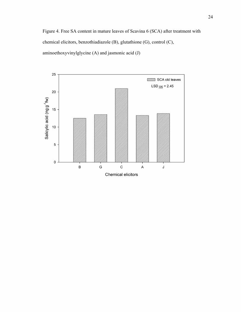
Salicylic acid contents of SCA flush leaves did not change after elicitor
application. Only free salicylic acid content of SCA old leaves decreased upon
application of all four chemical elicitors (Figure 4).
Free SA and SAG content of AME flush leaves did not change upon chemical
elicitor application.
Free salicylic acid content of AME fully developed leaves decreased upon
application of BTH, glutathione and jasmonic acid. SAG content of AME mature leaves
decreased upon application of BTH only.
Free SA content of IMC flush leaves decreased upon application of BTH only.
SAG content of IMC flush leaves decreased upon application of BTH and glutathione.
Free salicylic acid content of IMC fully developed leaves increased upon application of
AVG. SAG content of IMC mature leaves did not change upon application of elicitors.

23
Table 1. Leaf salicylic acid (SA) and salicylic acid glycoside (SAG) of four greenhouse-grown cacao genotypes IMC, AME, TSH and SCA. Leaf Age Cultivar SA (ng·g-1 fw) SAG (ng·g-1 fw) Total (SA+SAG) (ng·g-1 fw)
IMC 23.1a* 130.7ab** 153.8 AME 16.8b 99.7b 116.5 TSH 16.5b 157.5a 174.0 Flush
SCA 16.1b 167.2a 183.3
IMC 14.9b*** 81.6b**** 96.5 AME 16.3b 81.4b 97.7 TSH 14.0b 82.4b 96.4 Mature
SCA 21a 105.6a 126.6 Mean separation within columns and by leaf age.
* LSD.05 = 4.9 **LSD.05 = 48.2 *** LSD.05 = 4.0 ****LSD.05 = 19.8
24
Figure 4. Free SA content in mature leaves of Scavina 6 (SCA) after treatment with
chemical elicitors, benzothiadiazole (B), glutathione (G), control (C),
aminoethoxyvinylglycine (A) and jasmonic acid (J)

25
2.1.3.5. Levels of SA and SAG in leaves of field-grown trees after pathogen
inoculation
SA and SAG contents did not differ between the two cultivars tested (SCA and
CCO). SA and SAG contents decreased with leaf age; SA and SAG contents of L1 were
higher than L2, which in turn were higher than L3. Basidiospore inoculation did not alter
SA and SAG contents in either cultivar (Figure 5; Table 2). All the results from this
section about the levels of SA and SAG in leaves of field-grown trees after pathogen
inoculation are summarized in Table 2.

26
Figure 5. Salicylic acid levels in young leaves
Different letters indicate significant difference among leaf pairs (IL1, IL2, IL3, HL1,
HL2 and HL3) independent of genotype using F significance test at p 0.05.

27
Table 2. Leaf (L1) SA contents of field-grown trees inoculated (I) with basidiospores of M. perniciosa or uninoculated controls (C) and adjacent older leaf pairs L2 and L3* Tissue Cultivar Trt SA (ng·g-1 dw) SAG (ng·g-1 dw) SA+SAG (ng·g-1 dw)
I 217.4 1000.6 1218 SCA C 202.2 885.2 1087.4 I 204.2 720.3 924.5 Flush
CCO C 157.2 517.0 674.2 I 355.6 1711.8 2067.4 SCA C 311.9 1401.3 1713.2 I 313.6 1099 1412.6 L1
CCO C 232.6 743 975.6 I 232.7 921.5 1154.2 SCA C 223 812.6 1035.6 I 227.8 785.9 1013.7
L2 CCO C 165.7 499.3 665
I 64.2 368.6 432.8 SCA C 71.8 441.5 513.3 I 71.3 276.1 347.4
L3 CCO C 73.2 308.6 381.8
* No significant statistical differences were observed using the F test at P.05.

28
2.1.3.6. Changes in cacao procyanidin content throughout leaf development
The distribution of procyanidin oligomers, from monomer to decamer, found in
cacao leaves combining all four varieties, is shown in Figure 6. The average monomer
fraction including all leaf sizes was 21 mg·g-1 dw, about 30 % of total procyanidins. The
following oligomers averaged respectively, from dimer to decamer, 16, 12, 11, 10, 8, 6,
4, 3, and 2 % of the total procyanidin content.
When comparing procyanidin contents of old versus flush leaves, the latter have
significantly higher levels of both monomers and oligomers from dimer to decamer
(Figure 7).
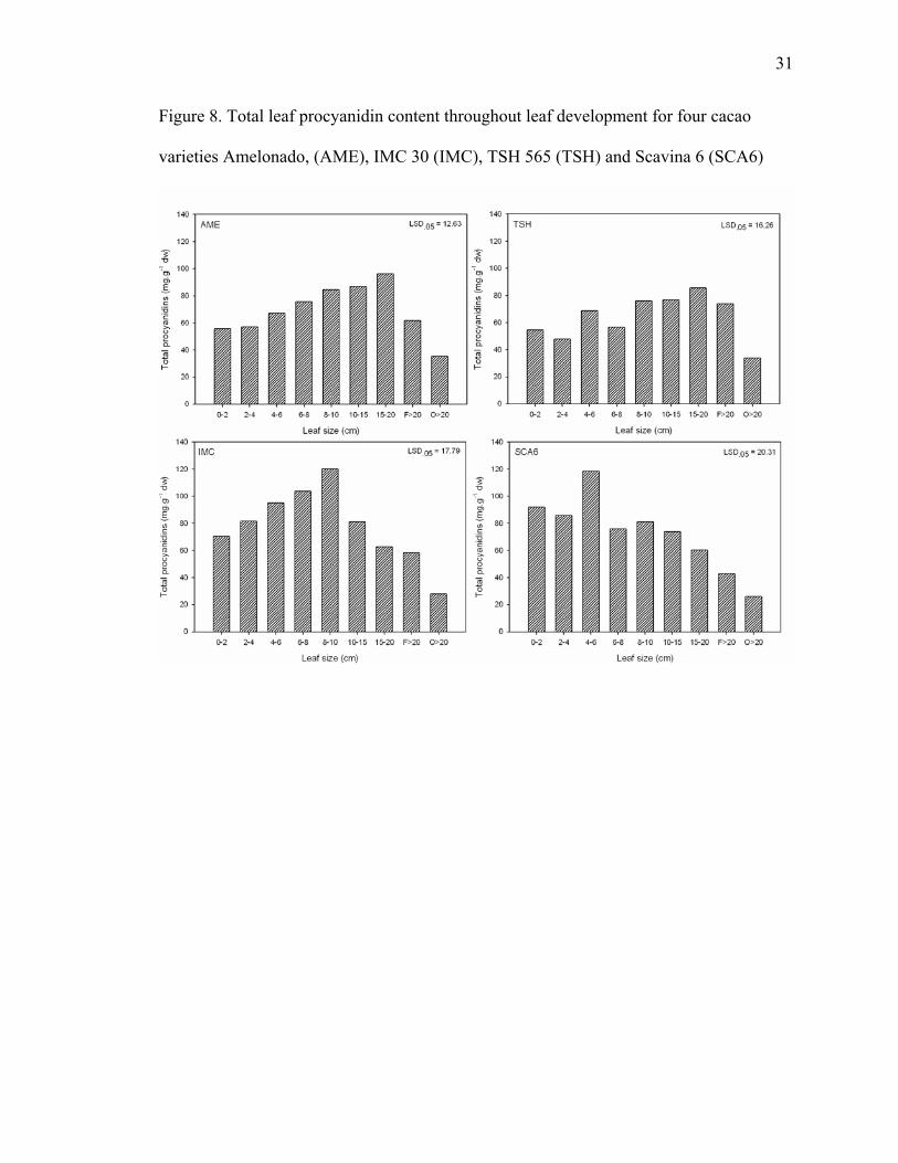
Total procyanidins of each leaf size for each cultivar is reported in Figure 8. AME
leaves < 2 cm contained 55.7 mg·g-1 dw and there was a linear increase to the maximum
of 96.1 mg·g-1 dw in leaves 15 to 20 cm long. Beyond that point procyanidin content
started to decrease and reached its lowest level in the mature leaves, which contained
35.3 mg·g-1 dw. Similarly, total procyanidin content of TSH leaves < 2 cm was 54.5
mg·g-1 dw and reached a maximum in leaves 15 to 20 cm long with 85.4 mg·g-1 dw. TSH
mature green leaves had the lowest level of total procyanidins with 33.7 mg·g-1 dw. IMC
leaves < 2 cm contained 70.4 mg·g-1 dw total procyanidins, which peaked at 120 mg·g-1
dw in leaves 8 to 10 cm long. As IMC leaves developed further, the procyanidin levels
decreased, reaching 27.7 mg·g-1 dw when leaves were fully mature.

29
Figure 6. Procyanidin content of cacao leaves

30
Figure 7. Comparison of total procyanidin content in flush and old leaves of cacao
31
Figure 8. Total leaf procyanidin content throughout leaf development for four cacao
varieties Amelonado, (AME), IMC 30 (IMC), TSH 565 (TSH) and Scavina 6 (SCA6)
32
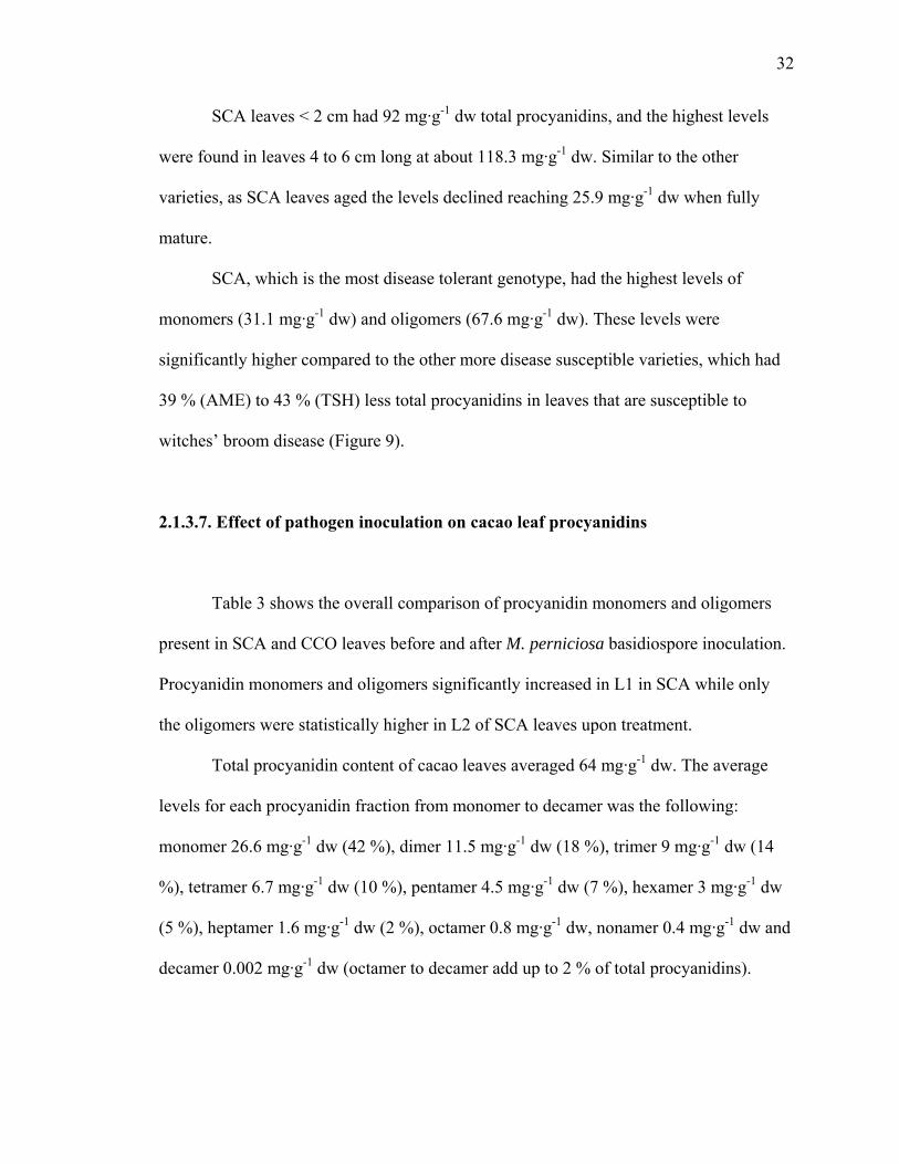
SCA leaves < 2 cm had 92 mg·g-1 dw total procyanidins, and the highest levels
were found in leaves 4 to 6 cm long at about 118.3 mg·g-1 dw. Similar to the other
varieties, as SCA leaves aged the levels declined reaching 25.9 mg·g-1 dw when fully
mature.
SCA, which is the most disease tolerant genotype, had the highest levels of
monomers (31.1 mg·g-1 dw) and oligomers (67.6 mg·g-1 dw). These levels were
significantly higher compared to the other more disease susceptible varieties, which had
39 % (AME) to 43 % (TSH) less total procyanidins in leaves that are susceptible to
witches’ broom disease (Figure 9).
2.1.3.7. Effect of pathogen inoculation on cacao leaf procyanidins
Table 3 shows the overall comparison of procyanidin monomers and oligomers
present in SCA and CCO leaves before and after M. perniciosa basidiospore inoculation.
Procyanidin monomers and oligomers significantly increased in L1 in SCA while only
the oligomers were statistically higher in L2 of SCA leaves upon treatment.
Total procyanidin content of cacao leaves averaged 64 mg·g-1 dw. The average
levels for each procyanidin fraction from monomer to decamer was the following:
monomer 26.6 mg·g-1 dw (42 %), dimer 11.5 mg·g-1 dw (18 %), trimer 9 mg·g-1 dw (14
%), tetramer 6.7 mg·g-1 dw (10 %), pentamer 4.5 mg·g-1 dw (7 %), hexamer 3 mg·g-1 dw
(5 %), heptamer 1.6 mg·g-1 dw (2 %), octamer 0.8 mg·g-1 dw, nonamer 0.4 mg·g-1 dw and
decamer 0.002 mg·g-1 dw (octamer to decamer add up to 2 % of total procyanidins).

33
Figure 9. Procyanidin content of cacao flush leaves susceptible to witches’
broom disease (< 6 cm) for four cacao varieties: AME, IMC, TSH and SCA

34
Table 3. Effect of inoculation with basidiospores of the pathogen Moniliophthora
perniciosa on the procyanidin content (mg·g-1 dw) of flush leaves of field-grown disease
resistant (SCA) and disease susceptible (CCO) cacao trees.
Cultivar SCA CCO Treatment Control Inoculated Control Inoculated Leaf pair Monomers Oligomers Monomers Oligomers Monomers Oligomers Monomers OligomersL1z 30.33 45.10 65.61 89.91 18.01 25.41 43.80 51.43 L2 17.17 28.50 39.03 65.85 13.30 24.20 38.65 45.36 L3 13.89 19.35 14.32 19.63 12.44 16.66 12.83 17.42
z inoculated leaf pair. LSD.05 = 30.06

35
F- test results presented in Table 4 indicate significant differences in the total
procyanidin content for basidospore treatment (Pr > F = 0.0036) and leaf age (Pr > F =
0.0021), as well as for comparisons with the monomer and oligomer fractions separately.
Although there is a trend for differences between cultivars when comparing the total
procyanidin content (Pr > F = 0.0930), the only statistically significant difference (Pr > F
= 0.0467) between cultivars was found when comparing the oligomer procyanidin
fraction. Procyanidin oligomers, which represented 58 % of total procyanidins in the
analyzed leaves, were statistically higher in SCA when compared to CCO.
Leaves from trees of both varieties inoculated with basidiospores had an average
of 84 mg·g-1 dw total procyanidins, which was significantly higher than the 44.1 mg·g-1
dw total procyanidin content of leaves from control trees (treatment significance
observed in Table 4).
Table 4 indicates a significant difference for the basidiospore treatment, but only
SCA inoculated leaves had higher total procyanidins than SCA control leaves. CCO
inoculated leaves did not differ statistically from CCO control leaves.
Taken together, all L1 samples from both cultivars, whether inoculated or not, had
on average 92.4 mg·g-1 dw procyanidin, L2 leaves had 68 mg·g-1 dw and did not differ
statistically from L1. In contrast, L3 leaves averaged 31.6 mg·g-1 dw procyanindins,
which was significantly lower than the two younger pairs.
Independent of cultivar, the first leaf pair of control branches had levels similar
(not statistically different) to the other two pairs; whereas in leaves from inoculated
branches, the inoculated pair (L1) and the adjacent leaf pair (L2) had a total procyanidin
content higher than the third leaf pair (L3). The inoculated leaf pair (L1) had statistically

36
higher total procyanidins than the L1 control, each with 125.4 mg·g-1 dw and 59.4 mg·g-1
dw respectively. Leaf pair 2 on the inoculated branch had statistically higher total
procyanidins than the L2 control, each with 94.4 mg·g-1 dw and 41.6 mg·g-1 dw
respectively. Leaf pair 3 on the inoculated branch had statistically similar total
procyanidins as the L3 control, each with 32.1 mg·g-1 dw and 31.2 mg·g-1 dw
respectively.
2.1.3.8. PCA analysis comparing caffeine, theobromine, (+)-catechin, (-)-epicatechin,
salicylic acid, and salicylic acid glycoside levels of flush leaves treated with elicitors
The variables 1 to 6 were theobromine, (+)-catechin, caffeine, (-)-epicatechin,
salicylic acid and salicylic acid glycoside. PCA 1 represented 60.61 % of total variation
and it was mainly explained by the contributions from variables 1, 2, 3, 4, 6, while, PCA
2 represented 16.18 % of total variation and its main contribution came from variable 5.
PCA 3 represented 11.59 % of total variation and the majority of the variation came from
variable 6. Plotting PCA 1 with PCA 2, PCA 1 with PCA 3 or PCA 2 with PCA 3
separated the samples in clusters by genotype. In figure 10, where PCA 1 was plotted
with PCA 2, AME and SCA clusters are grouped closer based on influence of the SA
variable of the PCA 2. In figure 11, which shows a PCA 1 versus PCA 3 plot, the SCA
cluster is separated from the AME cluster mostly based on the SAG contribution. PCA 2
and PCA 3 combined explain less then 18 % of the variation among the variables (Figure
12).

37
Table 4. Analysis of variance by F-test comparing leaf procyanidin contents under different treatments
Monomers F Value Pr > F Oligomers F Value Pr > F Total Proc. F Value Pr > F Model 2.81 0.0157 Model 3.12 0.0092 Model 2.99 0.0115
Replicates 0.27 0.7636 Replicates 0.39 0.6791 Replicates 0.34 0.7142 Treatment 11.27 0.0029 Treatment 9.78 0.0049 Treatment 10.60 0.0036 Leaf age 7.72 0.0029 Leaf age 8.58 0.0018 Leaf age 8.29 0.0021 Cultivar 1.62 0.2169 Cultivar 4.44 0.0467 Cultivar 3.08 0.0930 Trt*age 2.83 0.0808 trt*age 2.39 0.1146 trt*age 2.62 0.0953 Trt*cult 0.03 0.8543 trt*cult 0.69 0.4167 trt*cult 0.30 0.5871 Age*cult 0.88 0.4274 age*cult 1.25 0.3057 age*cult 1.07 0.3600
trt*age*cult 0.13 0.8804 Trt*age*cult 0.19 0.8292 trt*age*cult 0.11 0.8921 Numbers is bold are statistically significant.

38
Figure 10. PCA analysis (PCA 1 x PCA 2) of six metabolites (variables) present in four
cacao genotypes (AME, IMC, SCA and TSH) subjected to treatments with chemical
elicitors (A – aminoethoxyvinylglycine, B – benzothiadiazole, C – control, G –
gluthatione, J – jasmonic acid)

39
Figure 11. PCA analysis (PCA 1 x PCA 3) of six metabolites (variables) present in four
cacao genotypes (AME, IMC, SCA and TSH) subjected to treatments with chemical
elicitors (A – aminoethoxyvinylglycine, B – benzothiadiazole, C – control, G –
gluthatione, J – jasmonic acid)
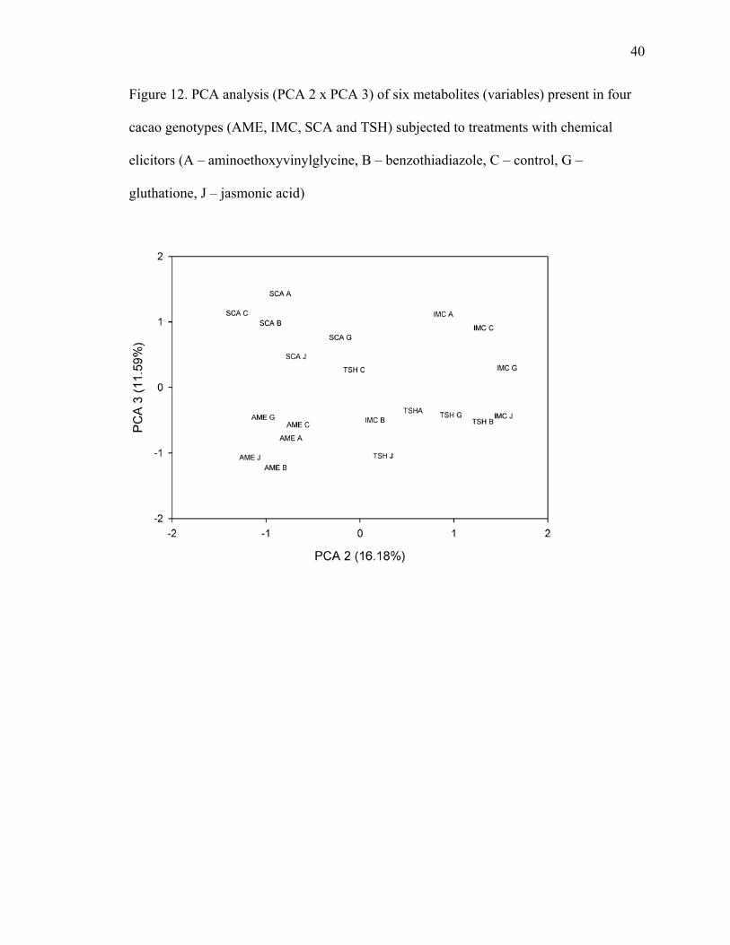
40
Figure 12. PCA analysis (PCA 2 x PCA 3) of six metabolites (variables) present in four
cacao genotypes (AME, IMC, SCA and TSH) subjected to treatments with chemical
elicitors (A – aminoethoxyvinylglycine, B – benzothiadiazole, C – control, G –
gluthatione, J – jasmonic acid)

41
2.1.4. DISCUSSION
2.1.4.1. Effect of chemical elicitors on purine alkaloids and procyanidin monomers
In order to measure flavonoids several spectrophotometric methods have been
employed including, Folin-Ciocalteu, acid/butanol assay, AlCl3 assay, TEAC, FRAP.
These are indirect measurements based upon the antioxidant ability of polyphenols
(Luximon-Ramma et al., 2002). Direct measurements can be achieved using HPLC.
Purine alkaloids and procyanidins have been analyzed simultaneously using reverse
phase (RP) HPLC (Tokusoglu and Unal, 2002). Following the same direct approach ,
caffeine, theobromine, (-)-epicatechin and (+)-catechin were analyzed concurrently by
RP-HPLC using a UV detector.
Theobromine levels (0.66 mg·g-1 dw) were found higher than caffeine levels (0.42
mg·g-1 dw) in cacao flush leaves. There were no differences in caffeine levels of flush
leaves among the studied genotypes. Theobromine was higher in leaves of TSH followed
by IMC, AME and SCA. While theobromine was 60 % in AME leaves it was 75 % of
total purine alkaloid in TSH and IMC leaves, but was only 25 % in SCA leaves; thus
caffeine levels were 40 % in AME, 75 % in SCA leaves and 25 % in TSH and IMC.
Caffeine is known to be toxic to M. perniciosa, while theobromine is not (Aneja and
Gianfagna, 2001). The higher proportion of caffeine in SCA leaves could be related to its
higher disease resistance against M. perniciosa when compared to the other genotypes.
However, Hammerstone et al. (1994) observed that caffeine levels are sometimes higher
sometimes lower than theobromine depending on the leaf age. They found that the

42
youngest leaf on a branch had 1.73 mg·g-1 dw and 0.56 mg·g-1 dw of theobromine and
caffeine respectively. With increasing leaf age, theobromine levels decreased but caffeine
levels went up (0.65 mg·g-1 dw caffeine when theobromine is 0.57 mg·g-1 dw) before
decreasing again to 0.28 mg·g-1 dw of caffeine and 0.15 mg·g-1 dw of theobromine in the
oldest leaves. This could suggest that as leaves age, theobromine is converted to caffeine
and then caffeine is later degraded to other compounds. Caffeine could have a defensive
role in the plant when in sufficient concentration. Koyama et al. (2003) observed that
more than 75 % of the purine alkaloids disappeared with leaf development and
determined that purine alkaloid biosynthesis occurs only in young cacao leaves further
suggesting a role in defense against some pathogens and insects, which preferentially
attack these younger tissues. By the time caffeine levels drop, the leaf has acquired other
defensive mechanisms and that is possibly the reason why older leaves are much less
susceptible to disease.
Leal et al. (2007) found differential gene expression of a caffeine synthase gene
and polyphenol biosynthesis related genes upon infection of cacao varieties with different
levels of resistance to witches’ broom disease. Scarpari et al. (2005) observed an increase
in purine alkaloids during witches’ broom development although they did not see changes
in polyphenol levels. Aneja and Gianfagana (2001) also observed an increase in caffeine
in cacao shoots infected with the fungus Moniliophthora perniciosa. Nojosa et al. (2003)
suggested that phenolic compounds are involved in defense mechanisms of cacao against
M. perniciosa because there are differences in the levels of total phenolics between
resistant and susceptible clones in young and mature tissues. In this experiment cacao

43
flush leaves were found to contain 2.33 mg·g-1 dw (-)-epicatechin and 1.15 mg·g-1 dw (+)-
catechin. There was no statistical significant difference between the studied cultivars.
Beno-Moualem et al. (2001) showed induction of (-)-epicatechin synthesis in avocado by
application of cytokinins. With grapes, BTH increased anthocyanin levels and improved
resistance against the fungal pathogen Botrytis cinerea (Iriti et al., 2004). Verica et al.
(2004) observed up-regulation of defense genes in cacao leaves treated with BTH.
During this project, the treatment with elicitors to leaves of greenhouse grown
trees induced accumulation of theobromine and (-)-epicatechin only. BTH induced (-)-
epicatechin in TSH leaves, and theobromine in AME leaves. AVG induced theobromine
in TSH leaves and glutathione induced (-)-epicatechin in SCA leaves.
Cell culture and protoplast have been shown to respond similarly to whole plant
systems when treated with elicitors (Gundlach et al., 1992; Edwards et al., 1991; Robbins
et al., 1991). The detached leaf experiment also allows for more replication, thus is a
controlled system unlike field or whole plant experiments which are subject to other
variables that could interfere in the results. However, the results obtained upon
application of chemical elicitors to cacao flush leaves did not show a clear pattern of
induction across genotypes nor an inductive effect that was compound specific.
Although, a detached leaf assay with cacao has shown positive results in the past
(Aneja and Gianfagna, 2001), for this study the results were variable. A chemical elicitor
treatment to intact leaves while still on the tree may provide results that are more
consistent.

44
2.1.4.2. Effect of chemical elicitors and pathogen inoculation in free SA and SAG of
cacao leaves
Plant response to infection, wounding or other stresses caused either by biotic or
abiotic factors is usually more effective in young, growing tissues. However, the salicylic
acid responses observed upon chemical elicitor application happened mainly in older
leaves. Older leaves are thicker and sturdier compared to the thin and tender young cacao
leaves. They seem to senesce later than the flush leaves, which wilt and start to turn
brownish at the edges much sooner when placed in the chemical elicitor solution after
detachment and cutting. Possibly, for this reason older leaves were able to response to
elicitors more effectively than young flush leaves. Except for young flush leaves of IMC
all other genotypes did not vary in their SA or SAG contents upon application of
chemical elicitors. IMC young leaves treated with BTH had reduced SA and SAG
content, while glutathione treatment induced a reduction in the SAG content only. For the
most part, chemical elicitor treatments reduced the SA contents of mature cacao leaves.
SAG content of TSH mature leaves was reduced upon application of AVG, while free SA
did not vary. Free SA content of SCA fully developed leaves was lower in all treatments,
while SAG did not change. In AME mature leaves, BTH caused a reduction of free SA
and SAG contents, while glutathione and jasmonic acid reduced free SA content of the
treated leaves. Only IMC mature leaves had free SA levels increased when treated with
AVG.
Plant disease resistance involves activation of a series of defense pathways that
try to counterattack the pathogen. However, it may not be desirable for plants to launch

45
defenses that are metabolically costly if they are not effective. Therefore, plants have
regulatory mechanisms to activate particular defenses according to the specific stimuli or
stresses that they are subjected to. Some of these pathways can be opposing, down
regulating each other, while others work in combination where the activation of one
triggers another. For some plants, including cacao, it may not be advantageous to activate
the SA pathway when attacked by necrotrophic pathogens. M. perniciosa is a
hemibiotrophic fungus, with a biotrophic and necrotrophic phase. By avoiding the SA
pathway cacao is preventing cell death. This allows the plant to avoid inducing the phase
transition of the fungus from biotroph to necrotroph, which at the same time may provide
for activation of other more effective defenses. This suggestion is supported by the
negative results observed in induction of SA and SAG levels in young leaves followed by
application with elicitors. While, SA and SAG levels increase in parallel after TMV
infection in tobacco (Hennig et al., 1993), young cacao leaves maintained the same SA
and SAG levels of the controls after M. perniciosa basidiospore inoculation, confirming
that the plant’s response to M. perniciosa infection is likely induced by a pathway other
than SA (Figure 5).
Although there was no increase in SA in the inoculated leaves, the molecule could
affect nearby tissues since it is mobile in the plant. This was not the case, as there was no
increase in free SA or SAG levels found in non-inoculated leaves on the same flush as the
inoculated ones (Figure 5).
Endogenous SA and SAG levels of both tested cultivars including the more
disease tolerant SCA and the more susceptible CCO, did not differ statistically (Figure 5),
reinforcing that SA levels are not indicative of resistance to M. perniciosa in cacao.

46
Although M. perniciosa preferentially infects the young meristematic tissue, both
varieties of cacao had the highest levels of free SA and SAG in the youngest tissues
independent of inoculation (Figure 5).
2.1.4.3. Leaf size experiment
Cacao flush leaves are thin and tender, and can be pale green or pink, and only
when fully expanded become dark green, thicker and sturdier. Among the varieties
studied, IMC 30 (IMC) was the only one with light green flush leaves, while the others,
Scavina 6 (SCA), TSH 565 (TSH) and Amelonado (AME) had pink flush leaves.
The distribution of procyanidin oligomers, from monomer to decamer, found in
cacao leaves combining all four varieties, averaged respectively, from monomer to
decamer, 30, 16, 12, 11, 10, 8, 6, 4, 3, and 2 % of the total procyanidin content. The
monomer fraction averaged 21 mg·g-1 dw. The levels of procyanidin monomers found in
the analyzed leaves was relatively similar to that found in cocoa beans as reported by
Kim and Keeney (1984) who found (-)-epicatechin ranging from 22 to 43 mg·g-1 dw in
defatted unfermented beans, and determined that the monomer made up about 35 % of
total bean procyanidins.
Fully developed leaves had significantly lower procyanidin contents than young
leaves (Figure 7). These results followed the same trend observed by Osman et al. (2004)
who measured only the monomer and found (-)-epicatechin content higher in the younger
leaves.

47
Morphological and physiological differences between mature and flush leaves are
evident and therefore analysis of cacao leaves in these two groups is obvious. However,
there may be significant differences within flush leaves of different developmental stages.
For that reason, flush leaves were separated into nine categories based on leaf size.
The levels of the very young leaves are intermediary and peak as leaves attain
lengths of 4 to 20 cm; but as the leaves increase in size and develop further, the
procyanidin content reaches its minimum.
Only the very young leaves up to about 5 cm are susceptible to infection by the
fungus Moniliophthora perniciosa, the causal agent of witches’ broom disease (Purdy
and Schimidt, 1996). Of the four cacao varieties used in this study, SCA was the most
disease tolerant variety, whereas AME was the most disease susceptible.
Both induced and constitutively present flavonoids have been associated with
plant resistance to biotic and abiotic stresses (Harborne and Williams, 2000). Andebrhan
et al. (1995) and Brownlee et al. (1990) demonstrated that purified cacao bean
procyanidins and cacao shoot extracts containing procyanidins have antifungal effects
against Moniliophthora perniciosa. Both extracts inhibited spore germination and germ
tube elongation; however there was no effect on the growth of the saprophytic mycelia.
Leaves of SCA, the most disease resistant of the studied genotypes, had
significantly higher levels of monomers and oligomers compared to the other more
disease susceptible genotypes, at a stage when cacao leaves are susceptible to witches’
broom disease (< 6 cm) (Figure 9).
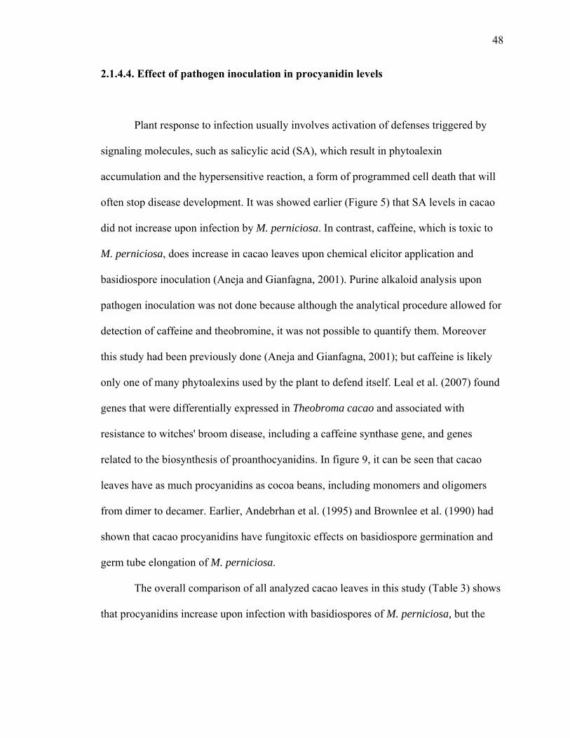
48
2.1.4.4. Effect of pathogen inoculation in procyanidin levels
Plant response to infection usually involves activation of defenses triggered by
signaling molecules, such as salicylic acid (SA), which result in phytoalexin
accumulation and the hypersensitive reaction, a form of programmed cell death that will
often stop disease development. It was showed earlier (Figure 5) that SA levels in cacao
did not increase upon infection by M. perniciosa. In contrast, caffeine, which is toxic to
M. perniciosa, does increase in cacao leaves upon chemical elicitor application and
basidiospore inoculation (Aneja and Gianfagna, 2001). Purine alkaloid analysis upon
pathogen inoculation was not done because although the analytical procedure allowed for
detection of caffeine and theobromine, it was not possible to quantify them. Moreover
this study had been previously done (Aneja and Gianfagna, 2001); but caffeine is likely
only one of many phytoalexins used by the plant to defend itself. Leal et al. (2007) found
genes that were differentially expressed in Theobroma cacao and associated with
resistance to witches' broom disease, including a caffeine synthase gene, and genes
related to the biosynthesis of proanthocyanidins. In figure 9, it can be seen that cacao
leaves have as much procyanidins as cocoa beans, including monomers and oligomers
from dimer to decamer. Earlier, Andebrhan et al. (1995) and Brownlee et al. (1990) had
shown that cacao procyanidins have fungitoxic effects on basidiospore germination and
germ tube elongation of M. perniciosa.
The overall comparison of all analyzed cacao leaves in this study (Table 3) shows
that procyanidins increase upon infection with basidiospores of M. perniciosa, but the
49
increase was only statistically significant for the more disease resistant genotype Scavina
6.
Even though only leaf pair one (L1) had been inoculated with basidiospores, the
next adjacent leaf pair (L2) also showed elevated levels of procyanidins indicating a
systemic induction of procyanidins. The systemic increase of procyanidins occurred
within 48 hours after inoculation with basidiospores, it was measured in the adjacent leaf
pair (L2) only, and the systemic signal did not reach the next leaf pair (L3).
The results indicate enhanced procyanidin leaf content as a mechanism of defense
used by T. cacao against infection by M. perniciosa. Moreover, Scavina 6, the more
disease tolerant genotype had constitutively higher levels of procyanidin oligomers than
CCO, which is more disease susceptible. These findings are consistent with the results of
Leal et al. (2007), who observed activation of proanthocyanidins biosynthesis genes
differentially expressed in genotypes with different levels of resistance to witches’ broom
disease. Selection of genotypes with high constitutive and inducible levels of
procyanidins, and the development of chemical treatments to increase procyanidin
content prior to infection, may reduce the incidence and severity of this disease in cacao.

50
2.1.5. CONCLUSIONS
Phytoalexins are one of several plant defenses against pathogens. Comparing
genotypes with different levels of disease resistance based on levels of single compounds
may not reflect their full defensive capacity because the effective response may require
concurrent production of several compounds. The measurements of caffeine,
theobromine, (-)-epicatechin and (+)-catechin, salicylic acid, and salicylic acid glucoside
levels of cacao flush leaves varied within varieties before and after treatment with
chemical elicitors. When analyzed separately it was impossible to determine whether any
compound or elicitor treatment had a predominant role in disease resistance. However,
the PCA analysis comparing the four studied cacao genotypes with different levels of
disease resistance, each treated with four chemical elicitors, based on the content of six
measured compounds, showed a trend in which the samples were clustered by genotype
rather than by elicitor treatment. This clustering demonstrated a stronger genotype
influence rather than elicitor treatment on the chemical profile content of cacao leaves.
This example of chemical profiling allowed for the separation of genotypes with different
levels of resistance based on their overall ability to produce phytoalexins.
In general, chemical elicitor or leaf inoculation with M. perniciosa basidiospores
did not alter leaf salicylic acid or salicylic acid glucoside contents of young flush leaves.
For some plants, including cacao, it may not be advantageous to activate the SA pathway
when attacked by necrotrophic pathogens. M. perniciosa is a hemibiotrophic fungus, with
a biotrophic and necrotrophic phase. By avoiding the SA pathway cacao is preventing
cell death. This allows the plant to avoid inducing the phase transition of the fungus from
51
biotroph to necrotroph, which at the same time may provide for activation of other more
effective defenses (Chaves and Gianfagna, 2006).
Cacao bean procyanidins have recently received much attention for their potential
to improve human health (Schroeter et al., 2006; Selmi et al., 2006). However, the
presence of these compounds in other plant parts as well as the role of these procyanidin
oligomers in plant health has not been investigated. Flavan-3-ol monomers and oligomers
from dimers to decamers were quantitatively determined by HPLC-FLD in leaves,
throughout development, of four Theobroma cacao varieties. The levels of the very
young leaves were intermediary and peaked as leaves attained lengths of 4 to 20
centimeters. As the leaves increased in size and developed further, the procyanidin
content reached its minimum. Cacao flush leaves had higher procyanidin content than
mature leaves.
Plant disease resistance involves activation of a series of defense pathways to
counterattack the pathogen. Understanding the defense mechanisms by which the plant
responds to pathogen attack will elucidate molecular markers useful for selecting
genotypes with higher disease resistance attributes, and/or suggest alternative disease
control strategies.
Witches’ broom disease tolerant cultivar Scavina 6 had the highest levels of
procyanidins when compared to Amelonado, TSH 565, and IMC 30, more disease
susceptible genotypes, at a stage when leaves are susceptible to infection. Moreover,
leaves of SCA 6 responded to M. perniciosa basidiospore infection by inducing
accumulation of procyanidins in both the infected and adjacent uninfected leaves of

52
actively growing shoots, demonstrating an inductive response effect both locally and
systemically.
Combined evidence for activation of procyanidin gene expression (Leal et al.,
2007), antifungal effects of procyanidins against the witches’ broom pathogen
(Andebrhan et al., 1995; Brownlee et al., 1990), higher endogenous levels of
procyanidins in leaves of a disease resistant genotype, including a peak in procyanidin
levels during leaf development at a stage when leaves are susceptible to witches’ broom
disease, and the local and systemic increase in procyanidins in cacao leaves of a disease
resistant genotype upon infection with basidiospores indicate a role for procyanidins in
defense against Moniliophthora perniciosa. Therefore, procyanidin content of flush
leaves is a potential marker for selecting for disease tolerance in cacao (Chaves and
Gianfagna, 2007).

53
2.2. ENDOPHYTES AND THEIR METABOLITES
2.2.1. INTRODUCTION
Endophytes are microorganisms that grow within plant tissues without causing
disease or negative impact on plant growth. What are the effects of the presence of the
endophytes on the plant? Do they protect against pathogens? There are examples of
endophytes associated with grasses that protect them against herbivory by producing
alkaloids (White and Morgan-Jones, 1996). Fine fescues inoculated with endophytes
suppressed dollar spot disease caused by Sclerotinia homeocarpa (Clarke et al., 2006). In
some tree species, it has also been shown that insects avoid leaves with high endophytic
incidence (Wilson and Carroll, 1997). In cacao, endophyte inoculation renders plants
more resistant against Phytophthora sp. (Arnold et al., 2003). As a result, endophytes
emerge as potential biological agents of disease control. In order to better understand and
fully utilize endophytes for disease control it is important to establish the mechanisms by
which they assist plants against pathogens.
Biological control using endophytes may occur through one or a combination of
the following processes: antibiosis, competition, induction of defenses, and parasitism.
Along with the ability to synthesize antimicrobial compounds, some endophytes produce
antimicrobial compounds that are also mammalian toxins. While these toxins may be
important in protecting the plants against pathogens, they are also a serious threat to
human health. Among the most toxic and commonly present compounds in foods and
feedstuffs are aflatoxins and ochratoxins. In general, contamination of plant products

54
such as grains, coffee berries or cocoa beans occurs post-harvest. However, the
opportunistic organisms that infect crops post-harvest could be present endophytically in
the plant during its development. Endophytes in some plant species can be transferred
vertically to the next generation through seeds or horizontally to other plants by both
spore and mycelium inoculation.
Before any endophytes can be seriously considered as biocontrol agents it must be
first determined whether they can produce any beneficial compounds or toxins. A
collection of endophytes found in coffee leaves (Coffea arabica), which consisted largely
of Aspergillus and Penicillium species (Eurotiales: Trichocomaceae), was used to
determine if the species and strains found as endophytes tended to be toxin producers.
Moreover, the possibility exists that parasitoids used for biocontrol of coffee berry borer
(CBB) and cocoa pod borer could carry spores of toxigenic fungi on their cuticle and
serve as a source of fungal infection. To study this interaction, fungi that were isolated
from a parasitoid wasp of the CBB were tested for ochratoxin production.
It has been proposed that some plant pathogens may have originated from
endophytic strains that acquired pathogenicity factors and became capable of causing
disease in the host (Aime and Phillips-Mora, 2005; Evans et al., 2003). At times, the
separation between an endophyte, a pathogen, and an opportunistic organism, which is
commonly a saprophyte (but given the right conducive environmental conditions can
colonize plants under stress and cause plant death), serves as a means of classification
because the difference from one role to another may be only temporal and circumstantial.
In order to study the interactions between endophytes and plant pathogenic fungi
and to identify a potential biocontrol organism, dual culture plate assays, covered culture

55
assays, and bioassay tests for volatile antimicrobials were used with the aim of
characterizing in vitro endophyte-pathogen interactions and possible antagonism. For the
identification of mycotoxins and other metabolites released in culture, fungi were grown
in different media, and after solvent extraction, samples were analyzed by HPLC and
GC-MS.

56
2.2.2. MYCOTOXINS PRODUCED BY COFFEE ENDOPHYTES
Penicillium and Aspergillus species cause molding and rotting of grains and beans
and are known to produce a diverse range of secondary metabolites along with toxins
such as aflatoxins and ochratoxins (OT) (Figure 13). Ochratoxin A (OTA, 7-carboxy-5-
cloro-8-hydroxy-3,4-dihydro-3R-methyliscoumarin-7-L-β-phenylalanine), one of the
most widespread mycotoxins, is found in a number of processed foods including coffee
and cocoa products (Bonhevi, 2004). Other compounds with similar chemical structure
have been identified. One such compound, ochratoxin B (OTB, 7-carboxy-8-hydroxy-
3,4-dihydro-3R-methylisocoumarin-7-L- β-phenylalanine), is a derivative of OTA that
appears to be involved in the biosynthetic pathway of OTA as a precursor and/or
metabolite (Harris and Mantle, 2001). Although OTB is not as toxic as OTA, both had
significant oral LD50 in day-old chicks of 54 mg·kg-1 and 3.3 mg·kg-1, respectively (Cole
and Cox, 1981).
Generally, crop contamination occurs post-harvest and the fungal infestation and
OT levels vary with the drying process of the harvested plant material (Taniwaki et al.,
2003). In order to avoid post harvest contamination, some important general protective
measures include maintaining low moisture content, low temperatures, and insect-free
conditions are recommended. Counter measures include quick air-drying or the use of
other aeration systems to remove moisture and heat.
Although air, soil, and machinery are the major sources of inoculum,
contamination can occur through other means. Penicillium spp. are common naturally
occurring epiphytes on cacao (Hoopen et al., 2003). Cacao pods wounded by, machete, or

57
predators such as rats and squirrels or by cocoa pod borers are usually found highly
contaminated with ochratoxigenic fungal species (Bastide et al., 2006). Endophytic
fungal associations have also been shown to be the source of toxigens (Bacon et al.,
2001). Similar problems occur in coffee plantations where coffee berries damaged by the
insect coffee berry borer (CBB) become contaminated with toxigenic fungi (Vega and
Mercadier, 1998).
Coffee and cacao while genetically unrelated are both tropical understory trees
that grow in similar environments and both produce caffeine. Some endophytes are found
in different tree species growing in the same environment (Arnold et al., 2003). Studies
have shown that caffeine, present in both coffee and cacao, can inhibit the production of
OT (Buchanan et al., 1981; Hasan, 1999). Caffeine inhibits toxin synthesis by restricting
the uptake of carbohydrates, which are ultimately used for toxin synthesis (Buchanan and
Lewis, 1984). Decaffeinated coffee beans have higher levels of toxins when compared to
regular beans, presumably because the higher caffeine content reduces the growth of
toxigenic contamination post-harvest (Soliman, 2002).
Since the endophytes from both coffee and cacao may be candidates for
biocontrol of diseases and pests, the objectives of the following analyses were to screen a
collection of fungi isolated as endophytes from (or associated with) coffee for their
ability to produce mycotoxins, as well as to determine the effect of the culture media and
caffeine on their ability to produce toxins.

58
Figure 13. Ochratoxins A and B
NH
OHOOC OH
Cl
O
O
CH3
HH
NH
OHOOC OH
O
O
CH3
HH
OTA OTB

59
2.2.2.1. MATERIALS AND METHODS
2.2.2.1.1. Fungal isolates
50 isolates comprising Penicillium spp. and Aspergillus spp. isolated from coffee
as endophytes, as well as fungi found associated with the coffee pathogen coffee berry
borer (Hypothenemus hampei) and a wasp (Prorops nasuta) parasitic to this pathogen,
were screened for the production of ochratoxins. An Aspergillus ochraceus isolate from
Dr. James F. White Jr. laboratory collection at Rutgers University, was used as a positive
control for OT.
2.2.2.1.2. Nutrient media
Isolates were grown in YES (2 % yeast extract and 20 % sucrose), YMA (yeast
malt agar, Sigma-Aldrich), and potato dextrose agar (PDA, Difco).
2.2.2.1.3. Ochratoxin analysis
Ochratoxins A and B were measured using the method described by Bragulat et
al. (2001). Three plugs (0.5 mm diameter x 0.5 mm length) of approximately 200 mg
containing media and fungal mycelia were sampled at different time intervals after
inoculation. Plugs were extracted with 0.8 mL methanol:formic acid (25:1) for one hour
at 25oC, then filtered through 13 mm nylon membrane filters. 10 uL of the filtrate were

60
injected in an HPLC equipped with a C18 reverse phase column (15 cm length x 4.6 mm
i.d.; pore size 5 um; 3/8 inches outside diameter). The mobile phase was composed of
acetonitrile:water:acetic acid (57:41:2), the flow rate was 0.8 mL·min-1, and the
fluorescence detector was set at excitation wavelength of 330 nm and emission
wavelength of 460 nm.
2.2.2.1.4. Aflatoxin analysis
Aspergillus oryzae NRRL 35191 isolated from coffee as an endophyte was
analyzed for the production of aflatoxins using an AgraStrip aflatoxin test (Romer Labs,
Union, MO), an immunochromatographic assay based on an inhibition immunoassay
format with a detection limit of 4 ppb.
2.2.2.2. RESULTS AND DISCUSSION
Two Aspergillus westerdijkiae were isolated from coffee berry borer, a major pest
in coffee plantations, and from a wasp, used as biological control agent against CBB
(Vega and Mercadier, 1998); both isolates were found to produce ochratoxins.
Ochratoxin A is known to have antiinsect activity (Wicklow et al., 1996). This
attribute is likely an advantage favoring selection of ochratoxigenic fungi associated with
plants challenged by insect pests, and could be beneficial to the plant if it affects the
insect pest. However, the ochratoxigenic fungus A. westerdijkiae was found associated
with both the insect pathogen and its parasite. Therefore, caution should be exercised to

61
avoid spreading the toxigenic fungus or introducing it along with the biological control
agent.
OT production has been shown to be inhibited by caffeine (Buchanan et al., 1981;
Hasan, 1999). However, some isolates show the ability to degrade caffeine and produce
higher levels of OTA when caffeine is added to the culture media (Tsubouchi et al.,
1985). Similarly, OT production by A. westerdijkiae increased more then ten times when
the fungus was grown in culture supplemented with 0.02 % caffeine. A. ochraceus also
produced more OT in media supplemented with caffeine (Table 5). It is possible that
toxigenic species in continuous contact with a high caffeine environment adapt and
develop the ability to degrade caffeine and stimulate OT production. Adaptation may
occur due to competition for organic nutrients. The bacteria Pseudomonas putida for
example, growing in soil under coffee plants has developed mechanisms to degrade
caffeine released by the plants (leaves, fruits and litter). While some microorganisms
degrade caffeine, others, including A. fumigatus, A. japonicus, and A. niger, have also
been reported to degrade ochratoxin A to less toxic products (Varga et al., 2000).
Buchanan et al. (1981) observed a 98 % inhibition of OTA production when 0.3 %
caffeine was present in the growing media. Tsubouchi et al. (1985) reported a strain of A.
ochraceus with decreased OTA production in the presence of caffeine, while a different
strain increased OTA production when grown under the same conditions. Suarez-Quiroz
et al. (2004) observed a peak in OTA production by A. ochraceus in a nutrient media with
0.1 % caffeine. After peaking, OTA levels dropped with increasing caffeine levels.

62
Table 5. Concentration of OTs (μg.g-1) produced by Aspergillus spp. (4 DAI) on media with and without caffeine
Fungi A. ochraceus A. westerdijkiae Media Caffeine OTA OTB OTA OTB
0% 10.96 (± 3.3) 3.23 (± 0.2) 0.25 (± 0.02) 0.31 (± 0.08) 0.02% 1.42 (± 0.3) 1.78 (± 0.2) 0.15 (± 0.06) 0.53 (± 0.03) PDA
1% 0.52 (± 0.3) 6.83 (± 2.5) 0 0 0% 4.04 (± 1.2) 3.55 (± 0.8) 0.26 (± 0.07) 0.27 (± 0.05)
0.02% 32.83 (± 1.7) 18.99 (± 10.7) 3.40 (± 0.5) 3.22 (± 0.3) YES 1% 8.13 (± 4.1) 63.64 (± 7.2) 0.22 (± 0.01) 0

63
During the current screening for ochratoxigenic fungi, 14 % of the tested isolates
(Table 6) were found to be toxigenic. Taniwaki et al. (2003) sampled a number of
Aspergillus isolates from coffee beans and of them, 75 % of the A. ochraceus, 3 % of the
A. niger, and 77 % of the A. carbonarius isolates produced OTA.
One of the isolates identified to the species level, A. oryzae, is very similar to the
aflatoxigenic fungus A. flavus. Although both species have the genes predicted to be
involved in the aflatoxin synthetic pathway, they are only expressed in A. flavus (Keller
et al., 2005). The analysis of culture filtrates of A. oryzae confirmed the absence of both
OT and aflatoxin sustaining the species identity.
The A. ochraceus and A. westerdjikiae fungal isolates were inoculated in YMA,
PDA, and YES to evaluate the effect of the media on ochratoxin production. Although
the OTA levels were different in each media, the toxin was present in all three. The same
was true for isolates that did not produce OT; when tested in the same three media, OT
was not present in any of them. Although the chance that these isolates produce OT in
another media cannot be ruled out, it is unlikely that the isolates negative for OT would
produce it in other media or that isolates positive for OT would not produce it in other
media.
A. ochraceus and both A. westerdijkiae isolates that produce OTA also produce
OTB. A. ochraceus produced at least 10 times more OTB in YES media when compared
to PDA (Table 5).
OT measurements were taken at different time intervals after inoculation. Isolates
that did not produce OT after one week of inoculation never produced OT. Among those
that produced OT, the levels increased with time and varied depending on the media. A.

64
ochraceus produced nearly the same amount of OTA at 4 days after inoculation (DAI)
and at 35 DAI when grown in PDA. However when grown in YES and YMA, OT
accumulated over time (Table 7). Accumulation of OTA over time was also seen with A.
westerdijkiae CBB9-1 and A. westerdijkiae W6-1 when grown in YMA and YES media.
When A. westerdijkiae CBB9-1 and A. westerdijkiae W6-1 were grown in PDA, OTA
was not detected at 4 DAI but it was present at 35 DAI.
A. ochraceus under the same conditions can produce 10 times more ochratoxin A
than A. westerdijkiae. OT production increased over time and can be measured as early as
4 DAI in YES and YMA for Aspergillus ochraceus and A. westerdijkiae CBB9-1.

65
Table 6. List of isolates analyzed for OT NRRL Reference # Identification Isolated from Country OT
1 35193 Aspergillus westerdjikiae Prorops nasuta Colombia +2 35197 Aspergillus westerdjikiae Coffee berry borer Colombia +3 32580 Penicillium brevicompactum Coffee USA4 32582 Penicillium brevicompactum Coffee USA5 32574 Penicillium brevicompactum Coffee USA6 32576 Penicillium brevicompactum Coffee USA7 35214 Penicillium brevicompactum Coffee USA8 32579 Penicillium brevicompactum Coffee USA +9 32600 Penicillium brevicompactum Coffee USA10 35599 Penicillium brocae Coffee USA11 35434 Penicillium citrinum Coffee USA12 35438 Penicillium citrinum Coffee USA13 35449 i4-20 Penicillium citrinum Coffee USA14 32592 Penicillium crustosum Coffee Guatemala15 35217 Penicillium crustosum Coffee Colombia16 35466 Penicillium cecidicola Coffee USA17 35451 Penicillium janthinellum Coffee USA18 35363 Penicillium coffea Coffee USA19 35367 Penicillium steckii Coffee USA20 35463 Penicillium steckii Coffee USA21 35448 i4-6 Penicillium citrinum Coffee USA22 35460 Aspergillus sp. Prorops nasuta23 35524 Aspergillus sp. Prorops nasuta24 35436 Aspergillus sp. Prorops nasuta25 35437 Aspergillus sp. Prorops nasuta26 35191 Aspergillus oryzae Coffee27 35185 Penicillium brocae Coffee Colombia28 35171 Penicillium olsonii Coffee USA29 Penicillium olsonii Coffee USA30 35186 Penicillium diversum Coffee USA31 Penicillium olsonii Coffee USA32 35167 Penicillium olsonii Coffee USA33 Penicillium olsonii Coffee USA34 35174 Penicillium olsonii Coffee USA35 Penicillium olsonii Coffee USA36 Penicillium olsonii Coffee USA37 32577 Penicillium olsonii Coffee USA +38 35166 Penicillium olsonii Coffee USA +39 35168 Penicillium olsonii Coffee USA40 35179 H81H2 Aspergillus sp.41 35213 H81P2 Penicillium olsonii42 35175 H81P4 Penicillium olsonii43 32583 Penicillium sclerotiorum Coffee USA44 32581 Penicillium olsonii Coffee USA45 35169 Penicillium olsonii Coffee USA46 35225 I1R22 Penicillium citrinum47 35176 M1S3 Aspergillus tamarii48 35178 Penicillium crustosum Coffee Mexico +49 35183 Penicillium oxalicum Coffee Colombia +50 35184 Penicillium brevicompactum Coffee Colombia

66
Table 7. Ochratoxin A production (OTA μg.g-1 media ± STD dev.) by A. ochraceus and
two A. westerdijkiae isolates from the parasitoid P. nasuta (W6-1) and the coffee berry
borer, Hypothenemus hampei (CBB9-1) at 4 and 35 days after inoculation (DAI)
OTA Media YMA PDA YES
Fungi DAI 4 35 4 35 4 35
A. ochraceus 1.4±0.06 4.6±1.10 5.4±0.66 6.1±1.26 2.5±0.14 26±2.60
A. westerdijkiae W6-1 0 0.7±0.04 0 0.6±0.10 0 1.1±0.35
A. westerdijkiae CBB9-1 0.4±0.10 0.6±0.42 0 0.9±0.04 2.5±0.4 3.5±0.56

67
2.2.2.3. CONCLUSIONS
Among all tested fungal isolates, two A. westerdjikiae were OT producers.
Although they produced less toxin than A. ochraceus, the levels were in the same order of
magnitude. The A. westerdjikiae isolates that produced OTA also produce OTB. It is
clear that media has an effect on OT production because quantitative differences were
observed, although no qualitative difference was seen. OT levels varied according to the
substrate and the presence of caffeine. Nutrient media containing higher levels of sugars
induced higher levels of toxin production. The inhibitory effect of caffeine on toxin
production is possibly related to restriction of carbohydrate uptake, previously suggested
by Buchanan and Lewis (1984). Increased OT production, when intermediate levels of
caffeine were added to the growing media, has also been reported in literature (Suarez-
Quiroz et al., 2005; Tsubouchi et al., 1985), although no explanation has been provided.
Caffeine below toxic levels may trigger toxin production while at higher levels
completely disrupt fungal metabolism. Some toxigenic fungi may be able to break down
caffeine and possibly use the degradation products as carbon and nitrogen sources.

68
2.2.3. Aspergillus oryzae FROM COFFEE, A NON-TOXIGENIC ENDOPHYTE
WITH THE ABILITY TO SYNTHESIZE KOJIC ACID IN CULTURE
Penicillium and Aspergillus are cosmopolitan fungi capable of synthesizing a
multitude of secondary metabolites with a broad range of functions (Cole and
Schweikert, 2003; Keller et al., 2005). Some of these metabolites are known to play a
role in the systems in which these fungi occur, and influence whether they are considered
pathogens, endophytes, or opportunistic organisms. At times, the delineation between
these categories is hazy and may be only temporal or circumstantial. For instance, an
endophyte can grow asymptomatically or cause disease depending on the developmental
stage of a plant or its physiological status (White and Morgan-Jones, 1996). Meanwhile,
opportunistic organisms exploit weakened plants to cause disease. Schulz and Boyle
(2005) suggested that these organisms exist on a continuum from saprophyte to
endophyte to pathogen.
Some A. oryzae strains are known to produce kojic acid (KA; 5-hydroxy-2-
hydroxymethyl-γ-pyrone; C6H6O4), a metabolite with insecticidal, antibacterial and
antifungal properties (Dowd, 1999; Dowd, 2002; Alverson, 2003; Burdock et al., 2001;
US Environmental Protection Agency, 1997). KA is also utilized in cosmetics as a skin
lightening agent. A. oryzae has been used for hundreds of years in the food industry for
the preparation of many Asian foods such as miso, sake and shoyu and is generally
regarded as safe (Bentley, 2006).

69
This study describes the identification of a non-toxigenic Aspergillus oryzae
endophyte isolated from coffee that has the ability to synthesize KA in culture. Moreover,
it has a potential to be inoculated and grow endophytically in cacao seedlings.
2.2.3.1. MATERIALS AND METHODS
2.2.3.1.1. Fungal isolate
Aspergillus oryzae (NRRL 35191) was found as an endophyte from coffee leaves,
see Vega et al. (2006) for endophyte isolation methodology. In order to confirm the
identity of this isolate, ITS and partial large subunit rDNA was amplified and sequenced.
The sequence from NRRL 35191 indicates inclusion in the A. oryzae/A. flavus species
group (Peterson et al., 2001). Because the ID gene of the A. flavus/A. oryzae complex is
insufficiently variable to clearly distinguish the two taxa, the OMT12 locus was also
amplified and sequenced as detailed by Geiser et al. (2000). This analysis was done by
S.W. Peterson, USDA, Peoria, IL, USA.
2.2.3.1.2. KA extraction from fungal culture
Yeast extract sucrose (YES) medium (2 % yeast extract and 20 % sucrose) was
inoculated with a 1x10⁷ spores mL-1 suspension of A. oryzae (NRRL 35191) and placed
in a shaker (VWR analog orbital shaker OS-500) at 100 rpm for 7 days at 25 oC. The
broth was filtered through glass wool and the filtrate was centrifuged at 10,000 x g for
70
five minutes. The pH of the supernatant was adjusted to 3.5 and extracted 3 times with 15
mL ethyl acetate. The organic phase was concentrated in vacuo.
2.2.3.1.3. Seedling inoculation and in planta extraction for KA and caffeine analysis
A cocoa pod from a tree grown at a Rutgers University greenhouse was cracked
open, the seeds cleaned of mucilage, and the seed coat removed. Seeds were immersed in
70 % ethanol for one minute and then submerged in 10 % bleach solution for one hour,
followed by rinsing 3 times with autoclaved water. Seeds were then placed inside a test
tube (25 cm in length x 3 cm in diameter) containing half strength potato dextrose agar
(PDA) with the radicle end up. Three week-old sterile cacao seedlings were inoculated
with 0.5 mL of a 1x10⁷ spores mL-1 A. oryzae suspension placed on the radicle. After 3
weeks, seedlings were surface sterilized by rinsing with tap water, immersed in 70 %
ethanol for one minute and 10 % bleach solution for one hour. The seedlings were then
rinsed three times with autoclaved water, and the stem and roots sections were plated on
PDA to assess for the presence of A. oryzae. Plates were kept at 25 oC. The remaining
plant parts were ground in liquid nitrogen and extracted with 150 mL methanol, stirring
overnight. The methanolic extract was filtered and concentrated in vacuo.

71
2.2.3.1.4. KA and caffeine analysis
The concentrated sample was derivatized with Tri-sil reagent (Pierce, Rockford,
IL) and analyzed by GC-MS (Figure 14). A KA commercial standard (Sigma-Aldrich, St.
Louis, MO) was converted with Tri-sil to its trymethylsylilated derivative and injected in
the GC-MS (Column DB-1; The initial temperature of 70 oC was held for two minutes,
then ramped up to 240 oC at 10 oC min-1, and held for 30 minutes). KA was identified by
matching the retention time of derivatized standard with the samples as well as by
comparison of their mass spectra. Caffeine was identified along with KA in the same
chromatogram by comparison of its mass spectra to the equipment’s library NIST 98.
2.2.3.2. RESULTS AND DISCUSSION
Distinguishing A. oryzae and A. flavus species from one another can be difficult
since both species have the genes predicted to be involved in the aflatoxin synthetic
pathway, but the aflatoxin pathway genes are expressed only in A. flavus (Keller et al.,
2005). When grown in YES media, Aspergillus oryzae NRRL 35191, did not produce
ochratoxin A or B or aflatoxins B1, B2, G1 and G2, but produced kojic acid, identified as
its trimethylsylilated derivative with the following fragmentation pattern 45(11), 73(58),
75(12), 147(23), 271(100), 272(26), 273(11), M+. 286(1) (Figure 15). The production of
koijc acid, and the absence of toxins, combined with the DNA analysis using the omt12
locus confirmed the identification of the endophyte as A. oryzae.

72
It has been hypothesized that A. oryzae evolved, after domestication, from A.
flavus, and may have lost certain features important to survival in the natural environment
(Kurtzman et al., 1986). However, our discovery of an A. oryzae isolate from healthy
coffee leaves suggests that it may indeed be capable of independent growth and
reproduction.
A. oryzae grew endophytically in cacao seedlings and the GC-MS profiles of the
seedling extracts revealed the presence of KA in the inoculated seedlings when compared
to the controls. A. oryzae inoculated seedlings also produced more caffeine than the non-
inoculated seedlings (Figure 16). Caffeine has been shown to be part of the defensive
mechanisms against pathogens in (Aneja and Gianfagna, 2001).
Aspergillus flavus grows saprophytically on a broad range of substrates but it is
known as an opportunistic post-harvest plant pathogen that can cause disease in nut trees,
cotton and corn. Although there are no reports of A. oryzae as a pathogen, we hypothesize
that in the studied interaction, the cacao plant may recognize the endophytic A. oryzae as
a pathogen and respond by producing caffeine, which is inhibitory to fungal growth
(Aneja and Gianfagna, 2001). This hypothesis supports the idea of the endophytic
continuum suggested by Schulz and Boyle (2005).
Due to the multitude of antipathogenic functions of KA, an endophytic non-
toxigenic A. oryzae isolate that has the ability to produce KA may be useful as a
protective agent against insects and pathogens. Although the mode of action of KA in
cacao needs further investigation, it may involve both induction of plant defenses and
direct antibiotic action against pathogens and insects. In addition, non-toxigenic
Aspergillus isolates can reduce contamination of food crops by toxic isolates through
73
competitive exclusion (Cleveland et al., 2003). With further testing, through the
inoculation of cacao seedlings and/or seeds, this isolate of A. oryzae has the potential to
be used for this purpose as well.
2.2.3.3. CONCLUSIONS
Aspergillus oryzae was isolated as an endophyte from coffee leaves and was
found to produce kojic acid (KA) in culture. When inoculated in cacao seedlings, A.
oryzae grew endophytically and synthesized KA in planta. Cacao seedlings inoculated
with A. oryzae produced higher levels of caffeine than non-inoculated ones. A. oryzae
may be a useful endophyte to introduce to cacao since it grows non-pathogenically and
induces the caffeine defense response that may make the plant more resistant to insects
and diseases.

74
Figure 14. Kojic acid measured by GC-MS as trimethylsylate derivative.
O
OHO
OH TMS O
O(CH3)3SiO
OSi(CH3)3
m.w. 286m.w. 142
Figure 15. MS spectra of kojic acid after derivatization with TMS
20 40 60 80 100 120 140 160 180 200 220 240 260 280 300 320 3400
2000
4000
6000
8000
10000
12000
14000
16000
18000
20000
22000
24000
26000
28000
30000
32000
34000
36000
m/z-->
Abundance
Scan 2335 (20.915 min): KATMS.D271
73
147
45
12828 20718316999113 243221 341286303 355

75
Figure 16. Overlaid extracted ion chromatograms of full scan analysis comparing cacao
control seedling and a seedling inoculated with A. oryzae. Ion m/z 194 is the molecular
ion and base peak of the mass spectrum of the caffeine molecule.
20.00 21.00 22.00 23.00 24.00 25.00 26.00 27.00 28.00 29.00 0
500000
1000000
1500000
2000000
2500000
3000000
Time-->
Abundanc e
Ion 194.00 (193.70 to 194.70): Control.DIon 194.00 (193.70 to 194.70): A.oryzae Inoc ula ted .D

76
2.2.4. MORE ENDOPHYTES FROM COFFEE WITH POTENTIAL
BIOCONTROL ACTIVITY AGAINST CACAO PATHOGENS
Cocoa pod borer (CPB), Conopomorpha cramerella, is the major cacao pest in
South East Asia and causes 20 to 30 % yield losses. Cocoa pod borer is a Lepidoptera
that bores through the pods to feed on the pulp, resulting in malformation of seeds and
premature ripening of pods. Moreover, the wounds serve as an entry point for
opportunistic organisms such as toxigenic fungi that contaminate beans and pose a threat
to human health. CPB control strategies include pod stripping and ethephon application
to eliminate flowers during the off-season and reduce inoculum (Prawoto and
Atmawinata, 1995). Pesticides and plastic bag sleeves covering the pods have also been
used. Biological control of CPB would be an interesting alternative based on the reduced
environmental pollution and risk of harming other insect species by avoiding the use of
pesticides.
In order to find a biological control agent, one alternative is to look for naturally
occurring antagonists. Another approach is to use organisms that have proven successful
in the control of other pathogens. For example, Trichoderma asperellum, Metarhizium
anisopliae, Paecilomyces fumosoroseus, and Beauveria bassiana, have all shown to be
pathogenic to fungi or insects (Kolombet et al., 2007; Vega et al., 1999; Shi and Feng,
2004; Wraight et al., 2000). Two important characteristics that the biocontrol agent must
possess are efficacy and persistency. In addition, a successful biocontrol agent might use
multiple mechanisms of pathogenesis and thrive under a wide range of environmental
conditions.

77
B. bassiana has been successfully employed against coffee berry borer (CBB), a
serious threat to coffee plantations. Although effective, Beauveria bassiana population
declines during the drier periods, and only shaded and humid portions of coffee fields
have a higher degree of parasitism (Costa et al., 2002). One hypothesis is that B. bassiana
populations might be declining due to the sum of antagonistic interactions with the
endophytes present in coffee. In order to better understand the interactions between B.
bassiana and endophytes, twelve coffee endophytes were tested in combination with two
B. bassiana isolates in bioassays.
2.2.4.1. MATERIALS AND METHODS
2.2.4.1.1. Fungal isolates
12 endophytes isolated from coffee leaves (Vega et al., 2006) as part of a survey
of fungal endophytes in coffee were submitted to in vitro bioassays. Among the identified
isolates tested against Beauveria bassiana were Trichoderma asperellum, Penicillium
steckii, P. citrinum, P. janthinellum and an actinomycete.

78
2.2.4.1.2. Media effect
The actinomycete isolate was grown in four different media: PDA (pH 5.3),
water-agar (2 % agar, pH 5.3), 1 % (1 % glucose, 1 % yeast extract, 2 % agar; pH 6.8)
and ISP (4 g·L-1 yeast extract, 10 g·L-1 malt extract, 4 g·L-1 dextrose, 20 g·L-1 agar; pH
5.9).
2.2.4.1.3. Dual culture plate assays
In this screening method, both the endophyte and the insect pathogen were
cultured simultaneously. The inoculation was performed by placing a 1 cm diameter agar
plug with mycelium 2.5 cm from the center of an agar plate containing nutrient media
(PDA or YES). A few days after inoculation, the plates were examined for fungal growth
inhibition. The plates were incubated at room temperature and checked for zones of
inhibition of mycelial growth when the fungal mycelium reached the edge of the plate.
Some fungal species displayed an irregular pattern of growth, which made measurement
of radial growth in culture difficult. Furthermore, when using plastic Petri dishes, the
static caused spores to scatter about the plate. In order to avoid this inconvenience, spores
were suspended in a solution made with 0.2 % melted agar and 0.05 % Tween 80. Spores
were then spotted on the plate using an inoculation loop (Pitt, 1973).
79
2.2.4.1.4. Cellophane covered media bioassay
A sheet of sterile cellophane was placed on top of PDA media. An endophyte was
then inoculated on the media on the center of the plate on top of the cellophane. The
culture was allowed to grow for a few days until it approached the margins of the
cellophane. The sheet was then peeled off, removing the endophyte. This was followed
by a second inoculation of the media with a plant pathogenic fungus at the center of the
plate. Observations of mycelial growth and development were recorded during the next
few days.
2.2.4.1.5. Bioassay test for volatile antimicrobials
This bioassay used physical isolation of fungi allowing only for volatiles to
interfere with the mycelial growth of the fungus being examined. Potato dextrose agar
(PDA) was poured into all quadrants of Petri plates with the base plate separated into four
quadrants, X-plate (Fisher Brand, USA). Each quadrant is 19 cm2 and holds
approximately 11 mL. A plug of agar containing an actively growing culture of
Beauveria bassiana isolate was placed into one quadrant. The plate was sealed and
incubated for four days at 23 oC prior to exposure of Trichoderma asperellum to the
volatile organic compounds arising in the plates. The dividing walls in the plate prevent
the diffusion of any compounds released in the media to have effects on the target
organism. Growth of the fungi was measured and compared to its growth on a control
plate (Strobel et al., 2001).

80
2.2.4.1.6. Analysis of the volatiles produced by endophytes
PDA was poured inside a two necked round bottom flask and allowed to solidify.
The media was then inoculated with a spore suspension of B. bassiana and T. asperellum
alone and in combination. The fungus was allowed to grow until it covered the media
inside the flask. Then air was passed through the system (50 mL. min-1) and the volatiles
were collected by a cartridge containing a Tenax resin. The volatiles were then desorbed
into the GC using a thermodesorption system and analyzed with the MS detector (Croft et
al., 1993).
GC-MS conditions: GC HP 6890 series, MSD HP 5973 NIST 98; column J and
W Scientific Inc. DB-1MS 50 m x 0.250 mm x 0.25 μm; initial temperature 70 oC held
for two minutes; 10 oC · min-1 until 240 oC held for 30 minutes; total run time 49 minutes;
helium gas flow 1 mL · min-1; injector 250 oC; MSD 280 oC; mass scan started at three
minutes and ranged from 20 amu to 550 amu, with a threshold of 150, scanning at 2.78
scans/second;
Desorption system conditions: short path thermal desorption model TD-4
Scientific Instrument Services Inc (Ringoes-NJ, USA). Gas purge time (helium
chromatographic grade-airgas) 30 seconds; injection time one minute; desorption time
five minutes; delay start 30 seconds; desorption temperature 250 oC; cooling trap
temperature - 40 oC (CO2 bone dry grade- siphon type) (from Airgas, Inc Radnor, PA,
USA).

81
2.2.4.1.7. Effect of caffeine on fungal growth
Nutrient media (PDA) was supplemented with different concentrations of caffeine
(0.01 %, 0.1 % and 1 %). Then coffee fungal endophytes and entomopathogenic fungi
Metarhizium anisopliae, Paecilomyces fumosoroseus, and Beauveria bassiana from a
USDA Beltsville, MD, collection (F.E. Vega), were inoculated on the plate and the
mycelial radial growth recorded.
2.2.4.2. RESULTS AND DISCUSSION
A group of endophytes isolated from coffee were subjected to different
concentrations of caffeine. The highest concentration of caffeine was inhibitory to growth
of all of the isolates, although some more than others, most isolates failed to grow in 1 %
caffeine. B. bassiana, Metarhizium anisopliae and Paecilomyces fumosoroseus isolates
had their mycelial growth inhibited and morphological changes were evident when
growing in high levels of caffeine in the media (Table 8 and Figures 17, 18, 19, 20, 21).
Among the tested isolates, Penicillium citrinum (I4-6), P. citrinum (I4-20) and Beauveria
bassiana IC-5486-Bb (Bb1) were the least inhibited by the presence of caffeine at 1 % in
the media. It is then expected that most endophytic fungi will have difficulties in
becoming established in crops where the caffeine levels are high. Therefore, when
considering the introduction of a fungal endophyte as a biological control agent, it is
preferable to use fungal isolates adapted to natural adverse environmental conditions,
particularly elevated caffeine concentrations that can be as high as 0.6 to 0.8 % in cacao

82
beans and 1 % (Coffea arabica) to 2.4 % (C. canephora) in coffee beans (Ashihara and
Crozier, 2001).
The three isolates that best responded to high caffeine levels in the media, B.
bassiana isolate Bb1, and the endophytic Penicillium citrinum and P. citrinum, were then
tested in dual plate assays.
When B. bassiana was challenged by P. citrinum (I4-6) and P. citrinum (I4-20),
its growth rate was reduced by 13 % and 25 % respectively. Meanwhile each of the
antagonists had its growth rate increased by 17 % (P. citrinum, I4-20) and 29 % (P.
citrinum, I4-6) when grown in dual plates with B. bassiana (Table 9).
The two Penicillium isolates exerted fungistatic effect against B. bassiana,
characterized by an inhibition zone between the B. bassiana and the endophyte on the
dual plate assay (Figure 22).
Trichoderma asperellum was another endophyte from coffee tested in bioassays
against B. bassiana. Rubini et al. (2005) had previously found Trichoderma sp. growing
endophytically in cacao tissues as well. Trichoderma species are usually considered soil
fungi that can also grow saprophytically in a number of substrates. In Africa, T.
asperellum is a mycoparasite of the cacao pathogen Phytophthora megakarya and has
been employed as biological control agent against this pathogen (Tondje et al., 2007).
Other species of Trichoderma such as T. harzianum produce hydrolytic enzymes that
may affect pathogens (Schirmböck et al., 1994; De Marco et al., 2003). An isolate of T.
harzianum from a diseased cacao pod produces nonanoic acid, which has fungitoxic
properties and, inhibits mycelial growth and spore germination of the cacao fungal
pathogens M. perniciosa and M. roreri (Aneja et al., 2006).

83
Table 8. Caffeine effect on growth rates of fungal entomopathogens expressed as %
increase or decrease (+ or -) compared to control
Fungal Entomopathogens Caffeine % Bb1 Bb4 Ma04 Ma06 Pr42
0.01 +7 +9 +2 -2 -1 0.1 -21 -3 +8 -34 -36 1 -68 -73 -86 -85 -88

84
Figure 17. Growth pattern of Beauveria bassiana isolate Bb1 in media containing
caffeine (0-1%)
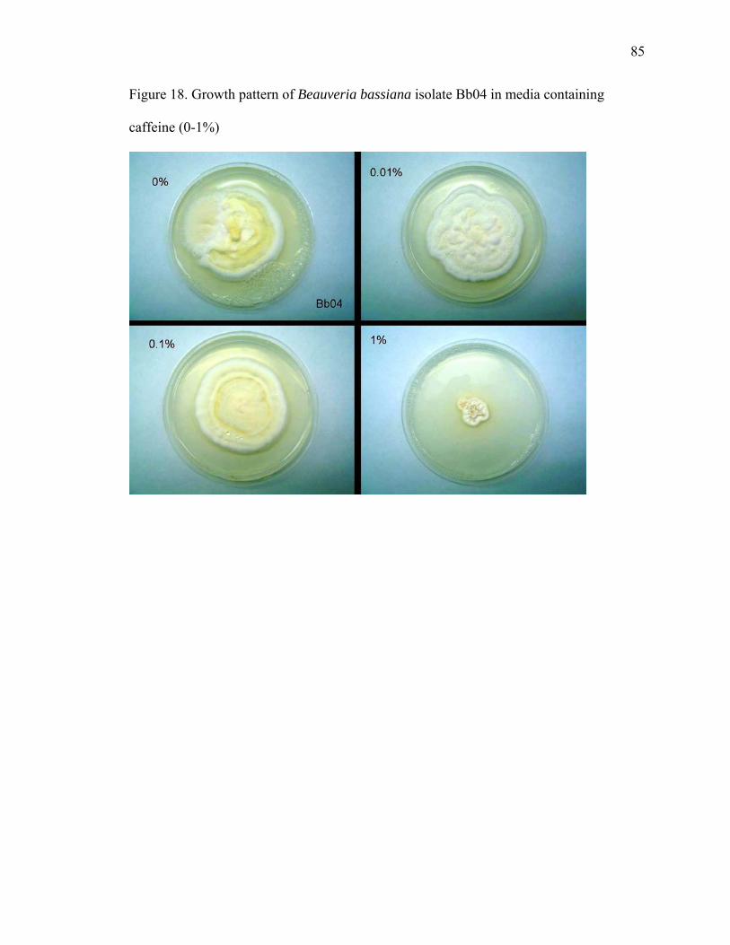
85
Figure 18. Growth pattern of Beauveria bassiana isolate Bb04 in media containing
caffeine (0-1%)

86
Figure 19. Growth pattern of Metarhizium anisopliae isolate MA04 in media containing
caffeine (0-1%)

87
Figure 20. Growth pattern of Metarhizium anisopliae isolate MA06 in media containing
caffeine (0-1%)
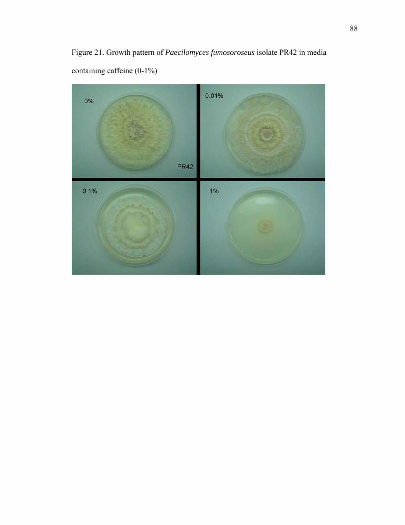
88
Figure 21. Growth pattern of Paecilomyces fumosoroseus isolate PR42 in media
containing caffeine (0-1%)

89
Table 9. Changes in fungal growth rate expressed in % increase (+) or decrease (-)
compared to controls (grown in PDA)
Cha
nges
in
grow
th ra
tes
1% c
affe
ine
2% y
east
ext
ract
an
d 20
% su
cros
e 1%
caf
fein
e, 2
% y
east
ex
trac
t and
20%
sucr
ose
Ant
agon
ist
Ant
agon
ist,
1% c
affe
ine,
2%
ye
ast e
xtra
ct a
nd 2
0% su
cros
e I4
-20
I4-6
I4
-20
I4-6
B.
bas
sian
a -8
7 -8
-7
5 -2
5 -1
3 -7
+4
P.
citr
inum
I4-2
0 - 1
1 +
102
+47
+17
+2
P. c
itrin
um I4
-6
-5
+168
+5
9 +2
9 +3
4

90
Figure 22. Dual plate assay between Penicillium citrinum and Beauveria bassiana
Ps Ps BbBbControl Control

91
T. asperellum was inoculated on top of a cellophane sheet on PDA media and was
allowed to grow for two days. Then the cellophane was peeled off removing Trichoderma
aperellum. Phytophthora palmivora was then inoculated in the center of the plate. Two
days later, P. palmivora controls were fast growing while P. palmivora inoculated in T.
asperellum pre-inoculated plates, failed to grow.
T. asperellum on dual plate assays exhibited mycoparasitism against B. bassiana
and three cacao pathogens, P. palmivora, Moniliophthora roreri and M. perniciosa
(Figure 23). Although T. asperellum is a fast growing isolate, its growth is completely
inhibited by caffeine. In addition, volatile compounds produced by B. bassiana seem to
have a fungistatic effect on T. asperellum. In an X-plate, B. bassiana was inoculated in ¾
of the plate while the remaining ¼ was inoculated with T. asperellum. In control plates,
T. asperellum was inoculated in ¼ of the plate and the other ¾ were left with media only.
In the control plate without B. bassiana, two days after inoculation, T. asperellum had
crawled up the walls and invaded the media in the other quadrants (Figure 24). In plates
where B. bassiana was present, T. asperellum was limited to the quadrant where it had
been inoculated.
Although it is known that Beauveria spp. produce a number of high molecular
weight bioactive compounds (Strasser et al., 2000), there is not much information about
their volatile compounds and their effects on other organisms that interact with
Beauveria.
Volatile profiles from B. bassiana, and T. asperellum grown separately and in
combination were collected in adsorbent resin Tenax TA (most common and
conventional adsorbent, Claeson et al., 2002) and analyzed by GC-MS (Table 10).

92
From B. bassiana, 31 compounds were identified. Also 32 volatile organic compounds
(VOCs) produced by T. asperellum were characterized. When the two fungi were co-
inoculated the VOC profile changed and a smaller number of VOCs (4) found were
unique only to the combination of B. bassiana and T. asperellum. One compound (M+.
m/z 222, retention time 16.4 minutes) that could not be identified was the most prominent
in the collection from the interaction between B. bassiana and T. asperellum. The
compound is in the sesquiterpene region and was not present in the volatile profile of
both fungi separately (Table 10). The identification of the majority of VOCs was done by
matching the retention time of the standards to the retention time of the samples or by
comparison to the equipment’s library.
The fungal VOCs identified were distributed among organic acids, aldehydes,
ketones, ethers, hydrocarbons and sesquiterpenes. Hexadecane and longipinene, only
found in the samples of Beauvera bassiana, are known antifungal volatiles produced by
fungi including Trichoderma harzianum that prevent development of several fungal soil
pathogens (Jannet et al., 2005).
Among the most common VOCs reported from fungal cultures are hydrocarbons,
ketones and terpenenoids. Many compounds found from B. bassiana and T. asperellum
cultures have been previously described as fungal metabolites, uncliding 2-heptanone, 2-
pentylfuran, acetic acid and hexanoic acid (Claeson et al., 2002). Other known microbial
VOCs present in B. bassiana and T. asperellum collections were dodecane, tridecane, and
2-ethyl-hexanal (Claeson and Sunesson, 2005). Caryophyllene and 2-nonanone only
found in the Trichoderma asperellum collection are among the volatile antimicrobial
compounds produced by Muscodor albus (Strobel et al., 2001).
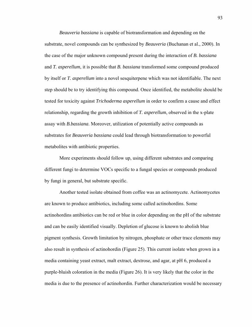
93
Beauveria bassiana is capable of biotransformation and depending on the
substrate, novel compounds can be synthesized by Beauveria (Buchanan et al., 2000). In
the case of the major unknown compound present during the interaction of B. bassiana
and T. asperellum, it is possible that B. bassiana transformed some compound produced
by itself or T. asperellum into a novel sesquiterpene which was not identifiable. The next
step should be to try identifying this compound. Once identified, the metabolite should be
tested for toxicity against Trichoderma asperellum in order to confirm a cause and effect
relationship, regarding the growth inhibition of T. asperellum, observed in the x-plate
assay with B.bassiana. Moreover, utilization of potentially active compounds as
substrates for Beauveria bassiana could lead through biotransformation to powerful
metabolites with antibiotic properties.
More experiments should follow up, using different substrates and comparing
different fungi to determine VOCs specific to a fungal species or compounds produced
by fungi in general, but substrate specific.
Another tested isolate obtained from coffee was an actinomycete. Actinomycetes
are known to produce antibiotics, including some called actinohordins. Some
actinohordins antibiotics can be red or blue in color depending on the pH of the substrate
and can be easily identified visually. Depletion of glucose is known to abolish blue
pigment synthesis. Growth limitation by nitrogen, phosphate or other trace elements may
also result in synthesis of actinohordin (Figure 25). This current isolate when grown in a
media containing yeast extract, malt extract, dextrose, and agar, at pH 6, produced a
purple-bluish coloration in the media (Figure 26). It is very likely that the color in the
media is due to the presence of actinohordin. Further characterization would be necessary

94
to confirm its identity. The molecule produced by the actinomycete isolate could also be
a new antibiotic with similar structure to actinohordin. Even if identified as the same
compound, the discovery of another isolate growing endophytically in coffee capable of
synthesizing an important antibiotic deserves further investigation.

95
Figure 23. Dual plate assays between T. asperellum (Tasp) and M. perniciosa (Mp), and
M. roreri (Mr)
Mr
Mp
Mr
MpControl
Control
Tasp
Tasp

96
Figure 24. X-plate assay between T. asperellum (Tasp) and B. bassiana (Bb1 and Bb2).
Bb1
Tasp TaspTasp
Bb2ControlBb1
Tasp TaspTasp
Bb2Control

97
Table 10. Volatiles produced by Beauveria bassiana and Trichoderma asperellum Rt. Organic Acids Origin Fragments m/z (Abundance%) Match (%)5.0 acetic acid T; B; X 29(10), 42(16), 43 (100), 45(90), M+. 60(66) 916.6 butanoic acid B 27(22), 39(16), 41(27), 42(27), 43(26), 45(18), 60(100), 73(36), M+.88(2) 937.9 pentanoic acid B 41(18), 43(12), 45(13), 55(12), 60(100), 73(39), 87(2), M+.100(<1) 869.3 hexanoic acid T; B 27(17), 29(14), 39(15), 41(30), 43(22), 45(17), 60(100), 73 (48), M+.116(<1) 86
11.6 2-ethyl hexanoic acid T 29(21), 41(39), 57(100), 73(23), 87(23), 88(95), 101(19), 116(15), M+.144(<1) 94Aldehydes and Ketones
6.4 hexanal T; B; 27(41), 29(49), 41(89), 43(75), 44(100), 56(93), 57(66), 72(24), M+. 100(1) 977.1 2-hexenal B 27(43), 39(61), 41(100), 42(57), 55(84), 57(46), 69(69), 83(54), M+.98(16) 977.4 4-heptanone T; B; 27(14), 39(10), 41(23), 43(100), 58(9), 71(99), 86(2), 99(3), M+. 114(22) 917.6 3-heptanone T; B; 27(14), 29(35), 41(22), 57(100), 72(20), 85(31), M+. 114(13) 947.7 2-heptanone T; B; X 27(10), 29(9), 39(10), 41(14), 43(100), 58(70), 59(12), 71(20), M+.114(8) 918.0 heptanal T; B; X 29(63), 41(94), 42(58), 43(100), 44(81), 55(66), 57(59), 70(84), M+.114(4) 968.1 3,4,5-trimethyl-2-cyclopenten-1-one B 53(18), 79(17), 81(21), 95(6), 96(5), 109(100), 110(7), 123(4), M+.124(47) 908.4 2-ethyl-2-hexenal T 39(43), 41(72), 43(49), 55(100), 67(51), 69(33), 97(84), 111(37), M+. 126(77) 838.7 2-ethyl-hexanal T; B; 27(17), 29(24), 41(43), 43(41), 57(100), 72(92), 85(5), M+.128(1) 918.9 benzaldehyde T; X 29(25), 39(7), 50(27), 51(45), 52(11), 77(100), 78(19), 105(98), M+.106 (99) 979.1 3,5-heptadienone T 43(33), 57(50), 99(100), 100(7), M+.128(27) 869.2 2-octanone T; B 27(7), 41(14), 43(100), 58(91), 59(17), 71(18), 85(7), 113(4), M+.128(5) 919.4 octanal B 27(42), 29(61), 41(90), 43(100), 56(68), 57(90), 69(35), 84(56), M+.128(1) 94
10.5 acetophenone T 51(24), 77(76), 105(100), M+. 120(33) 9510.6 3-ethyl-2-cyclopenten-1-one T 27(33), 39(42), 57(100), 67(46), 81(69), 95(13), 109(14), M+.110(64) 9310.9 2-nonanone T 43(74), 58(100), 59(24), 71(20), 82(4), 84(4), 85(4), 127(3), M+. 142(7) 9511.1 nonanal T; B; X 29(47), 41(84), 43(72), 44(47), 55(55), 56(60), 57(100), 70(39), M+. 142(<1) 9612.5 2-decanone T; B 29(15), 41(29), 43(98), 57(24), 58(100), 71(37), 85(8), 96(6), M+.156(6) 9512.7 decanal T; B; X 29(54), 41(98), 43(95), 44(51), 55(73), 57(100), 70(53), 71(49), M+.156(<1) 9513.9 2-undecanone T; B 43(92), 58(100), 71(37), 85(10), 95(4), 112(5), 127(3), 155(2), M+.170(5) 9514.2 undecanal T; X 29(48), 41(87), 43(100), 55(66), 57(84), 68(40), 71(41), 82(51), M+.170(<1) 98
Eters8.6 1-methoxy-4-methyl-benzene X 27(21), 39(25), 51(35), 77(48), 79(32), 91(15), 107(46), 121(79), M+.122(100) 909.0 3-methyl-2(5H)-furanone B 39(48), 41(100), 53(11), 69(75), M+.98(72) 949.5 2-pentyl-furan T; B; X 27(11), 39(11), 41(14), 53(17), 81(100), 82(26), 94(5), 95(6), M+.138(20) 94
10.1 5-ethyldihydro-2(3H)-furanone B 29(34), 42(20), 55(15), 56(19), 57(18), 70(12), 85(100), M+.114(2) 9412.6 2-n-heptylfuran T 27(10), 41(17), 53(16), 67(8), 81(100), 82(31), 95(21), M+.166(17) 93
Hydrocarbons12.8 dodecane B 29(26), 41(46), 43(83), 57(100), 71(51), 85(29), 112(5), 113(6), M+.170(5) 9114.1 1-tridecene T; B 41(79), 43(100), 55(79), 56(50), 57(67), 69(54), 70(43), 83(50), M+.182(22) 9514.3 tridecane T; B; X 27(13), 29(23), 41(45), 43(87), 55(26), 57(100), 71(57), 85(33), M+.184(4) 9815.5 1-tetradecene T 41(100), 43(71), 55(95), 56(67), 57(70), 69(70), 70(65), 83(74), M+.196(2) 9515.6 tetradecane T; B; X 29(21), 41(42), 43(80), 57(100), 71(59), 85(38), 99(9), M+.198(4) 9815.7 1-ethenyl-1-methyl-2,4-bis(1-methylethenyl)-cyclohexane B 41(66), 67(76), 68(73), 79(60), 81(100), 91(47), 93(96), 107(62), M+.204(2) 9916.8 1-pentadecene T; B 41(90), 43(100), 55(93), 56(54), 57(86), 69(68), 83(69), 97(56), M+.210(2) 9916.9 pentadecane T; B 29(18), 41(39), 43(73), 57(100), 71(63), 85(39), 99(13), M+.212(4) 9718.1 hexadecane B; X 41(36), 43(71), 57(100), 71(62), 85(44), 99(13), M+.226(4) 97
Sesquiterpenes15.8 isocaryophyllene B, X 69(45), 91(100), 93(66), 105(72), 107(51), 133(53), 161(48), M+.204(40) 9316.1 caryophyllene T 41(84), 69(100), 79(70), 91(75), 93(95), 105(52), 107(45), 133(79), M+.204(7) 9616.2 2,6-dimethyl-6-(4-methyl-3-pentenyl)-bicyclohept-2-ene T 41(56), 55(29), 69(45), 77(28), 91(39), 93(100), 107(31), 119(95), M+.204(2) 9816.4 unknown X15.3 longipinene B; X 41(80), 55(50), 77(36), 91(70), 105(59), 119(100), 133(45), M+.204 (19) 9317.0 unknonwn T 28(100), 41(92), 43(83), 55(58), 57(80), 93(58), 119(79), 121(52), M+.204 (11)17.2 cedrene X 65(), 105(), 133(), 161(), 189(), M+. 204() 9017.5 unknonwn X 105(), 129(), 145(100), 187(), M+. 202()
B - Beauveria bassianaT - Trichoderma asperellumX - Interation B. bassiana x T. asperellum

98
Figure 25. Chemical structure of Actinohordin
O
CO2H
O
CO2H
O
O
O
O
OH
OH
OH
OH
Actinorhodin
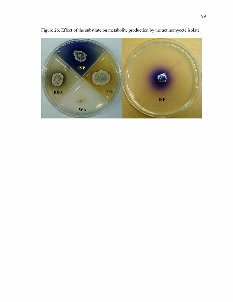
99
Figure 26. Effect of the substrate on metabolite production by the actinomycete isolate
PDA
WA
1%
ISP
ISP

100
2.2.4.3. CONCLUSIONS
Media composition has an effect on the metabolites produced by the fungi
studied. Caffeine, besides affecting the growth of the endophytes, affected their ability to
produce toxins.
Volatiles played a role in the interactions between the fungal isolates. Fast growth
rates may give advantage to some fungi, but their ability to grow under adverse
conditions and produce secondary metabolites is also important.
The interactions between studied microorganisms ranged from parasitism to
production of volatile organic compounds, antibiotics or toxins. The environment, to
which the organisms are subjected, influences their fitness and should be considered
when selecting biological control isolates. In a complex chemical warfare between
microorganisms, understanding the factors influencing their ability to produce secondary
metabolites enables researchers to provide favorable conditions for the target beneficial
organism to succeed.

101
2.2.5. ESTABLISHMENT OF Beauveria bassiana AS AN ENDOPHYTE IN
COCOA PODS
The entomopathogen Beauveria bassiana (Ascomycota: Hypocreales) has a
potential to be used in cacao since it has been shown to grow endophytically in
Theobroma cacao seedlings and is an efficient entomopathogen (Posada and Vega,
2005).
Several studies have tried to establish the fungal entomopathogen Beauveria
bassiana as an endophyte to control insect pests in different plants species (Bing and
Lewis, 1991; Jones, 1994; Wagner and Lewis, 2000; Leckie, 2002; Cherry et al., 2004;
Quesada-Moraga et al., 2006; Gómez-Vidal et al., 2006; Akello et al., 2007). The stigma
in a flower is a natural opening through which fungi can penetrate, as demonstrated by
plant pathogenic fungi such as Ustilago nuda and U. tritici in cereals and Monilinia
oxycocci in cranberries (McManus et al., 1999; Agrios, 2005). This experiment had two
objectives: (1) determine if Beauveria bassiana could infect cacao pods via conidial
spraying of the flowers; (2) determine if the fungus can colonize the pods as they
develop, possibly protecting them against insects and mycotoxigenic fungi.

102
2.2.5.1. MATERIALS AND METHODS
2.2.5.1.1. Counting spores
A hemocytometer was used to estimate the amount of spores per mL in a spore
suspension. Then the appropriate dilutions were made to obtain the desired concentration.
2.2.5.1.2. Application of Beauveria bassiana to cacao flowers and pods
A spore suspension of B. bassiana (1x10⁷ conidia mL-1) in 0.1 % Triton X-100
was applied with a hand held sprayer (Jet-Pak spray system, Sprayon Products, Inc.,
Cleveland, OH) to flowers of 15-year old greenhouse grown cacao trees. Beauveria
bassiana treatments were applied immediately after hand pollination of recently opened
flowers. Four trees were used for the inoculation with B. bassiana and four were used as
controls. The flowers used as control were sprayed with sterile autoclaved water plus 0.1
% Triton X-100. After spraying, flowers were individually covered with plastic bags. In
the case of the flowers sprayed with B. bassiana the increased moisture within the plastic
bag was expected to encourage germination of B. bassiana conidia. The experiment was
repeated four times; each time 120 flowers were treated, half with B. bassiana and half
with water. Pods and peduncles were evaluated for presence of B. bassiana on a monthly
basis after the treatments were applied (for a total of 5 months). The pods were divided
in four sections and cut open; tissue samples were taken at random from the outer and
inner part of each section of the pod (Figure 27).

103
Pod tissues were sterilized by submerging in 0.5 % bleach for two minutes, 70 %
ethanol for two minutes, and then rinsed in sterile water. Tissues were dried with sterile
tissue paper and the edges were removed, followed by plating 3x4 mm sections in 5 cm
Petri dishes with YMA plus antibiotics (Posada and Vega, 2005).
2.2.5.2. RESULTS AND DISCUSSION
In the B. bassiana inoculation experiment, from a total of 480 flowers that were
hand pollinated and sprayed, only 40 set fruit (8.33 %). Usually the fruit set achieved
with hand pollination ranges from 30-50 %, in this experiment the fruit set was
surprisingly lower. The B. bassiana treatment could have affected fruit set, although the
controls (sprayed with water and surfactant) were also low. Perhaps even the spray with
water and surfactant after pollination could have affected the flowers, or washed the
pollen away and reduced the chance of fruit setting. Another control where flowers were
hand pollinated and not sprayed at all would have helped to determine whether the
spraying with water and surfactant influenced fruit set or not. Out of the 40 fruits formed,
10 wilted and only 30 resulted in pods that could be sampled. For cacao, this is a typical
percentage of fruit that fail to develop to maturity. For B. bassiana treatment, 16 pods
were sampled, while for controls, 14 pods were sampled (Table 11).
B. bassiana was recovered from the outer section of the pods in the second and
third evaluation, two and three months post-inoculation. In the second evaluation, one
pod out of four (25 %) was positive for B. bassiana, which was isolated from the third

104
quarter of the pod, and the fungus grew from one of the six pieces of tissue placed on
YMA (Figure 27).
In the third evaluation, one out of five pods (20 %) was positive for B. bassiana,
which was recovered from the fourth quarter of the pod, and the fungus grew in four of
the six pieces of tissue placed on YMA. Tissues from a peduncle attached to the pod were
positive for B. bassiana in the second evaluation. The evaluations of surface sterilized
water did not yield any microorganisms thus assuring that the external surface of tissues
was sterile.
This is the first report of the successful establishment of a fungal entomopathogen
in fruits, as an endophyte, inoculated via flowers. This finding demonstrates a novel
method for inoculating plants with a biological control agent. The recovery of B.
bassiana from the pod and peduncle tissues demonstrates its ability to colonize and move
through the pod. Further evaluations need to be done in an attempt to increase the B.
bassiana establishment efficiency. The data indicate that although it is possible to
establish B. bassiana via floral inoculation, the lower fruit set and poor percentage of
successful inoculations need to be overcome before B. bassiana in the pods can be
considered for testing as a novel biocontrol method against the cocoa pod borer.

105
2.2.5.3. CONCLUSIONS
B. bassiana was isolated from various sections of the cacao pod and peduncle two
and three months after the flowers had been hand-pollinated and sprayed in the
greenhouse with a B. bassiana conidial suspension. These results indicate that spraying
flowers with conidia can result in the establishment of a fungal entomopathogen as an
endophyte, and that the fungus can move within the pod as it grows. This finding could
lead to an innovative way to manage insect pests in biocontrol programs.

106
Figure 27. Cocoa pod sections and parts sampled for determining the presence of
Beauveria bassiana
Cocoa pod
Cocoa pod
outer part
Cocoa pod Inner part
10 9
4 3 2 1
8 7 6 5
Basal Apical
Peduncle
First quarter
Second quarter
Third quarter
Fourth quarter
Cocoa pod
Cocoa pod
outer part
Cocoa pod Inner part
10 9
4 3 2 1
8 7 6 5
Basal Apical
Peduncle
First quarter
Second quarter
Third quarter
Fourth quarter

107
Table 11. Percentage of B. bassiana recovered from pods sprayed with a B. bassiana spore suspension after flower pollination
B. bassiana Treatment Control Pod Peduncle Pod+Ped Pod Peduncle Pod+Ped
Endophytic Fungi
+Bb Inside
+Bb Outside
+Bb Ins+Out +Bb +Bb
-Bb Inside
-Bb Outside
-Bb In+Out -Bb -Bb
First evaluation B. bassiana 0.0 0.0 0.0 0.0 0.0 0.0 0.0 0.0 0.0 0.0
Second evaluation B. bassiana 0.0 0.8 0.8 0.8 1.7 0.0 0.0 0.0 0.0 0.0
Third evaluation B. bassiana 0.0 5.2 5.2 0.0 5.2 0.0 0.0 0.0 0.0 0.0
Fourth evaluation B. bassiana 0.0 0.0 0.0 0.0 0.0 0.0 0.0 0.0 0.0 0.0
Fifth evaluation B. bassiana 0.0 0.0 0.0 0.0 0.0 0.0 0.0 0.0 0.0 0.0

108
2.3. PATHOGEN
2.3.1. INTRODUCTION
Among the pathogens attacking cacao, those affecting pods deserve primary
consideration due to direct crop losses. The three major fungal pod diseases of economic
relevance are Witches’ Broom (WB), Frosty Pod (FP) and Black Pod (BP). The causal
agents for these diseases are Moniliophthora perniciosa, Moniliophthora roreri and
Phytophthora sp. respectively. BP disease, although present throughout the world, is a
major problem in Africa where species of Phytophthora that are more virulent infect
cacao. WB and FP are present only in the Americas; FP does not occur in Brazil at this
time, but only in the northwestern portion of South America and in Central America. WB
disease, the major constraint for cocoa bean production in South America and the
Caribbean, a serious threat to the other cacao growing regions of the world still free of
the pathogen, is the focus of this study.
Witches’ broom disease caused by Moniliophthora (= Crinipellis) perniciosa
(Stahel) Aime & Phillips-Mora, belongs to kingdom Fungi, phylum Basidiomycota, class
Basidiomycetes, order Agaricales, family Tricholomataceae. M. perniciosa is a
hemibiotrophic fungus that infects young meristematic tissue (actively growing shoots,
flowers and developing fruits) and grows intercellularly as a monokaryotic biotroph.
Subsequent to infection, the plant develops abnormal growth patterns including
hypertrophy and hyperplasia, with excessive and unorganized growth of apical parts, loss
of apical dominance, and proliferation of clustered short lateral shoots with undeveloped

109
leaves that resemble a broom. Throughout this period, the fungus becomes necrotrophic
(dikaryotic) and infected plant parts turn necrotic. Once plant tissues are dead,
basidiocarps emerge and release spores that start a new infection cycle (Figure 28).
Control strategies commonly employed against cacao pathogens include use of
resistant cultivars, pruning and pod removal or pod stripping, fungicide applications and
use of biological control agents. Phytosanitation is more successful with frosty pod
disease because only pods have to be removed, whereas with witches’ broom, infected
branches, which are not always easily accessible, must be removed as well. Although
there are disease tolerant cacao varieties, the genetic and physiological variability within
the pathogen, as well as the absence of a genetic cross resistance to multiple diseases,
makes cacao selection difficult. Therefore, a better understanding of the interactions
between pathogens and cacao will help define more efficient disease management
practices. In view of identifying the biochemical mechanisms of pathogenesis and disease
resistance, M. perniciosa isolates were grown in culture media for the analysis of fungal
metabolites and enzymes with possible roles in the disease process; the pathogen was
also challenged with plant metabolites and fungal endophyte extracts.

110
Figure 28. Moniliophthora perniciosa basidiocarps formed in cherelles (young fruit of
cacao) from a farm in Pichilingue, Ecuador

111
2.3.2. pH EFFECT ON GROWTH OF M. perniciosa AND M. roreri
M. perniciosa and M. roreri were grown in PDA at different pH levels in order to
determine the optimum pH for growth of these pathogens in culture. M. roreri grew well
on media with pH levels ranging from 5.6 to 4.4. At pH 3.8 its growth was completely
inhibited (Figure 29). M. perniciosa had its growth rate slowed at pH 4.4, and this
inhibition was stronger at pH 3.8 (Figure 30). The results show that relatively low pH is
inhibitory to the growth of these cacao pathogens, presenting a potential mechanism of
control.

112
Figure 29. Effect of pH on the growth of M. roreri
Days
0 5 10 15 20
Rad
ial m
ycel
ium
gro
wth
(cm
)
0
1
2
3
4
5
pH 3.8 pH 4.4 pH 5.0 pH 5.6

113
Figure 30. Effect of pH on the growth of M. perniciosa
Days
0 5 10 15 20
Rad
ial m
ycel
ium
gro
wth
(cm
)
0
1
2
3
4
5
pH 3.8 pH 4.4 pH 5.0pH 5.6

114
2.3.3. ANASTOMOSIS GROUPS OF Moniliophthora perniciosa ISOLATES
M. perniciosa have been found associated with other plant species such as
Bignoniaceae lianas, Solanum paniculatum, and Heteropterys acutifolia. M. perniciosa
isolates have been classified according to host range: C-type pathogenic to cacao, L-type
associated with Bignoniaceae lianas, S-type found in Solanaceus hosts, and H-type
associated with the shrub H. acutifolia. The C-types have been further divided by
geographic region. The C- and S-biotypes are non-outcrossing and form broom
symptoms on hosts, whereas the L-biotype is outcrossing and asymptomatic. Wheeler
and Mepsted (1988) also classified isolates based on their ability to induce symptoms.
Phylogenetic analysis of several regions of the rRNA locus revealed near identity
between C- and S-biotype isolates from diverse loci, but the L- and H-biotypes formed
separate groupings. Differences could not be found among M. perniciosa C-type isolates
using 20 random amplified polymorphic DNA (RAPD) markers, while vegetative
compatibility experiments showed genetic differences within basidiospores from the
same basidiocarp when single spore cultures were plated together. It is likely that a small
number of genes control vegetative compatibility. Therefore, RAPD banding patterns that
target relatively few areas of the genome possibly did not include the genes involved in
vegetative compatibility (Andebrhan, 1994). Andebrhan et al. (1999) determined that M.
perniciosa isolates found in Bahia belong to two groups based on RAPD analysis.
Here ten M. perniciosa isolates collected from infected brooms in Bahia, Brazil
were subjected to vegetative compatibility assays in order to distinguish isolates and find

115
correlations between mycelial characteristics or other aspect of fungal physiology that
could be linked to pathogenicity.
2.3.4. RESULTS AND DISCUSSION
A single M. perniciosa isolate, when inoculated at two sides of a Petri plate, has
the two fronts of mycelial growth merge in a homogeneous mycelial mass. The same
growth pattern occurs when two vegetatively compatible isolates are co-inoculated on a
plate (Figure 31). However, when mycelia of two incompatible isolates come upon each
other, a pigmented thick layer forms at the point where they merge (Figure 32). A third
type of interaction is an intermediate one, in which a pigmented layer is absent in
between the isolates and they do not merge as evenly as in the compatible case (Figure
33). Andebrhan et al. (1999) determined that there are two groups of isolates infecting
cacao in the Bahian region where these isolates were collected.
Based on the results of the vegetative compatibility experiments, three groups of
isolates were observed (Figure 34). One group was composed of isolates PS10, PS12,
16avd1 and Hrvd1, which were compatible among each other and partially compatible
with isolates of group 2, composed of Alf42, PS9 and 1130. Isolates from group 2 had
intermediate compatibility among each other. Both groups 1 and 2 were incompatible
with isolates of the third group, 278, 1103 and PS6, which were compatible among each
other. This set of isolates also varied in growth rates that ranged from 0.3 to 0.6 cm/day,
where isolates from group 1 had higher rates (Figure 35).

116
Figure 31. Vegetatively compatible isolates (1103 left and 278 right)

117
Figure 32. Vegetatively incompatible (PS6 left and Alf42 right)

118
Figure 33. Intermediate vegetative compatibility (PS9 left and PS12 right)

119
Figure 34. Vegetative compatibility groups of M. perniciosa isolates (Red –
Incompatible; Yellow – Intermediate compatibility; Green – Compatible)
Groups PS10 16avd1 PS12 Hrvd1 Alf42 PS9 1130 278 PS6 1103PS10
16avd1I PS12
Hrvd1Alf42
II PS91130278
III PS61103
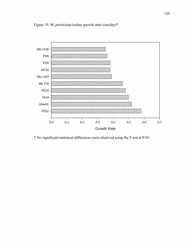
120
Figure 35. M. perniciosa isolate growth rates (cm/day)*
Growth Rate
0.0 0.1 0.2 0.3 0.4 0.5 0.6 0.7
PS12
16avd1
Hrvd
PS10
Mp 278
Mp 1103
Alf 42
PS9
PS6
Mp 1130
* No significant statistical differences were observed using the F test at P.05.

121
2.3.5. METABOLITES PRODUCED BY M. perniciosa
Although most steps of M. perniciosa infection biology are known (Frias et al.,
1991), a few key points remain unclear. The symptoms appear to be hormonally induced,
yet application of hormones to uninfected actively growing leaves fails to induce broom
development. Another aspect not well understood that could be linked to symptom
development, is when and how the pathogen switches from a biotrophic to a necrotrophic
phase. Scarpari et al. (2005) conducted a thorough study of the biochemical changes in
cacao during the development of witches’ broom; however, salicylic acid (SA) levels
were not analyzed. SA, an extensively documented plant hormone, triggers systemic
acquired resistance (Durrant and Dong, 2004), thermogenesis (Raskin et al., 1989), and
regulates coupled cell enlargement and cell death (Rate et al., 1999; Vanacker et al.,
2001). In cacao, SA and chemical elicitors, including an SA analogue, benzothiadiazole,
induce chemical changes similar to those observed in infected tissues when applied to
young shoots (Aneja and Gianfagna, 2001). The hypothesis here is that chemical changes
leading to broom development (cell enlargement and death) involve regulation via the SA
pathway and that the fungus may be the source of SA.

122
2.3.5.1. MATERIALS AND METHODS
2.3.5.1.1. Chemical reagents
All solvents used were HPLC grade. BSA (N,O-bis(trimethylsilyl)-acetamide)
(Pierce, USA) was used as derivatizing reagent prior to GC-MS analysis. SA was used as
the standard for HPLC and GC-MS analyses.
2.3.5.1.2. Fungal cultures
M. perniciosa isolates 278, Alf42, 1103, PS12, 16avd1, 1130, Hrvd1, PS9, PS6
and PS10 collected in Bahia, Brazil, from diseased cacao tissues were grown from
mycelia in potato dextrose broth for three weeks. The mycelia were then filtered off using
glass wool, and the broth was lyophilized and stored at -80 oC until analysis.
2.3.5.1.3. Plant material
Infected brooms and healthy cacao shoots were collected, freeze-dried and stored
at -80 oC.

123
2.3.5.1.4. SA extraction and HPLC analyses
SA extraction and HPLC analyses followed the method of Verberne et al. (2002).
A Shimadzu LC-10AT equipped with a RF-10A spectrofluorometric detector and a C18
column (140 mm x 4.6 mm x 0.5 μm particle size) was used. The fluorescence detector
was set at an excitation wavelength of 305 nm and emission of 407 nm; the flow rate was
0.8 mL·min-1 using a mobile phase of 90 % 0.2 M sodium acetate buffer pH 5.5 and 10 %
methanol.
2.3.5.1.5. GC-MS analyses
The samples analyzed by HPLC were dried in nitrogen, resuspended in
acetonitrile and derivatized with BSA (reaction vial was heated at 70 oC for 15 minutes).
Samples were injected in the GC without further purification. GC-MS analyses were
performed using a Hewlet Packard (HP) gas chromatograph 6890 series, equipped with
an Agilent column DB-1MS (60 m x 250 μm i.d. x 0.25 microns film thickness) using
helium (chromatographic grade, from Airgas, NJ, USA) as carrier gas with a flow rate of
1 mL·min -1 . Oven temperature was set at 70 oC, held for two minutes and increased to
240 oC at 10 oC·min -1 and held for 30 minutes. Spectral data was obtained from an HP
mass spectrometer 5973 series with ionization energy of 70 eV and mass scan range from
40 to 550 amu at 1.97 scans·s -1. The MS source temperature was set at 230 oC.
Compounds were identified by comparison to commercially available standards
or the equipment’s library (NIST 98).

124
2.3.5.1.6. SA application to cacao tissues
SA was applied to young and old leaves, flowers, trunk and fruits (2 months old)
of cacao using a hand held sprayer. Plant parts were sprayed until their surface was
completely covered with solution. Spray solution contained 20 mM of SA in distilled
water and 0.05 % Tween 80. Control spray solution contained 0.05 % Tween 80 in
distilled water.
2.3.5.1.7. Statistical analysis
Statistical analysis of the data was performed using SAS version 9.1 for Windows
(SAS Institute, USA).
2.3.5.2. RESULTS AND DISCUSSION
M. perniciosa is a hemibiotrophic fungus, with a biotrophic and necrotrophic
phase. As such, cell death assists the pathogen to complete its life cycle. For this reason,
cacao avoids the SA pathway. In summary, SA levels in young leaves treated with
elicitors were not statistically different from control treatments. Young cacao leaves
maintained the same SA levels as the controls after pathogen inoculation, locally and
systemically. Endogenous SA levels of both tested cultivars, including the more disease
tolerant SCA and the more susceptible CCO, did not differ statistically. Cacao had the
125
highest levels of SA in the youngest tissues, which are known to be susceptible to
disease.
Some pathogens produce plant growth hormones (auxin, ethylene) and derivatives
(coronatine, a JA analogue) as virulence factors that may act by disrupting the tightly
regulated plant defense metabolism, altering physiological processes and favoring the
pathogen to thrive. All tested M. perniciosa isolates, when grown in liquid culture media,
produced SA at varying levels ranging from about 0.3 to 2.2 ng·g -1 of fresh weight of
mycelia (Figure 36). There was an inverse relationship between growth rate in vitro and
SA content in planta. Vegetative compatible isolates from group 1, were the fastest
growing isolates, and produced the lowest average content of SA.
SA was identified by matching the retention time of the standard with the
compound in the sample at specific excitation and emission wavelengths. A calibration
curve was prepared by injecting different concentrations of the commercially available
SA standard. In order to confirm SA’s identity, samples extracted for the HPLC analysis
were examined by GC-MS. GC-MS runs also confirmed the presence of two other aryl
compounds, mandelic acid and phenyllactic acid (Figure 37). SA was identified as its
trimethylsilylated derivative obtained after reaction with BSA. The retention time and
mass spectra of the compound present in the fungal extract were identical to the
trimethylsilylated SA standard. The major fragment ions were: m/z 73(78.2), 91(11.4),
135(28.2), 149(7.4), 179(8.6), 209(6.8), 267(100), 268(22.4), 269(10.3) and M+. 282(<1)
(Figure 37). Another phenolic compound characterized by GC-MS was 2-hydroxy-2-
phenylacetic acid (also known as mandelic acid) with the distinctive fragmentation: m/z
73(65.3), 147(33.1), 163(11.3), 179(100), 253(15.2), 281(2.5) and M+. 296(<1) (Figure

126
37). A third compound identified was 2-hydroxy-3-phenylpropanoic acid (also known as
phenyllactic acid) with the following fragmentation pattern: m/z 59(5.5), 73(100),
147(74.5), 193(93.7), 205(5.9), 220(19.7), 267(14.1), 295(10.8) and M+. 310(<1) (Figure
37). Identification of the two latter compounds was based on their fragmentation pattern
and by comparison to the equipment’s library. These molecules have been previously
identified as intermediates in the metabolic pathway of the white rot fungus, Bjerkandera
adusta, leading to synthesis of aryl metabolites (Lapadatescu et al., 2000), and have been
found in the basidiomycete Oudemansiella mucida (Zouchova et al., 1982). These SA
derivatives could be intermediates in the synthesis of SA as well as active compounds
that could interfere with the plant defenses by mimicking SA. The jasmonic acid
pathway, primarily responsible for defense responses against herbivores, is negatively
regulated by SA (Kunkel and Brooks, 2002; Thaler et al., 2002). Therefore, activation of
SA may render the plant susceptible to attack from other pathogens by what Cui et al.,
(2005) called systemic induced susceptibility (SIS). In the SIS activated by SA, the
pathogen alters a defensive pathway to induce vulnerability to pathogen-vectoring
herbivores that would, in one hypothesis, increase the spread of the pathogen. In addition,
Arabidopsis thaliana mutants impaired in the JA pathway are more susceptible to
necrotrophic fungal pathogens including Alternaria brassicicola, Botrytis cinerea, and
Pythium sp. (Kunkel and Brooks, 2002). Based on these results, one hypothesis for
further investigation in cacao is that SA blocks the JA pathway, rendering the plant
susceptible to M. perniciosa infection.
There are evolutionary speculations that M. perniciosa evolved from an
endophyte to a pathogen. It may have acquired the mechanism of producing SA to alter

127
plant defenses to its advantage, and as a consequence, modified itself from a biotrophic
and endophytic organism to a hemibiotrophic pathogen.
At a certain stage in its development, the fungus may be the source of the SA,
which stimulates cell growth and enlargement, producing the broom symptoms. The
rising SA levels induce cell death and the brooms become necrotic. The symptoms
remain localized in the young tissues due to their sensitivity to SA. When high
concentrations of SA were applied to different plant parts, only flowers and young leaves
showed necrosis and abnormal growth (Figure 38). In addition, the analyzed broom tissue
had SA levels five times higher than healthy tissue (Figure 39). Healthy shoots had 224.2
ng of SA·g -1 dry weight stem tissue, while brooms had statistically higher (F p < 0.01)
levels of 1,212.3 ng of SA·g -1 dry weight stem tissue. Although the possibility that the
higher SA levels are produced by the plant cannot be ruled out, the results suggest that
the fungus promotes an increase in SA levels, which may play a role in pathogenesis
(Figure 40) and provide a target for disease control.
The fungal isolates used in this study were collected from diseased cacao tissue.
Thus, it is possible that non-pathogenic strains do not produce SA and therefore have a
symbiotic interaction with cacao.
Following Chaves and Gianfagna (2006), Kilaru et al. (2007) obtained similar
results that support and confirm the findings described above. Moreover, the mechanism
of pathogenicity by M. perniciosa likely involves cell death regulated by calcium oxalate
and hydrogen peroxide (Ceita et al., 2007). Plant resistance seems to be related with the
ability of the plant to avoid calcium oxalate accumulation and by activation of hydrogen
peroxide quenching enzymes (Ceita et al., 2007).

128
Figure 36. Salicylic acid produced by M. perniciosa isolates in culture
Salicylic acid produced in culture
SA (ng.g-1f.w.)
0.0 0.5 1.0 1.5 2.0 2.5
M. p
erni
cios
a is
olat
es
PS10
PS9
16avd1
Hrvd1
PS12
1103
PS6
Alf42
1130
278
A
A B C
CC
A
BC
A B CA B CA B C
AB
Salicylic acid produced in culture
SA (ng.g-1f.w.)
0.0 0.5 1.0 1.5 2.0 2.5
M. p
erni
cios
a is
olat
es
PS10
PS9
16avd1
Hrvd1
PS12
1103
PS6
Alf42
1130
278
A
A B C
CC
A
BC
A B CA B CA B C
AB

129
Figure 37. Mass spectra of trimethylsilylated fungal metabolites salicylic acid, mandelic
acid and phenyllactic acid

130
OTM S
OTM S
O
m /z 2 9 6
OTM S
O
OTM S
m /z 2 8 2
OTM S
O
OTM S
m /z 3 1 0

131
Figure 38. Salicylic acid treatment to young cacao leaves

132
Figure 39. HPLC chromatogram of salicylic acid in brooms and healthy
stems. *Different letters following means indicate significant difference
at p < 0.01.

133
Figure 40. Flow chart with proposed role for salicylic acid in pathogenicity
Moniliophthora perniciosa
Theobroma cacao
Salicylic acid
Necrotroph
Biotroph
Infection
Caffeine
Defenses
Cell DeathCell Growth
Symptoms JA
Moniliophthora perniciosa /
Pathways:AdvanceAvoidedBlocked
Pathways:AdvanceAvoidedBlocked
Procyanidins

134
2.3.5.3. CONCLUSIONS
Salicylic acid (SA), phenyllactic acid and mandelic acid were identified as
products of fungal metabolism by M. perniciosa during the dikaryotic growth stage.
Levels of SA were quantified in infected and uninfected plant tissues as well as in
filtrates of fungal cultures. When SA levels were measured in plant tissues, infected
brooms had five times more SA compared to healthy shoots. Moreover, application of SA
to leaves induced curling and necrosis. In contrast, leaf inoculation with basidiospores or
application of chemical elicitors did not alter leaf SA content, suggesting that in this
infection system, the actively growing mycelia is the source of the SA rather than the
plant.
When plants respond to infection by necrotrophic pathogens through the
activation of cell death, the pathogen growth is accelerated instead of retarded. The
necrotrophic pathogens B. cinerea and S. sclerotiorum induced oxidative burst and cell
death in Arabdopsis, and their pathogenicity was directly related to the generation and
accumulation of superoxide and hydrogen peroxide (Govrin and Levine, 2000). For
necrotrophic fungal pathogens, including M. perniciosa, SA may be a component of
pathogenesis and not part of the defense response of plants. Therefore, it is possible that
plants capable of responding to infection more effectively by activating cell death, when
challenged by a necrotrophic pathogen, tend to be more disease susceptible. This can
explain why cultivars tolerant to non-necrotrophic pathogens are susceptible to
necrotrophs like M. perniciosa.

135
2.3.6. M. perniciosa ENZYMES INVOLVED IN DISEASE
Lipoxygenases (LOX; EC 1.13.11.12) are iron-containing dioxygenases widely
distributed in plants, animals and fungi. In plants, lipoxygenases are known to be active
in defense response. LOX gene expression is regulated by different effectors such as JA,
abscisic acid, and by different forms of stress, such as wounding, water deficiency, or
pathogen attack. In mushrooms and other fungi such as Penicillium sp., the combined
activities of LOX and HPLS (hydroperoxide lyase) convert linoleic acid and linolenic
acid into volatile C8 and non-volatile C10 compounds. One of these C8 compounds, 1-
octen-3-ol, responsible for the characteristic mushroom aroma, is also known to be an
insect attractant (Ritchie, 1994) and an inhibitor of spore germination (Chitarra, 2003).
Another metabolite of the LOX pathway produced along with 1-octen-3-ol is the
bioactive substance 10-oxo-trans-8-decenoic acid (ODA). ODA is a fungal hormone that
stimulates mycelial growth, stipe lengthening, and basidiocarp initiation (Champavier,
2000; Money, 2002). It is also very inhibitory to other fungi such as Penicillium spp. and
Aspergillus spp. (Mau and Li, 2002; Okull et al., 2003). The lypoxygenase pathway
involved in the development of basidiomycetes, including the witches’ broom pathogen,
is a possible target for plant defenses because blocking the production of ODA may
prevent basidiocarp development and the formation of infective basidiospores.
It has been shown in previous sections that procyanidins are part of cacao’s
biochemical defensive response to pathogens. (-)-Epicatechin and other flavonoids have
been reported to inhibit various lipoxygenases such as rabbit reticulose 15-lipoxygenase-
1, recombinant human 5-lipoxygenase and soybean 15-lipoxygenases (Sadik et al., 2003).

136
It is possible that cacao polyphenols may target fungal lipoxygenases to inhibit fungal
reproduction. M. perniciosa, however, is successful in causing disease in cacao.
Therefore, it must have its own defenses against procyanidins.
In a pathosystem involving avocado and the hemibiotroph Colletotrichum
gloeosporoioides, Guetsky et al. (2005) found that laccase secretion by C.
gloeosporoioides lowers (-)-epicatechin levels, which in turn increases lipoxygenase
activity, metabolizing an antifungal compound (conjugated diene, 1-acetoxy-2-hydroxy-
4-oxo-heneicosa-12,15-diene). This contributes to the activation of quiescent infections,
thus inducing susceptibility in avocado. Furthermore, C. gloeosporioides isolates that
showed reduced capability to metabolize (-)-epicatechin were not able to colonize ripe
fruits that naturally lack the antifungal compound. Laccase activity has been reported to
be associated with normal mycelial growth in a range of fungi and in the development of
rhizomorphs and sclerotia in certain basidiomycete and ascomycete taxa, as well as with
basidiome production. In some basidiomycetes, expression of laccase genes and/or
accumulation of laccase occur during vegetative growth, but both expression and activity
decline following basidiome initiation.
Laccases (EC 1.10.3.2), polyphenol oxidases (PPO), catechol oxidase (EC
1.10.3.1) and tyrosinases (EC 1.14.18.1), are phenolic oxidative enzymes that cause
browning in many fruits and vegetables, and can be produced by a range of fungi.
Polyphenol oxidases are also often referred to as tyrosinase, phenolase, catechol
oxidase, or catecholase. PPO are copper containing enzymes (Chen et al., 1991) that
catalyze the hydroxylation of monophenols to o-diphenols and oxidate o-diphenols to o-
quinones. These products may condense and react non-enzymatically with other phenolic
137
compounds, amino acids, proteins, and other cellular constituents to produce colored
polymers and pigments.
Basidiomycetes have several oxidases that can use phenolic substrates. In
Agaricus bisporus, tyrosinase, laccase, and peroxidase were detected. Tyrosinase was
present in larger amounts and was the main enzyme responsible for browning reactions
(Zhang and Flurkey, 1997; Jolivet et al., 1998).
These enzymes have considerable overlap in terms of their substrate affinities.
Any substrate with characteristics similar to a p-diphenol can be oxidized by laccases. In
addition, some fungal laccases can also oxidize monophenols and ascorbic acid (Mayer,
2002). They can be differentiated from one another by testing the effects specific
substrates and inhibitors on enzyme activity. For example, L-tyrosine (L-3-[4-
hydroxyphenyl]-alanine) is not used by laccases but by tyrosinases only.
Laccase is a copper containing enzyme that oxidizes phenolic compounds to
reactive semiquinones that, depending upon the degree of auto-oxidation can yield
quinines, which in turn polymerize forming insoluble complexes. The reactive
semiquinone may also react with O2 to yield a superoxide radical (O2-.) that may lead to a
depolymerization processes. This contrasts with other polyphenoloxidases that lead to
formation of quinines rather than semiquinones. Distinguishing the products of these
reactions may be very complicated due to the variability of the products formed, and it
requires highly sophisticated and complex analytical procedures and equipment.
Spectrophotometric measurements of enzyme activity using different substrates,
however, are advantageous in that they are straightforward and require only basic
instrumentation.

138
Here phenoloxidative enzymes secreted in the growing media by M. perniciosa
isolates were extracted and analyzed with different substrates and inhibitors to determine
their activity and possible role in pathogenesis.

139
2.3.6.1. MATERIALS AND METHODS
2.3.6.1.1. Fungal cultures
Fungi were stored in 15 % glycerol at -80 oC to preserve viability and limit
variations due to mutation, a consequence of continuous subculturing.
2.3.6.1.2. Effects of ODA on M. perniciosa
M. perniciosa was inoculated in media containing 10-oxo-trans-8-decenoic acid
(ODA) alone and in combination with 1-octen-3-ol. In addition, two endophytes used as
biological control agents of M. perniciosa were challenged with ODA.
2.3.6.1.3. M. perniciosa growth assay in culture supplemented with (-)-epicatechin
M. perniciosa growth was evaluated in culture media supplemented with either (-
)-epicatechin, the main polyphenol in cacao, or kojic acid, a PPO inhibitor and fungal
metabolite, and both compounds combined.

140
2.3.6.1.4. (-)-Epicatechin degradation
(-)-Epicatechin degradation was analyzed by RP-HPLC using a fluorescence
detector. Mobile phase A (90:8:2) (water:methanol:acetic acid) and mobile phase B
(90:8:2) (methanol:water:acetic acid) were used in a gradient starting at 100 % A. At five
minutes, A concentration was 75 % and B 25 %, at ten minutes A and B were 50 %, and
at 20 minutes A was 25 % and B 75 %. The flow rate was 0.8 mL·min-1. The fluorescence
detector was set at ex. 290 nm and em. 320 nm, while the UV detector scanned from 200
to 400 nm (Zhu et al., 2003).
2.3.6.1.5. Growth of M. perniciosa in culture broth for enzyme extract
Ten plugs, 0.6 cm in diameter containing mycelia of M. perniciosa growing on
potato dextrose agar (PDA) were placed in 250 mL erlenmeyer flasks containing 100 mL
of potato dextrose broth (PDB). The flasks were placed on a shaker at 100 rpm for three
weeks. Mycelial balls were filtered off, washed with deionized water, and both broth and
mycelia were freeze-dried. The freeze-dried samples were combined with ten times the
volume of ice cold extraction buffer containing 1 mM phenylmethylsulphonylfluoride
(PMSF) in 0.01M phosphate buffer. The suspension was vortexed for one minute and left
on ice for five minutes. It was then centrifuged at 4 oC for ten minutes at 8,000 x g. An
aliquot of the supernatant was separated for use to determine protein content. The
remainder of the supernatant was used as the enzymatic extract.

141
2.3.6.1.6. Protein determination
Protein was determined by the Bradford method using bovine serum albumin as a
standard. Using Bio-Rad dye reagent, the absorbance was measured at 595 nm (Bradford,
1976).
2.3.6.1.7. Gel electrophoresis
Non-denaturing gels were prepared using the Mini Protean (Bio-Rad) according
to the method of Laemmli (Laemmli, 1970). Molecular weights were determined by
using standards (Bio-Rad). The marker used was the wide molecular weight range from
Sigma (6,500-205,000). Concentrations were adjusted to the sample with lowest protein
content and 5 μL of a 60 % sucrose solution was added to 20 μL of the adjusted samples.
Then the polyacrilamide gel (6 % stacking gel and 12 % resolving gel) was loaded with
25 μL of sample and ran at 150 V, using a running buffer consisting of 150 g.L-1 tris
base, 720 g.L-1 of glycine, pH 8.8. Gels were rinsed with buffer (50 mM sodium
phosphate buffer pH 6.8), then stained using coomassie brilliant blue or (-)-epicatechin
solution in buffer phosphate pH 6.8. To test the inhibitors, gels were soaked in a solution
containing the inhibitor in buffer phosphate pH 6.8 before immersion in the substrate
solution.

142
2.3.6.1.8. Enzyme assays
Enzyme activity was determined at 25 oC using 2,2’-azino-bis-ethylbenthiazoline
(ABTS), 3,4-dihydroxy-L-phenylalanine (L-dopa), tyrosine, (-)-epicatechin, and cacao
seed extract as substrates, in 0.01 M citrate buffer at pH 4 to 5.5 and sodium phosphate
buffer at pH 6 to 9. Oxidation of ABTS was monitored at 420 nm (Shi et al., 2002), (-)-
epicatechin at 390 nm, and L-dopa at 475 nm (Baurin et al., 2002).
2.3.6.1.9. Polyphenoloxidase inhibition
The enzyme assays described above were also performed in the presence of a
polyphenoloxidase inhibitor, kojic acid, which is a natural compound produced by one of
the endophytes isolated from coffee and described in previous sections of the dissertation.

143
2.3.6.2. RESULTS AND DISCUSSION
The actual role of ODA on fungal development is not certain, and the effects vary
according to the concentration applied and the fungal species. ODA improved M.
perniciosa basidiocarp formation (Hebbar, P.K. unpublished results). This fungal
hormone, when present in the growing media did not stimulate mycelial growth of M.
perniciosa significantly. The addition of 1-octen-3-ol alone or in combination with ODA
also did not significantly alter M. perniciosa mycelial growth (Table 12). Perhaps in the
case of M. perniciosa, ODA acts by influencing fruiting body formation and stipe
elongation and is not as important for mycelial growth. When tested against Trichoderma
stromaticum and T. koningopsis, which are used as biological control agents against M.
perniciosa, ODA inhibited growth rates slightly. The LOX enzyme responsible for
production of ODA was extracted from mycelia of M. perniciosa and identified in a
protein gel by comparison to commercial LOX (Figure 41). Further investigation
comparing the levels of this enzyme in basidiocarps and mycelia may help clarify the role
of ODA and LOX in fungal development.
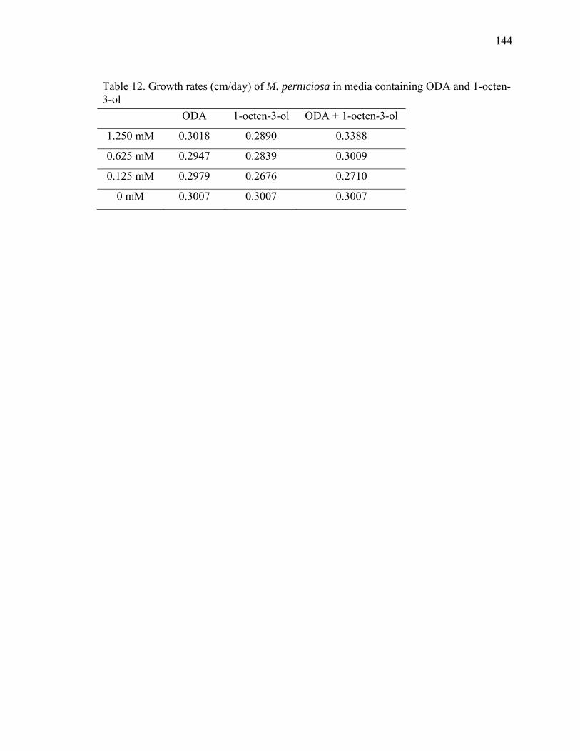
144
Table 12. Growth rates (cm/day) of M. perniciosa in media containing ODA and 1-octen-3-ol
ODA 1-octen-3-ol ODA + 1-octen-3-ol
1.250 mM 0.3018 0.2890 0.3388
0.625 mM 0.2947 0.2839 0.3009
0.125 mM 0.2979 0.2676 0.2710
0 mM 0.3007 0.3007 0.3007

145
Figure 41. Protein gel of M. perniciosa mycelial extract compared to commercial soybean
LOX
146
M. perniciosa inoculated on culture media (PDA) supplemented with (-)-
epicatechin had similar growth rates to that of PDA alone (Figure 42). In addition, the
presence of the fungus induced browning/darkening of the media containing (-)-
epicatechin (Figure 43). No such browning effect was seen in the absence of the
polyphenol in the media. The browning observed in the media indicates the oxidation of
polyphenols through the action of fungal polyphenoloxidases. Kojic acid, a
polyphenoloxidase inhibitor, when applied to the media, slightly inhibited growth of M.
perniciosa. However, when applied in combination with (-)-epicatechin, the inhibition
was stronger and almost no browning of the media occurred (Figures 42 and 43).
The oxidation of (-)-epicatechin seen in the plate assay was determined by
measuring the degradation of (-)-epicatechin. Two M. perniciosa isolates (PS10 and 278)
inoculated in PDB containing (-)-epicatechin completely degraded the polyphenol within
hours of inoculation (Figure 44).
Mycelial growth of M. perniciosa is inhibited in media containing procyanidins
and kojic acid combined, and no browning is observed. Therefore, it suggests that when
phenoloxidases from M. perniciosa are inhibited by kojic acid, the polyphenols from
cacao are now available to inhibit the growth of the pathogen. Similar to C.
gloeosporioides in avocado (Guetsky et al., 2005), M. perniciosa virulence or
pathogenicity may be reduced if the ability to oxidize cacao polyphenols is impaired.
Determinations of pH optima for the fungal enzymatic extract secreted in the
media compared to that of commercial PPO and laccase were made in an attempt to
distinguish the oxidative enzyme involved in the polyphenol degradation (Figure 45).

147
Figure 42. Growth rates of M. perniciosa in media containing (-)-epicatechin and KA
Time (days)
0 2 4 6 8 10 12 14
Rad
ial m
ycel
ial g
row
th (c
m)
0
2
4
6
8
10(-)-epicatechin 1.5 mM(-)-epicatechin 1.5 mM + kojic acid 5 mMkojic acid 5 mMcontrol

148
Figure 43. M. perniciosa growth assay in the presence of (-)-epicatechin (bottom left),
kojic acid (top right), (-)-epicatechin and KA (bottom right), and media alone (top left)

149
Figure 44. (-)-Epicatechin degradation by M. perniciosa isolates
Time (hours after inoculation)
0 20 40 60 80 100
(-)-e
pica
tech
in c
once
ntra
tion
(mg. m
L-1)
0.0
0.2
0.4
0.6
0.8
1.0
PS10 278

150
Figure 45. Comparison of pH optima for activity of commercial laccase (LAC), PPO and
M. perniciosa (Mp) extracellular enzyme extract using (-)-epicatechin as substrate at
25oC
pH
4 5 6 7 8
Rat
e of
reac
tion
0.00
0.01
0.02
0.03
0.04
0.05
0.06 PPO Mp LAC

151
All three analyzed enzymes had a peak in activity at pH 5.5. While laccase
activity only peaked at pH 5.5, PPO and M. perniciosa extract also peaked at pH 7.
It is possible that both enzymes are present in the fungal extract, and it is also possible
that the commercial PPO may not be completely pure and contain laccases, as shown by
Rescigno et al. (2007).
Studies show variation in the optimum pH and the relative activity of the phenol
oxidative enzyme towards different substrates. In Guetsky et al. (2005) findings, laccase
activity was highest at pH 6 and laccase gene expression was highest in the presence of (-
)-epicatechin at pH 5.6.
Gauillard and Richard-Forget (1997) analyzed PPO extracts of pears and found
the molecular mass to be 43,000 Da. Fan and Flurkey (2004) identified PPO isoforms
from mushroom without latent activity below pH 5.5. Crude extracts had isoforms
ranging from 50 to 230 kDa (after denaturing SDS PAGE), while purified isoforms had
mw of 70 kDa.
M. perniciosa extracts showed bands at around 45 kDa, while the commercial
PPO showed a protein band around 84 kDa (Figure 46). Clearly shown on another
activity gel (Figure 47), the fungal enzymatic extracts exhibited a protein band very
distant from the commercial PPO when revealed with an (-)-epicatechin solution. The
fungal enzyme maintained its activity after heating at 60 oC for 5 minutes. But when
placed under denaturing conditions by heating at 100 oC for 5 minutes, activity was
completely abolished.
When a commercial laccase, commercial PPO, and the fugal enzymatic extract
were compared in the same activity gel, the laccase and the fungal extract exhibited
152
bands that co-migrated to the same point (Figure 48). Although there was some staining
at the commercial PPO lane at the same place as the other two enzymes, the stronger
band for PPO migrated more slowly than the other two (Figure 48). It is possible that
commercial PPO has different isoforms or that it has some laccase contamination.
Enzymatic extracts of M. perniciosa isolates and commercial enzymes responded
differently according to the substrate used in the enzyme activity assays. While all three
enzymes reacted with (-)-epicatechin, only commercial laccase and M. perniciosa media
extract oxidized ABTS, and only commercial PPO oxidized tyrosine (Figure 49). It is
most likely that the enzyme from M. perniciosa is a laccase rather than a polyphenol
oxidase; however, it is not possible to rule out the presence of PPO in fungal extracts due
to the two peaks of pH optima for the fungal extracts.
When using cacao’s main polyphenol, (-)-epicatechin, as susbtrate, all tested M.
perniciosa isolates induced browning, which indicated oxidized (-)-epicatechin. This
suggests that in order to avoid the plant polyphenols, the fungus secretes
polyphenoloxidative enzymes that oxidize these compounds. If not oxidized, polyphenols
could block fungal lipoxygenases, preventing ODA (basidiocarp hormone) synthesis that
ultimately could lead to preventing basidiocarp formation.
In order to avoid browning a few mechanisms can be used, including reduction of
quinones formed during oxidation, or the inhibition or inactivation of PPO (Hsu et al.,
1988). Reducing agents, such as ascorbic acid, cysteine, and sulfur dioxide reduce the
ortho-quinone to their ortho-phenol precursor. This mechanism however, is temporary
because the reducing agents themselves are irreversibly oxidized during the process.
Kojic acid, a polyphenoloxidase inhibitor, seems to act through a few different
153
mechanisms. It functions as a reducing agent, as an inhibitor to the enzyme, or by
interfering with the oxygen uptake required for the enzymatic browning (Son, 2001;
Burdock, 2001; Chen et al., 1991).
The temporary inhibition of enzyme activity caused by ascorbic acid was
observed when enzymatic extracts of M. perniciosa were incubated with (-)-epicatechin
and different concentrations of ascorbic acid (Figure 50).
When kojic acid, ascorbic acid, and citric acid were added, each one separately at
3.33 mM, to M. perniciosa enzymatic extract and 3.33 mM of (-)-epicatechin, all
inhibitors reduced polyphenoloxidase activity (Table 13).
Table 14 shows the rate of oxidation using different concentrations of L-dopa as
substrate. Cacao procyanidin extract containing monomers and oligomers from dimer to
decamer were also oxidized by the fungal enzymatic extract.

154
Figure 46. Protein gels with commercial polyphenol oxidase (PPO) and media and
mycelial extracts of M. perniciosa (isolate PS12). Left panel stained with commassie blue
and right panel with (-)-epicatechin.

155
Figure 47. Activity gel stained by immersing non-denaturing protein gel in an (-)-
epicatechin solution
CommercialPPO PS12 PS9 562 PS12 PS9 562
MyceliaMediaCommercialPPO PS12 PS9 562 PS12 PS9 562
MyceliaMedia

156
Figure 48. Activity gels comparing commercial laccase (LAC) and PPO to M. perniciosa
enzyme extract (Mp)

157
Figure 49. Enzyme activity of M. perniciosa, PPO and laccase using ABTS as substrate
Time (minutes)
0 2 4 6 8
Rea
ctio
n ra
te
0.0
0.2
0.4
0.6
0.8
Mp PPO LAC
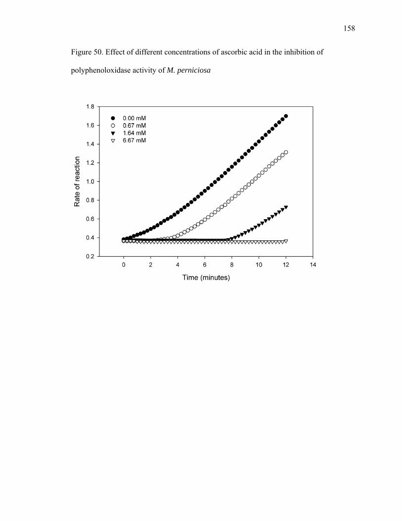
158
Figure 50. Effect of different concentrations of ascorbic acid in the inhibition of
polyphenoloxidase activity of M. perniciosa

159
Table 13. Effect of polyphenoloxidase inhibitors, kojic acid (KA), ascorbic acid (AA) and citric acid (CA) tested at 3.33 mM on enzyme activity using (-)-epicatechin as substrate
% Inhibiton No inhibitor 0
KA 50% AA 100% CA 100%
Table 14. Rates of oxidation of L-dopa using M. perniciosa enzymatic extract Substrate concentration Reaction rate
4.17 mM 4.4 μmol L-1·min-1
3.33 mM 4.1 μmol L-1·min-1
1.67 mM 3.4 μmol L-1·min-1
0.83 mM 2.4 μmol L-1·min-1
160
2.3.6.3. CONCLUSIONS
Although the fungal hormone ODA did not affect mycelial growth, further
investigation is needed to determine whether the lipoxygenase pathway is important for
fungal development. Laccase is one of the polyphenoloxidative enzymes produced by M.
perniciosa as a virulence or pathogenicity factor and is therefore a potential target for
disease control. The main procyanidin found in cacao, (-)-epicatechin, can prevent
pathogen growth in the presence of a polyphenoloxidase inhibitor that blocks the ability
of the fungus to detoxify polyphenols, rendering them toxic to fungal growth. Application
of polyphenoloxidase inhibitors to cacao may affect the ability of the pathogen to thrive
in plants with high levels of polyphenols. In future studies, other polyphenoloxidase
inhibitors should be tested. In addition, mutant lines of the pathogen could be developed,
specifically with mutations in genes that are found to encode for polyphenoloxidases. The
mutants would then be used to inoculate cacao, where disease symptoms and (-)-
epicatechin levels would be monitored.

161
3. PRODUCTION
3.1. INTRODUCTION
Crop yield had been the major focus of cacao research until the late 1980’s, when
fungal disease outbreaks began to cause severe crop losses. Brazil, once the number two
producer and exporter of cacao beans in the world, has changed its status to importer to
match domestic industrial demands due to major reduction in crop production caused by
witches’ broom disease. However, even if losses due to pathogens persist in restricting
production, it is imperative to revisit the issues of low fruit set and low yields and
determine ways to improve pod production in order to maximize the potential of the
cacao producing countries around the world.
Cacao has a cauliflorous habit with flowers growing from the trunk. Flowers are
perfect with both male and female parts. The androecium consists of five stamens, each
bearing two anthers, and five staminodes (sterile stamens). The gynoecieum is composed
of an ovary with five united carpels, and a stile and stigma forming the pistil. The
perianth consists of five sepals and five modified petals, the petals each bearing a ligule.
Cacao flowers begin to open in late afternoon, and continue throughout the
evening so that the flower is fully open just before dawn the next day. The stigma is
receptive for pollination during morning hours, and pollen remains viable for 24 hours. If
the flower is not pollinated it abscises within 48 hours.
One factor limiting cacao yields is pollination. The ratio of fruit set per number of
flowers produced is very low (Young, 1994). Trees typically bear about 4,500 flowers in
162
a six-month period, with less than 1 % of naturally pollinated flowers yielding fruits
(Ibrahim and Hussein, 1987). Hand pollination can increase fruit set, confirming that
natural pollination is a limiting factor for fruit yield. Therefore, efforts to enhance yields
must focus on increasing pollination efficiency.
Cacao trees can be self compatible or self incompatible and pollination by flying
insects results in approximately 25 to 50 % of cross pollination on self compatible trees
(Wood and Lass, 1987). The major natural cacao pollinators are ceratopogonids,
commonly referred as biting midges and gall midges from the dipteran family
Ceratopogonidae, genus Forcipomyia and Culicoides. These insects seem to be attracted
to fragrance emitted from cacao flowers (Young, 1994). Erickson et al. (1992) suggest
that Theobroma spp. exhibit floral thermogenicity to assure thermal stability, essential for
the release of specific ratios of floral volatile compounds. The increase in flower
temperature is likely related to increased respiration levels using the alternative oxidase
respiratory pathway (cyanide-resistant respiration) (Seymour, 2001), known to be
triggered by salicylic acid (SA) (Raskin et al., 1989).
Volatile organic compounds (VOC), temperature, respiration rates, and salicylic
acid content of flowers of greenhouse grown cacao trees were measured in an effort to
further investigate floral attractants and better understand their regulatory mechanisms.
Fruit set upon hand pollination was also determined.
163
3.2. MATERIALS AND METHODS
3.2.1. Volatile Organic Compounds
Flowers collected at 9:00 AM were placed in a 250 mL two-necked round bottom
flask. The flask consisted of a closed system with air pumped in through one neck at a
flow rate of 50 mL· min-1 and out through the other neck, passing through a moisture trap
(composed of cotton plugs and drierite) into an adsorbent resin (Tenax) cartridge, for a
collection time of 30 minutes. VOCs adsorbed in the Tenax columns were desorbed using
a short-path thermal desorption system (Scientific Instrument Services) (Hartman et al.,
1993) into a cryotrap prior to GC-MS analysis.
GC-MS conditions: GC HP 6890 series, MSD HP 5973 NIST 98; column J and
W Scientific Inc. DB-1MS 50 m x 0.250 mm x 0.25 μm; initial temperature 70 oC held
for two minutes; 10 oC · min-1 until 240 oC held for 30 minutes; total run time 49 minutes;
helium gas flow 1 mL · min-1; injector 250 oC; MSD 280 oC; mass scan started at three
minutes and ranged from 20 amu to 550 amu, with a threshold of 150, scanning at 2.78
scans/second;
Desorption system conditions: short path thermal desorption model TD-4
Scientific Instrument Services Inc (Ringoes-NJ, USA). Gas purge time (helium
chromatographic grade-airgas) 30 seconds; injection time one minute; desorption time
five minutes; delay start 30 seconds; desorption temperature 250 oC; cooling trap
temperature - 40 oC (CO2 bone dry grade- siphon type) (from Airgas, Inc Radnor, PA,
USA).

164
In order to test volatiles emitted from different flower parts, 150 TSH 565 flowers
were separated into three parts: petals, sepals, and reproductive components (including,
stamens, ovary, pistil, staminodes) and their VOCs were analyzed separately. Flower
pedicel was discarded.
Flowers were also treated with 200 μM salicylic acid aqueous solution, then
enclosed to collect their VOCs.
3.2.2. Temperature measurements
Temperature measurements were taken from closed and open flowers from three
different T. cacao genotypes, Amelonado, TSH 565 and IMC 30, while the flowers were
still on the trees. The trees were grown in greenhouses under environmentally controlled
conditions. Temperature measurements of three different plant tissue surfaces, flowers
(open and closed), bark, and leaves (young and mature) from nine trees of the three
mentioned genotypes, were taken twice a day (at 9:00 AM and 5:30 PM) for seven days.
Temperatures were measured using a portable infrared thermometer model Raynger ST3
(standard laser) from Raytek Corporation. Ambient temperature was also recorded for the
seven day period.
3.2.3. Respiration
Open and closed cacao flowers were collected at 9:00 AM. Tomato flowers were
used as an external control for comparing levels of respiration. Cacao flowers were then
165
treated with SHAM (salicylhydroxamic acid), a specific inhibitor of the alternative
oxidase pathway, potassium cyanide (KCN), used as inhibitor of the cytochrome oxidase
pathway, and salicylic acid (SA), known to trigger the alternative oxidase pathway. A 10
mM solution of SHAM was prepared in 0.3 % ethanol, a 1 mM solution of KCN was
prepared in a 20 mM HEPES buffer pH 8, following the method described by Millennar
et al. (2001), and a 200 μM SA aqueous solution was prepared.
For the chemical treatments, flowers were placed in a 50 mL plastic syringe
containing SHAM, KCN, or SA solution. A vacuum was created inside the syringe, and
the plunger was pulled to force the solution to penetrate the flower. The flowers were
then rinsed with deionized water and placed in a 50 mL filtering flask with a rubber
stopper on top and a septum on the side arm, which were sealed with Teflon tape. The
flasks were placed on a shaker and gas samples were taken with a plastic syringe and
hypodermic needle that was pierced on a rubber stopper after collection in order to avoid
gas leakage from the syringe. Two samples of each flask were taken every 15 minutes for
two hours, one sample for CO2, the other for O2 analysis. The gas samples were then
injected in a Shimadzu GC 8A IT gas chromatograph with two injectors connected to two
columns, one for CO2 (Alltech; Length 6ft; O.D. 1/8”; I.D. 0.085”; stationary phase –
PORAPAK Q; mesh range 80/100) and the other for O2 (Supelco; 3’ x 14”; stationary
phase – molecular sieve; mesh range 60/80). The column temperature was set to 30 oC,
and the thermal conductivity detector (TCD) set a 100 mA.

166
3.2.4. Salicylic acid analysis
Cacao flowers from Amelonado, IMC 30 and TSH 565 were used in this
experiment. Buds and open cacao flowers were sampled before and after spraying with
1mM SHAM solution.
SA and SAG analysis was previously described on page 15.
3.2.5. Hand pollination
Recently open flowers were pollinated at 9:00 AM using a forceps. Staminodes
surrounding the stigma were clipped off to facilitate access to the stigma. All stamens
were removed and used to self-pollinate each flower by rubbing the anthers against the
stigma.

167
3.3. RESULTS
3.3.1. Volatile organic compounds
47 VOCs produced by cacao flowers were identified (Table 15). The compounds
belonged to the chemical classes: aldehyde, ketone, alcohol, amide, organic acid,
sesquiterpene and hydrocarbon. The most abundant group was the hydrocarbon with 29
compounds varying from 8 to 17 carbons, while alcohol, amide and sesquiterpene each
had only one representative compound identified. Although the analysis focused on
qualitative information, it was observed that 1-pentadecene, followed by 7-tetradecene,
tridecane, and pentadecane were the compounds found in higher amounts. Salicylic acid
treatment of the flowers induced the production of long chain organic acids, tetradecanoic
acid, pentadecanoic acid, 3-hexadecenoic acid and hexadecanoic acid, not normally
present otherwise.
All three, sepals, petals, and reproductive parts produced volatile organic
compounds. In order to compare the relative levels of volatiles emitted by each flower
segment, the 12 most abundant compounds were selected. 3-hexadecene, 1-pentadecene
and pentadecane were produced in higher levels in the sepals, while undecane, dodecane,
2-methy-dodecane, 6-tridecene, tridecane, 7-tetradecene, and tetradecane tended to be
higher in petals. 4-dodecene and 3-methyl-tridecane were produced at similar levels in
the three measured flower parts (Figures 51, 52 and 53).

168
Table 15. Volatile organic compounds produced by cacao flowers R.t. Aldehydes Fragments m/z (Abundance%) Match %6.5 hexanal 27(42), 29(52), 41(81), 43(63), 44(100), 56(87), 57(67), M+. 100(2) 957.18 2-hexenal 29(58), 39(74), 41(100), 42(63), 43(70), 55(84), 57(54), 69(66), 83(56), M+.98(21) 977.9 heptanal 27(50), 29(66), 41(100), 42(59), 43(98), 44(93), 55(68), 57(58), 70(94), M+.114(2) 969.5 octanal 29(50), 41(93), 42(42), 43(100), 44(61), 55(52), 56(69), 57(77), 81(39), M+.138(5) 9111.2 nonanal 29(55), 41(99), 43(83), 44(50), 55(59), 56(59), 57(100), 69(36), 70(35), M+. 142(<1) 93
12.65 decanal 29(55), 41(100), 43(90), 44(47), 55(69), 57(93), 68(38), 70(43), 71(38),M+. 156(<1) 838.9 benzaldehyde 28(80), 43(34), 51(44), 77(100), 105(85), 106(85), M+.116(3) 96
Ketones7.6 2-heptanone 27(12), 29(18), 39(13), 41(19), 43(100), 58(62), 59(10), 71(17), M+.114(11) 837.7 1-(1-cyclohexen-1-yl)-ethanone 39(31), 43(100), 53(17), 55(13), 79(29), 81(85), 109(31), M+.124(22) 909.9 5-ethenyldihydro-5-methyl-2(3H)-furan29(34), 42(20), 55(15), 56(19), 57(18), 70(12), 85(100), M+.114(2) 81
Alcohols10 2-ethyl-1-hexanol 28(28), 41(42), 43(40), 55(31), 57(100), 70(22), 83(22), M+.(130)(<1) 91
Amides8.3 N,N-diethylformamide 27(21), 28(24), 30(84), 44(46), 58(100), 86(35), M+.101(83) 93
Organic acids9.2 hexanoic acid 27(27), 39(26), 41(49), 43(53), 45(20), 60(100), 73 (48), M+.116(<1) 7819.8 tetradecanoic acid 28(33), 41(98), 43(100), 55(83), 57(72), 60(65), 69(46), 71(37), 73(92), M+.228(20) 9821 pentadecanoic acid 29(45), 41(94), 43(100), 55(82), 57(72), 60(91), 69(40), 71(47), 73(82), M+. 242(26) 9122 9-hexadecenoic acid 29(39), 41(100), 43(86), 55(98), 57(51), 67(39), 69(59), 73(42), 83(46),M+.254(2) 92
22.3 hexadecanoic acid 29(35), 41(89), 43(100), 55(78), 57(70), 60(73), 69(43), 71(35), 73(87), M+.256(26) 98Sesquiterpenes
16.1 longifolene 28(100), 29(17), 32(17), 41(30), 43(30), 55(27), 57(19), M+.(204) 99Hydrocarbons
6.7 octane 41(44), 43(100), 55(22), 56(25), 57(33), 70(13), 71(24), 85(33), M+.114(10) 878.1 nonane 28(76), 29(37), 31(31), 41(47), 43(100), 57(72), 85(22), M+. 128(4) 939.8 decane 28(60), 29(28), 41(50), 43(100),56(25), 57(80), 71(31), 85(24), M+.142(6) 9510.3 indene 28(27), 39(12), 41(12), 63(12), 89(13), 115(94), M+.116(100) 9410.4 3,7-dimethyl-1,3,7-octatriene 27(19), 39(29), 41(35), 53(19), 77(38), 79(47), 80(36), 91(49), 93(100), M+.136(7) 9610.8 2-methyl-decane 29(23), 41(50), 42(24), 43(100), 57(85), 71(53), 75(42), 85(27), M+. 156(1) 9611.1 4-undecene 28(100), 32(32), 41(35), 43(30), 55(44), 57(30), 69(27), M+.154(8) 9611.3 undecane 41(55), 43(97), 55(24), 56(23), 57(100), 71(42), 85(26), M+.156(5) 9612.6 3-dodecene 28(57), 41(77), 43(57), 55(100), 56(55), 57(39), 69(70), 70(51), 83(33), M+.168(22) 9812.7 4-dodecene 28(100), 29(48), 41(82), 43(82), 55(88), 56(50), 57(57), 69(54), 70(51), M+.168(12) 9812.8 dodecane 29(28), 41(56), 43(95), 57(100), 71(54), 85(27), 112(6), M+.170(4) 9613.8 2-methyl-dodecane 28(38), 29(34), 41(70), 43(100), 55(42), 57(92), 71(46), 85 (31), 125(29), M+. 184(1) 9514 2-undecanone 29(13), 41(27), 43(90), 58(100), 59(34), 71(26), M+. 170(5) 91
14.1 6-tridecene 41(71), 43(52), 55(100), 56(58), 57(37), 69(68), 70(49), 83(46), M+.182(22) 9814.3 tridecane 41(46), 43(83), 55(21), 57(100), 71(57), 85(34), M+.184(5) 9815.2 2-methyl-tridecane 28(49), 41(51), 43(99), 57(100), 71(54), 85(36), M+.198(1) 9615.3 3-methyl-tridecane 29(23), 41(42), 43(47), 56(27), 57(100), 71(41), 85(31), M+. 198(1) 9615.4 7-tetradecene 41(87), 43(68), 55(100), 56(63), 57(48), 69(93), 70(58), 83(61), 97(43), M+.196(23) 9915.5 1-tetradecene 29(47), 41(100), 43(95), 55(91), 56(56), 57(76), 70(47), 71(38), 83(44), M+.196(10) 9915.6 tetradecane 41(45), 43(82), 57(100), 71(59), 85(37), 99(8), 112(4), M+.198(4) 9816.5 2-methyl-tetradecane 41(49), 43(100), 55(26), 56(20), 57(96), 69(16), 70(11), 71(53), 85(37), M+.212(1) 9516.6 1,6-tridecadiene 28(24), 29(30), 39(30), 41(100), 43(59), 54(59), 55(84), 56(20), 57(31), M+.180(7) 9416.7 1-pentadecene 41(75), 43(66), 55(97), 56(63), 57(55), 69(100), 70(71), 83(89), 97(76), M+.210(46) 9316.9 pentadecane 29(21), 41(46), 43(82), 57(100), 71(62), 85(41), 99(10), 113(5), M+.212(4) 9617.9 3-hexadecene 41(87), 43(83), 55(100), 56(60), 57(61), 69(85), 70(56), 83(69), 97(55), M+. 224(18) 9918 1-hexadecene 28(40), 41(54), 43(88), 55(51), 56(26), 57(35), 69(37), 71(100), 83(29), M+.224(3) 96
18.1 hexadecane 29(25), 41(49), 43(82), 57(100), 71(62), 85(38), 99(10), M+.226(4) 9819 8-heptadecene 41(91), 43(90), 55(100), 56(57), 57(69), 69(81), 70(55), 83(74), 97(60), M+.238(16) 99
19.2 heptdecane 29(22), 41(43), 43(78), 55(20), 56(18), 57(100), 70(16), 85(33), M+.240(3) 96

169
Figure 51. Levels of tridecane, 7-tetradecene and 1-pentadecene emitted by petals, sepals
and reproductive parts of cacao flowers
tridecane 7-tetradecene 1-pentadecene
Are
a
0
1e+9
2e+9
3e+9
4e+9
5e+9
6e+9
7e+9
Petals Sepals Reproductive

170
Figure 52. Levels of undecane, dodecane, 6-tridecene, tetradecane and pentadecane
emitted by petals, sepals and reproductive parts of cacao flowers
undecane dodecane 6-tridecene tetradecane pentadecane
Are
a
0
5e+7
1e+8
2e+8
2e+8
3e+8
3e+8
Petals Sepals Reproductive

171
Figure 53. Levels of 4-dodecene, 2-methyl-dodecane, 3-methyl-tridecane and 3-
hexadecene emitted by petals, sepals and reproductive parts of cacao flowers
4-dodecene 2-methyl-dodecane 3-methyl-tridecane 3-hexadecene
Are
a
0
1e+7
2e+7
3e+7
4e+7Petals Sepals Reproductive

172
3.3.2. Temperature measurements
Measurements indicated that flower temperatures in the morning (24.13 ºC) were
1.91 ºC higher than the surrounding bark and 3.00 ºC higher than nearby leaves, but 1.08
ºC lower than the ambient temperature (Table 16). When the temperature data was
analyzed for differences between open and closed flowers and young versus old leaves at
different times of the day, no differences were observed (data not shown). In the
afternoon, flower temperature (22.76 ºC) was higher than leaf temperature by 0.9 ºC but
lower than bark and ambient temperatures (Table 17). All tissues measured had
temperatures lower than ambient temperature. Salicylic acid treatment increased flower
temperature by 2 ºC.

173
Table 16. Temperature measurements of flowers, background, and plant surfaces of T. cacao in the morning and afternoon (SE, standard error)
Mean temperature (ºC) ± SE 9:00 AM 5:30 PM Daily average
Ambient 25.21 ± 0.49 (n=5) 23.52 ± 0.93 (n=5) 24.80 ± 0.59 (n=10)
Flower 24.13 ± 0.12 (n=365) 22.19 ± 0.24 (n=79) 23.79 ± 0.11 (n=444)
Bark 22.22 ± 0.25 (n=60) 22.76 ± 0.47 (n=43) 22.44 ± 0.19 (n=103)
Leaves 21.08 ± 0.12 (n=78) 21.29 ± 0.16 (n=118) 21.21 ± 0.11 (n=196)

174
3.3.3. Respiration
Respiration rate was determined using gas chromatography (Figure 54) to
measure the increase in CO2 and decrease in O2 in the head space where flowers were
enclosed. Open flowers respired more than closed ones, expressed in Figure 55 as μmoles
of CO2 emitted per gram of flower dry weight. No differences were observed in the levels
of flower respiration among the three cacao varieties investigated (data not shown).
SHAM lowered the levels of CO2 evolution in closed and open flowers,
suggesting the presence of the alternative oxidase pathway (Figures 56 and 57). When
KCN was added, the levels of respiration were dramatically reduced; the remaining
respiration present may have been due to the alternative oxidase pathway (Figures 56 and
57).
Tomato flowers had respiration rates similar to cacao flowers (Table 17).
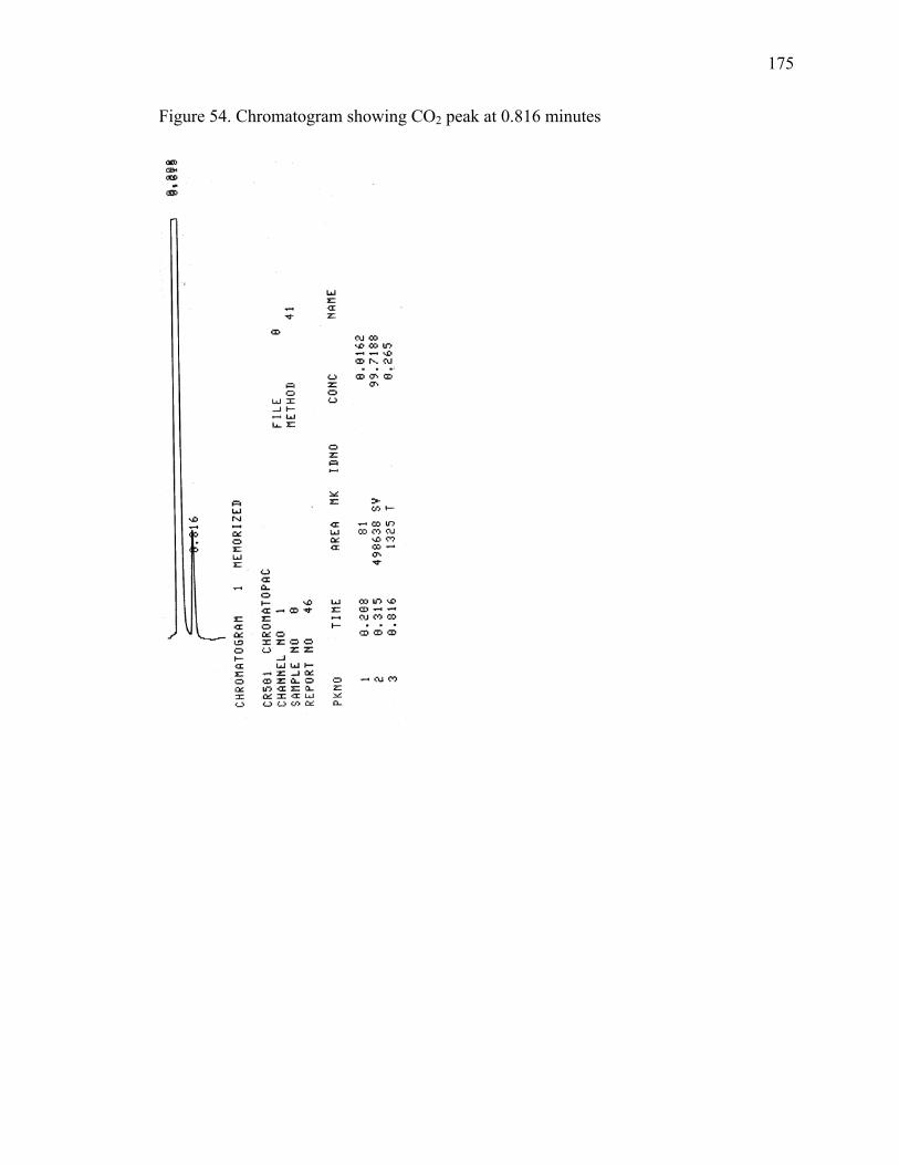
175
Figure 54. Chromatogram showing CO2 peak at 0.816 minutes
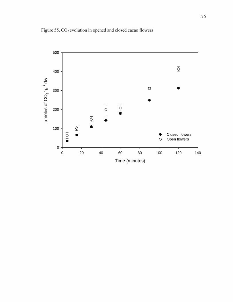
176
Figure 55. CO2 evolution in opened and closed cacao flowers
Time (minutes)
0 20 40 60 80 100 120 140
μ mol
es o
f CO
2 . g-1
dw
0
100
200
300
400
500
Closed flowersOpen flowers
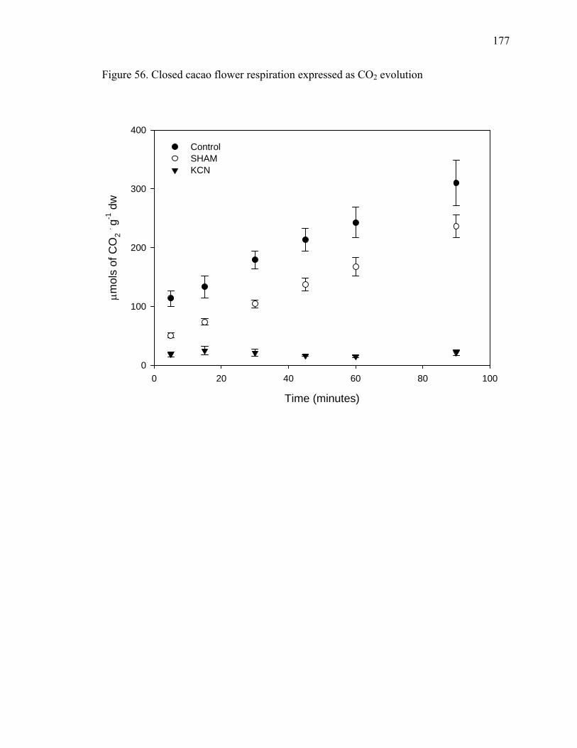
177
Figure 56. Closed cacao flower respiration expressed as CO2 evolution
Time (minutes)
0 20 40 60 80 100
μ mol
s of
CO
2 . g-1 d
w
0
100
200
300
400
Control SHAM KCN

178
Figure 57. Open cacao flower respiration expressed as CO2 evolution
Time (minutes)
0 20 40 60 80 100
μ mol
es o
f CO
2 . g-1
dw
0
50
100
150
200
250
300
350
ControlSHAMKCN

179
Table 17. Percentage of accumulated CO2 produced by enclosed cacao and tomato flowers
% CO2 Hours Cacao Tomato
0 0.21 0.2 0.5 0.35 0.38 1 0.59 0.66
1.5 0.71 0.75 2 0.53 0.72 16 1.71 4.02 40 9.91 12.85

180
3.3.4. Salicylic acid analysis
Salicylic acid and salicylic acid glucoside were measured in buds and open cacao
flowers before and after spraying with 1mM SHAM solution (Table 18). LSD analysis of
the data, showed significant differences for SHAM treatment, cultivar, and the interaction
treatment versus cultivar for SA and SAG contents (Table 19).
Buds and open flowers did not differ in their SA and SAG contents (Table 19).
While flowers of all analyzed genotypes, Amelonado, TSH 565 and IMC 30 had higher
SA and SAG levels after treatment with SHAM (Table 20). Amelonado had the lowest
SAG content and TSH had the highest SA content among the three genotypes (Table 21).

181
Table 18. Salicylic acid (SA) and salicylic acid glucoside (SAG) contents of open and closed cacao flowers before and after treatment with SHAM
SA (ng.g-1 fw) SAG (ng.g-1 fw) Flower Cultivar - sham + sham -sham +sham AME 22.3 123.9 90.1 351.8 TSH 22.5 549.7 84.4 1042.5 Bud IMC 24.3 74.0 105.6 1014.5 AME 22.6 77.7 81.2 238.0 TSH 24.1 380.6 141.0 1037.1 Open IMC 21.2 65.1 131.8 683.4
Table 19. LSD analysis of the SA and SAG contents of cacao flowers SA F Value Pr > F SAG F Value Pr > F
Model 24.59 <0.0001 Model 17.37 <0.0001 Replicates 0.51 0.6058 Replicates 0.76 0.4810 Treatment 109.67 <0.0001 Treatment 142.6 <0.0001 Flower age 4 0.0579 Flower age 1.49 0.2345
Cultivar 48.52 <0.0001 Cultivar 19.96 <0.0001 Trt*age 3.92 0.0604 trt*age 2.88 0.1039 Trt*cult 48.17 <0.0001 trt*cult 16.9 <0.0001 Age*cult 1.81 0.1872 age*cult 1.01 0.3819
trt*age*cult 2 0.1596 trt*age*cult 0.81 0.4564 Numbers in bold are statistically significant

182
Table 20. Effect of treatment on salicylic acid (SA) and salicylic acid glucoside (SAG) contents of cacao flowers by cultivar AME SA Mean LSD.05 = 22.08 SAG Mean LSD.05=55.37 -sham 22.461 B 85.65 B +sham 100.796 A 294.92 A
TSH SA Mean LSD.05 = 140.86 SAG Mean LSD.05 = 403.57
-sham 23.29 B 112.7 B +sham 465.14 A 1039.8 A
IMC SA Mean LSD.05 = 18.26 SAG Mean LSD.05 = 90.71
-sham 22.727 B 118.68 B +sham 73.271 A 846.34 A Mean separation within columns and by treatment.
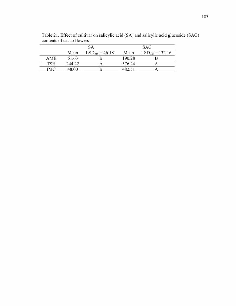
183
Table 21. Effect of cultivar on salicylic acid (SA) and salicylic acid glucoside (SAG) contents of cacao flowers
SA SAG Mean LSD.05 = 46.181 Mean LSD.05 = 132.16
AME 61.63 B 190.28 B TSH 244.22 A 576.24 A IMC 48.00 B 482.51 A

184
3.3.5. Hand pollination
Out of 132 flowers pollinated, 57 set fruit (43 %) and of these, 17 wilted, resulting
in 40 pods reaching maturity. This represented 30 % of the total flowers pollinated and 70
% of those that set fruit.
3.4. DISCUSSION
Young and Severson (1994) had been the only ones to report the volatile profile
of cacao flowers. They collected these volatiles using steam distillation, a procedure
which may lead to artifacts and may extract compounds that are not naturally emitted by
the flowers. Using the Tenax cartridges, only naturally emitted volatiles, which
accumulate in the headspace of the container enclosing the flowers, are blown out with
the air which is pumped in the vessel and then adsorbed in the Tenax resin as they come
out.
Although, a few of the VOCs identified here had been reported by Young and
Severson (1994), here many more were identified including, hexanal, 2-hexenal,
heptanal, octanal, benzaldehyde, 2-heptanone, 1-(cyclohexen-1-yl)-ethanone, 5-
ethenyldihydro-5-methyl-2(3H)-furanone, n,n-diethylformamide, hexanoic acid,
longifolene, octane, nonane, indene, 3,7-dimethyl-1,3,7-octantriene, 2-methyldecane, 4-
undecene, 3-dodecene, 4-dodecene, 2-methyl-dodecane, 2-undecanone, 2-
methyltridecane, 3-methyltridecane, 1,6-tridecadiene (Table 15).

185
When the most abundant VOCs from cacao flowers, pentadecene, pentadecane
and a few other, were tested individually in insect attraction trials, none was effective
(Young, 2007 ). However, flower extracts placed in insect traps were successful in
attracting more cacao pollinators compared to controls (Young, 2007). It is possible that
compounds present in smaller amounts not previously identified and therefore not
utilized in the insect attraction trials are the actual attractants of cacao pollinators.
Regarding the temperature measurements, the results suggest that cacao flowers
regulate their temperature using the alternative oxidase pathway, thereby corroborating
previous reports that indicate cacao flowers can increase their temperature to levels
higher than their surrounding background tissues during morning hours. Cacao flower
temperatures were 1.91 ºC higher than the surrounding bark, similar to findings by
Erickson et al. (1992) who observed cacao flowers with 2.1 ºC higher than the bark.
Cacao flowers, due to their cauliflorous nature, are more exposed to shaded conditions
and lower temperatures than leaves. Therefore, the fact that flower temperature is higher
than bark and leaves reinforces the presence of floral thermoregulation. The current
results also support the idea that greater differences between flower temperature and their
surrounding background tissues occur at morning hours when natural pollination is
thought to occur (Erickson et al., 1992).
Measurements showed no differences between temperatures of open and closed
flowers, but flower temperature differed from other plant parts as well as the ambient
temperature. Flower temperature also differed at certain times of the day (Table 16).
Nonetheless, the differences in temperature, although small, are biologically relevant.
Honeybees, for example, are able to differentiate thermal differences down to 0.25 oC
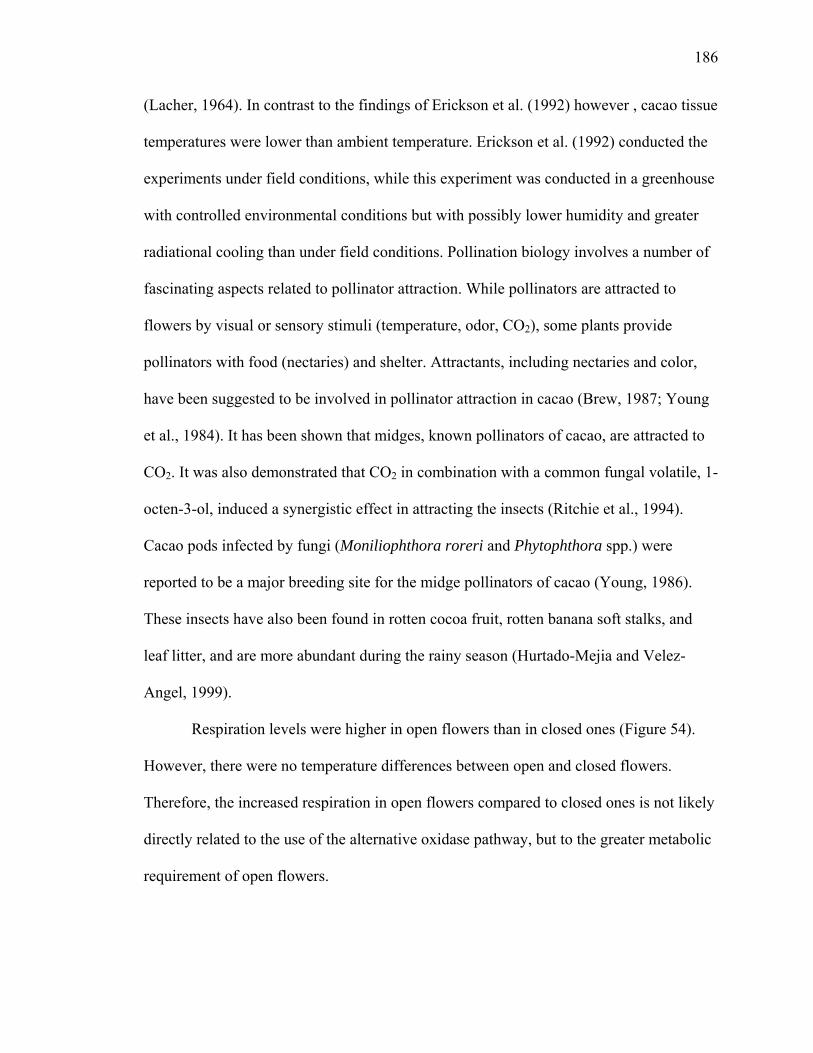
186
(Lacher, 1964). In contrast to the findings of Erickson et al. (1992) however , cacao tissue
temperatures were lower than ambient temperature. Erickson et al. (1992) conducted the
experiments under field conditions, while this experiment was conducted in a greenhouse
with controlled environmental conditions but with possibly lower humidity and greater
radiational cooling than under field conditions. Pollination biology involves a number of
fascinating aspects related to pollinator attraction. While pollinators are attracted to
flowers by visual or sensory stimuli (temperature, odor, CO2), some plants provide
pollinators with food (nectaries) and shelter. Attractants, including nectaries and color,
have been suggested to be involved in pollinator attraction in cacao (Brew, 1987; Young
et al., 1984). It has been shown that midges, known pollinators of cacao, are attracted to
CO2. It was also demonstrated that CO2 in combination with a common fungal volatile, 1-
octen-3-ol, induced a synergistic effect in attracting the insects (Ritchie et al., 1994).
Cacao pods infected by fungi (Moniliophthora roreri and Phytophthora spp.) were
reported to be a major breeding site for the midge pollinators of cacao (Young, 1986).
These insects have also been found in rotten cocoa fruit, rotten banana soft stalks, and
leaf litter, and are more abundant during the rainy season (Hurtado-Mejia and Velez-
Angel, 1999).
Respiration levels were higher in open flowers than in closed ones (Figure 54).
However, there were no temperature differences between open and closed flowers.
Therefore, the increased respiration in open flowers compared to closed ones is not likely
directly related to the use of the alternative oxidase pathway, but to the greater metabolic
requirement of open flowers.
187
Plant respiration is the source of ATP and provides carbon intermediates used in
the synthesis of many cellular constituents. Plant respiration can occur through the
cytochrome pathway and the alternative pathway, which constitute the respiratory
electron-transport pathways of plant mitochondria. The cyanide resistant respiration
enzyme, alternative oxidase, responsible for the alternative pathway, catalyzes a four
electron reduction of oxygen to water and is specifically inhibited by salicylhydroxamic
acid (SHAM). When electrons pass to the alternative pathway from the ubiquinone pool
two site of energy conservation (complexes III and IV) are bypassed and no ATP is
formed. Since there is no energy conservation site on the alternative pathway between
ubiquinone and oxygen, the free energy that would normally be stored as ATP is lost as
heat (Moore and Siedow, 1991). This heat induces in some plants volatilization of
compounds that serve as insect attractants.
Thermoregulation seems to occur through the alternative oxidase pathway since
SA increased respiration rates while SHAM reduced them (Figure 58). Moreover, flowers
treated with SA had an increase in temperature of up to 2 oC.
In this experiment the respiratory quotient (RQ; RQ = O2rate/CO2rate) observed
was greater than one, for control flowers and flowers treated with SA, indicating that
organic acids contribute to the substrates used for respiration (Table 22) (Hopkins and
Huner, 2004).
Interestingly flowers treated with 200 μM of salicylic acid maintained their
normal appearance, whereas control flowers after 84 hours of treatment were brown
(Figure 59). SA may be acting on the inhibition of ethylene biosynthesis and/or reversing
the effects of abscisic acid (Aneja et al., 1999).
188
Figure 58. Effect of SA, SA plus SHAM, SHAM and KCN treatments on cacao flower
respiration
Control SA SA + SHAM SHAM KCN
μmol
es o
f gas
. g-1 d
w . m
in-1
-3
-2
-1
0
1
2
3 CO2
O2

189
Table 22. Respiratory quotient (RQ) of flowers treated with different chemicals Control SA SA + SHAM SHAM KCN
RQ 1.2 1.3 0.75 0.63 0.65
Figure 59. Flowers treated with 200 μM of salicylic acid (3 on the left) compared to
controls (3 on the right), after 84 hours of treatment
190
The presence of SA in cacao flowers is another evidence for the alternative
respiration present in cacao flowers. SA and SAG contents of open cacao flowers did not
differ from buds. Although there were differences among cultivars, Amelonado had the
lowest levels of SAG of the three, while TSH 565 had the highest SA levels. SAG is
though to be a storage form of SA. When present in higher levels in the self-incompatible
gentopytes (TSH and IMC) this storage compound could be involved in producing more
SA to affect the release of floral attractants (CO2 and volatiles) more effectively than in
Amelonado (self-compatible) to due the incompatible genotypes’ higher dependency on
cross-pollination.
The alternative respiratory pathway is specifically inhibited by SHAM and
triggered by SA. The application of SHAM to open and closed flowers induced an
accumulation of SA and SAG. One explanation for the SA accumulation is that flowers
keep producing SA in order to activate the alternative pathway, which fails to occur due
to the presence of the inhibitor. The other option is that SHAM is degraded to SA in
planta. This hypothesis have been suggested by Chivasa et al., (1997) who observed an
induction of PR-1 protein accumulation upon application of SHAM, similar to the effect
caused by SA application.

191
3.5. CONCLUSIONS
Many new volatile organic compounds released by cacao flowers were identified.
It is possible that these compounds, some of which are present in smaller amounts than
compounds previously reported in the literature, can contribute to the attraction of cacao
pollinators. Cacao flowers raised their temperatures higher than background tissues
during morning hours when pollination is known to occur. Flower temperature regulation
occurs through the alternative respiration pathway. Endogenous and inducible SA and
SAG were found in cacao flowers, and their levels varied between self-compatible and
self-incompatible genotypes suggesting a possible role in pollination biology.
Further studies, such as electroantennograms and wind tunnels, using the newly
identified volatiles in combination with CO2 are the next steps in trying to correlate the
identified compounds with pollinator attraction.
Purification of alternative oxidase enzyme and determination of the effects of SA
and SHAM on its expression levels should be performed to continue the investigation on
the alternative respiration pathway in cacao flowers.

192
4. PROCESSING
4.1. INTRODUCTION
Although pests and diseases, the reliance on pollinators, and poor fruit set all
contribute to lower crop yields, bean quality will always be one of the main concerns in
cacao production systems. The principal criterion for assessing the quality of cocoa for
manufacturers is flavor. Flavor is developed and assessed after roasting, but the plant
genotype, as well as the fermentation and drying processes are factors that influence the
composition of aroma precursor compounds.
Once ripe, cacao pods are harvested, cracked open with a machete, and the beans,
naturally covered with a mucilaginous pulp, are stacked in a pile. Banana leaves are then
placed on top of the pile in order to avoid wetting from constant rain. In some cocoa-
growing regions, beans are placed in heaps, boxes, baskets or trays. Every other day the
bean pile is mixed or turned to provide aeration and to allow uniform fermentation of all
beans. After a few days (commonly varies from three to seven days), beans are spread out
and dried under the sun, then roasted, ground and processed.
During fermentation, sucrose is converted into glucose and fructose, which are
then converted into alcohol and acids by yeasts and bacteria. Acids migrate from the pulp
to the beans, killing the embryo, disrupting cell walls, and liberating their contents
(Schwan and Wheals, 2004). Enzymes are also active during fermentation, oxidizing
polyphenols to quinones, which then polymerize or bind to proteins, giving beans a dark
color. Maillard reactions, non-enzymatic chemical reactions between amino acids and

193
sugars, also may contribute to the browning of the beans. However, this reaction is
thought to occur after fermentation and drying, during roasting (Hansen et al., 1998).
Currently, chocolate manufacturers are rediscovering the health benefits of cocoa
and increasing the levels of cacao in their products. In effect, they are increasing the
procyanidin contents of their products and therefore turning candies into nutraceutical
products. Health benefits associated with the consumption of cacao bean products are
mainly derived from procyanidins. These compounds are potent antioxidants, and a
substantial amount of research indicates a series of beneficial properties including their
action as vasodilating agents shown to improve cardiovascular function (Keen et al.,
2005; Schroeter et al., 2006).
Fermentation is essential to produce precursor aroma compounds that, after
roasting, become the familiar flavors of cocoa powder and chocolate. However, during
the fermentation process, cocoa polyphenols are oxidized and the health benefits of the
cocoa bean are lost. Although some oxidation of cocoa polyphenols is necessary due to
their contribution to the bitter taste of unfermented beans, it would be desirable to limit
this process, particularly for products designed for the health food market.
Most of the work on cacao bean fermentation was done in the 1950’s and 1960’s.
With the new analytical tools available today, more accurate data can be obtained,
particularly on the procyanidin contents of beans. In this section, research on the effect of
adding an inhibitor of polyphenol oxidases to a cacao bean fermentation under laboratory
conditions is described to determine if losses of polyphenols during fermentation could
be limited without affecting the sugar and amino acid content needed for the development
of flavor precursors.

194
4.2. MATERIALS AND METHODS
4.2.1. Fermentation
Cacao pods from greenhouse grown Amelonado trees were harvested, cracked
open and the mucilaginous beans placed in 250 mL beakers. The beakers were placed in
an incubator at 40 oC and bakers’ yeast was added as a fermentation starter culture. An
aqueous solution of kojic acid (10 mM) was added along with the yeast to one of the
beakers while the control received only yeast and water. Every day the pile of beans was
turned and transferred to another beaker. Periodically bean samples were removed for
chemical analysis during the time course of the experiment. After 14 days beans were
removed from the incubator freeze-dried and kept at - 80 oC.
4.2.2. Effect of kojic acid on yeast
Yeast suspensions applied to the cocoa bean fermentation were plated on PDA
media containing 10 mM of KA and on PDA without KA.
4.2.3. Bean sample preparation
Seed coat was removed from freeze-dried beans before grinding them in a mortar
and pestle into a fine powder. Beans were defatted with hexane, extracted with

195
acetone:water:acetic acid (70:29.5:0.5), filtered, and concentrated, prior to the different
chemical analyses.
4.2.4. Sugar analysis
Sugars were analyzed using the anthrone method measuring the absorbance at 620
nm and using sucrose as standard (Yemm and Willis, 1954).
4.2.5. Amino acids analysis
Total amino acids were analyzed using the ninhydrin method, absorbance
measured at 570 nm and used phenylalanine as standard (Rosen, 1957).
4.2.6. Procyanidin analysis
Procyanidins were measured using the method described on page 17.

196
4.3. RESULTS AND DISCUSSION
The major flavonoids in cocoa beans are procyanidins, oligomers of flavan-3-ols
ranging from monomers to decamer (Kelm et al., 2006). Unfermented cocoa beans
contain approximately 6 to 8% (dry weight basis) of phenolic compounds and the major
phenolic compound in cocoa bean is (-)-epicatechin with 2 % dw (Wollgast and Anklam,
2000; Porter et al., 1991). Kim and Keeney (1984) found 21.89 to 43.27 mg.g-1 of (-)-
epicatechin in dry defatted unfermented samples and dry defatted fermented beans had 2
to 10 mg.g-1. According to Bonhevi and Coll (1997), optimally fermented cocoa beans
should have a maximum total polyphenol content of 58 mg.g-1, tannins 31 mg.g-1 and (-)-
epicatechin 3 mg.g-1.
Cacao beans have a strong polyphenol oxidase system capable of removing 80 %
of the total polyphenols in 15 minutes (Forsyth, 1952). Misnawi et al. (2003) showed that
partly fermented cocoa enriched with polyphenoloxidases increased significantly
procyanidin degradation, resulting in lower levels of procyanidins in the beans.
Levanon and Rossetini (1965) showed that polyphenolic compounds and sugars
decreased over time during the fermentation process. Ardhana and Fleet (2003) found
lower sugar levels in fermented beans compared to unfermented beans. In contrast, Brito
et al. (2000) found an increase in the sugar content of fermented beans compared to
unfermented ones. Rohan and Stewart (1967) observed a loss of almost 100 % of
reducing sugars and 50 % of the free amino acids and during the roasting process of
cocoa. Meanwhile, Rohan (1964) found that the amount of free amino acids present in
cocoa beans increases during fermentation.

197
On page 69, it was shown the identification of kojic acid, a natural compound
synthesized in fermentation cultures of Aspergillus oryzae, isolated as an endophyte from
coffee. This natural product is commercially available and used in the food processing
industry, and in the cosmetic industry as an antioxidant (Bentley, 2006). KA inhibits PPO
from a number of organisms including a PPO of the cacao pathogen M. perniciosa (See
page 152). When KA was added to cacao bean fermentations conducted in the laboratory,
there were dramatic visual differences in the appearance of the cacao beans during
fermentation. KA treated beans were lighter in color with more purple anthocyanin
pigment evident, whereas untreated beans were darker in color (Figure 60). Cut tests
indicate that the oxidation of cocoa polyphenols is reduced (Figure 61). Yet KA had no
effect on growth of the yeast (Figure 62).
Although total procyanidins decreased during fermentation in the treated and
control beans, kojic acid treated beans by the end of the experiment had about two-fold
higher procyanidin levels that non-treated beans (Figure 63). The monomer fraction
including (-)-epicatechin and (+)-catechin decreased from 153.6 mg.g-1 in unfermented
beans to 22.4 mg.g-1 in untreated beans and 42.1 mg.g-1 in KA treated beans six days into
fermentation. While oligomers (dimer to decamer) levels were 42 mg.g-1 in non-treated
beans they were 99 mg.g-1 in KA treated beans six days into fermentation.
Amino acids levels increased during fermentation (Figure 64), while sugars levels
decreased (Figure 65). KA treated beans had higher levels of both sugars and amino acids
when compared to control beans (Figures 64 and 65).
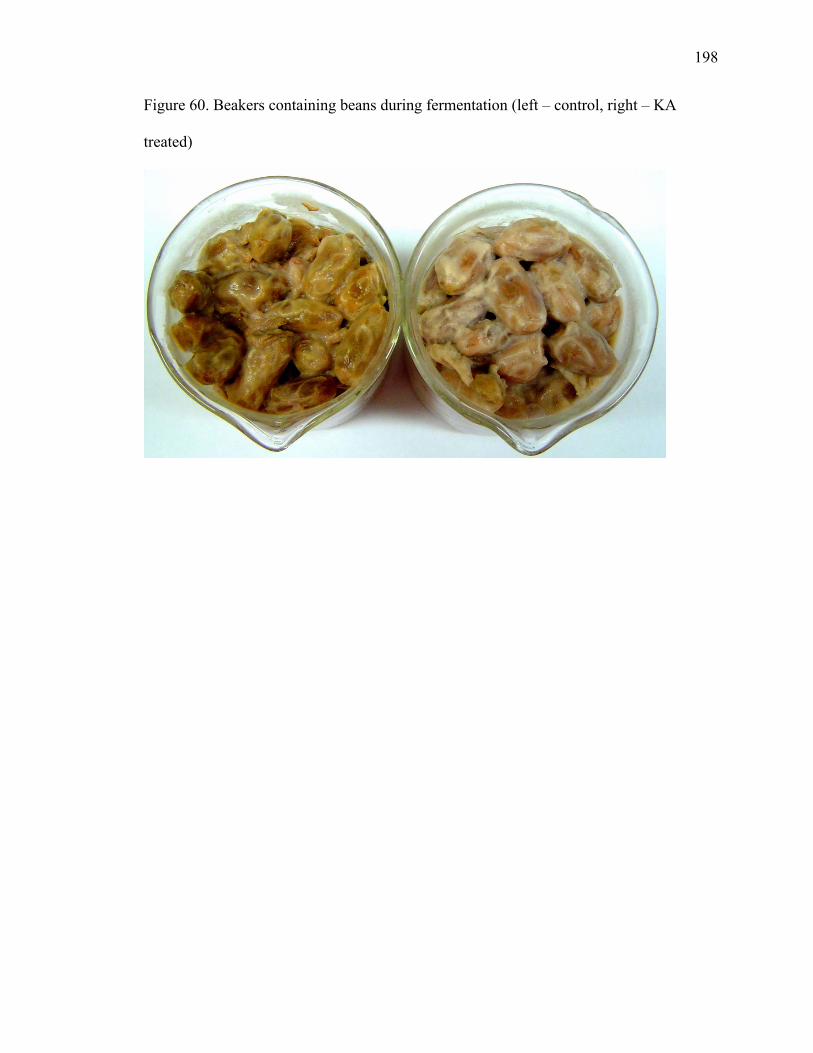
198
Figure 60. Beakers containing beans during fermentation (left – control, right – KA
treated)

199
Figure 61. Cut test showing on the left control beans and on the right KA treated beans
after 7 days of fermentation.

200
Figure 62. Yeast streaked on media containing 10 mM of kojic acid in PDA (right)
compared to PDA alone (left)

201
Figure 63. Total procyanidin content of beans during fermentation*
Time (days)
0 2 4 6 8 10 12 14 16
Pro
cyan
idin
s (m
g. g-1 d
w)
0
50
100
150
200
250
300
ControlKojic acid
* No significant statistical differences were observed using the F test at P.05.

202
Figure 64. Total amino acids in cacao beans during fermentation
Time (days)
0 2 4 6 8 10 12 14 16
Am
ino
acid
s(m
g of
phe
nyla
lani
ne e
quiv
. . g-1
of b
eans
dw
)
0.8
1.0
1.2
1.4
1.6
1.8
2.0
2.2
2.4
2.6
2.8
KA Control
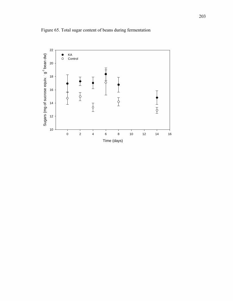
203
Figure 65. Total sugar content of beans during fermentation
Time (days)
0 2 4 6 8 10 12 14 16
Sug
ars
(mg
of s
ucro
se e
quiv
. . g-1
bean
dw
)
10
12
14
16
18
20
22KA Control
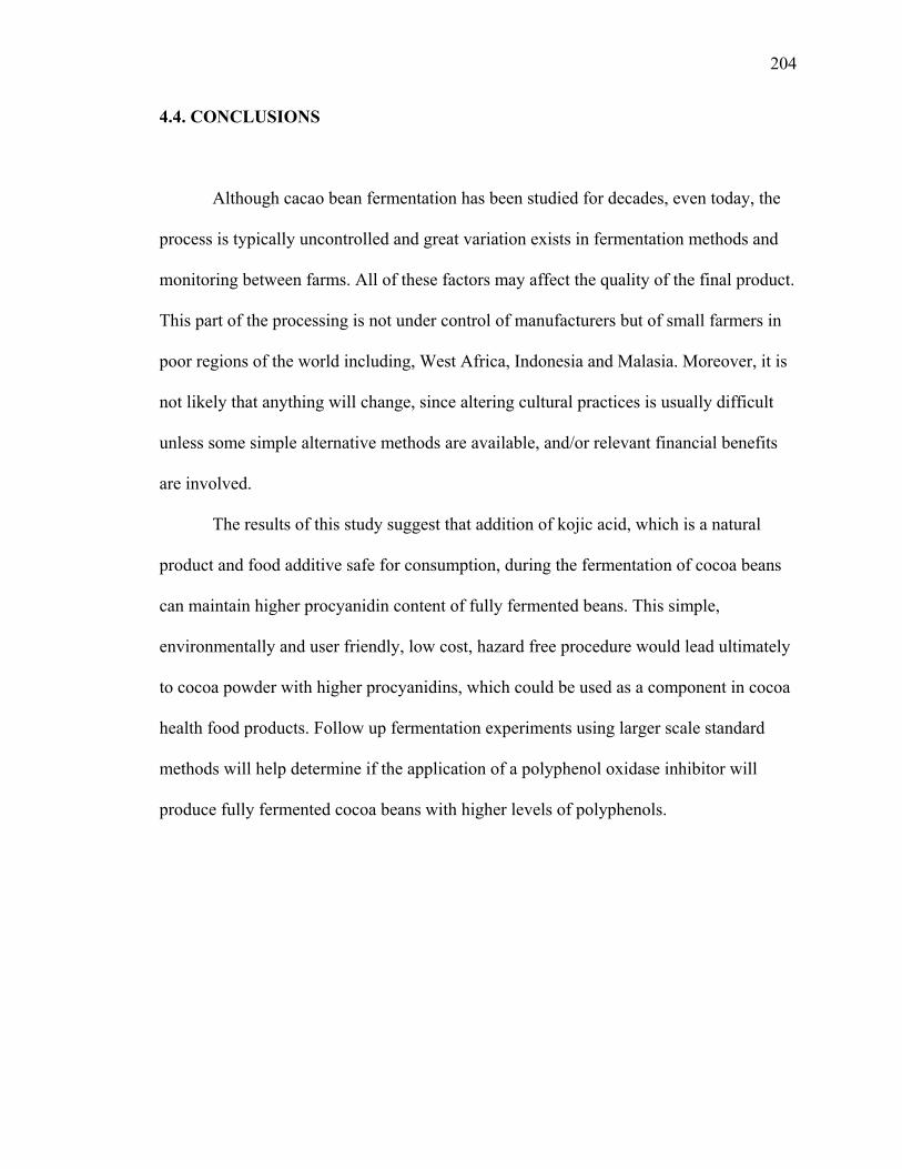
204
4.4. CONCLUSIONS
Although cacao bean fermentation has been studied for decades, even today, the
process is typically uncontrolled and great variation exists in fermentation methods and
monitoring between farms. All of these factors may affect the quality of the final product.
This part of the processing is not under control of manufacturers but of small farmers in
poor regions of the world including, West Africa, Indonesia and Malasia. Moreover, it is
not likely that anything will change, since altering cultural practices is usually difficult
unless some simple alternative methods are available, and/or relevant financial benefits
are involved.
The results of this study suggest that addition of kojic acid, which is a natural
product and food additive safe for consumption, during the fermentation of cocoa beans
can maintain higher procyanidin content of fully fermented beans. This simple,
environmentally and user friendly, low cost, hazard free procedure would lead ultimately
to cocoa powder with higher procyanidins, which could be used as a component in cocoa
health food products. Follow up fermentation experiments using larger scale standard
methods will help determine if the application of a polyphenol oxidase inhibitor will
produce fully fermented cocoa beans with higher levels of polyphenols.

205
5. SUMMARY AND CONCLUDING REMARKS
During this project, problems associated with three main aspects of cacao were
investigated: disease, production and processing. The dissertation was divided into 3
sections: (1) Studies on plant-endophyte-pathogen interactions allowed for the
identification of new possible mechanisms of disease control; (2) Studies on cacao flower
physiology indicated ways to improve pollination and therefore increase fruit set and crop
yield; and (3) Investigations on the fermentation step of cacao processing permitted
discovery of a method for maintaining higher levels of compounds valued by cacao
manufacturers for chocolate production.
Plant defenses, including, endogenous and inducible levels of caffeine,
theobromine, (-)-epicatechin and (+)-catechin, salicylic acid, salicylic acid glucoside
were evaluated in genotypes with different levels of disease resistance in order to
determine a broad spectrum defense response, which may require concurrent production
of several compounds. Cluster analysis demonstrated a stronger genotype influence rather
than elicitor treatment effect on the chemical profile of cacao leaves. This example of
chemical profiling allowed the separation of genotypes with different levels of resistance
based on their overall ability to produce phytoalexins.
Flavan-3-ol monomers and oligomers, from dimers to decamers, were
quantitatively determined by HPLC-FLD in leaves throughout development in four
Theobroma cacao genotypes. The levels of the very young leaves were intermediary and
peaked as leaves attained lengths of 4 to 20 centimeters. As the leaves increased in size

206
and developed further, the procyanidin content reached its minimum. Cacao flush leaves
had higher procyanidin content than mature leaves.
Witches’ broom disease tolerant genotype Scavina 6 had the highest levels of
procyanidins when compared to Amelonado, TSH 565, and IMC 30, more disease
susceptible genotypes, at a stage when leaves are susceptible to infection. Moreover,
leaves of SCA 6 responded to M. perniciosa basidiospore infection by inducing
accumulation of procyanidins in both the infected and adjacent uninfected leaves of
actively growing shoots, demonstrating an inductive response effect both locally and
systemically.
Combined evidence for activation of procyanidin gene expression (Leal et al.
2007), antifungal effects of procyanidins against the witches’ broom pathogen
(Andebrhan et al., 1995; Brownlee et al., 1990), higher endogenous levels of
procyanidins in leaves of a disease resistant genotype, including a peak in procyanidin
levels during leaf development at a stage when leaves are susceptible to witches’ broom
disease, and the local and systemic increase in procyanidins in cacao leaves of a disease
resistant genotype upon infection with basidiospores indicate a role for procyanidins in
defense against Moniliophthora perniciosa. Therefore, procyanidin content of flush
leaves is a potential marker for selecting for disease tolerance in cacao.
Laccase is one of the polyphenoloxidative enzymes produced by M. perniciosa as
a virulence or pathogenicity factor and is therefore a potential target for disease control.
The main procyanidin found in cacao, (-)-epicatechin, can prevent pathogen growth in the
presence of a polyphenoloxidase inhibitor that blocks the ability of the fungus to detoxify
polyphenols, rendering them toxic to fungal growth. Application of polyphenoloxidase

207
inhibitors to cacao may affect the ability of the pathogen to thrive in plants with high
levels of polyphenols.
Salicylic acid (SA), phenyllactic acid and mandelic acid were identified as
products of fungal metabolism by M. perniciosa. Levels of SA were quantified in
infected and uninfected plant tissues as well as in filtrates of fungal cultures. Infected
brooms had five times more SA compared to healthy shoots. Moreover, application of SA
to leaves induced curling and necrosis. In contrast, leaf inoculation with basidiospores or
application of chemical elicitors did not alter leaf SA content, suggesting that in this
infection system, the actively growing mycelia is the source of the SA rather than the
plant. For necrotrophic fungal pathogens, including M. perniciosa, SA may be a
component of pathogenesis and not part of the defense response of plants. Therefore, it is
possible that plants capable of responding to infection more effectively by activating cell
death, when challenged by a necrotrophic pathogen, tend to be more disease susceptible.
Among all tested endophytic fungal isolates, two Aspergillus westerdjikiae were
found to be ochratoxin producers. Media composition had an effect on the metabolites
produced by these fungi. Caffeine, besides affecting the growth of the endophytes,
affected their ability to produce toxins. Caffeine below toxic levels may trigger toxin
production, while at higher levels completely disrupts fungal metabolism. Some toxigenic
fungi may be able to break down caffeine and possibly use the degradation products as
carbon and nitrogen sources.
An A. oryzae isolate was found to produce kojic acid (KA) in culture. When
inoculated in cacao seedlings, A. oryzae grew endophytically and synthesized KA in
planta. Cacao seedlings inoculated with A. oryzae produced higher levels of caffeine than

208
non-inoculated ones. A. oryzae may be a useful endophyte to introduce to cacao since it
grows non-pathogenically and induces the caffeine defense response that may improve
plant resistance to pathogens.
The interactions between studied microorganisms ranged from parasitism to
production of volatile organic compounds, antibiotics or toxins. The environment to
which the organisms are subjected influences their fitness and should be considered when
selecting biological control isolates. In a complex chemical warfare between
microorganisms, understanding the factors influencing their ability to produce secondary
metabolites enables researchers to provide favorable conditions for the target beneficial
organism to succeed.
B. bassiana was isolated from various sections of the cacao pod and peduncle two
and three months after the flowers had been hand-pollinated and sprayed in the
greenhouse with a B. bassiana conidial suspension. These results indicate that spraying
flowers with conidia can result in the establishment of a fungal entomopathogen as an
endophyte, and that the fungus can move within the pod as it grows. This finding could
lead to an innovative way to manage insect pests in biocontrol programs.
Many new volatile organic compounds released by cacao flowers were identified.
It is possible that these compounds, some of which present in smaller amounts than
compounds previously reported in the literature, can contribute to the attraction of cacao
pollinators. Cacao flowers raised their temperatures higher than background tissues
during morning hours when pollination is known to occur. Flower temperature regulation
occurs through the alternative respiration pathway. Endogenous and inducible SA and

209
SAG were found in cacao flowers, and their levels varied between self compatible and
self incompatible genotypes suggesting a possible role in pollination biology.
The fermentation step of cacao processing is much uncontrolled and demands
improvements. Addition of kojic acid, which is a natural product food additive safe for
consumption, to fermenting cocoa beans allowed for reduced procyanidin losses in fully
fermented beans. This simple, environmentally and user friendly, low cost, hazard free
procedure would lead ultimately to cocoa powder with higher procyanidins, which could
be used as a component in cocoa health food products.
Cocoa is and has always been a highly valued crop in the world. Old medicinal
properties are being rediscovered, bringing about new applications for cacao. Yet cacao
production is limited by factors such as pathogens and low productivity. The basis for
unraveling these issues lies in identifying their mechanisms. One approach is through the
analysis of the complex chemical interactions between the plant itself and various
pathogens affecting cacao production.
In nature, many compounds serve multiple purposes. Exploring known
compounds for novel activities uncovers new involvements for these metabolites. Cacao
procyandins are not only important in human health but participate in the plant’s immune
system. It is critical to maintain a holistic perspective of the various agricultural,
processing, and manufacturing aspects of the crop in order to determine its success.

210
REFERENCES Agrios G.N. (2005) Plant Pathology, 5th ed. Academic Press, New York. 922 p. Aime M.C. and Phillips-Mora W. (2005) The causal agents of witches’ broom and frosty pod rot of cacao (chocolate, Theobroma cacao) form a new lineage of Marasmiaceae. Mycologia 97(5):1012-1022. Akello J., Dubois T., Gold C.S., Coyne D., Kakavuma J. and Paparu P. (2007) Beauveria bassiana (Balsamo) Vuillemin as an endophyte in tissue culture banana (Musa spp.). Journal of Invertebrate Pathology 96(1):34-42. Alverson J. (2003) Effects of mycotoxins, kojic acid and oxalic acid, on biological fitness of Lygus hesperus (Heteroptera: Miridae). Journal of Invertebrate Pathology 83:60-62. Andebrhan T., Hammerstone J.F.Jr., Romanczyk L.J.Jr. and Furtek D.B. (1995) Sensitivity of Crinipellis perniciosa to procyanidins from Theobroma cacao L. Physiological and Molecular Plant Pathology 46:339-348. Andebrhan T. and Furtek D.B. (1994) Random amplified polymorphic DNA (RAPD) analysis of Crinipellis perniciosa isolates from different hosts. Plant Pathology 43:1020-1027. Andebrhan T., Figueira A., Yamada M.M., Cascardo J. and Furtek D.B. (1999) Molecular fingerprinting suggests two primary outbreaks of witches’ broom disease (Crinipellis perniciosa) of Theobroma cacao in Bahia, Brazil. European Journal of Plant Pathology 105:167-175. Aneja M., Gianfagna T. and Ng E. (1999) The roles of abscisic acid and ethylene in the abscission and senescence of cocoa flowers. Plant Growth Regulation 27:149-155. Aneja M. and Gianfagna T.J. (2001) Induction and accumulation of caffeine in young, actively growing leaves of cocoa (Theobroma cacao L.) by wounding or infection with Crinipellis perniciosa. Physiological and Molecular Plant Pathology 59:13-16. Aneja M., Gianfagna T.J. and Hebbar P.K. (2006) Trichoderma harzianum produces nonanoic acid, an inhibitor of spore germination and mycelial growth of two cacao pathogens. Physiological and Molecular Plant Pathology, 67(6):304-307. Ardhana M.M. and Fleet G.H. (2003) Microbial ecology of cocoa bean fermentations in Indonesia. International Journal of Food Microbiology 86:87-99. Arnold A.E., Mejia L.C., Kyllo D., Rojas E.I., Maynard Z., Robbins N., and Herre EA. (2003) Fungal endophytes limit pathogen damage in a tropical tree. Proceedings of the National Academy of Sciences USA 100(26):15649-15654.

211
Ashihara H. and Crozier A. (2001) Caffeine: a well known but little mentioned compound in plant science. Trends in Plant Science 6(9):407-413. Bacon C.W., Yates I.E., Hinton D.M. and Meredith F. (2001) Biological control of Fusarium moniliforme in maize. Environmental Health Perspectives 109(2):325-332. Bastide P., Fourny G., Durand N., Petithuguenin P., Guyot B., Gilmour M. and Lindblom M. (2006) Identification of ochratoxin A sources during cocoa post-harvest processing: influence of harvest quality and climatic factors. 15th International Cocoa Research Conference, San Jose, Costa Rica. Baurin N., Arnoult E., Scior T., Do Q.T. and Bernard P. (2002) Preliminary screening of some tropical plants for anti-tyrosinase activity. Journal of Ethnopharmacology 82:155-158. Beno-Moualem D., Vinokur Y. and Prusky D. (2001) Cytokinin increase epicatechin content and fungal decay resistance in avocado fruits. Journal of Plant Growth Regulation 20:95-100. Bentley R. (2006) From miso, sake and shoyu to cosmetics: a century of science for kojic acid. Natural Product Reports 23:1046-1062. Bing L. and Lewis L.C. (1991) Suppression of Ostrinia nubilalis (Hübner) (Lepidoptera: Pyralidae) by endophytic Beauveria bassiana (Balsamo) Vuill. Environmental Entomology 20:1207-1211. Bonhevi J.S. and Coll F.V. (1997) Evaluation of bitterness and astringency of polyphenolic compounds in cocoa powder. Food Chemistry 60(3):365-370. Bonhevi J.S. (2004) Occurrence of ochratoxin A in cocoa products and chocolate. Journal of Agricultural and Food Chemistry 52:6347-6352. Bradford M.M. (1976) A rapid and sensitive method for the quantitation of microgram quantities of protein utilizing the principle of protein-dye binding. Analytical Biochemistry 72:248-254. Bragulat M.R., Abarca M.L. and Cabanes F.J. (2001) An easy screening method for fungi producing ochratoxin A in pure culture. International Journal of Food Microbiology, 71:139-144. Brew A.H. (1987) The effect of colours and specific features of floral parts on the pollination of cocoa by ceratopogonid midges in Ghana. Proceedings of the Tenth International Cocoa Research Conference, Santo Domingo, Dominican Republic, 17-23 May 1987. Cocoa Producers’ Alliance, Lagos, Nigeria, London, UK. p.307-310.
212
Brito E.S., Garcia N.H.P., Gallao M.I., Cortelazzo A.L., Fevereiro P.S., and Braga M.R. (2000) Structural and chemical changes in cocoa (Theobroma cacao L.) during fermentation, drying and roasting. Journal of Food Science and Agriculture 81:281-288. Brownlee H.E., Mceuen A.R., Hedger J. and Scott I.M. (1990) Anti-fungal effects of cocoa tannin on the witches’ broom pathogen Crinipellis perniciosa. Physiological and Molecular Plant Pathology 36:39-48. Buchanan G.O., Williams L.A.D. and Reese P.B. (2000) Biotransformation of cadinane sesquiterpenes by Beauveria bassiana ATCC 7159. Phytochemistry 54:39-45. Buchanan R.L. and Lewis D.F. (1984) Caffeine inhibition of aflatoxin synthesis: probable site of action. Applied and Environmental Microbiology 47(6):1216-1220. Buchanan R.L., Tice G. and Marino D. (1981) Caffeine inhibition of ochratoxin A production. Journal of Food Science 47:319-321. Burdock G.A., Soni M.G. and Carabin I.G. (2001) Evaluation of health aspects of kojic acid in food. Regulatory Toxicology and Pharmacology 33:80-101. Ceita G.O., Macedo J.N.A., Santos T.B, Alemanno L., Gesteira A.S., Micheli F., Mariano A.C., Gramacho K.P., Silva D.C., Meinhardt L., Mazzafera P., Pereira G.A.G. and Cascardo J.C.M. (2007) Involvement of calcium oxalate degradation during programmed cell death in Theobroma cacao tissues triggered by the hemibiotrophic fungus Moniliophthora perniciosa. Plant Science 173:106-117. Champavier Y., Pommier M.-T., Arpin A. and Pellon G. (2000) 10-oxo-trans-8-decenoic acid (ODA): production, biological activities, and comparison with other hormone-like substances in Agaricus bisporus. Enzyme and Microbial Technology 26:243-251. Chatt E.M. (1954) Cocoa – cultivation, processing, analysis. Vol. III in Economic crops by Kertesz ZI. Interscience Publishers, New York. 302p. Chaves F.C. and Gianfagna T.J. (2007) Necrotrophic phase of Moniliophthora perniciosa causes salicylic acid accumulation in infected stems of cacao. Physiological and Molecular Plant Pathology 69:104-108. Chaves F.C. and Gianfagna T.J. (2007) Cacao leaf procyanidins increase locally and systemically in response to infection by Moniliophthora perniciosa basidiospores. Physiological and Molecular Plant Pathology 70:174-179. Chen J.S., Wei C. and Marshall M.R. (1991) Inhibition mechanism of kojic acid on polyphenol oxidase. Journal of Agricultural and Food Chemistry 39(11)1897-1901. Cherry A.J., Banito A., Djegui D. and Lomer C. (2004) Suppression of the stem-borer Sesamia calamistis (Lepidoptera; Noctuidae) in maize following seed dressing, topical

213
application and stem injection with African isolates of Beauveria bassiana. International Journal of Pest Management 50:67-73. Chitarra G.S. (2003) Germination inhibitors of fungal spores: identification and mode of action. Ph.D. thesis Wageningen University, Wageningen, The Netherlands. 120p. Chivasa S., Murphy A.M., Naylor M. and Carr J.P. (1997) Salicylic acid interferes with tobacco mosaic virus via a novel salicylhydroxamic acid-sensitive mechanism. The Plant Cell 9:547-557. Claeson A.-S. and Sunesson A.-L. (2005) Identification using versatile sampling and analytical methods of volatile compounds from Streptomyces albidoflavus grown on four humid building materials and one synthetic medium. Indoor Air 15(Supplement 9):41-47. Claeson A.-S., Levin J.-O., Blomquist G. and Sunesson A.-L. (2002) Volatile metabolites from microorganisms grown on humid building mateials and synthetic media. Journal of Environmental Monitoring 4:667-672. Clarke B.B., White J.F.Jr., Hurley R.H., Torres M.S., Sun S. and Huff DR. (2006) Endophyte-mediated suppression of dollar spot disease in fine fescues. Plant Disease 90:994-998. Cleveland T.E., Dowd P.F., Desjardins A.E., Bhatnagar D. and Cotty P.J. (2003) United States Department of Agriculture–Agricultural Research Service research on pre-harvest prevention of mycotoxins and mycotoxigenic fungi in U.S. crops. Pest Management Science 59:629-642. Cole R.J. and Cox R.H. (1981) Handbook of toxic fungal metabolites. Academic Press; 937p. Cole R.J. and Schweikert M.A. (2003) Handbook of secondary fungal metabolites. Academic Press, San Diego, CA. Costa J.N.M., Silva R.B., Ribeiro P.A. and Garcia A. (2002) Ocorrencia de Beauveria bassiana (Bals.) Vuill. em broca-do-cafe (Hypothenemus hampei, Ferrari) no estado de Rondonia, Brasil. Acta Amazonica 32(3):517-519. Croft K.P.C., Juttner F. and Slusarenko A.J. (1993) Volatile products of the lipoxygenase pathway evolved from Phaseolus vulgaris (L.) leaves inoculated with Pseudomonas syringae pv phaseolicola. Plant Physiology 101:13-24. Cui J., Bahrami A.K., Pringle E.G., Hernandez-Guzman G., Bender C.L., Pierce N.E., and Ausubel F.M. (2005) Pseudomonas syringae manipulates systemic plant defenses against pathogens and herbivores. Proceedings of the National Academy of Sciences USA 102(5):1791-1796.

214
De Marco J.L., Valadares-Inglis M.C. and Felix C.R. (2003) Production of hydrolytic enzymes by Trichoderma isolates with antagonistic activity against Crinipellis perniciosa the causal agent of witches’ broom of cocoa. Brazilian Journal of Microbiology 34:33-38. Dowd P.F. (1999) Relative inhibition of insect phenoloxidase by cyclic fungal metabolites from insect and plant pathogens. Natural Toxins 7:337-341. Dowd P.F. (2002) Antiinsectan compounds derived from microorganisms. In: Koul O, Dhaliwal GS (eds.), Microbial Biopesticides, Taylor & Francis, New York; p.13-115. Duke J.A. (2007) Phytochemical and Ethnobotanical Databases. http://www.ars-grin.gov/duke/; Then click on ethnobotanical uses; then type Theobroma cacao. Durrant W.E. and Dong X. (2004) Systemic acquired resistance. Annual Reviews of Phytopathology 42:185-209. Edwards R., Blount J.W. and Dixon R.A. (1991) Glutathione and elicitation of the phytoalexin response in legume cell cultures. Planta 184(3):403-409. Erickson E.H.Jr., Erickson B.J. and Young A.M. (1992) Evidence of floral thermoregulation in Theobroma cacao and related species. Bee Science 2(2):82-87. Evans H.C., Holmes K.A. and Reid A.P. (2003) Phylogeny of the frosty pod rot pathogen of cocoa. Plant Pathology 52:476-485. Fan Y. and Flurkey W.H. (2004) Purification and characterization of tyrosinase from gill tissue of Portabella mushrooms. Phytochemistry 65:671-678. Fao (2005) data. http://faostat.fao.org/site/567/default.aspx; then select production quantity, cocoa beans, year 2005 and the producing country. Figueira A., Janick J. and Goldsbrough P. (1992) Genome size and DNA polymorphism in Theobroma cacao. Journal of the American Society for Horticultural Science 117(4):673-677. Forsyth W.G.C. (1952) Cacao polyphenolic substances 2. Changes during fermentation. Biochemistry Journal 51:516-520. Frias G.A., Purdy L.H. and Schmidt R.A. (1991) Infection biology of Crinipellis perniciosa on vegetative flushes of cacao. Plant Disease 75(6):552-556. Gauillard F. and Richard-Forget F. (1997) Polyphenoloxidases from Williams pear (Pyrus communis L., cv Williams): activation, purification and some properties. Journal of the Science of Food and Agriculture 74:49-56.

215
Geiser D.M., Dorner J.W., Horn B.W. and Taylor J.W. (2000) The phylogenetics of mycotoxin and sclerotium production in Aspergillus flavus and Aspergillus oryzae. Fungal Genetics and Biology 31:169-179. Gómez-Vidal S., Lopez-Llorca L.V., Jansson H.B. and Salinas J. (2006) Endophytic colonization of date palm (Phoenix dactylifera L.) leaves by entomopathogenic fungi. Micron 37:624-632. Govrin E.M. and Levine A. (2000) The hypersensitive response facilitates plant infection by the necrotrophic pathogen Botrytis cinerea. Current Biology 10:751-757. Guetsky R., Kobiler I., Wang X., Perlman N., Gollop N., Avila-Quezada G., Hadar I. and Prusky D. (2005) Metabolism of the flavonoid epicatechin by laccase of Colletotrichum gloeosporoioides and its effect on pathogenicity on avocado fruits. Phytopathology 95:1341-1348. Gundlach H., Muller M.J., Kutchan T.M. and Zenk M.H. (1992) Jasmonic acid is a signal transducer in elicitor-induced plant cell cultures. Proceedings of the National Academy of Sciences USA 89:2389-2393. Hammerstone J.F., Romanczyk L.J. and Aitken W.M. (1994) Purine alkaloid distribution within Herrania and Theobroma. Phytochemistry 35(5):1237-1240. Hansen C.E., del Olmo M. and Burri C. (1998) Enzyme activities in cocoa beans during fermentation. Journal of the Science of Food and Agriculture 77:273-281. Harborne J.B. and Williams C.A. (2000) Advances in flavonoid research since 1992. Phytochemistry 55:481-504. Harris J.P. and Mantle P.G. (2001) Biosynthesis of ochratoxins by Aspergillus ochraceus. Phytochemistry 58:709-716. Hartman T.G., Lech J., Salinas J., Rosen RT. and Ho C.T. (1993) Flavor characterization using adsorbent trapping thermal desorption or direct thermal desorption-gas chromatography and gas chromatography-mass spectrometry. In C-T Ho, CH Manly, eds, Flavor Measurements. Dekker, New York, pp 37-60. Hasan H.A. (1999) Role of caffeine and tannin in anti-toxigenic proerties of coffee and tea. Cryptogamie Mycologie 20(1):17-21. Hennig J., Malamy J., Grynkiewicz, Indulski J. and Klessig D.F. (1993) Interconversion of the salicylic acid signal in its glucoside in tobacco. The Plant Journal 4(4):593-600. Hoopen G.M.T., Rees R., Aisa P., Stirrup T. and Krauss U. (2003) Population dynamics of epiphytic mycoparasites of the genera Clonostachys and Fusarium for the biocontrol
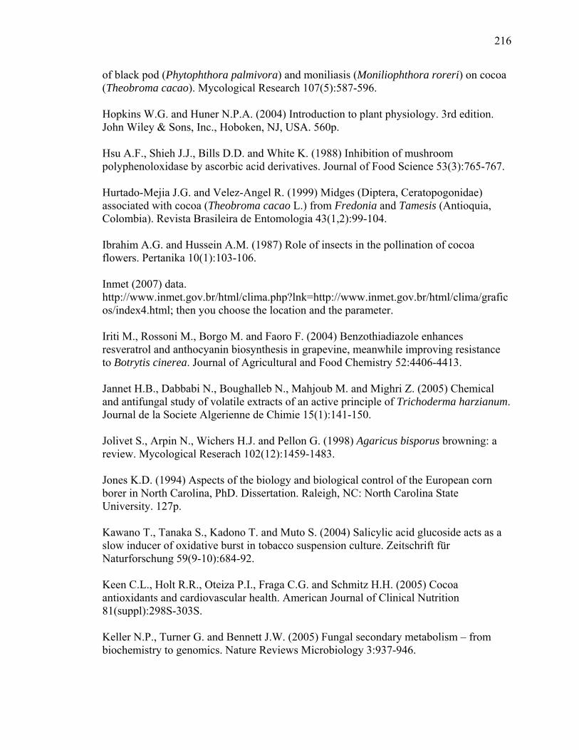
216
of black pod (Phytophthora palmivora) and moniliasis (Moniliophthora roreri) on cocoa (Theobroma cacao). Mycological Research 107(5):587-596. Hopkins W.G. and Huner N.P.A. (2004) Introduction to plant physiology. 3rd edition. John Wiley & Sons, Inc., Hoboken, NJ, USA. 560p. Hsu A.F., Shieh J.J., Bills D.D. and White K. (1988) Inhibition of mushroom polyphenoloxidase by ascorbic acid derivatives. Journal of Food Science 53(3):765-767. Hurtado-Mejia J.G. and Velez-Angel R. (1999) Midges (Diptera, Ceratopogonidae) associated with cocoa (Theobroma cacao L.) from Fredonia and Tamesis (Antioquia, Colombia). Revista Brasileira de Entomologia 43(1,2):99-104. Ibrahim A.G. and Hussein A.M. (1987) Role of insects in the pollination of cocoa flowers. Pertanika 10(1):103-106. Inmet (2007) data. http://www.inmet.gov.br/html/clima.php?lnk=http://www.inmet.gov.br/html/clima/graficos/index4.html; then you choose the location and the parameter. Iriti M., Rossoni M., Borgo M. and Faoro F. (2004) Benzothiadiazole enhances resveratrol and anthocyanin biosynthesis in grapevine, meanwhile improving resistance to Botrytis cinerea. Journal of Agricultural and Food Chemistry 52:4406-4413. Jannet H.B., Dabbabi N., Boughalleb N., Mahjoub M. and Mighri Z. (2005) Chemical and antifungal study of volatile extracts of an active principle of Trichoderma harzianum. Journal de la Societe Algerienne de Chimie 15(1):141-150. Jolivet S., Arpin N., Wichers H.J. and Pellon G. (1998) Agaricus bisporus browning: a review. Mycological Reserach 102(12):1459-1483. Jones K.D. (1994) Aspects of the biology and biological control of the European corn borer in North Carolina, PhD. Dissertation. Raleigh, NC: North Carolina State University. 127p. Kawano T., Tanaka S., Kadono T. and Muto S. (2004) Salicylic acid glucoside acts as a slow inducer of oxidative burst in tobacco suspension culture. Zeitschrift für Naturforschung 59(9-10):684-92. Keen C.L., Holt R.R., Oteiza P.I., Fraga C.G. and Schmitz H.H. (2005) Cocoa antioxidants and cardiovascular health. American Journal of Clinical Nutrition 81(suppl):298S-303S. Keller N.P., Turner G. and Bennett J.W. (2005) Fungal secondary metabolism – from biochemistry to genomics. Nature Reviews Microbiology 3:937-946.
217
Kelm M.A., Johnson J.C., Robbins R.J., Hammerstone J.F. and Schmitz H.H. (2006) High-performance liquid chromatography separation and purification of cacao (Theobroma cacao L.) procyanidins according to degree of polymerization using a diol stationary phase. Journal of Agricultural and Food Chemistry 54(5):1571-1576. Kilaru A., Bailey B.A. and Hasenstein K.H. (2007) Moniliophthora perniciosa produces hormones and alters endogenous auxinand salicylic acid in infected cocoa leaves. Federation of European Microbiological Societies, Microbiology Letters 274:238-244. Kim H. and Keeney P.G. (1984) (-)-Epicatechin content in fermented and unfermented cocoa beans. Journal of Food Science 49:1090-1092. Kim J.Y., Kim D.G., Lee S.H. and Lee I. (2006) Wound-induced expression of the ferulate 5-hydroxylase gene in Camptotheca acuminate. Biochimica et Biophysica Acta 1760:182-190. Kolombet L. V., Zhigletsova S.K., Kosareva N.I., Bystrova E.V., Derbyshev V.V., Krasnova S.P. and Schisler D. (2007) Development of an extended shelf-life, liquid formulation of the biofungicide Trichoderma asperellum. World Journal of Microbiology and Biotechnology doi 10.1007/s11274-007-9449-9. Koyama Y., Tomoda Y., Kato M. and Ashihara H. (2003) Metabolism of purine bases, nucleosides and alkaloids in theobromine-forming Theobroma cacao leaves. Plant Physiology and Biochemistry 41:977-984. Kunkel B.N. and Brooks D.M. (2002) Cross talk between signaling pathways in pathogen defense. Current Opinion in Plant Biology 5:325-331. Kunz W., Schurter R. and Maetzke T. (1997) The chemistry of benzothiadiazole plant activators. Pesticide Science 50:275-282. Kurtzman C.P., Smiley M.J., Robnett C.J. and Wicklow D.T. (1986) DNA relatedness among wild and domesticated species of the Aspergillus flavus group. Mycologia 78(6):955-959. Lacher von V. (1964) Electrophysiological investigations of the separate receptors for smell, carbon dioxide, relative humidity and temperature of the antennae of the worker bee and the drone (Apis melifera L.). Journal of Comparative Physiology A 48(6):587-623. Laemmli U.K. (1970) Cleavage of structural proteins during the assembly of the head of bacteriophage T4. Nature 227:680-685. Lapadatescu C., Ginies C., Quere J.-L.L. and Bonnarme P. (2000) Novel scheme for biosynthesis of aryl metabolites from L-phenylalanine in the fungus Bjerkandera adusta. Applied and Environmental Microbiology 66(4):1517-1522.

218
Leal G.A.Jr., Albuquerque P.S.B. and Figueira A. (2007) Genes differentially expresses in Theobroma cacao associated with resistance to witche’s broom disease caused by Crinipellis perniciosa. Molecular Plant Pathology 8(3):279-292. Leckie B.M. (2002) Effects of Beauveria bassiana mycelia and metabolites incorporated into synthetic diet and fed to larval Helicoverpa zea, and detection of endophytic Beauveria bassiana in tomato plants using PCR and ITS. M.S. thesis. The University of Tennessee, Knoxville. Levanon Y. and Rossetini S.M.O. (1965) A laboratory study of farm processing of cocoa beans for industrial use. Journal of Food Science 30(4):719-722. Luximon-Ramma A., Bahorun T., Soobrattee M.A. and Auoma O.I. (2002) Antioxidant activities of phenolic, proanthocyanidin, and flavonoid components in extracts of Cassia fistula. Journal of Agricultural and Food Chemistry 50:5042-5047. Mantle P.G. (2002) Risk assessment ant the importance of ochratoxins. International Biodeterioration and Biodegradation 50:143-146. Mau J.-L. and Li J.-Y. (2002) Effect of 10-oxo-trans-8-decenoic acid on the growth and enzyme activities of Aspergillus niger and A. oryzae. Fungal Science 17(3,4):65-76. Mayer A.M. and Staples R.C. (2002) Laccase: new functions for an old enzyme. Phytochemistry 60:551-565. McManus P.S., Best V.M. and Voland R.P. (1999) Infection of cranberry flowers by Monilinia oxycocci and evaluation of cultivars for resistance to cottonball. Phytopathology 89:1127-1130. Millenaar F.F., Gonzales-Meler M.A., Fiorani F., Welschen R., Ribas-Carbo M., Siedow J.N., Wagner A.M. and Lamber H. (2001) Regulation of alternative oxidase activity in six wild monocotyledonous species. An in vivo study at the whole root level. Plant Physiology 126:376-387. Misnawi, Jinap S., Jamilah B. and Nazamid S. (2003) Effects of incubation and polyphenol oxidase enrichment on color, fermentation index, procyanidins and adstringency of unfermented and partly fermented cocoa beans. International Journal of Food Science and Technology 38:285-295. Money N.P. (2002) Mushrooms stem cells. Bioessays 24:949-952. Moore A.L. and Siedow J.N. (1991) The regulation and nature of the cyanide-resistant alternative oxidase of plant mitochondria. Biochimica et Biophysica Acta 1059:121-140.

219
Nathanson J.A. (1984) Caffeine and related methyl-xanthines: possible naturally occurring pesticides. Science 226:184-187. Niki T., Mitsuhara I., Seo S., Ohtsubo N. and Ohashi Y. (1998) Antagonistic effect of salicylic acid and jasmonic acid on the expression of pathogenesis-related (PR) protein genes in wounded mature tobacco leaves. Plant Cell Physiology 39(5):500-507. Nojosa G.B.A., Resende M.L.V., Aguilar M.A.G., Bezerra K.M.T. and Anhert D.E. (2003) Componentes fenolicos e enzimas oxidativas em clones de Theobroma cacao resistentes e suscetiveis a Crinipellis perniciosa. Fitopatologia Brasileira 28(2):148-154. Okull D.O., Belman R.B. and Gourama H. (2003) Antifungal activity of 10-oxo-trans-8-decenoic acid and 1-octen-3-ol against Penicillium expansum in potato dextrose agar medium. Journal of Food Protection 66(8):1503-1505. Osman H., Nasarudin R. and Lee SL. (2004) Extracts of cocoa (Theobroma cacao L.) leaves and their antioxidation potential. Food Chemistry 86: 41-46. Pearson L.J. and Marth E.H. (1990) Behavior of Listeria monocytogenes in the presence of methylxanthines - caffeine and theobromine. Journal of Food Protection 53:47-50. Pena-Cortés H., Albrecht T., Prat S., Weiler E.W. and Willmitzer L. (1993) Aspirin prevents wound-induced gene expression in tomato leaves by blocking jasmonic acid biosynthesis. Planta 191(1):123-128. Peterson S.W., Ito Y., Horn B.W. and Goto T. (2001) Aspergillus bombycis, a new aflatoxigenic species and genetic variation in its sibling species, A. nomius. Mycologia 93:689-703. Pieterse C.M.J. and van Loon L.C. (1999) Salicylic acid-independent plant defence pathways. Trends in Plant Science 4(2):52-58. Pitt J.I. (1973) A laboratory guide to common Penicillium species. Commonwealth Scientific and Industrial Research Organization Division of Food Processing. Porter L.J., Ma Z. and Chan B.G. (1991) Cacao procyanidins: major flavanoids and identification of some minor metabolites. Phytochemistry 30(5):1657-1663. Posada F. and Vega F.E. (2005) Establishment of the fungal entomopathogen Beauveria bassiana (Ascomycota: Hypocreales) as an endophyte in cocoa seedlings (Theobroma cacao). Mycologia 97(6):1195-1200. Prawoto A.A. and Atmawinata O. (1995) Influence of ethephon on flowering and fruiting of cocoa (Theobroma cacao L.). Pelita Perkebunan 11(1):1-21.

220
Purdy L.H. and Schimidt R.A. (1996) Status of cacao witches’ broom: biology, epidemiology, and management. Annual Reviews in Phytopathology 34:573-594. Quesada-Moraga E., Landa B.B., Muñoz-Ledesma J., Jiménez-Diáz R.M. and Santiago-Álvarez C. (2006) Endophytic colonization of opium poppy, Papaver somniferum, by an entomopathogenic Beauveria bassiana strain. Mycopathologia 161:323-329. Raskin I., Turner I.M. and Melander W.R. (1989) Regulation of heat production in the inflorescences of an Arum lily by endogenous salicylic acid. Proceeding of the National Academy of Sciences USA 86:2214-2218. Rate D.N., Cuenca J.V., Bowman G.R., Guttman D.S., Greenberg J.T. (1999) The gain-of-function arabidopsis acd6 mutant reveals novel regulation and function of the salicylic acid signaling pathway in controlling cell death, defenses, and cell growth. The Plant Cell 11:1695-1708. Rescigno A., Zucca P., Flurkey A., Inlow J. and Flurkey W.H. (2007) Identification and discrimination between some contaminant enzyme activities in commercial preparations of mushroom tyrosinase. Enzyme and microbial technology 41:620-627. Resende M.L.V., Nojosa G.B.A., Cavalcanti L.S., Aguilar M.A.G., Silva L.H.C.P., Perez J.O., Andrade G.C.G., Carvalho G.A. and Castro R.M. (2002) Induction of resistance in cocoa against Crinipellis perniciosa and Verticilium dahalie by acilbenzolar-S-methyl (ASM). Plant Pathology 51:621-628. Ritchie S.A., Essen P.H.A.van, Kemme J.A., Kay B.H. and Allaway D. (1994) Response of biting midges (Diptera: Ceratopogonidae) to carbon dioxide, octenol, and light in southeastern Queensland, Australia. Journal of Medical Entomology 31(5):645-648. Robbins M.P., Hartnoll J. and Morris P. (1991) Phenylpropanoid defence responses in transgenic Lotus corniculatus 1. Glutathione elicitation of isoflavan phytoalexins in transformed root cultures. Plant Cell Reports 10(2):59-62. Rohan T.A. (1964) The precursors of chocolate aroma: a comparative study of fermented and unfermented cocoa beans. Journal of Food Science 29(4):456-459. Rohan T.A and Stewart T. (1967) Precursors of chocolate aroma: studies in the degradation of amino acids during the roasting of accra cocoa beans. Journal of Food Science 32:625-628. Rosen H. (1957) A modified nynhydrin colorimetric analysis for amino acids. Archives of Biochemistry and Biophysics 67:10-15. Rubini M.R., Silva-Ribeiro R.T., Pomella A.W.V., Maki C.S., Araújo W.L., Santos D.R. and Azevedo J.L. (2005) Diversity of endophytic fungal community of cacao

221
(Theobroma cacao L.) and biological control of Crinipellis perniciosa, causal agent of witches' broom disease. International Journal of Biological Sciences 1:24-33. Sadik C.D., Sies H. and Schewe T. (2003) Inhibition of 15-lipoxygenases by flavonoids: structure-activity relations and mode of action. Biochemical Pharmacology 65:773-781. Saltveit M.E. (2005) Aminoethoxyvinylglycine (AVG) reduces ethylene and protein biosynthesis in excised discs of mature-green tomato pericarp tissue. Postharvest Biology and Technology 35:183-190. Sano H., Seo S., Koizumi N., Niki T., Iwamura H. and Ohashi Y. (1996) Regulation by cytokinins of endogenous levels of jasmonic and salicylic acids in mechanically wounded tobacco. Plant and Cell Physiology 37(6):762-769. Scarpari L.M., Meinhardt L.W., Mazzafera P., Pomella A.W.V., Schiavinato M.A., Cascardo J.C.M. and Pereira G.A.G. (2005) Biochemical changes during the development of witches’ broom: the most important disease of cocoa in Brazil caused by Crinipellis perniciosa. Journal of Experimental Botany 56(413):865-877. Schirmböck M., Lorito M., Wang I.L., Hayes C.K., Arisan-Atac I., Scala F., Harman G.E. and Kubicek C.P. (1994) Parallel formation and synergism of hydrolytic enzymes and peptaibol antibiotics, molecular mechanisms involved in the antagonistic action of Trichoderma harzianum against phytopathogenic fungi. Applied Environmental Microbiology 60:4364-4370. Schroeter H., Heiss C., Balzer J., Kleinbongard P., Kenn C.L., Hollenberg N.K., Sies H., Kwik-Uribe C., Schmitz H.H. and Kelm M. (2006) (-)-Epicatechin mediates beneficial effects of flavanol-rich cocoa on vascular function in humans. Proceedings of the National Academy of Sciences USA 103(4):1024-1029. Schulz B. and Boyle C. (2005) The endophytic continuum. Mycological Research 109:661-686. Schwan R.F. and Wheals A.E. (2004) The microbiology of cocoa fermentation and its role in chocolate quality. Critical Reviews in Food Science and Nutrition 44:1-17. Selmi C., Mao T.K., Keen C.L., Schmitz H.H. and Gershwin E. (2006) The anti-inflammatory properties of cocoa flavanols. Journal of Cardiovascular Pharmacology 47(2):S163-S171. Seymour R.S. (2001) Biophysics and physiology of temperature regulation in thermogenic flowers. Bioscience Reports 21(2):223-236. Shi W.B. and Feng M.G. (2004) Lethal effect of Beauveria bassiana, Metarhizium anisopliae, and Paecilomyces fumosoroseus on the eggs of Tetranychus cinnabarinus

222
(Acari: Tetranychidae) with a description of a mite egg bioassay system. Biological Control 30(2):165-173. Shi Y.-L., Benzie I.F.F. and Buswell J.A. (2002) Role of tyrosinase in the genoprotective effect of the edible Mushroom, Agaricus bisporus. Life Science 70:1595-1608. Soliman K.M. (2002) Incidence, level, and behavior of aflatoxins during coffee bean roasting and decaffeination. Journal of Agricultural and Food Chemistry 50:7477-7481. Son S.M., Moon K.D. and Lee C.Y. (2001) Inhibitory effects of various antibrowning agents on apple slices. Food Chemistry 73:23-30. Strasser H., Abendstein D., Stuppner H. and Butt T.M. (2000) Monitoring the distribution of secondary metabolites produced by the entomogenous fungus Beauveria brongniartii with particular reference to oosporein. Mycological Research 104 (10):1227-1233. Strobel G.A., Dirkse E., Sears J. and Markworth C. (2001) Volatile antimicrobials from Muscodor albus, a novel endophytic fungus. Microbiology 147:2943-2950. Suarez-Quiroz M.L., Gonzalez-Rios O., Barel M., Guyot B., Schorr-Galindo S. and Guiraud J.P. (2004) Effect of chemical and environmental factors on Aspergillus ochraceus growth and toxigenesis in green coffee. Food Microbiology 21:629-634. Taniwaki M.H., Pitt J.I., Teixeira A.A. and Iamanaka B.T. (2003) The source of ochratoxin A in Brazilian coffee and its formation in relation to processing methods. International Journal of Food Microbiology 82:173-179. Tellez-Tellez M., Diaz-Godinez G. and Sanchez C. (2003) Physiology of a colony of Pleurotus pulmonarius grown on medium overlaid with a Cellophane membrane. Applied Microbiology and Biotechnology 63:212-216. Thaler J.S., Karban R., Ullman D.E., Boege K. and Bostock R.M. (2002) Cross-talk between jasmonate and salicylate plant defense pathways: effects on several plant parasites. Oecologia 131:227-235. Tokusoglu O. and Unal M.K. (2002) Optimized method for simultaneous determination of cathechin, gallic acid, and methylxanthine compounds in chocolate using RP-HPLC. European Food Research and Technology 215:340-346. Tondje P.R., Roberts D.P., Bon M.C., Widmer T., Samuels G.J., Ismaiel A., Begoude A.D., Tchana T., Nyemb-Tshomb E., Ndoumbe-Nkeng M., Bateman R., Fontem D., Hebbar K.P. (2007) Isolation and identification of mycoparasitic isolates of Trichoderma asperellum with potential for suppression of black pod disease of cacao in Cameroon. Biological Control 43:202-212.

223
Tsubouchi H., Terada H., Yamamoto K., Hisada K. and Sakabe Y. (1985) Caffeine degradation and increased ochratoxin A production by toxigenic strains of Aspergillus ochraceus isolated from green coffee beans. Mycopathologia 90:181-186. US Environmental Protection Agency (EPA) (1997) Biotechnology program under toxic substances control act. Aspergillus oryzae, Final risk assessment, February, 1997. Vanacker H., Lu H., Rate D.N. and Greenberg J.T. (2001) A role for salicylic acid and NPR1 in regulating cell growth in Arabidopsis. The Plant Journal 28(2):209-216. Varga J., Rigo K. and Teren J. (2000) Degradation of ochratoxin A by Aspergillus species. International Journal of Food Microbiology 59:1-7. Vega F.E. and Mercadier G. (1998) Insects, coffee and ochratoxin A. Florida Entomologist 81(4):543-544. Vega F.E., Jackson M.A. and McGuire M.R. (1999) Germination of conidia and blastospores of Paecilomyces fumosoroseus on the cuticle of the silverleaf whitefly, Bemisia argentifolii. Mycopathologia 147(1):33-35. Vega F.E., Posada F., Gianfagna T.J., Chaves F.C. and Peterson S.W. (2006) An insect parasitoid carrying an ochratoxin producing fungus. Naturwissenschaften 93: 297-299. Vega F.E., Posada F., Peterson S.W., Gianfagna T.J. and Chaves F. (2006) Penicillium species endophytic in coffee plants and ochratoxin A production. Mycologia 98(1):37-48. Verberne M.C., Brouwer N., Delbianco F., Linthorst H.J.M., Bol J.F., Verpoorte R. (2002) Method for the extraction of the volatile compound salicylic acid from tobacco leaf material. Phytochemical Analysis 13:45-50. Verica J.A., Maximova S.N., Strem M.D., Carlson J.E., Bailey B.A. and Guiltinan M.J. (2004) Isolation of ESTs from cacao (Theobroma cacao L.) leaves treated with inducers of the defense response. Plant Cell Reports 23:404-413. Wagner B.L. and Lewis L.C. (2000) Colonization of corn, Zea mays, by the entomopathogenic fungus Beauveria bassiana. Applied Environmental Entomology 66:3468-34. Waite J.H. (1976) Calculating extinction coefficients for enzymatically produced o-quinones. Analytical Biochemistry 75:211-218. Wheeler B.E.J. and Mepsted R. (1988) Pathogenic variability amongst isolates of Crinipellis perniciosa from cocoa (Theobroma cacao). Plant Pathology 37:475-488. White J.F.Jr. and Morgan-Jones G. (1996) Morphological and physiological adaptations of Balansiae and trends in the evolution of grass endophytes. In: Endophytic fungi in
224
grasses and woody plants: systematics, ecology, and evolution. Eds. Redlin SC, Carris LM. APS Press; p.133-154. Wicklow D.T., Dowd P.F., Alfatafta A.A. and Gloer J.B. (1996) Ochratoxin A: an antiinsectan metabolite from the sclerotia of Aspergillus carbonarius NRRL 369. Canadian Journal of Microbiology 42(11):1100-1103. Wilson D. and Carroll G.C. (1997) Avoidance of high-endophyte space by gall-forming insects. Ecology 78(7):2153-2163. Wollgast J. and Anklam E. (2000) Review on polyphenols in Theobroma cacao: changes in composition during the manufacture of chocolate and methodology for identification and quantitation. Food Research International 33:423-447. Wood G.A. and Lass R.A. (1987) Cocoa. 4th Ed. Longman Scientific and Technical. 620p. Wraight S.P., Carruthers R.I., Jaronski S.T., Bradley C.A., Garza C.J. and Galaini-Wraight S. (2000) Evaluation of the entomopathogenic fungi Beauveria bassiana and Paecilomyces fumosoroseus for microbial control of the silverleaf whitefly, Bemisia argentifolii. Biological Control 17(3):203-217. Yemm E.W. and Willis A.J. (1954) The estimation of carbohydrates in plant extracts by Anthrone. Biochemistry 57:508-514. Young A.M. (1986) Habitat differences in cocoa tree flowering, fruit set, and pollinator availability in Costa Rica. Journal of Tropical Ecology 2(2):163-186. Young A.M. (2007) The chocolate tree – a natural history of cacao. University Press of Florida, Gainsville, FL. 218p. Young A.M. and Severson D.W. (1994) Comparative analysis of stem distilled floral oils of cacao cultivars (Theobroma cacao L., Sterculiaceae) and attraction of flying insects: implications for a Theobroma pollination syndrome. Journal of Chemical Ecology 20(10):2687-2703. Young A.M., Schaller M. and Strand M. (1984) Floral nectaries and trichomes in relation to pollination in some species of Theobroma and Herrania (Sterculiaceae). American Journal of Botany 71(4):466-480. Zhang X. and Flurkey W.H. (1997) Phenoloxidases in Portabella mushrooms. Journal of Food Science 62(1):97-100. Zheng X.-Q., Koyama Y., Nagai C. and Ashihara H. (2004) Biosynthesis, accumulation and degradation of theobromine in developing Theobroma cacao fruits. Journal of Plant Physiology 161:363-369.

225
Zhu Q.Y., Hammerstone J.F., Lazarus S.A., Schmitz H.H., and Keen C.L. (2003) Stabilizing effect of ascorbic acid on flavan-3-ols and dimeric procyanidins from cocoa. Journal of Agricultural and Food Chemistry 51:828-833. Zouchova Z., Wurst M., Nerud F. and Musilek V. (1982) Metabolism of aromatic acids in the antibiotic-producing basidiomycete Oudemansiella mucida. Folia Microbiologica 27(6):446-449.

226
CURRICULUM VITA
FÁBIO C. CHAVES
EDUCATION 2008 Ph. D. Plant Biology, Dept. of Plant Biology and Pathology, Rutgers University 2003 M. S. Agrochemistry, Dept. of Chemistry, Federal University of Viçosa, Brazil 2001 B. S. Chemistry, Catholic University of Pelotas, Brazi 2001 B. S. Agronomy, Federal University of Pelotas, Brazil PUBLICATIONS Chaves F.C. and Gianfagna T.J. (2007) Cacao leaf procyanidins increase locally and systemically in response to infection by Moniliophthora perniciosa basidiospores. Physiological and Molecular Plant Pathology 70:174-179. Kalkunte S.S., Singh A.P., Chaves F.C., Gianfagna T.J., Pundir V.S., Jaisawal A.K., Vorsa N. and Sharma S. (2007) Antidepressant and antistress activity of GC-MS characterized lipophilic extracts of Ginkgo biloba leaves. Phytotherapy Research 21(11):1061-1065. Chaves F.C. and Gianfagna T.J. (2006) Necrotrophic phase of Moniliophthora perniciosa causes salicylic acid accumulation in infected stems of cacao. Physiological and Molecular Plant Pathology 69:104-108. Chaves F.C., Barbosa L.C.A., Demuner A.J. and Silva A.A. (2006) New helminthosporal analogues with plant-growth regulatory properties synthesized via oxyallyl cation Zeitschrift für Naturforschung 61b, 1287-1294. Vega F.E., Posada F., Gianfagna T.J., Chaves F.C. and Peterson S.W. (2006) An insect parasitoid carrying and ochratoxin producing fungus. Naturwissenchaften 93:297-299. Vega F.E., Posada F., Peterson S.W., Gianfagna T.J. and Chaves F.C. (2006) Penicillium species endophytic in coffee plants and ochratoxin A production. Mycologia 98(1):37-48.

227
RESEARCH, TEACHING and PROFESSIONAL EXPERIENCE 2007-Present Research scientist at Mars Inc. at the Mars Center for Cocoa Science, in Bahia, Brazil. 2003-present PhD program at Rutgers University 2005 Part time lecturer of Principles of Biology at Rutgers University 2001-2003 Masters program at Federal University of Viçosa 2000 High school teacher of chemistry in Pelotas, RS 2000 Laboratory assistant in the Plant Disease Diagnosis laboratory at Embrapa, Pelotas, RS 1999 Laboratory assistant in the Plant Biology Department, Rutgers University. 1998 CAPES fellow for international exchange program, Plant Science Major, Cook College, Rutgers University. 1995-1997 Laboratory Assistant in the Food Chemistry Laboratory, Department of Science and Technology for Agroindustry, Federal Unversity of Pelotas