Braunwalds heart disease review and assessment.
311
description
Â
Transcript of Braunwalds heart disease review and assessment.

Use of the current edition of the electronic version of this book (eBook) is subject to the terms of the nontransferable, limited license granted on expertconsult.inkling.com. Access to the eBook is limited to the first individual who redeems the PIN, located on the inside cover of this book, at expertconsult.inkling.com and may not be transferred to another party by resale, lending or other means.
Any screen. Any time. Anywhere.Activate the eBook version of this title at no additional charge.
Unlock your eBook today.1 Visit expertconsult.inkling.com/redeem
2 Scratch off your code
3 Type code into “Enter Code” box
4 Click “Redeem”
5 Log in or Sign Up
6 Go to “My Library”
It’s that easy!
Expert Consult eBooks give you the power to browse and find content, view enhanced images, share notes and highlights—both online and offline.
For technical assistance: email [email protected] call 1-800-401-9962 (inside the US) call +1-314-447-8200 (outside the US)
Scan this QR code to redeem your eBook through your mobile device:
e.

Designed as companions to the bestsellingBraunwald’s Heart Disease: A Textbook ofCardiovascular Medicine, the Braunwaldsuite includes titles covering everythingfrom valvular heart disease to lipidologyto hypertension, ensuring you’re equippedwith all the cardiology resources youneed for e� ective care.
Visit us.elsevierhealth.com/BraunwaldFamily to shop the entire collection today!
The family that keeps families together.
Better Together.
NEW!NEW!

Shop online at us.elsevierhealth.com/BraunwaldFamily
Stay current with recent developments inthe � eld, improved patient managementstrategies, and new drug therapies andimplantable devices that will make adi� erence in your patients’ lives.
EXPLORE the entire Braunwald family of resources!
This reference contains focused chapters on how to utilize cutting-edge interventional technologies, with an emphasis on the latest protocols and standards of care.
From basic science to pathogenesis of atherothrombotic disease, this reference o� ers unparalleled coverage and expert guidance on lipidology in a straightforward, accessible, and user-friendly style.
Diabetes inCardiovascular Disease: A Companion to Braunwald’s Heart DiseaseMcGuire978-1-4557-5418-2
Heart Failure: A Companion to Braunwald’s Heart Disease, 3rd EditionMann & Felker978-1-4557-7237-7
CardiovascularIntervention:A Companion to Braunwald’s Heart DiseaseBhatt978-0-323-26219-4
Clinical Lipidology: A Companion to Braunwald’s Heart Disease, 2nd EditionBallantyne978-0-323-28786-9
This interdisciplinary resource bridges the gap between the cardiology and endocrinology communities of scientists and care providers, and highlights the emerging scienti� cand clinical topics that are relevant forcardiologists, diabetologists/endocrinologists, and the extended diabetes care team.
Valvular Heart Disease: A Companion to Braunwald’s Heart Disease, 4th EditionOtto & Bonow978-1-4557-4860-0
Give your patients the most accuratediagnoses, the best possible heart disease treatment options, and the expert care they deserve with this indispensable resource for your everyday practice.
Cardiovascular Therapeutics :A Companion to Braunwald’s Heart Disease, 4th EditionAntman978-1-4557-0101-8
Manage cardiovascular problems moree� ectively with this comprehensiveresource, which addresses pharmacological, interventional, and surgical management approaches for each type of cardiovascular disease.
Vascular Medicine: A Companion to Braunwald’s Heart Disease, 2nd EditionCreager, Beckman, & Loscalzo978-1-4377-2930-6
Make the most of today’s innovative medical therapies, advances in vascular imaging, and new drugs to improve your patients’cardiovascular health.
Hypertension: A Companion to Braunwald’s Heart Disease, 2nd EditionBlack & Elliott978-1-4377-2766-1
This respected cardiology reference covers everything you need to know to e� ectively manage the chronic problems of yourhypertensive patients.
Clinical Arrhythmology and Electrophysiology: A Companion to Braunwald’s Heart Disease, 2nd EditionIssa, Miller, & Zipes978-1-4557-1274-8
With its unique, singular focus on the clinical aspect of cardiac arrhythmias, this title makes it easy to apply today’s most up-to-date guidelines for diagnosis and treatment.
Acute CoronarySyndromes: A Companion to Braunwald’s Heart Disease, 2nd EditionTheroux978-1-4160-4927-2
Dr. Pierre Theroux and his team of expert contributors present advances in diagnostic and imaging techniques, such as biomarkers, nuclear cardiology, echocardiography, and multislice CT; secondary prevention; and new antiplatelet, anti-ischemic, and gene therapies.
Preventive Cardiology: A Companion to Braunwald’s Heart DiseaseBlumenthal, Foody & Wong978-1-4377-1366-4
Address the prevention and risk strati� cation of cardiovascular disease so that you can delay the onset of disease and moderate the e� ects and complications.
Mechanical Circulatory Support: A Companion to Braunwald’s Heart DiseaseKormos & Miller978-1-4160-6001-7
Access the clinically relevant information you need to e� ectively use this therapy to treat and manage end-stage cardiovascular disease.

BRAUNWALD’S HEART DISEASE REVIEW AND ASSESSMENT

BRAUNWALD’S HEART DISEASE REVIEW AND ASSESSMENT10TH EDITION
Leonard S. Lilly, MDProfessor of MedicineHarvard Medical SchoolChief, Brigham and Women’s/Faulkner CardiologyBrigham and Women’s HospitalBoston, Massachusetts

1600 John F. Kennedy Blvd.Ste 1800Philadelphia, PA 19103-2899
BRAUNWALD’S HEART DISEASE REVIEW AND ASSESSMENT, TENTH EDITION
ISBN: 978-0-323-34134-9
Copyright © 2016 by Elsevier, Inc. All rights reserved.
No part of this publication may be reproduced or transmitted in any form or by any means, electronic or mechanical, including photocopying, recording, or any information storage and retrieval system, without permission in writing from the publisher. Details on how to seek permission, and further information about the Publisher’s permissions policies and our arrangements with organizations such as the Copyright Clearance Center and the Copyright Licensing Agency can be found at our website: www.elsevier.com/permissions.
This book and the individual contributions contained in it are protected under copyright by the Publisher (other than as may be noted herein).
Notices
Knowledge and best practice in this field are constantly changing. As new research and experience broaden our understanding, changes in research methods, professional practices, or medical treatment may become necessary.
Practitioners and researchers must always rely on their own experience and knowledge in evaluating and using any information, methods, compounds, or experiments described herein. In using such information or methods they should be mindful of their own safety and the safety of others, including parties for whom they have a professional responsibility.
With respect to any drug or pharmaceutical products identified, readers are advised to check the most current information provided (i) on procedures featured or (ii) by the manufacturer of each product to be administered, to verify the recommended dose or formula, the method and duration of administration, and contraindications. It is the responsibility of practitioners, relying on their own experience and knowledge of their patients, to make diagnoses, to determine dosages and the best treatment for each individual patient, and to take all appropriate safety precautions.
To the fullest extent of the law, neither the Publisher nor the authors, contributors, or editors assume any liability for any injury and/or damage to persons or property as a matter of products liability, negligence or otherwise, or from any use or operation of any methods, products, instructions, or ideas contained in the material herein.
Previous editions copyrighted 2012, 2008, 2006, 2001, 1997, 1992, and 1989.
Library of Congress Cataloging-in-Publication DataBraunwald’s heart disease : review and assessment / [edited by] Leonard S. Lilly.—Tenth edition. p. ; cm. title: Heart disease review and assessment “Study guide designed to accompany the tenth edition of Braunwald’s heart disease: a textbook of cardiovascular medicine, edited by Dr. Douglas Mann, Dr. Douglas Zipes, Dr. Peter Libby, and Dr. Robert Bonow”—Preface. Includes bibliographical references. ISBN 978-0-323-34134-9 (pbk. : alk. paper) I. Lilly, Leonard S., editor. II. Braunwald’s heart disease. Tenth edition. Guide to (work): III. Title: Heart disease review and assessment. [DNLM: 1. Heart Diseases—Examination Questions. WG 18.2] RC669.2 616.1′20076—dc23
2015004713
Printed in the United States of America
Last digit is the print number: 9 8 7 6 5 4 3 2 1
Content Strategist: Dolores MeloniContent Development Specialist: Jennifer EhlersPublishing Services Manager: Catherine JacksonSenior Project Manager: Rachel E. McMullenDesign Direction: Xiaopei Chen

vii
Contributors
Marc P. Bonaca, MDCardiovascular DivisionBrigham and Women’s HospitalBoston, MassachusettsSections IV and V
Akshay Desai, MDCardiovascular DivisionBrigham and Women’s HospitalBoston, MassachusettsSection II
Neal K. Lakdawala, MDCardiovascular DivisionBrigham and Women’s HospitalBoston, MassachusettsSection III
Bradley A. Maron, MDCardiovascular DivisionBrigham and Women’s Hospital;Department of CardiologyBoston VA Healthcare SystemBoston, MassachusettsSection IV
Amy Miller, MD, PhDCardiovascular DivisionBrigham and Women’s HospitalBoston, MassachusettsSection I
Fidencio Saldaña, MDCardiovascular DivisionBrigham and Women’s HospitalBoston, MassachusettsSection IV
Victor Soukoulis, MD, PhDDivision of Cardiovascular MedicineUniversity of VirginiaCharlottesville, VirginiaSection I
Garrick Stewart, MDCardiovascular DivisionBrigham and Women’s HospitalBoston, MassachusettsSection II
Neil Wimmer, MDCardiovascular DivisionBrigham and Women’s HospitalBoston MassachusettsSection III

ix
Preface
Review and Assessment is a comprehensive study guide designed to accompany the tenth edition of Braunwald’s Heart Disease: A Textbook of Cardiovascular Medicine, edited by Dr. Douglas Mann, Dr. Douglas Zipes, Dr. Peter Libby, and Dr. Robert Bonow. It consists of more than 700 questions that address key topics in the broad field of car-diovascular medicine. A detailed answer is provided for each question, often comprising a “mini-review” of the subject matter. Each answer refers to specific pages, tables, and figures in Braunwald’s Heart Disease and in most cases to additional pertinent citations. Topics of greatest clinical relevance are emphasized, and subjects of particular impor-tance are intentionally reiterated in subsequent questions for reinforcement.
Review and Assessment is intended primarily for cardiol-ogy fellows, practicing cardiologists, internists, advanced medical residents, and other professionals wishing to review contemporary cardiovascular medicine in detail. The subject matter is suitable to help prepare for the Sub-specialty Examination in Cardiovascular Disease offered by the American Board of Internal Medicine.
All questions and answers in this book were designed specifically for this edition of Review and Assessment. I am grateful for the contributions by my colleagues at Brigham and Women’s Hospital who expertly authored new questions and updated material carried forward from the
previous edition: Dr. Marc Bonaca, Dr. Akshay Desai, Dr. Neal Lakdawala, Dr. Bradley Maron, Dr. Amy Miller, Dr. Fidencio Saldaña, Dr. Victor Soukoulis, Dr. Garrick Stewart, and Dr. Neil Wimmer. I acknowledge with great apprecia-tion Dr. Sara Partington and Dr. Alfonso Waller for submit-ting new noninvasive images, and the following colleagues provided additional material or support to this edition: Dr. Ron Blankstein, Dr. Sharmila Dorbala, Dr. Dan Halpern, and Dr. Raymond Kwong. I also warmly thank the Brigham and Women’s Hospital team of cardiac ultrasonographers, led by Jose Rivero, who expertly obtained and alerted us to several of the images that appear in this book.
It has been a pleasure to work with the editorial and production departments of our publisher, Elsevier, Inc. Spe-cifically, I thank Ms. Jennifer Ehlers, Ms. Dolores Meloni, and Ms. Rachel McMullen for their expertise and profes-sionalism in the preparation of this edition of Review and Assessment.
Finally, I am extremely thankful to my family for their support and patience during the often-long hours required to prepare this text.
On behalf of the contributors, I hope that you find this book a useful guide in your review of cardiovascular medicine.
Leonard S. Lilly, MDBoston, Massachusetts

xi
SECTION I (Chapters 1 to 20)Fundamentals of Cardiovascular Disease; Genetics and Personalized Medicine; Evaluation of the Patient 1Amy Miller, Victor Soukoulis, and Leonard S. Lilly
Questions 1Answers, Explanations, and References 45
SECTION II (Chapters 21 to 40)Heart Failure; Arrhythmias, Sudden Death, and Syncope 81Akshay Desai, Garrick Stewart, and Leonard S. Lilly
Questions 81Answers, Explanations, and References 103
SECTION III (Chapters 41 to 61)Preventive Cardiology; Atherosclerotic Cardiovascular Disease 141Neal K. Lakdawala, Neil Wimmer, and Leonard S. Lilly
Questions 141Answers, Explanations, and References 159
SECTION IV (Chapters 62 to 75)Diseases of the Heart, Pericardium, and Pulmonary Vascular Bed 195Fidencio Saldaña, Bradley A. Maron, Marc P. Bonaca, and Leonard S. Lilly
Questions 195Answers, Explanations, and References 223
SECTION V (Chapters 76 to 89)Cardiovascular Disease in Special Populations; Cardiovascular Disease and Disorders of Other Organs 271Marc P. Bonaca and Leonard S. Lilly
Questions 271Answers, Explanations, and References 279
Contents

1
SECTION I(CHAPTERS 1 TO 20)
Fundamentals of Cardiovascular Disease; Genetics and Personalized Medicine; Evaluation of the Patient
Amy Miller, Victor Soukoulis, and Leonard S. Lilly
Directions:For each question below, select the ONE BEST response.
QUESTION 1
A 54-year-old African-American man with a history of hypertension and hypercholesterolemia undergoes a tread-mill exercise test using the standard Bruce protocol. He stops at 11 minutes 14 seconds because of fatigue, at a peak heart rate of 152 beats/min, and peak systolic blood pres-sure of 200 mm Hg. The diastolic blood pressure declines by 5 mm Hg during exercise. During recovery, the systolic blood pressure decreases to 15 mm Hg below his preexer-cise pressure. There are no ischemic changes on the ECG during or after exercise. Which of the following is correct?A. His peak systolic blood pressure during exercise exceeds
that normally observedB. The change in diastolic blood pressure during exercise
is indicative of significant coronary artery diseaseC. This test is nondiagnostic owing to an inadequate peak
heart rateD. These results are consistent with a low prognostic risk of
a coronary eventE. The postexercise reduction in systolic blood pressure is
suggestive of severe coronary artery disease
QUESTION 2
A 62-year-old man is noted to have an extra heart sound shortly after S2. Which of the following is not a possible cause of that sound?A. Opening snapB. Third heart soundC. Ejection clickD. Tumor plopE. Pericardial knock
QUESTION 3
A state-of-the-art blood test has been developed for the rapid, noninvasive diagnosis of coronary artery disease.
The assay has a sensitivity of 90% and a specificity of 90% for the detection of at least one coronary stenosis of >70%. In which of the following scenarios is the blood test likely to be of most value to the clinician?A. A 29-year-old man with exertional chest pain who has
no cardiac risk factorsB. A 41-year-old asymptomatic premenopausal womanC. A 78-year-old diabetic woman with exertional chest pain
who underwent two-vessel coronary stenting 6 weeks ago
D. A 62-year-old man with exertional chest pain who has hypertension, dyslipidemia, and a 2-pack-per-day smoking history
E. A 68-year-old man with chest discomfort at rest accom-panied by 2 mm of ST-segment depression in the inferior leads on the ECG
QUESTION 4
A murmur is auscultated during routine examination of an 18-year-old asymptomatic college student, at the second left intercostal space, close to the sternum. The murmur is crescendo-decrescendo, is present through-out systole and diastole, and peaks simultaneously with S2. It does not change with position or rotation of the head. Which of the following best describes this murmur?A. This is a continuous murmur, most likely a venous hum
commonly heard in adolescentsB. This is a continuous murmur resulting from mixed aortic
valve diseaseC. This is a continuous murmur due to a congenital shunt,
likely a patent ductus arteriosusD. Continuous murmurs of this type can only be congenital;
murmurs due to acquired arteriovenous connections are purely systolic
E. This murmur, the result of left subclavian artery ste-nosis, is not considered continuous, because a con-tinuous murmur can result only from an arteriovenous communication
1

2
I
CH
APT
ERS
1 TO
20
A. Simvastatin and erythromycinB. Sildenafil and nitroglycerinC. Pravastatin and ketoconazoleD. Cyclosporine and St. John’s wortE. Digoxin and verapamil
QUESTION 10
Each of the following conditions is a contraindication to exercise stress testing EXCEPTA. Symptomatic hypertrophic obstructive cardiomyopathyB. Advanced aortic stenosisC. Acute myocarditisD. Abdominal aortic aneurysm with transverse diameter of
5.5 cmE. Unstable angina
QUESTION 11
A 42-year-old woman with hypertension and dyslipidemia underwent a 1-day rest-stress exercise myocardial perfu-sion single-photon emission computed tomography (SPECT) study with technetium-99m imaging to evaluate symptoms of “atypical” chest pain. Her resting ECG showed left ventricular hypertrophy. She exercised for 12 minutes 30 seconds on the standard Bruce protocol and attained a peak heart rate of 155 beats/min. She developed a brief sharp parasternal chest pain during the test that resolved quickly during recovery. Based on the images in Figure 1-1, which of the following statements is correct?A. The SPECT myocardial perfusion images are diagnostic
of transmural myocardial scar in the distribution of the mid–left anterior descending coronary artery
B. The anterior wall defect on the SPECT images is likely an artifact due to breast tissue attenuation
C. Thallium-201 would have been a better choice of radio-tracer to image this patient
D. Gated SPECT imaging cannot differentiate attenuation artifacts from a true perfusion defect
E. A transmural scar is associated with reduced wall motion but normal wall thickening on gated SPECT imaging
QUESTION 12
Which of the following statements regarding the second heart sound (S2) is TRUE?A. Earlier closure of the pulmonic valve with inspiration
results in physiologic splitting of S2
B. Right bundle branch block results in widened splitting of S2
C. Paradoxical splitting of S2 is the auscultatory hallmark of an ostium secundum atrial septal defect
D. Fixed splitting of S2 is expected in patients with a right ventricular electronically paced rhythm
E. Severe pulmonic valvular stenosis is associated with a loud P2
QUESTION 13
A 56-year-old asymptomatic man with a history of hyperten-sion and cigarette smoking is referred for a screening
QUESTION 5
Unequal upper extremity arterial pulsations are often found in each of the following disorders EXCEPTA. Aortic dissectionB. Takayasu diseaseC. Supravalvular aortic stenosisD. Subclavian artery atherosclerosisE. Subvalvular aortic stenosis
QUESTION 6
A 58-year-old woman with metastatic breast cancer pres-ents with exertional dyspnea and is found to have a large circumferential pericardial effusion, jugular venous distention, and hypotension. Which of the following echo-cardiographic signs is likely present?A. Collapse of the right ventricle throughout systoleB. Exaggerated decrease in tricuspid inflow velocity during
inspirationC. Exaggerated decrease in mitral inflow velocity during
inspirationD. Exaggerated increase in left ventricular outflow tract
velocity during inspirationE. Markedly increased E/A ratio of the transmitral Doppler
velocity profile
QUESTION 7
Which of the following statements about pulsus paradoxus is correct?A. Inspiration in normal individuals results in a decline of
systolic arterial pressure of up to 15 mm HgB. Accurate determination of pulsus paradoxus requires
intra-arterial pressure measurementC. Pulsus paradoxus in tamponade is typically accompa-
nied by the Kussmaul signD. Pulsus paradoxus is unlikely to be present in patients
with significant aortic regurgitation, even in the presence of tamponade
E. Pulsus paradoxus is common in patients with hypertro-phic cardiomyopathy
QUESTION 8
A 57-year-old man with a history of hypertension and elevated LDL cholesterol presents to the emergency room with the acute onset of substernal chest pressure, dyspnea, and diaphoresis. His blood pressure is 158/96 and the heart rate is 92 bpm. Physical examination reveals clear lung fields and no cardiac gallop or murmurs. The ECG shows sinus rhythm with a prominent R wave in lead V2, 0.5 mm of ST elevation in lead III, and 2 mm of horizontal ST depression in leads V1-V3. Which of the following would be diagnostically useful to plan a course of action?A. Repeat the ECG with right-sided precordial leadsB. Repeat the ECG with V7-V9 leadsC. Await results of serum cardiac biomarkersD. Obtain a chest CT to assess for pulmonary embolism
QUESTION 9
Each of the following combinations has the potential for significant pharmacologic interaction and drug toxicity EXCEPT

Fun
dam
entals o
f Card
iovascu
lar Disease; G
enetics an
d Perso
nalized
Med
icine; Evalu
ation
of th
e Patient
3
1
QUESTION 15
Which of the following statements regarding cardiac cath-eterization is TRUE?A. The risk of a major complication from cardiac catheter-
ization is 2.0% to 2.5%B. The incidence of contrast-induced nephrotoxicity in
patients with renal dysfunction is decreased with intra-venous administration of mannitol before and after the procedure
C. High osmolar nonionic contrast agents demonstrate a reduced incidence of adverse hemodynamic reactions compared with low osmolar ionic contrast agents
D. One French unit (F) is equivalent to 0.33 mmE. Retrograde left-sided heart catheterization is generally a
safe procedure in patients with tilting-disc prosthetic aortic valves
QUESTION 16
A 75-year-old woman was brought to the cardiac catheter-ization laboratory in the setting of an acute myocardial infarction. She had presented with chest pain, epigastric discomfort, and nausea. Physical examination was perti-nent for diaphoresis, heart rate 52 beats/min, blood pressure 85/50 mm Hg, jugular venous distention, and slight bilateral pulmonary rales. Coronary angiography demonstrated ostial occlusion of a dominant right coronary artery, without significant left-sided coronary artery disease. The present-ing ECG likely showed all of the following features EXCEPTA. ST-segment elevation in leads II, III, and aVFB. ST-segment depression in leads V1 and V2
C. Sinus bradycardiaD. ST-segment depression in lead V4R
exercise treadmill test. After 7 minutes on the standard Bruce protocol, he is noted to have 1 mm of flat ST-segment depression in leads II, III, and aVF. He stops exercising at 9 minutes because of leg fatigue and breathlessness. The peak heart rate is 85% of the maximum predicted for his age. The ST segments return to baseline by 1 minute into recovery. Which of the following statements is correct?A. This test is conclusive for severe stenosis of the proximal
right coronary arteryB. His risk of death due to an acute myocardial infarction
during the next year is >50%C. He should proceed directly to coronary angiographyD. The test predicts a 25% risk of cardiac events over the
next 5 years, most likely the development of anginaE. This is likely a false-positive test
QUESTION 14
In which of the following clinical scenarios do ST-segment depressions during standard exercise testing increase the diagnostic probability of significant coronary artery disease?A. A 56-year-old man with left bundle branch block and a
family history of premature coronary diseaseB. A 45-year-old woman with diabetes and hypertension,
with left ventricular hypertrophy on her baseline ECGC. A 76-year-old woman with new exertional dyspnea,
a history of cigarette smoking, and a normal baseline ECG
D. A 28-year-old woman with pleuritic left-sided chest pain after a gymnastics class
E. A 63-year-old man with exertional dyspnea on beta-blocker, digoxin, and nitrate therapies
FIGURE 1-1
End diastolic frame
Stress
Rest
Stress
Rest
Stress
Rest
End systolic frame

4
I
CH
APT
ERS
1 TO
20
A. Hypokalemia causes peaked T wavesB. Hyperkalemia causes QRS narrowing and increased P
wave amplitudeC. Hypomagnesemia is associated with monomorphic ven-
tricular tachycardiaD. Hypocalcemia causes prolongation of the QT intervalE. Severe hypocalcemia has been associated with the pres-
ence of a J wave (Osborn wave)
QUESTION 20
For which of the following scenarios is the diagnostic sensitivity of standard exercise testing sufficient to forego additional imaging with either nuclear scintigraphy or echocardiography?A. A 53-year-old woman with hypertension and left ven-
tricular hypertrophy by echocardiography who has developed exertional chest pressure
B. A 74-year-old man with a history of cardiomyopathy with a normal baseline electrocardiogram on angiotensin-converting enzyme inhibitor, beta-blocker, and digoxin therapies
C. A 37-year-old asymptomatic woman with incidentally detected left bundle branch block
D. A 44-year-old male smoker with Wolff-Parkinson-White syndrome and a family history of coronary artery disease with new exertional chest discomfort
E. A 53-year-old man with hyperlipidemia, a normal base-line ECG, and sharp, fleeting chest pains
QUESTION 21
Which of the following statements about the ECG depicted in Figure 1-2 is correct?A. The basic rhythm is wandering atrial pacemakerB. The 5th QRS complex on the tracing is likely a premature
ventricular beatC. The Ashman phenomenon is present and it occurs
because the refractory period is directly related to the length of the preceding RR interval
D. The bundle of His is the likely anatomic location of con-duction delay in the 5th beat because it has the longest refractory period of conduction tissue
QUESTION 22
The timing of an “innocent” murmur is usuallyA. Early systolicB. PresystolicC. MidsystolicD. HolosystolicE. Early diastolic
QUESTION 17
Using Doppler echocardiography methods, the following values are obtained in a patient with a restrictive ventricu-lar septal defect (VSD) and mitral regurgitation: systolic transmitral flow velocity = 5.8 m/sec and systolic flow velocity at the site of the VSD = 5.1 m/sec. The patient’s blood pressure is 144/78 mm Hg. The estimated right ven-tricular systolic pressure is (choose the single best answer)A. 20 mm HgB. 30 mm HgC. 40 mm HgD. 50 mm HgE. Not able to be determined from the provided
information
QUESTION 18
A 68-year-old woman with a history of diabetes and ciga-rette smoking is admitted to the hospital with the new onset of shortness of breath with exertion, and orthopnea. She describes having experienced a “muscle ache” in her ante-rior chest 10 days earlier that lasted several hours and has not recurred. Her blood pressure is 109/88, the heart rate is 102 bpm, and she is afebrile. Her exam reveals an elevated JVP, bibasilar crackles, and 1+ pitting edema of both ankles. On auscultation, there is a new II/VI early systolic murmur between the left sternal border and apex. The ECG reveals sinus tachycardia with inferior Q waves that were not present on a tracing 6 months earlier. The chest x-ray is consistent with pulmonary edema. She is admitted to the hospital and a transthoracic echocardiogram is obtained that is technically limited due to her body habitus. It reveals a left ventricular ejection fraction of 60% with inferior wall hypokinesis. The mitral valve is not well-visualized but appears thickened and there is an anteriorly directed jet of mitral regurgitation that is difficult to quantitate. Diuretic therapy is initiated.
Which of the following is the next most reasonable approach in her management?A. Urgent coronary angiography with planned percutane-
ous coronary interventionB. Nuclear stress testing to evaluate for ongoing ischemiaC. Transesophageal echocardiography and surgical
consultationD. Conservative long-term management with aspirin,
diuretic, ACE inhibitor, and beta-blocker therapiesE. Urgent right heart catheterization to evaluate for a left-
to-right shunt
QUESTION 19
Which of the following statements regarding altered elec-trolytes and electrocardiographic abnormalities is TRUE?
FIGURE 1-2 From Marriott HJL: Rhythm Quizlets: Self Assessment. Philadelphia, 1987, Lea & Febiger, p 14.
V1

Fun
dam
entals o
f Card
iovascu
lar Disease; G
enetics an
d Perso
nalized
Med
icine; Evalu
ation
of th
e Patient
5
1D. Restrictive cardiomyopathyE. Hyperthyroidism
QUESTION 26
A 32-year-old woman, a native of India, is referred by her primary care physician for further evaluation of dyspnea on exertion. On examination, both an opening snap and mid-diastolic rumble are appreciated at the apex. An echocar-diogram is obtained. The transmitral Doppler tracing shown in Figure 1-4 permits accurate assessment of each of the following EXCEPTA. The presence of mitral stenosisB. The presence, but not the severity, of mitral regurgitationC. The transmitral diastolic pressure gradientD. The etiology of the valvular lesionE. The mitral valve area
QUESTION 27
A 37-year-old woman with no significant past medical history presents to the emergency department with acute shortness of breath and pleuritic chest pain. Her only medi-cation is an oral contraceptive. Her exam is notable for sinus tachycardia. A chest CT shows subsegmental pulmo-nary emboli, and she is started on anticoagulation therapy. An echocardiogram is performed, which demonstrates the McConnell sign as well as mild tricuspid regurgitation with the following values:Peak systolic velocity across the tricuspid valve = 3 m/secIVC diameter = 1.9 cm with <50% collapse with inspiration
Which of the following statements is correct?A. The McConnell sign refers to localized dyskinesis of the
right ventricular apex in patients with acute pulmonary embolism
B. The Kussmaul sign may result from acute pulmonary embolism
C. This patient’s estimated pulmonary artery systolic pres-sure is 64 mm Hg
D. This patient’s right atrial pressure should be estimated as ~15 mm Hg
QUESTION 23
Which of the following statements about the jugular venous wave form is correct?A. The Kussmaul sign is pathognomonic for constrictive
pericarditisB. The c wave is a reflection of ventricular diastole and
becomes visible in patients with diastolic dysfunctionC. The x descent is less prominent than the y descent in
cardiac tamponadeD. Phasic declines in venous pressure (the x and y descents)
are typically more prominent to the eye than the positive pressure waves (the a, c, and v waves)
E. Cannon a waves indicate intraventricular conduction delay
QUESTION 24
Which of the following statements regarding the measure-ment of cardiac output is correct?A. In the thermodilution method, cardiac output is
directly related to the area under the thermodilution curve
B. The thermodilution method tends to underestimate cardiac output in low-output states
C. In the presence of tricuspid regurgitation, the thermodi-lution method is preferred over the Fick technique for measuring cardiac output
D. A limitation of the Fick method is the necessity of mea-suring oxygen consumption in a steady state
E. Cardiac output is directly proportional to systemic vas-cular resistance
QUESTION 25
Which of the following conditions is associated with the Doppler transmitral inflow pattern shown in Figure 1-3?A. Gastrointestinal hemorrhageB. Constrictive pericarditisC. Normal aging
FIGURE 1-3

6
I
CH
APT
ERS
1 TO
20
FIGURE 1-5 Courtesy of RC. Gilkeson, MD, Case Western Reserve University, Cleveland, Ohio.
A B
FIGURE 1-4
QUESTION 28
Which of the following statements is TRUE regarding the response of healthy older adults to aerobic exercise?A. Ventricular stroke volume decreases with age such that
there is an age-related fall in cardiac output during exercise
B. Systolic and diastolic blood pressures each rise signifi-cantly during aerobic exercise
C. A decline in beta-adrenergic responsiveness contributes to a fall in the maximum heart rate in older individuals
D. A normal adult’s cardiac output doubles during maximum aerobic exercise
E. Maximum aerobic capacity does not change signifi-cantly with age in sedentary individuals
QUESTION 29
Physiologic states and dynamic maneuvers alter the char-acteristics of heart murmurs. Which of the following state-ments is correct?A. In acute mitral regurgitation, the left atrial pressure rises
dramatically so that the murmur is heard only during late systole
B. Rising from a squatting to a standing position causes the murmur of mitral valve prolapse to begin later in systole
C. The diastolic rumble of mitral stenosis becomes more prominent during the strain phase of a Valsalva maneuver
D. The murmur of aortic stenosis, but not mitral regurgita-tion, becomes louder during the beat after a premature ventricular contraction
E. The murmur of acute aortic regurgitation can usually be heard throughout diastole
QUESTION 30
Which of the following statements regarding the computed tomograms of the chest shown in Figure 1-5 is TRUE?A. The patient’s disorder should be managed medically,
with surgical intervention considered only if there is evidence of secondary organ involvement
B. The left common carotid artery is spared by this processC. The sensitivity of computed tomography for the diagno-
sis of this condition is >95%D. Fewer than 50% of patients with this condition will report
chest pain

Fun
dam
entals o
f Card
iovascu
lar Disease; G
enetics an
d Perso
nalized
Med
icine; Evalu
ation
of th
e Patient
7
1C. The standard Bruce protocol is characterized by only
small increases in oxygen consumption between stagesD. A fall in systolic blood pressure during exercise is associ-
ated with severe coronary artery diseaseE. An optimal graded treadmill exercise test rarely requires
more than 5 minutes of exercise on the Bruce protocol
QUESTION 34
Which of the following patients is LEAST likely to have a cardiac cause of his/her recent onset of dyspnea?A. An active 54-year-old man with a congenitally bicuspid
aortic valve who has recently noticed shortness of breath walking his usual 18 holes of golf
B. A 70-year-old woman who sustained an anterior myocar-dial infarction 1 year ago with a left ventricular ejection fraction of 50% at that time. She has not had recurrent angina but has noted dyspnea during her usual house-work over the past 2 months
C. A 46-year-old woman with a history of asymptomatic rheumatic mitral stenosis who recently noticed irregular palpitations and shortness of breath while climbing stairs
D. A 38-year-old woman with a previously asymptomatic ostium secundum atrial septal defect, now 8 months pregnant, who has noted shortness of breath during her usual weekly low-impact aerobics class
E. A 22-year-old man with trisomy 21 and a heart murmur who has described shortness of breath carrying grocery bundles over the past 3 months
QUESTION 35
A 68-year-old man with a history of diabetes, hypertension, and hyperlipidemia presents to the emergency department via ambulance, complaining of crushing substernal chest pain. Emergency Medical Services personnel report that anterior ST segments were elevated on the ECG en route. Which of the following electrocardiographic findings is LEAST likely in this patient experiencing an acute anterior ST-segment elevation myocardial infarction?A. ST-segment elevation in leads V2 to V5
B. Shortened QT intervalC. New right bundle branch block
E. Transesophageal echocardiography is necessary to confirm the diagnosis
QUESTION 31
Which of the following statements regarding ST-segment changes during exercise testing is TRUE?A. The electrocardiographic localization of ST-segment
depression predicts the anatomic territory of coronary obstructive disease
B. The J point is the proper isoelectric reference point on the ECG
C. J point depression during exercise is diagnostic for sig-nificant cardiac ischemia
D. Persistence of ST-segment depression for 60 to 80 milli-seconds after the J point is necessary to interpret the electrocardiographic response as abnormal
E. ST-segment depression must be present both during exercise and in recovery to be interpreted as abnormal
QUESTION 32
An ECG is obtained as part of the routine preoperative evaluation of an asymptomatic 45-year-old man scheduled to undergo wrist surgery. The tracing is shown in Figure 1-6 and is consistent withA. Right ventricular hypertrophyB. Left posterior fascicular blockC. Reversal of limb lead placementD. Left anterior fascicular block and counterclockwise
rotationE. Dextrocardia with situs inversus
QUESTION 33
Which of the following statements is TRUE regarding exer-cise test protocols?A. Regardless of the exercise protocol, the heart rate and
systolic and diastolic blood pressures all must increase substantially to achieve a valid test
B. Bicycle, treadmill, and arm ergometry protocols all produce approximately equal heart rate and blood pres-sure responses
FIGURE 1-6
I
II
aVR
aVL
III aVF
V1
V2
V3
V4
V5
V6

8
I
CH
APT
ERS
1 TO
20
QUESTION 39
Which of the following statements is TRUE regarding prog-nosis as determined by myocardial perfusion imaging?A. Patients with normal perfusion in the presence of angio-
graphically documented coronary artery disease have very low rates of cardiac events (<1% per year).
B. Thallium imaging results in less breast attenuation arti-fact compared with technesium-99m sestamibi
C. Transient ischemic dilatation of the left ventricle and lung uptake of the nuclear tracer imply the presence of minor coronary artery disease
D. The combination of clinical and cardiac catheterization data is more predictive of subsequent cardiac events than the combination of clinical and myocardial perfu-sion data
E. The risk of future cardiac events is unrelated to the number or extent of myocardial perfusion defects
QUESTION 40
A previously healthy 28-year-old man presented to the hos-pital because of 1 month of progressive exertional dyspnea, weakness, and weight loss. One day before hospitalization he was unable to climb one flight of stairs because of shortness of breath. On examination, he appeared fatigued with mild respiratory distress. His blood pressure was 110/70 mm Hg without pulsus paradoxus. His heart rate was 110 beats/min and regular. The jugular veins were dis-tended without the Kussmaul sign. Pulmonary auscultation revealed scant bibasilar rales. The heart sounds were distant. There was mild bilateral ankle edema. As part of the evaluation during hospitalization, he underwent cardiac magnetic resonance imaging. A short-axis view at the midventricular level is shown in Figure 1-7. Which of the following is the most likely diagnosis?A. Pericardial malignancyB. Chronic organized pericardial hematomaC. Constrictive pericarditisD. Extracardiac tumor compression of the heart
D. ST-segment depression in leads III and aVFE. Hyperacute T waves in the precordial leads
QUESTION 36
All of the following statements regarding nuclear imaging and acute myocardial infarction (MI) are true EXCEPTA. The size of the resting myocardial perfusion defect after
acute MI correlates with the patient’s prognosisB. Increased lung uptake of thallium-201 at rest correlates
with an unfavorable prognosisC. Submaximal exercise imaging soon after MI is a better
predictor of late complications than adenosine myocar-dial perfusion imaging
D. Technetium-99m sestamibi imaging can be used to assess the effectiveness of thrombolytic therapy
E. Measuring infarct size by technetium-99m sestamibi imaging before discharge from the hospital is a reliable way to predict subsequent ventricular remodeling
QUESTION 37
A 61-year-old man presents for a treadmill exercise test because of intermittent chest pain. He believes he had a “small heart attack” in the past but is unsure. He has a history of prior tobacco use and his father died of a myo-cardial infarction at age 68. His baseline ECG shows normal sinus rhythm with Q waves in the inferior leads. At 6 minutes into the Bruce protocol he develops mild anterior chest heaviness and the ECG demonstrates ST elevation in leads I, aVL, V5, and V6. Which of the following statements regard-ing ST-segment elevation during exercise testing is correct?A. ST-segment elevation during exercise testing is a common
finding in patients with coronary artery diseaseB. ST-segment elevation in a lead that contains a pathologic
Q wave at baseline indicates severe myocardial ischemiaC. The electrocardiographic leads that manifest ST-segment
elevation during exercise localize the anatomic regions of ischemia
D. ST-segment elevation that develops during exercise is usually a manifestation of benign early repolarization
E. ST-segment elevation during exercise is commonly asso-ciated with the development of complete heart block
QUESTION 38
Which of the following statements regarding coronary calcium assessment by electron beam tomography (EBT) is TRUE?A. The amount of calcium on EBT strongly correlates
with the severity of coronary disease detected by angiography
B. Patients who benefit most from screening with EBT are those at a high risk for coronary events based on tradi-tional risk factors
C. The absence of coronary calcium completely excludes the presence of severe obstructive coronary artery stenosis
D. Interpretation of the calcium score is independent of the patient’s age and gender
E. A coronary calcium score higher than the median confers an increased risk of myocardial infarction and death FIGURE 1-7
LV
RV

Fun
dam
entals o
f Card
iovascu
lar Disease; G
enetics an
d Perso
nalized
Med
icine; Evalu
ation
of th
e Patient
9
1E. Congenital partial absence of the pericardium with
cardiac herniation
QUESTION 41
Which of the following statements regarding intracardiac shunts is correct?A. A left-to-right shunt should be suspected if the difference
in oxygen saturation between the superior vena cava (SVC) and the pulmonary artery is 3% or more
B. Oxygen saturation in the SVC is normally higher than that in the inferior vena cava
C. In a suspected atrial septal defect with left-to-right flow, mixed venous O2 content should be measured at the level of the pulmonary artery
D. A pulmonic-to-systemic blood flow ratio (Qp/Qs) >1 indi-cates a net right-to-left shunt
E. Pulmonary artery oxygen saturation exceeding 80% should raise the suspicion of a left-to-right shunt
QUESTION 42
A 46-year-old man with dyspnea on exertion is noted to have a systolic ejection murmur along the left sternal border. An echocardiogram is obtained. Figure 1-8 shows Doppler pulsed-wave interrogation of the left ventricular outflow tract, recorded from the apex. Which of the following rec-ommendations would be most appropriate?A. Strict fluid restrictionB. Avoid volume depletionC. Aortic valve replacementD. Bed rest
QUESTION 43
Which of the following statements regarding echocardiog-raphy in pericardial disease is correct?A. Small pericardial effusions tend to accumulate anterior
to the heartB. Up to 100 mL of pericardial fluid is present in normal
individualsC. In cardiac tamponade, right ventricular diastolic col-
lapse occurs less frequently if pulmonary hypertension is present
D. In the presence of a pericardial effusion, right atrial dia-stolic indentation is a more specific sign of cardiac
FIGURE 1-8 FIGURE 1-9
LV
RV
tamponade than early diastolic collapse of the right ventricle
E. Transthoracic echocardiography is superior to chest computed tomography as a means to accurately measure pericardial thickness
QUESTION 44
Which of the following statements regarding nuclear imaging in cardiac disease is TRUE?A. The use of single-photon emission computed tomogra-
phy (SPECT) with electrocardiographic gating has no impact on the specificity of nuclear testing in women with attenuation artifacts
B. Exercise nuclear stress imaging, rather than pharmaco-logic stress testing, is the preferred diagnostic modality for patients with left bundle branch block
C. The presence of reversible defects on pharmacologic stress perfusion imaging before non–cardiac surgery predicts an increased risk of perioperative cardiac events, but the magnitude of risk is not related to the extent of ischemia
D. Cardiovascular event rates are similar in diabetics compared with nondiabetics for any given myocardial perfusion abnormality
E. Viability of noncontracting myocardium can be accu-rately evaluated by thallium-201 imaging
QUESTION 45
A 45-year-old woman was referred for exercise echocar-diography because of a history of intermittent chest pain. She has a strong family history of premature coronary artery disease but no other atherosclerotic risk factors. The exer-cise echocardiogram achieved the desired heart rate goal and demonstrated a focal wall motion abnormality of the left ventricular anterior wall at rest, which was unchanged at maximum exercise. A subsequent cardiac magnetic reso-nance study was performed to characterize the myocardial tissue in that region. A delayed image taken after intrave-nous administration of gadolinium is shown in Figure 1-9.

10
I
CH
APT
ERS
1 TO
20
D. Sustained ventricular tachycardiaE. Failure to increase systolic blood pressure by at least
10 mm Hg
QUESTION 49
Which of the following statements regarding the ausculta-tory findings in aortic stenosis is TRUE?A. Initial squatting decreases the intensity of the murmurB. The murmur is increased in intensity during the strain
phase of the Valsalva maneuverC. In patients with premature ventricular contractions,
aortic stenosis can be differentiated from mitral regurgi-tation because there is beat-to-beat variation in the inten-sity of the aortic stenosis murmur while the intensity of the mitral regurgitation remains constant
D. Respiration typically has a prominent effect on the inten-sity of the murmur
QUESTION 50
A 59-year-old business executive presents because of epi-sodes of retrosternal chest discomfort that does not radiate. It is an aching, burning sensation, occurring most frequently at night, occasionally awakening the patient shortly after he has fallen asleep. It does not occur while walking or climb-ing stairs. His internist prescribed nitroglycerin, which he has taken infrequently. However, it does relieve his pain, usually within 10 to 20 minutes. The previous day during a luncheon meeting he had a severe episode while present-ing a new financial plan; the discomfort seemed to lessen when he sat down and finished lunch. The most likely explanation for his chest discomfort isA. Prinzmetal anginaB. Esophageal reflux and spasmC. PericarditisD. Unstable angina pectorisE. Biliary colic
QUESTION 51
A 44-year-old man with diabetes and a strong family history of premature coronary artery disease underwent cardiac evaluation because of episodes of exertional substernal chest pressure. His resting ECG demonstrated normal sinus rhythm and borderline left ventricular hypertrophy. During exercise myocardial perfusion imaging, he developed his typical chest discomfort and stopped at 03:20 minutes of the standard Bruce protocol, at a peak heart rate of 105 beats/min (60% of his age-predicted maximal heart rate). The systolic blood pressure decreased by 20 mm Hg at peak exercise. Based on the myocardial perfusion images in Figure 1-10, each of the following statements is true EXCEPTA. There is evidence of reversible ischemia in the territory
of the left anterior descending coronary arteryB. There is transient dilatation of the left ventricle after
exercise stress, and this finding is a marker of extensive and severe coronary artery disease
C. The increased lung uptake of the radiotracer evident on stress imaging is indicative of elevated left ventricular filling pressure
What is the most likely cause of the anterior wall motion abnormality?A. Transient myocardial ischemia due to a significant coro-
nary artery stenosisB. Prior myocardial infarctionC. MyocarditisD. Infiltrative cardiomyopathyE. Breast attenuation artifact
QUESTION 46
Which of the following statements concerning the echocar-diographic evaluation of aortic stenosis is TRUE?A. The peak-to-peak gradient measured at cardiac catheter-
ization routinely exceeds the peak instantaneous aortic valve pressure gradient assessed by Doppler echocardiography
B. Patients with impaired left ventricular function may have severe aortic stenosis, as determined by the continuity equation, despite a peak outflow velocity between 2 and 3 m/sec
C. Among echocardiographic-Doppler techniques, the most accurate transaortic valve flow velocity in aortic stenosis is determined by pulse-wave Doppler imaging
D. The greatest degree of error in the calculation of aortic valve area using the continuity equation resides in inac-curate measurement of the transaortic valve flow velocity
E. The mean aortic valve gradient measured by Doppler echocardiography is nearly always higher than the mean gradient measured by cardiac catheterization
QUESTION 47
Which of the following statements regarding the assess-ment for intracardiac shunts during cardiac catheterization is correct?A. In normal subjects, there should be no difference in O2
content in different portions of the right atriumB. Atrial septal defect, anomalous pulmonary venous drain-
age, and ruptured sinus of Valsalva aneurysm all are associated with a significant step-up in O2 saturation between the right atrium and the right ventricle
C. Because of the normal variability in O2 saturation, shunts with pulmonary-to-systemic flow ratios (Qp/Qs) ≤1.3 at the level of the pulmonary artery or right ventricle may escape detection by oximetry run analyses
D. When a shunt is bidirectional, its magnitude can be cal-culated as the difference between the pulmonary and systemic blood flows (Qp—Qs) as determined using the Fick equation
E. In patients with a pure right-to-left shunt, the Qp/Qs ratio should be >1.0
QUESTION 48
Each of the following findings during an exercise test is associated with multivessel (or left main) coronary artery disease EXCEPTA. Early onset of ST-segment depressionB. Persistence of ST-segment changes late into the recovery
phaseC. ST-segment elevation in lead aVR

Fun
dam
entals o
f Card
iovascu
lar Disease; G
enetics an
d Perso
nalized
Med
icine; Evalu
ation
of th
e Patient
11
1
FIGURE 1-10
TID Ratio: 1.37
SA (Apex –> Base)
Defect Blackout Map
GATED STRESS [Rec GATED STRESS [Recon Reversibility
GATED STRESS GATED REST
HLA (Post –> Ant)
VLA (Sep –> Lat)
Str
Rst
Str
Rst
Str
Rst
D. There is increased right ventricular tracer uptake on the post-stress images, which is a specific marker of multi-vessel or left main coronary disease
E. The test results are inconclusive owing to failure to achieve the target heart rate
QUESTION 52
Which of the following statements about the transaortic valve Doppler flow tracing shown in Figure 1-11 is TRUE?A. The probability of critical aortic stenosis in this patient
is very lowB. The estimated peak transaortic valvular gradient is 90 to
100 mm HgC. Aortic insufficiency is severeD. Based on the Doppler findings, premature closure of the
mitral valve is likelyE. The echocardiogram likely reveals normal left ventricu-
lar wall thickness
QUESTION 53
Each of the following statements regarding abnormalities of the extremities in cardiac conditions is true EXCEPT
A. Arachnodactyly is associated with Marfan syndromeB. A thumb with an extra phalanx commonly occurs in
Turner syndromeC. Quincke sign is typical of chronic aortic regurgitationD. Osler nodes are tender, erythematous lesions of the
fingers and toes in patients with infective endocarditisE. Differential cyanosis is typical of patent ductus arterio-
sus with a reversed shunt
QUESTION 54
Each of the following is commonly associated with the disorder illustrated in Figure 1-12 EXCEPTA. Tricuspid regurgitationB. Patent foramen ovaleC. Wolff-Parkinson-White syndromeD. Systemic hypertensionE. Atrial fibrillation
QUESTION 55
Which of the following statements is TRUE regarding the echocardiographic evaluation of suspected infective endocarditis?

12
I
CH
APT
ERS
1 TO
20
A. A reduced-volume brachial pulse with a late systolic peak is the most characteristic arterial finding on physical examination in patients with severe aortic stenosis
B. A bisferious pulse is characterized by a systolic and then a diastolic peak and is typical of mixed mitral valve disease
C. The carotid artery is the blood vessel used to best appre-ciate the contour, volume, and consistency of the periph-eral vessels
D. In coarctation of the aorta, the femoral pulse demon-strates a later peak than the brachial pulse
E. The normal abdominal aorta is palpable both above and below the umbilicus
QUESTION 57
Each of the following statements regarding cardiac cathe-terization is true EXCEPTA. When catheterization is performed from the groin, the
risk of retroperitoneal hemorrhage is decreased when the femoral artery puncture is made below the inguinal ligament
B. An INR <2.2 is acceptable for radial artery catheterizationC. Patients with shellfish allergy are at greater risk of intra-
venous contrast reactions than patients with other food allergies
D. Pseudoaneurysm formation is more likely to occur if the femoral artery puncture is made below the bifurcation of the common femoral artery
QUESTION 58
Which of the following statements regarding the use of cardiopulmonary exercise testing in patients with conges-tive heart failure is TRUE?
FIGURE 1-11
5.0 m/Sec
FIGURE 1-12
TV
A. After successful antibiotic therapy, previously detected vegetations should not be visible by echocardiography
B. Bacterial vegetations are most commonly located on the downstream, lower-pressure side of a valve
C. Serial echocardiograms should be obtained during anti-biotic therapy, even if clinical improvement is evident
D. Functional and structural consequences of valvular infection are rarely observed by transthoracic echo-cardiographic evaluation, such that a transesophageal study is always mandatory
E. When endocarditis is suspected, the absence of vegeta-tions on a transthoracic echocardiogram is reassuring and should turn the diagnostic evaluation elsewhere
QUESTION 56
Which of the following statements is TRUE regarding exami-nation of the arterial pulse?

Fun
dam
entals o
f Card
iovascu
lar Disease; G
enetics an
d Perso
nalized
Med
icine; Evalu
ation
of th
e Patient
13
1
A. The presence of systolic anterior motion of the mitral valve is consistent with dynamic outflow tract obstruction
B. Systolic notching of the aortic valve on M-mode examina-tion is typical in patients with outflow tract obstruction
C. Normal septal thickness can be present in patients with HCM
D. Myocardial relaxation velocities measured by tissue Doppler imaging are typically normal
QUESTION 62
Each of the following statements regarding cardiac hemo-dynamics is true EXCEPTA. The x descent of the right atrial pressure wave form
represents relaxation of the atrium and downward tugging of the tricuspid annulus by right ventricular contraction
B. In the left atrium, in contrast to the right atrium, the v wave is more prominent than the a wave
C. A prominent y descent is typical of constrictive pericarditis
D. Tricuspid stenosis results in a prominent y descent
QUESTION 63
Which of the following statements regarding the effects of maneuvers on the auscultation of cardiac murmurs is TRUE?A. In patent ductus arteriosus, the diastolic phase of the
murmur is softened by isometric handgrip
A. A peak oxygen consumption <14 mL/kg/min identifies patients who would benefit from cardiac transplantation
B. Patients with ejection fractions <20% consistently have peak oxygen consumptions <10 mL/kg/min, and exer-cise testing is of little utility in this population
C. The exercise limitation in severe heart failure is due primarily to an inability to raise the heart rate
D. Exercise training in congestive heart failure patients improves functional capacity but has no effect on abnor-malities of autonomic and ventilatory responsiveness or increased lactate production
E. Results of exercise testing are rarely useful when making clinical decisions about heart failure patients, such as timing of cardiac transplantation
QUESTION 59
Magnetic resonance imaging is a superior imaging modality in the assessment of each of the following clinical scenarios EXCEPTA. Diagnosis of iron overload cardiomyopathy in a pediat-
ric patient with beta-thalassemia major and congestive heart failure
B. Diagnosis of arrhythmogenic right ventricular cardiomy-opathy in a 24-year-old man who recently survived a cardiac arrest
C. Diagnosis of aortic coarctation in a 17-year-old girl with hypertension and radial-femoral artery delay on physical examination
D. Serial evaluation of left ventricular function in a 54-year-old woman with metastatic breast cancer receiving doxorubicin chemotherapy
E. Diagnosis of renal artery stenosis in a 78-year-old man with refractory hypertension
QUESTION 60
Figure 1-13 shows the post-test probability of coronary artery disease (CAD) as a function of the pretest probability of CAD and results of exercise electrocardiography—either a positive [(+) ST, red bars] or negative [(−) ST, blue bars] response. Four different patient examples are plotted. Which of the following statements is correct?A. Stress testing should be pursued in the 45-year-old man
with atypical chest pain because, if positive, the test will have the best positive predictive value of the cases shown
B. Stress testing should be pursued in the 55-year-old man with typical chest pain because, if negative, the test will have the best negative predictive value of the cases shown
C. The positive and negative predictive values cannot be determined for these patients from the given information
D. A 45-year-old asymptomatic man with a positive stress test is less likely to have CAD than is a man of the same age with atypical chest pain and a negative stress test
E. The pretest probability of coronary artery disease in a 45-year-old man depends solely on the presence of symptoms
QUESTION 61
Each of the following statements concerning imaging find-ings in hypertrophic cardiomyopathy (HCM) is true EXCEPT
FIGURE 1-13
100
80
60
40
20
00 20 40
Pre-test (clinical) probability of CAD (%)
60
(–) ST
(+) ST
45 y
/o M
. Asy
mpt
omat
ic, n
o ris
k fa
ctor
s
Pos
t-te
st p
roba
bilit
y of
CA
D (
%)
45 y
/o M
. Asy
mpt
omat
ic. H
BP
, ↑C
hol,
D.M
.
45 y
/o M
. Aty
pica
l che
st p
ain
55 y
/o M
. Typ
ical
ang
ina
80 100

14
I
CH
APT
ERS
1 TO
20
QUESTION 66
Each of the following statements regarding coronary artery anatomy is true EXCEPTA. At cardiac catheterization, the left main coronary artery
is best visualized in the anteroposterior projection with slight caudal angulation
B. A ramus intermedius branch is present in more than 25% of people
C. The left circumflex artery is the dominant vessel in 45% of people
D. The most densely vascularized area of the heart is the interventricular septum
E. The abnormality shown in Figure 1-15 is the most common type of coronary congenital abnormality that is hemodynamically significant
QUESTION 67
A 25-year-old asymptomatic man presents for routine physi-cal examination with his new primary care physician. The physician notes that the patient is tall with unusually long limbs and pectus excavatum. There is no family history of Marfan syndrome. Which of the following is among the “major criteria” for the diagnosis of Marfan syndrome?A. Mitral valve prolapseB. Mild pectus excavatumC. Joint hypermobilityD. Descending aortic aneurysmE. Ectopia lentis
QUESTION 68
Which one of the following echocardiographic findings suggests that aortic regurgitation is severe?A. Diastolic flow reversal in the descending thoracic aortaB. Premature closure of the aortic valveC. Pressure half-time of the aortic regurgitation Doppler
spectrum of 500 millisecondsD. A color Doppler regurgitant jet that extends to the tips of
the papillary musclesE. The left ventricular outflow tract systolic gradient is
64 mm Hg
B. The murmur of hypertrophic obstructive cardiomyopa-thy becomes softer with standing or during a Valsalva strain maneuver
C. The murmur of a ventricular septal defect decreases with isometric handgrip
D. Isometric handgrip decreases the diastolic murmur of aortic regurgitation
E. The diastolic murmur of mitral stenosis becomes louder with exercise
QUESTION 64
A 62-year-old previously healthy man is brought to the emergency department because of severe headache and dizziness. He has no chest pain or dyspnea. He takes no medications. His blood pressure is 186/98 mm Hg; his heart rate is 56 beats/min and regular. The presenting ECG is shown in Figure 1-14. Which of the following actions is appropriate?A. Initiate antiplatelet therapy with aspirin and clopidogrelB. Initiate antithrombotic therapy with heparinC. Initiate anti-ischemic therapy with intravenous nitroglyc-
erin and a beta blockerD. Obtain a head computed tomographic scanE. Proceed directly to cardiac catheterization if ST-
segment/T wave abnormalities fail to quickly normalize with anti-ischemic therapy
QUESTION 65
Each of the following statements about diastolic murmurs is true EXCEPTA. Diastolic murmurs are classified according to their time
of onset as early diastolic, mid-diastolic, or late diastolicB. In aortic regurgitation due to aortic root dilatation, the
murmur typically radiates to the right sternal borderC. It is possible to differentiate the murmur of acute severe
aortic regurgitation from that of chronic aortic regurgita-tion at the bedside
D. Late diastolic (presystolic) accentuation of the murmur indicates that the patient is in atrial fibrillation
E. The Graham Steell murmur begins in early diastole after a loud P2
FIGURE 1-14
I
II
aVR
aVL
III aVF
V1
V2
V3
V4
V5
V6

Fun
dam
entals o
f Card
iovascu
lar Disease; G
enetics an
d Perso
nalized
Med
icine; Evalu
ation
of th
e Patient
15
1
D. The development of atrioventricular dissociation in a patient taking digitalis is a likely indication of digitalis toxicity
E. Ventricular premature beats are common but are not highly specific for the presence of digitalis toxicity
QUESTION 72
An 82-year-old man presents after a recent non–ST- elevation myocardial infarction. Coronary angiography had revealed severe three-vessel disease with 100% occlusion of the proximal left anterior descending (LAD) coronary artery, 100% mid–right coronary artery occlusion, and a 70% stenosis of the proximal left circumflex coronary artery. Echocardiography demonstrated akinesis of the entire anterior wall, septum, and mid- and apical antero-lateral wall, with an estimated left ventricular ejection fraction of 20%. Myocardial viability was evaluated using cardiac positron emission tomography (PET) with rest rubidium-82 (82Rb flow tracer) and 18F-labeled fluorode-oxyglucose (18FFDG glucose metabolism tracer) as shown in Figure 1-16. The images show a large region of PET perfusion metabolism mismatch in the mid-LAD distribu-tion. Each of the following statements about myocardial viability is true EXCEPTA. This finding is consistent with the presence of hibernat-
ing (viable) myocardiumB. Radionuclide techniques are more sensitive than
measurement of inotropic contractile reserve by dobuta-mine echocardiography for the detection of viable myocardium
C. Inotropic contractile reserve measured by dobutamine echocardiography is more specific than radionuclide techniques for predicting functional recovery after revascularization
D. Survival benefit associated with revascularization of hibernating myocardium has been demonstrated in ran-domized clinical trials
E. The transmural extent of myocardial scar can be assessed accurately using gadolinium-enhanced cardiac magnetic resonance imaging
QUESTION 69
Each of the following statements regarding pharmacologic agents used in myocardial perfusion stress testing is true EXCEPTA. Patients who cannot perform exercise can be adequately
evaluated for coronary artery disease (CAD) using vaso-dilating medications and nuclear scintigraphy
B. Dipyridamole blocks the cellular uptake of adenosine, an endogenous vasodilator
C. During perfusion stress testing, administration of adenos-ine or dipyridamole commonly provokes myocardial ischemia in patients with CAD
D. Radiopharmaceutical agents should be injected 1 to 2 minutes before the end of exercise
E. Dobutamine is an alternative pharmacologic agent for stress testing of patients with contraindications to ade-nosine and dipyridamole
QUESTION 70
Each of the following statements regarding the auscultatory findings of mitral stenosis is correct EXCEPTA. The opening snap (OS) is an early diastolic soundB. A long A2-OS interval implies severe mitral stenosisC. In atrial fibrillation, the A2-OS interval varies with cycle
lengthD. The “snap” is generated by rapid reversal of the position
of the anterior mitral leafletE. The presence of an opening snap implies a mobile body
of the anterior mitral leaflet
QUESTION 71
True statements about digitalis-induced arrhythmias include all of the following EXCEPTA. Ventricular bigeminy with varying morphology and
regular coupling is a sign of digitalis toxicityB. Nonparoxysmal junctional tachycardia is a common
digitalis-induced arrhythmiaC. Atrial tachycardia with block is diagnostic of digitalis
toxicity
FIGURE 1-15
A B

16
I
CH
APT
ERS
1 TO
20
D. If a systolic thrill is present, it is most often located in the second right intercostal space in HCM and at the apex in AS
E. Squatting increases the intensity of the murmur of HCM
QUESTION 74
Which of the following statements is correct regarding the oral anticoagulants dabigatran, rivaroxaban, and apixaban in the treatment of patients with atrial fibrillation?
QUESTION 73
Which of the following statements regarding physical find-ings that distinguish the murmur of aortic stenosis (AS) from the murmur of hypertrophic cardiomyopathy (HCM) is TRUE?A. The strain phase of the Valsalva maneuver decreases the
intensity of the murmurs of both AS and HCMB. The carotid upstroke in HCM is more brisk than in ASC. The murmurs of AS and HCM both radiate to the carotid
arteries
FIGURE 1-16
ANT
INF
SEP LAT
ANT
INF
SEP LAT
ANT
INF
SEP LAT
ANT
INF
SEP
REST(G)
REST(G) 82 Rb-flow
F18 FDG – Metabolism
APEX
BASE
SEP LAT
REST(G)
APEX
BASE
SEP LAT
FDG(G)
Inferior AnteriorHorizontal axis
Septal
Apical Short axis
LateralVertical axis
FDG(G)
FDG(G)
LAT

Fun
dam
entals o
f Card
iovascu
lar Disease; G
enetics an
d Perso
nalized
Med
icine; Evalu
ation
of th
e Patient
17
1D. Left main or severe multivessel coronary artery diseaseE. Normal coronary arteries; the images demonstrate breast
attenuation artifact
QUESTION 76
Each of the following statements regarding pulsus alter-nans in patients with marked LV dysfunction is true EXCEPTA. It is usually associated with electrical alternans of the
QRS complexB. It is more readily detected in the femoral as compared
with radial arteriesC. It can be detected by sphygmomanometryD. It can be elicited by the assumption of erect postureE. It is common for patients with pulsus alternans also to
have an S3 gallop
QUESTION 77
Which of the following statements regarding exercise testing is TRUE?
A. These agents are as effective as warfarin for prevention of thromboemboli in patients with atrial fibrillation and mechanical heart valves
B. These drugs can be used safely in patients with advanced renal disease
C. Rivaroxaban has a shorter half life than apixaban and dabigatran
D. For patients whose INR levels on warfarin have varied due to noncompliance, rivaroxaban is an excellent alter-native given its once-daily dosing
QUESTION 75
A 73-year-old woman with exertional angina is referred for a standard Bruce protocol exercise tolerance test with thallium-201 single-photon emission computed tomogra-phy. Her nuclear images are shown in Figure 1-17. What is the likely diagnosis?A. Dilated cardiomyopathyB. Single-vessel coronary artery disease involving the left
circumflex arteryC. Prior inferior myocardial infarction with high-grade
stenosis of the right coronary artery
FIGURE 1-17
StrAC
SA (Apex→Base)
HLA (INF→ANT)
VLA (SEP→LAT)
RstAC
RstAC
RstAC
RstAC
StrAC
StrAC
StrAC

18
I
CH
APT
ERS
1 TO
20
QUESTION 81
Each of the following conditions is often associated with a prominent R wave in electrocardiographic lead V1 EXCEPTA. Right ventricular hypertrophyB. Wolff-Parkinson-White syndromeC. Duchenne muscular dystrophyD. Left anterior fascicular blockE. Misplacement of the chest leads
QUESTION 82
The hemodynamic tracing illustrated in Figure 1-18 is asso-ciated with each of the following features EXCEPTA. A large systolic pressure gradient between the left ven-
tricular midcavity and aortaB. A bifid aortic pulse contourC. Increased ventricular stiffness resulting in an elevated
left ventricular end-diastolic pressureD. A delayed rise in the carotid artery pulsationE. No clinical improvement with aortic valve replacement
QUESTION 83
Each of the following statements regarding axis positions of the heart and findings on the ECG is correct EXCEPTA. A “horizontal” heart results in a tall R wave in lead aVLB. “Clockwise rotation” refers to a delayed transition zone
in the precordial leadsC. In patients with a “vertical” heart, the QRS complex is
isoelectric in lead ID. “Counterclockwise rotation” mimics left ventricular
hypertrophyE. When all six limb leads show isoelectric complexes,
it is not possible to calculate the axis in the frontal plane
QUESTION 84
Which of the following statements concerning the cardiac catheterization laboratory evaluation of valve orifice areas is TRUE?A. Valve area as calculated by the Gorlin formula is inversely
proportional to the flow across the valve
A. Frequent ventricular ectopy in the early postexercise phase predicts a worse long-term prognosis than ectopy that occurs only during exercise
B. Patients who develop QT interval prolongation during exercise testing are good candidates for class IA antiar-rhythmic drugs
C. The appearance of sustained supraventricular tachycar-dia during exercise testing is diagnostic of underlying myocardial ischemia
D. Exercise-induced left bundle branch block is not predic-tive of subsequent cardiac morbidity and mortality
E. Tachyarrhythmias are commonly precipitated during exercise testing in patients with Wolff-Parkinson-White syndrome
QUESTION 78
Each of the following statements regarding extra systolic sounds is true EXCEPTA. Ejection sounds are high-frequency “clicks” that occur
early in systoleB. Ejection sounds due to a dilated aortic root have a similar
timing as those associated with aortic valvular diseaseC. The ejection sound associated with pulmonic stenosis
decreases in intensity during inspirationD. Aortic ejection sounds vary with respiration, occurring
later in systole during inspirationE. The bedside maneuver of standing from a squatting posi-
tion causes the click of mitral valve prolapse to occur earlier in systole
QUESTION 79
Which of the following statements regarding the ECG in chronic obstructive lung disease with secondary right ven-tricular hypertrophy is correct?A. The mean QRS axis is typically <15°B. The amplitude of the QRS complex is abnormally high
in the precordial leadsC. Even mild right ventricular hypertrophy produces diag-
nostic electrocardiographic abnormalitiesD. A deep S wave in V6 is typicalE. Precordial lead transition is typically rotated in a coun-
terclockwise fashion (early transition)
QUESTION 80
Each of the following statements regarding shunt detection is true EXCEPTA. When a “physiologic” shunt is present, arterial oxygen
saturation normalizes with administration of 100% oxygen
B. Methods of shunt detection include oximetry, echocar-diography, radionuclide imaging, and magnetic reso-nance imaging
C. Among the sources of right atrial venous blood, the infe-rior vena cava has the lowest oxygen saturation
D. Although the sensitivity of oximetry for shunt detection is low, most clinically relevant left-to-right shunts can be detected using this method
E. The Flamm formula is used to estimate mixed venous oxygen content proximal to a left-to-right shunt at the right atrial level FIGURE 1-18
ECG
20
40
60
80
100
120
140
160
180
Pre
ssur
e (m
m H
g)
Time
LV Mid-cavity/aorta
SubaorticLV outflow tract/aorta

Fun
dam
entals o
f Card
iovascu
lar Disease; G
enetics an
d Perso
nalized
Med
icine; Evalu
ation
of th
e Patient
19
1exercise test because of chronic hip pain. He undergoes an adenosine PET vasodilator stress test, images from which are shown in Figure 1-19. What is the correct interpretation of this study?A. No perfusion defectsB. A partially reversible defect of the entire inferior wallC. A severe predominantly reversible defect of the anterior
wallD. A fixed defect of the anterior wall without reversibilityE. Fixed defects of the apex and lateral walls
QUESTION 86
A 40-year-old man presents to his physician with shortness of breath on exertion, peripheral edema, and arthritis of his hands. On examination, his vital signs are normal. His
B. The presence of valvular regurgitation will result in a falsely high calculated valve area because actual flow across the valve is less than the flow calculated from the systemic cardiac output
C. Calculation of mitral valve area typically relies on sub-stitution of a confirmed pulmonary capillary wedge pres-sure for left atrial pressure
D. Valve area calculation is more strongly influenced by errors in the pressure gradient measurement than by errors in cardiac output measurement
QUESTION 85
A 56-year-old man who underwent coronary artery bypass graft surgery 6 years ago has experienced exertional chest discomfort in recent months. He is not able to perform an
FIGURE 1-19
StrAC
SA (Apex→Base)
HLA (INF→ANT)
VLA (SEP→LAT)
RstAC
StrAC
RstAC
StrAC
RstAC

20
I
CH
APT
ERS
1 TO
20
QUESTION 90
Each of the following statements regarding the interpreta-tion of exercise electrocardiography is true EXCEPTA. The presence of right bundle branch block decreases
the sensitivity of exercise electrocardiography for the diagnosis of myocardial ischemia
B. ST-segment depressions in the inferior leads during exer-cise testing are specific for significant right coronary artery disease
C. The location of ST-segment elevations during exercise testing predicts the anatomic site of clinically advanced coronary stenosis
D. Features that predict high-risk coronary disease include 2-mm ST-segment depressions during exercise or ST- segment depressions that persist >5 minutes during the recovery phase
E. Digoxin therapy is associated with false-positive findings of exercise electrocardiography even if the baseline ST segments are normal
QUESTION 91
A patient underwent echocardiography as part of the evalu-ation of exertional dyspnea. Figure 1-20 displays an image from the continuous-wave Doppler interrogation across the mitral valve, obtained from the apical long-axis view. Each of the statements below is true EXCEPTA. The early diastolic peak velocity of 2.7 m/sec is within
the normal rangeB. There is an abnormally delayed decline of the transmi-
tral velocity signal during diastoleC. Significant mitral stenosis is presentD. Abnormal transmitral systolic blood flow is demon-
stratedE. With color Doppler imaging, the extent of mitral regurgi-
tation can be underestimated if the regurgitant jet is directed along the left atrial wall
QUESTION 92
Which of the following statements regarding commonly used quality improvement strategies is correct?A. Iterative PDSA (Plan-Do-Study-Act) cycles are most suc-
cessful when goals are subjective, allowing the quality improvement team to think creatively without being con-strained by a discrete goal
sclerae are icteric and his skin has a bronzed hue. Lung examination demonstrates rales at the bases; the carotids are of normal upstroke. The cardiac impulse is displaced laterally and there is an audible S3. His abdomen is dis-tended, with evidence of hepatosplenomegaly and ascites. There is peripheral pitting edema. Laboratory studies reveal a serum glucose level of 225 mg/dL and a transferrin satura-tion of 70%. Which of the following statements about this condition is TRUE?A. It is inherited as an autosomal dominant conditionB. Cardiac involvement results in a mixed dilated and
restrictive cardiomyopathyC. Early cardiac death is common, due primarily to acceler-
ated atherosclerosisD. Ventricular hypertrophy with increased QRS voltages is
the most common electrocardiographic findingE. Echocardiography often shows a thickened ventricle
with a “granular sparkling” appearance
QUESTION 87
A 56-year-old woman presents for routine evaluation. On examination, a systolic murmur is noted. Which of the fol-lowing responses to maneuvers would be suggestive of mitral valve prolapse as the cause of the murmur?A. With isometric handgrip, the murmur starts earlier in
systole and becomes louderB. With standing from a supine position, the murmur begins
later in systoleC. Carotid sinus massage increases the intensity of the
murmurD. Valsalva maneuver causes the murmur to arise earlier in
systoleE. Squatting from a standing position moves the onset of
the murmur earlier in systole
QUESTION 88
Which of the following statements regarding the effect of the potassium concentration on the ECG is TRUE?A. The earliest electrocardiographic sign of hyperkalemia
is a reduction in P wave amplitudeB. Deep symmetric T wave inversions are characteristic of
early hyperkalemiaC. Hyperkalemia predisposes to digitalis-induced tachyar-
rhythmiasD. Prominent U waves are a characteristic feature of
hyperkalemiaE. QRS complex widening is common in severe
hyperkalemia
QUESTION 89
Each of the following conditions can result in significant electrocardiographic Q waves in the absence of infarction EXCEPTA. Left bundle branch blockB. Left ventricular dilatation with posterior rotation of the
heartC. Electrocardiographic lead misplacementD. AcidosisE. Wolff-Parkinson-White syndrome FIGURE 1-20

Fun
dam
entals o
f Card
iovascu
lar Disease; G
enetics an
d Perso
nalized
Med
icine; Evalu
ation
of th
e Patient
21
1and palmar keratosis. Electrocardiography demonstrates T wave inversions in leads V1-V3. An ambulatory (Holter) elec-trocardiographic monitor captures runs of ventricular tachycardia with a left bundle morphology and superior axis. A signal-averaged ECG demonstrates late potentials. Echocardiography demonstrates a mildly dilated right ven-tricle with reduced systolic function; the left ventricle appears structurally normal. Which of the following state-ments is TRUE about this condition?A. The majority of patients with this disorder have an abnor-
mality of the ryanodine receptorB. Endomyocardial biopsy establishes the diagnosis with
high sensitivityC. This patient likely has a mutation in the plakoglobin
geneD. This condition is transmitted in an autosomal dominant
fashionE. Noncaseating granulomas are likely present in the right
ventricular myocardium
QUESTION 95
A 62-year-old man with ischemic cardiomyopathy under-went routine generator change of his implanted cardio-verter defibrillator (ICD) 2 weeks ago. He now presents with mildly tender erythema and scant purulent drainage at the defibrillator pocket. He is afebrile, otherwise feels well, and blood cultures grow no organisms. The defibrillator leads were implanted 11 years prior. Which of the following would be the best approach to management?A. The generator should be explanted. A new generator
should be implanted, using the old leads, after a period of antibiotic therapy
B. The generator should be explanted. After a period of antibiotic therapy, a new system should be implanted on the contralateral side, leaving the abandoned leads in place
C. The entire generator and lead system should be removed, and a new system implanted on the contralateral side after a period of antibiotic therapy
D. Since the infection appears localized, a course of intra-venous antibiotic therapy alone should be attempted; if evidence of infection recurs after antibiotics, then device extraction should be considered.
E. Needle aspiration of the pocket should be performed to isolate the organism responsible for the apparent infec-tion prior to treatment decisions
Directions:Each group of questions below consists of lettered headings followed by a set of numbered questions. For each ques-tion, select the ONE lettered heading with which it is most closely associated. Each lettered heading may be used once, more than once, or not at all.
QUESTIONS 96 TO 100
Match each of the following clinical scenarios to the most likely cause of syncope:A. Ventricular tachycardiaB. High-degree atrioventricular block
B. The Lean approach was originally developed by the Institute for Healthcare Improvement
C. The Lean approach focuses on high level concepts, avoiding getting bogged down in the details of a process
D. The Lean approach focuses on reducing unnecessary variation in a process
E. Six Sigma is an iterative process of Define, Measure, Analyze, Improve, and Control
QUESTION 93
A 25-year-old man died suddenly while jogging, and a post-mortem examination was performed. A histologic section of left ventricular myocardium is shown in Figure 1-21. Which of the following statements is TRUE?A. The histologic findings are of normal myocardium sub-
jected to chronic vigorous exerciseB. This condition is inherited as an autosomal dominant
traitC. This is a disease of plasma membrane protein synthesisD. The greatest risk to affected patients is the development
of complete heart blockE. One specific mutation has been identified that accu-
rately predicts sudden cardiac death in the majority of patients with this disorder
QUESTION 94
A 28-year-old woman presents for evaluation after a synco-pal episode. Her family history is notable for sudden death in an older sibling. Physical examination reveals woolly hair
FIGURE 1-21

22
I
CH
APT
ERS
1 TO
20
C. EpilepsyD. Neurocardiogenic syncopeE. Hysterical fainting
96. A 73-year-old man with a remote history of myocar-dial infarction feels the onset of palpitations while driving, then awakens having driven his car into a ditch, unaware of what has transpired
97. A 25-year-old woman on chronic antiseizure medi-cation becomes warm, diaphoretic, and very pale after donating blood, then suffers frank syncope while seated upright in a chair. After being helped to the floor, she awakens embarrassed and alert
98. A 73-year-old woman with recent episodes of diz-ziness begins to feel lightheaded while seated at church, then within seconds turns pale and slumps to the floor with a few clonic jerks. She regains consciousness 1 minute later, completely aware of where she is and asks what has happened. When an ambulance arrives, her blood pressure is 108/70 mm Hg and the heart rate is 60 beats/min.
99. A 32-year-old man with a history of prior syncope notices an odd odor, after which he falls to the
ground. He awakens 3 minutes later, confused and disoriented, and is found to be incontinent of urine.
100. An 18-year-old Army recruit falls to the ground while standing at attention for 20 minutes during his first week of basic training. He immediately awakens, feels a bit groggy, but quickly is able to rejoin his squad.
QUESTIONS 101 TO 104
For each clinical scenario, select the most likely ECG from the four tracings shown in Figure 1-22:
101. A 19-year-old male college student with exertional lightheadedness and a harsh systolic murmur that intensifies after standing from a squatting position
102. A 56-year-old woman with sudden onset of pleu-ritic chest discomfort and dyspnea
103. A 36-year-old man with sharp inspiratory precor-dial chest discomfort that radiates to the left shoulder
104. A 71-year-old alcoholic man with epigastric dis-comfort after 18 hours of intermittent vomiting

Fun
dam
entals o
f Card
iovascu
lar Disease; G
enetics an
d Perso
nalized
Med
icine; Evalu
ation
of th
e Patient
23
1
FIGURE 1-22
A
I
II
aVR
aVL
III aVF
V1
V2
V3
V4
V5
V6
B
I
II
aVR
aVL
III aVF
V1
V2
V3
V4
V5
V6
C
I
II
aVR
aVL
III aVF
V1
V2
V3
V4
V5
V6
D
I
II
aVR
aVL
III aVF
V1
V2
V3
V4
V5
V6

24
I
CH
APT
ERS
1 TO
20
QUESTIONS 105 TO 109
Match the most appropriate descriptive phrase to each angiogram shown in Figure 1-23:
105. Right anterior oblique (RAO) projection: left ante-rior descending (LAD) artery, demonstrating myo-cardial bridging with narrowing in systole and near-normal caliber in diastole
106. Left anterior oblique (LAO) projection: right coro-nary arteriogram demonstrating anomalous origin
of the left circumflex artery from the right coronary sinus
107. Collateral vessels arising from the distal RCA and supplying an occluded LAD artery
108. Right coronary arteriogram demonstrating diffuse coronary spasm and restoration of normal caliber with introduction of nitroglycerin
109. A dilated left circumflex artery and subsequent coronary sinus opacification due to a congenital coronary fistula
FIGURE 1-23
DiastoleDiastole
A (diastole)
SystoleSystole
A (systole)
B

Fun
dam
entals o
f Card
iovascu
lar Disease; G
enetics an
d Perso
nalized
Med
icine; Evalu
ation
of th
e Patient
25
1
E (Injection 1) E (Injection 2)
C D
FIGURE 1-23, cont’d

26
I
CH
APT
ERS
1 TO
20
FIGURE 1-24
C D
LV
LA
LA
RV
A
RA
RV
B
QUESTIONS 110 TO 113
For each clinical scenario, match the most likely computed tomogram in Figure 1-24:
110. A 53-year-old woman with exertional dyspnea, recurrent transient ischemic attacks, lightheaded-ness with sudden changes in position, and a 15-pound weight loss over the past 6 months
111. A 21-year-old man with recurrent syncope112. A 69-year-old woman with recent myocardial
infarction and subsequent stroke113. A 71-year-old man with jugular venous distention,
ascites, and marked peripheral edema
QUESTIONS 114 TO 117
For each condition, match the appropriate pattern of left ventricular (LV) filling as recorded by Doppler of diastolic mitral flow velocities (E wave = early diastolic filling; A wave = period of atrial contraction; normal LV deceleration time in early diastole is >190 milliseconds):A. E wave > A wave, LV deceleration time >190 millisecondsB. E wave > A wave, LV deceleration time <190 millisecondsC. E wave < A wave, LV deceleration time >200 milliseconds
D. E wave ≫ A wave, LV deceleration time <150 milliseconds
114. Restrictive cardiomyopathy115. Normal pattern116. Pseudonormalized pattern117. Impaired LV diastolic relaxation
QUESTIONS 118 TO 121
For each of the chest radiographs shown in Figure 1-25, match the most appropriate cardiac diagnosis:
118. Mitral stenosis119. Aortic regurgitation120. Atrial septal defect121. Pericardial effusion
QUESTIONS 122 TO 125
Match each description below to the most appropriate cardiac rhythm:A. Atrial tachycardiaB. Atrial flutter

Fun
dam
entals o
f Card
iovascu
lar Disease; G
enetics an
d Perso
nalized
Med
icine; Evalu
ation
of th
e Patient
27
1
FIGURE 1-25 A, C, and D from Miller SW: Cardiac imaging: the requisites, ed 2, Philadelphia, 2005. Elsevier. B, From Daves ML: Cardiac roentgenology. Chicago, 1981, Year Book Publishers, p 470.
A
CC DD
B
FIGURE 1-26
1 – Specificity
Test B
Test A
Sen
sitiv
ity
1.0
1.00.0
0.0
0.5
0.5
C. Sinus rhythmD. Atrioventricular nodal reentrant tachycardiaE. Atrial fibrillation
122. P waves are negative in lead aVR and upright in leads I, II, and aVF
123. Rhythm can be due to automaticity, reentry, or trig-gered mechanisms
124. Macroreentrant mechanism in the right atrium125. The initial P wave of the tachycardia is usually
different than the subsequent P waves
QUESTIONS 126 TO 129
For the receiver-operating curve (ROC) for two diagnostic tests shown in Figure 1-26, match the following:A. 11%B. 40%

28
I
CH
APT
ERS
1 TO
20
B
I
II
aVR
aVL
III aVF
V1
V2
V3
V4
V5
V6
A
I
II
aVR
aVL
III aVF
V1
V2
V3
V4
V5
V6
FIGURE 1-27
C. 59%D. 47%E. Test AF. Test B
126. The false-positive rate of Test A at a sensitivity of 98%
127. The sensitivity of Test A at a specificity of 98%128. The positive predictive value of Test A with a sen-
sitivity of 98%, for a population with disease preva-lence of 50/1000
129. The superior screening test
QUESTIONS 130 TO 133
For each clinical scenario, select the appropriate ECG from those provided in Figure 1-27:
130. A 49-year-old man with chronic renal failure and progressive fatigue
131. A 37-year-old man with a recent viral syndrome and sharp anterior chest pain that worsens when he changes position
132. A 59-year-old man with severe lightheadedness133. A 38-year-old woman with perioral and peripheral
cyanosis, digital clubbing, and a history of cardiac surgery as a child

Fun
dam
entals o
f Card
iovascu
lar Disease; G
enetics an
d Perso
nalized
Med
icine; Evalu
ation
of th
e Patient
29
1
C
I
II
aVR
aVL
III aVF
V1
V2
V3
V4
V5
V6
D
I
II
aVR
aVL
III aVF
V1
V2
V3
V4
V5
V6
FIGURE 1-27, cont’d

30
I
CH
APT
ERS
1 TO
20
ECHOCARDIOGRAMS
Directions:Each of the still-frame echocardiographic images below is introduced by a brief clinical scenario. For each image, comment on the major abnormal findings:
FIGURE 1-28
FIGURE 1-29
RVOT
LA
RA
FIGURE 1-30
LV
Ao
LA
134. A 62-year-old man who sustained a myocardial infarc-tion 1 month ago (Figure 1-28)
135. A 33-year-old woman with an early systolic click (Figure 1-29)
FIGURE 1-31
LV
Ao
LA
+
+4.2 cm
136. A 26-year-old man with a loud asymptomatic systolic murmur (Figure 1-30)
137. A tall, thin 31-year-old woman with a diastolic murmur (Figure 1-31)

Fun
dam
entals o
f Card
iovascu
lar Disease; G
enetics an
d Perso
nalized
Med
icine; Evalu
ation
of th
e Patient
31
1138. A 59-year-old woman with a systolic murmur
(Figure 1-32)
FIGURE 1-32
RV LV
RA
A B
80
40
0
40
80
FIGURE 1-33
139. Doppler tissue imaging in a 54-year-old man with mul-tiple myeloma, exertional dyspnea, and peripheral edema (Figure 1-33)

32
I
CH
APT
ERS
1 TO
20
140. A 44-year-old woman with an acute stroke (Figure 1-34)
FIGURE 1-34
RV
RV
RA
LV
FIGURE 1-35
LVLA
RVRA
FIGURE 1-36
LV
LA
Aorta
141. A 78-year-old woman with atrial fibrillation (Figure 1-35)
142. A 66-year-old man with dyspnea (Figure 1-36)

Fun
dam
entals o
f Card
iovascu
lar Disease; G
enetics an
d Perso
nalized
Med
icine; Evalu
ation
of th
e Patient
33
1ELECTROCARDIOGRAMS
ELECTROCARDIOGRAM RESPONSE FORM (FOR USE WITH QUESTIONS 143-167)
GENERAL FEATURES□ 1. Normal ECG□ 2. Normal variant□ 3. Incorrect electrode placement□ 4. Artifact
P WAVE ABNORMALITIES□ 5. Right atrial
abnormality/enlargement□ 6. Left atrial abnormality/
enlargement
ATRIAL RHYTHMS□ 7. Sinus rhythm□ 8. Sinus arrhythmia□ 9. Sinus bradycardia (<60)□ 10. Sinus tachycardia (>100)□ 11. Sinus pause or arrest□ 12. Sinoatrial exit block□ 13. Atrial premature complexes□ 14. Atrial parasystole□ 15. Atrial tachycardia□ 16. Atrial tachycardia, multifocal□ 17. Supraventricular tachycardia□ 18. Atrial �utter□ 19. Atrial �brillation
AV JUNCTIONAL RHYTHMS□ 20. AV junctional premature
complexes□ 21. AV junctional escape complexes□ 22. AV junctional rhythm/tachycardia
VENTRICULAR RHYTHMS□ 23. Ventricular premature
complex(es)□ 24. Ventricular parasystole□ 25. Ventricular tachycardia (3 or
more consecutive complexes) □ 26. Accelerated idioventricular
rhythm□ 27. Ventricular escape complexes or
rhythm□ 28. Ventricular �brillation
AV CONDUCTION□ 29. AV block, 1°□ 30. AV block, 2° — Mobitz type I
(Wenckebach)□ 31. AV block, 2° — Mobitz type II□ 32. AV block, 2:1□ 33. AV block, 3°□ 34. Wolff-Parkinson-White pattern□ 35. AV dissociation
ABNORMALITIES OF QRS VOLTAGE OR AXIS□ 36. Low voltage□ 37. Left axis deviation (>-30°)□ 38. Right axis deviation (>+100°)□ 39. Electrical alternans
VENTRICULAR HYPERTROPHY□ 40. Left ventricular hypertrophy□ 41. Right ventricular hypertrophy□ 42. Combined ventricular
hypertrophy
INTRAVENTRICULAR CONDUCTION□ 43. RBBB, complete□ 44. RBBB, incomplete□ 45. Left anterior fascicular block□ 46. Left posterior fascicular block□ 47. LBBB, complete□ 48. LBBB, incomplete□ 49. Intraventricular conduction
disturbance, nonspeci�c type□ 50. Functional (rate-related)
aberrancy
Q WAVE MYOCARDIAL INFARCTION
AGE RECENT, OR PROBABLY ACUTE
AGE INDETERMINATE, OR PROBABLY OLD
Anterolateral □ 51. □ 52.
Anterior or anteroseptal
□ 53. □ 54.
Lateral □ 55. □ 56.
Inferior □ 57. □ 58.
Posterior □ 59. □ 60.
ST, T, U WAVE ABNORMALITIES□ 61. Normal variant, early
repolarization□ 62. Normal variant, juvenile T waves□ 63. Nonspeci�c ST and/or T wave
abnormalities□ 64. ST and/or T wave abnormalities
suggesting myocardial ischemia□ 65. ST and/or T wave abnormalities
suggesting myocardial injury□ 66. ST and/or T wave abnormalities
suggesting electrolyte disturbances
□ 67. ST and/or T wave abnormalities secondary to hypertrophy
□ 68. Prolonged Q-T interval□ 69. Prominent U waves
CLINICAL DISORDERS□ 70. Brugada syndrome□ 71. Digitalis toxicity□ 72. Torsades de pointes□ 73. Hyperkalemia□ 74. Hypokalemia□ 75. Hypercalcemia□ 76. Hypocalcemia□ 77. Dextrocardia, mirror image□ 78. Chronic lung disease□ 79. Acute cor pulmonale including
pulmonary embolus□ 80. Pericardial effusion□ 81. Acute pericarditis□ 82. Hypertrophic cardiomyopathy□ 83. Central nervous system disorder□ 84. Hypothermia
PACEMAKER FUNCTION□ 85. Atrial or coronary sinus pacing□ 86. Ventricular demand pacemaker
(VVI), normally functioning□ 87. Dual-chamber pacemaker (DDD),
normally functioning□ 88. Pacemaker malfunction, not
constantly capturing (atrium or ventricle)
□ 89. Pacemaker malfunction, not constantly sensing (atrium or ventricle)
□ 90. Biventricular pacing or cardiac resynchronization therapy

34
I
CH
APT
ERS
1 TO
20
Directions:Each of the 12-lead ECGs below is introduced by a brief clinical description of the patient. For each ECG, perform a systematic reading. Consider the rhythm, rate, axis, and intervals and whether atrioventricular conduction disturbances are present. Then determine if criteria are met for atrial or ventricular hypertrophy, intraventricular conduction distur-bances, or prior myocardial infarction. Continue by noting abnormalities of the ST segment and T waves. Conclude by suggesting a clinical diagnosis compatible with each tracing. You may use the electrocardiographic response form (p. 33) and its numerical codes, representative of that used by the American Board of Internal Medicine Certification Examination, as a framework.
FIGURE 1-37
FIGURE 1-38
143. A 70-year-old woman presents to the emergency department with severe chest pain and dyspnea (Figure 1-37)
144. A 66-year-old man with a history of cigarette smoking, who has not received medical care for many years, presents to his new primary care physician for a routine examination (Figure 1-38)

Fun
dam
entals o
f Card
iovascu
lar Disease; G
enetics an
d Perso
nalized
Med
icine; Evalu
ation
of th
e Patient
35
1
FIGURE 1-39
145. A 28-year-old man with a lifelong heart murmur (Figure 1-39)
FIGURE 1-40
I
II
II
Loc 95614 25 mm/sec 10.0 mm/mV F ~ W 0.50–40
aVR
aVL
III aVF
V1
V2
V3
V4
V5
V6
146. An 85-year-old woman who comes for a routine appointment with her cardiologist (Figure 1-40)

36
I
CH
APT
ERS
1 TO
20
FIGURE 1-41
FIGURE 1-42
I
II
aVR
aVL
III aVF
V1
V2
V3
V4
V5
V6
FIGURE 1-43
147. A 47-year-old man with episodes of syncope (Figure 1-41)
148. A 63-year-old man with a rapid heart rate (Figure 1-42)
149. A 78-year-old woman with a history of a heart murmur presents with intermittent dyspnea and lightheaded-ness (Figure 1-43)

Fun
dam
entals o
f Card
iovascu
lar Disease; G
enetics an
d Perso
nalized
Med
icine; Evalu
ation
of th
e Patient
37
1
FIGURE 1-44
II
II
aVR
aVL
III aVF
V1
V2
V3
V4
V5
V5
V6
150. A 21-year-old woman with palpitations and presyn-cope (Figure 1-44)
FIGURE 1-45
I
II
aVR
aVL
III aVF
V1
V2
V3
V4
V5
V5
V6
151. A 48-year-old woman with nausea (Figure 1-45)

38
I
CH
APT
ERS
1 TO
20
FIGURE 1-46
FIGURE 1-47
FIGURE 1-48
I
II
III
V1
aVR
aVL
aVF
V1
V2
V3
V4
V5
V6
152. A 61-year-old man admitted with frequent dizziness (Figure 1-46)
153. A 65-year-old man who underwent coronary artery bypass graft surgery 24 hours ago (Figure 1-47)
154. A 74-year-old man with an irregular pulse (Figure 1-48)

Fun
dam
entals o
f Card
iovascu
lar Disease; G
enetics an
d Perso
nalized
Med
icine; Evalu
ation
of th
e Patient
39
1
FIGURE 1-49
I
II
III
V1
aVR
aVL
aVF
V1
V2
V3
V4
V5
V6
FIGURE 1-50
I
II
III
aVR
aVL
aVF
V1
V2
V3
V4
V5
V6
155. A 28-year-old man presents for a pre-employment physical examination (Figure 1-49)
156. A 66-year-old woman with renal failure, palpitations, and lightheadedness (Figure 1-50)
FIGURE 1-51
157. A 63-year-old man admitted for elective orthopedic surgery (Figure 1-51)
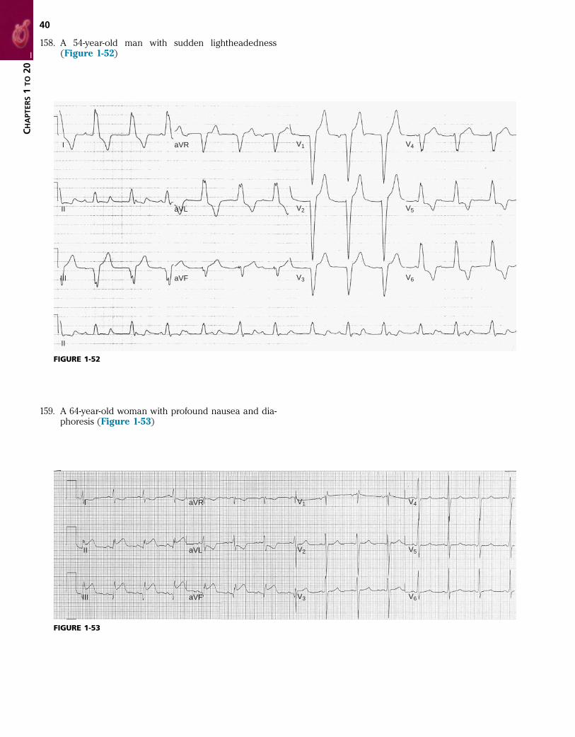
40
I
CH
APT
ERS
1 TO
20
FIGURE 1-52
I
II
II
III
aVR
aVL
aVF
V1
V2
V3
V4
V5
V6
FIGURE 1-53
I
II
III
aVR
aVL
aVF
V1
V2
V3
V4
V5
V6
158. A 54-year-old man with sudden lightheadedness (Figure 1-52)
159. A 64-year-old woman with profound nausea and dia-phoresis (Figure 1-53)

Fun
dam
entals o
f Card
iovascu
lar Disease; G
enetics an
d Perso
nalized
Med
icine; Evalu
ation
of th
e Patient
41
1160. A 55-year-old man with long-standing hypertension
(Figure 1-54)
FIGURE 1-54
I
II
III
V1
aVR
aVL
aVF
V1
V2
V3
V4
V5
V6
FIGURE 1-55
I
II
III
aVR
aVL
aVF
V1
V2
V3
V4
V5
V6
161. An asymptomatic 36-year-old man presents for an insurance physical examination (Figure 1-55)

42
I
CH
APT
ERS
1 TO
20
FIGURE 1-56
I
II
III
II
aVR
aVL
aVF
V1
V2
V3
V4
V5
V6
FIGURE 1-57
I
II
III
aVR
aVL
aVF
V1
V2
V3
V4
V5
V6
162. A 23-year-old woman referred to the cardiology clinic because of a murmur and abnormal ECG (Figure 1-56)
163. A 51-year-old woman with discrete episodes of presyn-cope (Figure 1-57)

Fun
dam
entals o
f Card
iovascu
lar Disease; G
enetics an
d Perso
nalized
Med
icine; Evalu
ation
of th
e Patient
43
1
FIGURE 1-58
I
II
II
III
aVR
aVL
aVF
V1
V2
V3
V4
V5
V6
FIGURE 1-59
164. A 72-year-old man with palpitations after coronary artery bypass surgery (Figure 1-58)
165. A 78-year-old man with a long history of cigarette smoking and paroxysmal atrial fibrillation (Figure 1-59)

44
I
CH
APT
ERS
1 TO
20
FIGURE 1-60
FIGURE 1-61
166. A 69-year-old man with a history of dilated cardiomy-opathy (Figure 1-60)
167. An elderly nursing home resident with fatigue (Figure 1-61)

45
SECTION I(CHAPTERS 1 TO 20)
Fundamentals of Cardiovascular Disease; Genetics and Personalized Medicine; Evaluation of the Patient
ANSWER TO QUESTION 1
D (Braunwald, pp. 156, 161, 162)The normal systolic blood pressure response during exer-cise is a progressive increase to a peak value between 160 and 200 mm Hg. The higher end of this range is more commonly observed in older patients; in general, black patients tend to have a higher systolic blood pressure response to exercise than white patients. A failure to increase systolic blood pressure to at least 120 mm Hg, or a decline in systolic blood pressure during exercise, is abnormal. Such exertional hypotension occurs in 3% to 9% of patients and is suggestive of underlying multivessel or left main coronary artery disease. Other causes of a decline in systolic blood pressure, or a failure to increase systolic blood pressure with exercise, include cardiomyopathy, vasovagal reactions, ventricular outflow obstruction, hypo-volemia, arrhythmias, and prolonged vigorous exercise. Subjects who demonstrate hypotension in the postexercise period are much less likely to have advanced underlying coronary artery disease; about 3% of normal subjects younger than 55 years of age demonstrate such a response.
In normal subjects, diastolic blood pressure does not change significantly during exercise. A large change in diastolic blood pressure is uncommon and has not been shown to correlate with underlying coronary artery disease.
The age-related maximum predicted heart rate (MPHR) is estimated from the following formula:
MPHR age in years= −220 ( )
which in this patient would be 166 beats/min. The peak heart rate he achieved during the test was 152 beats/min, or 92% of the MPHR (i.e., 152/166 beats/min). An achieved heart rate of ≥85% MPHR is indicative of an adequate diagnostic workload.
Predictors of low prognostic coronary risk in his case include his very good functional capacity (having achieved stage IV of the Bruce protocol) and lack of cardiopulmo-nary symptoms or ST-segment changes during the test.
BIBLIOGRAPHYFroelicher VF, Myers J: Exercise and the heart, ed 5, Philadelphia, 2006,
Elsevier Saunders.
ANSWER TO QUESTION 2
C (Braunwald, pp. 103-104; Figure 11-8)S2 coincides with closure of the aortic and pulmonic valves and marks the onset of diastole at the bedside. Several abnormal heart sounds may follow S2. The opening snap of mitral stenosis is a high-frequency sound that occurs shortly after S2. It is generated when the superior bowing of the anterior mitral leaflet during systole rapidly reverses direc-tion toward the left ventricle in early diastole, owing to the high left atrial (LA) pressure. The delay between the aortic component of S2 and the opening snap corresponds to the left ventricular (LV) isovolumic relaxation time. As mitral stenosis becomes more severe, this phase shortens because of the greater LA pressure, and the interval between S2 and the opening snap becomes less. Other sounds that occur shortly after S2 are associated with the rapid filling phase of diastole. These include the third heart sound (S3), which is a low-frequency sound that is thought to be caused by sudden limitation of LV expansion during brisk early dia-stolic filling. An S3 is normal in children and young adults, but the presence of this sound beyond age 40 is abnormal and reflects flow into a dilated ventricle or an increased volume of flow in early diastole, as may occur in mitral regurgitation. A tumor “plop” may be auscultated when an atrial myxoma, attached to the interatrial septum by a long stalk, moves into and obstructs the mitral or tricuspid valve orifice during early diastole. In constrictive pericarditis, a pericardial “knock” may be heard during the rapid filling phase of early diastole as the high-pressure atria rapidly decompress into relatively noncompliant ventricles.
An ejection click is an early systolic sound that represents opening of an abnormal semilunar valve, characteristically a bicuspid aortic valve.
ANSWER TO QUESTION 3
D (Braunwald, p. 35; Table 13-10)In addition to accuracy and reliability, the performance of a diagnostic test depends on its ability to distinguish between the presence and absence of disease. Test performance depends on its sensitivity and specificity, as well as the prevalence of disease in the population of patients to be studied or the pretest probability of disease in a particular

46
I
CH
APT
ERS
1 TO
20
such as Takayasu disease. In supravalvular aortic stenosis there may be selective streaming of the arterial jet toward the innominate artery and right arm, leading to higher pres-sures in that extremity. This is not the case, however, with subvalvular or valvular aortic stenosis. Valvular aortic ste-nosis leads to pulsus parvus et tardus, a slowly rising and weak pulse best appreciated by palpation of the carotid arteries. Coarctation of the aorta in adults usually involves the aorta distal to the origin of the left subclavian artery and leads to higher blood pressure in the upper extremities compared with the legs; the arm pulses and pressures are typically equal.
BIBLIOGRAPHYLane D, et al: Inter-arm differences in blood pressure: When are they
clinically significant? J Hypertens 20:1089, 2002.
ANSWER TO QUESTION 6
C (Braunwald, pp. 1640-1645; Figures 71-8 and 71-9)This patient with metastatic breast cancer and a large peri-cardial effusion has clinical findings consistent with cardiac tamponade.1 Tamponade physiology results when an accu-mulation of pericardial effusion causes equilibration of intrapericardial and intracardiac pressures.2,3 In addition to the presence of an echo-free space surrounding the heart, characteristic echocardiographic and Doppler findings reflect the aberrant physiology of this disorder. Collapse of the right ventricle during early diastole occurs because the abnormally elevated pericardial pressure transiently exceeds right ventricular (RV) pressure at that phase of the cardiac cycle. Indentation of the right atrial wall during diastole is a more sensitive marker of increased pericardial pressure but is less specific for tamponade physiology than RV collapse and tends to occur earlier in the course of hemodynamically significant pericardial effusion. Cardiac tamponade is associated with exaggerated ventricular inter-dependence, a phenomenon manifested at the bedside by pulsus paradoxus. The Doppler correlate of pulsus para-doxus is amplified respirophasic variation of flow across the right- and left-sided cardiac valves. This includes a prominent inspiratory decrease in flow velocity across the mitral and aortic valves (see the transmitral tracing in Figure 1-62), whereas inspiration causes a prominent
patient. Sensitivity and specificity are characteristics of the diagnostic test that are not altered by disease prevalence or pretest probability. Sensitivity is the percentage of patients with disease who will be correctly identified by the test. Specificity is the percentage of patients without disease who will be correctly identified as disease free by the test. Posi-tive predictive value is the probability that a positive test correctly identifies the presence of disease. Negative predic-tive value is the probability that a negative test correctly identifies the absence of disease. A perfect diagnostic test has a positive predictive value of 100% (no false-negative results) and a negative predictive value of 100% (no false-positive results).
For a diagnostic test with moderately high sensitivity and specificity, the test will perform best in a population of patients with an intermediate pretest probability of disease (patient D). In patients with a low pretest probability of disease (patients A and B), the positive predictive value of the test is low and there will be a large number of false-positive tests that may prompt unnecessary testing and procedures. In patients with an extremely high pretest probability of disease (patients C and E), the negative pre-dictive value of the test is low and the possibility that a nega-tive result represents a false negative is unacceptably high.
ANSWER TO QUESTION 4
C (Braunwald, pp. 104-106; Tables 11-5 and 11-8)The term continuous applies to murmurs that begin in systole and continue without interruption into part or all of diastole. The murmur described here, that of a patent ductus arteriosus, is the classic continuous murmur, peaking in intensity just before or after S2, then decreasing in intensity during diastole, sometimes disappearing before the subsequent first heart sound. Continuous murmurs may be congenital or acquired and can be caused by (1) an aortopulmonary shunt, such as patent ductus arteriosus; (2) an arteriovenous shunt, including arteriovenous fistulas, coronary artery fistulas, or rupture of an aortic sinus of Valsalva aneurysm into a right heart chamber; (3) con-stricted arterial vessels (e.g., a femoral arterial atheroscle-rotic stenosis); (4) turbulence in nonconstricted arteries (e.g., the “mammary souffle,” an innocent flow murmur heard during late pregnancy and the puerperium over the lactating breast and augmented by light pressure with the stethoscope); or (5) venous murmurs, such as a cervical venous hum, an often “rough” sounding murmur present in healthy children and young adults. The cervical hum may be accentuated by deforming the internal jugular vein with rotation of the head. It is augmented during pregnancy and in disease states in which there is increased venous flow, such as thyrotoxicosis.
The combined murmurs of aortic stenosis and regurgita-tion have distinct systolic and diastolic components and do not constitute a continuous murmur.
ANSWER TO QUESTION 5
E (Braunwald, pp. 100-101)Reduced or unequal arterial pulsations may occur in the arms of patients with atherosclerosis affecting the subcla-vian arteries, aortic dissection, and uncommon arteritides FIGURE 1-62
E

47
1Fun
dam
entals o
f Card
iovascu
lar Disease; G
enetics an
d Perso
nalized
Med
icine; Evalu
ation
of th
e Patient
coronary syndrome rather than pulmonary embolism, based on the presenting clinical symptoms.
BIBLIOGRAPHYKhan JN, Chauhan A, Mozdiak E, et al: Posterior myocardial infarction:
are we failing to diagnose this? Emerg Med J 29:15-18, 2012.
ANSWER TO QUESTION 9
C (Braunwald, pp. 76-77, 82; Tables 9-1 to 9-3)All drugs prescribed to achieve a particular clinical benefit also have the potential for toxicity. Many factors determine the likelihood of drug toxicity, including the pharmacoki-netic and pharmacodynamic properties of the drug and its target, genetic variability in the patient’s response to the drug, and drug–drug interactions.
Many medications are metabolized by isoforms of the cytochrome P-450 (CYP) enzyme system, which are expressed in the liver and other tissues. Ketoconazole, erythromycin, and clarithromycin (but not azithromycin) are examples of drugs that inhibit CYP3A4 and 3A5. Because these P-450 isoforms are responsible for metabolism of sim-vastatin, atorvastatin, and lovastatin, combined therapy with such inhibitors may increase the likelihood of myopa-thy due to these statins.1 Pravastatin is not metabolized by the CYP3A system, and thus the risk of myopathy is not increased in the presence of CYP3A inhibitors.
St. John’s wort induces activity of CYP3A and results in decreased cyclosporine levels.2
Sildenafil, a selective inhibitor of phosphodiesterase type 5 prescribed to treat erectile dysfunction, potentiates the vasodilatory effect of nitrates. Administration of nitrates within 24 hours of sildenafil use has been associated with profound hypotension.
Verapamil inhibits the P-glycoprotein–mediated efflux of digoxin into bile and urine and may contribute to digoxin toxicity.
REFERENCES1. Sathasivam S, Lecky B: Statin induced myopathy. BMJ 337:a2286,
2008.2. Yarnell E, Abascal K: Interaction of herbal constituents with cyto-
chrome P450 enzymes. Altern Complement Med 13:239, 2007.
ANSWER TO QUESTION 10
D (Braunwald, p. 156; Table 13-1)Treadmill exercise testing is a safe procedure with an associated mortality of <0.01% and risk of myocardial infarction of 0.04%.1 The risk of a procedure-related com-plication is determined by the clinical characteristics of the patient to be studied. Patients with high-grade obstruc-tion of the left ventricular outflow tract, such as those with hypertrophic obstructive cardiomyopathy or critical aortic stenosis, are at an increased risk of a procedural complication owing to the inability of cardiac output to compensate for peripheral vasodilatation during exercise. Patients with unstable angina should not be subjected to the high myocardial oxygen demands of exercise and should be referred for coronary angiography instead. Acute myocarditis is associated with an increased risk of exercise-associated sudden death.
reciprocal increase in flow velocity across the tricuspid and pulmonic valves.
A marked increase of the E/A ratio of the mitral valve inflow velocity is a finding typical of constrictive pericardi-tis, not cardiac tamponade.
REFERENCES1. Roy CL, Minor MA, Brookheart MA, et al: Does this patient with a
pericardial effusion have cardiac tamponade? JAMA 297:1810, 2007.
2. Maisch B, Ristic A, Pankuweit S: Evaluation and management of pericardial effusion in patients with neoplastic disease. Prog Cardiovasc Dis 53:157, 2010.
3. Spodick DH: Acute cardiac tamponade. N Engl J Med 349:684, 2003.
ANSWER TO QUESTION 7
D (Braunwald, pp. 100, 104-105)Pulsus paradoxus is an exaggeration of the normal ten-dency for arterial pulse strength to fall with inspiration and can be measured easily and accurately at the bedside with a sphygmomanometer. A decline of more than 8 to 10 mm Hg with inspiration is considered abnormal and can be observed in a variety of conditions. Pulsus para-doxus is characteristic of patients with cardiac tamponade, is seen in approximately one third of patients with chronic constrictive pericarditis, and is noted as well in patients with wide intrapleural pressure swings (e.g., bronchial asthma and emphysema), pulmonary embolism, preg-nancy, extreme obesity, and hypovolemic shock.
Notably, aortic regurgitation augments left ventricular diastolic pressure and tends to prevent pulsus paradoxus even in the presence of tamponade.
The Kussmaul sign manifests as inappropriate augmenta-tion of the jugular venous pressure during inspiration and implies the presence of constrictive pericarditis, not iso-lated cardiac tamponade.
ANSWER TO QUESTION 8
B (Braunwald, pp. 118, 141-142, 149; Tables 12-1 and 12-9)The clinical presentation is concerning for an acute coro-nary syndrome and rapid evaluation of the ECG is critical to detect whether an acute ST-segment elevation myocar-dial infarction (STEMI) is present, which would warrant prompt coronary revascularization. The ECG in this patient is not diagnostic for STEMI, with <1 mm of ST elevation in only one lead (III). However, the presence of ST depres-sions and a prominent R wave in the anterior precordial leads should raise suspicion for an acute ST elevation MI in the posterior territory. Since standard leads are not placed on the patient’s back, myocardial injury from the posterior wall is typically identified as mirror-image ECG findings in the opposite anatomic position, namely leads V1-V3. In this circumstance, posteriorly placed ECG leads (V7-V9) placed from the posterior axillary line to the left border of the spine would be helpful to evaluate for true posterior ST elevations.
Right-sided precordial leads are useful to detect right ventricular involvement in the setting of an inferior STEMI. Awaiting the results of cardiac biomarkers would delay care. Diagnostic tests should focus first on an acute

48
I
CH
APT
ERS
1 TO
20
increased flow through the right-sided cardiac chambers and an increase in pulmonary vascular capacitance, con-tributing to a widened split of S2. On inspiration, augmen-tation of the systemic venous return is counterbalanced by a reciprocal decrease in the volume of the left-to-right shunt, such that right ventricular filling and the timing of P2 relative to A2 do not change, resulting in the fixed splitting.
When valvular stenosis restricts opening of a cardiac valve, the decreased excursion of the leaflets reduces the intensity of the closure sound. Thus, in pulmonic stenosis, the pulmonic component of S2 becomes softer.
ANSWER TO QUESTION 13
D (Braunwald, pp. 162-163, 164, 165)Electrocardiographic changes during exertion in an asymp-tomatic patient must be interpreted in light of the pretest likelihood of coronary disease. An exercise-induced ST- segment abnormality is an independent predictor of future cardiac events in men with and without conventional risk factors for coronary disease, although the risk is greatest among the former. However, over 5 years of follow-up, only one in four such patients will actually develop symptoms of cardiac disease, most commonly angina. Because the patient described in this question is asymptomatic and demonstrates good exercise capacity, there is no need for immediate aggressive intervention such as cardiac cathe-terization. Appropriate recommendations for this patient with asymptomatic coronary artery disease would include aggressive risk factor modification: smoking cessation, control of hypertension, the addition of aspirin, 81 to 325 mg/d, and statin therapy.
The distribution of ST-segment depressions during exer-cise testing correlates poorly with the location of coronary stenoses. Conversely, the location of ST-segment elevations, when present, does correlate well with the anatomic lesion causing ischemia.
BIBLIOGRAPHYGreenland P, Alpert JS, Beller GA, et al: 2010 ACCF/AHA guideline for
the assessment of cardiovascular risk in asymptomatic adults: executive summary. A report of the American College of Cardiology Foundation/American Heart Association Task Force on Practice Guidelines. Circulation 122:2748, 2010.
Lauer M, Freolicher ES, Williams M, et al: Exercise testing in asymptom-atic adults: a statement for professionals from the American Heart Association Council on Clinical Cardiology, Subcommittee on Exer-cise, Cardiac Rehabilitation, and Prevention. Circulation 112:771, 2005.
ANSWER TO QUESTION 14
C (Braunwald, pp. 165-166, 170; Tables 13-11, 13G-1, and 16G-1)Several clinical situations affect the ST segment and impair the diagnostic utility of the standard exercise ECG. These include the presence of left bundle branch block (LBBB), left ventricular hypertrophy, ventricular preexcitation (Wolff-Parkinson-White syndrome), and digitalis therapy. In these situations, other aspects of the exercise test, such as exercise duration, presence or absence of symptoms, and abnormal blood pressure or heart rate responses, may still
Despite the theoretical risk of aortic rupture due to increased wall stress, treadmill exercise testing may be safely performed in patients with an abdominal aortic aneurysm.2 In contrast, aortic dissection is a contraindica-tion to the stress of exercise testing.
REFERENCES1. Myers J, Arena R, Franklin B, et al: Recommendations for clinical
exercise laboratories: a scientific statement from the American Heart Association. Circulation 119:3144, 2009.
2. Hartley RA, Pichel AC, Grant SW, et al: Preoperative cardiopulmo-nary exercise testing and risk of early mortality following abdominal aortic aneurysm repair. Br J Surg 99:1539-1546, 2012.
ANSWER TO QUESTION 11
B (Braunwald, pp. 271-272, 279-280, 301; Figure 16-12)The images show a fixed defect in the mid and apical ante-rior wall segments with preserved wall motion and thicken-ing as evident from the end-diastolic and end-systolic frames. These findings are most consistent with imaging artifact due to breast tissue attenuation.
Attenuation artifacts are a common source of error in single-photon emission computed tomography (SPECT). Regional wall motion and wall thickening should be assessed on the electrocardiographic gated SPECT data in any region that shows a fixed perfusion defect. Although a transmural myocardial scar would be associated with reduced wall motion and wall thickening, attenuation arti-fact is more likely where there is a fixed perfusion defect with normal regional wall motion and wall thickening.
Thallium-201 is a lower-energy radiotracer that results in more attenuation artifacts than technetium-99m imaging, and hence would be an inferior choice in this case.
BIBLIOGRAPHYIskandrian AE, Garcia EV: Atlas of nuclear cardiology, Philadelphia,
2012, Elsevier Saunders, pp 21-27.
ANSWER TO QUESTION 12
B (Braunwald, p. 103)The normal second heart sound (S2) consists of two parts, an earlier aortic component and a later pulmonic compo-nent. During inspiration, the increased filling of the right ventricle prolongs the ejection phase of the right side of the heart leading to delayed closure of the pulmonic valve. This is the predominant factor in normal inspiratory splitting of the S2. Right bundle branch block delays right ventricular activation and ejection and is therefore associated with widened splitting of S2. Conditions in which left ventricular activation is late, such as left bundle branch block or right ventricular pacing, cause closure of the aortic valve to be delayed. In that setting, the pulmonic valve closure sound actually precedes that of the aortic valve. Then, during inspiration (and prolongation of right ventricular ejection), the delayed closure of the pulmonic valve narrows the timing between the two sounds, a situation known as paradoxical splitting.
Fixed splitting of the S2 is typical of an uncomplicated ostium secundum atrial septal defect. In this condition, closure of the pulmonic valve is delayed because of the

49
1Fun
dam
entals o
f Card
iovascu
lar Disease; G
enetics an
d Perso
nalized
Med
icine; Evalu
ation
of th
e Patient
BIBLIOGRAPHYLevine GN, Bates ER, Blankenship JC, et al: 2011 ACCF/AHA/SCAI
guideline for percutaneous coronary intervention: a report of the American College of Cardiology Foundation/American Heart Asso-ciation Task Force on Practice Guidelines and the Society for Cardiovascular Angiography and Interventions. J Am Coll Cardiol 58:e44, 2011.
ANSWER TO QUESTION 16
D (Braunwald, p. 118; Figures 12-38 and 12-40)Proximal occlusion of a dominant right coronary artery (RCA) leads to infarction of the left ventricular (LV) inferior wall but often also involves the posterior wall, the right ventricle, and portions of the conduction system, which are all supplied by branches of the RCA. ST-segment elevation in leads II, III, and aVF is the sine qua non of transmural infarction of the inferior wall. If the posterior wall is involved, ST-segment depression is usually evident in V1 and V2, reflecting a current of injury on the side of the heart oppo-site those leads (if unipolar leads were placed on the patient’s back overlying the posterior wall, ST-segment ele-vation would be observed instead). Very proximal occlu-sion of the RCA is often accompanied by right ventricular (RV) infarction because the RV arterial branch arises near the origin of that vessel. If RV infarction is present, right-sided precordial electrocardiographic leads, particu-larly V4R, often demonstrate ST-segment elevation as well (Figure 1-63). Sinus bradycardia is common in the setting of acute myocardial infarction, especially in inferior or pos-terior infarction. This arrhythmia, particularly when accom-panied by hypotension, may arise from stimulation of cardiac vagal afferent fibers, which are prominent in the inferoposterior left ventricle. Sinus bradycardia may also be a vasovagal response to the severe chest pain in acute myocardial infarction or reflect ischemia of the sinoatrial artery, which arises from the RCA in 60% of the population. Another potential cause of bradycardia in this patient is the development of atrioventricular (AV) block because of either vagal stimulation or ischemia of the AV node. The AV node is supplied by the AV nodal artery, which arises from the RCA 85% of the time.
ANSWER TO QUESTION 17
C (Braunwald, pp. 183, 204-205)One of the most clinically important applications of Doppler technology is the estimation of pressure gradients across stenotic orifices or septal defects in the cardiovascular system. The Bernoulli equation relates the pressure differ-ence across a narrowed area to the convective accelera-tion, flow acceleration, and viscous friction. By modifying the Bernoulli equation, a more clinically useful simplified formula is derived. The simplified Bernoulli equation states that the pressure difference across a flow-limiting orifice = 4V2, where V is the peak velocity distal to the obstruction. In this question, the patient’s systolic blood pressure is 144 mm Hg, which, in the absence of left ventricular (LV) outflow obstruction, is also the LV systolic pressure. If the ventricular septal defect flow is 5.1 m/sec, then using the modified Bernoulli equation, the pressure gradient across the ventricular septal defect is 4 × (5.1)2 = 104 mm Hg. The
provide useful diagnostic information. However, in the pres-ence of these baseline electrocardiographic abnormalities, concurrent imaging (e.g., nuclear scintigraphy or echocar-diography) is frequently required when more specific diag-nostic information is needed. In the case of LBBB, a vasodilator pharmacologic (e.g., adenosine) stress test with myocardial perfusion imaging helps to avoid artifactual septal perfusion defects compared with exercise protocols.
In patients with a low prior probability of significant coro-nary artery disease, such as a young woman without signifi-cant cardiac risk factors, the development of ST-segment depression on exercise testing is more often a nonspecific false-positive result than an indicator of previously unde-tected coronary artery disease.
BIBLIOGRAPHYFihn SD, Gardin JM, Abrams J, et al: 2012 ACCF/AHA/ACP/AATS/PCNA/
SCAI/STS Guideline for the diagnosis and management of patients with stable ischemic heart disease: a report of the American College of Cardiology Foundation/American Heart Association Task Force on Practice Guidelines, and the American College of Physicians, Ameri-can Association for Thoracic Surgery, Preventive Cardiovascular Nurses Association, Society for Cardiovascular Angiography and Interventions, and Society of Thoracic Surgeons. J Am Coll Cardiol 60:e44, 2012.
Gibbons RJ, Balady GJ, Bricker JT, et al: ACC/AHA 2002 guideline update for exercise testing: summary article. A report of the American College of Cardiology/American Heart Association Task Force on Practice Guidelines (Committee to Update the 1997 Exercise Testing Guidelines). J Am Coll Cardiol 40:1531, 2002.
ANSWER TO QUESTION 15
D (Braunwald, pp. 366-367, 385, 1912-1914; Table 19-5)Diagnostic cardiac catheterization is a relatively safe pro-cedure, with an overall risk of a major complication of <1%. Mortality rates related to the procedure depend on the population studied and range from 0.08% to 0.75%. The risk of myocardial infarction is approximately 0.05%, and neurologic complications occur in 0.03% to 0.2% of patients. The incidence of acute renal dysfunction in patients with baseline renal insufficiency can be most effectively decreased with intravenous saline administra-tion before and after the procedure. The addition of man-nitol or furosemide to saline infusion has been shown to worsen renal outcomes in patients receiving an intravenous contrast agent.
Nonionic low osmolar contrast agents reduce the likeli-hood of adverse hemodynamic and electrophysiologic reactions during angiography. They also reduce the inci-dence of contrast-induced nephropathy in patients with baseline renal insufficiency, with or without diabetes. Of note, in patients with normal renal function, there is no advantage of low osmolar agents over ionic agents in the prevention of nephrotoxicity.
Cardiac catheters are available in many sizes, shapes, and lengths. The outer diameter of the catheter is specified using French units (F), where 1 F is equal to 0.33 mm.
Patients with tilting-disc prosthetic aortic valves should not undergo retrograde left-sided heart catheterization because of the risk of catheter entrapment, occlusion of the valve, or possible dislodgment of the disc with embolization.

50
I
CH
APT
ERS
1 TO
20
FIGURE 1-64
FIGURE 1-63
I aVR V1R V4R
II aVL V2R V5R
III aVF V3R V6R
right ventricular systolic pressure can then be calculated by subtracting that gradient from the LV systolic pressure: 144−104, or 40 mm Hg.
BIBLIOGRAPHYArmstrong WF, Ryan T: Feigenbaum’s echocardiography, ed 7, Philadel-
phia, 2010, Lippincott Williams & Wilkins, pp 217-240.
ANSWER TO QUESTION 18
C (Braunwald, pp. 110-111, 204-205; Figures 11-9 and 14-31)This patient’s history is concerning for an undetected acute myocardial infarction (MI) 10 days earlier, including the prolonged chest “muscle ache,” new Q waves in the inferior leads, and inferior wall motion abnormalities on echocar-diography. The lack of acute intervention places the patient at risk for post-MI mechanical complications, including acute mitral regurgitation (MR) due to dysfunction or rupture of a papillary muscle, ventricular septal defect (VSD), and free wall rupture. The brief systolic murmur on exam in the setting of new-onset heart failure should raise suspicion for acute MR. Although chronic MR typically pro-duces a holosystolic murmur at the apex, in acute MR there may only be a soft, early systolic murmur due to the rapid systolic increase in pressure in the unprepared and non-compliant left atrium (LA) that attenuates the pressure gra-dient between the left ventricle and LA responsible for the murmur.
The presumed recent right coronary artery occlusion in this patient would impair the blood supply to the postero-medial papillary muscle. Dysfunction or rupture of this pap-illary muscle often produces MR in an eccentric direction, which may be underestimated by transthoracic echocar-diography, especially in this patient with poor acoustic windows. Given the high suspicion for papillary muscle involvement resulting in acute MR, a transesophageal echo-cardiogram would be appropriate, with cardiac surgical consultation for consideration of urgent valve repair. In most cases, coronary revascularization would be addressed with bypass grafting at the time of valve surgery rather than with percutaneous intervention.
A nuclear stress test would not address the urgent mechanical process in this patient’s situation. The location of the murmur and echocardiographic findings are not con-sistent with a VSD such that urgent right heart catheteriza-tion alone would not be the best immediate approach.
BIBLIOGRAPHYNishimura RA, Otto CM, Bonow RO, et al: 2014 AHA/ACC guideline for
the management of patients with valvular heart disease: executive summary: a report of the American College of Cardiology/American Heart Association Task Force on Practice Guidelines. J Am Coll Cardiol 63:e57, 2014.
ANSWER TO QUESTION 19
D (Braunwald, pp. 146-148)Many electrolyte disturbances result in characteristic elec-trocardiographic manifestations. Decreased extracellular calcium in hypocalcemia prolongs phase 2 of the action potential (AP), thereby lengthening the AP duration and the QT interval. The long QT interval in hypocalcemia is characteristically flat (i.e., isoelectric), without the concave configuration of many drug-induced prolonged QT states. Increased extracellular calcium shortens the ventricular action potential and the duration of the QT interval. The appearance of a J wave (also known as an Osborn wave—see arrow in Figure 1-64), an extra deflection at the junc-tion of the QRS complex and the ST segment that is typically

51
1Fun
dam
entals o
f Card
iovascu
lar Disease; G
enetics an
d Perso
nalized
Med
icine; Evalu
ation
of th
e Patient
American Association for Thoracic Surgery, Preventive Cardiovas-cular Nurses Association, Society for Cardiovascular Angiography and Interventions, and Society of Thoracic Surgeons. J Am Coll Cardiol 60:e44, 2012.
Fletcher GF, Ades PA, Kligfield P, et al: Exercise standards for testing and training: a scientific statement from the American Heart Association. Circulation 128:873, 2013.
ANSWER TO QUESTION 21
C (Braunwald, pp. 135-136; Figure 12-28)The underlying rhythm is atrial fibrillation. The Ashman phenomenon (as exemplified by the 5th QRS complex in the tracing) represents conduction aberrancy caused by changes in the preceding cycle length. Because the dura-tion of the refractory period is a function of the immediate preceding cycle length, the longer the preceding cycle, the longer the ensuing refractory period and the more likely that the next impulse will be conducted with delay. Nor-mally the length of the refractory periods of the conduction system components are as follows: right bundle branch > left bundle branch = atrioventricular node ≫ His bundle. Therefore, it would be unusual for the bundle of His to be the site of conduction delay and, as is commonly the case, the aberrant beat on this tracing demonstrates right bundle branch block morphology in lead V1.
ANSWER TO QUESTION 22
C (Braunwald, pp. 104, 106; Table 11-5; Figure 11-10)Innocent (normal) systolic murmurs are related to intracar-diac flow rates and are usually loudest in midsystole. They may be caused by normal vibrations of the pulmonary leaf-lets, or exaggeration of normal ejection vibrations within the pulmonary artery, or be associated with sclerosis at the base of the aortic valve leaflets in the absence of significant valvular stenosis. The normal mammary souffle, heard over the breasts of women in late pregnancy or during lactation, may also be midsystolic in timing or continuous. Careful auscultation usually reveals a time delay between S1 and onset of this murmur, which is the transit time of flow from the proximal aorta to the mammary arteries.
ANSWER TO QUESTION 23
D (Braunwald, pp. 98-99; Figures 11-2, 11-3)A great deal of information about right-sided heart hemo-dynamics can be ascertained from the jugular venous pres-sure wave forms. The a wave results from venous distention due to right atrial contraction; the x descent reflects atrial relaxation and downward descent of the base of the right atrium during right ventricular (RV) systole. The c wave is an inconstant positive deflection in the jugular venous pulse that interrupts the x descent and corresponds to ven-tricular contraction. The v wave results from right atrial filling during ventricular systole when the tricuspid valve is closed, and the y descent occurs after the tricuspid valve opens and right atrial pressure declines. It is easier for an observer to see the x and y descents than the positive pres-sure waves (a, c, and v waves) in the neck because the former produce larger excursions. An elevated jugular venous pressure reflects increased right atrial pressure. During inspiration, the jugular venous pressure normally
observed in severe hypothermia, has been reported with severe hypercalcemia.1
Hyperkalemia causes a specific sequence of electro-cardiographic changes depending on its severity. The earli-est manifestation is narrow, peaked T waves. The QT interval is usually decreased at that time because of short-ened action potential duration. Progressive hyperkalemia reduces the resting membrane potentials in both the atria and ventricles, thus inactivating sodium channels. The net result is to slow depolarization and reduce action potential conduction velocity. The ECG shows widening of the QRS complex with a decrease in P wave amplitude. PR-segment prolongation may also occur. Very marked hyperkalemia leads to slow, undulating ventricular flutter (a “sine wave” appearance) followed by eventual asystole. Hypokalemia, in contrast, manifests primarily as ST-segment depressions with flattened T waves and U wave prominence. Because of prolongation of the QT interval there is a propensity for polymorphic ventricular tachycardia (torsades de pointes).
The effects of magnesium on the surface ECG are not as well characterized. Magnesium deficiency may predispose to prolongation of the QT interval, primarily as a result of a prolonged U wave (QU interval), and polymorphic ventricu-lar tachycardia.
REFERENCE1. Otero J, Lenihan DJ: The “normothermic” Osborn wave induced by
severe hypercalcemia. Tex Heart Inst J 27:316, 2000.
ANSWER TO QUESTION 20
E (Braunwald, pp. 165-166, 170; Table 13G-1; see also Answer to Question 14)Meta-analyses show that the sensitivity and specificity of the exercise ECG for the detection of coronary artery disease (CAD) are 68% and 77%, respectively. The sensitiv-ity for diagnosis is impaired by resting ST-segment depres-sion or other repolarization abnormalities, including left ventricular hypertrophy, digitalis therapy (even if digitalis effect is not evident on the resting ECG), left bundle branch block, and preexcitation syndromes. In addition, severe hypertension, hyperventilation, and hypokalemia may distort the ST segment, leading to false-positive electrocar-diographic interpretations. One of the main diagnostic values of the standard exercise test is to exclude CAD in patients with chest pain with a low pretest likelihood of disease based on age and gender who have a normal resting ECG. If such a patient achieves the maximum heart rate without ST-segment deviations and a normal blood pressure response, it is very unlikely that CAD is the cause of chest pain. For most other populations, the exercise ECG is limited by the suboptimal sensitivity and specificity. Both myocardial perfusion imaging and stress echocardiogra-phy increase the sensitivity and specificity of exercise testing in patients with resting electrocardiographic abnor-malities. In particular, the negative predictive value of both of these techniques is excellent.
BIBLIOGRAPHYFihn SD, Gardin JM, Abrams J, et al: 2012 ACCF/AHA/ACP/AATS/PCNA/
SCAI/STS Guideline for the diagnosis and management of patients with stable ischemic heart disease: a report of the American College of Cardiology Foundation/American Heart Association Task Force on Practice Guidelines, and the American College of Physicians,

52
I
CH
APT
ERS
1 TO
20
The Fick technique is based on the principle that cardiac output is equal to the oxygen consumption divided by the difference in oxygen content between arterial and mixed venous blood. That is,
Cardiac outputO Consumption
A VO Difference=
−2
2
The Fick technique is more accurate than thermodilution in patients with low cardiac outputs; however, its main limi-tation is in measuring true oxygen consumption in a steady state. Many laboratories use an “assumed” oxygen con-sumption by considering the patient’s age, gender, and body surface area. Inaccuracy in the oxygen consumption measurement can result in substantial variability in reported cardiac outputs.
Systemic vascular resistance (SVR) is derived by dividing the difference between the mean aortic and right atrial (RA) pressures by the systemic cardiac output (and then multiplying by a constant to convert to the commonly used units of dynes∙sec∙cm−5):
SVRMean Aortic Pressure Mean RA Pressure
Systemic cardiac out= × −
80pput
The pulmonary vascular resistance (PVR) is obtained by dividing the difference between the mean pulmonary artery (PA) and left atrial (LA) pressures by the pulmonic cardiac output (then multiplying by the same constant).The pulmo-nary capillary wedge (PCW) pressure is commonly used as a surrogate for left atrial pressure:
PVRMean PA Pressure Mean LA or PCW Pressure
Pulmonic cardiac= × −
80( )ooutput
In the absence of intracardiac shunts, the systemic and pulmonic cardiac outputs should be the same.
ANSWER TO QUESTION 25
C (Braunwald, p. 192; Figures 14-17 and 14-18)Pulsed Doppler interrogation of mitral valve inflow is useful in identifying disorders of left ventricular (LV) diastolic function. In normal adults, the early (E wave) velocity exceeds the late (A wave) velocity such that the normal E/A ratio is >1.2. The figure accompanying this question illus-trates an abnormal pattern of mitral inflow, with an E/A ratio <1.0. Although this pattern may be present in patients who have documented abnormalities of LV relaxation, it can also occur as a result of normal aging.1 In fact, most people older than age 70 have an E/A ratio <1.0. In addition, the pattern of diastolic mitral inflow is load dependent. If a patient with the illustrated pattern of impaired relaxation were administered intravenous volume, the mitral inflow pattern could change to the “pseudonormalized” form, with an E/A ratio >1.0, without any alteration in the intrinsic relaxation properties of the ventricle. Caution should thus be used when inferring a diastolic or relaxation abnormal-ity from the mitral inflow pattern. Although none of the available echocardiographic indices of diastolic function is infallible, and even stepwise approaches using the mitral inflow pattern, Doppler tissue imaging, and assessment of pulmonary venous inflow can result in internally incon-sistent results,2 echocardiographic measures of diastolic
declines as intrathoracic pressure becomes more negative. The Kussmaul sign is a paradoxical rise in the height of the venous pressure during inspiration. It reflects an inability of the right-sided chambers to accept additional volume, typical of constrictive pericarditis, but may also be observed in patients with right-sided heart failure, severe cor pulmo-nale, or tricuspid stenosis.
The a wave becomes more prominent in conditions that increase the resistance to right atrial contraction, such as RV hypertrophy, pulmonary hypertension, or tricuspid ste-nosis. Amplified “cannon” a waves are evident during any situation that causes atrioventricular dissociation, because the right atrium contracts against a closed tricuspid valve, at least intermittently.
In constrictive pericarditis, the y descent is rapid and deep because the very earliest phase of diastolic RV filling is unimpeded. In contrast, in cardiac tamponade, the y descent is blunted and it is the x descent that is most prominent.
ANSWER TO QUESTION 24
D (Braunwald, pp. 377-379; Figures 19-11 and 19-12)There is no completely accurate method for measuring cardiac output in the cardiac catheterization laboratory. Two commonly used methods are the thermodilution and the Fick techniques. The former involves injection of a bolus of fluid (i.e., saline or dextrose) into the proximal port of a right-sided balloon flotation (e.g., Swan-Ganz) catheter, after which alterations in temperature are measured at the distal end of the catheter. The change in the temperature over time is then plotted to derive the cardiac output, which is inversely related to the area under the thermodilution curve (Figure 1-65). In low cardiac output states there is a larger area under the curve owing to the longer time required for the temperature curve to return to its baseline. However, this technique tends to overestimate cardiac output in the setting of low output states, because the dis-sipation of the cooler temperature to the surrounding cardiac structures results in a reduction in the total area under the curve. In patients with severe tricuspid regurgita-tion, the back and forth flow across the tricuspid valve also creates significant error in measurement, producing a falsely low cardiac output by this technique.
FIGURE 1-65
Normal cardiac output
High cardiac output Low cardiac output
Improper injection technique

53
1Fun
dam
entals o
f Card
iovascu
lar Disease; G
enetics an
d Perso
nalized
Med
icine; Evalu
ation
of th
e Patient
The cause of mitral stenosis in the vast majority of adults is rheumatic heart disease. Occasionally, other causes can be identified, such as congenital abnormalities of the valve or heavy calcification that restricts transval-vular flow. The transvalvular Doppler pattern cannot dis-tinguish the etiology of mitral stenosis, but two-dimensional echocardiographic imaging is usually diagnostic in this regard.
ANSWER TO QUESTION 27
B (Braunwald, pp. 98, 183, 196, 233; Figure 14-76; Tables 14-8 and 14G-1)The McConnell sign is a distinct regional wall motion abnor-mality associated with acute pulmonary embolism in which the right ventricular (RV) midwall is dyskinetic, with sparing of the apex. The Kussmaul sign refers to a rise in jugular venous pressure (or its failure to decrease) with inspiration, which occurs in states of reduced RV compliance and right-sided volume overload. It is typically associated with con-strictive pericarditis, but is also seen in pulmonary embolism, RV infarction, and restrictive cardiomyopathy.
The general calculation of a pressure gradient across a cardiac valve is determined by the simplified Bernoulli equation:
∆ Pressure velocity= ×4 2
In this patient, the pressure gradient between the RV and right atrium (RA) is 36 mm Hg (4 × 32). The right ventricular pressure can then be calculated as this pressure gradient plus the RA pressure. The right atrial pressure can be esti-mated from the echocardiographic diameter of the IVC and its respiratory variation (see Braunwald, Table 14-8). In this case, the IVC diameter is normal (<2.1 cm), but there is <50% inspiratory collapse, consistent with a right atrial pres-sure of 5 to 10 mm Hg. With a right atrial pressure of 5 to 10 mm Hg and a pressure gradient of 36 mm Hg between the RA and RV, the right ventricular systolic pressure can then be estimated at 41 to 46 mm Hg. In the absence of pulmonic stenosis, the pulmonary artery (PA) systolic pres-sure is equal to the right ventricular systolic pressure, such that the estimated PA systolic pressure in this patient is also 41 to 46 mm Hg.
BIBLIOGRAPHYDouglas PS, Garcia MJ, Haines DE, et al: ACCF/ASE/AHA/ASNC/HFSA/
HRS/SCAI/SCCM/SCCT/SCMR 2011 Appropriate use criteria for echocardiography: a report of the American College of Cardiology Foundation Appropriate Use Criteria Task Force, American Society of Echocardiography, American Heart Association, American Society of Nuclear Cardiology, Heart Failure Society of America, Heart Rhythm Society, Society for Cardiovascular Angiography and Inter-ventions, Society of Critical Care Medicine, Society of Cardiovascu-lar Computed Tomography, and Society for Cardiovascular Magnetic Resonance Endorsed by the American College of Chest Physicians. J Am Coll Cardiol 57:1126, 2011.
ANSWER TO QUESTION 28
C (Braunwald, pp. 155-156, 160-161; Table 13-9)With normal aerobic exercise, the heart rate and systolic blood pressure both rise. Diastolic blood pressure, however,
function nonetheless provide clinically important diagnos-tic and prognostic information.3
Constrictive pericarditis and restrictive cardiomyopathy are clinical situations in which the bulk of ventricular filling occurs in early diastole. In these conditions there is a high E/A ratio, often >2.0. Hyperthyroidism and hemorrhage each result in hyperdynamic states in which diastolic relax-ation is enhanced, such that reversal of the E/A ratio would be unusual.
REFERENCES1. Paulus WJ, et al: How to diagnose diastolic heart failure: a consensus
statement on the diagnosis of heart failure with normal left ven-tricular ejection fraction by the Heart Failure and Echocardiography Associations of the European Society of Cardiology. Eur Heart J 28:2539, 2007.
2. Tschope C, Paulus WJ: Doppler echocardiography yields dubious estimates of left ventricular diastolic pressures. Circulation 120:810, 2009.
3. Little WC, Ho JK: Echocardiographic evaluation of diastolic function can be used to guide care. Circulation 120:802, 2009.
ANSWER TO QUESTION 26
D (Braunwald, pp. 184, 216-216, 1473; Figures 14-38, 14-39, 63-21)The figure accompanying this question displays continuous Doppler interrogation of transmitral valvular flow obtained from the apical four-chamber transducer position. During diastole, the deceleration of transmitral inflow is prolonged, consistent with a persistent pressure gradient between the left atrium and left ventricle (i.e., mitral stenosis). In systole, a faint signal of mitral regurgitation (MR) is seen as a down-ward velocity spectrum. To determine the extent of MR noninvasively, simultaneous two-dimensional echocardiog-raphy and color Doppler interrogation would be required. However, the presented Doppler spectrum of mitral inflow is sufficient to determine the severity of mitral stenosis, including the transmitral pressure gradient and the valve area, as described below.
The mitral valve area can be calculated noninvasively by three distinct noninvasive echo-Doppler methods. First, if an adequate two-dimensional short-axis image can be obtained in diastole, the valve orifice can be traced and the valve area measured directly using planimetry. The second method (which can be applied to the provided figure) uti-lizes the Doppler pressure half-time (PHT), which calculates the time in milliseconds required for the diastolic transmi-tral pressure gradient to decline to one half of its peak value. Because of the relationship between velocity and pressure, this requires determining the time it takes for the peak diastolic velocity to fall to the peak velocity divided by the square root of 2. Most modern echocardiographic machines calculate the PHT automatically once the Doppler profile is traced on the screen, using the following equation:
Mitral valve area PHT= ÷220
This relationship becomes less accurate when there is marked MR or aortic regurgitation, which interferes with the measured pressure gradient across the mitral valve. The third method to calculate the mitral valve area utilizes the continuity equation, which is based on the principle that the volume rate of flow through the heart is constant.

54
I
CH
APT
ERS
1 TO
20
ANSWER TO QUESTION 30
C (Braunwald, p. 372, 1288-1302)This computed tomogram demonstrates a type A aortic dissection involving the ascending (large arrow, A) and descending (small arrows, A) thoracic aorta. In addition, the dissection extends into the aortic arch vessels (large arrows, B), the left common carotid artery (small arrow, B), and the innominate artery (arrowhead, B). Dissection membranes are clearly visualized in the involved segments. Acute aortic dissection is often lethal; survival depends on prompt clinical recognition and definitive imaging of the aorta. Stanford type A aortic dissections involve the ascending aorta. Type B aortic dissections do not involve the ascending aorta. The most common symptom of acute aortic dissection is chest or back pain, reported in up to 96% of patients. The pain is typically sudden in onset and severe, often with a “ripping” or “tearing” quality. The patient discussed in this question may have also presented with pulse deficits or neurologic symptoms, given the branch artery involvement demonstrated on the computed tomogram. Consensus guidelines regarding the diagnosis and management of thoracic aortic dissections were pub-lished in 2010. Computed tomography (CT) and magnetic resonance imaging (MRI) have outstanding sensitivity for the diagnosis of aortic dissection, on the order of 96% to 100%. Transesophageal echocardiography (TEE) is unnec-essary in this case, but it is also an excellent imaging modality for the diagnosis of aortic dissection (sensitivity of ~98% and specificity of 94% to 97%), with the caveat that visualization of the distal ascending aorta and proxi-mal aortic arch may be limited by interference by the trachea and bronchus. Conversely, the sensitivity of stan-dard transthoracic echocardiography for aortic dissection is relatively poor (59% to 85%). Aortography is rarely nec-essary and has inferior diagnostic capabilities for aortic dissection compared with CT and MRI. Because of its rapid availability in most emergency departments, CT is the initial diagnostic test of choice for suspected aortic dissection in many centers.
All patients with acute aortic dissection should receive immediate parenteral therapy for hypertension, typically an intravenous beta-blocker to additionally provide rate control and reduce aortic wall stress, after which vasodila-tors can be added as needed to further control blood pressure. Patients with acute type A aortic dissection should be referred for emergent surgery. Surgery is also the treatment of choice for patients with acute type B aortic dissection complicated by vital organ compromise, rupture or impending aortic rupture, retrograde extension into the ascending aorta, or Marfan syndrome. Patients with uncomplicated acute type B aortic dissections can typically be managed initially with parenteral agents for blood pressure control, in-hospital monitoring, and serial imaging of the aorta to assess for retrograde extension or other indications for surgery, and then conversion to oral antihypertensive therapy.
BIBLIOGRAPHYHiratzka LF: 2010 ACCF/AHA/AATS/ACR/ASA/SCA/SCAI/SIR/STS/SVM
Guidelines for the diagnosis and management of patients with thoracic aortic disease: executive summary. J Am Coll Cardiol 55:1509, 2010.
typically does not change significantly. During isometric exercise, both systolic and diastolic blood pressures may increase. In normal individuals, the cardiac output rises fourfold to sixfold above basal levels during maximum exer-cise. The physiologic response to physical activity often becomes attenuated as an individual ages. The heart rate response to exercise is blunted in the elderly, and the predicted maximum heart rate decreases with age (esti-mated by 220 minus age in years). This is due in part to decreased beta-adrenergic responsiveness in older indi-viduals. The average stroke volume is preserved in normal older adults, and the observed decline in maximum cardiac output is due primarily to the blunted heart rate response. Maximal aerobic capacity ( �VO2 max) declines 8% to 10% per decade in sedentary individuals, such that there is a 50% fall between ages 30 and 80. Thus, the exercise proto-col chosen to test elderly individuals should take into account predicted limitations in exertional capacity.
BIBLIOGRAPHYFletcher GF, Ades PA, Kligfield P, et al: Exercise standards for testing
and training: a scientific statement from the American Heart Asso-ciation. Circulation 128:873, 2013.
ANSWER TO QUESTION 29
D (Braunwald, pp. 104-105, 1453, 1461-1462, 1473, 1484-1485, 1495; Table 11-6)The intensity of a heart murmur is related to the pressure gradient and rate of flow across the responsible orifice. Physiologic changes or bedside maneuvers that alter the driving pressure gradient or the rate of flow lead to audible changes in murmur intensity. In acute mitral regurgitation (MR), flow is directed backward from the left ventricle into a relatively noncompliant left atrium, leading to a rapid increase in left atrial pressure during systole. Because this abolishes the pressure gradient between the left ven-tricle and left atrium in late systole, the murmur of acute MR is often present only in early systole. Similarly, in acute aortic regurgitation, the left ventricular (LV) diastolic pressure rises rapidly, leading to cessation of the diastolic murmur in mid to late diastole, as LV and aortic pressures equalize. In patients with mitral valve prolapse, the aus-cultatory findings vary prominently with physiologic altera-tions. The valve physically prolapses into the left atrium, and the associated click/murmur commences when the reduction of LV volume during contraction reaches the point at which the mitral leaflets fail to coapt. Maneuvers that decrease LV volume, such as standing from a squat-ting position, cause the valve prolapse, the click, and the murmur to all occur earlier in systole. In mitral stenosis, the diastolic rumbling murmur increases with any maneu-ver that augments transvalvular flow and decreases in situations that reduce transmitral flow, such as the strain phase of the Valsalva maneuver.
The systolic murmurs of aortic valvular stenosis and MR are sometimes difficult to distinguish. However, the inten-sity of aortic stenosis varies from beat to beat when the duration of diastole is not constant, as in atrial fibrillation or after a premature contraction. The murmur of MR is not affected in this manner, because the changes in driving pressure between the left ventricle and the left atrium are smaller.

55
1Fun
dam
entals o
f Card
iovascu
lar Disease; G
enetics an
d Perso
nalized
Med
icine; Evalu
ation
of th
e Patient
normal progression of the R waves in the precordial leads is not consistent with that diagnosis (in dextrocardia, R wave progression would be reversed). Rather, the tracing is most consistent with right and left arm lead reversal, a common error of lead placement. In this situation, the recordings from leads aVL and aVR are interchanged and the complexes in lead I are the mirror image of what would be expected in that lead had the limb leads been placed correctly. Figure 1-66 here shows the ECG from the same patient with the leads placed correctly.
ANSWER TO QUESTION 33
D (Braunwald, pp. 155-156, 157-159, 162)An understanding of the differences between stress testing protocols is required to choose the appropriate study for a specific patient. Six to 12 minutes of progressive exercise, leading to a level of maximal oxygen consumption, pro-vides the greatest diagnostic and prognostic information. An optimal exercise study is characterized by an appropri-ate rise in both heart rate and systolic blood pressure. The diastolic blood pressure may fall, rise, or stay the same, depending on the protocol used and the workload achieved. When systolic blood pressure falls during exer-cise, it is often indicative of severe underlying coronary artery disease (typically three-vessel or left main disease) or severe left ventricular (LV) contractile dysfunction. Other potential causes include LV outflow obstruction (e.g., advanced aortic stenosis or hypertrophic obstructive car-diomyopathy) and hypovolemia.
Treadmill protocols are the most commonly used form of stress testing and are characterized by achievement of high maximum heart rates and oxygen consumption. Bicycle protocols are sometimes better tolerated by deconditioned patients because of the ramped nature of the test and achieve maximum heart rates similar to, but maximal oxygen consumption less than, treadmill tests. Arm crank ergometry can be useful for patients who cannot perform leg exercise, such as patients with severe peripheral arterial disease. Arm protocols typically produce a higher heart rate and blood pressure response for a given workload than leg exercise protocols; however, the maximum heart rate achieved during arm ergometry testing is typically only about 70% of that achieved during treadmill or bicycle tests.
ANSWER TO QUESTION 31
D (Braunwald, pp. 162-164; Figure 13-3)ST-segment displacement is the primary means by which ischemia is detected during exercise testing. ST-segment depression is the most common form of abnormal response, whereas ST-segment elevation in a lead without pathologic Q waves is found only in about 1% of patients with obstruc-tive coronary disease. J point depressions with upsloping ST segments are a normal finding during exercise. Patients with ischemic heart disease usually display true ST-segment depression with a horizontal or downsloping configuration. The correct isoelectric reference point for the ST segment is the TP segment. However, because this segment is short-ened during exercise, the PQ junction is typically chosen as the reference point instead. In normal individuals who have an early repolarization pattern on the resting ECG, the chronically elevated J point usually returns to baseline during exercise. In this setting, any ST-segment deviations during exercise should be referenced to the PQ junction, not to the original J point position. Ischemic ST-segment depressions may develop only during exercise or may occur during exercise and persist into recovery. In 10% of patients, ischemic changes are observed only during the recovery phase. The onset of ST-segment changes during recovery occurs more commonly in asymptomatic individuals com-pared with those with symptomatic coronary artery disease. An abnormal ST-segment response is considered to be 0.10 mV (1 mm) or greater of J point depression, with a flat or downsloping ST segment that remains depressed to 0.10 mV or greater at 80 milliseconds after the J point.
Although ST-segment elevation is helpful to localize isch-emia to particular coronary territories, the location of ST-segment depression during exercise does not accurately predict the responsible coronary anatomy.
ANSWER TO QUESTION 32
C (Braunwald, pp. 121, 122-123, 129-130, 143; Table 12-5)This ECG demonstrates extreme right-axis deviation of the QRS complex and an abnormally rightward P wave axis (the P waves are inverted in leads I and aVL). Although this pattern might suggest dextrocardia with situs inversus, the
FIGURE 1-66
I
II
III
aVR
aVL
aVF
V1
V2
V3
V4
V5
V6

56
I
CH
APT
ERS
1 TO
20
into adulthood. The new onset of exertional dyspnea in a patient with trisomy 21 warrants a full cardiac evaluation, including careful echocardiographic study.
ANSWER TO QUESTION 35
B (Braunwald, pp. 136-143; Figure 12-35)Early recognition of myocardial infarction (MI) is critical to take full advantage of emergent percutaneous revascular-ization or fibrinolytic therapy. The earliest electrocardio-graphic finding in acute ST-elevation MI is ST-segment elevation and hyperacute (tall, positive) T waves overlying the affected region of myocardium. Reciprocal ST-segment depressions are often noted in leads overlying the opposite cardiac territories. In the absence of reperfusion therapy, T wave inversions become evident in the leads overlying the region of infarction over a matter of hours, accompanied by Q wave development. In the case of an anterior Q wave MI, the early ST-segment deflections become apparent in the anterior precordial leads, whereas ST-segment depressions are often present in the inferior leads. A normal ECG at the time of presentation does not exclude an acute infarction; however, it is unlikely that the ECG would remain normal over hours in the presence of ongoing transmural ischemia. Acute infarction affecting portions of the conduction system may produce a new bundle branch block. The presence of a new right bundle branch block (RBBB) does not obscure the diagnosis of an acute MI, because the ST-segment ele-vations in the precordium should remain interpretable. However, the ST-segment and T wave changes that accom-pany a new left bundle branch block (LBBB) typically mask the ST-segment and T wave changes of acute infarction, making the diagnosis more difficult. In a patient with a convincing history of prolonged ischemic chest discomfort, a new LBBB is an acceptable criterion of acute infarction. Interestingly, appearance of a new RBBB during an acute anterior MI has been shown to portend a worse prognosis than a new LBBB in fibrinolytic1 and interventional2 studies.
A shortened QT interval is typical of hypercalcemia, not acute MI.
REFERENCES1. Wong CK, Stewart RA, Gao W, et al: Prognostic differences between
different types of bundle branch block during the early phase of acute myocardial infarction: insights from the Hirulog and Early Reperfusion or Occlusion (HERO)-2 trial. Eur Heart J 27:21, 2006.
2. Kurisu S, Inoue I, Kawagoe T, et al: Right bundle-branch block in anterior acute myocardial infarction in the coronary intervention era: acute angiographic findings and prognosis. Int J Cardiology 116:57, 2007.
ANSWER TO QUESTION 36
C (Braunwald, pp. 272-274, 279, 282-283, 295-296, 305)Myocardial perfusion imaging with either thallium-201– or technetium-99m–labeled compounds (e.g., technetium-99m sestamibi) is useful for the detection of myocardial ischemia and infarction (MI), to characterize infarct size (which predicts future ventricular remodeling), and to determine the effectiveness of acute revascularization. Nuclear imaging is also useful for early risk stratification after an acute MI. The size of the resting myocardial
Maximal oxygen consumption and minute ventilation are also lower for arm cycling than for leg exercise. The stan-dard treadmill Bruce protocol is the most commonly used, and a large diagnostic and prognostic database has been accumulated with this regimen. The primary limitation of the Bruce protocol is the large increase in oxygen consump-tion from one stage to the next, which limits the utility of this protocol in elderly, deconditioned, or ill individuals. In such patients a more gradual regimen, such as the Naugh-ton or Weber protocol, using 1- to 2-minute stages with 1-MET increases per stage, or a ramped bicycle protocol with small increases in workload each minute, is typically better tolerated.
BIBLIOGRAPHYGibbons RJ, Balady GJ, Bricker JT, et al: ACC/AHA 2002 guideline update
for exercise testing: summary article. A report of the American College of Cardiology/American Heart Association Task Force on Practice Guidelines (Committee to Update the 1997 Exercise Testing Guidelines). J Am Coll Cardiol 40:1531, 2002.
ANSWER TO QUESTION 34
D (Braunwald, pp. 95-96, 110-112, 1399, 1407-1409, 1411)The development of new exertional dyspnea in a patient with cardiac disease may herald progression or a change in the clinical syndrome. Each of the patients in this ques-tion has a known, previously asymptomatic cardiac lesion and has recently developed shortness of breath with physi-cal activities. A congenitally bicuspid aortic valve often becomes progressively stenotic with age, and symptoms associated with aortic stenosis (angina, syncope, heart failure) frequently develop in mid to late adulthood. A patient with a history of myocardial infarction who presents with new dyspnea without recurrent angina is likely to have developed left ventricular (LV) dysfunction due to ventricu-lar remodeling. Other potential contributing factors include dyspnea as an anginal equivalent or a superimposed arrhythmia. New irregular palpitations in a patient with a history of rheumatic heart disease may indicate superim-posed atrial fibrillation. This is a poorly tolerated complica-tion in patients with mitral stenosis because of the abbreviated LV diastolic filling period during rapid heart rates.
Most young adults with an uncomplicated ostium secun-dum atrial septal defect (ASD) have normal exercise toler-ance. Previously asymptomatic ostium secundum ASDs, in the absence of pulmonary hypertension, are typically well tolerated during pregnancy. Although problems may arise because of paradoxical embolism in the setting of lower extremity venous thrombosis, symptoms of ventricular dys-function or reversed shunting across the ASD are rare. Exer-tional dyspnea in the third trimester of pregnancy in such patients is more likely the result of impaired diaphragmatic excursion from the increasing size of the uterus.
The most common causes of morbidity and mortality in trisomy 21 (Down syndrome) are congenital heart defects, which are present in 40% to 50% of patients. The most char-acteristic abnormalities are endocardial cushion defects, such as ostium primum ASD and “cleft” mitral valve with mitral regurgitation. Approximately one third of the con-genital heart lesions are complex defects that are detected early, but simpler cardiac anomalies may remain unnoticed

57
1Fun
dam
entals o
f Card
iovascu
lar Disease; G
enetics an
d Perso
nalized
Med
icine; Evalu
ation
of th
e Patient
cardiovascular risk factors and for whom an abnormal scan will have an impact on clinical management. There is cur-rently no role for serial assessment of coronary calcification with EBT in clinical practice.
BIBLIOGRAPHYGreenland P, Alpert JS, Beller GA, et al: 2010 ACCF/AHA guideline for
assessment of cardiovascular risk in asymptomatic adults: a report of the American College of Cardiology Foundation/American Heart Association Task Force on Practice Guidelines. J Am Coll Cardiol 56:e50, 2010.
ANSWER TO QUESTION 39
A (Braunwald, pp. 274-275, 276-279, 280, 298, 301; Figure 16-31)Myocardial perfusion imaging provides an important source of prognostic information in patients with coronary artery disease (CAD). Stress myocardial perfusion imaging has been shown to be a powerful predictor of subsequent cardiac events. The combination of clinical and myocardial perfusion data is more predictive than the combination of clinical and cardiac catheterization data. Indeed, even when angiographic CAD is present, a normal myocardial perfusion study confers a very low risk of a subsequent cardiovascular event (<1% per year).
Stress perfusion defects in multiple locations correspond-ing to multiple vascular territories are suggestive of left main or three-vessel coronary artery disease. Other indica-tors of high-risk coronary artery disease include large defects, transient pulmonary uptake of tracer, and left ven-tricular cavity dilatation with exercise. The severity of a myocardial perfusion defect can be assessed in terms of both its size and the extent of its reversibility. The severity of defects as well as their number and size are important indicators of prognosis. The predictive value of myocardial perfusion imaging is independent of the imaging technique (planar or single-photon emission computed tomography) and the imaging agent used (e.g., thallium-201– or technetium-99m–based compounds), with one important exception. There is a decreased likelihood of significant breast attenuation artifact with the use of technetium-99m–based agents, with ECG-gated SPECT imaging.
BIBLIOGRAPHYHendel RC, Berman DS, DiCarli MF, et al: ACCF/ASNC/ACR/AHA/ASE/
SCCT/SCMR/SNM 2009 Appropriate use criteria for radionuclide imaging. J Am Coll Cardiol 53:2201, 2009.
Klocke FJ, Baird MG, Lorell BH, et al: ACC/AHA/ASNC guidelines for the clinical use of cardiac radionuclide imaging: a report of the Ameri-can College of Cardiology/American Heart Association Task Force on Practice Guidelines (ACC/AHA/ASNC Committee to Revise the 1995 Guidelines for the Clinical Use of Radionuclide Imaging). J Am Coll Cardiol 42:1318, 2003.
ANSWER TO QUESTION 40
A (Braunwald, pp. 330-331)Cardiac magnetic resonance (CMR) imaging is an excellent technique to define pericardial anatomy and abnormalities. It is highly sensitive for the detection of pericardial fluid, masses, and pericardial thickening. The T1-weighted spin-echo CMR image in this case demonstrates diffuse
perfusion defect correlates with prognosis: the larger the defect, the worse the outcome.
Certain resting image patterns seen after acute MI, such as increased lung uptake of thallium-201, have been associated with an unfavorable prognosis because they are indicative of impaired left ventricular function. Lastly, phar-macologic stress testing with nuclear imaging after an acute MI has been shown to be safe and to predict in-hospital and late cardiac complications better than submaximal exer-cise stress imaging. Gated single-photon emission com-puted tomography (SPECT) myocardial perfusion imaging allows for determination of left ventricular function, which adds additional prognostic information in the management of the postinfraction patient.
BIBLIOGRAPHYIskandrian AE, Garcia EV: Atlas of nuclear cardiology, Philadelphia,
2012, Elsevier Saunders, pp 200-213.
ANSWER TO QUESTION 37
C (Braunwald, pp. 163, 164; Table 13-24; see also Answer to Question 31)The development of ST-segment elevation during exercise testing is predictive of the presence of transmural ischemia owing to vasospasm or a high-grade coronary narrowing. It is very uncommon, occurring in only about 1% of patients with obstructive coronary artery disease. In contrast to ST-segment depression, the development of ST-segment elevation is useful in localizing the anatomic site of isch-emia and typically correlates with a perfusion defect on imaging studies. In patients with early repolarization, the normal response is for elevated J points to return to base-line during exercise.
ST-segment elevation during exercise testing does not have predictive significance when it occurs in leads that contain pathologic Q waves. In that situation, it may rep-resent a region of myocardial scar with a resting wall motion abnormality. There is no direct association between exercise-induced ST-segment elevation and the develop-ment of conduction system abnormalities.
ANSWER TO QUESTION 38
E (Braunwald, pp. 345-348)Assessment of coronary artery calcification by electron beam tomography (EBT) using electrocardiographic gating is a technique to screen for coronary artery disease (CAD). Although coronary artery calcification is a surrogate marker for coronary atherosclerotic plaque, the correlation between the amount of coronary calcium and the actual angio-graphic severity of the CAD is weak. The complete absence of coronary calcification on EBT has a strong negative pre-dictive value for high-grade coronary stenosis but does not completely rule out the presence of significant CAD. The Agatston score is the most frequently used system for report-ing the severity of coronary artery calcifications, and refer-ence data sets for interpretation are stratified by age and gender. Annual rates of myocardial infarction or cardiovas-cular death rise with increasing Agatston scores, even in patients with similar Framingham risk scores. Coronary EBT is of most clinical value for patients who are at inter-mediate risk for coronary events based on traditional

58
I
CH
APT
ERS
1 TO
20
mixed venous saturation must be determined proximal to the shunt and can be estimated by the Flamm formula, measuring oxygen content in the SVC and IVC:
Mixed venousoxygencontent
SVC O content IVC O content= +3 14
2 2( ) ( )
The ratio of pulmonic to systemic blood flow (Qp/Qs) is used to determine the significance of an intracardiac shunt. A Qp/Qs <1 indicates net right-to-left shunting. A Qp/Qs of 2.0 or more indicates a large left-to-right shunt that gener-ally requires repair. A Qp/Qs of 1.0 to 1.5 indicates a small left-to-right shunt.
ANSWER TO QUESTION 42
B (Braunwald, pp. 207-208, 1578-1582; Figure 14-33)The pulsed-wave Doppler spectrum in the illustration dem-onstrates late systolic acceleration of flow characteristic of left ventricular outflow tract (LVOT) obstruction, typically seen in patients with hypertrophic cardiomyopathy. This abnormal Doppler signal (“dagger” pattern) is detected in the LVOT and peaks significantly later than that of valvular aortic stenosis, owing to the dynamic nature of the obstruc-tion. Because the disorder is at the subvalvular level, there is no therapeutic role for aortic valve surgery.
The LVOT obstruction may be worsened by any action that decreases left ventricular volume and narrows the distance between the interventricular septum and the anterior mitral leaflet, including volume depletion. Bed rest has no specific role in the management of hypertro-phic cardiomyopathy.
ANSWER TO QUESTION 43
C (Braunwald, pp. 226-228, 330-331, 1640-1642; Figures 71-8 and 71-9)Echocardiography is an excellent technique to detect and grossly quantify the volume of pericardial effusions. The normal pericardial space contains 35 to 50 mL of serous fluid between the visceral and parietal pericardial layers. Small pathologic effusions tend to accumulate posteriorly, external to the left ventricular free wall, because of the effects of gravity. Larger effusions tend to circumscribe the heart. Certain echocardiographic features are good indica-tors of the presence of cardiac tamponade physiology in patients with pericardial effusion. These include early dia-stolic collapse of the right ventricle, which indicates the presence of elevated intrapericardial pressure (see Braun-wald, Figure 71-8). However, right ventricular (RV) diastolic collapse may be absent in clinical tamponade if pulmonary hypertension or RV hypertrophy is present, because these forces oppose RV indentation. Diastolic invagination of the right atrium (see Braunwald, Figure 71-9) is a more sensi-tive, but less specific, marker of tamponade physiology. Doppler interrogation can also provide clues to the pres-ence of tamponade, including exaggerated respiratory variation of transvalvular velocities (see Answer to Ques-tion 6).
Transthoracic echocardiographic imaging is often inad-equate in direct assessment of the thickness of the peri-cardium; transesophageal echocardiography, computed
encasement of the heart by a structure within the pericar-dium that has intermediate signal intensity resembling myo-cardial tissue (labeled with asterisks in Figure 1-67). The features are most consistent with pericardial malignancy.
Biopsy of the affected region demonstrated pericardial angiosarcoma. This is a rare primary pericardial malig-nancy that arises from the pericardial vasculature and typi-cally does not metastasize. However, it usually proliferates widely throughout the pericardial cavity and may invade the myocardium. This case was considered inoperable at surgery and the patient died 4 days after presentation.
BIBLIOGRAPHYHundley WG, et al: ACCF/SCCT/ACR/AHA/ASNC/NASCI/ SCMR 2010
Expert consensus document on cardiovascular magnetic resonance: a report of the American College of Cardiology Foundation Task Force on Expert Consensus Documents. J Am Coll Cardiol 56:2614, 2010.
ANSWER TO QUESTION 41
E (Braunwald, pp. 381-382)In a normal individual, systemic and pulmonary cardiac outputs are approximately equal. In the presence of an intracardiac shunt, blood flows abnormally between the pulmonary and systemic circulations. Although many shunts are detected by noninvasive means, certain findings during right and left heart catheterization may point to unexpected intracardiac communications. For example, suspicion for a left-to-right shunt should be raised if the pulmonary artery oxygen saturation exceeds 80% or if the difference in oxygen saturation between the superior vena cava (SVC) and the pulmonary artery is 8% or more. A right-to-left shunt should be considered if the systemic arterial saturation is <93% without any other reason.
Normally, the oxygen saturation in the inferior vena cava (IVC) is higher than that in the SVC. Mixed venous satura-tion is most accurately determined in the pulmonary artery, because complete mixing of venous return has occurred at that level. However, when assessing a transatrial shunt, the
FIGURE 1-67
LV
RV
**
*

59
1Fun
dam
entals o
f Card
iovascu
lar Disease; G
enetics an
d Perso
nalized
Med
icine; Evalu
ation
of th
e Patient
tomography, and magnetic resonance imaging are more accurate techniques for this purpose.
ANSWER TO QUESTION 44
E (Braunwald, pp. 280-283, 298, 299-300, 306-307, 310; Figure 16-12)Although nuclear stress testing is commonly used in the evaluation of women with suspected coronary artery disease (CAD), there is the possibility of breast attenuation artifact in female patients. The use of technetium-99m–based agents with single-photon emission computer tomog-raphy imaging and electrocardiographic gating reduce the likelihood of a false-positive study due to such artifact, thereby increasing the specificity of nuclear testing in women. Breast attenuation artifact typically appears as a fixed defect of the anterior or anterolateral wall. If this defect is due to artifact rather than prior infarction, the involved segment(s) will demonstrate normal wall motion on the gated images (see answer to Question 11).
Patients with left bundle branch block (LBBB) may dem-onstrate artifactual exercise-induced perfusion defects, especially in the septal and anteroapical regions. Pharma-cologic stress testing with adenosine or dipyridamole mini-mizes the incidence of such artifacts. Thus it is recommended that patients with complete LBBB on the ECG be evaluated by vasodilatory agents, rather than an exercise protocol, to avoid false-positive results.
Nuclear imaging is a useful modality for preoperative risk stratification before noncardiac surgery, especially for patients at intermediate clinical risk.1 For example, dipyri-damole stress testing with perfusion imaging is predictive of perioperative cardiac events and the magnitude of risk correlates with the extent of ischemia. Patients with diabe-tes are at increased risk for CAD and its complications, and perfusion defects during testing predict higher event rates in diabetics compared with nondiabetics.2
In patients with CAD, assessment of myocardial viability may be of great importance in defining revascularization options. Modalities for the determination of myocardial viability include thallium-201 imaging, magnetic resonance imaging, dobutamine echocardiography, and positron emission tomography.3
REFERENCES1. Fleisher LA, Beckman JA, Brown KA, et al: ACC/AHA 2007 guidelines
on perioperative cardiovascular evaluation and care for noncardiac surgery. Circulation 116:e418, 2007.
2. Young LH, Wackers FJ, Chyun DA, et al: Cardiac outcomes after screening for asymptomatic coronary artery disease in patients with type 2 diabetes. JAMA 301:1547, 2009.
3. Partington SL, Kwong RY, Dorbala S: Multimodality imaging in the assessment of myocardial viability. Heart Fail Rev 16:381, 2011.
ANSWER TO QUESTION 45
B (Braunwald, pp. 324-326)In this delayed cardiac magnetic resonance (CMR) image, the midseptum and midanterior segments of the left ven-tricle demonstrate subendocardial late enhancement (the bright area indicated by the white arrows in Figure 1-68) that involves approximately half of the transmural thickness of the myocardium. This finding, as well as the matching
FIGURE 1-68
LV
RV
wall motion abnormality by echocardiography, is most consistent with a prior nontransmural myocardial infarc-tion (MI). The presence of MI (acute or old) can be deter-mined accurately using the protocol of late enhancement, which involves injecting intravenous gadolinium and per-forming CMR after a delay. Gadolinium is an extracellular contrast agent that only minimally enters normal myocar-dial cells. However, disrupted myocardium after an MI allows expansion of the volume of distribution and delayed entry of the contrast agent into the affected region. A sub-endocardial distribution is indicative of myocardial infarc-tion. Both conventional CMR and late gadolinium enhancement are useful in assessing myocardial viability, to help determine whether a segment of poorly contracting myocardium would benefit from mechanical revasculariza-tion. For example, improved wall thickening with low-dose dobutamine CMR correlates well with the presence of viable myocardium. With the late gadolinium enhancement technique, a transmural extent of MI (i.e., region of late enhancement) of <50% is also predictive of functional recovery after revascularization.
BIBLIOGRAPHYHundley WG, Bluemke DA, Finn JP, et al: ACCF/ACR/AHA/NASCI/SCMR
2010 Expert consensus document on cardiovascular magnetic reso-nance: a report of the American College of Cardiology Foundation Task Force on Expert Consensus Documents. J Am Coll Cardiol 55:2614, 2010.
ANSWER TO QUESTION 46
B (Braunwald, pp. 217-219, 379, 1453; Figures 14-48, 14-49, and 19-13)As with most stenotic valvular lesions, echocardiographic evaluation of aortic stenosis is accurate and clinically useful. The morphology of the valve can be examined by two-dimensional imaging to assess for congenital or

60
I
CH
APT
ERS
1 TO
20
equation. The ratio of pulmonary to systemic blood flow (Qp/Qs) is used to determine the significance of a shunt. A Qp/Qs of <1.5 indicates a small left-to-right shunt. A Qp/Qs of 2.0 or more indicates a large left-to-right shunt that generally warrants percutaneous or surgical closure. Because of normal variability in O2 saturation, left-to-right shunts with Qp/Qs ≤1.3 at the pulmonary artery or right ventricular levels and those with Qp/Qs <1.5 at the atrial level may not be detected. A Qp/Qs of <1 indicates a net right-to-left shunt. For unidirectional shunts, the magnitude of the shunt can also be expressed by the difference Qp−Qs. From this calcula-tion, it can be deduced that a negative value will occur with pure right-to-left shunts.
BIBLIOGRAPHYMoscucci M, editor: Grossman and Baim’s cardiac catheterization,
angiography, and intervention, ed 8, Baltimore, 2014, Lippincott Williams & Wilkins, pp 261-271.
ANSWER TO QUESTION 48
C (Braunwald, pp. 161-165)The exercise ECG is very likely to be abnormal in patients with severe coronary artery disease (CAD). There are several abnormalities that are suggestive of multivessel CAD and an adverse prognosis. These include the early onset of ischemic ST-segment depression, such as during the first stage of a standard Bruce protocol. In addition, ST-segment depression of ≥2 mm (0.20 mV) involving five or more leads, or persisting ≥5 minutes into recovery, is suggestive of more severe underlying coronary atheroscle-rosis. Exercise-induced ST-segment elevation is also con-sistent with multivessel CAD, except in lead aVR, which may demonstrate ST-segment elevation in a variety of cir-cumstances, including less severe coronary disease. A failure to increase systolic blood pressure by 10 mm Hg, or a sustained decrease in systolic blood pressure of 10 mm Hg or more, is suggestive of multivessel (or left main) CAD and an adverse prognosis. Multifocal premature ventricular contractions occurring during exercise is not correlated with extensive disease, but reproducible sus-tained or symptomatic ventricular tachycardia is highly suggestive of multivessel CAD.
ANSWER TO QUESTION 49
C (Braunwald, pp. 104, 106; Table 11-6)Squatting increases venous return to the heart and ventricu-lar stroke volume. The resultant augmented flow and turbu-lence across the stenotic aortic valve increase the intensity of the murmur. In distinction, during the early phase of the Valsalva maneuver, venous return (and therefore stroke volume) is decreased so that the intensity of the murmur of aortic stenosis (AS) lessens.
The systolic murmur of AS can be confused with mitral regurgitation. However, if the patient has an irregular rhythm or premature ventricular contractions, beat-to-beat variations in diastolic filling of the left ventricle result in changes of the intensity of the AS murmur, whereas the murmur of MR does not demonstrate such variability. Res-piration has little effect on the intensity of left-sided heart murmurs, including AS, but can accentuate most right-sided murmurs, including tricuspid regurgitation.
rheumatic abnormalities or age-related changes of valvular architecture. The peak outflow velocity is then measured using continuous-wave Doppler imaging. Important aortic stenosis usually results in outflow velocities of 3.5 m/sec or greater, which is out of the range for accurate quantifica-tion by pulsed-wave Doppler imaging. From the continuous-wave Doppler measurement, both the peak and mean gradients can be determined. The peak instantaneous gradient measured by Doppler imaging reflects the true maximum pressure difference between the left ventricle and aorta. The peak-to-peak gradient measured in the cath-eterization laboratory has no actual physiologic basis and does not directly correspond to the echocardiographically measured values—the peak instantaneous gradient rou-tinely exceeds the peak-to-peak valve gradient. Conversely, the mean gradients determined noninvasively and in the catheterization laboratory show excellent agreement, because they both measure the difference in pressure between the left ventricular (LV) and aorta averaged throughout systole.
The aortic valve area can be calculated using the conti-nuity equation (see Braunwald, Figure 14-49), comparing the volume rate of flow across the left ventricular outflow tract (LVOT) with that through the aortic valve. The greatest potential error introduced into this calculation resides in inaccurate measurement of the LVOT diameter. In practice, a peak transaortic valvular gradient >50 mm Hg correlates with clinically significant aortic stenosis. Similarly, a patient with normal LV contractile function and a low gradient is unlikely to have significant stenosis. However, in the pres-ence of impaired LV function, a more modest gradient (cor-responding to transvalvular velocities of 2 to 3 m/sec) may be clinically important, and calculation of the aortic valve area by the continuity equation can be very helpful in this case.
BIBLIOGRAPHYBaumgartner H, Hung J, Bermejo J, et al: Echocardiographic assessment
of valve stenosis: EAE/ASE recommendations for clinical practice. J Am Soc Echocardiogr 22:1, 2009.
Nishimura RA, Otto CM, Bonow RO, et al: 2014 AHA/ACC guideline for the management of patients with valvular heart disease: execu-tive summary: a report of the American College of Cardiology/American Heart Association Task Force on Practice Guidelines. J Am Coll Cardiol 63:e57, 2014.
ANSWER TO QUESTION 47
C (Braunwald, pp. 381-382)Detection and localization of intracardiac shunts is possible at catheterization by a traditional oximetry run, in which samples are drawn at numerous sites in the right side of the heart and adjacent vessels. The technique involves mea-surement of O2 saturations to identify a significant step-up between consecutive chambers. Using averaged samples, an O2 saturation step-up of ≥8% is typically necessary to diagnose a left-to-right shunt at the atrial level, whereas one ≥5% suffices at the level of the right ventricle or pulmonary artery (O2 content in different portions of the right atrium may vary by as much as 2%, reflecting the streaming of blood that enters from the superior vena cava, the inferior vena cava, and the coronary sinus).
The data obtained in the course of an oximetry run may be used to quantify shunt size. Pulmonary and systemic blood flows can be calculated using the standard Fick

61
1Fun
dam
entals o
f Card
iovascu
lar Disease; G
enetics an
d Perso
nalized
Med
icine; Evalu
ation
of th
e Patient
ANSWER TO QUESTION 52
B (Braunwald, pp. 182-183, 217-220)The Doppler tracing was obtained across the left ventricular outflow tract of a patient with combined aortic stenosis and aortic insufficiency. The tracing shows a characteristic delayed onset of peak velocity, consistent with significant aortic stenosis. The pressure gradient across the aortic valve can be calculated using the modified Bernoulli equa-tion (pressure gradient = 4 × V2). The peak velocity across the aortic valve of approximately 4.8 m/sec in the figure corresponds to an instantaneous peak systolic gradient of 92 mm Hg. In general, when the aortic flow velocity is >4 m/sec, the probability of critical aortic stenosis is high. In contrast, an aortic flow velocity <3 m/sec is usually asso-ciated with only mild aortic stenosis. For aortic flow veloci-ties that are intermediate, echocardiographic calculation of the aortic valve area or additional hemodynamic data are often needed.
The diastolic flow on this tracing represents aortic insuf-ficiency. Severe aortic insufficiency is associated with a rapidly declining flow velocity, whereas mild aortic insuf-ficiency demonstrates a gradually declining velocity. In this case, the decline in diastolic velocity is gradual and the degree of aortic insufficiency is likely mild. Premature dia-stolic closure of the mitral valve may be observed in patients with severe, acute aortic insufficiency owing to the greatly elevated diastolic left ventricular (LV) pressure; it would not be expected in this patient given the mild degree of aortic insufficiency present.
Additional findings on the echocardiogram that may help assess the severity of aortic valve disease include measure-ments of LV size and thickness. In this patient with severe aortic stenosis, one would expect to find concentric LV hypertrophy.
BIBLIOGRAPHYNishimura RA, Otto CM, Bonow RO, et al: 2014 AHA/ACC guideline for
the management of patients with valvular heart disease: executive summary—a report of the American College of Cardiology/American Heart Association Task Force on Practice Guidelines. J Am Coll Cardiol 63:e57, 2014.
ANSWER TO QUESTION 53
B (Braunwald, pp. 97, 1397, 1399, 1414, 1461, 1528)Many congenital and acquired cardiac diseases are associ-ated with abnormalities of the extremities. Arachnodactyly—abnormally long and slender digits—is a manifestation of Marfan syndrome. A common finding in arachnodactyly is that when a clenched fist is made around the thumb the latter extends beyond the ulnar side of the hand (the “thumb sign”). Characteristic extremity abnormalities associated with Turner syndrome include short stature and bowed arms. It is the Holt-Oram syndrome that consists of an atrial septal defect and skeletal abnormalities, including deformi-ties of the radius and ulna, and a “fingerized” thumb (i.e., the thumb has an extra phalanx).
Quincke sign, systolic flushing of the nail beds, is common in chronic aortic regurgitation and other conditions with a widened pulse pressure.
Osler nodes are small, tender erythematous skin lesions observed primarily on the pads of the fingers and toes but
ANSWER TO QUESTION 50
B (Braunwald, pp. 95-96, 1057-1059; Table 50-1)This man has classic findings of chest discomfort caused by esophageal reflux and spasm. Differentiation of esopha-geal disorders from ischemic heart disease can be difficult, because the sensations are often located in similar areas and both can be associated with emotional stress. However, although each condition may produce substernal burning, features in this case are more suggestive of an esophageal rather than cardiac origin. These include a prolonged con-tinuous ache, a discomfort that is primarily retrosternal but does not radiate toward the arms, and the fact the symptom is not precipitated by exercise but occurs while recumbent. Classically, esophageal disease associated with regurgitation causes “water brash,” a taste in the mouth consistent with regurgitation of gastric contents. Frequently, patients with esophageal spasm experience some relief with nitroglycerin. Unlike angina due to myocardial isch-emia, however, esophageal pain is often relieved by milk, antacids, or food. Biliary colic also may be confused with angina pectoris. It is usually caused by a rapid rise in biliary pressure due to obstruction of the cystic or bile ducts. Thus, the pain is usually abrupt in onset and steady in nature and lasts from minutes to hours. In many cases, the discomfort is described as colicky. It should be sus-pected when a history of dyspepsia, fatty food intolerance, and indigestion is present.
The chest discomfort of pericarditis is typically described as sharp, stabbing, or knife-like. Pain due to pericarditis is often localized in the substernal or apical areas and can radiate to the neck or left shoulder. The pain is often wors-ened by deep breathing or lying in a supine position. Patients may find relief from the chest discomfort of peri-carditis by sitting up and leaning forward.
ANSWER TO QUESTION 51
E (Braunwald, pp. 160-162, 276-279)The perfusion images display a large region of reversible ischemia in the anteroseptal territory, in the distribution of the left anterior descending coronary artery. In addition, compared with the rest images, there are additional postex-ercise abnormalities, including dilatation of the left ven-tricular (LV) cavity (best seen on the short-axis images), increased right ventricular tracer uptake (most evident in the horizontal long-axis images), and increased lung uptake. Transient dilatation of the LV cavity after stress is associated with extensive and severe coronary artery disease. It is thought to represent diffuse subendocardial ischemia resulting in an apparent LV dilatation, rather than true cavity enlargement. Increased pulmonary radiotracer uptake after stress is a marker of elevated LV end-diastolic pressure and is predictive of a poor prognosis. It is usually associated with multivessel coronary artery disease (CAD), depressed LV function, and extensive ischemia. Increased right ventricular tracer uptake on the post-stress images, compared with the rest images, is a specific marker of severe multivessel or left main disease. Finally, a fall in systolic blood pressure of 10 mm Hg or more during exer-cise is also predictive of left main or three-vessel CAD. In this case, all of these findings are present, along with a large and severe reversible perfusion defect, indicative of severe CAD despite failure to achieve the target heart rate.

62
I
CH
APT
ERS
1 TO
20
transthoracic study for detection of small vegetations in endocarditis. However, there is little additional diagnostic yield of TEE when a high-quality transthoracic study is completely normal, without evidence of valve thickening or pathologic regurgitation. Echocardiography also plays a critical role in the identification of structural and func-tional impairments that result from endocarditis. The degree of valvular destruction and regurgitation can be assessed, particularly with TEE, although, again, TEE is not mandatory if a high-quality transthoracic study pro-vides all relevant information. TEE is more sensitive than transthoracic echocardiography for identification of myo-cardial abscess formation, valvular perforation, chordal rupture, and endocarditis of prosthetic valves.
Echocardiography can assist in determining whether cor-rective surgical intervention is appropriate in endocarditis, but the decision to operate should be made primarily on clinical grounds. Factors that predict poor outcome and favor earlier surgical intervention include a perivalvular abscess, intractable heart failure due to valve dysfunction, very large (>1 cm) and hypermobile vegetations, recurrent embolic events despite antibiotic therapy, infection by aggressive organisms, or persistent bacteremia (see Braun-wald, Tables 64-5 and 64G-3).
It is not necessary to routinely repeat echocardiography in the presence of clinical improvement, nor is it cost effec-tive to do so. Residual vegetation often is evident despite bacteriologic cure, and the patient’s clinical status, as well as repeat blood cultures after completion of an appropriate antibiotic course, should dictate further clinical decision making.
BIBLIOGRAPHYBaddour LM, Wilson WR, Bayer AS, et al: Infective endocarditis: diag-
nosis, antimicrobial therapy, and management of complications—a statement of healthcare professionals from the Committee on Rheumatic Fever, Endocarditis, and Kawasaki Disease, Council on Cardiovascular Disease in the Young, and the Councils on Clinical Cardiology, Stroke, and Cardiovascular Surgery and Anesthesia, American Heart Association—endorsed by the Infectious Diseases Society of America. Circulation 111:e394, 2005.
Nishimura RA, et al: ACC/AHA 2008 Guideline update on valvular heart disease: focused update on infective endocarditis. A report of the American College of Cardiology/American Heart Association Task Force on Practice Guidelines endorsed by the Society of Cardiovas-cular Anesthesiologists, Society for Cardiovascular Angiography and Interventions, and Society of Thoracic Surgeons. J Am Coll Cardiol 52:676, 2008.
ANSWER TO QUESTION 56
D (Braunwald, pp. 98-99, 100-101, 1579; Figures 11-2 and 11-5)The volume and contour of the arterial pulse depend in part on the left ventricular stroke volume, the ejection velocity, and the compliance and capacity of the arterial system. Pulsus parvus et tardus—a small pulse with a delayed sys-tolic peak—is characteristically seen in severe aortic steno-sis and is best appreciated by palpating the carotid artery rather than a peripheral vessel such as the brachial artery. In patients with severe aortic stenosis and congestive heart failure, the delayed upstroke is not usually evident, leaving only pulsus parvus.
also on the palms of the hands and soles of the feet. They result from infective microemboli in patients with endocar-ditis. In contrast, Janeway lesions are slightly raised, non-tender, hemorrhagic lesions on the palms of the hands and soles of the feet that may also appear in patients with infec-tive endocarditis. Both types of lesions were more common in the preantibiotic era.
Differential cyanosis is the condition in which the hands and fingers are pink but the feet and toes are cyanotic. It is typical of patent ductus arteriosus with pulmonary hyper-tension and a reversed shunt, in which case desaturated blood is directed to the lower body.
ANSWER TO QUESTION 54
D (Braunwald, pp. 769, 1431-1432; Figure 14-100)The echocardiographic image is an apical four-chamber view of a patient with Ebstein anomaly. Ebstein anomaly is a congenital malformation of the tricuspid valve character-ized by elongation and tethering of the anterior leaflet (“sail-like” appearance) and apical displacement of a diminutive septal leaflet. This anomaly results in conver-sion of a portion of the right ventricle into an “atrialized” right ventricle. There is typically severe right atrial enlarge-ment. Because of the structural deformity of the tricuspid valve, varying degrees of tricuspid regurgitation, and occa-sionally tricuspid stenosis, are present. The symptomatic presentation of patients with Ebstein anomaly depends on the severity of the valvular regurgitation and the presence of additional congenital heart lesions.
Ebstein anomaly is associated with a number of other congenital heart defects, including patent foramen ovale or atrial septal defect in approximately 50% of patients. Up to 25% of patients with Ebstein anomaly have an acces-sory conduction pathway (Wolff-Parkinson-White pattern on the ECG), which is typically right-sided. Atrial arrhyth-mias, particularly atrial fibrillation and atrial flutter, are common.
Although coarctation of the aorta can rarely be associ-ated with Ebstein anomaly, systemic hypertension is not a common finding among patients with this disorder.
BIBLIOGRAPHYConnolly HM: Ebstein’s anomaly. In Warnes CA, editor: Adult congenital
heart disease, Oxford, 2009, Blackwell Publishing.Attenhofer CH, et al: Congenital heart disease for the adult cardiologist:
ebstein’s anomaly. Circulation115:277, 2007.
ANSWER TO QUESTION 55
B (Braunwald, pp. 235-236, 1526-1527, 1531; Tables 64-5 and 64G-3)Echocardiography plays a key role in the diagnosis and evaluation of infective endocarditis. The echocardio-graphic hallmark is the presence of a valvular vegetation, which is a collection of thrombus, necrotic valvular debris, inflammatory material, and bacteria. Typically, vegetations are located on the downstream, lower-pressure side of the valve; however, large and aggressive lesions may involve both surfaces of the valve. Transesophageal echo-cardiography (TEE) is more sensitive than standard

63
1Fun
dam
entals o
f Card
iovascu
lar Disease; G
enetics an
d Perso
nalized
Med
icine; Evalu
ation
of th
e Patient
ANSWER TO QUESTION 58
A (Braunwald, pp. 155-156, 159-160, 171, 481)Exercise testing and exercise training play a critical role in the care of patients with advanced congestive heart failure. It has been demonstrated that peak oxygen consumption and anaerobic threshold, measured during cardiopulmo-nary exercise testing, provide independent prognostic infor-mation in this population and are superior to measures such as ejection fraction or functional class in predicting outcome. Patients with severely depressed ejection frac-tions exhibit a wide range of exercise capacities, with some being near normal, and exercise testing can be critical to quantifying the true functional limitation. A peak oxygen consumption of <14 mL/kg/min has been shown to predict poor survival and to identify a population in whom mortal-ity is improved by cardiac transplantation. Patients at this level of impairment demonstrate a profound exercise limita-tion, with maximal exercise capacity being required for activities such as walking, golf, or raking leaves. Such a patient would be unable to complete stage I on a standard Bruce protocol. Conversely, patients who achieve a peak oxygen consumption of >14 mL/kg/min have a mortality rate similar to patients who have undergone transplantation and would be less likely to benefit from that intervention.
The exercise limitation in patients with congestive heart failure is correlated most strongly with alterations in skele-tal muscle metabolism. Abnormalities in autonomic and ventilatory responsiveness, increased lactate production, and inability to augment cardiac output are also contribut-ing factors. All of these limitations improve with exercise training, and long-term moderate exercise has been shown to benefit functional capacity, reduce symptoms, and enhance quality of life.
BIBLIOGRAPHYYancy CW, Jessup M, Bozkurt B, et al: 2013 ACCF/AHA Guideline for
the management of heart failure: a report of the American College of Cardiology Foundation/American Heart Association Task Force on Practice Guidelines. Circulation 128:e240, 2013.
ANSWER TO QUESTION 59
D (Braunwald, pp. 323-334, 1552, 1559)Cardiac magnetic resonance (CMR) imaging is a powerful noninvasive tool for the diagnosis of several heart and vascular disorders. In recent years, there has been signifi-cant growth in the use of CMR for clinical and research applications due to a number of factors, including outstand-ing image quality, reproducibility, and attractive safety features (i.e., no need for ionizing radiation exposure or iodinated contrast). CMR is well suited for identification of congenital heart lesions and cardiac tumors, assessment of myocardial infarction and viability, and characterization of cardiomyopathies. CMR has been used to identify the presence of iron overload in patients with beta-thalassemia and to guide chelation therapy. CMR can define the struc-ture and function of the right ventricle and is useful in the diagnosis of arrhythmogenic right ventricular cardio-myopathy. CMR can also be useful in the evaluation of stenotic and regurgitant valves when echocardiographic windows are inadequate. The use of CMR for pharmaco-logic myocardial perfusion imaging and dobutamine stress
Pulsus bisferiens, characterized by two systolic peaks, occurs when a large stroke volume is ejected rapidly from the ventricle. It is typical of pure aortic regurgitation and of aortic regurgitation combined with aortic stenosis. It is also seen in hypertrophic cardiomyopathy with dynamic outflow obstruction.
The peripheral pulse rate of rise, contour, and volume is best appreciated by palpation of the brachial artery. The carotid pulse provides the most accurate representation of the central aortic pulse.
In coarctation of the aorta, the carotid and brachial pulses are usually bounding and rapidly rising and have large volumes. Conversely, the lower extremity pulses, such as at the femoral artery, have reduced systolic and pulse pressures with a slow rate of rise and a late peak. The femoral artery delay can be appreciated by palpating the brachial and femoral arteries simultaneously.
The normal aorta is often palpable only above the umbi-licus. If the aorta is palpable below the umbilicus, an abdominal aortic aneurysm should be suspected.
BIBLIOGRAPHYVlachopoulos C, O’Rourke M: Genesis of the normal and abnormal
pulse. Curr Prob Cardiol 25:297, 2000.
ANSWER TO QUESTION 57
C (Braunwald, pp. 366, 368-371, 385)Coronary arteriography was first performed in 1959 by Sones. It has subsequently become the most commonly used invasive procedure in cardiovascular medicine, and more than 2 million cardiac catheterization procedures are performed annually in the United States. Common vascular entry sites are the femoral, brachial, and radial artery approaches. To decrease the risk of retroperitoneal hemor-rhage when using the femoral artery approach, the punc-ture of the common femoral artery should be made below the inguinal ligament but proximal to the bifurcation of the superficial femoral and profunda arterial branches. If the puncture site is proximal to the inguinal ligament, it may be difficult to establish hemostasis with manual compression. If the puncture site is distal to the bifurcation, there is an increased risk for the formation of a pseudoaneurysm after sheath removal.
For patients on anticoagulation, warfarin should gener-ally be held for 3 days prior to catheterization and the INR should be <1.8 at the time of the procedure to mini-mize bleeding when the femoral artery approach is used. An INR <2.2 is acceptable for radial artery access. Dabi-gatran should be held for 24 hours prior to the procedure in patients with normal renal function, and at least 48 hours before when the estimated GFR is <50 mL/min. Rivaroxaban and apixaban should also be held the day prior to the procedure.
Contrary to common belief, patients with a history of shellfish allergy are not at higher risk of contrast media reactions than patients with other types of food allergies. The allergens in shellfish are non–iodine-containing pro-teins (e.g., tropomyosin).1
REFERENCE1. Kaufman E, et al: Reflections in internal medicine: mythmaking in
medical education and medical practice. Eur J Intern Med 24:222, 2013.

64
I
CH
APT
ERS
1 TO
20
The Doppler spectral mitral inflow pattern in HCM can be variable but usually reflects the abnormal diastolic filling of the hypertrophied ventricle. Usually there is evidence of a diminished E wave and prominent A wave. Abnormalities of diastolic function can also be identified by Doppler tissue imaging, which typically demonstrates reduced early diastolic mitral annular velocities (E′) as a measure of impaired myocardial relaxation.
BIBLIOGRAPHYLosi M-A, et al: Echocardiography in patients with hypertrophic car-
diomyopathy: usefulness of old and new techniques in diagnosis and pathophysiological assessment. Cardiovasc Ultrasound 8:7, 2010.
ANSWER TO QUESTION 62
D (Braunwald, pp. 367-368; Figure 19-10; Table 19-4)As summarized in the Answer to Question 23, the right atrial pressure wave form contains three positive deflec-tions termed the a, c, and v waves. The a wave represents atrial systole and occurs after the P wave on the ECG. The x descent represents the relaxation of the atrium and down-ward tugging of the tricuspid annulus by right ventricular (RV) contraction. The c wave interrupts the x descent and represents the protrusion of the closed tricuspid valve into the right atrium. The v wave represents the passive venous filling of the atrium, which occurs during ventricular systole. The height of the v wave reflects atrial compliance. In the left atrium, as opposed to the right atrium, the v wave is generally more prominent than the a wave. The y descent follows the v wave, and it represents right atrial emptying after the tricuspid valve opens.
Conditions that blunt the right atrial y descent include cardiac tamponade, ventricular ischemia, and tricuspid ste-nosis. Conversely, constrictive pericarditis is associated with prominence of the y descent, because the very earliest phase of diastolic ventricular filling is unimpeded in this condition.
Blunting of the x descent can be observed in the pres-ence of atrial fibrillation or atrial ischemia.
ANSWER TO QUESTION 63
E (Braunwald, pp. 102-106, 1579; Table 11-6)Dynamic auscultation is the technique of altering circula-tory dynamics with physiologic maneuvers and then deter-mining the effect on cardiac murmurs. Typical maneuvers include changes in respiration, the Valsalva maneuver, squatting or standing, and isometric exercise.
Patent ductus arteriosus causes a continuous murmur that is loudest at the second left intercostal space. The dia-stolic phase of this murmur is increased by isometric hand-grip as a result of augmented systemic vascular resistance.
Hypertrophic obstructive cardiomyopathy is associated with a harsh, crescendo–decrescendo systolic murmur best heard between the apex and left sternal border. Actions that reduce left ventricular (LV) size, such as standing or the strain phase of a Valsalva maneuver, bring the anterior mitral leaflet and the interventricular septum into closer proximity, thus intensifying the murmur. These maneuvers
testing is a promising clinical application. Although CMR can accurately measure global left ventricular function with excellent reproducibility, echocardiography (or radionu-clide ventriculography) remains the superior option for the patient described in answer D, in light of cost and patient comfort issues (e.g., breath holding, confined space, and relatively lengthy scan times for CMR).
Magnetic resonance angiography (MRA) is an excellent imaging technique for assessment of the aorta and periph-eral arteries, and coarctation of the aorta can be readily characterized by this technique. MRA has also been vali-dated for the noninvasive diagnosis of renal artery stenosis and is the screening test of choice for this condition at many medical centers.
BIBLIOGRAPHYHundley WG, Bluemke DA, Finn JP, et al: ACCF/ACR/AHA/NASCI/SCMR
2010 Expert consensus document on cardiovascular magnetic reso-nance: a report of the American College of Cardiology Foundation Task Force on Expert Consensus Documents. J Am Coll Cardiol 55:2614, 2010.
ANSWER TO QUESTION 60
D (Braunwald, p. 35, 165-166)The figure demonstrates the impact of pretest probability on prognostic information that can be determined from a positive or negative stress test. The test provides the greatest impact on patients whose pretest probability is in an inter-mediate range—in such patients, a negative test results in a much lower post-test probability than pretest probability, whereas a positive test results in a much higher post-test probability, relative to the pretest probability. For patients who have a high pretest probability, the post-test probability that disease is present remains high even if the test is nega-tive (i.e., there is a high likelihood that the result is a false negative). In such patients, the negative predictive value of the test is low. Similarly, for patients who have a low pretest probability, the positive predictive value of the test is low.
ANSWER TO QUESTION 61
D (Braunwald, pp. 192-193, 207-208, 1574-1579)In hypertrophic cardiomyopathy (HCM) with outflow tract obstruction, the anterior leaflet of the mitral valve is abnor-mally drawn toward the hypertrophied interventricular septum during systole. This systolic anterior motion (SAM) of the valve can be readily identified by echocardiography (see Braunwald, Figure 14-33) and plays an integral role in the development of outflow tract obstruction in this condition. In addition, characteristic systolic notching of the aortic valve is often evident, particularly by M-mode recordings, owing to the dynamic nature of the obstruction. Asymmetric hypertrophy of the interventricular septum is common in HCM and is readily identifiable by echocar-diography. However, pathologic localized hypertrophy in this condition can instead be confined to the apex, lateral, or inferior segments, typically without dynamic outflow obstruction (see Braunwald, Figure 66-2). Cardiac magnetic resonance imaging is complementary to echocardiography by visualizing regions that are difficult to assess by echo-cardiography (e.g., the anterolateral free wall).

65
1Fun
dam
entals o
f Card
iovascu
lar Disease; G
enetics an
d Perso
nalized
Med
icine; Evalu
ation
of th
e Patient
murmurs begin at a clear interval after S2. When the murmur of AR radiates selectively to the right sternal border (and not the left sternal border), it implies that aortic root dilata-tion, rather than aortic valve disease, is the cause.
The murmur of acute severe AI differs importantly from the murmur of chronic severe AR. The high-pitched murmur of chronic AR begins with the aortic component of S2 and has an early peak and a dominant decrescendo pattern throughout diastole. In contrast, in acute severe AR (e.g., caused by infective endocarditis or aortic dissection), the diastolic murmur is of short duration and tends to be soft, because the aortic diastolic pressure rapidly equilibrates with the steep rise in diastolic left ventricular pressure.
Presystolic accentuation of diastolic murmurs, owing to atrial activation and a transient increase in driving pressure, is typical of patients with mitral or tricuspid stenosis who are in sinus rhythm. Such accentuation is absent in atrial fibrillation because of the lack of orga-nized atrial contraction.
When PR develops in the setting of pulmonary hyperten-sion, the murmur begins with a loud P2 and may last throughout diastole (the Graham Steell murmur).
ANSWER TO QUESTION 66
C (Braunwald, pp. 399-404, 408-412)The left main coronary artery arises from the superior portion of the left aortic sinus. It ranges from 3 to 6 mm in diameter and is up to 10 mm in length. This vessel is best visualized in the anteroposterior projection with slight caudal angulation. The left main coronary artery bifurcates into the left anterior descending artery (LAD) and the left circumflex artery (LCx). In up to 37% of patients, however, the left main trifurcates into a third vessel known as the ramus intermedius, which lies between the LAD and the LCx. The right coronary artery is the dominant vessel in 85% of patients, supplying the posterior descending artery. The LCx artery is the dominant vessel 15% of the time. The interventricular septum is the most densely vascularized area of the heart. It is supplied by septal branches of the LAD that interconnect with septal branches from the pos-terior descending artery, producing a network of potential collateral channels. There are a number of congenital anomalies that can cause myocardial ischemia. Coronary artery fistulas (abnormal communications between a coro-nary artery and a cardiac chamber or major vessel) com-prise the most common congenital coronary abnormality that is of hemodynamic significance. About half of the patients with such an anomaly are asymptomatic, but the remainder develop complications including heart failure, infective endocarditis, ischemia, or rupture of an aneu-rysm. The figure demonstrates a congenital coronary fistula, arising from branches of the LAD and circumflex coronary arteries, that drains into the left ventricle (see right anterior oblique view in A and left anterior oblique view in B). Other congenital anomalies that may result in myocardial isch-emia include anomalous origin of the left coronary artery from the pulmonary artery, congenital coronary stenosis, and anomalous origin of either coronary artery from the contralateral sinus of Valsalva.
BIBLIOGRAPHYAngelini P: Coronary artery anomalies: an entity in search of an identity.
Circulation 115:1296, 2007.
are useful in differentiating hypertrophic obstructive car-diomyopathy from a fixed orifice obstruction (i.e., aortic stenosis), in which the murmur softens with standing or the Valsalva maneuver.
The murmur of an uncomplicated ventricular septal defect is holosystolic because LV systolic pressure and sys-temic resistance exceed right ventricular systolic pressure and pulmonary resistance from the beginning to the end of systole. Isometric handgrip increases systemic vascular resistance and may further intensify the murmur.
Aortic regurgitation is a diastolic murmur best heard with the patient sitting forward and holding a deep expiration. The murmur may be accentuated by maneuvers that increase the arterial pressure, such as isometric handgrip.
The diastolic murmur of mitral stenosis is a low-pitched, rumbling murmur best heard at the apex. Maneuvers that increase the rate of transmitral flow, including exercise, accentuate the murmur.
ANSWER TO QUESTION 64
D (Braunwald, pp. 146, 1580; Figure 12-46)Although the ST-segment and T wave abnormalities on this ECG may suggest myocardial ischemia, the deeply inverted T waves are typical of acute cerebrovascular conditions, including subarachnoid hemorrhage. Given the presenting symptoms of headache, nausea, and dizziness, a cranial imaging study such as computed tomography should be obtained rapidly. In this scenario, antiplatelet agents and antithrombotic therapy should be withheld until intracra-nial bleeding has been excluded. In the absence of findings to suggest unstable myocardial ischemia, anti-ischemic therapies and cardiac catheterization are not initially appro-priate. Electrocardiographic abnormalities are present in a large percentage of patients with acute cerebral events. These abnormalities may include tachyarrhythmias or bradyarrhythmias, conduction disturbances, repolarization abnormalities that resemble myocardial ischemia, prolon-gation of the QT interval, and prominent U waves. The mechanisms responsible for such electrocardiographic changes are unknown but appear to be related to abnormal autonomic nervous system function. Myocardial damage with release of serum markers (cardiac-specific troponins, CK-MB) and subendocardial hemorrhage can actually occur in the setting of acute severe cerebrovascular disease, believed to be related to the release of excessive local myocardial catecholamines.
In the appropriate clinical situation, diffuse deep T wave inversions are also found in some patients with hypertro-phic cardiomyopathy. Giant inverted T waves in the midpre-cordial leads are particularly characteristic of the apical form of hypertrophic cardiomyopathy.
ANSWER TO QUESTION 65
D (Braunwald, p. 104; Table 11-5; Figure 11-8)Diastolic murmurs are classified according to their time of onset as early diastolic, mid-diastolic, or late diastolic. Early diastolic murmurs include aortic regurgitation (AR) and pulmonic regurgitation (PR). AR begins with the aortic component of the second heart sound (S2) and PR starts with the pulmonic component of S2. Mid and late diastolic

66
I
CH
APT
ERS
1 TO
20
findings must be visually integrated, because no one crite-rion has sufficient accuracy for quantification. In aortic regurgitation (AR), findings that suggest marked elevation of left ventricular (LV) end-diastolic pressure are among the more predictive indicators of lesion severity. Unlike mitral regurgitation, in which the overall jet size correlates with the degree of regurgitation, the size and depth of penetra-tion of the AR color Doppler signal are less strongly corre-lated with the magnitude of the lesion. This is in part due to merging of the color jet signals of AR with normal mitral inflow into the left ventricle. Measurements of the jet width may also be misleading, because it may rapidly widen after passing through the more restricted valvular orifice, giving a falsely severe appearance. Careful imaging of the regur-gitant signal in the parasternal short-axis view often gives the best sense of true jet width.
In the presence of severe AR there is rapid elevation of LV diastolic pressure such that the velocity of retrograde flow into the left ventricle quickly decays, resulting in a shortened pressure half-time. In general, a pressure half-time <250 milliseconds correlates with severe AR. In addi-tion, the rapid rise in diastolic LV pressure can force the mitral valve to close prematurely. Such premature closure is often best visualized by M-mode imaging, in which the mitral leaflets coapt together before the subsequent QRS complex appears.
Although the depth of the Doppler signal in the left ventricle correlates poorly with the severity of AR, in advanced AR such flow typically extends past the tips of the papillary muscles and into the apex. Finally, detection of diastolic flow reversal in the descending thoracic aorta, imaged from the suprasternal notch, is a sign of moderate-to-severe regurgitation.
Elevated LV outflow tract systolic gradients are a sign of systolic obstruction and do not provide direct information about the severity of AR.
BIBLIOGRAPHYLancellotti P, Tribouilloy C, Hagendorff A, et al: European Association
of Echocardiography recommendations for the assessment of val-vular regurgitation: I. Aortic and pulmonary regurgitation (native valve disease). Eur J Echocardiogr 11:223, 2010.
ANSWER TO QUESTION 69
C (Braunwald, pp. 290-292)Several pharmacologic stress-testing agents are available for patients who cannot exercise because of orthopedic limitations, neurologic conditions, or peripheral arterial disease. Dipyridamole acts by inhibiting the cellular uptake and intracellular breakdown of adenosine, increasing the concentration of the latter in the circulation. The subse-quent increased activation of adenosine A2a receptors results in vasodilatation and increased coronary blood flow in healthy coronary arteries. However, atherosclerotic vessels tend to be maximally dilated distal to the site of significant stenosis at baseline, such that adenosine does not cause further dilatation in those territories. Therefore, perfusion imaging with either adenosine or dipyridamole in a patient with clinically significant CAD reveals regions of relatively hypoperfused myocardium adjacent to normal zones of increased myocardial blood flow, with little change in myocardial oxygen demand. As a result, in a “positive”
ANSWER TO QUESTION 67
E (Braunwald, p. 97; Table 57-2)The clinical diagnosis of Marfan syndrome has historically depended on characteristic abnormalities of the skeletal, cardiovascular, and ocular systems. Genetic testing for mutations in the FBN1 gene has not been required for routine assessment. Since 1996, the Ghent criteria have been the standard for diagnosis and rely on “major” and “minor” manifestations. Major criteria include the following:Skeletal findings: at least four of the following: pectus
carinatum, pectus excavatum requiring surgery, arm span/height ratio >1.05, positive thumb sign (distal phalanx protrudes beyond clenched fist) and wrist sign (thumb and fifth finger overlap while encircling the wrist), scoliosis >20° or spondylolisthesis, reduced elbow extension <170°, pes planus (flat feet), and pro-trusio acetabulae
Cardiovascular findings: Dilation of sinuses of Valsalva and ascending aorta, or ascending aortic dissection
Ocular finding: Ectopia lentis (lens dislocation identified by slit lamp examination)
Central nervous system finding: Lumbosacral dural ectasia (identified by computed tomography or magnetic reso-nance imaging)
Family or genetic history: First-degree relative who meets diagnostic criteria or who has known Marfan mutationMinor criteria include lesser skeletal abnormalities (joint
hypermobility, nonsurgical pectus excavatum, high arched palate, and facial features such as malar hypoplasia), mitral valve prolapse, and dilation/dissection of the descending thoracic/abdominal aorta before age 50.
In the absence of a family history, the diagnosis of Marfan syndrome requires one major manifestation in two of the above organ systems, with involvement (major or minor criterion) in a third organ system, or if a mutation known to cause Marfan syndrome is found, the diagnosis is based on one major criterion plus involvement (major or minor criterion) in a second organ system.
In this patient’s case, ectopia lentis is a major criterion, the other choices are minor criteria.
In 2010, an international expert committee proposed revised diagnostic nosology for Marfan syndrome that emphasizes the importance of the cardiovascular mani-festations of this condition. By the new criteria, in the absence of a family history of Marfan syndrome, the com-bination of aortic root dilatation and ectopic lentis is suf-ficient to establish the diagnosis. In the absence of either of these findings, the identification of a known FBN1 muta-tion or a combination of systemic findings is required for confirmation.
BIBLIOGRAPHYLoeys BL, Dietz HC, Braverman AC, et al: The revised Ghent nosology
for the Marfan syndrome. J Med Genet 47:476, 2010.
ANSWER TO QUESTION 68
A (Braunwald, pp. 214-216, 219-220, 1462-1463)Assessment of the severity of regurgitant valvular lesions by Doppler echocardiography is less straightforward than assessment of stenotic conditions. Often a combination of

67
1Fun
dam
entals o
f Card
iovascu
lar Disease; G
enetics an
d Perso
nalized
Med
icine; Evalu
ation
of th
e Patient
ANSWER TO QUESTION 71
C (Braunwald, pp. 146, 697-698, 702-703, 756, 786, 788, 791, 797e.1)Many arrhythmias may result from digitalis excess. These include bradyarrhythmias related to the markedly enhanced vagal tone (e.g., sinus bradycardia or arrest, or atrioventric-ular [AV] nodal blocks) and tachyarrhythmias attributed to delayed afterdepolarization-triggered activity (e.g., atrial, junctional, and ventricular tachycardias). One of the most common manifestations of excess digitalis is the appear-ance of ventricular premature beats (VPBs). However, because these are morphologically similar to VPBs of other causes, they are not highly specific for digitalis toxicity. When ventricular bigeminy occurs, varying morphology of the VPBs is suggestive of digitalis excess. Some forms of ventricular tachycardia are also more indicative of digitalis toxicity as the cause, including ventricular tachycardia with exit block and bidirectional ventricular tachycardia. Atrial tachycardia with block may be caused by excess digitalis but can also occur in the setting of structural heart disease without digitalis toxicity. The rhythm must be distinguished from atrial flutter; and because the amplitude of the atrial depolarization may be low, this rhythm is sometimes diffi-cult to recognize. Nonparoxysmal junctional tachycardia (see Braunwald, Figures 12-74 and 37-8) is highly suggestive of digitalis excess, although other causes of this rhythm must be excluded, including myocardial ischemia, recent cardiac surgery, and myocarditis. The term nonparoxysmal refers to the gradual appearance and disappearance of the rhythm. The majority of patients with this arrhythmia dem-onstrate AV dissociation due to acceleration of the AV junc-tional pacemaker. AV dissociation that appears in the course of digitalis therapy should be considered a sign of digitalis intoxication until proven otherwise.
ANSWER TO QUESTION 72
D (Braunwald, pp. 272-274, 284-286, 292-295, 301, 306-309)Myocardial perfusion and integrity of cell membranes can be assessed using thallium-201 or technetium-99m radio-tracers. However, flow and metabolism can be simultane-ously assessed with positron emission tomography using rubidium-82– or nitrogen-13–labeled ammonia as a flow tracer, and fluorine-18 fluorodeoxyglucose as a metabolic tracer. A mismatch pattern (preserved metabolic activity despite reduced flow), as in this case, is indicative of the presence of viable myocardium. Conversely, a true myocar-dial scar would demonstrate reduced flow and a matched reduction in metabolism (a perfusion-metabolism match). Additional modalities used to assess for viability include dobutamine echocardiography (which is more specific but less sensitive than radionuclide techniques) and delayed hyperenhancement assessed by gadolinium-enhanced magnetic resonance imaging, which has excellent resolu-tion and can accurately estimate the transmural extent of myocardial scar.1
Observational studies have suggested that patients with ischemic cardiomyopathy and large areas of viable myocardium identified by noninvasive cardiac imaging have superior rates of survival, more substantial improve-ments in left ventricular function, a greater reduction in
test, the infusion of dipyridamole or adenosine results in heterogeneity of myocardial perfusion but does not actually provoke myocardial ischemia in the majority of patients with CAD, unlike standard exercise stress testing. Ischemic ST-segment depression may occur in 10% to 15% of patients receiving these vasodilators and tends to correlate with the presence of multiple perfusion defects and extensive coro-nary artery disease. In contrast, chest pain without electro-cardiographic changes is a common symptom after infusion of these agents, is likely due to stimulation of adenosine A1 receptors, and is of no prognostic significance. Of note, xanthine derivatives (e.g., theophylline and caffeine) compete for adenosine receptors and consumption of sub-stances that contain these agents (e.g., coffee) before the test can result in a false-negative study.
Dobutamine is an alternative pharmacologic agent when adenosine or dipyridamole should not be used, as in patients with bronchospastic pulmonary disease (because stimulation of the adenosine A2b receptor can produce bronchospasm). Dobutamine increases myocardial oxygen demand by augmenting myocardial contractility, heart rate, and blood pressure. The increase in coronary blood flow is similar to that in physical exercise but less than that caused by dipyridamole or adenosine.
In myocardial perfusion imaging, radiotracer agents such as technetium-99m sestamibi or thallium-201 should be injected 1 to 2 minutes before the end of exercise. It is important to maintain the elevated heart rate and blood pressure to allow accumulation of the radiotracer at a “steady” ischemic state.
BIBLIOGRAPHYMiller DD: Physiologic and pharmacologic stressors. In Dilsizian V,
Narula J, editors: Atlas of nuclear cardiology, ed 4, New York, 2013, Springer, pp 111-144.
ANSWER TO QUESTION 70
B (Braunwald, pp. 102-105, 110)In mitral stenosis, the opening snap is an early diastolic sound generated when superior systolic bowing of the ante-rior mitral valve leaflet is rapidly reversed toward the left ventricle in early diastole, as a result of the high left atrial pressure. The presence of an opening snap implies a mobile body of the anterior mitral leaflet. The first heart sound tends to be quite loud in mitral stenosis for a similar reason: it relates to the abrupt systolic movement of the body of the anterior mitral leaflet, which was recessed into the left ven-tricle throughout diastole because of the elevated left trial pressure.
The timing of the A2-OS interval relates to the severity of mitral stenosis. When mitral stenosis is advanced and left atrial pressure is therefore high, left atrial and left ventricu-lar pressures equilibrate earlier in diastole, resulting in a shorter A2-OS interval. This interval varies in atrial fibrilla-tion according to the previous cycle length. During rela-tively short cycles, left atrial pressure is higher (because less atrial emptying can occur) and the A2-OS interval lessens.
BIBLIOGRAPHYPerloff JK: Physical examination of the heart and circulation, ed 4,
Shelton, Conn, 2009, People’s Medical Publishing House.

68
I
CH
APT
ERS
1 TO
20
The new oral anticoagulants are not approved for patients with prosthetic heart valves. Indeed, dabigatran resulted in more thromboembolic complications and greater bleeding risk compared to warfarin in the RE-ALIGN trial of patients with mechanical valves.2
The dose of each of these agents must be adjusted in patients with renal dysfunction, and should not be used in severe renal disease. Despite its once daily dosing in patients with atrial fibrillation, rivaroxaban actually has a shorter half-life (5 to 9 hours) than do apixaban or dabiga-tran (10 to 16 hours), which are administered twice daily. All of these agents lose most of their anticoagulant effect after 24 hours of discontinuation, so they do not offer an advantage in patients with drug noncompliance.
REFERENCES1. Ruff CT, Giugliano RP, Braunwald E, et al: Comparison of the efficacy
and safety of the new oral anticoagulants with warfarin in patients with atrial fibrillation: a meta-analysis of randomized trials. Lancet 383:955, 2014.
2. Eikenboom JW, Connolly SJ, Brueckmann M, Granger CB, Kappetein AP, et al: Dabigatran versus warfarin in patients with mechanical heart valves. N Engl J Med 369:1206 2013.
ANSWER TO QUESTION 75
D (Braunwald, pp. 276-280, 288; see also Answer to Question 51)These single-photon emission computed tomographic images demonstrate transient ischemic dilatation of the left ventricle during stress. The size of the left ventricular (LV) cavity appears larger in the exercise images than in the rest images. This phenomenon is typically due to extensive exercise-induced ischemia with transient LV dysfunction and dilatation. In some patients, the appearance of LV chamber dilatation is actually due to the development of global subendocardial ischemia with reduced tracer uptake along the internal border of the left ventricle. As a result, there is the appearance of a larger chamber without true chamber dilatation. Regardless of the precise underlying mechanism, transient LV dilatation is a high-risk marker of severe ischemia and correlates with extensive coronary artery disease (i.e., multivessel or left main coronary artery) on angiography.1 Similarly, increased lung uptake is another marker of extensive coronary artery disease and indicates increased risk for an adverse cardiac event.1
LV dilatation that is present both at rest and with exertion would be typical for dilated cardiomyopathy. Nuclear images of a patient with chest pain due to single-vessel disease of the left circumflex artery would likely demon-strate a reversible isolated perfusion defect at the lateral or posterior wall. For the patient with prior inferior myocardial infarction, a fixed inferior wall defect, with or without partial reversibility, would be expected. Breast attenuation artifact typically appears as a fixed defect of the anterior or anterolateral wall. A number of techniques have been devel-oped to reduce the likelihood of a false-positive test due to breast attenuation artifact, including the use of technetium-99m–based agents and electrocardiographic gating.
REFERENCE1. Holly TA, Abbott BG, Al-Mallah M, et al: ASNC Imaging guidelines
for nuclear cardiology procedures: single photon-emission com-puted tomography. J Nucl Cardiol 17:941, 2010.
symptoms of heart failure, and better exercise tolerance after revascularization than patients with large areas of nonviable myocardium. However, the prospective, random-ized STICH trial failed to show a difference in outcome with revascularization in patients with or without viable myocardium.2
REFERENCES1. Partington SL, Kwong RY, Dorbala S: Multimodality imaging in the
assessment of myocardial viability. Heart Fail Rev 16:381, 2011.2. Bonow RO, Maurer G, Lee KL et al: Myocardial viability and survival
in ischemic left ventricular dysfunction. N Engl J Med 364:1617, 2011.
ANSWER TO QUESTION 73
B (Braunwald, pp. 104-106, 111, 1452-1453, 1579; Figure 11-5)Both aortic stenosis (AS) and hypertrophic cardiomyopa-thy (HCM) cause harsh systolic murmurs, but these condi-tions frequently can be differentiated by careful physical examination. For example, the carotid upstroke quality is different (see Braunwald, Figure 11-5). In AS there is fixed outflow obstruction and the carotid upstrokes are dimin-ished in amplitude and delayed (pulsus parvus et tardus). In contrast, the carotid upstroke in HCM is initially brisk, then diminishes in midsystole as the left ventricular (LV) outflow gradient becomes more pronounced. Another dif-ferentiating feature is the pattern of radiation of the murmur. In AS, the systolic murmur radiates to the carotid arteries, which is not the rule in HCM. If a systolic thrill is present, it is usually located in the second right intercostal space in AS, whereas it is more likely to be felt at the fourth left intercostal space in HCM. Dynamic bedside maneuvers are helpful to distinguish AS from HCM. Physiologic maneuvers that enhance contractility increase the intensity of both murmurs. Maneuvers that reduce LV filling (e.g., strain phase of Valsalva maneuver or standing from a squatting position) decrease the intensity of the AS murmur, but in HCM the decreased intraventricular volume causes LV dynamic outflow obstruction to intensify and the murmur of HCM becomes louder. The murmur of HCM diminishes in intensity with maneuvers that augment LV filling, such as sudden squatting from the standing position.
ANSWER TO QUESTION 74
C (Braunwald, pp. 803-804)The direct thrombin inhibitor dabigatran, and the factor Xa inhibitors rivaroxaban and apixaban, are non–vitamin K antagonist oral anticoagulants approved by the FDA for the prevention of stroke in patients with nonvalvular atrial fibrillation. Each of these agents has been shown to be non-inferior, or superior, to warfarin in efficacy and safety.1 Advantages of these agents over warfarin include a fixed dosing regimen (no need to monitor laboratory tests to assess degree of anticoagulation), rapid onset of action such that bridging therapy with parenteral agents is not neces-sary, fewer drug interactions, and no important food interac-tions. Disadvantages compared to warfarin include higher cost, lack of a laboratory test to accurately monitor compli-ance, and difficulty in reversing the anticoagulant effect in case of bleeding, as no antidotes are available at this time.

69
1Fun
dam
entals o
f Card
iovascu
lar Disease; G
enetics an
d Perso
nalized
Med
icine; Evalu
ation
of th
e Patient
such as Wolff-Parkinson-White (WPW) syndrome, only rarely experience tachyarrhythmias during exercise testing, because antegrade conduction through the atrioventricular node is favored by the catecholamine response to exercise. As such, the delta wave disappears during exercise in 20% to 50% of patients with WPW syndrome.
Exercise testing is also useful to assess the response and risks of antiarrhythmic drug therapy. This is particularly important for patients on class IC antiarrhythmic agents, such as propafenone and flecainide, because QRS widen-ing during exercise on such drugs is predictive of a proar-rhythmic effect and reentrant ventricular tachycardia. In addition, prolongation of the QT interval of >10 millisec-onds during exercise identifies patients at particularly high risk of a proarrhythmic effect on class IA antiarrhythmic agents.
The development of left bundle branch block (LBBB) during exercise is predictive of subsequent progression to permanent LBBB. Patients with exercise-induced LBBB also have a threefold increase in the risk of death or major cardiac events compared with patients without this abnormality.
BIBLIOGRAPHYFletcher GF, Ades PA, Kligfield P, et al: Exercise standards for testing
and training: a scientific statement from the American Heart Asso-ciation. Circulation 128:873, 2013.
Gibbons RJ, Balady GJ, Bricker JT, et al: ACC/AHA 2002 guideline update for exercise testing: summary article. A report of the American College of Cardiology/American Heart Association Task Force on Practice Guidelines (Committee to Update the 1997 Exercise Testing Guidelines). J Am Coll Cardiol 40:1531, 2002.
ANSWER TO QUESTION 78
D (Braunwald, pp. 103, 1495; Figures 11-7, 11-8, and 63-40; Table 11-6)Ejection sounds are high-frequency “clicks” that occur in early systole. They may be either aortic or pulmonic in origin, require a mobile valve for their generation, and begin at the exact time of maximal opening of the semilu-nar valve in question. If the valve is structurally abnormal,
ANSWER TO QUESTION 76
A (Braunwald, pp. 100-101; Figure 11-6)Pulsus alternans (Figure 1-69) is a sign of marked left ventricular dysfunction characterized by alternating strong and weak ventricular contractions, which results in alternat-ing intensity of the peripheral pulses. It is thought to reflect cyclic changes in intracellular calcium levels and action potential duration. It is more easily detectable in the femoral than in the brachial, radial, or carotid arteries and can be observed with sphygmomanometry by slowly deflating the blood pressure cuff below the systolic level. It can be elic-ited with maneuvers that decrease venous return, such as assumption of erect posture. Because patients with pulsus alternans generally have markedly reduced ventricular con-tractile function, an S3 gallop sound is usually present.
Pulsus alternans would only rarely be accompanied by electrical alternans of the QRS complex on the ECG. The latter is more likely to be found in patients with large peri-cardial effusions.
ANSWER TO QUESTION 77
A (Braunwald, pp. 164, 169-170)Exercise testing is often helpful in the assessment of patients with known or suspected arrhythmias. In select patients, particularly those with exercise-induced palpitations, exer-cise testing is a crucial component of the evaluation. In others, exercise testing can be an important adjunct to ambulatory monitoring and invasive electrophysiologic testing. In association with exercise testing, ventricular arrhythmias may occur during the recovery period, in part because circulating catecholamines continue to increase for several minutes after exertion. In fact, frequent ventricu-lar ectopy in the early postexercise phase predicts a worse long-term cardiac prognosis than ectopy that occurs only during exercise. Supraventricular tachycardia during exer-cise testing occurs in 4% to 10% of normal individuals and in up to 40% of patients with underlying heart disease. Sustained supraventricular tachycardia develops in only 1% to 2% of patients and is not diagnostic for underlying ischemic heart disease. Patients with known preexcitation,
FIGURE 1-69 Pulsus alternans in a patient with severe left ventricular systolic dysfunction. The systolic pressure varies from beat to beat independently of the respiratory cycle. The rhythm is sinus throughout.
200
100
0
∧
∨

70
I
CH
APT
ERS
1 TO
20
and hyperinflation of the lungs. These include reduced amplitude of the QRS complex, right-axis deviation, and delayed transition in the precordial leads, which may be sufficiently prominent to simulate anterior myocardial infarction (see Braunwald, Figure 12-20).
For right ventricular hypertrophy (RVH) to be evident on the ECG, it must be severe enough to overcome the oppos-ing effects of the larger left ventricular forces. In patients with COPD, additional electrocardiographic abnormalities that support the presence of true RVH include (1) marked right-axis deviation (>110°); (2) deep S waves in the lateral precordial leads; and (3) an S1Q3T3 pattern (S wave in lead I, prominent Q in lead III, inverted T waves in the inferior leads).
ANSWER TO QUESTION 80
C (Braunwald, pp. 184, 331, 381-382; see also Answer to Question #41)Detection and quantification of shunts within the cardiac chambers or great vessels can be accomplished by cardiac catheterization, echocardiography, radionuclide scintigra-phy, and magnetic resonance imaging. Shunt evaluation by cardiac catheterization involves utilizing oximetry in mul-tiple locations and calculation of pulmonary and systemic blood flow. Comparison of pulmonary and systemic blood flows helps to establish the presence and magnitude of the shunt, whereas oximetry in multiple locations helps to localize the site of abnormal flow. “Physiologic” shunting, such as occurs in hypoventilation, pulmonary edema, and cardiogenic shock, should be correctable with the admin-istration of 100% oxygen. Failure to correct with 100% oxygen suggests an anatomic shunt.
A shortcoming of oximetric shunt detection is its lack of sensitivity, although most clinically relevant shunts can be detected using this method. When performing an oximetry run, multiple sites in the inferior vena cava (IVC), superior vena cava (SVC), and right atrium must be sampled because the oxygen saturation from these sites may vary widely. The IVC, because of the relatively low renal oxygen consump-tion, usually has the highest oxygen saturation. Conversely, the coronary sinus delivers venous blood with a very low oxygen saturation. The Flamm formula is used to estimate the mixed venous oxygen content (Mvo2) proximal to a left-to-right shunt at the right atrial level:
MvoSVC O content IVC O content
22 23 1
4= +( ) ( )
ANSWER TO QUESTION 81
D (Braunwald, p. 129; Figure 12-19; Table 12-9)The normal QRS complex in lead V1 consists of a small R wave, representing initial septal depolarization, followed by an S wave, which is inscribed as the bulk of electrical forces swing in the direction of the left ventricle.
In right ventricular hypertrophy, the increased right-sided forces cause abnormally tall R waves in leads V1 and V2, deep S waves in the lateral precordial leads, and right-axis deviation. The Wolff-Parkinson-White (WPW) syndrome, a form of preexcitation, is characterized by a short PR interval and an initial slur (delta wave) with prolongation of the QRS complex. When WPW is caused by a posterior or lateral left
the ejection sound is believed to be caused by the abrupt halting of valve opening at its maximum level of ascent in early systole. If the sound is associated with a structurally normal valve, it is called a “vascular” ejection sound (e.g., associated with a dilated aortic root), in which case the origin of the sound is not clearly defined. In valvular pul-monic stenosis, the ejection sound is loudest during expira-tion. With inspiration, increased venous return augments atrial systole and results in partial opening of the pulmonic valve before ventricular systole commences. In contrast, with expiration, the pulmonic valve is forced to open from a fully closed position, thus generating a louder ejection sound as the valve’s systolic movement is suddenly halted. Aortic ejection sounds do not vary with respiration.
The click of mitral valve prolapse occurs in mid or late systole and coincides with maximal systolic excursion of the prolapsed leaflet(s) into the left atrium. The generation of the click has been attributed to sudden tensing of the redundant leaflets and elongated chordae tendineae. Maneuvers that decrease left ventricular volume, such as the strain phase of the Valsalva maneuver or standing from a squatting position, move the click earlier in systole (Figure 1-70).
ANSWER TO QUESTION 79
D (Braunwald, pp. 129, 144, 1687; Table 12-45; Figure 12-20)Chronic obstructive pulmonary disease (COPD) causes several electrocardiographic abnormalities related to changes of the position of the heart in the chest
FIGURE 1-70 From Shaver JA, Leonard JJ, Leon DF: Examination of the heart: IV auscultation of the heart. Dallas, 1990, American Heart Associa-tion, p 13.
Supine
Standing
Squatting
S1S2
C
S1S2
C
S1S2
C

71
1Fun
dam
entals o
f Card
iovascu
lar Disease; G
enetics an
d Perso
nalized
Med
icine; Evalu
ation
of th
e Patient
deep. The transition zone represents the lead in which the R wave height surpasses the depth of the S wave and is usually localized to lead V3 or V4. An early transition (termed coun-terclockwise rotation as viewed from under the diaphragm) is present when the height of the R wave is greater than the depth of the S wave prior to lead V3. Conversely, a delayed transition (clockwise rotation) is present when the S wave depth exceeds the height of the R wave beyond lead V4.
ANSWER TO QUESTION 84
C (Braunwald, pp. 380-381)The pioneering work of Gorlin and Gorlin provided equa-tions to calculate cardiac valve areas based on the mea-sured transvalvular pressure gradient and blood flow. The valve area is proportional to
F
K P× ∆
in which F represents flow across the valve, ΔP is the mean pressure gradient across the orifice, and K is an empirical constant for the valve in question. In the calculation of mitral valve area, a confirmed pulmonary capillary wedge pressure is typically substituted for the left atrial pressure.
There are assumptions and potential pitfalls in the deter-mination of valve area by catheterization techniques. Because flow in the Gorlin equation is assumed to be sys-temic (forward) cardiac output, the presence of regurgita-tion across the valve in question results in a falsely low value for F in the Gorlin equation, and therefore the calcu-lated valve area may be underestimated. In such cases, the calculated valve area actually represents the lower limit of the true valve area.
Accurate and simultaneous determinations of cardiac output and mean pressure gradient are essential in the determination of a stenotic orifice area. Both of these mea-surements are subject to error. Because it is the square root of the mean pressure gradient that is used in the Gorlin formula, errors in measurement of the cardiac output have a proportionately greater influence on the calculated valve area.
ANSWER TO QUESTION 85
C (Braunwald, pp. 288-292; see also Answer to Question 69)Exercise testing is the preferred stress modality to evaluate for coronary artery disease because it allows a correlation between exertional symptoms and objective findings of ischemia and provides substantial prognostic information. However, many patients are not capable of attaining a suf-ficient level of exercise (because of physical limitations or poor conditioning) and undergo phamacologic stress testing instead. The most widely used agents for pharmaco-logic stress testing are (1) coronary arteriolar vasodilators (adenosine, dipyridamole, and regadenoson) and (2) adrenergic agents such as dobutamine. During pharmaco-logic perfusion positron emission tomographic scintigraphy of a normal individual there is homogeneous uptake of the radiotracer (e.g., rubidium-82) throughout the myocardium in both the exercise and resting states. The images in the case of this patient demonstrate severe vasodilator-induced ischemia (hypoperfusion) of the anterior and anteroseptal
ventricular (LV) accessory pathway, the delta wave is posi-tive in lead V1, associated with prominence of the R wave. A tall R wave in the right precordial leads is also typical of Duchenne muscular dystrophy. In that disorder, myocardial dystrophy of the posterobasal and contiguous lateral LV wall results in deep Q waves in leads I, aVL, and V5 and V6, with reciprocal changes (tall R waves) in leads V1 and V2. Accidental placement of the right precordial leads too low on the chest wall is a common technical error that results in prominent R waves in leads V1 and V2.
In left anterior fascicular block, the amplitude of the R waves in the right precordial leads is often diminished and deep S waves are present in leads V5 and V6 because of the anterosuperiorly directed late QRS forces.
ANSWER TO QUESTION 82
D (Braunwald, pp. 98-99, 207, 1578, 1585-1587; Figure 11-5)The hemodynamic pressure tracing is consistent with hyper-trophic obstructive cardiomyopathy (HCM). In the tracing, a large gradient exists between the midcavity of the left ven-tricle and the aorta as shown during the first three full beats. As the catheter is withdrawn into the subaortic left ventricu-lar (LV) outflow tract, the gradient is no longer observed (last three beats). In patients with valvular aortic stenosis, the pressure gradient would persist in the latter location.
A bifid aortic pulse contour is demonstrated in the tracing, with a notch on the upstroke (“spike-and-dome” configuration) characteristic of HCM. This is in contrast to delayed rise of the aortic pressure tracing (and therefore carotid artery pulsation) expected in patients with valvular aortic stenosis. In addition, LV diastolic dysfunction is common in HCM, owing to impaired LV compliance, which leads to a prominent LV a wave and elevation of end-diastolic pressure, as shown on the tracing (~20 mm Hg in this example).
There is no role for aortic valve replacement in the treatment of HCM. In selected patients with HCM and severe symptomatic obstruction due to septal hypertrophy, surgical myomectomy or alcohol septal ablation may be beneficial.
ANSWER TO QUESTION 83
D (Braunwald, pp. 114-119)The normal mean QRS complex in the frontal plane ranges from −30° to +90°. A mean axis more positive than +90 is referred to as right-axis deviation, whereas an axis more negative than −30° is left-axis deviation. The axis is indeter-minate (i.e., cannot be calculated in the frontal plane) if all six limb leads record isoelectric QRS complexes (i.e., equal positive and negative deflections).
The designation “vertical” heart is applied when the mean QRS complex is near +90°, in which case the QRS recording in lead I would be isoelectric. A “horizontal” heart refers to a mean axis near 0°, in which case there is a tall R wave in leads I and aVL and an isoelectric QRS complex in aVF.
The normal QRS complex in precordial leads V1 and V2 shows small R waves with more prominent S waves, reflect-ing early septal depolarization followed by activation of the posteriorly located left ventricle. Proceeding from V1 to V6, the R wave becomes gradually taller and the S wave less

72
I
CH
APT
ERS
1 TO
20
will also delay the click and murmur due to the increased venous return and augmented LV volume.
Carotid sinus massage is not generally helpful in differ-entiating the murmur of MVP from other valvular abnor-malities. This maneuver can be useful to slow the heart rate in a tachycardic patient to help characterize extra heart sounds.
ANSWER TO QUESTION 88
E (Braunwald, pp. 115-147; Figure 12-40; see also Answer to Question 19)Hyperkalemia is associated with a distinctive sequence of electrocardiographic abnormalities, beginning with nar-rowing and peaking (tenting) of the T wave, with a short-ened QT interval. Progressive hyperkalemia reduces atrial and ventricular resting membrane potentials, which inacti-vates sodium channels and decreases conduction velocity. At that stage, the QRS complex begins to widen and the P wave amplitude decreases. Complete loss of observed P waves may occur. In that situation, sinus rhythm typically persists but the P wave amplitude is so small that it is not recorded on the surface ECG. Very marked hyperkalemia leads to asystole, sometimes preceded by a slow undulatory (sinusoidal wave) pattern on the ECG caused by marked QRS widening.
Hypokalemia, in contrast, is associated with hyperpolar-ization of the resting membrane potential and increased action potential duration. This results in resting ST-segment depression and QT interval prolongation. The T waves often become flat, whereas U waves become prominent. The QT prolongation predisposes to torsades de pointes. Hypoka-lemia, not hyperkalemia, predisposes to digitalis-associated tachyarrhythmias.
ANSWER TO QUESTION 89
D (Braunwald, pp. 143-144; Table 12-10)Noninfarction Q waves can be produced by any condition that results in (1) abnormal heart position, (2) altered ventricular conduction, (3) ventricular enlargement, or (4) myocardial damage or replacement. Wolff-Parkinson-White syndrome is an example of altered ventricular con-duction in which early activation of the inferoposterior wall by a bypass tract results in inferior Q waves and a pseudoin-farction pattern. Similarly, left bundle branch block is fre-quently associated with noninfarction Q waves due to altered ventricular activation. Any condition that displaces the mass of the left ventricle posteriorly, such as cardiomy-opathy with ventricular dilatation, or chronic obstructive pulmonary disease, is associated with Q waves in the right-sided chest leads and poor R wave progression across the precordium. Incorrect superior placement of the right pre-cordial chest leads can also result in the appearance of Q waves in those leads.
Acidosis leads to hyperkalemia with the characteristic changes of that condition (see Answer to Question 88) but not to the development of Q waves.
ANSWER TO QUESTION 90
B (Braunwald, pp. 160-164; Table 13G-2)Several abnormalities on the baseline ECG greatly limit the utility of standard exercise electrocardiography. These
walls. That is, there is a large region of hypoperfusion on the stress images that is predominantly reversible: it almost completely fills in on the resting images. A fixed defect is one that is present and unchanged on both exercise and rest images and corresponds to a region of prior myocardial infarction or scar. This patient subsequently underwent cardiac catheterization. A 90% stenosis at the anastomosis of the left internal mammary artery to the left anterior descending artery was found, and successful percutaneous intervention was performed.
ANSWER TO QUESTION 86
B (Braunwald, p. 97, 1570)This patient has an advanced form of hemochromatosis, a disease that leads to abnormal deposition of iron in tissues. The most common form of hemochromatosis is inherited as an autosomal recessive disorder due to a muta-tion in the HFE gene that codes for a transmembrane protein responsible for regulating iron uptake in the intes-tine and liver. Acquired hemochromatosis is the result of excess iron load due to an underlying disease process (e.g., thalassemia) or increased ingestion of iron. Normal body content of iron is maintained by the proper absorption of iron in the intestines. In hemochromatosis, mucosal absorp-tion is inappropriately high, which leads to elevated plasma iron levels and increased transferrin saturation. The extra iron load is deposited in multiple organs, including the heart, pancreas, and liver. The classic presentation there-fore is that of “bronze diabetes” due to excess iron deposi-tion in the dermis and pancreas. Other findings include hypogonadism (impaired hypothalamic-pituitary function), arthropathy (often the small joints of the hands), and cir-rhosis. The severity of cardiac involvement in hemochro-matosis varies widely and may culminate in heart failure due to a mixed dilated and restrictive cardiomyopathy. Electrocardiographic findings may include supraventricular arrhythmias, varying degrees of atrioventricular block, and low QRS voltage. Diagnosis is based on the history, an elevated plasma iron level, normal or low total iron-binding capacity (TIBC), and markedly increased transferrin satura-tion (iron:TIBC ratio) and serum ferritin levels. Treatment options include phlebotomy and chelating agents (e.g., desferrioxamine).
BIBLIOGRAPHYVan Bokhoven MA, Van Deirsem CT, Swinkels DW: Diagnosis and man-
agement of hereditary haemochromatosis. BMJ 342:c7251, 2011.
ANSWER TO QUESTION 87
D (Braunwald, pp. 102-106, 1495; Figures 11-7 and 63-40; Table 11-6)Physiologic maneuvers that alter left ventricular (LV) filling can aid in the bedside diagnosis of mitral valve prolapse (MVP). Any maneuver that reduces LV volume, such as the Valsalva maneuver or standing from a supine position, causes the valve to prolapse into the left atrium sooner, such that the click and onset of the murmur occur earlier in systole and the murmur may become louder. Conversely, actions that augment impedance to LV emptying, such as isometric handgrip, increase the LV volume and thus delay the click and murmur. Squatting from a standing position

73
1Fun
dam
entals o
f Card
iovascu
lar Disease; G
enetics an
d Perso
nalized
Med
icine; Evalu
ation
of th
e Patient
are peripherally directed along the left atrial wall, rather than centrally located, may underestimate the severity of MR as determined by Doppler techniques.
ANSWER TO QUESTION 92
E (Braunwald, p. 47)Quality improvement initiatives are increasingly used in healthcare settings, employing several approaches. A com-monly used model is PDSA (Plan-Do-Study-Act) in which goals, metrics, and changes to implement are set, and then tested in an iterative PDSA cycle (see Braunwald, Figure 5-2) This model works best with goals that are measurable, allowing quantification of the effects of a given iteration. The Lean approach was developed at Toyota as a means to improve the efficiency of automobile production. Lean is focused on reducing wasteful processes (including over-production, unnecessary motion, and uneven production). In healthcare, this approach relies on mapping the essential components of complex processes and empowering all team members to identify targets and areas of potential waste. Whereas Lean focuses on eliminating waste, Six Sigma focuses on reducing unnecessary variation in care delivery, and reducing error rates to less than six standard deviations below average. Six Sigma’s iterative process (Define, Measure, Analyze, Improve, and Control) is a varia-tion on the PDSA theme, adding the “control” element to underscore the need for continued monitoring of care processes after successfully reducing variability.
BIBLIOGRAPHYTing HH, Shojania KG, Montori VM, Bradley EH: Key issues in outcomes
research. Quality improvement: science and action. Circulation 119:1962, 2009.
ANSWER TO QUESTION 93
B (Braunwald, pp. 831, 1574-1582)The microscopic specimen demonstrates marked myocyte disarray typical of hypertrophic cardiomyopathy (HCM). HCM is a genetic disorder caused by more than 900 muta-tions in genes that encode sarcomeric proteins. It is trans-mitted in an autosomal dominant fashion, and specific mutations are often unique to affected families. More than 50% of all HCM mutations occur in genes that encode β-myosin heavy chain or cardiac myosin binding protein C.
The risk of most concern for patients with HCM is sudden cardiac death. It results from ventricular tachycardia/fibrillation and may occur at any age but is most often a complication in adolescents and young adults with HCM. The risk is greatest in those with specific clinical markers, for whom primary prevention with an implantable cardioverter-defibrillator is often appropriate, particularly in younger patients: (1) a family history of HCM-related death, especially if sudden; (2) unexplained syncope; (3) hypoten-sive or attenuated blood pressure response on exercise testing; (4) multiple, prolonged nonsustained bursts of ven-tricular tachycardia on ambulatory electrocardiographic monitoring; and (5) marked left ventricular hypertrophy (wall thickness ≥30 mm).
BIBLIOGRAPHYHo CY: Genetics and clinical destiny: Improving care in hypertrophic
cardiomyopathy. Circulation 122:2430, 2010.
patterns include preexcitation syndromes, paced ventricu-lar rhythms, left ventricular hypertrophy, and left bundle branch block. Digoxin can accentuate ischemic exercise-induced ST-segment changes, even if the resting ECG is without evidence of ST-segment depression.
Right bundle branch block (RBBB) is typically associ-ated with T wave and ST-segment changes in the anterior precordial leads. Exercise-induced ST-segment depression in these leads (V1 to V4) is a common finding in patients with RBBB and is nondiagnostic for ischemia. Thus, RBBB decreases the sensitivity of the test; however, the ST seg-ments in leads V5 and V6, and in leads II and aVF, remain interpretable in patients with this conduction disorder.
ST-segment depressions typically are not predictive of the anatomic site of ischemia. In contrast, ST-segment eleva-tions in leads that do not contain pathologic Q waves are very specific for exercise-induced ischemia and are predic-tive of the anatomic location of ischemia.
Certain findings on exercise electrocardiography are indicative of severe (left main or multivessel) coronary artery disease and should be considered “high risk” markers. These include ≥2-mm ST-segment depressions (particularly at low levels of exercise), ST-segment depres-sions in more than five leads, ST-segment depressions that persist more than 5 minutes during the recovery phase, ST-segment elevations, and hypotension during exercise (see also Answer to Question 48).
BIBLIOGRAPHYFletcher GF, Ades PA, Kligfield P, et al: Exercise standards for testing
and training: a scientific statement from the American Heart Asso-ciation. Circulation 128:873, 2013.
ANSWER TO QUESTION 91
A (Braunwald, pp. 181-184, 212-216, 1473-1474, 1485-1486)The continuous-wave Doppler profile demonstrates com-bined mitral stenosis (MS) and mitral regurgitation (MR). In the figure, diastolic flow toward the transducer (into the left ventricle) is represented by velocities above the base-line while flow away from the transducer (into the left atrium) is indicated by velocities below the baseline. In a normal individual there would be no flow across the valve during systole and the peak diastolic velocity would be <1.3 m/sec. The presence of MS results in a high diastolic velocity (usually >1.5 m/sec). In addition, the persistent dia-stolic pressure gradient between the left atrium and left ventricle in patients with MS results in an abnormally delayed decline of the transmitral velocity signal, as shown in this case. The severity of mitral stenosis can be deter-mined from Doppler measurements by calculating the pres-sure decline half-time (see Answer to Question 26).
Two-dimensional echocardiography and Doppler inter-rogation are also very useful techniques for assessing mitral regurgitation and determining its cause. The continuous-wave Doppler examination shown here demonstrates abnormal systolic flow directed retrograde into the left atrium. Doppler color flow imaging (not shown in this example) is one of the best echocardiographic methods to determine the magnitude of MR, which can be estimated by the area of the visualized regurgitant jet. When mitral regurgitation is severe, Doppler interrogation demonstrates reversal of systolic flow in the pulmonary veins. MR jets that

74
I
CH
APT
ERS
1 TO
20
staphyloccoccal species. Patients with evidence of an infec-tion of a CIED infection, including localized pocket infec-tions without evidence of systemic involvement, should undergo complete removal of the system hardware (gen-erator and leads). The risk of transvenous lead extraction increases the longer the lead has been in place. However, when performed by an experienced operator, this proce-dure can be performed with low morbidity rates.
BIBLIOGRAPHYBaddour LM, Epstein AE, Erickson CC, et al: Update on cardiovascular
implantable electronic device infections and their management: a scientific statement from the American Heart Association. Circula-tion 121:458, 2010.
Wilkoff BL, Love CJ, Byrd CL, et al: Transvenous lead extraction: Heart Rhythm Society expert consensus on facilities, training, indications, and patient management. Heart Rhythm 6:1085, 2009.
ANSWERS TO QUESTIONS 96 TO 100
96–A, 97–D, 98–B, 99–C, 100–D (Braunwald, pp. 96, 861-865; Table 40-4)Cardiac syncope is usually of rapid onset and is not pre-ceded by an aura. Patients typically regain consciousness promptly with a clear sensorium. In any patient with a history of coronary artery disease (CAD), cardiac causes of syncope should be carefully investigated, even when the presenting history is somewhat atypical. Neurologic syncope is sometimes preceded by an aura, is more char-acteristically associated with incontinence and tongue biting, and is notable for a clouded sensorium with slow clearing after return of consciousness. Seizure-like activity may occur at any time cerebral perfusion is impaired and is not particularly helpful in distinguishing different types of syncope.
Distinguishing cardiac syncope due to tachyarrhythmias (usually ventricular tachycardia) from that due to bradyar-rhythmias can be difficult on clinical grounds alone. Patients with a history of CAD are at increased risk for ventricular tachyarrhythmias. Bradyarrhythmias are more common in patients with a history of conduction abnormalities.
A neurocardiogenic (or vasovagal) cause accounts for approximately 50% of syncopal episodes. It may be pre-cipitated by emotional distress, fear, pain, or extreme fatigue or may occur in the setting of diminished venous return with a reduced stroke volume. Each of these situ-ations results in high catecholamine activity with sym-pathetic stimulation of the heart. In susceptible individuals, the resultant hypercontractility excessively stimulates cardiac mechanoreceptors (vagal afferent C fibers), which then leads to sympathetic withdrawal, vasodilatation, and bradycardia. When extreme, this activation leads to frank syncope.
Hysterical fainting is typically not accompanied by a change in pulse, blood pressure, or skin color. It is often associated with paresthesias of the hands or face, hyper-ventilation, dyspnea, and other manifestations of acute anxiety.
BIBLIOGRAPHYRosanio S, Schwarz E, Ware D, et al. Syncope in adults: systematic
review and proposal of a diagnostic and therapeutic algorithm. Int J Cardiol 162:149, 2013.
Maron BJ: Sudden death in hypertrophic cardiomyopathy. J Cardiovasc Trans Res 2:368, 2009.
Watkins H, Ashrafian H, Redwood C: Inherited cardiomyopathies. N Engl J Med 364:1643, 2011.
ANSWER TO QUESTION 94
C (Braunwald, pp. 833, 1557-1560)This patient has a form of arrhythmogenic right ventricular cardiomyopathy (ARVC), a condition in which fibrofatty replacement of myocardium, most commonly of the right ventricle, leads to ventricular arrhythmias and sudden cardiac death. ARVC results from mutations in genes that encode components of cardiac desmosomes, cell mem-brane structures that maintain structural and functional contacts between neighboring myocytes. ARVC can occur as an isolated cardiomyopathy or as a syndromic disorder. In Naxos syndrome, ARVC is accompanied by woolly, kinky hair and palmar–plantar keratosis. It arises from autosomal recessive mutations (in distinction to isolated ARVC, which is transmitted in an autosomal dominant fashion) in the gene that encodes plakoglobin, a cytoplasmic desmosomal protein. Patients with Carvajal syndrome display similar hair and skin findings, but the cardiomyopathic findings pre-dominantly affect the left ventricle. The patient in this ques-tion has predominantly right ventricular involvement and therefore has Naxos syndrome.
Major diagnostic criteria for ARVC include (1) RV enlarge-ment and dysfunction visualized by echocardiography, cardiac magnetic resonance imaging, or angiography; (2) fibrofatty replacement of myocardium on endomyocardial biopsy (however, there is a high false-negative rate of diag-nosis because of sampling error and because the RV septum [the region sampled by the bioptome] may not display characteristic changes); (3) electrocardiographic abnormalities including inverted T waves in V1 to V3, an epsilon wave between the QRS complex and the T wave in leads V1 to V3, late potentials on signal averaged elec-trocardiography, nonsustained or sustained ventricular tachycardia with a left bundle branch morphology, and superior axis; and (4) a confirmed family history of ARVC in a first-degree relative.
BIBLIOGRAPHYCox MGPJ, van der Zwaag PA, van der Werf C, et al: Arrhythmogenic
right ventricular dysplasia/cardiomyopathy. Circulation 123:2690, 2011.
Marcus FI, McKenna WJ, Sherril D, et al: Diagnosis of arrhythmogenic right ventricular cardiomyopathy/dysplasia (ARVC/D). Circulation 121:1533, 2010.
Romero J, Mejia-Lopez E, Manrique C, et al: Arrhythmogenic right ventricular cardiomyopathy (ARVC/D): a systematic literature review. Clin Med Insights Cardiol 7:97, 2013.
ANSWER TO QUESTION 95
C (Braunwald, pp. 739, 1526-1527, 1543-1544)Cardiac implanted electronic device (CIED) infections are increasing in incidence, and are more common after a generator change than at the time of initial implantation. The organisms responsible for early infections are typically gram positive cocci derived from the skin, predominantly

75
1Fun
dam
entals o
f Card
iovascu
lar Disease; G
enetics an
d Perso
nalized
Med
icine; Evalu
ation
of th
e Patient
The most prevalent hemodynamically significant con-genital coronary artery anomaly is the coronary arteriove-nous fistula (see D). When the fistula drains into any of several areas—the coronary sinus, superior vena cava, pulmonary artery, or a right-sided cardiac chamber—a left-to-right shunt is created. When present in infancy and child-hood, approximately half of such patients develop symptoms of congestive heart failure, but the majority of these patients come to attention because of the presence of a loud continuous murmur.
Anomalous coronary origins are present in 1% to 5% of patients undergoing coronary angiography. Ectopic origin of the right coronary artery (RCA) is present in approxi-mately 2% of patients. Ectopic origin of the left circumflex from the right coronary cusp (see C) occurs in <1% of patients. Other abnormal coronary origins occur even less frequently.
ANSWERS TO QUESTIONS 110 TO 113
110–D, 111–B, 112–C, 113–A (Braunwald, pp. 208, 238, 327, 330, 333-334, 1557-1560, 1865; Figure 14-25)Computed tomography (CT) provides high-resolution mor-phologic imaging of the heart and is very useful in the diagnosis of a number of cardiovascular disorders. The 53-year-old woman with dyspnea, embolic events, posi-tional lightheadedness, and weight loss has a left atrial myxoma, the most common primary cardiac tumor. In this case, the myxoma is identified as a large left atrial mass attached to the interatrial septum (arrows in D of the figure). CT and magnetic resonance imaging (MRI) can supple-ment echocardiography in the assessment of cardiac tumors because of excellent spatial resolution.1 The young man with recurrent syncope has arrhythmogenic right ven-tricular (RV) cardiomyopathy. The computed tomogram in B demonstrates a dilated right ventricle and aneurysmal bulging of the RV free wall with a scalloped appearance (arrows). Fatty infiltration, fibrosis, and wall motion abnor-malities may also be visualized on imaging studies.2 The 69-year-old woman with prior myocardial infarction has suffered a cerebral thromboembolism due to the left ven-tricular apical thrombus demonstrated in C. The x-ray atten-uation of the thrombus is markedly different from that of the surrounding myocardium.
The computed tomogram in A demonstrates a thickened pericardium with prominent calcification, typical of con-strictive pericarditis, manifest by findings of right-sided heart failure in the clinical scenario presented. Diffuse or localized pericardial thickness >4 mm on cardiac CT is found in the majority of patients with pericardial con-striction. Either CT or MRI may be used to image thick-ened pericardium in suspected constrictive disease; however, pericardial calcification cannot be directly visu-alized by MRI.3
REFERENCES1. Hoey ET, Mankad K, Puppala S, et al: MRI and CT appearances of
cardiac tumors in adults. Clin Radiol 64:1214, 2009.2. Murphy DT, Shine SC, Cradock A, et al: Cardiac MRI in arrhythmo-
genic right ventricular cardiomyopathy. AJR Am J Roentgenol 194:W299, 2010.
3. Yared K, Baggish AL, Picard MH, et al: Multimodality imaging of pericardial diseases. JACC Cardiovasc Imaging 3:650, 2010.
ANSWERS TO QUESTIONS 101 TO 104
101–B, 102–A, 103–D, 104–C (Braunwald, pp. 102-106, 112, 127-129, 130, 144, 145, 146-148; 1579-1580)The ECG of the young man with exertional chest discomfort and a systolic murmur demonstrates marked left ventricular hypertrophy with a “strain” pattern. In this case, the systolic murmur is due to dynamic left ventricular outflow obstruc-tion because of hypertrophic cardiomyopathy. Such a murmur becomes louder with maneuvers that decrease left ventricular preload, such as standing from a squatting posi-tion or during the strain phase of the Valsalva maneuver. The 56-year-old woman with pleuritic chest discomfort and dyspnea has a hemodynamically significant pulmonary embolism. Her ECG demonstrates borderline sinus tachy-cardia, an incomplete right bundle branch block, and an S1Q3T3 pattern in the limb leads. The latter reflects a deep S wave in lead I, and a prominent Q wave with T wave inver-sion in lead III, a pattern that can be observed in individuals with hemodynamically significant pulmonary embolism. However, the sensitivity of this finding is low and it is observed in only a minority of such patients. The most common electrocardiographic findings of hemodynami-cally important pulmonary embolism are sinus tachycardia and T wave inversions in the anteroseptal leads that may mimic anterior myocardial ischemia. The 36-year-old man with sharp inspiratory chest discomfort has acute pericar-ditis. His ECG demonstrates diffuse ST-segment elevations. There is also subtle PR-segment depression in the limb leads (especially lead II) and slight PR-segment elevation in lead aVR. All are common findings in patients with acute pericarditis. The elderly alcoholic man with vomiting and epigastric discomfort has developed hypokalemia and hypomagnesemia. His ECG is consistent with these electro-lyte derangements, including a prolonged QT (actually QU) interval and low-voltage T waves.
ANSWERS TO QUESTIONS 105 TO 109
105–A, 106–C, 107–B, 108–E, 109–D (Braunwald, pp. 396, 408-412)Coronary arteriography remains the benchmark to assess coronary anatomy. Coronary artery spasm (in the figure see E, Injections 1 & 2) may be due to organic vascular disease or may be induced by the mechanical stimulation of the artery by the catheter tip. In 1% to 3% of patients who do not receive vasodilators (e.g., nitroglycerin) before arteriog-raphy, spasm may be observed. Whereas the major coro-nary arteries usually pass along the epicardial surface of the heart, occasional short segments tunnel into the myocar-dium, leading to myocardial bridging, as demonstrated in the figure in A (diastole and systole). Such bridging has been identified in about 5% of human hearts at autopsy. Angio-graphic identification of a myocardial bridge usually is most common in the left anterior descending (LAD) artery.
Intercoronary collateral vessels in the interventricular septum are normally <1 mm in diameter, are characterized by moderate tortuosity, and tend to serve as connections between numerous septal branches of the LAD and smaller posterior septal branches that arise from the posterior descending artery. Recruitment and development of such collateral vessels due to occlusion of the LAD artery are shown in part B of the figure.

76
I
CH
APT
ERS
1 TO
20
stenosis tends to be more difficult to recognize on plain chest films. Abnormalities in the shape of the heart, although sometimes present, tend to be subtle. Significant left ven-tricular dilatation occurs only with myocardial failure in end-stage aortic stenosis. Although calcification of the aortic valve is common in aortic stenosis, it may not be appreciated on routine chest films. Similarly, routine views may not visualize the post-stenotic dilatation of the ascend-ing aorta that often occurs in this condition.
The ostium secundum type of atrial septal defect (ASD; see D) is a congenital cardiac lesion that may be first identi-fied in adults. Findings on the chest radiograph include dila-tation of the main pulmonary artery, enlargement of the right ventricle, and a generalized increase in the pulmonary vascularity. Right atrial enlargement may be present. Although the chest film in a patient with a secundum ASD may be similar to that of the patient with mitral stenosis, in the latter condition there is usually left atrial enlargement (as noted in image A) and redistribution of pulmonary blood flow with dilatation of upper lobe vessels and constriction of the vessels at the lung bases. In contrast, when significant left-to-right shunting is present in an ASD, all the pulmonary vessels—including those at the bases—are dilated.
The presence of pericardial effusion (see B) leads to a characteristic set of changes in the chest x-ray. With increas-ing volumes of pericardial fluid, enlargement of the cardiac silhouette with smoothing out and loss of the normal cardiac contours occurs, leading to a symmetrically dis-tended, flask-shaped cardiac shadow. Although such a pattern may be seen with the generalized dilatation that occurs in heart failure, the appearance of the pulmonary hila distinguishes between these two conditions. In pericar-dial effusion, the pericardial sac tends to cover the shadows of the hilar vessels as it is further distended. In contrast, the failing heart is usually associated with abnormally promi-nent hilar vessels and pulmonary vascular congestion.
ANSWERS TO QUESTIONS 122 TO 125
122–C, 123–A, 124–B, 125–D (Braunwald, pp. 121, 663, 753-756; Figure 37-4)Sinus rhythm is initiated by impulses from the sinus node at rates between 60 and 100 beats/min. Because the wave of depolarization spreads downward from the right atrium toward the left atrium and ventricles, the P wave is upright in leads I, II, and aVF and is negative in lead aVR. The rate of atrial tachycardia is generally 150 to 200 beats/min, with a P wave contour that is different from that of the normal sinus P wave. This rhythm occurs most commonly in patients with structural heart disease (coronary artery disease, cor pulmonale, digitalis intoxication) but can also occur in normal hearts. Potential mechanisms of atrial tachycardia include automaticity, triggered activity, and reentry. Focal atrial tachycardia due to automaticity gener-ally accelerates after its initiation, but the initiating P wave has the same contour as subsequent ones. In contrast, in reentrant rhythms such as atrioventricular nodal reentrant tachycardia, the first P wave is usually different in shape compared with subsequent P waves because the tachycar-dia is initiated by a premature atrial complex, whereas sub-sequent P waves derive from retrograde atrial activation.
Atrial flutter is a macroreentrant atrial rhythm. In the typical form (type I flutter) the reentrant pathway circulates
ANSWERS TO QUESTIONS 114 TO 117
114–D, 115–A, 116–B, 117–C (Braunwald, pp. 193-195; Figures 14-17 and 14-18)Normal left ventricular (LV) filling, as recorded by Doppler imaging of diastolic mitral flow velocities, is characterized by a rapid early diastolic phase (the E wave), followed by late additional filling during atrial contraction (the A wave). The relative contribution of early versus late filling is expressed as the E/A ratio. Normally this ratio is >1 and the time required for LV deceleration in early diastole is >190 milliseconds. These relationships are altered in states of abnormal LV filling.
When impaired LV diastolic relaxation is present there is a reduced diastolic gradient between the left atrium and left ventricle, resulting in a decreased early LV filling (E wave) with a reversed E/A ratio of <1, and the early decel-eration time may be prolonged.
The pseudonormalized pattern is observed in patients with more severe diastolic impairment. In this situation there is restoration of the normal early diastolic LV pressure gradient due to elevated left atrial pressure. Thus, the E wave is taller than the A wave, but the LV deceleration time is more rapid compared with normal (see Braunwald, Figure 14-18).
The restrictive pattern of LV filling is seen in patients with infiltrative disease and other forms of restrictive cardiomy-opathy. Because of the markedly elevated left atrial pres-sure, there is enhanced early filling of the left ventricle such that early mitral inflow (E wave) is much greater than the atrial (A wave) contribution to filling and the LV decelera-tion time is shortened. In this situation, atrial contraction often contributes little to LV filling and the A wave may be barely detected.
BIBLIOGRAPHYNageuh SF, Appleton CP, Gillebert TC, et al: Guidelines and standards:
Recommendations for the evaluation of left ventricular diastolic function by echocardiography. J Am Soc Echocardiogr 22:107, 2009.
ANSWERS TO QUESTIONS 118 TO 121
118–A, 119–C, 120–D, 121–B (Braunwald, pp. 266-269, 1400, 1407-1409, 1463, 1474, 1642-1645; Figure 15-3)The chest radiograph in patients with abnormalities of the mitral valve commonly displays left atrial enlargement, whether the lesion is mitral stenosis or mitral regurgitation. The characteristic chest film in mitral stenosis (see A) dis-plays a heart that is often normal in size, except for the enlargement of the left atrium, which is even more promi-nent in patients with atrial fibrillation. Calcification of the mitral valve may also be visible. Severe mitral stenosis is commonly accompanied by pulmonary hypertension, which may be associated with right ventricular dilatation on the chest radiograph. With advanced mitral stenosis there may be pulmonary vascular redistribution or frank interstitial edema.
A number of findings may be present on the chest radio-graph in patients with aortic regurgitation (see C). Enlarge-ment of the left ventricle results in displacement of the cardiac apex downward, to the left, and posteriorly. In addi-tion, the ascending aorta may be dilated. In contrast, aortic

77
1Fun
dam
entals o
f Card
iovascu
lar Disease; G
enetics an
d Perso
nalized
Med
icine; Evalu
ation
of th
e Patient
False Positives = Number unaffected − True Negatives = 950 − 570 = 380
Positive Predictive Value
True PositivesTrue Positives False Po
=+ ssitives
×
=+
× =
100
4949 380
100 11%
BIBLIOGRAPHYMcPherson RA, Pincus MR: Henry’s clinical diagnosis and management
by laboratory methods, ed 22, Philadelphia, 2011, Elsevier.
ANSWERS TO QUESTIONS 130 TO 133
130–D, 131–A, 132–C, 133–B (Braunwald, pp. 133-135, 146-147, 1638-1639, 1687)The patient with renal failure has developed hyperkalemia. The ECG was obtained when the serum potassium level was 7.2 and shows the characteristic narrow, peaked T waves of hyperkalemia (see D in the figure). The QT interval is shortened relative to the baseline, and there is mild QRS widening. The P wave amplitude is normal at this time but would decrease with progressive, untreated hyperkalemia. The patient with positional, sharp chest discomfort has acute viral pericarditis, manifest on the ECG by diffuse ST-segment elevation, PR-segment depression, and a resting tachycardia (see A). The ECG of the man with lightheaded-ness demonstrates complete heart block. Although the initial portion of the rhythm may suggest 2:1 second-degree atrioventricular block (see C), there is actually no relation-ship between the P waves and QRS complexes (note that the PR interval varies). The 38-year-old woman has congeni-tal heart disease with partially corrective surgery as a child and has subsequently developed pulmonary hypertension with a right-to-left shunt through an atrial septal defect, resulting in systemic oxygen desaturation. The ECG shows findings of right ventricular (RV) hypertrophy, including right-axis deviation in the frontal plane, deep S waves, and abnormally small R waves in the left-sided leads, with rever-sal of normal R wave progression across the precordium (see B). The rSR′ pattern in lead V1 is consistent with RV conduction delay.
ANSWERS TO QUESTIONS 134 TO 142
(Braunwald, Chapter 14)134. Transthoracic echocardiogram, apical four-chamber
view in systole. There is left ventricular enlargement with a thinned region of the distal septum and apex, which bulges outward and contains a large thrombus. There is a pericardial effusion external to the left ven-tricle. Diagnosis: Left ventricular aneurysm with thrombus and pericardial effusion.
135. Transthoracic echocardiogram, parasternal short-axis view at the aortic valve level. A bicuspid aortic valve is present (10-o’clock to 4-o’clock orientation). Diagno-sis: Congenital bicuspid aortic valve.
136. Transthoracic echocardiogram, parasternal long-axis view. Turbulent color flow across the interventricular septum is consistent with a left-to-right shunt through
in a counterclockwise direction in the right atrium, con-strained anteriorly by the tricuspid annulus and posteriorly by the crista terminalis and Eustachian ridge. The atrial rate during typical atrial flutter is 250 to 350 beats/min. The ECG shows recurring regular sawtooth flutter waves often best visualized in leads II, III, and aVF. In typical counterclock-wise flutter, the flutter waves are inverted (negative) in these leads.
ANSWERS TO QUESTIONS 126 TO 129
126–B, 127–C, 128–A, 129–F (Braunwald, pp. 35, 165-166, 174-176, 276; Table 13-10; Figure 16-9)Test selection depends in part on the goal of the test (i.e., is the test intended to select a subpopulation with increased probability of disease for further testing [a screening test], or is the purpose to definitively rule out the presence of disease [a confirmatory test])? Sensitivity quantifies a test’s ability to detect disease when present, whereas specificity quantifies a test’s ability to rule out disease. As demon-strated in the receiver operating curve, sensitivity and speci-ficity are inversely related—as sensitivity increases, the specificity of the test decreases, and vice versa. Sensitivity and specificity can be derived from the graph: the dashed vertical line denotes 98% specificity, at which point Test A has a sensitivity of ~59%, but Test B has a sensitivity of only ~47%. Test A is thus a better test to confirm the presence of disease (i.e., better sensitivity at the high level of specificity required for a confirmatory test). In contrast, an ideal screening test has high sensitivity while minimizing false positives (i.e., maximizing specificity). In the high-sensitivity range, Test B has a higher specificity (specificity ~70% at a sensitivity of 98%, denoted by the dashed line) than does Test A (specificity ~60% at the same sensitivity of 98%), making Test B the better screening test.
SensitivityTrue Positives
True Positives False Negatives=
+
SpecificityTrue Negatives
True Negatives False Positives=
+
Sensitivity and specificity are specific to a test, whereas positive predictive value and negative predictive value reflect the characteristics not only of the test but also of the population in which the test is being used.
Positive Predictive Value
True PositivesTrue Positives False Po
=+ ssitives
×100
Negative Predictive Value
True NegativesTrue Negatives False Ne
=+ ggatives
×100
For the population in question with a disease prevalence of 50/1000,
True Positives = Number of people affected × Sensitivity = 50 × 0.98 = 49
False Negatives = Number affected − True Positives = 50 − 49 = 1
True Negatives = Number unaffected × Specificity = 950 × 0.6 = 570

78
I
CH
APT
ERS
1 TO
20
effusions accumulate anterior to the descending aorta). Diagnosis: Dyspnea due to large pleural effusion.
ANSWERS TO QUESTIONS 143 TO 167
The numbers in brackets refer to features on the Electrocar-diographic Response form on page 33.
143. [7] Normal sinus rhythm (rate 89)[36] Low voltage (borderline)[53] Acute anterior Q wave myocardial infarction[65] Anterior ST-segment elevation consistent with
myocardial injury(Patient presented with an acute anterior STEMI)
144. [7] Normal sinus rhythm (rate 74)[6] Left atrial abnormality[43] Right bundle branch block[58] Inferior Q waves consistent with old MI(The ECG suggests patient has had prior inferior MI,
and right bundle branch block)
145. [7] Normal sinus rhythm (rate 83)[38] Right-axis deviation[41] Right ventricular hypertrophy[43] Right bundle branch block(The patient has pulmonic stenosis with right ven-
tricular hypertrophy)
146. [9] Sinus bradycardia (rate 55)[89] Inappropriate ventricular paced beats (failure to
sense and pseudofusion beat [complex 11])[63] Nonspecific inferior T wave abnormality[69] Prominent U waves(Pacemaker malfunction)
147. [7] Normal sinus rhythm (rate 67)[34] Wolff-Parkinson-White syndrome (Type B)
148. [18] Atrial flutter with 2:1 atrioventricular conduction, ventricular rate 160 (the flutter waves are seen best in the inferior leads)
a perimembranous ventricular septal defect. Diagno-sis: Congenital ventricular septal defect.
137. Transthoracic echocardiogram, parasternal long-axis view. There is dilatation of the aortic root. Diagnosis: Patient has aortic root dilatation because of Marfan syndrome.
138. Transthoracic echocardiogram, apical four-chamber view, with color Doppler interrogation (A). The blue (retrograde flow) color Doppler signal extends to the level of the pulmonary veins, consistent with severe mitral regurgitation. In B, pulsed Doppler interroga-tion of a pulmonary vein confirms reversed systolic flow at that level (the systolic Doppler signal below the baseline indicates flow from the left atrium into the pulmonary vein). Diagnosis: Severe mitral regurgitation.
139. Doppler tissue imaging (DTI) at the lateral mitral annulus. The early diastolic relaxation velocity (the deflection below the baseline after the electrocardio-graphic T wave) is recorded at 7.3 cm/sec. Values <8 cm/sec are consistent with impaired diastolic relaxation. An example of a normal DTI is shown in Figure 1-71. Diagnosis: Diastolic dysfunction, due in this case to cardiac amyloidosis.
140. Transthoracic echocardiogram, apical four-chamber view. Images are before (left) and after (right) injec-tion of agitated saline into an antecubital vein (a “bubble study”). A redundant and mobile interatrial septum bulges into the left atrium. Passage of “bubbles” into the left side of the heart is consistent with a right-to-left shunt. Diagnosis: Atrial septal aneurysm with likely patent foramen ovale and right-to-left shunt (possible paradoxical embolism as the cause of the stroke).
141. Transthoracic echocardiogram, subcostal view. There is a thickened interatrial septum that spares the fossa ovalis. Diagnosis: Lipomatous hypertrophy of the atrial septum.
142. Transthoracic echocardiogram, parasternal long-axis view. There is a large left-sided pleural effusion posterior to the left ventricle (note that the effusion extends posterior to the descending aorta; pericardial
FIGURE 1-71

79
1Fun
dam
entals o
f Card
iovascu
lar Disease; G
enetics an
d Perso
nalized
Med
icine; Evalu
ation
of th
e Patient
159. [7] Normal sinus rhythm (rate 84)[44] Incomplete right bundle branch block[57] Acute inferior myocardial infarction
160. [7] Normal sinus rhythm (rate 66)[29] First-degree atrioventricular block[43] Right bundle branch block[45] Left anterior fascicular block[40] Left ventricular hypertrophy
161. [7] Normal sinus rhythm (atrial rate ~84)[33, 22] Third-degree atrioventricular block with junc-
tional escape rhythm(Patient has congenital complete heart block related
to maternal systemic lupus erythematosus)
162. [7] Normal sinus rhythm (rate 90)[38] Borderline right-axis deviation[6] Left atrial abnormality[49] Right ventricular conduction delay[41] Possible right ventricular hypertrophy[63] Nonspecific ST-segment/T wave abnormality in
anterior leads(A 23-year-old woman with a secundum atrial septal
defect)
163. [7] Normal sinus rhythm (rate 82)[68] Prolonged QT interval(Patient with congenital prolongation of the QT inter-
val whose subsequent ambulatory monitor showed periods of polymorphic ventricular tachycardia as the cause of her presyncopal episodes)
164. [19] Atrial fibrillation with rapid ventricular response (ventricular rate ~140)
[37] Borderline left-axis deviation[47] Left bundle branch block
165. [9] Sinus bradycardia (rate 58)[36] Low voltage[37] Left-axis deviation[58] Inferior Q waves—old inferior MI[68] Prolonged QT interval(Patient has COPD and QT interval is prolonged
because of amiodarone therapy)
166. [19] Atrial fibrillation[90] Biventricular pacing (cardiac resynchronization
therapy)
167. [4] Baseline artifact due to resting tremor[7] Sinus rhythm (rate 70)Otherwise normal tracing(Patient has Parkinson disease)
149. [17] Supraventricular tachycardia(Note retrograde P waves between QRS and T wave,
best seen in leads V3-V6)
150. [19] Rapid atrial fibrillation (ventricular rate ~220)[37] Left-axis deviation[34] Wolff-Parkinson-White syndrome(Patient has Wolff-Parkinson-White syndrome with
atrial fibrillation and rapid conduction through the accessory pathway, which is probably located in the left posteroseptal region)
151. [8] Sinus arrhythmia[30] Type I second-degree atrioventricular block
(Wenckebach)[43] Right bundle branch block(Patient experienced a vagal reaction during an
uncomfortable medical procedure)
152. [8] Sinus arrhythmia[36] Low voltage of native beats[37] Left-axis deviation (borderline)[25] Nonsustained ventricular tachycardia(This man has bursts of nonsustained ventricular
tachycardia triggered by paced beats)
153. [85] Atrial paced rhythm[81] Diffuse ST elevation consistent with acute
pericarditis(Patient has findings of postoperative pericarditis)
154. [19] Atrial fibrillation (ventricular rate ~60)[43] Right bundle branch block[38] Right-axis deviation[46] Left posterior fascicular block
155. [9] Sinus bradycardia (rate 58)Counterclockwise rotation[61] Early repolarization(Diffuse ST-segment elevation is <25% height of the
T wave and there is no PR-segment depression, which helps distinguish early repolarization from pericarditis)
156. [10] Borderline sinus tachycardia (rate 100)[49] Intraventricular conduction delay[66, 73] Peaked T waves consistent with hyperkalemia(This patient with chronic renal failure presented with
a serum potassium level of 7.5 mmol/L)
157. [7] Sinus rhythm (rate 78)[77] Dextrocardia(Patient has total situs inversus and is asymptomatic)
158. [10] Sinus tachycardia (atrial rate 106)[33, 27] Third-degree atrioventricular block with ven-
tricular escape rhythm (or possibly AV junctional rhythm with left bundle branch block [22])

This page intentionally left blank

81
SECTION II(CHAPTERS 21 TO 40)
Heart Failure; Arrhythmias, Sudden Death, and Syncope
Akshay Desai, Garrick Stewart, and Leonard S. Lilly
Directions:For each question below, select the ONE BEST response.
QUESTION 168
All of the following statements about natriuretic peptides are true EXCEPTA. Circulating levels of both atrial natriuretic peptide and
brain natriuretic peptide (BNP) are elevated in patients with heart failure
B. Plasma BNP level is useful in distinguishing cardiac from noncardiac causes of dyspnea in the emergency depart-ment setting
C. Elevated plasma BNP levels predict adverse outcomes in patients with acute coronary syndromes
D. Prohormone BNP is cleaved into the biologically inactive N-terminal (NT) proBNP and biologically active BNP
E. Circulating levels of NT-proBNP levels decrease with age
QUESTION 169
Which of the following statements about diuretics in heart failure is true?A. Mannitol is an effective diuretic in cardiac surgical
patients with decompensated heart failureB. Aldosterone receptor antagonists lessen the tendency of
ACE inhibitors to cause hyperkalemiaC. Loop diuretics often result in hypokalemia and meta-
bolic acidosisD. The effectiveness of loop diuretic agents is reduced by
nonsteroidal anti-inflammatory drugs
QUESTION 170
Which of the following conditions is likely to precipitate symptomatic heart failure in patients with previously com-pensated left ventricular contractile dysfunction?A. Atrial fibrillationB. Marked sinus bradycardiaC. Atrioventricular dissociationD. Right ventricular apical pacingE. All of the above
QUESTION 171
An 80-year-old women with a history of hypertension and type 2 diabetes is hospitalized because of progres-sive exertional dyspnea and orthopnea. Her examination is notable for an elevated jugular venous pressure (JVP) to the angle of the jaw, pitting peripheral edema with warm extremities, normal blood pressure, and clear mental status. After 4 days of treatment with a loop diuretic she appears clinically euvolemic with a JVP of 7 cm H2O. However, the serum creatinine value has risen from 1.6 mg/dL at the time of admission to 2.3 mg/dL. Each of the following statements about this patient’s con-dition is correct EXCEPTA. Diabetes and hypertension predispose to the develop-
ment of cardiorenal syndromeB. Worsening renal function during hospitalization for
acute heart failure is an important predictor of early hospital readmission and mortality
C. Decreased renal venous pressure contributes to the car-diorenal syndrome
D. High-dose loop diuretic therapy activates neurohor-mones that contribute to the cardiorenal syndrome
E. A disproportionate rise in blood urea nitrogen compared with serum creatinine is a sign of renal hypoperfusion
QUESTION 172
Each of the following statements regarding therapy for heart failure with reduced ejection fraction is correct EXCEPTA. Angiotensin-converting enzyme (ACE) inhibitors improve
survival in heart failure more than the combination of hydralazine plus isosorbide dinitrate
B. Digoxin therapy decreases hospitalizations and mortal-ity in patients with chronic heart failure
C. Angiotensin II receptor blockers provide morbidity and mortality benefits comparable with ACE inhibitors in patients with heart failure
D. Spironolactone reduces mortality in patients with class III to IV heart failure symptoms
E. The aldosterone antagonist eplerenone reduces mortal-ity in patients with class II to III heart failure
2

82
II
CH
APT
ERS
21 T
O 4
0
D. Gram-negative pneumoniaE. Posterior wall myocardial infarction
QUESTION 175
Each of the following statements about laboratory findings in heart failure is true EXCEPTA. Serum electrolyte values are usually normal in patients
with untreated heart failure of short durationB. Contributors to hyponatremia in heart failure include
dietary sodium restriction, diuretic therapy, and an ele-vated circulating vasopressin level
C. Elevated serum aspartate aminotransferase levels may accompany congestive hepatomegaly due to heart failure
D. Acute hepatic venous congestion due to heart failure may produce a syndrome that closely resembles viral hepatitis
E. Pulmonary capillary wedge pressures of 13 to 17 mm Hg are commonly responsible for pulmonary vascular redis-tribution and interstitial edema on the chest radiograph
QUESTION 176
Each of the following statements about myocardial contrac-tion is true EXCEPTA. Beta1-adrenergic stimulation increases the concentration
of intracellular calciumB. Beta1-adrenergic stimulation promotes production of
intracellular cyclic guanosine monophosphateC. Interaction of calcium with troponin C is essential for
myocyte contractionD. Myosin molecules are tethered to the Z line by the protein
titinE. The sarcoplasmic reticulum plays a key role in the
release and uptake of calcium
QUESTION 177
A 45-year-old woman who underwent orthotopic cardiac transplantation because of a familial dilated cardiomyopa-thy presents 2 months later for routine post-transplant sur-veillance. An endomyocardial biopsy is obtained (Figure 2-2). Each of the following statements is true EXCEPT
FIGURE 2-1 From Kumar V, Abbas AK, Fausto N, editors: Robbins and Cotran Pathologic Basis of Disease, ed 7, Philadelphia, 2005, Elsevier, p 615.
FIGURE 2-2 From Kumar V, Abbas AK, Fausto N, editors: Robbins and Cotran Pathologic Basis of Disease, ed 7, Philadelphia, 2005, Elsevier, p 615.
QUESTION 173
Each of the following statements about cardiac transplanta-tion is true EXCEPTA. Use of the immunosuppressant agent cyclosporine
improves long-term outcomes after cardiac transplantation
B. Younger patients have better survival rates than older patients after cardiac transplantation
C. Cardiac transplant patients lack the chronotropic response necessary to support the hemodynamic demands of exercise
D. Endomyocardial biopsy is the most reliable current tech-nique to assess allograft rejection
E. Risk of death from malignancy rises with time after trans-plantation whereas mortality from infection declines
QUESTION 174
A 78-year-old nursing home resident is admitted via the emergency department because of fever and disorienta-tion. His physical examination demonstrates minimal bibasilar rales and no jugular venous distention, abnormal heart sounds, or peripheral edema. The ECG shows sinus tachycardia. Notable laboratory results include an elevated white blood cell count, low platelet count, and prolonged prothrombin time. The urine sediment contains numerous polymorphonuclear leukocytes. A cardiology consultation is obtained for evaluation of the chest radiograph shown in Figure 2-1. The most likely explanation for the accompany-ing chest radiographic findings is:A. Left ventricular failureB. Pneumococcal pneumoniaC. Acute respiratory distress syndrome

Heart Failu
re; Arrh
ythm
ias, Sud
den
Death
, and
Synco
pe
83
2QUESTION 181
A 56-year-old man with ischemic cardiomyopathy and an ejection fraction (EF) of 25% comes for an outpatient clinic visit complaining of dyspnea and fatigue with minimal exer-tion (New York Heart Association class III). His medical regimen includes lisinopril, carvedilol, eplerenone, and furosemide, as well as aspirin and atorvastatin. Resting elec-trocardiography reveals sinus rhythm with a QRS complex duration of 160 milliseconds and left bundle branch mor-phology. He is being considered for implantation of a biven-tricular pacemaker. Which of the following statements about cardiac resynchronization therapy (CRT) is correct?A. CRT reduces mortality in patients with class III or IV
heart failure only when combined with an implantable cardioverter-defibrillator (ICD)
B. A patient with right bundle branch block is as likely to respond to CRT as a patient with left bundle branch block and similar QRS duration
C. CRT improves myocardial performance without increas-ing myocardial oxygen consumption
D. Echocardiographic measures of dyssynchrony are part of the standard selection criteria for CRT
QUESTION 182
True statements about the syndrome of circulatory shock include all of the following EXCEPTA. The clinical signs of shock reflect a decrease in blood
flow to multiple organsB. Electrocardiographic signs of myocardial ischemia may
appear in patients with apparently normal hearts owing to a reduction in regional coronary blood flow
C. Vasodilatory shock is associated with an excess level of circulating vasopressin
D. Vasodilatory shock is the final common pathway for long-lasting and severe shock of any cause
E. During circulatory shock, cerebral blood flow is often protected at the expense of splanchnic and renal perfusion
QUESTION 183
Which of the following statements about peripheral edema in heart failure is correct?A. Edema in heart failure correlates well with the level of
systemic venous pressureB. Peripheral edema may be detected when extracellular
fluid volume has increased by as little as 1 to 2 litersC. Rupture of the skin with extravasation of fluid is more
likely to occur with chronic volume overload than with the sudden development of edema
D. In patients with hemiplegia due to a cerebrovascular accident, edema is usually more apparent on the para-lyzed side
QUESTION 184
A 68-year-old woman with long-standing hypertension pres-ents to the emergency department because of progressive exertional dyspnea and fatigue over the past several months. She notes that it is increasingly difficult to put on her shoes because of ankle edema and is uncomfortable sleeping on
A. Endomyocardial biopsy is the most reliable technique to assess for this complication
B. This disorder is caused by preexisting recipient antibod-ies to allogeneic antigens on the vascular endothelium of the donor organ
C. Lymphocyte infiltration and myocyte necrosis are the most important biopsy features in the diagnosis of this disorder
D. Most episodes of this complication arise less than 6 months after transplantation
E. Pulsed corticosteroids are the therapy of choice for this finding
QUESTION 178
All of the following statements are true EXCEPTA. Major determinants of cardiac output are heart rate and
ventricular stroke volumeB. The myocardium extracts oxygen from blood nearly
maximally at rest such that the coronary sinus oxygen saturation is <40%
C. During strenuous exercise, O2 consumption can increase up to 18-fold
D. The peak O2 consumption and the anaerobic threshold as measured by cardiopulmonary exercise testing are highly reproducible when measured days or weeks apart
E. A measured peak O2 consumption of 20 mL/min/kg at exercise testing indicates severe functional impairment
QUESTION 179
Which of the following statements regarding structural and hemodynamic left ventricular (LV) parameters in chronic pressure- and volume-overload states is correct?A. LV systolic stress is proportional to intracavitary pressure
and chamber radiusB. Eccentric hypertrophy is characteristic of pressure
overloadC. Concentric hypertrophy is characteristic of volume
overloadD. LV wall thickness is greater in volume-overload states
than in pressure-overload states
QUESTION 180
Each of the following statements concerning therapy of patients with left ventricular (LV) dysfunction is true EXCEPTA. Amiodarone reduces mortality in patients with class II
or III heart failure who have a left ventricular ejection fraction <35%
B. Implantation of a cardioverter-defibrillator is indicated in patients with the combination of LV dysfunction and unexplained syncope or resuscitated cardiac arrest
C. Patients with LV dysfunction and a transient or correct-able cause of ventricular tachycardia remain at high risk for sudden death
D. Prophylactic implantation of a cardioverter-defibrillator is effective in reducing mortality in patients with coro-nary artery disease and severe LV dysfunction
E. Use of dronedarone in patients with moderate or severe heart failure is associated with increased mortality

84
II
CH
APT
ERS
21 T
O 4
0
peak oxygen consumption of 10 mL/kg/min during maximal effort. His past medical history is notable for prior placement of an implantable cardioverter-defibrillator (ICD) for primary prevention of sudden cardiac death, and prostatectomy 1 year earlier for adenocarcinoma. For the last 6 months he has been intolerant of beta-adrenergic blockers because of hypotension. He has a supportive family, adheres to therapeutic recommenda-tions, and does not use tobacco.
Physical examination reveals a blood pressure of 92/78 mm Hg, resting heart rate of 106 beats/min, body mass index of 26 kg/m2, jugular venous pressure of 10 cm H2O, clear lungs, an S3 apical gallop, and cool extremities but no evidence of hepatomegaly or ascites. Laboratory studies are notable for the following: sodium, 126 mEq/L; potassium, 4.6 mEq/L; blood urea nitrogen, 34 mg/dL; and creatinine 2.5 mg/dL. Liver function tests and the complete blood cell count are normal. Electrocardiography shows sinus tachycardia and a QRS complex duration of 96 milliseconds. Echocardiography demonstrates a dilated left ventricle with an ejection fraction of 20%, mild mitral regurgitation, and normal right ventricular size and function.
Which of the following is the most appropriate consider-ation for this patient?A. Cardiac resynchronization therapyB. Urgent listing for cardiac transplantationC. Listing for combined heart-kidney transplantationD. Implantation of a left ventricular assist deviceE. Implantation of a biventricular assist device
QUESTION 187
All of the following statements regarding the cardiac cycle are true EXCEPTA. The third heart sound (S3) corresponds to rapid early
diastolic filling of the ventriclesB. The absence of an a wave on the right atrial pressure
tracing is typical of atrial fibrillationC. Isovolumic ventricular contraction corresponds to the
period between mitral valve closure and aortic valve opening
D. The v wave on the right or left atrial pressure tracing occurs before the T wave on the ECG
E. The mitral valve opens in diastole when the left ventricu-lar pressure falls below the left atrial pressure
QUESTION 188
Which of the following statements about physical findings in heart failure is true?A. Hydrothorax in heart failure is most often bilateral, but
when unilateral it is usually confined to the right side of the chest
B. The absence of pulmonary rales on examination excludes the presence of an elevated pulmonary capil-lary pressure
C. Hepatomegaly typically follows the development of overt peripheral edema
D. The absence of peripheral edema indicates the lack of volume overload and systemic venous congestion
E. In left ventricular failure, P2 is often reduced in intensity
one pillow due to a cough while recumbent. She denies chest discomfort or palpitations. Her medications include atenolol and hydrochlorothiazide.
On examination, she is an overweight woman with heart rate of 70 beats/min, respirations of 20 breaths/min, and blood pressure of 170/90 mm Hg. The jugular venous pres-sure is 14 cm H2O. Examination of the chest is notable for bibasilar rales. The cardiovascular examination reveals a normal S1, physiologically split S2, and S4 gallop. There is no audible murmur. The extremities demonstrate pitting edema to the midcalf bilaterally.
The ECG reveals sinus rhythm with voltage criteria for left ventricular hypertrophy. The chest radiograph shows a normal cardiac silhouette with bilateral pleural effusions with mild interstitial pulmonary edema. Echocardiography demonstrates concentric left ventricular hypertrophy. The estimated ejection fraction is 70%.
Which of the following pharmacologic agents has been shown to consistently improve survival in this condition?A. DigoxinB. PerindoprilC. SpironolactoneD. CandesartanE. None of the above
QUESTION 185
A 40-year-old man is admitted to the hospital with progres-sive exertional dyspnea, fatigue, and palpitations. He has no significant past medical history and was well until 4 weeks ago, when he developed flu-like symptoms with low-grade fever, myalgias, pharyngitis, and cough. Clinical examination is consistent with decompensated heart failure. The electrocardiogram shows first-degree AV block and T wave inversions in the anterior precordial leads. Echocardiography at the time of admission reveals normal left ventricular (LV) size, global hypokinesis with an ejec-tion fraction (EF) of 40%, and no valvular abnormalities.
Viral myocarditis is suspected and lisinopril, metoprolol and furosemide are administered. However, the patient’s symptoms rapidly progress, accompanied by frequent epi-sodes of nonsustained ventricular tachycardia. IV amioda-rone is initiated but ventricular tachycardia episodes continue. His blood pressure is 88/40 mm Hg, the heart rate is 115 bpm. Echocardiogram is repeated on the third hospi-tal day: LV contractile function is now severely decreased, with an estimated EF of 15%.
Which of the following management strategies would be most appropriate?A. Place an implantable cardioverter-defibrillator (ICD).B. Treat with high-dose methylprednisoloneC. Add intravenous milrinoneD. Administer intravenous immune globulinE. Perform an endomyocardial biopsy and consult cardiac
surgery regarding mechanical circulatory support
QUESTION 186
A 72-year-old diabetic man with long-standing dilated cardiomyopathy presents for evaluation of dyspnea at rest. He has been hospitalized three times within the past year for decompensated heart failure and recently underwent a cardiopulmonary exercise test showing a

Heart Failu
re; Arrh
ythm
ias, Sud
den
Death
, and
Synco
pe
85
2a II/VI apical holosystolic murmur. His extremities are warm and peripheral pulses are symmetric and full. The chest radiograph shows a normal cardiac silhouette with interstitial pulmonary edema.
Echocardiography reveals a reduced LV ejection fraction (LVEF) of 34% with anteroapical akinesis. No apical throm-bus is visualized. Moderate mitral regurgitation is present. Brief runs of nonsustained ventricular tachycardia are noted on the first hospital day.
His current regimen includes aspirin 81 mg daily, clopido-grel 75 mg daily, atorvastatin 80 mg daily, metoprolol succi-nate 50 mg daily, and lisinopril 10 mg daily.
Which of the following interventions prior to discharge would further reduce his mortality risk?A. Implantation of a defibrillatorB. Valsartan 80 mg dailyC. Eplerenone 25 mg dailyD. Amiodarone 200 mg dailyE. Warfarin adjusted to an international normalized ratio
(INR) of 2 : 3
QUESTION 192
The circulatory support device seen in the radiograph in Figure 2-3 has been shown to significantly improve sur-vival and quality of life in select patients with advanced heart failure. Common adverse events associated with this therapy include all of the following EXCEPTA. StrokeB. Driveline infectionC. Pump rotor failureD. Gastrointestinal bleedingE. Right ventricular failure
QUESTION 193
A 56-year-old woman with atrial fibrillation and nonisch-emic dilated cardiomyopathy is admitted to the hospital
QUESTION 189
A 66-year-old man with a history of diabetes and hyperten-sion presents for evaluation of exertional dyspnea. He denies associated chest discomfort but frequently awakens from sleep with shortness of breath. On examination, he has prominent jugular venous distention, a regular heart rhythm with an apical S4 gallop, bibasilar rales, hepato-megaly, and mild bilateral pedal edema. Electrocardiogra-phy reveals sinus rhythm at a rate of 94 beats/min without ST-segment deviations or pathologic Q waves. Echocardiog-raphy is notable for a left ventricular ejection fraction of 25% and akinesis of the anterior wall. Subsequent coronary angi-ography reveals severe three-vessel coronary artery disease with distal targets suitable for surgical revascularization. Which of the following statements about this patient’s isch-emic cardiomyopathy is correct?A. Coronary artery bypass grafting (CABG) is superior to
medical therapy only if angina is presentB. In this patient’s case, dobutamine echocardiography
could be used to differentiate anterior wall infarction from hibernating myocardium
C. CABG improves quality of life and survival more than medical therapy only if >50% of the myocardium is shown to be viable
D. Stunned myocardium refers to persistent contractile dys-function caused by chronically reduced coronary blood flow
E. Surgical ventricular reconstruction should be performed along with CABG because the anterior wall is akinetic
QUESTION 190
All of the following statements about digitalis toxicity are true EXCEPTA. Lidocaine is a useful agent in treating arrhythmias due
to digitalis excessB. Second- and third-degree atrioventricular blocks in this
setting often respond to atropineC. Recurrence of digitalis toxicity may occur 24 to 48 hours
after the administration of antidigoxin immunotherapyD. Direct-current cardioversion may precipitate ventricular
arrhythmias in patients with digitalis intoxication and should be avoided
E. Dialysis is effective in cases of massive overdose
QUESTION 191
A 57-year-old man is admitted to the hospital with 2 hours of severe chest pain. His initial electrocardiogram shows 3-mm ST-segment elevations and small Q waves in leads V1-V4. Emergency coronary angiography reveals occlusion of the proximal left anterior descending artery (LAD) without collateral vessel flow. The left circumflex and right coronary arteries are patent without significant obstruction. Primary percutaneous coronary intervention of the LAD restores Thrombolysis in Myocardial Infarction (TIMI) grade 3 flow.
Following coronary intervention his physical examina-tion reveals a blood pressure of 120/70 mm Hg and heart rate of 70 bpm. The jugular venous pressure (JVP) is not elevated. On auscultation of the chest bibasilar crackles are audible without wheezes. Cardiac examination reveals a normal apical impulse, a prominent fourth heart sound, and FIGURE 2-3

86
II
CH
APT
ERS
21 T
O 4
0
QUESTION 195
Each of the following conditions is associated with the development of pulmonary edema EXCEPTA. Increased pulmonary venous pressureB. High altitudeC. Increased plasma oncotic pressureD. EclampsiaE. Heroin overdose
QUESTION 196
True statements about cardiac physical findings in patients with heart failure include all of the following EXCEPTA. Cardiomegaly is usually absent in primary restrictive
cardiomyopathyB. Elevated jugular venous pressure and an S4 gallop in
patients with heart failure are each associated with a poor prognosis
C. Pulsus alternans results from variation of the stroke volume, likely owing to incomplete recovery of contract-ing myocardial cells
D. Low-grade fever may occur in advanced heart failure in the absence of underlying infection
E. Sleep-disordered breathing is common in patients with heart failure
QUESTION 197
True statements regarding the relationship between perfu-sion pressure and airway pressure in the upright lung include all of the following EXCEPTA. Pulmonary artery pressure is greater than alveolar pres-
sure at the lung apicesB. Pulmonary venous pressure exceeds alveolar pressure
at the lung basesC. Measurement of pulmonary capillary wedge pressure is
most meaningful in lung zones in which pulmonary venous and arterial pressures exceed alveolar pressure
D. Pulmonary vascular redistribution on the chest radio-graph occurs when there is a relative reduction in
with acute decompensated heart failure. Her past medical history is notable for type 2 diabetes, chronic renal insufficiency, and gout. Her admission blood pressure is 90/76 mm Hg with an irregular heart rate of 94 bpm and her examination is notable for pulmonary and sys-temic congestion. Treatment is initiated with a continuous IV furosemide infusion at 10 mg/hr after an intravenous bolus. However, her urine output remains <70 mL/hr and her serum creatinine increases from 1.4 mg/dL to 2.1 mg/dL over the first 48 hours of hospitalization. Short runs of nonsustained ventricular tachycardia are noted on telemetry.
Which of the following would be most appropriate in the management of this patient?A. Addition of dopamine 2 mcg/kg/min by continuous IV
infusionB. Addition of milrinone 0.25 mcg/kg/min by continuous IV
infusionC. Addition of sodium nitroprusside 20 mcg/min by con-
tinuous IV infusionD. Insertion of a pulmonary artery catheter for assessment
of central hemodynamicsE. Initiation of bedside ultrafiltration
QUESTION 194
Figure 2-4 displays posteroanterior and lateral chest radio-graphs of a patient with an idiopathic cardiomyopathy, symptomatic heart failure, a left ventricular ejection frac-tion <0.35, and left bundle branch block on the ECG. All of the following statements about the pictured device are true EXCEPTA. Implantation of this device reduces left ventricular
dimensions and mitral regurgitationB. Placement of this type of device is associated with
improved survivalC. A complication of device implantation is phrenic nerve
stimulation with diaphragmatic pacingD. Minimization of this form of ventricular pacing is impor-
tant to prevent progressive heart failureE. QRS duration on the surface ECG is an important predic-
tor of clinical benefit
FIGURE 2-4
A B

Heart Failu
re; Arrh
ythm
ias, Sud
den
Death
, and
Synco
pe
87
2B. The IAB should be timed to deflate during the isovolu-
metric phase of left ventricular contractionC. Inflation of the IAB should be timed with aortic valve
closure on the arterial pressure waveformD. Aortic valve stenosis is a strict contraindication to the
use of an IABE. In the SHOCK II trial IAB therapy did not reduce 30-day
mortality in comparison to medical therapy among patients with acute myocardial infarction complicated by cardiogenic shock for whom early revascularization was planned
QUESTION 203
All of the following statements regarding therapy for patients with heart failure are true EXCEPTA. Angiotensin receptor blocking drugs are less effective
than angiotensin-converting enzyme (ACE) inhibitors in the reduction of mortality in patients with heart failure
B. ACE inhibitors are indicated in patients with heart failure and left ventricular dysfunction irrespective of the func-tional New York Heart Association classification
C. Digoxin has been shown to decrease heart failure hospitalizations
D. Spironolactone has been shown to decrease mortality in patients with class III to IV symptoms
E. Regular physical exercise does not reduce mortality in patients with chronic heart failure
QUESTION 204
A 52-year-old businessman presents to the office complain-ing of increasing fatigue and shortness of breath. He has also recently noticed that he is more comfortable sleeping on three pillows. He denies chest discomfort or pleuritic pain. His only medications are hydrochlorothiazide, 25 mg daily, and atenolol, 50 mg daily, for hypertension of 10 years’ duration, with good control. His past medical history includes an appendectomy. He smokes 1
2 pack of cigarettes per day. He drinks whiskey socially and admits to two mar-tinis at lunchtime each day. There is no family history of heart disease.
On examination, his heart rate is 104 beats/min, res-pirations are 20 breaths/min, and blood pressure is 134/84 mm Hg. There are no hypertensive changes in the fundi. There are bibasilar rales over the lower third of the lung fields; the carotid upstrokes are normal. The apical impulse is laterally displaced and sustained. S1 and S2 are normal. There is a loud S4 and a moderately loud S3. There is a grade 2/6 holosystolic murmur that radiates to the axilla. The remainder of the examination findings are normal except for a trace of pedal edema. The chest radio-graph shows left ventricular (LV) enlargement. The ECG is consistent with LV hypertrophy. The most likely cause for this man’s heart failure isA. HypertensionB. Alcoholic cardiomyopathyC. Coronary atherosclerosisD. Hypertrophic cardiomyopathyE. Excessive beta-blocker dosage
QUESTION 205
Each of the following statements regarding dysrhythmias is true EXCEPT
perfusion of the bases with a relative increase in apical perfusion
QUESTION 198
Which of the following is NOT likely to be a complication of cyclosporine therapy in the cardiac transplant recipient?A. Gingival hyperplasiaB. MyelosuppressionC. HypertensionD. TremorE. Hirsutism
QUESTION 199
All of the following statements about acute heart failure are correct EXCEPTA. Most patients with acute heart failure present with
normal or elevated blood pressureB. Milrinone does not improve in-hospital mortalityC. Serum vasopressin levels are elevated in acute heart
failure and contribute to hyponatremia, a marker of poor prognosis
D. Tolvaptan, a vasopressin receptor antagonist, reduces the risk of death and heart failure rehospitalization
E. Noninvasive ventilation in patients with acute pulmo-nary edema does not reduce short-term mortality com-pared with oxygen alone
QUESTION 200
All of the following statements about patients with symp-tomatic heart failure are true EXCEPTA. Plasma norepinephrine level is usually elevatedB. Serum B-type natriuretic peptide is elevatedC. Cardiac beta-adrenergic receptor density is increasedD. Serum aldosterone level is elevatedE. The circulating level of tumor necrosis factor-alpha is
increased
QUESTION 201
All of the following statements regarding post–cardiac transplantation complications are true EXCEPTA. Infectious complications are responsible for approxi-
mately 20% of deaths during the year after transplanta-tion
B. Allograft coronary artery disease is the most significant factor limiting long-term survival
C. The propensity for allograft rejection decreases with time
D. Cytomegalovirus infection is associated with develop-ment of post-transplantation lymphoproliferative disorder
E. Transplant recipients have an increased incidence of cancer compared with age-matched controls
QUESTION 202
All of the following statements regarding intra-aortic balloon (IAB) counterpulsation are true EXCEPTA. The tip of the IAB should be positioned just distal to the
left subclavian artery

88
II
CH
APT
ERS
21 T
O 4
0
B. Defibrillators reduce the risk of sudden death from arrhythmia in patients with nonischemic cardiomyopa-thy and left ventricular (LV) ejection fraction (EF) ≤35%
C. For survivors of out-of-hospital cardiac arrest not associ-ated with a myocardial infarction, the risk of recurrent cardiac arrest at 1 year is about 30%
D. Prophylactic defibrillator implantation is appropriate for prevention of sudden death in patients with left ventricu-lar dysfunction (LV EF ≤ 30%) and prior myocardial infarction
E. Amiodarone is the most appropriate long-term prophy-lactic strategy for patients with hypertrophic cardiomy-opathy and a prior history of syncope
QUESTION 208
True statements regarding atrioventricular (AV) block include all of the following EXCEPTA. In first-degree AV block, the intensity of the first heart
sound is increasedB. The conduction abnormality in Mobitz type I second-
degree heart block with normal QRS complex duration is almost always at the level of the AV node, proximal to the His bundle
C. In typical Mobitz type I second-degree heart block, the RR interval progressively shortens over consecutive beats, until a beat is dropped
D. In Mobitz type II second-degree heart block, the PR inter-vals are constant prior to the nonconducted P wave
E. The ventricular escape rate in acquired complete heart block is usually <40 beats/min
QUESTION 209
True statements about the use of ambulatory electrocardio-graphic monitoring in the detection of cardiac arrhythmias include all of the following EXCEPTA. A long-term loop recorder is frequently useful for diag-
nosis in patients with frequent symptoms and an unre-vealing Holter recording
A. The prevalence of premature ventricular complexes increases with age
B. In the absence of structural heart disease, detection of premature ventricular complexes has no impact on survival
C. Class IC antiarrhythmic agents are the drugs of choice for suppression of premature ventricular complexes after myocardial infarction
D. Most concealed accessory pathways are located between the left ventricle and the left atrium
E. A concealed accessory pathway should be suspected in narrow-complex tachycardias when the retrograde P wave occurs after completion of the QRS complex
QUESTION 206
A 56-year-old man with a history of hypercholesterolemia and smoking is referred to the cardiology clinic for preop-erative risk assessment before an orthopedic procedure. He denies any cardiovascular symptoms and exercises regu-larly. His ECG is shown in Figure 2-5. All of the following statements are true EXCEPTA. This syndrome is thought to account for 40% to 60% of
all cases of idiopathic ventricular fibrillationB. Genetic mutations in the sodium channel have been
identified in some families with this syndromeC. Implantable cardioverter-defibrillators are appropriate
therapy for preventing sudden deathD. Antiarrhythmic therapy with procainamide reliably pre-
vents ventricular arrhythmias in this syndromeE. Screening of family members for this condition is
recommended
QUESTION 207
True statements regarding the prevention of sudden cardiac death include all of the following EXCEPTA. The therapy of choice for survivors of sudden cardiac
death is implantation of a cardioverter-defibrillator
FIGURE 2-5
I aVR V1 V4
II aVL V2 V5
III aVF V3 V6

Heart Failu
re; Arrh
ythm
ias, Sud
den
Death
, and
Synco
pe
89
2A. A dual-chamber system must be present to cause PMTB. Intact atrial sensing is required for PMTC. Premature ventricular contractions frequently initiate
PMTD. Shortening the postventricular atrial refractory period
will prevent PMTE. Retrograde P waves are typically present
QUESTION 213
A 76-year-old man with diabetes mellitus and exertional angina is found to have three-vessel coronary artery disease and a left ventricular ejection fraction of 40%. He undergoes successful coronary artery bypass grafting surgery. A rhythm strip obtained on the second postoperative day is shown in Figure 2-6. Preoperative administration of each of the following therapies has been shown to prevent this dysrhythmia after cardiac surgery EXCEPTA. AmiodaroneB. AtorvastatinC. SotalolD. DigoxinE. Metoprolol
QUESTION 214
Each of the following statements about procainamide is true EXCEPTA. At therapeutic concentrations, procainamide prolongs
the QRS complex duration on the surface ECGB. Procainamide may accelerate the ventricular rate in
patients with atrial flutterC. Procainamide suppresses conduction in the accessory
pathway of patients with Wolff-Parkinson-White syn-drome
D. A positive antinuclear antibody in procainamide-treated patients signals a drug-induced lupus syndrome
E. Rapid intravenous administration of procainamide may precipitate hypotension
QUESTION 215
Each of the following statements about invasive cardiac electrophysiologic study is true EXCEPTA. Of the common arrhythmic causes of syncope, tachyar-
rhythmias are most reliably initiated in the electrophysi-ology laboratory, followed by sinus node abnormalities and His-Purkinje block
B. Supraventricular tachycardias are characterized by a His-ventricular interval that is shorter than that recorded during normal sinus rhythm
B. Type I second-degree atrioventricular block is commonly present in normal subjects
C. Sinus bradycardia with rates as low as 35 beats/min and sinoatrial exit block may be found in persons without cardiac disease
D. Long-term monitoring of patients with a history of atrial fibrillation indicates that symptomatic atrial fibrillation episodes occur more commonly than asymptomatic episodes
E. The frequency of ventricular premature beats after myo-cardial infarction increases over the first several weeks
QUESTION 210
All of the following statements regarding syncope are true EXCEPTA. Cardiac causes account for 10% to 20% of syncopal
episodesB. Syncope of cardiac origin is associated with a 30% 1-year
mortalityC. The most common causes of syncope are vascular in
origin, including reflex-mediated syncope and ortho-static hypotension
D. Supraventricular tachycardia has been identified as a common cause of syncope
E. The cause of syncope can be identified in a large per-centage of patients based on history and physical exami-nation alone
QUESTION 211
True statements about permanent pacemakers include all of the following EXCEPTA. AAIR pacing is appropriate for patients with sinus node
dysfunction and intact atrioventricular (AV) conductionB. Symptomatic Wenckebach AV block is an indication for
permanent pacingC. Pacemaker syndrome can be manifest in any pacing
mode in which there is AV dissociationD. Medically refractory hypertrophic cardiomyopathy is a
class I indication for the placement of a permanent dual-chamber pacemaker
E. A pacemaker mode-switching option is beneficial for patients with paroxysmal supraventricular rhythm disturbances
QUESTION 212
All of the following statements regarding pacemaker-mediated tachycardia (PMT) are true EXCEPT
FIGURE 2-6
V1
V5
II

90
II
CH
APT
ERS
21 T
O 4
0
A. Aspirin 325 mg dailyB. Warfarin, dose adjusted to achieve INR 2.5-3.5C. Dabigatran 150 mg BIDD. Warfarin, dose adjusted to achieve INR 2.0-3.0E. Aspirin 162 mg daily plus clopidogrel 75 mg daily
QUESTION 219
Which of the following statements concerning atrial fibril-lation (AF) is correct?A. AF develops in <20% of patients after cardiac surgeryB. Patients who have been in AF for less than 72 hours can
be safely cardioverted without the need for prolonged anticoagulation
C. Restoration of sinus rhythm results in superior clinical outcomes compared with chronic rate control and anti-coagulation in patients with asymptomatic AF
D. A chronically rapid ventricular response rate in AF results in impaired left ventricular systolic function
E. The incidence of AF declines after age 60
QUESTION 220
Each of the following statements regarding the cardiac con-duction system is true EXCEPTA. The sinus node is innervated with postganglionic adren-
ergic and cholinergic nerve terminalsB. In 60% of people, the arterial supply to the atrioventricu-
lar node is derived from a branch of the left circumflex artery
C. The conduction system in the upper muscular interven-tricular septum receives its blood supply from branches of the anterior and posterior descending arteries
D. Inhibition of the delayed rectifier K+ current (Ikr) has been implicated in the acquired form of the long QT syndrome
E. The resting transmembrane potential of the cardiac myocyte is close to the equilibrium potential of potassium
QUESTION 221
Which of the following statements regarding amiodarone for the treatment of cardiac arrhythmias is correct?A. The mean terminal half-life of amiodarone is 6 monthsB. Among patients with systolic heart failure, amiodarone
is of comparable efficacy as implantable cardioverter-defibrillator therapy in the prevention of sudden cardiac death
C. Corneal deposits develop in <10% of patients who are treated with amiodarone for more than 6 months
D. Amiodarone can result in hyperparathyroidismE. Amiodarone-induced pulmonary toxicity may develop
within the first week of therapy
QUESTION 222
Which of the following statements regarding the mecha-nisms of cardiac arrhythmias is correct?A. In the common form of atrioventricular (AV) nodal reen-
trant tachycardia, anterograde conduction occurs down the “fast” pathway
C. Patients with sinus node dysfunction often have atrioven-tricular nodal conduction abnormalities
D. Sinus node recovery time is defined as the difference between the spontaneous sinus node cycle length prior to pacing and the duration to the first spontaneous sinus response after termination of pacing
E. Accessory pathways are most commonly left lateral in location
QUESTION 216
True statements about the use of adenosine in the manage-ment of cardiac arrhythmia include all of the following EXCEPTA. Adenosine administration may aid in the diagnosis of
wide QRS complex tachycardiasB. Slow, peripheral intravenous administration of 6 to 12 mg
of adenosine is useful in terminating supraventricular tachycardias involving the atrioventricular node
C. Patients with heart transplants may have an exaggerated response to adenosine
D. Adenosine may be ineffective in patients who have con-sumed caffeine
E. Flushing, dyspnea, and chest pressure are all common side effects of adenosine therapy
QUESTION 217
A 34-year-old teacher is referred for consultation because of intermittent palpitations and found to have paroxysmal atrial fibrillation (AF) on ambulatory electrocardiography. His pertinent co-morbid conditions include mild obesity, controlled hypertension, and untreated obstructive sleep apnea. Ethanol and caffeine consumptions are minimal. Echocardiography demonstrates normal left ventricular size and function and mild left atrial enlargement. Exercise treadmill testing reveals no evidence of ischemia. Despite initiation of metoprolol and flecainide, symptomatic AF persists and the patient is referred for catheter ablation. Which of the following statements about ablation for AF is correct?A. A trial of antiarrhythmic drug therapy is mandatory
before performing AF ablationB. The efficacy of catheter ablation for paroxysmal AF is
higher than for persistent AFC. A successful procedure requires focal ablation of prema-
ture atrial beats originating deep within the pulmonary veins
D. Ablation in the posterior left atrium is associated with a risk of atrial-tracheal fistula
E. Catheter ablation eliminates the need for chronic antico-agulation in subsequently asymptomatic individuals
QUESTION 218
A 76-year-old woman is evaluated by her cardiologist because of recently recognized paroxysmal atrial fibrilla-tion (AF). She has a history of aortic stenosis for which she underwent bioprosthetic aortic valve replacement 5 years ago, systemic hypertension, diabetes, and left hip replace-ment after a mechanical fall 6 months ago. Which of the following would be the most appropriate antithrombotic regimen for this patient?

Heart Failu
re; Arrh
ythm
ias, Sud
den
Death
, and
Synco
pe
91
2B. In orthodromic AV reentrant tachycardia, the activation
wave travels via the accessory pathway to the ventricles then retrograde via the AV node to the atria
C. In most patients with Wolff-Parkinson-White syndrome, the accessory pathway conducts more rapidly than the normal AV node but takes longer to recover excitability
D. Abnormal automaticity is the most common mechanism of atrial flutter
E. All forms of ventricular tachycardia arise from abnormal automaticity
QUESTION 223
A 14-year-old boy suffers two episodes of sudden syncope during gym class while jogging and is referred for further evaluation. His paternal uncle had died suddenly during physical exercise at age 20. Laboratory evaluation includes an ECG that shows normal sinus rhythm with a normal QTc interval. Cardiac magnetic resonance imaging reveals a structurally normal heart. CT angiography demonstrates normal coronary anatomy. He undergoes treadmill exercise testing, and the rhythm displayed in Figure 2-7 is noted after 3 minutes of exercise. What is the first-line treatment for this condition?A. MagnesiumB. Dual-chamber pacemakerC. Beta-blocker and an implantable cardioverter-defibrillatorD. Exercise training with adequate hydrationE. Cardiac sympathectomy
QUESTION 224
A 72-year-old man with nonischemic dilated cardiomyopa-thy, left ventricular ejection fraction 38%, New York Heart Association class III symptoms, and chronic kidney disease (creatinine = 2.8 mg/dL) presents with symptomatic parox-ysmal atrial fibrillation. Electrocardiography (ECG) when in sinus rhythm shows a corrected QT interval of 442 mil-liseconds. Which of the following is the most appropriate initial oral antiarrhythmic regimen to maintain sinus rhythm in this patient?A. Outpatient initiation of dofetilide 250 mcg twice dailyB. Outpatient initiation of sotalol 120 mg twice dailyC. Inpatient initiation of disopyramide extended release
200 mg every 12 hoursD. Inpatient initiation of dronedarone 400 mg twice daily
with close ECG monitoringE. Outpatient oral amiodarone 200 mg three times daily for
2 weeks followed by 200 mg daily
QUESTION 225
A sample tracing from an electrophysiologic study in a patient with a narrow-complex tachycardia is displayed in Figure 2-8. What is the diagnosis?A. Sinus tachycardiaB. Atrial flutterC. Typical (slow-fast) atrioventricular (AV) nodal reentrant
tachycardia
D. Orthodromic AV reentrant tachycardia using an acces-sory pathway
E. Atrial tachycardia
QUESTION 226
A 75-year-old woman presents following an episode of syncope. She was watching television with her husband when she suddenly lost consciousness and was unrespon-sive for approximately 20 seconds. She awoke slightly con-fused but otherwise felt well. Her past medical history is unremarkable. The baseline ECG demonstrates right bundle branch block and left anterior fascicular block. An electro-physiologic study was performed, and a panel from that procedure is shown in Figure 2-9. Which of the following is the most appropriate recommendation?A. Catheter ablation for atrioventricular nodal reentrant
tachycardiaB. Beta-blocker therapyC. Neurologic consultationD. Permanent pacemaker implantationE. Coronary angiography with anticipated percutaneous
revascularization
QUESTION 227
All of the following statements about distinguishing ven-tricular tachycardia (VT) from a supraventricular tachycar-dia (SVT) with aberrant conduction in a patient with a wide-complex rapid rhythm are true EXCEPTA. The presence of fusion beats supports the diagnosis of
VTB. Termination of the tachycardia by vagal maneuvers is
consistent with an SVTC. The presence of atrioventricular dissociation implies
VTD. Concordance of the QRS complexes in the precordial
leads is more suggestive of SVT than VTE. A past history of myocardial infarction makes the diag-
nosis of VT significantly more likely
QUESTION 228
A 53-year-old man with symptomatic atrial fibrillation pres-ents for elective outpatient cardioversion. For the past 3 months he has been taking appropriately dosed rivaroxa-ban as an anticoagulant, and diltiazem for rate control. Which of the following statements about external electrical cardioversion is correct?A. Administration of intravenous ibutilide before electrical
cardioversion facilitates successful conversion of atrial fibrillation to normal sinus rhythm
B. Electrical cardioversion is most effective at terminating tachycardias that arise from enhanced automaticity
C. Repeated shocks at the same energy level increase chest wall impedance
D. Rivaroxaban can be safely discontinued 7 days after suc-cessful cardioversion if sinus rhythm is achieved
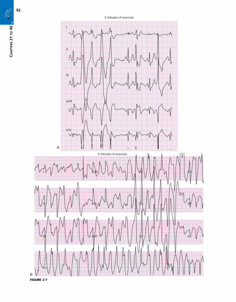
92
II
CH
APT
ERS
21 T
O 4
0
FIGURE 2-7
A
I
II
aVR
aVL
III
3 minutes of exercise
B
I
II
aVR
aVL
5 minutes of exercise
V1 V4
V2 V5
III
V1
aVF V3 V6

Heart Failu
re; Arrh
ythm
ias, Sud
den
Death
, and
Synco
pe
93
2
FIGURE 2-8
I
RA
HBE
CS
IIIIIV1
HA A AV
V
H HAV V
AH V
AH
AHV
S1 S1 S2
500 250 330
300
A
A VA A
A A A A A A
FIGURE 2-9
V1
V5
His distal
His mid
His proximal
RV
QUESTION 229
Each of the following predisposes to the arrhythmia shown in Figure 2-10 EXCEPTA. Congenital severe bradycardiaB. HypokalemiaC. Loss of function mutation in the SCN5A geneD. Tricyclic antidepressant overdoseE. Disopyramide
QUESTION 230
A 60-year-old man presents with presyncope and is found to be in a wide-complex tachycardia at 140 beats/min. An electrophysiologic study is performed and tachycardia is induced, as shown in Figure 2-11. The mechanism of this tachycardia isA. Atrioventricular reciprocating tachycardia utilizing an
accessory pathwayB. Atrioventricular nodal reentrant tachycardia with aber-
rant ventricular conductionC. Ventricular tachycardiaD. Atrial flutter with aberrant ventricular conductionE. Sinus tachycardia
QUESTION 231
Which of the following statements is correct regarding the electrophysiologic abnormality depicted in Figure 2-12?A. It is associated with syncope and sudden deathB. When present with an inferior myocardial infarction, it
is an indication for temporary pacingC. It typically reflects block at the level of the atrioventricu-
lar node, proximal to the His bundleD. Carotid sinus massage typically abolishes this
abnormality
QUESTION 232
Permanent cardiac pacing is appropriate for all of the fol-lowing patients EXCEPTA. A 35-year-old man with asymptomatic type II second-
degree atrioventricular (AV) block and sinus bradycar-dia at 38 beats/min
B. A 70-year-old man with left ventricular hypertrophy, per-sistent fatigue, and lightheadedness, with marked first-degree AV block (PR interval = 0.36 second)
C. A 57-year-old man with acquired asymptomatic third-degree AV block

94
II
CH
APT
ERS
21 T
O 4
0
FIGURE 2-10
FIGURE 2-11
V1
V5
HRA
His distal
His proximal
RV apex
D. A 40-year-old woman with congenital complete AV block
E. A 56-year-old marathon runner with calf cramps and 3.4-second pauses during sleep
QUESTION 233
A 62-year-old man underwent pacemaker insertion 4 years ago because of marked bradycardia. As part of his exercise program he has been using a rowing machine. Recently, he
has had several episodes of near-syncope that have occurred only during such exercise. An ambulatory ECG (Holter monitor) demonstrated the rhythm strip shown in Figure 2-13 during one near-syncopal episode while rowing. Which of the following is correct?A. There is evidence of a single-chamber pacing systemB. There is inappropriate inhibition of ventricular pacingC. There is undersensing of atrial activityD. There is lack of capture of the ventriclesE. There is lack of capture of the atria
QUESTION 234
The ECG in Figure 2-14 is compatible with all of the follow-ing diagnoses EXCEPTA. Lead insulation breachB. HyperkalemiaC. Lead dislodgmentD. Loose set screwE. Impending battery depletion
QUESTION 235
Which of the following statements regarding sinus node function is correct?A. Atropine enhances cardioinhibitory input to the sinus
nodeB. Sinus arrest is defined as a pause that is an exact multiple
of the PP interval of the underlying rhythmC. Vasodepressor carotid sinus hypersensitivity is defined
as asystole exceeding 2 seconds during carotid sinus massage
D. In the respiratory form of sinus arrhythmia, the PP inter-val cyclically shortens during inspiration
QUESTION 236
Which of the following statements regarding arrhythmo-genic right ventricular cardiomyopathy (ARVC) is TRUE?

Heart Failu
re; Arrh
ythm
ias, Sud
den
Death
, and
Synco
pe
95
2
E. A 68-year-old woman with hypertension and left atrial enlargement by echocardiography
QUESTION 238
Each of the following statements regarding the antiarrhyth-mic drug dronedarone is true, EXCEPTA. Its electrophysiologic properties are similar to
amiodaroneB. It is safe for use in patients with heart failure symptomsC. The prevalence of thyroid toxicity is lowD. QT prolongation is typical during use but only rarely
results in proarrhythmiaE. Acute hepatic failure is a potential complication
QUESTION 239
A 30-year-old woman presents because of recurrent epi-sodes of paroxysmal tachycardia with dyspnea and presyn-cope. A maternal uncle died suddenly at age 27. An electrophysiologic study is performed. The intracardiac electrograms at baseline (A) and during tachycardia (B)
A. Ventricular tachycardia in patients with ARVC typically has a right bundle branch block morphology
B. ARVC is more common in womenC. Fatty or fibrofatty infiltration of the right ventricle is the
pathologic hallmarkD. Pathologic changes do not occur in the left ventricleE. Radiofrequency ablation is successful at preventing
ventricular tachycardia
QUESTION 237
Of the following patients with atrial fibrillation, who is the best candidate for aspirin rather than an anticoagulant for stroke prophylaxis?A. A 78-year-old man with diabetes but no structural heart
diseaseB. A 50-year-old woman with dilated cardiomyopathy and
controlled symptoms of heart failureC. A 52-year-old man with hypertension and lower-extremity
claudicationD. A 45-year-old man with a history of obesity and smoking
but no structural heart disease
FIGURE 2-12
A 240H
V
A 280H
V
A 290H
V
A295H
V
A295H
V
A A325H
V
RA
V1
HBEA 240
H V
FIGURE 2-13
aVF
DDD 70/min 25 mm/s
FIGURE 2-14
VV V

96
II
CH
APT
ERS
21 T
O 4
0
C. Sudden loud acoustic events are a common trigger of syncope in LQT3 patients
D. Cardiac events during sleep are common in patients with LQT2
QUESTION 241
All of the following statements regarding sudden cardiac death (SCD) are true EXCEPTA. There are over 300,000 SCDs annually in the United
StatesB. The peak incidence of SCD among adults is between the
ages of 45 and 75 yearsC. Hereditary causes of SCD include hypertrophic cardio-
myopathy, long QT syndrome, arrhythmogenic right ven-tricular cardiomyopathy, and the Brugada syndrome
D. SCD is more common in women than in men
are shown in Figure 2-15. Of the following choices, the most appropriate therapy isA. Ablation of the atrioventricular nodal slow pathwayB. Ablation of an accessory pathwayC. Implantation of an automatic cardioverter-defibrillatorD. Direct-current cardioversion followed by long-term anti-
coagulation with warfarinE. No further therapy is required
QUESTION 240
Which of the following statements about congenital long QT syndromes (LQTS) is TRUE?A. Most forms of LQTS result from mutations in genes that
code for proteins in cardiac calcium channelsB. LQT1 patients experience a high frequency of cardiac
events during swimming
FIGURE 2-15
Baseline
HRA
CS proximal
CS
CS
CS
CS distal
His distal
His proximal
RV apex
A
B
V1
V5
Tachycardia
HRA
CS proximal
CS
CS
CS
CS distal
His distal
His proximal
RV apex
V1
V5

Heart Failu
re; Arrh
ythm
ias, Sud
den
Death
, and
Synco
pe
97
2
QUESTION 245
Which of the following patients is the most appropriate candidate for placement of an implantable cardioverter defibrillator for primary prevention of sudden cardiac death?A. 45-year-old man 1 week after acute anterior myocardial
infarction with left ventricular ejection fraction (LVEF) 35% and New York Heart Association (NYHA) class III symptoms
B. 66-year-old woman with a history of nonischemic dilated cardiomyopathy and LVEF 30% despite medical therapy with an ACE-inhibitor and beta-blocker for the past 6 months
C. 22-year-old man who presents with chest pain and mildly reduced LVEF of 50% who has a normal surface ECG but evidence of myocarditis on cardiac MR imaging
D. 78-year-old man with history of ischemic cardiomyopa-thy, LVEF 30%, end-stage renal disease on dialysis, and recently diagnosed metastatic non–small cell lung cancer
E. 52-year-old with history of alcoholism with newly recog-nized LVEF of 20% and NYHA class II symptoms
QUESTION 246
Which of the following statements regarding cardiac pacing modes is TRUE?A. Ventricular inhibited pacing (VVI) restores and main-
tains atrioventricular (AV) synchronyB. VVI pacing provides rate responsiveness in the chrono-
tropically incompetent patientC. Single-chamber triggered pacing (AAT or VVT) increases
the drain on the pacemaker batteryD. Atrial inhibited pacing (AAI) is an appropriate mode of
pacing for patients with AV nodal dysfunctionE. Dual-chamber pacing and sensing with inhibition and
tracking (DDD) is the preferred mode of pacing for patients in atrial fibrillation
QUESTION 247
All of the following statements regarding cardiac pacemak-ers are true EXCEPTA. Hyperkalemia results in pacing and sensing threshold
abnormalitiesB. Lead dislodgment or inadequate initial lead placement
should be suspected if true undersensing is presentC. Cellular telephones do not cause clinically important
pacemaker interference during normal use
E. An intraventricular conduction abnormality on the ECG is a stronger predictor of SCD than findings of left ventricular hypertrophy
QUESTION 242
All of the following are likely in a patient with the ECG shown in Figure 2-16 EXCEPTA. A gradual increase in atrial rate with the administration
of digoxinB. An irregular atrial rateC. Precipitation of the arrhythmia by hypokalemiaD. Absence of underlying cardiac disease in the majority
of casesE. Frequent ventricular premature beats
QUESTION 243
Which of the following statements regarding sudden cardiac death (SCD) in patients with coronary artery disease is correct?A. SCD accounts for 10% of all coronary artery disease
related deathsB. Compared to Caucasians, African Americans have
a higher age-adjusted incidence of sudden cardiac death
C. The most common mechanism of cardiac arrest is asystole
D. The outcome of patients with bradycardic/asystolic out-of-hospital cardiac arrest is better than if ventricular tachycardia is the initial arrhythmia
E. Successfully resuscitated ventricular fibrillation during the first 48 hours of an acute MI identifies individuals at increased risk of future SCD and warrants ICD therapy
QUESTION 244
All of the following statements regarding electrophysiologic testing are true EXCEPTA. A prolonged His-ventricular (HV) interval identifies
patients at increased risk of developing atrioventricular (AV) block
B. The HV interval has low sensitivity but high specificity for predicting the development of complete AV block
C. A prolonged sinus node recovery time (SNRT) is very specific for identifying patients with sinus node dysfunction
D. The sensitivity of the SNRT in identifying sinus node dysfunction is 95%
FIGURE 2-16
20+181614121086420

98
II
CH
APT
ERS
21 T
O 4
0
FIGURE 2-18
aVR
aVL
aVF
V1
V2
V3
V4
V5
V6
I
II
III
V1
FIGURE 2-17
D. Right bundle branch block is the expected electrocar-diographic pattern during right ventricular pacing
E. Pseudofusion on the surface ECG is identified by an appropriately timed pacing stimulus that does not alter the morphology of a superimposed intrinsic QRS
QUESTION 248
The finding on the chest radiograph shown in Figure 2-17 would be associated with which of the following measure-ments on device interrogation?A. High-voltage threshold, high lead impedanceB. Low-voltage threshold, high lead impedance
C. High-voltage threshold, low lead impedanceD. Low-voltage threshold, low lead impedanceE. High-voltage threshold, normal lead impedance
QUESTION 249
The electrocardiographic abnormality displayed in Figure 2-18 is associated with all of the following EXCEPTA. An absence of underlying structural heart disease in
most adultsB. Beneficial response to treatment with verapamilC. An association with Ebstein’s anomalyD. A higher prevalence in menE. A right posteroseptal pathway
QUESTION 250
A 65-year-old diabetic man with a history of myocardial infarction presents to your office for evaluation. He is known to have a left ventricular ejection fraction of 30% with an anterior wall motion abnormality. He is comfortable at rest but reports dyspnea with simple household activities and cannot ascend a flight of stairs without stopping to catch his breath. He does not describe chest discomfort and there is no evidence of reversible ischemia by exercise scintigra-phy. His current medical regimen includes carvedilol, 25 mg twice daily; lisinopril, 20 mg daily; furosemide, 40 mg daily; spironolactone, 25 mg daily; and digoxin, 0.125 mg daily. His physical examination reveals blood pressure of 90/50 mm Hg, heart rate of 70 beats/min, normal jugular venous pressure, and clear lungs to auscultation. The apical cardiac impulse is laterally displaced toward the anterior axillary line. On auscultation there is a normal S1, paradoxically split S2, a soft apical S3 gallop, and a grade 3/6 holosystolic murmur at the apex that radiates to the

Heart Failu
re; Arrh
ythm
ias, Sud
den
Death
, and
Synco
pe
99
2
C. Pacemaker implantationD. Radiofrequency catheter ablationE. Defibrillator implantation
QUESTION 252
All of the following statements are true about the antiar-rhythmic drug dofetilide EXCEPTA. It has significant renal excretionB. It prolongs the QT interval in a dose-dependent fashionC. It is unsafe in patients with prior myocardial infarctionD. Patients must be admitted to the hospital for drug
initiationE. It should not be used in patients taking verapamil
QUESTION 253
A 26-year-old construction worker comes to the emergency department on the morning of January 2 in atrial fibrilla-tion. He states that for the past few months he has had occasional episodes of palpitations, almost always on Mondays. His vital signs include heart rate of 140 beats/min, respirations of 16 breaths/min, and blood pressure of 160/95 mm Hg. His physical examination is unremarkable except for a soft systolic murmur. While being observed in the hospital, he spontaneously reverts to normal sinus rhythm. The most likely precipitating cause isA. CaffeineB. CocaineC. AlcoholD. HypertensionE. Mitral valve prolapse
QUESTIONS 254 TO 257
For each of the following descriptions, match the appropri-ate disorders:A. Jervell and Lange-Nielsen syndromeB. Romano-Ward syndrome
axilla. There is no peripheral edema. His ECG is shown in Figure 2-19. Which of the following is the most appropriate approach to device therapy in this patient?A. No device implantation is indicatedB. Implantation of a cardioverter-defibrillator is warrantedC. Implantation of a combined cardiac resynchronization-
defibrillator system is appropriateD. Refer the patient for echocardiography to assess
dyssynchrony and, if positive, implant a cardiac resynchronization-defibrillator
E. Implantation of a cardiac-resynchronization pacemaker without defibrillation capability is most appropriate
QUESTION 251
The electrophysiologic study tracing in Figure 2-20 is obtained from a 28-year-old man with palpitations, recur-rent syncope, and a structurally normal heart. Which of the following is the most appropriate therapy?A. AtenololB. Verapamil
FIGURE 2-19
FIGURE 2-20 From Prystowsky EN, Browne KF, Zipes DP: J Am Coll Cardiol 1:468, 1983.
I
II
III
V1
HRA
HBE
RV
HV = 45 ms A H V
AP
A V AH = 90 ms

100
II
CH
APT
ERS
21 T
O 4
0
266. Acetazolamide267. Metolazone268. Torsemide269. Eplerenone
QUESTIONS 270 TO 273
For each clinical condition, match the most appropriate pacemaker mode:A. VATB. VVIRC. DDDD. DDDRE. AAIR
270. A 58-year-old man with tachycardia-bradycardia syndrome who develops symptomatic sinus brady-cardia with beta-blocker therapy
271. A 70-year-old woman with long-standing atrial fibrillation who complains of dizziness and is found on examination to have a ventricular rate of 30 beats/min
272. A 62-year-old man with complete heart block after aortic valve surgery
273. A 45-year-old man with symptomatic sinoatrial exit block and an appropriate junctional escape rhythm
QUESTIONS 274 TO 277
Several conditions can cause dilated cardiomyopathy (DCM) with electric instability, defined as conduction disease or arrhythmia out of proportion to the degree of left ventricular dysfunction. For each clinical description, match the corresponding etiology of DCM with electric instability:A. Cardiac sarcoidosisB. Giant cell myocarditisC. Chagas diseaseD. Arrhythmogenic right ventricular cardiomyopathyE. Cardiolaminopathy
274. A 30-year-old man with a history of syncope who is found to have low-amplitude deflections on the ST segment in the right precordial leads and abnormal desmosomes on cardiac immunohistochemistry
275. A 38-year-old man with gastrointestinal dysmotility, complete heart block, and an apical aneurysm
276. A 50-year-old woman with a history of restrictive lung disease who presents with syncope in the setting of first-degree atrioventricular block and right bundle branch block
277. A 42-year-old woman with rapidly deteriorating cardiac function, frequent bursts of ventricular tachycardia, and widespread myocyte necrosis on endomyocardial biopsy
QUESTIONS 278 TO 282
For each of the following conditions, match the correspond-ing clinical presentations of syncope:A. A 20-year-old woman “blacked out” during phlebotomy
for a routine blood test
C. Right ventricular outflow tract ventricular tachycardiaD. Brugada syndrome
254. Autosomal recessive disorder associated with sen-sorineural deafness
255. Typically, the ECG reveals a right bundle branch block morphology with ST-segment elevation in the anterior precordial leads
256. Autosomal dominant long QT syndrome with normal hearing
257. Left bundle branch block with an inferior axis
QUESTIONS 258 TO 261
Match the following antiarrhythmic drug actions with the appropriate Vaughan-Williams drug classification:A. Predominantly block potassium channels and prolong
repolarizationB. Predominantly block beta-adrenergic receptorsC. Predominantly block slow calcium channels (ICa.L)D. Reduce the rate of rise of the action potential upstroke
(Vmax) and prolong the action potential durationE. Block sodium channels but shorten the action potential
duration and do not reduce Vmax
258. Class IA drugs259. Class II drugs260. Class III drugs261. Class IV drugs
QUESTIONS 262 TO 265
Assume that you have decided to prescribe antiarrhythmic drug therapy to prevent recurrent episodes of atrial fibrilla-tion for each of the patients described below. Match each patient with the most appropriate antiarrhythmic from the provided list:A. FlecainideB. SotalolC. AmiodaroneD. Mexiletine
262. A 64-year-old man with remote history of anterior myocardial infarction and left ventricular ejection fraction of 50%
263. A 74-year-old woman with long-standing hyperten-sion and left ventricular hypertrophy (wall thick-ness = 1.6 cm)
264. A 58-year-old with nonischemic cardiomyopathy and class III congestive heart failure
265. A 46-year-old otherwise healthy man with no struc-tural heart disease
QUESTIONS 266 TO 269
For each of the following diuretic agents, match the appro-priate adverse effect:A. OtotoxicityB. GynecomastiaC. Metabolic acidosisD. HypercalcemiaE. Hyperkalemia

Heart Failu
re; Arrh
ythm
ias, Sud
den
Death
, and
Synco
pe
101
2B. A 65-year-old woman lost consciousness after arm
exercisesC. A 35-year-old man sustained syncope during exercise
and has a systolic murmur that intensifies on standing upright
D. A 74-year-old man experiences sudden syncope while shaving
E. A 28-year-old woman with recurrent episodes of breath-lessness, lightheadedness, and syncope after changes in body position
278. Hypertrophic cardiomyopathy279. Subclavian steal syndrome280. Vasovagal syncope281. Carotid sinus hypersensitivity282. Left atrial myxoma
QUESTIONS 283 TO 286
For each condition capable of precipitating high-output cardiac failure, match the appropriate physical findings:A. HyperthyroidismB. BeriberiC. Arteriovenous fistulaD. Carcinoid syndromeE. Osler-Weber-Rendu syndrome
283. Nicoladoni-Branham sign284. Hepatomegaly and abdominal bruits285. Means-Lerman scratch286. Paresthesias and painful glossitis
QUESTIONS 287 TO 290
For each of the following descriptions, match the appropri-ate medication:
END-DIASTOLIC VOLUME (mL/m2)
STROKE VOLUME (mL/m2)
LV MASS (g/m2)
Normal = 70 45 92
A. 84 44 172
B. 193 92 200
C. 199 37 145
D. 70 40 80
A. One of the least lipid-soluble beta-blockersB. Cardioselective beta-blocker with intrinsic sympathomi-
metic activityC. Beta-blocker with alpha-blocking activityD. Noncardioselective beta-blocker with intrinsic sympa-
thomimetic activity
287. Atenolol288. Carvedilol289. Acebutolol290. Pindolol
QUESTIONS 291 TO 294
For each disease state, match the appropriate left ventricu-lar (LV) volume and mass data:
291. Aortic valve stenosis with peak systolic gradient >30 mm Hg
292. Myocardial disease (primary dilated cardiomyopa-thy)
293. Aortic regurgitation with regurgitant flow >30 mL per beat
294. Mitral valve regurgitation with regurgitant flow >20 mL per beat

This page intentionally left blank

103
SECTION II(CHAPTERS 21 TO 40)
Heart Failure; Arrhythmias, Sudden Death, and Syncope
ANSWER TO QUESTION 168
E (Braunwald, pp. 458, 478, 491; Figure 22-7, A)Natriuretic peptides are of importance in the diagnosis, and assessment of prognosis, in patients with heart failure. The natriuretic peptide system consists of five structurally similar peptide hormones: atrial natriuretic peptide (ANP), urodilantin (an isoform of ANP), brain-type natriuretic peptide (BNP), C-type natriuretic peptide, and dendroaspis natriuretic peptide (DNP).1 ANP is released by atrial myo-cytes in response to acute increases in atrial pressure. Pro-hormone BNP is released in response to hemodynamic stress from ventricular dilatation, hypertrophy, or increased wall tension. Prohormone BNP is cleaved by a circulating endoprotease into two polypeptides: the inactive N-terminal proBNP (NT-proBNP), 76 amino acids in length, and the biologically active peptide BNP, 32 amino acids in length.2
Circulating levels of ANP and BNP are elevated in patients with heart failure. Both ANP and BNP promote vasodilata-tion and natriuresis, thereby counteracting the salt- and water-retaining effects of the adrenergic, renin-angiotensin-aldosterone, and vasopressin systems. BNP and NT-proBNP levels rise with increasing age and worsening renal func-tion. In contrast, BNP levels have an inverse relationship with body mass index.
CNP is derived predominantly from endothelial cells in the peripheral vasculature. The precise roles of CNP, urodilantin, and DNP in cardiovascular physiology remain unclear.
A large, multicenter study of patients in the emergency department undergoing evaluation for acute dyspnea showed that a BNP level of >100 pg/mL is 90% sensitive and 76% specific in identifying a cardiac etiology.3 A BNP level >400 pg/mL rendered the diagnosis of heart failure likely. Analogous results were found for NT-proBNP—patients in acute heart failure had mean levels >4000 pg/mL compared with 130 pg/mL in those without heart failure.4
A substudy of the TACTICS-TIMI 18 trial demonstrated that an elevated BNP level in patients presenting with acute coronary syndromes is associated with a higher risk of subsequent congestive heart failure and death.5
REFERENCES1. Daniels LB, Maisel AS: Natriuretic peptides. J Am Coll Cardiol
50:2357,2007.
2. BraunwaldE:Biomarkers inheartfailure.NEnglJMed358:2148,2008.
3. MaiselAS,KrishnaswamyP,NowakRM,etal:RapidmeasurementofB-typenatriuretic peptide in theemergencydiagnosisof heartfailure.NEnglJMed347:161,2002.
4. Januzzi JL, Jr, Camargo CA, Anwaruddin S, etal: The N-terminalpro-BNP investigation of dyspnea in the emergency department(PRIDE)study.AmJCardiol95:948,2005.
5. MorrowDA,deLemosJA,SabatineMS,etal:EvaluationofB-typenatriuretic peptide for risk assessment in unstable angina/non-ST-elevation myocardial infarction: B-type natriuretic peptideand prognosis in TACTICS-TIMI 18. J Am Coll Cardiol 41:1264, 2003.
ANSWER TO QUESTION 169
D (Braunwald, pp. 520-523; Figure 25-10; Table 25-7)Mannitol is an inert osmotic agent that expands the extra-cellular fluid volume; its use is contraindicated in patients with decompensated heart failure.
Mineralocorticoid receptor antagonists (spironolactone and eplerenone) act at the renal distal convoluted tubule and cortical collecting tubule to reduce sodium reabsorp-tion and inhibit K+ and H+ excretion. Although only weak diuretics, they have been shown to reduce morbidity and mortality in systolic heart failure.1 However, aldosterone receptor antagonists may contribute to clinically significant hyperkalemia, particularly when used in combination with angiotensin-converting enzyme (ACE) inhibitors and/or angiotensin II receptor blockers (ARBs).2
Loop diuretics, such as furosemide, torsemide, and bumetanide, are inhibitors of the Na+/K+/2Cl− cotransporter in the thick ascending limb of the loop of Henle. Inhibition of this cotransporter markedly increases the fractional excretion of sodium and chloride. However, the delivery of large amounts of sodium and fluid to the distal nephron increases K+ and H+ secretion, leading to hypokalemia and metabolic alkalosis.
Nonsteroidal anti-inflammatory agents (NSAIDs) may impair renal function and diminish the action of diuretics by inhibiting the production of vasodilating prostaglandins. All NSAIDs, including aspirin, have this potential effect, which can contribute to diuretic resistance in individuals with an initially favorable diuretic response.3

104
II
CH
APT
ERS
21 T
O 4
0
resulting from impaired cardiac output and excessive loop diuretic therapy.
Renal function is an important prognostic indicator in patients with acute heart failure syndromes. Impaired base-line renal function, and worsening renal function during hospitalization or early after discharge, are potent predic-tors of adverse outcomes, including early readmission rates and mortality.3 Elevated, not reduced, systemic venous pres-sure appears to be an important contributing factor to the development of cardiorenal syndrome, because venous congestion contributes to elevated intra-abdominal pres-sure that can impair glomerular filtration.4
An increase in blood urea nitrogen (BUN) out of propor-tion to serum creatinine is a sign of renal hypoperfusion, resulting from low forward cardiac output or intravascular volume depletion because of high doses of intravenous diuretics. The neurohormonal activation accompanying renal hypoperfusion increases proximal sodium and urea reabsorption, while creatinine handling is unaffected, leading to an elevated BUN/creatinine ratio.
REFERENCES1. GheorghiadeM,PangPS:Acuteheartfailuresyndromes.JAmColl
Cardiol53:557,2009.2. BockJS,GottliebSS:Cardiorenalsyndrome:newperspectives.Cir-
culation121:2592,2010.3. DammanK,NavisG,VoorsAA,etal:Worseningrenalfunctionand
prognosis in heart failure: systematic review and meta-analysis. JCardFail13:599,2007.
4. Mullens W, Abraham Z, Francis GS, etal: Importance of venouscongestionforworseningofrenalfunctioninadvanceddecompen-satedheartfailure.JAmCollCardiol53:589,2009.
ANSWER TO QUESTION 172
B (Braunwald, pp. 527-532, 534)The effectiveness of digoxin was studied in the DIG trial, which enrolled 6800 patients with class I to III heart failure, with a mean left ventricular (LV) ejection fraction of 28%.1 Over 37 months of follow-up there was a significant decrease in heart failure hospitalizations but no significant improve-ment in total mortality with digoxin therapy compared with placebo.
Angiotensin-converting enzyme (ACE) inhibitors are the cornerstone of therapy for patients with chronic heart failure with reduced ejection fraction or asymptomatic LV dysfunction because of their favorable hemodynamic effects and unsurpassed benefit on long-term mortality. The Vasodilator Heart Failure Trial II (V-HEFT II) compared the ACE inhibitor enalapril with the combination of hydralazine plus isosorbide dinitrate in the treatment of chronic moder-ate congestive heart failure. Survival in the enalapril-treated group proved superior.2
Aggregate clinical data indicate that ARBs are as effective as ACE inhibitors in reducing HF-related morbidity and mortality. Candesartan significantly reduced the primary composite of cardiovascular death or hospital admission for heart failure among ACE-inhibitor intolerant patients enrolled in the Candesartan Heart Failure: Assessment of Reduction in Mortality and Morbidity trial (CHARM-Alternative Trial).3 The VALIANT trial randomized patients with LV dysfunction after myocardial infarction to valsar-tan, captopril, or both and found similar benefit with the ACE inhibitor or ARB in reducing mortality.4 Thus ARBs are
REFERENCES1. ZannadF,McMurrayJJV,KrumH,etal:Eplerenoneinpatientswith
systolic heart failure and mild symptoms. N Engl J Med 364:11,2011.
2. Juurlink DN, Mamdani MM, Lee DS, etal: Rates of hyperkalemiaafter publication of the RandomizedAldactone Evaluation Study. NEnglJMed351:543,2004.
3. YancyCW,JessupM,BozkurtB,etal:ACCF/AHAguidelineforthemanagementofheartfailure:a reportoftheAmericanCollegeofCardiology Foundation/American HeartAssociationTask Force onPracticeGuidelines.Circulation62(e147):2013,2013.
ANSWER TO QUESTION 170
E (Braunwald, pp. 491, 536, 547; Figure 24-5; Table 25-6)Cardiac arrhythmias are common in patients with structural heart disease, can contribute to worsening intracardiac hemodynamics, and may precipitate acute decompensated heart failure.1 Cardiac output is dependent on maintenance of adequate stroke volume and heart rate. Tachyarrhythmias increase myocardial oxygen demand and reduce the time available for ventricular filling in diastole, compromising ventricular stroke volume and cardiac output.
Since stroke volume is compromised in patients with left ventricular dysfunction, maintenance of cardiac output is largely dependent on an adequate heart rate. Thus, exces-sive heart rate slowing (i.e., bradyarrhythmias) may also depress cardiac output.
Dissociation between atrial and ventricular contraction (as in high-grade atrioventricular block) reduces the atrial contribution to ventricular filling, impairing subsequent stroke volume and cardiac output in patients with systolic or diastolic dysfunction. Abnormal intraventricular conduc-tion, as in ventricular tachycardia or right ventricular apical pacing, may impair myocardial performance because of the loss of synchronized ventricular contraction.2 Optimiza-tion of atrioventricular and ventriculoventricular synchrony is the primary mechanism of benefit from cardiac resyn-chronization therapy (biventricular pacing) in patients with left ventricular dysfunction and heart failure.3
REFERENCES1. BenzaRL,TallajJA,FelkerGM,etal:Theimpactofarrhythmias in
acuteheartfailure.JCardFail10:279,2004.2. WangNC,MaggioniAP,KonstamMA,etal:Clinicalimplicationsof
QRSdurationinpatientshospitalizedwithworseningheartfailureandreducedleftventricularejectionfraction.JAMA299:2656,2008.
3. AbrahamWT:Cardiacresynchronizationtherapyisimportantforallpatientswithcongestiveheartfailureandventriculardyssynchrony.Circulation114:2692,2006.
ANSWER TO QUESTION 171
C (Braunwald, pp. 488, 495, 526; Figures 24-4, 25-5)The cardiorenal syndrome is characterized by worsening renal function during a heart failure hospitalization, or soon after discharge, despite symptomatic improvement with diuretic therapy and maintenance of adequate intravascu-lar volume.1,2 Underlying chronic kidney disease (e.g., asso-ciated with diabetes or hypertension) increases the risk of cardiorenal syndrome, as does neurohormonal activation

105
2Heart Failu
re; Arrh
ythm
ias, Sud
den
Death
, and
Synco
pe
intrinsic Frank-Starling mechanism to augment cardiac output. Circulating catecholamine levels rise as exercise ensues, providing a delayed chronotropic response. This mechanism allows near-normal hemodynamic function at rest, and the capacity to support at least moderate exercise in a large majority of long-term survivors. Of note, beta-blockers may impair the heart rate response and should be used cautiously after heart transplantation.
As no reliable serologic markers for allograft rejection have been identified, right ventricular endomyocardial biopsy remains the gold standard for this diagnosis. Biop-sies are typically performed through a transjugular vein approach and are carried out on a routine schedule during the post-transplantation period and over the life of the trans-plant. High-grade rejection may be signaled by the develop-ment of clinical heart failure, atrial arrhythmias, low QRS voltage on the ECG, or echocardiographic evidence of left ventri cular dysfunction; however, definitive diagnosis or identification of early or subtle lower-grade rejection requires histologic examination of the myocardium.
Allograft rejection and infection are the most common causes of death early after transplant. The risk of infection diminishes with time as the intensity of immunosuppres-sion therapy is reduced. In contrast, the risk of fatal malig-nancy increases progressively in the years thereafter. The most common fatal early malignancies are post-transplant lymphoproliferative disorders and lung cancer.
REFERENCES1. Stehlik J, Edwards LB, KucheryavayaAY, etal:The Registry of the
Internal Society of Heart and Lung Transplantation: 29th officialadulthearttransplantreport–2012.JHeartLungTransplant31:1052,2012.
2. Grady KL, Naftel DC, Young JB, etal: Patterns and predictors ofphysicalfunctionaldisabilityat5to10yearsafterhearttransplanta-tion.JHeartLungTransplant26:1182,2007.
ANSWER TO QUESTION 174
C (Braunwald, p. 476)Many medical and surgical conditions are associated with a chest radiographic pattern consistent with pulmonary edema, as in the accompanying radiograph. The physical examination of this patient was nearly normal, without dra-matic chest examination findings, jugular venous disten-tion, or a third heart sound. This suggests that, in this patient, the edema was primarily caused not by an altera-tion in the Starling forces across pulmonary capillaries but rather by abnormal permeability of the alveolar-capillary membrane. This situation is consistent with acute respira-tory distress syndrome (ARDS). Among the conditions that have been associated with ARDS are pneumonia, inhaled toxins, circulating foreign substances (including bacterial endotoxins), aspiration of gastric contents, acute radiation pneumonitis, release of endogenous vasoactive substances, disseminated intravascular coagulation, shock lung in asso-ciation with nonthoracic trauma, and acute hemorrhagic pancreatitis.
Whereas the pathophysiology of ARDS is incompletely understood, it is likely that epithelial injury leading to increased permeability of the alveolar-capillary barrier is a critical initiating event. An imbalance between proin-flammatory cytokines and anti-inflammatory mediators likely contributes to recruitment of neutrophils, release of
currently accepted as appropriate substitutes for patients with LV dysfunction who are intolerant of ACE inhibitors.5 The incremental benefit of ARBs in patients already treated with ACE inhibitors is less clear.
In the RALES trial, the aldosterone antagonist spironolac-tone was shown to reduce mortality when added to stan-dard heart failure therapy in patients with class III to IV heart failure with an ejection fraction <35%.6 In the EMPHA-SIS trial, eplerenone, a selective aldosterone antagonist, significantly reduced mortality compared with placebo when added to standard heart failure therapy in patients with class II or III systolic heart failure.7
REFERENCES1. TheDigitalisInvestigationGroup:Theeffectofdigoxinonmortality
andmorbidityinpatientswithheartfailure.NEnglJMed336:525,1997.
2. CohnJN,JohnsonG,ZeischeS,etal:Acomparisonofenalaprilwithhydralazine-isosorbidedinitrateinthetreatmentofchronicconges-tiveheartfailure.NEnglJMed325:303,1991.
3. GrangerCB,McMurrayJJ,YusufS,etal:Effectsofcandesartan inpatientswithchronicheartfailureandreducedleft-ventricularsys-tolic function intolerant to angiotensin-converting-enzyme inhibi-tors:TheCHARM-Alternativetrial.Lancet362:772,2003.
4. PfefferMA,McMurrayJJV,VelazquezEJ,etal:Valsartan,captopril,orbothinpatientswithmyocardialinfarctioncomplicatedbyheartfailure,leftventriculardysfunction,orboth.NEnglJMed349:2003,1893.
5. YancyCW,JessupM,BozkurtB,etal:2013ACCF/AHAguidelineforthemanagementofheartfailure:areportoftheAmericanCollegeofCardiologyFoundation/AmericanHeartAssociationTaskForceonPracticeGuidelines.Circulation62:e147,2013.
6. PittB,ZannadF,RemmeWJ,etal:Theeffectofspironolactoneonmorbidityandmortality inpatientswithsevereheartfailure.Ran-domizedAldactone Evaluation Study Investigators. N Engl J Med341:709,1999.
7. ZannadF,McMurrayJJ,KrumH,etal:Eplerenoneinpatientswithsystolic heart failure and mild symptoms. N Engl J Med 364:11,2011.
ANSWER TO QUESTION 173
C (Braunwald, pp. 582-588)Long-term outcomes after transplantation improved dra-matically with the introduction of the calcineurin inhibitor cyclosporine as the cornerstone of immunosuppression. The median survival of all cardiac transplant patients is now >10 years, with a median survival of 13 years among those patients who are alive 1 year after transplant.1 By the first year after transplantation, 90% of surviving patients report no functional limitations.2
The selection of candidates for transplantation is a complex process that integrates assessment of hemody-namics, comorbidities that might limit post-transplantation survival, and psychosocial factors that may impair compli-ance. Optimal candidates for heart transplantation are younger patients with advanced heart failure, minimal comorbid illness or end-organ damage, a history of adher-ence to prescribed medical therapy, and a strong family and social network.
The transplanted heart is denervated and relies on atypi-cal adaptive mechanisms to meet varying demands for cardiac output. As a result of increased venous return at the onset of exercise, the transplanted heart responds with the

106
II
CH
APT
ERS
21 T
O 4
0
ANSWER TO QUESTION 176
B (Braunwald, pp. 432-433, 443; Figures 21-5, 21-14)The binding of a beta-adrenergic agonist to its myocyte receptor initiates a complex system of messengers within the sarcolemma and cytosol of the cell. The beta1 receptor is coupled, via G-proteins, to activation of adenylate cyclase and formation of cyclic adenosine monophosphate (cAMP). This molecule acts via protein kinases to phosphorylate proteins and enzymes within the cell. Such action stimulates enhanced entry of calcium ions through voltage-dependent L-type calcium channels, followed by additional calcium-induced calcium release from the sarcoplasmic reticulum. The rise in cytosolic calcium increases calcium–troponin C interaction, a necessary step for subsequent contraction. The activated troponin C binds tightly to the inhibitory molecule troponin I, thus removing inhibition of actin-myosin cross-bridge formation, and contraction ensues.
Whereas cAMP is a second messenger for the beta-adrenergic system, another cyclic nucleotide, cyclic guano-sine monophosphate (cGMP), acts as a second messenger during cholinergic stimulation. In vascular smooth muscle, cGMP acts as an intracellular messenger after nitric oxide stimulation.
Titin is a large protein that provides elasticity and supports the myosin molecule by tethering it to the myocyte Z line.
BIBLIOGRAPHYOpieLH:HeartPhysiology:FromCelltoCirculation,ed4,Philadelphia,
2004,LippincottWilliams&Wilkins.
ANSWER TO QUESTION 177
B (Braunwald, pp. 585-586; Table 28-3)Rejection is a ubiquitous concern after solid organ trans-plantation. It results from cell- or antibody-mediated allograft injury owing to recognition of the allograft as non-self tissue. Three major types of rejection have been identi-fied: hyperacute, acute, and chronic. Hyperacute rejection occurs within minutes to hours after heart transplantation and is mediated by preexisting antibodies to allogeneic antigens on the vascular endothelium that fix complement. This results in occlusion of graft vasculature and over-whelming graft failure. In contrast, the biopsy specimen in this question shows a dense lymphocytic infiltrate and myocyte damage typical of acute cellular allograft rejection. Acute cellular rejection is a T-cell–mediated process that develops in the first weeks to 6 months after transplantation. Chronic rejection, or late graft failure, is an irreversible dete-rioration of graft function years after transplant mediated by antibodies or progressive graft loss from ischemia.
Risk factors for rejection include female gender, black recipient race, recipient-positive cytomegalovirus serology, prior infections, and the number of HLA mismatches.1 Patients who do not experience acute rejection within the first 6 months after transplantation have a lower incidence of late rejection.
Although progress has been made in novel noninvasive serologic approaches, such as gene expression profiling, to identify acute rejection,2 endomyocardial biopsy remains the diagnostic gold standard. The procedure is performed under fluoroscopic or echocardiographic guidance using a
toxic inflammatory substances, loss of endothelial integrity, reduced surfactant production, and impaired removal of edema from the alveolar space.
BIBLIOGRAPHYWare LB, Matthay MA: Clinical practice: acute pulmonary edema.
NEnglJMed353:2788,2005.
ANSWER TO QUESTION 175
E (Braunwald, p. 477)A variety of laboratory abnormalities may be noted in patients with congestive heart failure. Alterations in serum electrolyte values usually occur only after patients have begun treatment or in more long-standing, severe cases of heart failure. Hyponatremia is seen in up to 25% of patients with acute heart failure decompensation and is an important negative prognostic indicator.1 It may be present for a variety of reasons, including sodium restric-tion, intensive diuretic therapy, a decrease in the ability to excrete water related to reductions in renal blood flow and glomerular filtration rate (GFR), and elevations in the concentration of circulating vasopressin.2 Hypokalemia may result from aggressive diuretic therapy. Conversely, hyperkalemia may occasionally occur in patients with severe heart failure who have marked reductions in GFR or who are receiving aldosterone receptor antagonists (spironolactone or eplerenone).
Congestive hepatomegaly due to “backward” failure and cardiac cirrhosis from long-standing heart failure is often accompanied by impaired hepatic function, reflected by abnormal circulating liver enzymes. In acute hepatic venous congestion, severe jaundice may result, with bilirubin levels as high as 15 to 20 mg/dL, dramatic elevations of serum aspartate aminotransferase levels, and prolongation of the prothrombin time. Although the clinical and laboratory pro-files of such an event may resemble viral hepatitis, the diagnosis of hepatic congestion is confirmed by rapid nor-malization of these values with successful treatment of heart failure. In patients with long-standing heart failure and secondary severe hepatic damage, albumin synthesis may become impaired. Rarely, more severe sequelae may occur, including hepatic hypoglycemia, fulminant hepatic failure, and hepatic coma.
Elevations in pulmonary capillary pressure are reflected by the appearance of the vasculature on the chest radio-graph. With minimal elevations (i.e., ~13 to 17 mm Hg), early equalization in the size of the vessels in the apices and bases is first discernible. It is not until greater pressure elevations occur (~18 to 20 mm Hg) that actual pulmonary vascular redistribution occurs. When pressure exceeds 20 to 25 mm Hg, frank interstitial pulmonary edema is usually observed. Importantly, however, in patients with chronic left ventricular failure, higher pulmonary pressures can be accommodated with few clinical and radiologic signs of congestion due to enhanced lymphatic drainage.
REFERENCES1. MohammedAA,vanKimmenadeRR,RichardsM,etal:Hyponatre-
mia,natriureticpeptides,andoutcomesinacutelydecompensatedheart failure: results from the International Collaborative ofNT-proBNPStudy.CircHeartFail3:354,2010.
2. DeLucaL,KleinL,UdelsonJE,etal:Hyponatremiainpatientswithheartfailure.AmJCardiol96:19L,2005.

107
2Heart Failu
re; Arrh
ythm
ias, Sud
den
Death
, and
Synco
pe
functional capacity. A peak oxygen consumption of >20 mL/kg/min reflects mild or no functional impairment. Con-versely, a peak oxygen consumption of <12 mL/kg/min indi-cates severe impairment and a poor prognosis. Serial tests can be performed over time to assess the response to phar-macologic therapies in patients with heart failure and can help guide the need for aggressive interventions such as cardiac transplantation or placement of a ventricular assist device.
REFERENCE1. BaladyGJ,ArenaR,SietsemaK,etal:Clinician’sguidetocardiopul-
monary exercise testing inadults: a scientific statement from theAmericanHeartAssociation.Circulation122:191,2010.
ANSWER TO QUESTION 179
A (Braunwald, p. 462; Figure 22-8)By the law of Laplace, LV wall stress (σ) is directly propor-tional to intracavitary pressure and chamber radius and inversely proportional to wall thickness (σ = [Pressure × radius]/wall thickness). Ventricular hypertrophy is therefore an adaptive mechanism that serves to reduce ventricular wall stress. Myocardial hypertrophy and remodeling pro-ceed in different patterns based on the timing and nature of the provocative stimulus. When the primary stimulus is pressure overload (as in aortic stenosis or hypertension), an increase in wall stress during ventricular systole triggers the addition of new myofibrils in parallel, leading to wall thick-ening at the expense of chamber size, in a pattern of con-centric hypertrophy. When the primary stimulus is volume overload (as in chronic mitral regurgitation), increased wall stress during ventricular diastole triggers the replication of sarcomeres in series, elongation of myocytes, and ventricu-lar cavity dilatation, in a pattern of eccentric hypertrophy. Therefore, although both chronic pressure and volume overload are associated with a compensatory increase in LV mass, the pattern of hypertrophy is distinct in each case.
With the use of echocardiography, concentric hypertro-phy can be distinguished from eccentric hypertrophy visu-ally, or on the basis of the ratio between wall thickness and the LV internal diameter during diastole (i.e., the relative wall thickness = [2 × posterior wall thickness]/LV internal dimension). A relative wall thickness <0.45 suggests eccen-tric hypertrophy, whereas a higher ratio is more consistent with concentric hypertrophy.
ANSWER TO QUESTION 180
A (Braunwald, pp. 551-552)Amiodarone has been studied extensively in patients with left ventricular (LV) dysfunction. It effectively suppresses ventricular and supraventricular arrhythmias and appears to be safe for use in this subset of patients. However, convincing evidence of mortality reduction has not been shown. In the SCD-HeFT study of patients with class II to III heart failure and a left ventricular ejection fraction of 35% or less, amiodarone did not improve survival compared with placebo.1
Dronedarone is a derivative of amiodarone that shares its electrophysiologic properties but does not contain iodine and is associated with lower rates of lung and thyroid toxic-ity. Dronedarone is approved to facilitate maintenance of
bioptome inserted percutaneously via the right internal jugular vein. Potential complications include pneumotho-rax, transient rhythm disturbances, myocardial perforation, and tricuspid regurgitation.
The most important feature of the post-transplant biopsy specimen is the detection of lymphocyte infiltration and the presence of myocyte necrosis. A revised continuum scale has been established for grading cardiac biopsies from no rejection (grade OR) to diffuse damaging inflammatory cell infiltrates with encroachment of myocytes and disruption of normal cell architecture (grade 3R).3 Appropriate therapy for acute cellular rejection depends on the timing and sever-ity of the rejection episode. Episodes that occur within the first 3 months, or episodes that are moderate to severe, are initially treated with pulsed-dose methylprednisolone. If steroid therapy is ineffective, then more aggressive therapy with OKT3 monoclonal antibody or ATGAM (horse antithy-mocyte globulin) may be necessary.4
REFERENCES1. Kobashigawa JA, StarlingRC,MehraMR,etal:Multicenter retro-
spectiveanalysisofcardiovascularriskfactorsaffectinglong-termoutcome of de novo cardiac transplant recipients. J Heart LungTransplant25:1063,2006.
2. PhamMX,TeutebergJJ,KfouryAG,etal:Geneexpressionprofilingforrejectionsurveillanceaftercardiactransplantation.NEnglJMed362:1890,2010.
3. Stewart S, Winters GL, Fishbein MC, etal: Revision of the 1990working formulation for the standardization of nomenclature in thediagnosisofheart rejection. JHeartLungTransplant24:1710,2005.
4. JessupM,BrozenaS:State-of-the-artstrategiesforimmunosuppres-sion.CurrOpinOrganTransplant12:536,2007.
ANSWER TO QUESTION 178
E (Braunwald, pp. 447-448, 450, 481)The four primary determinants of cardiac output (CO) are (1) heart rate; (2) preload, which is closely related to left ventricular (LV) end-diastolic volume; (3) afterload, which is closely related to aortic impedance (i.e., the sum of the external factors that oppose ventricular ejection); and (4) contractility, a fundamental property of cardiac muscle that reflects the level of activation of cross-bridge formation. Preload, afterload, and contractility determine the ventricu-lar stroke volume (SV), and cardiac output = Heart rate × SV.
Unlike other organs, the myocardium extracts oxygen from blood nearly maximally. Thus, even at rest, the oxygen saturation in coronary venous blood (measured at the coro-nary sinus) is quite low, usually <40%.
The body’s oxygen consumption at peak exercise is an indirect measure of the CO.1 The peak oxygen uptake ( �VO2 max) is defined as the value achieved when �VO2 plateaus despite a continued increase in the intensity of exercise. The anaerobic threshold is indicated by the �VO2 at which carbon dioxide production starts to rise, resulting in an increase in the � �V VCO O2 2/ ratio. Both the peak oxygen con-sumption and the anaerobic threshold are reproducible when measured days or weeks apart. During exercise, the CO of a normal heart increases up to sixfold and the body’s oxygen consumption can increase up to 18-fold.
In patients with left ventricular dysfunction, cardiopul-monary exercise tests are often performed to determine

108
II
CH
APT
ERS
21 T
O 4
0
under further mechanical disadvantage by reducing con-tractility and ventricular filling and prolonging the duration of mitral regurgitation.1 Cardiac resynchronization therapy (CRT), also known as biventricular pacing, serves to better coordinate left ventricular (LV) contraction, leading to improved ventricular performance and reduced LV filling pressures.
CRT improves ventricular function without increasing myocardial energy consumption, in contrast to the effects of inotropic agents such as dobutamine. CRT may also reverse LV remodeling over time, reducing LV mass and end-diastolic dimension while increasing the ejection fraction.
The CARE-HF trial demonstrated that CRT alone, without a defibrillator, reduces mortality in patients with classes III and IV heart failure, left ventricular ejection fraction ≤35%, and ventricular dyssynchrony.2 CRT combined with an implantable cardioverter-defibrillator (CRT-D) has been shown to reduce death and hospitalization rates in patients with classes II and III heart failure and LV systolic dysfunc-tion.3 The benefit of CRT has recently been extended to patients with less severe (class I-II) heart failure, left ventricular dysfunction, and QRS widening based on the MADIT-CRT trial.4
Approximately 25% of patients receiving CRT under current indications are nonresponders. Contributing factors to nonresponse include coronary venous anatomy that precludes optimal LV lead placement, the presence of ventricular scar, or suboptimal atrioventricular or ventricu-loventricular activation timing. The greatest benefit of CRT appears to accrue to patients with QRS widening >150 ms and left bundle branch block morphology, compared to those with lesser QRS widening or with right bundle branch block or nonspecific intraventricular conduction delay.5
Echocardiography can identify mechanical dyssyn-chrony, but has not proved useful in selecting patients for successful CRT.6
REFERENCES1. JarchoJA:Biventricularpacing.NEnglJMed355:288,2006.2. Cleland JG, Daubert JC, Erdmann E, etal: The effect of cardiac
resynchronizationonmorbidityandmortalityinheartfailure.NEnglJMed352:1539,2005.
3. Tang AS, George GA, Talajic M, etal: Cardiac-resynchronizationtherapyformild-to-moderateheartfailure.NEnglJMed363:2385,2010.
4. Moss AJ, Hall WJ, Cannon DS, etal: Cardiac resynchronizationtherapy for the prevention of heart failure events. N Engl J Med361:1329,2009.
5. ZarebaW,KleinH,Cygankiewica I, etal: EffectivenessofCardiacResynchronizationTherapybyQRSMorphology in theMulticenterAutomaticDefibrillatorImplantationTrial—CardiacResynchroniza-tionTherapy(MADIT-CRT).Circulation123:1061,2011.
6. Chung ES, Leon AR, Tavazzi A, etal: Results of the Predictor ofResponse toCardiac ResynchronizationTherapy (PROSPECT) trial.Circulation117:2608;2008.
ANSWER TO QUESTION 182
C (Braunwald, p. 494)The hallmark of circulatory shock is a marked reduction of blood flow to vital organs. This occurs either because of a dramatic fall in cardiac output (as a result of cardiac
sinus rhythm in patients with atrial fibrillation or atrial flutter. It should not be prescribed in class III to IV heart failure because its use was associated with increased mor-tality in such patients in the ANDROMEDA trial.2
The results of several randomized, controlled clinical trials support the benefit of implantable cardioverter-defibrillator therapy in reducing mortality among patients with LV dysfunction. The Antiarrhythmics Versus Implant-able Defibrillators (AVID) study randomized patients with reduced LV ejection fraction and prior resuscitated cardiac arrest, or symptomatic sustained ventricular tachy-cardia (VT), to therapy with amiodarone or an implantable cardioverter-defibrillator (ICD). ICD therapy was associated with a 29% reduction in all-cause mortality compared with amiodarone. A concurrent registry of patients with transient or correctable causes of VT or ventricular fibrillation not enrolled in the primary trial revealed that the risk of mortal-ity in these patients remains comparable with those with primary VT or ventricular fibrillation.3 ICD therapy is there-fore recommended as the standard of care for secondary prevention of sudden cardiac death or symptomatic ven-tricular tachyarrhythmias in patients with LV dysfunction.
The data for ICD efficacy in primary prevention of sudden cardiac death are also compelling. The results of the Multi-center Automatic Defibrillator Implantation II Trial (MADIT II) confirm that, in patients with coronary artery disease, prior myocardial infarction, and LV ejection fraction ≤30%, prophylactic implantation of a defibrillator reduces mortal-ity by 31% relative to conventional medical therapy.4 The results of the Sudden Cardiac Death in Heart Failure Trial (SCD-HeFT) have extended the benefits of ICD therapy to patients with nonischemic cardiomyopathy.1 Thus, current ACC/AHA guidelines recommend that patients with mild to moderate heart failure and an LV ejection function ≤35% on optimal medical therapy are potential candidates for ICD implantation.5
REFERENCES1. BardyGH, LeeKL,MarkDB, etal:Amiodaroneor an implantable
defibrillatorinadvancedchronicheartfailure.NEnglJMed352:225,2005.
2. Kober L,Torp-Pederson C, McMurray JJ, etal: Increased mortalityafter dronedarone therapy for severe heart failure. N Engl J Med358:2678,2008.
3. WyseDG,FriedmanPL,BrodskyMA,etal:Life-threateningventricu-lararrhythmiasduetotransientorcorrectablecauses:highriskfordeathinfollowup.JAmCollCardiol38:1718,2001.
4. MossAJ, ZarebaW, HallWJ, etal: Prophylactic implantation of adefibrillatorinpatientswithmyocardialinfarctionandreducedejec-tionfraction.NEnglJMed346:877,2002.
5. TracyCM,EpsteinAE,DarbarD,etal:2012ACCF/AHA/HRSFocusedUpdateofthe2008guidelinesforDevice-BasedTherapyofCardiacRhythmAbnormalities—areportoftheAmericanCollegeofCardi-ologyFoundation/AmericanHeartAssociationTaskForceonPracticeGuidelines and the Heart Rhythm Society. Circulation 126:1784,2012.
ANSWER TO QUESTION 181
C (Braunwald, pp. 547-551)Ventricular dyssynchrony is defined as a QRS duration >120 milliseconds on the surface ECG regardless of the QRS mor-phology. Ventricular conduction delay alters the timing of ventricular activation and places an already failing ventricle

109
2Heart Failu
re; Arrh
ythm
ias, Sud
den
Death
, and
Synco
pe
including advanced age, a history of hypertension, and elevated body mass index as well as clinical signs and symptoms of decompensated heart failure.
In contrast to heart failure with a reduced EF, there are only limited data from prospective, randomized clinical trials to guide appropriate management of this large popula-tion of patients. Although aggressive management of hyper-tension in this disorder is beneficial,1 no specific therapy has been clearly associated with a survival benefit. In the subgroup of HF-PEF patients enrolled in the Digitalis Inves-tigators Group (DIG) trial, digoxin did not improve the com-posite primary end point of heart failure hospitalization or cardiovascular mortality.2
The PEP-CHF3 trial randomized a population of patients with heart failure, normal or near-normal EF, and age >70 years to treatment with the angiotensin-converting enzyme inhibitor perindopril or placebo and failed to demonstrate a benefit in the primary end point of all-cause mortality or unplanned heart failure hospitalizations.
The CHARM-Preserved trial randomized patients with chronic heart failure and EF >40% to treatment with the angiotensin receptor blocker candesartan or placebo.4 Fewer hospitalizations for heart failure occurred in the can-desartan group, but cardiovascular survival was not improved. The I-PRESERVE trial studied patients >60 years old with symptomatic heart failure and EF ≥45%. After ran-domization to irbesartan or placebo there was no significant effect on mortality or hospitalizations from a cardiovascular cause.5
The recently reported Treatment of Preserved Cardiac Function with an Aldosterone Antagonist (TOPCAT) trial examined the role of spironolactone in patients with HF-PEF and found no impact on the composite of cardiovascular death, heart failure hospitalization, or aborted cardiac arrest relative to placebo; there was a statistically significant reduction in heart failure hospitalizations, however, with spironolactone treatment.6 In addition, post hoc analysis suggested geographic differences in results in that patients from the Americas with HF-PEF benefited from spironolac-tone but those from the countries of Russia and Georgia did not, possibly reflecting variation in clinical characteristics of the patients or in technical performance of the study.
REFERENCES1. Jessup ML, Abraham WT, Casey DE, etal: 2009 focused update:
ACCF/AHAGuidelinesfortheDiagnosisandManagementofHeartFailure inAdults: a report of theAmerican College of CardiologyFoundation/American Heart Association Task Force on PracticeGuidelines:developedincollaborationwiththeInternationalSocietyforHeartandLungTransplantation.Circulation119:1977,2009.
2. AhmedA,RichMW,FlegJL,etal:Effectsofdigoxinonmorbidityandmortalityisdiastolicheartfailure:Theancillarydigitalisinves-tigationgrouptrial.Circulation114:397,2006.
3. Cleland JG,TenderaM,Adamus J, etal:Theperindopril in elderlypeople with chronic heart failure (PEP-CHF) study. Eur Heart J27:2338,2006.
4. Yusuf S, Pfeffer MA, Swedberg K, etal: Effects of candesartan inpatientswithchronicheartfailurepreservedleft-ventricularejectionfraction.TheCHARM-PreservedTrial.Lancet362:777,2003.
5. Massie BM, Carson PE, McMurray JJ, etal: Irbesartan in patientswith heart failure and preserved ejection fraction. N Engl J Med359:2456,2008.
6. PittB,PfefferMA,AssmannSF,etal:Spironolactoneforheartfailurewithpreservedejectionfraction.NEnglJMed370:1383,2014.
pump failure or massive volume loss) or as a result of an acute loss of systemic vascular tone (due to sepsis, ana-phylaxis, or spinal cord injury). Regulatory mechanisms permit maintenance of cerebral and coronary blood flow, at the expense of splanchnic and renal perfusion, until the most advanced stages. A decrease in mental alertness or the development of electrocardiographic abnormalities (including ischemic ST-segment and T wave changes) is an ominous sign and reflects progressive cerebral or regional myocardial hypoperfusion.
Sepsis is the most frequent cause of vasodilatory shock, but loss of vascular tone is also the final common pathway for severe and long-lasting shock of any cause. In patients with marked hypotension and decreased tissue perfusion due to hemorrhagic or cardiogenic shock, for example, correction of the initial problem may not cure the hypoten-sion because excessive vasodilatation has supervened. The common mechanisms responsible for the vasodilatation and resistance to vasopressor medications that occur in vasodilatory shock include activation of adenosine triphosphate–sensitive potassium channels in the plasma membrane of vascular smooth muscle, activation of the inducible form of nitric oxide synthase, and deficiency of the hormone vasopressin. Exogenous vasopressin adminis-tration in patients with vasodilatory shock may therefore be of clinical usefulness in improving arterial pressure.
BIBLIOGRAPHYLandryDW,OliverJA:Thepathogenesisofvasodilatoryshock.NEngl
JMed345:588,2001.
ANSWER TO QUESTION 183
D (Braunwald, pp. 475-476; Table 23-4)Although peripheral edema is a common and important physical finding in congestive heart failure, its presence does not correlate well with the level of systemic venous pressure. The excess volume of extracellular fluid is a more important determinant of edema. In adults, a minimum of 4 liters of excess extracellular fluid volume usually must accumulate before peripheral edema develops. In patients with chronic left ventricular failure and low cardiac output, peripheral edema may develop in the presence of normal or minimally elevated systemic venous pressure because of a gradual but persistent accumulation of extracellular fluid volume.
Edema generally accumulates in dependent portions of the body such as the ankles or feet of ambulatory patients or the sacrum of bedridden patients. As heart failure pro-gresses, edema becomes more severe and may become massive and generalized (anasarca). In rare instances, especially when edema develops suddenly and severely, frank rupture of the skin with extravasation of fluid may result. Edema is usually more marked on the paralyzed side of patients with hemiplegia; unilateral edema may also result from venous obstruction.
ANSWER TO QUESTION 184
E (Braunwald, pp. 569-570; Figure 27-11)This patient’s presentation is most consistent with the syn-drome of heart failure with preserved ejection fraction (HF-PEF). She exhibits many of the typical demographic features

110
II
CH
APT
ERS
21 T
O 4
0
as permanent “destination therapy” in patients for whom cardiac transplantation is not feasible. Such devices may also be used as a “bridge to recovery” in rare cases of an identifiable, potentially reversible cause of cardiac decom-pensation, such as acute myocarditis, postcardiotomy syn-drome, or peripartum cardiomyopathy. Because this patient is ineligible for transplantation, destination therapy would be the most appropriate mechanical support strategy.
The Randomized Evaluation of Mechanical Assistance for the Treatment of Congestive Heart Failure (REMATCH) trial evaluated patients with NYHA class IV symptoms and left ventricular ejection fraction <25%, most of whom were on continuous intravenous inotropes.1 REMATCH demon-strated the superiority of an early-generation pulsatile left ventricular assist device (LVAD) compared with medical therapy, but by 2 years of follow-up only 23% of the patients with the assist devices were still alive.
The HeartMate II, a next-generation rotary LVAD providing continuous blood flow, is approved by the U.S. Food and Drug Administration both as a bridge to transplantation and as destination therapy. Continuous-flow pumps are relatively low profile devices that allow implantation in smaller patients and offer mechanical durability but do expose patients to the risk of stroke, infection, and bleeding. The HeartMate II LVAD confers an actuarial survival of 68% at 1 year and 58% at 2 years, while dramatically improving the quality of life in the majority of patients.2,3 The Heartware ventricular assist device (HVAD) is a smaller, intrapericardial, continuous-flow pump that is an alternative to the HeartMate II for use as a bridge to cardiac transplant, but is currently not approved for destination therapy.4 Of note, patients being considered for destination LVAD therapy must have ade-quate native right ventricular (RV) function, because post-operative RV failure is associated with high mortality.
Although mechanical biventricular support is also avail-able as a bridge to transplantation, it is not currently approved as destination therapy. Because this patient has adequate right ventricular function by echocardiography, he should be well supported by an isolated LVAD.
Finally, palliative and hospice level care play important roles in the care of stage D heart failure patients for whom these advanced options are not practical and there are no reasonable prospects for improvement in quality or length of life.
REFERENCES1. RoseEA,GelijnsAC,MoskowitzAJ,etal:Long-termmechanicalleft
ventricular assistance for end-stage heart failure. N Engl J Med345:1435,2001.
2. SlaughterMS,RogersJG,MilanoCA,etal:Advancedheartfailuretreatedwithcontinuous-flowleftventricularassistdevice.NEnglJMed361:2241,2009.
3. WilsonSR,MudgeGH,StewartGC,etal:Evaluationforaventricularassist device: selecting the appropriate candidate. Circulation119:2225,2009.
4. AaronsonKD,SlaughterMS,MillerLW,etal:Useofanintrapericar-dial,continuous-flow,centrifugalpump inpatientsawaitinghearttransplantation.Circulation125:3191,2012.
ANSWER TO QUESTION 187
D (Braunwald, pp. 446-447; Figure 21-17)The Wiggers diagram of the cardiac cycle graphically dem-onstrates the important temporal relationships between the
ANSWER TO QUESTION 185
E (Braunwald, pp. 1598, 1600)The clinical presentation of new-onset heart failure in the context of a viral prodrome suggests myocarditis. While most cases of myocarditis with antecedent viral infection are related to lymphocytic myocarditis, the rapid and ful-minant progression of disease with worsening ventricular function, hypotension, ventricular arrhythmias, and hypo-perfusion in this case suggest giant cell myocarditis (GCM). In patients with suspected GCM, endomyocardial biopsy is indicated to clarify the pathologic diagnosis.
Fulminant lymphocytic myocarditis generally carries a favorable prognosis if the patient can be supported through the period of acute illness.1 Immune suppression has gener-ally not been effective in facilitating myocardial recovery. GCM, by contrast, carries a very poor prognosis with a mean transplant-free survival of only 5.5 months. Combina-tion immunosuppressive therapy including cyclosporine, azathioprine, and corticosteroids has been demonstrated to prolong the time to transplantation or death once the diagnosis is confirmed. However, due to the rapid progres-sion and poor survival, urgent mechanical circulatory support as a bridge to cardiac transplantation is the treat-ment of choice.2,3
REFERENCES1. Cooper LT, Berry GJ, Shabetai R, For the Multicenter Giant Cell
Myocarditis Study Group Investigators: idiopathic giant-cellmyocarditis—naturalhistoryandtreatment.NEnglJMed336:1860,1997.
2. McCarthyRE,BoehmerJP,HrubanRH,etal:Long-termoutcomeoffulminantmyocarditisascomparedwithacute(nonfulminant)myo-carditis.NEnglJMed342:690,2000.
3. CooperLT:Myocarditis.NEnglJMed360:1526,2009.
ANSWER TO QUESTION 186
D (Braunwald, pp. 592-594, 595-596)The patient in this vignette has New York Heart Association (NYHA) class IV and American College of Cardiology/American Heart Association stage D congestive heart failure. He is not a candidate for cardiac resynchronization therapy because the QRS complex duration is normal (see Answer to Question 181). Repeated hospitalizations for heart failure, hyponatremia, intolerance to beta-adrenergic blockade, and a markedly reduced peak oxygen uptake of 10 mL/kg/min are all markers of poor prognosis and, taken together, predict mortality >50% in the next year. Such a patient should be considered for advanced therapies, including heart transplantation and mechanical circulatory support. Given the limitations of donor supply, cardiac transplantation is generally reserved for younger patients with few comorbid conditions. In addition, because of renal insufficiency and the recent diagnosis of cancer, this patient is unlikely to be eligible for transplantation (either heart alone or combined heart-kidney). Furthermore, the typi-cally long waiting time for a donor heart renders transplan-tation an impractical immediate solution for this patient with a very limited predicted short-term survival.
Durable mechanical circulatory devices (i.e., ventricular assist devices) can extend survival and enhance the quality of life in patients with advanced heart failure. Mechanical support can be used as a “bridge to transplantation” and also

111
2Heart Failu
re; Arrh
ythm
ias, Sud
den
Death
, and
Synco
pe
fibrillation, organized atrial contraction is absent and a discrete a wave is usually not evident.
With the onset of the QRS complex on the surface ECG, ventricular systole begins and the LV pressure begins to rise. When LV pressure exceeds that in the left atrium, the mitral valve closes, producing the first heart sound (S1). Ventricular pressure continues to rise at constant ventricu-lar volume (isovolumic contraction) until it exceeds aortic pressure. At this point, the aortic valve opens and rapid ejection begins. After contraction, as the pressure in the left ventricle falls below the aortic pressure, ejection ceases and the aortic valve closes, generating the second heart sound (S2). Isovolumic relaxation then occurs, beginning ventricular diastole again.
The v waves on the right and left atrial pressure tracings correspond to venous return to the atria when the tricuspid
electrical and mechanical events of cardiac contraction and relaxation (Figure 2-21). Nearly simultaneous events occur in the right and left sides of the heart during the cardiac cycle; here we concentrate on the left-sided events. Filling of the left ventricle begins in early diastole when the left ventricular (LV) pressure falls below left atrial pressure and the mitral valve opens. The subsequent early phase of rapid ventricular filling corresponds to the timing of the third heart sound (S3), which may be audible when filling pressures are increased (as in patients with heart failure).
As the pressures in the left atrium and ventricle equalize, ventricular filling slows. Filling is then augmented at the end of diastole owing to atrial contraction, which generates the a wave on the left atrial pressure tracing. Temporally, the a wave occurs just after the P wave on the surface ECG and coincides with the fourth heart sound (S4). In atrial
FIGURE 2-21 From Opie LH: Heart Physiology: From Cell to Circulation. Philadelphia, Lippincott Williams & Wilkins, 2004; copyright L. H. Opie, 2004.
Heartsounds
P P
ECG
The Lewis or Wiggers Cycle
0 800 msec
JVP
a
g a b c d e f g a
f
e
d
cb
iso
iso
a
g
MO
AO
Aorticclosure
c
v
Q S
T
Cardiologicsystole
Atrialpressure
Aorticpressure
Ventricularpressure
S4 S3
M1 A2
T1 P2
Crossover

112
II
CH
APT
ERS
21 T
O 4
0
persistent contractile dysfunction caused by a chronically reduced blood supply, usually a result of multivessel CAD. In such regions, irreversible damage has not occurred and ventricular function can improve with restoration of ade-quate blood flow through revascularization.3 Myocardial stunning refers to reversible postischemic contractile dys-function that persists for a prolonged period after reper-fusion. It is thought to be caused by the generation of oxygen-derived free radicals or the transient loss of contrac-tile filament sensitivity to calcium. This patient’s anterior wall akinesis may have resulted from cell death due to prior infarction, or it may represent hibernating myocardium. Viability testing with thallium perfusion imaging, positron emission tomography, or dobutamine echocardiography can confirm whether the anterior wall is viable or perma-nently scarred.
Surgical ventricular reconstruction (SVR) involves the resection of an aneurysmal or akinetic segment after trans-mural myocardial infarction and reconstruction with a patch. The exclusion of thinned, scarred myocardium restores an elliptical shape to the left ventricle with the goal of diminishing wall stress, mitral regurgitation, and residual ischemia. However, the results of the SVR portion of the STICH trial indicate that routine performance of SVR at the time of coronary artery bypass surgery is not associated with improved clinical outcomes.4
REFERENCES1. YancyCW,JessupM,BozkurtB,etal:2013ACCF/AHAguidelinefor
themanagementofheartfailure:areportoftheAmericanCollegeofCardiologyFoundation/AmericanHeartAssociationTaskForceonPracticeGuidelines.Circulation62:e147,2013.
2. BonowRO,MaurerG,LeeKL,etal:Myocardialviabilityandsurvivalin ischemic left ventricular dysfunction. N Engl J Med 364:1617,2011.
3. CarniciPG,PrasadSK,RinoldiOE:Stunning,hibernation,andassess-mentofmyocardialviability.Circulation117:103,2008.
4. MichlerRE,RouleauJL,Al-KhalidiHR,etal:InsightsfromtheSTICHtrial: change in left ventricular size after coronary artery bypassgraftingwithorwithoutsurgicalventricularreconstruction.JThoracCardiovascSurg146:1139,2013.
ANSWER TO QUESTION 190
E (Braunwald, p. 534)Disturbances of cardiac rhythm are important and poten-tially life-threatening complications of cardiac glycoside administration. Side effects can be minimized by maintain-ing therapeutic levels of digoxin between 0.5 and 1.0 ng/mL. Although overt digitalis toxicity is usually manifest at serum levels >2.0 ng/mL, adverse drug effects may occur at lower levels, particularly if hypokalemia or hypomag-nesemia is present. Digitalis toxicity may be manifest by neurologic symptoms (visual abnormalities, confusion), gastrointestinal disturbance (nausea, vomiting), or ventric-ular or supraventricular tachycardias and bradyarrhyth-mias. Common rhythm disturbances include junctional or ventricular ectopic beats, varying grades of atrioventricular (AV) block, accelerated AV junctional rhythm, paroxysmal atrial tachycardia with block, excessively slow ventricular response to atrial fibrillation, or bidirectional ventricular tachycardia. The class IB antiarrhythmic lidocaine may be helpful in managing ventricular arrhythmias due to digoxin toxicity, and vagally mediated AV block in this setting often
and mitral valves are closed. The peak of the v wave is usually inscribed after electrical repolarization of the ven-tricles, so that it follows the T wave on the ECG.
ANSWER TO QUESTION 188
A (Braunwald, pp. 475-476; Table 23-3; see also Answer to Question 183)Because the pleural veins drain into both the systemic and pulmonary venous beds, hydrothorax (pleural effusion) may develop when there is marked elevation of pressure in either venous system. Heart failure–related pleural effusions are usually bilateral, but when they are unilateral they are usually present on the right side. When hydrothorax devel-ops, dyspnea becomes more marked because of a further reduction in vital capacity. The absence of pulmonary rales does not exclude considerable elevation of the pulmonary capillary pressure, especially in patients with chronic heart failure who may have well-developed lymphatic drainage.
Hepatomegaly is often present in patients with heart failure before the development of overt peripheral edema. When it develops rapidly with acute congestion, the liver may be tender as a result of rapid capsular distention.
Peripheral edema often does not correlate well with the degree of systemic venous congestion. Usually, a substan-tial gain of extracellular fluid volume (>4 L in adults) must occur before peripheral edema develops.
With the development of left ventricular failure, pulmo-nary artery pressures rise and the pulmonic component of the second heart sound is accentuated. A systolic murmur of mitral regurgitation may also be audible owing to ven-tricular cavity dilatation.
ANSWER TO QUESTION 189
B (Braunwald, pp. 575-577, 581; Figures 28-3, 28-5)This patient has ischemic cardiomyopathy due to multives-sel obstructive coronary artery disease (CAD). Patients with left ventricular (LV) dysfunction and heart failure due to CAD may benefit from coronary artery bypass grafting (CABG) regardless of whether angina is present. Many factors should be considered before referring such a patient for CABG, including the coronary anatomy, the degree of LV dysfunction, the severity of heart failure symptoms, and the magnitude of comorbid medical conditions. Because the benefits of revascularization are thought to result in part from improvement in blood flow to previously underper-fused but still viable myocardium, testing to assess cellular viability has been utilized to identify patients likely to derive greatest benefit from CABG.1 In patients with ischemic car-diomyopathy, the weight of evidence has suggested that significant myocardial viability (≥25%) predicts improve-ment in survival and quality of life with CABG over medical therapy alone. However, the myocardial viability substudy of the STICH trial called this conclusion into question, as assessment of viability did not identify patients with a sur-vival benefit with CABG compared with medical therapy alone. Thus the role of testing for viability is still evolving.2
Segments of markedly hypokinetic or akinetic myocar-dium identified by resting echocardiography may contain substantially viable myocardium if those areas are simply hibernating or stunned. Myocardial hibernation refers to
113
2Heart Failu
re; Arrh
ythm
ias, Sud
den
Death
, and
Synco
pe
LVADs have a smaller profile, lower weight, and greater durability than earlier-generation pulsatile LVADs and very low mechanical failure rates. The HeartMate II continuous-flow LVAD is approved by the U.S. Food and Drug Admin-istration for use both as a bridge to cardiac transplantation and as permanent destination therapy in patients not eligible for transplantation. In a pivotal destination therapy trial, patients with a HeartMate II had an actuarial survival of 68% at 1 year and 58% at 2 years, a significant improve-ment over the earlier-generation pulsatile device.1
Despite improved pump design and survival, disabling strokes remain a common complication of continuous-flow LVAD therapy, with rates approaching 10% per year. Device-related infections are the second leading cause of death after cardiac failure. Bacterial pathogens dominate and are found most commonly in the blood or the percutane-ous driveline. Device endocarditis requires systemic anti-biotics and device explantation. Gastrointestinal bleeding is more common with continuous-flow LVADs compared with earlier-generation pulsatile pumps and may be related to either an acquired von Willebrand factor defect (sec-ondary to shear-related damage of high-molecular-weight von Willebrand factor multimers) or to enhanced develop-ment of gastrointestinal arteriovenous malformations (pos-sibly as a consequence of low pulse pressure). Bleeding complications may be further exacerbated by the require-ment for both antiplatelet and anticoagulant therapy to prevent device-related thromboembolism.2
LVAD therapy is associated with a high rate of right-sided heart failure, which may prolong hospitalization and reduces survival. Right-sided heart failure may be the con-sequence of underlying right-sided heart disease and LVAD-related hemodynamic alterations. A continuous-flow LVAD generates increased right-sided venous return and can produce interventricular septal shifts that can further impair right ventricular performance.3 Therefore, patients with pre-operative right ventricular dysfunction may not be candi-dates for isolated left-sided mechanical support.
The device in the upper left chest in the radiograph is an implantable cardioverter-defibrillator.
REFERENCES1. SlaughterMS,RogersJG,MilanoCA,etal:Advancedheartfailure
treatedwithcontinuous-flowleftventricularassistdevice.NEnglJMed361:2241,2009.
2. SlaughterMS,PaganiFD,RogersJG,etal:Clinicalmanagementofcontinuous-flow left ventricular assist devices in advanced heartfailure.JHeartLungTransplant29:S1,2010.
3. KormosRL,Teuteberg JJ,PaganiFD,etal:Rightventricular failureinpatients with theHeartMate II continuous-flow left ventricularassist device: incidence, risk factors, and effect on outcomes. JThoracCardiovascSurg139:1316,2010.
ANSWER TO QUESTION 193
D (Braunwald, p. 488; see also Answer to Question 171)Cardiorenal syndrome, or coexistent cardiac and renal dysfunction, is common in patients with heart failure.1 Furthermore, 15-30% of patients develop worsening renal insufficiency during a heart failure hospitalization. This patient has several risk factors for deterioration of renal function with heart failure treatment including older age, diabetes, and baseline renal dysfunction. Management of
responds to therapeutic doses of atropine. Conversely, direct-current cardioversion can precipitate serious ventric-ular arrhythmias in patients with overt digitalis toxicity.
Owing to its significant binding to plasma proteins and its large volume of distribution, digoxin is not efficiently removed by dialysis. However, in cases of life-threatening overdose, antidigoxin immunotherapy can prove lifesaving. Doses of purified antidigoxin antigen-binding fragments (Fab) are administered on the basis of estimated dose of digoxin ingested, or total body burden. Although rare, recurrence of toxicity can occur, usually 24 to 48 hours after administration of antidigoxin immunotherapy.
BIBLIOGRAPHYBaumanJL,DidomenicoRJ,GalanterWL:Mechanisms,manifestations,
and managements of digoxin toxicity in the modern era. Am JCardiovascDrugs6:77,2006.
ANSWER TO QUESTION 191
C (Braunwald, p. 1118)Current ACC/AHA guidelines for the management of ST elevation myocardial infarction recommend a number of predischarge pharmacologic interventions to reduce mortality risk including antiplatelet agents (aspirin plus a platelet P2Y12 receptor antagonist), a beta-blocker, a high-dose HMG-CoA reductase inhibitor, and an angiotensin-converting enzyme inhibitor, especially when LV dysfunction is present.1 For acute MI patients with LVEF ≤40% and clini-cal heart failure or diabetes, the addition of a mineralocor-ticoid receptor antagonist is recommended based on the results of the EPHESUS trial.2 This study showed that com-pared to placebo, the mineralocorticoid antagonist eplere-none resulted in a 15% decrease in death from any cause and reduced hospitalizations for cardiovascular events.
Implantation of a defibrillator should be deferred until 40 days after a myocardial infarction based on the results of the DINAMIT trial.3 The other answer choices (amiodarone, warfarin, or supplementing an ACE inhibitor with the angio-tensin receptor antagonist valsartan) have not demon-strated mortality benefits in the post-MI setting.
REFERENCES1. O’GaraPT,KushnerFG,AscheimDD,etal:2013ACCF/AHAGuide-
line for the Management of ST-Elevation Myocardial Infarction: areportoftheAmericanCollegeofCardiologyFoundation/AmericanHeartAssociationTaskForceonPracticeGuidelines.JACCJAmCollCardiol61:e78,2013.
2. PittB,RemmeW,ZannadF,etal:Eplerenone,aselectivealdoste-roneblocker,inpatientswithleftventriculardysfunctionaftermyo-cardialinfarction.NEnglJMed348:1309,2003.
3. Hohnloser SH, Kuck KH, Dorian P, etal: DINAMIT Investigators:Prophylactic use of an implantable cardioverter-defibrillator afteracutemyocardialinfarction.NEnglJMed351:2481,2004.
ANSWER TO QUESTION 192
C (Braunwald, pp. 595-596)This radiograph displays a continuous-flow left ventricular assist device (LVAD) in the center of the figure. This rotary, axial-flow pump is implanted below the diaphragm, with an inflow cannula inserted in the LV apex and an outflow cannula connected to the ascending aorta. Continuous-flow

114
II
CH
APT
ERS
21 T
O 4
0
improvement after CRT, especially among patients with left bundle branch block in sinus rhythm.
Because the left ventricular lead is typically placed over the surface of the left ventricle via a coronary sinus branch, inadvertent phrenic nerve stimulation and diaphragmatic pacing may complicate device implantation.
To achieve the clinical and hemodynamic benefits of CRT, biventricular pacing must be continuous. This is in distinction to isolated right ventricular apical pacing, a mode that can contribute to dyssynchrony and a higher risk of heart failure symptoms.2
REFERENCES1. Tang AS, Wells GA, Talajic M, etal: Cardiac-resynchronization
therapyformild-to-moderateheartfailure.NEnglJMed363:2385,2010.
2. YuCM,ChanJY,ZhangQ,etal:Biventricularpacinginpatientswithbradycardiaandnormalejectionfraction.NEnglJMed361:2123,2009.
ANSWER TO QUESTION 195
C (Braunwald, pp. 486-487; Figure 24-2)Pulmonary edema is initiated by an imbalance of Starling forces. Such an imbalance results from (1) increased pul-monary capillary pressure, (2) decreased plasma oncotic pressure, (3) increased negativity of interstitial pressure, or (4) increased interstitial oncotic pressure. An increased pulmonary venous pressure (e.g., due to mitral stenosis or left ventricular failure) raises the pulmonary capillary pres-sure and can result in pulmonary edema.
High-altitude pulmonary edema may occur in individuals who rapidly ascend to altitudes >2500 meters and then perform strenuous physical exercise before they have become acclimated.1 The pathogenesis appears to involve hypoxic pulmonary vasoconstriction leading to increased capillary pressure. Symptoms respond quickly to descent to a lower altitude or to administration of a high inspiratory concentration of oxygen.
Acute pulmonary edema may develop in women with preeclampsia or eclampsia, most commonly in the post-partum period.2 Multiple factors likely contribute, including the increased afterload of acute systemic hypertension, hypervolemia, hypoalbuminemia, and increased vascular permeability.
Heroin overdose is a recognized cause of pulmonary edema. The mechanism is not known but may relate to an alveolar-capillary membrane leak induced by the drug.
REFERENCES1. ScherrerU,RexhajE,JayetPY,etal:Newinsightsinthepathogen-
esisofhigh-altitudepulmonaryedema.ProgCardiovascDis52:485,2010.
2. Bauer ST, Cleary KL: Cardiopulmonary complications of pre-eclampsia.SeminPerinatol33:158,2009.
ANSWER TO QUESTION 196
B (Braunwald, pp. 475-476, 537)Systolic dysfunction sets into motion a cascade of neuro-hormonal events that promote ventricular remodeling and progressive left ventricular cavity enlargement. As a con-sequence, cardiomegaly is common in patients with heart
her apparent diuretic resistance would be aided by delinea-tion of her intravascular hemodynamics with a pulmonary artery (PA) catheter. While there is no role for routine PA catheterization in the management of acute decompen-sated heart failure (ADHF), current guidelines recommend consideration of hemodynamic assessment in patients who are refractory to initial therapy, including high-dose or com-bination diuretics.2
Adding a low-dose inotrope has not been shown to improve short-term outcomes in heart failure, and may exac-erbate this patient’s ventricular arrhythmia. Empiric admin-istration of nitroprusside without arterial monitoring is inadvisable given the patient’s hypotension. Ultrafiltration as a rescue strategy in this context would not be a preferred option based on the results of the CARESS study.3 This randomized trial of patients with ADHF, worsened renal function, and congestion compared a strategy of ultrafiltra-tion with stepped pharmacologic therapy. Compared to patients treated pharmacologically, those randomized to ultrafiltration showed higher creatinine levels, no significant change in weight, and more adverse effects (e.g., renal failure, bleeding, and IV catheter complications).
REFERENCES1. ShahRV,GivertzMM:Managingacuterenalfailureinpatientswith
acutedecompensatedheartfailure:thecardiorenalsyndrome.CurrHeartFailRep6:176,2009.
2. YancyC,JessupM,BozkurtB,etal:2013ACCF/AHAguidelineforthemanagementofheartfailure.JAmCollCardiol62:e147,2013.
3. BartBA,Goldsmith SR, LeeKL,etal:Ultrafiltration indecompen-sated heart failure with cardiorenal syndrome. N Engl J Med367:2296,2012.
ANSWER TO QUESTION 194
D (Braunwald, pp. 547-551)The chest radiographs illustrate a biventricular pacing system, implanted for the purpose of cardiac resynchroni-zation therapy (CRT). Three pacing leads are present, including standard leads in the right atrium and right ven-tricle and a third lead placed via the coronary sinus into a lateral marginal vein for left ventricular (LV) pacing. For patients with LV ejection fraction ≤0.35, prolonged QRS duration on the surface ECG, and symptomatic heart failure (class II to IV) despite optimal medical therapy, there is compelling evidence that CRT, either alone or with defibril-lator capability, is associated with improvements in func-tional capacity, quality of life, and mortality. For example, the RAFT trial of CRT in patients with class II to III heart failure, ejection fraction ≤0.30, and prolonged QRS complex demonstrated a 25% reduction in all-cause mortality and a 32% reduction in heart failure hospitalizations, confirming the results previously shown in MADIT-CRT.1
Improvements in ventricular mechanical delay with CRT are associated with reductions in the end-systolic volume index and mitral regurgitation and an augmented LV ejec-tion fraction. These data support a potential “reverse remodeling” benefit of CRT in patients with heart failure and reduced ejection fraction.
To date, the QRS complex duration (a marker of delayed electrical activation) is the most extensively validated criterion in selection of patients for CRT. Although it is an imperfect surrogate for mechanical ventricular dyssyn-chrony, QRS complex duration predicts acute hemodynamic

115
2Heart Failu
re; Arrh
ythm
ias, Sud
den
Death
, and
Synco
pe
pressure rather than the true left atrial pressure. In most spontaneously breathing patients, a flow-directed balloon-tipped catheter is naturally directed to zone 3 (because these areas are best perfused) and the pulmonary artery occlusion pressure is a truer estimate of the left atrial pressure.
Pulmonary vascular redistribution on the chest radio-graph reflects a relative reduction of perfusion of the bases and a relative increase in apical perfusion. This phenome-non is likely due to compression of vessels at the lung bases owing to dependent edema in that zone.
ANSWER TO QUESTION 198
B (Braunwald, p. 584)The calcineurin inhibitors (cyclosporine and tacrolimus) interfere with T-cell activation and have become the cor-nerstone of immunosuppression in solid organ transplanta-tion. Despite reducing episodes of rejection and prolonging survival after transplantation, cyclosporine use is associ-ated with a number of potential complications. Hyperten-sion and nephrotoxicity are common, and cyclosporine levels must be monitored carefully to limit progressive renal failure. Adverse gastrointestinal tract side effects include hepatotoxicity and cholelithiasis, leading to dose-dependent abnormalities in liver function tests. Fine tremors, paresthesias, and occasionally seizures are poten-tial neurologic side effects of cyclosporine therapy. Many patients who receive cyclosporine develop hypertrichosis (hirsutism) or gingival hyperplasia. The latter complication is reported to occur more frequently in those treated simul-taneously with nifedipine. Myelosuppression in transplant patients is most commonly associated with azathioprine, not cyclosporine.
Tacrolimus has a different side-effect profile. Hirsutism and gingival hyperplasia do not occur with tacrolimus, and this drug is associated with a lower incidence of hyperten-sion and dyslipidemia than cyclosporine. However, hyper-glycemia and neurologic toxicity may be more common with tacrolimus.
BIBLIOGRAPHYLindenfeld J, Miller GG, Shakar SF, etal: Drug therapy in the heart
transplant recipient: II. immunosuppressive drugs. Circulation110:3858,2004.
ANSWER TO QUESTION 199
D (Braunwald, pp. 490, 493, 502, 503)Acute heart failure syndromes (AHFS) are responsible for >1 million hospitalizations annually in the United States. Systolic blood pressure is usually normal or high (≥180 mm Hg) in patients with AHFS regardless of ejection fraction, likely related to enhanced sympathetic tone. Fewer than 10% of patients are hypotensive (<90 mm Hg), usually in association with advanced left ventricular (LV) systolic dysfunction and reduced cardiac output. Patients with low cardiac output may benefit hemodynamically from infusion of intravenous inotropes with vasodilating properties, such as the phosphodiesterase-3 inhibitor milrinone for short-term support. However, milrinone use has not been shown to improve hospital mortality rates and can be associated with hypotension, arrhythmias, and myocardial ischemia.
failure and reduced systolic function. However, many patients develop heart failure without systolic dysfunction. This group of patients with heart failure and preserved ejec-tion fraction comprises a diverse group of patients with primary diastolic heart failure, pericardial disease, valvular heart disease, hypertrophic heart disease, or primary restrictive cardiomyopathy. A clue to identifying these syn-dromes is the presence of signs and symptoms of heart failure in the absence of cardiomegaly.
Analysis of the Studies of Left Ventricular Dysfunction (SOLVD) treatment trial has demonstrated that the physical examination carries important prognostic information in patients with heart failure. In particular, elevated jugular venous pressure and a third heart sound are each indepen-dently associated with adverse outcomes, including pro-gression of heart failure.1
Pulsus alternans is characterized by a regular rhythm with an alternating strong and weak peripheral pulse. It signifies advanced myocardial disease and is likely the result of cyclic alteration in left ventricular stroke volume due to incomplete myocardial recovery after contraction. It often disappears with successful treatment of heart failure.
The presence of fever in heart failure should always raise the possibility of underlying infection, pulmonary infarction, or infective endocarditis. In severe heart failure, low-grade fever may be seen as a consequence of cutaneous vasocon-striction and impairment of heat loss from sustained adren-ergic nervous system activation. Sleep-disordered breathing is common in heart failure and is an independent risk factor for death and need for cardiac transplantation. Central sleep apnea, commonly manifested by Cheyne-Stokes breathing, is present in approximately 40% of patients with a reduced LV ejection fraction, whereas obstructive sleep apnea is present in another 10%. Sleep-disordered breathing causes recurrent apnea-related hypoxemia and sleep arousal, con-tributing to chronic neurohormonal activation, peripheral vasoconstriction, and reduced cardiac performance.2
REFERENCES1. DraznerMH,Rame JE,StevensonLW,DriesDL:Prognostic impor-
tanceofelevatedjugularvenouspressureandathirdheartsoundinpatientswithheartfailure.NEnglJMed345:574,2001.
2. SharmaB,McSharryD,MalhotraA:Sleepdisorderedbreathing inpatientswithheartfailure:pathophysiologyandmanagement.CurrTreatOptionsCardiovascMed13:506,2011.
ANSWER TO QUESTION 197
A (Braunwald, p. 367)The lung can be divided into three zones on the basis of the relationship between pulmonary arterial, alveolar, and pulmonary venous pressures. In zone 1 (at the apex), the alveolar pressure exceeds both pulmonary arterial and pul-monary venous pressures, so that there is ventilation without perfusion (dead space). In zone 2, alveolar pressure exceeds the venous but not the arterial pressure at some stage of the respiratory cycle. In zone 3, the alveolar pressure does not exceed either arterial or venous pressures. Zone 3 alveoli are therefore the best perfused and in the upright patient are situated in the dependent areas of the lung.
Measurement of the pulmonary capillary wedge pressure using a balloon-tipped catheter will be misleading if the catheter tip is wedged in a zone 1 or zone 2 arterial branch. In these zones, the wedge pressure measures alveolar

116
II
CH
APT
ERS
21 T
O 4
0
vasodilatation and natriuresis. Circulating levels of inflam-matory cytokines, including tumor necrosis factor-alpha, are also increased in heart failure and may contribute to the cachexia seen in patients with end-stage disease.2
In typical patients with heart failure, there is progressive downregulation of cardiac beta-adrenergic receptors, pro-portional to disease severity, likely mediated by increased circulating norepinephrine levels.
REFERENCES1. Floras JS: Sympathetic nervous system activation in human heart
failure:clinicalimplicationsofanupdatedmodel.JAmCollCardiol54:375,2009.
2. AnkerSD,vonHaehlingS:Inflammatorymediatorsinchronicheartfailure:anoverview.Heart90:464,2004.
ANSWER TO QUESTION 201
D (Braunwald, pp. 585-588)Mortality from infection accounts for approximately 20% of deaths over the first year after transplantation.1 Infections in the first postoperative month tend to involve nosocomial bacterial and fungal pathogens. Later post-transplantation infections are more diverse and involve opportunistic infec-tions such as cytomegalovirus (CMV), herpes simplex, Candida, Pneumocystis jiroveci (formerly Pneumocystis carinii), Nocardia, and Toxoplasma gondii.
CMV infection is one of the most frequent post-transplantation infections. CMV-negative recipients who receive a CMV-positive allograft are at highest risk, but prior seropositivity does not fully protect against this complication. Prophylactic therapy with trimethoprim-sulfamethoxazole (TMP-SMZ) is prescribed to prevent infections by Pneumocystis and T. gondii. For patients allergic to TMP-SMZ, oral atovaquone prophylaxis against Pneumocystis pneumonia is commonly prescribed.
Allograft rejection is a very important potential compli-cation of heart transplantation, but the likelihood is sub-stantially reduced with effective immunosuppression. Immunologic tolerance to the donor organ develops in the recipient over time, making rejection less likely, which permits a gradual decrease in the intensity of immunosup-pressive drugs.
More than 1 year from heart transplantation, the leading cause of death is the development of coronary artery disease in the allograft. It is pathologically distinct from typical atherosclerosis and is associated with intimal hyperplasia and smooth muscle cell proliferation that lead to progressive obliteration of the vessel lumen and loss of tertiary branching.2 Patients with CMV infection appear to be at higher risk. The diagnosis of this complication is established by surveillance angiography, which is typi-cally performed on an annual basis in patients after transplantation.
Transplant recipients have a markedly increased inci-dence of various cancers compared with age-matched con-trols. This increased risk is related to the intensity and chronicity of immunosuppression therapy. Skin cancers are the most common malignancy after transplantation, fol-lowed by lymphoproliferative disorders (which are associ-ated with Epstein-Barr virus infection, not CMV). Other common malignancies after transplantation include adeno-carcinomas of the prostate, lung, bladder, and kidney.
It should be used only for patients who do not respond to diuretics and noninotropic vasodilators.1
Vasopressin (antidiuretic hormone) mediates (1) vaso-constriction via binding to the V1a receptor on vascular smooth muscle, and (2) free water retention via the V2 receptor at the renal collecting duct. Vasopressin levels are elevated in both acute and chronic heart failure and are thought to be a major contributor to hyponatremia, an adverse prognostic marker. Tolvaptan, an orally available V2 receptor antagonist, has been shown to improve the pulmonary capillary wedge pressure and to normalize serum sodium concentrations. However, the Efficacy of Vasopressin Antagonism in Heart Failure Outcome Study with Tolvaptan (EVEREST) trial failed to demonstrate a reduction in mortality or rehospitalizations for heart failure with use of this agent.2
Initial management of AHFS with noninvasive ventilation reduces respiratory distress and improves LV function by lowering afterload. Noninvasive mask ventilation may be administered by continuous positive airway pressure (CPAP) or noninvasive intermittent positive-pressure venti-lation (NIPPV). The Three Interventions in Cardiogenic Pul-monary Oedema (3CPO) Trial demonstrated that noninvasive ventilation improves dyspnea, hypercapnia, and acidosis compared with standard therapy but does not reduce mortality or the need for intubation in patients with pulmonary edema.3
REFERENCES1. FelkerGM,BenzaRL,ChandlerAB,etal:Heartfailureetiologyand
responsetomilrinoneindecompensatedheartfailure:resultsfromtheOPTIME-CHFstudy.JAmCollCardiol41:997,2003.
2. KonstamMA,GheorghiadeM,Burnett JC, Jr, etal: Effectsoforaltolvaptan inpatientshospitalized forworseningheart failure: theEVERESToutcometrial.JAMA297:1319,2007.
3. MasipJ,MebazaaA,FilippatosGS:Noninvasiveventilationinacutecardiogenicpulmonaryedema.NEnglJMed359:2068,2008.
ANSWER TO QUESTION 200
C (Braunwald, pp. 454-457, 465; Figure 22-1)Neurohormonal activation is an important aspect of heart failure syndromes.1 Although initially a beneficial compen-satory response to falling cardiac output, sustained neuro-hormonal activation ultimately contributes to ventricular remodeling and heart failure progression. Neurohormonal modulation is consequently a cornerstone of modern heart failure treatment.
Reduced cardiac output in patients with heart failure triggers adrenergic nervous system stimulation and increased norepinephrine (NE) release. The degree of ele-vation in the plasma NE concentration correlates with the severity of left ventricular dysfunction, and plasma NE levels are a potent predictor of mortality in heart failure patients. Low forward cardiac output also activates the renin-angiotensin system, increasing circulating levels of angiotensin II and aldosterone and promoting salt and water retention. Increased preload and afterload in the failing heart contribute to mechanical atrial and ventricular stretch, which triggers the release of natriuretic peptides. These peptides, in particular atrial natriuretic peptide and B-type natriuretic peptide (BNP), promote a compensatory

117
2Heart Failu
re; Arrh
ythm
ias, Sud
den
Death
, and
Synco
pe
ANSWER TO QUESTION 203
A (Braunwald, pp. 527-530, 534, 535)The annual mortality rate of patients with ventricular dys-function and heart failure ranges from 2% to 5% in asymp-tomatic patients (class I) to greater than 25% in those who are severely symptomatic (class IV).
Many clinical trials have confirmed that angiotensin-converting enzyme (ACE) inhibitors should be the mainstay of treatment for patients with left ventricular (LV) dysfunc-tion. In patients with asymptomatic LV dysfunction (class I), ACE inhibitors slow progression to symptomatic heart failure. In patients who have established symptomatic heart failure (class II to IV), ACE inhibitors significantly reduce mortality. For individuals who are intolerant of ACE inhibi-tors, an angiotensin receptor blocker (ARB) or the combina-tion of hydralazine plus long-acting nitrates should be considered, because both of these strategies have shown outcome benefits in patients with symptomatic heart failure. The CHARM trial demonstrated that ARB therapy results in comparable outcomes as ACE inhibition in patients with chronic heart failure.1 In heart failure patients of African-American descent, data from the A-HeFT trial show that addition of hydralazine plus isosorbide dinitrate to standard therapy results in improved clinical end points.2 Beta-blockers (in particular carvedilol or metoprolol succinate) should also be prescribed to heart failure patients in stable condition (those without substantial fluid retention or recent episodes of acute decompensation requiring inotro-pic therapy).
The DIG trial demonstrated that digoxin decreases hos-pitalizations for heart failure in patients with class II to IV symptoms but has no effect on overall mortality. The RALES trial showed that the addition of spironolactone to standard heart failure therapy decreases mortality in patients with advanced (class III to IV) symptoms.3
Historically, patients with chronic heart failure were instructed to avoid physical exercise and to rest in bed. This practice is no longer recommended because regular exer-cise has been shown to improve functional capacity. In addition, exercise may improve excessive neurohormonal activation and quality of life. However, moderate levels of exercise have not been demonstrated to improve the natural history of heart failure.4
REFERENCES1. PfefferMA,SwedbergK,GrangerCB,etal:Effectsofcandesartan
on mortality and morbidity in patients with chronic heart failure:theCHARM-Overallprogramme.Lancet362:759,2003.
2. TaylorAL,ZiescheS,YancyC,etal:Combinationofisosorbidedini-trate and hydralazine in blacks with heart failure. N Engl J Med351:2049,2004.
3. PittB,ZannadF,RemmeWJ,etal:Theeffectofspironolactoneonmorbidityandmortalityinpatientswithsevereheartfailure.NEnglJMed341:709,1999.
4. FlynnKE,PenaIL,WhellanDJ,etal:Effectsofexercisetrainingonhealth status in patients with chronic heart failure: HF-ACTIONrandomizedcontrolledtrial.JAMA301:1451,2009.
ANSWER TO QUESTION 204
B (Braunwald, p. 1603)This 52-year-old man has symptoms and signs of heart failure. There are several possible contributors to his left
REFERENCES1. Stehlik J, Edwards LB, KucheryavayaAY, etal:The Registry of the
InternationalSocietyforHeartandLungTransplantation:29thoffi-cial adult heart transplant report–2012. J Heart Lung Transplant31:1052,2012.
2. SchmaussD,WeisM:Cardiacallograftvasculopathy:recentdevel-opments.Circulation117:2131,2008.
ANSWER TO QUESTION 202
D (Braunwald, p. 595)The intra-aortic balloon (IAB) is a catheter-mounted counterpulsation device that is most commonly inserted percutaneously through the common femoral artery and positioned in the descending aorta just distal to the left subclavian artery. The balloon timing should be adjusted such that inflation occurs at the dicrotic notch of the arterial pressure waveform, which coincides with the timing of aortic valve closure. The resultant diastolic rise in aortic pressure increases coronary blood flow. The IAB is timed to deflate during the isovolumic phase of left ventricular (LV) contraction. That relative reduction of afterload decreases peak LV pressure and myocardial oxygen consumption.
IAB counterpulsation improves aberrant hemodynamics in cardiogenic shock after cardiac surgery, acute myocar-dial infarction (MI), or during high-risk coronary interven-tions.1 IAB therapy is indicated for stabilization of patients compromised by mechanical complications of acute MI such as mitral regurgitation or ventricular septal defect. IAB may also be useful as a treatment of refractory angina or ventricular arrhythmias and as a means to stabilize criti-cally ill patients awaiting cardiac transplantation before insertion of a ventricular assist device. Of note, the SHOCK II trial showed that IAB therapy did not reduce 30-day mortality in comparison to medical therapy among patients with acute myocardial infarction complicated by cardio-genic shock for whom early revascularization was planned.2 Absolute contraindications to the use of an IAB include aortic valve insufficiency and aortic dissection. An IAB should not be inserted via the femoral artery in patients with an abdominal aortic aneurysm or severe calcific aor-toiliac or femoral arterial disease.
The complication rate of IAB usage ranges from 5% to 47%. Major complications include limb ischemia, aortic dis-section, aortoiliac laceration or perforation, and deep wound infection.
In recent years, novel temporary percutaneous cardiac assist devices that can directly augment cardiac output have begun to replace IAB counterpulsation for the treat-ment of cardiogenic shock.3
REFERENCES1. Sjauw KD, EngstromAE,Vis MM, etal:A systematic review and
meta-analysisofintra-aorticballoonpumptherapyinST-elevationmyocardialinfarction:shouldwechangetheguidelines?EurHeartJ30:459,2009.
2. ThieleH,ZeymerU,NeumannFJ,etal:Intraaorticballoonsupportfor myocardial infarction with cardiogenic shock. N Engl J Med367:1287,2012.
3. NaiduSS:Novelpercutaneouscardiacassistdevices:thescienceofand indications for hemodynamic support. Circulation 123:533,2011.

118
II
CH
APT
ERS
21 T
O 4
0
Approximately 30% of patients with paroxysmal supraven-tricular tachycardias referred for electrophysiologic study are found to have a concealed accessory pathway, most commonly between the left ventricle and the left atrium. These concealed pathways conduct unidirectionally from the ventricles to the atria but not in the opposite direction. Thus, the ventricle is not preexcited and the ECG does not demonstrate a delta wave during normal sinus rhythm. Nonetheless, concealed pathways may participate in reen-trant AV tachycardias. This mechanism should be suspected during tachycardias when the QRS complex is of normal width (due to anterograde conduction down the atrioven-tricular node) and the retrograde P wave occurs after com-pletion of the QRS complex, in the ST segment or T wave.
ANSWER TO QUESTION 206
D (Braunwald, pp. 625, 781)The ECG demonstrates right bundle branch block with prominent ST-segment elevation in the anterior precordial leads, typical of Brugada syndrome. This condition can lead to sudden cardiac death due to ventricular fibrillation despite the fact that the heart is structurally normal.1 The clinical presentation is distinguished by male predomi-nance and appearance of arrhythmic events at an average age of 40 years. It is believed that this syndrome accounts for 40% to 60% of cases of idiopathic ventricular fibrillation. The mechanism that produces the electrocardiographic abnor-malities and development of ventricular fibrillation remains unknown. In many families the syndrome segregates in an autosomal dominant fashion and is associated with loss of function mutations in the sodium channel SCN5A.2 Screening of family members, including an ECG, is therefore important. Pharmacologic therapies are not effective in treating ventricular arrhythmias associated with Brugada syndrome. Administration of procainamide may be useful in bringing out the typical electrocardiographic phenotype in patients with a history of aborted sudden death and an equivocal ECG. Implantation of a defibrillator is the therapy of choice for prevention of sudden cardiac death.
REFERENCES1. MizusawaY,WildeAAM:Brugada syndrome.CircArrhythElectro-
physiol5:606,2012.2. TesterDJ,AckermanMJ:Genetictestingforpotentiallylethal,highly
treatable inherited cardiomyopathies/channelopathies in clinicalpractice.Circulation123:1021,2011.
ANSWER TO QUESTION 207
E (Braunwald, pp. 551-552; see also Answer to Question 180)The preferred therapy for survivors of cardiac arrest at risk for recurrence is an implantable cardioverter-defibrillator (ICD), rather than antiarrhythmic drug therapy.1 Among survivors of out-of-hospital cardiac arrest not associated with a myocardial infarction, the risk of recurrent cardiac arrest after 1 year is 30% and after 2 years it is about 45%. In the AVID trial, ICD implantation resulted in a 27% relative risk reduction in total mortality over 2 years of follow-up.
A number of studies directed at the primary prevention of sudden cardiac death (SCD) in high-risk patients have been reported. In patients with left ventricular (LV)
ventricular (LV) dysfunction based on the clinical history. The history of smoking and hypertension put him at high risk for coronary artery disease, but there is no evidence of angina or prior myocardial infarction by history or ECG. Long-standing, severe, uncontrolled hypertension may ulti-mately lead to heart failure owing to progressive LV hyper-trophy and diastolic dysfunction; however, this patient’s hypertension has been well controlled in recent years. The evidence by chest radiograph and physical examination for an enlarged heart with decompensated heart failure is most consistent with a dilated cardiomyopathy. His confessed alcohol use is moderate; however, patients with excessive alcohol intake may underreport their true consumption. Of the choices presented, the best unifying diagnosis is alco-holic cardiomyopathy.
Heavy alcohol consumption is the leading cause of nonischemic dilated cardiomyopathy in the United States for both men and women. In general, alcoholic cardiomy-opathy is associated with heavy alcohol consumption, although the precise amount of alcohol that is “safe” for any given individual is variable. Excessive alcohol consumption causes abnormalities of both systolic and diastolic function as well as progressive LV cavity enlargement. Frequently, individuals with alcoholic cardiomyopathy develop atrial fibrillation or ventricular arrhythmias.
There is no definitive diagnostic test for alcoholic cardio-myopathy. As a result, it is often recommended that patients with dilated cardiomyopathy abstain completely from alcohol consumption. Total cessation in the early stages of the disease frequently leads to resolution of the manifesta-tions of congestive heart failure and the return of the heart size to normal. Continued alcohol consumption leads to further myocardial damage and fibrosis, with the develop-ment of refractory heart failure. Patients with alcoholic car-diomyopathy benefit from treatment with standard medical therapies for heart failure, including angiotensin-converting enzyme inhibitors and beta-blockers. Because nutritional deficiencies are common in alcoholics and may contribute to myocardial dysfunction, vitamin supplementation (in particular, thiamine) should be considered.
BIBLIOGRAPHYGeorgeA,FigueredoVM:Alcoholic cardiomyopathy:a review. JCard
Fail17:844,2011.
ANSWER TO QUESTION 205
C (Braunwald, pp. 760-761, 771-772)Premature ventricular complexes (PVCs) are common and the prevalence increases with age. Their frequency may be exacerbated by a variety of factors, including electrolyte imbalances (especially hypokalemia and hypomagnese-mia), infection, hypoxia, and excessive use of tobacco, caf-feine, or alcohol. In the absence of structural heart disease, isolated PVCs have no impact on survival. Conversely, after myocardial infarction (MI), PVCs identify patients who are at increased risk for ventricular tachycardia or sudden death. In the Cardiac Arrhythmia Suppression Trial (CAST), the use of class IC antiarrhythmic drugs (encainide and flecainide) to suppress asymptomatic ventricular arrhyth-mias after acute MI was associated with an increased rate of death, and such agents should not be used in the setting of coronary artery disease.

119
2Heart Failu
re; Arrh
ythm
ias, Sud
den
Death
, and
Synco
pe
In third-degree heart block there is complete AV dissocia-tion. As a result, the ventricular rate is governed not by the atrial rate but by an independent ventricular escape pace-maker. In acquired forms of third-degree heart block, the ventricular rate is usually <40 beats/min. The ventricular rate tends to be faster in patients with congenital complete heart block, about 50 beats/min.
ANSWER TO QUESTION 209
D (Braunwald, pp. 666-668)Prolonged ambulatory electrocardiographic (Holter) moni-toring of patients engaged in normal daily activity is useful to document the nature and frequency of underlying cardiac arrhythmias and to correlate a patient’s symptoms with rhythm disturbances. Although significant rhythm disturbances are uncommon in healthy persons, a variety of arrhythmias, including sinus bradycardia (with rates as low as 35 beats/min), sinus arrhythmia, sinoatrial exit block, type I second-degree atrioventricular block (espe-cially during sleep), and junctional escape complexes may be seen in normal persons. In addition, the prevalence of arrhythmias in normal subjects increases with older age. Persons with ischemic heart disease, especially those recovering from acute myocardial infarction, exhibit ven-tricular premature beats (VPBs) when long-term recordings of the heart rhythm are obtained. The frequency of VPBs increases over the first several weeks after infarction and decreases about 6 months after infarction. Frequent and complex ventricular ectopy is associated with a twofold to fivefold increased risk of sudden cardiac death after myocardial infarction.
Long-term electrocardiographic recordings are useful for the detection of underlying rhythm disturbances in patients with hypertrophic cardiomyopathy and mitral valve pro-lapse, as well as in patients who have unexplained syncope or transient cerebrovascular symptoms. In normal subjects and in patients with underlying rhythm disturbances, the cardiac rhythm may vary dramatically from one long-term recording period to the next.
On many occasions, the relatively brief period of record-ing provided by standard Holter monitoring (24-48 hours) may be inadequate to identify an abnormal rhythm respon-sible for a patient’s symptoms. Longer-term monitoring in such patients, using an external event recorder, may help to establish a diagnosis. For patients with very infrequent symp-toms, implantable loop recorders may be appropriate as they can remain in place for months or years. These devices have proven useful in establishing a diagnosis in patients with recurrent syncope and prior unrevealing evaluations.
Long-term monitoring has demonstrated a high fre-quency of asymptomatic recurrences of atrial fibrillation in patients treated for that condition. The frequency of asymptomatic episodes exceeds that of symptomatic epi-sodes, which has important implications for the risk of discontinuing anticoagulant medications.
BIBLIOGRAPHYEpstein AE, DiMarco JP, Ellenbogen KA, etal: 2012 ACC/AHA/HRS
focused update incorporated into theACC/AHA/HRS 2008 guide-lines fordevice-based therapyof cardiac rhythmabnormalities:areportoftheAmericanCollegeofCardiologyFoundation/AmericanHeartAssociationTaskForceonPracticeGuidelinesandtheHeartRhythmSociety.Circulation127:e283,2013.
dysfunction (ejection fraction ≤30%) and prior history of myocardial infarction, prophylactic ICD implantation is associated with a reduction in all-cause mortality.2 Defibril-lator implantation also reduces death rates in patients with symptomatic systolic heart failure from nonischemic causes. In the SCD-HeFT trial of patients with class II to III heart failure and LV ejection fraction ≤35%, ICD implanta-tion reduced overall mortality by 23% and was superior to amiodarone therapy.2
ICDs are also an appropriate consideration in primary and secondary prevention of SCD in high-risk individuals with hypertrophic cardiomyopathy. High-risk features include a history of syncope, a family history of SCD, the presence of marked LV hypertrophy (wall thickness >30 mm), and the finding of nonsustained ventricular tachy-cardia on noninvasive monitoring.3
REFERENCES1. EpsteinAE,DiMarco JP,EllenbogenKA, etal:ACC/AHA/HRS2008
guidelines fordevice-based therapyof cardiac rhythm abnormali-ties: a report of theACC/AHATask Force on Practice Guidelines.Circulation117:e350,2008.
2. BardyGH, LeeKL,MarkDB, etal:Amiodaroneor an implantablecardioverter-defibrillatorforcongestiveheartfailure.NEnglJMed352:225,2005.
3. Maron BJ, Spirito P, Shen WK, etal: Implantable cardioverter-defibrillatorsandpreventionofsuddencardiacdeath inhypertro-phiccardiomyopathy.JAMA298:405,2007.
ANSWER TO QUESTION 208
A (Braunwald, pp. 792-796)Atrioventricular (AV) block is present when atrial impulses are conducted to the ventricles with abnormal delay or are not conducted at all. There are three categories. First-degree AV block is present when the PR interval is prolonged (>0.20 second) in a constant fashion and every atrial impulse con-ducts to the ventricle. In second-degree heart block, some impulses fail to conduct from the atria to the ventricles. Second-degree block is divided into two groups: Mobitz type I and Mobitz type II. In the former type (also termed Wenckebach block), the PR interval progressively increases (and the RR interval usually progressively shortens) until an atrial impulse fails to conduct to the ventricles. In Mobitz type II block, the PR intervals are constant and without warning there is intermittent failure of an atrial impulse to conduct to the ventricles. Third-degree heart block is present when all atrial impulses fail to conduct to the ventricles such that the atrial and the ventricular rhythms are inde-pendent of one another.
In first-degree heart block, the delay between atrial and ventricular contraction allows the leaflets of the mitral and tricuspid valves to drift toward a partially closed position prior to ventricular systole. Therefore, the intensity of the first heart sound is diminished. First-degree heart block and Mobitz type I second-degree heart block often arise in normal healthy adults and well-trained athletes, owing to increased vagal tone.
Mobitz type I second-degree heart block with a normal QRS duration almost always occurs at the level of the AV node, proximal to the His bundle. Conversely, type II second-degree heart block, especially when accompanied by a bundle branch block QRS morphology, usually reflects a more serious abnormality in the His-Purkinje system.

120
II
CH
APT
ERS
21 T
O 4
0
chamber that is being paced (O = none, A = atrium, V =ventricle, D = dual [both atrium and ventricle are paced]); the second position reflects the chamber being sensed (O, A, V, and D as above); the third position corresponds to the response to sensing (O = none, T = triggered, I = inhibited, D = dual); and the fourth position reflects programmability and rate modulation (e.g., O = none, R = rate modulation). As an example, a patient with a dual-chamber, rate-adaptive pacemaker would have a DDDR code.
Rate-adaptive pacemakers incorporate a sensor that can modulate the pacing rate independently of intrinsic cardiac activity by monitoring physiologic processes such as physi-cal activity or minute ventilation. Mode switching is a useful feature in which the pacemaker mode automatically changes (e.g., from DDDR to DDIR) in response to inappro-priate rapid atrial rhythms. This is particularly beneficial for patients with paroxysmal supraventricular tachyarrhyth-mias, such as atrial fibrillation or flutter, to avoid rapid ventricular pacing during those episodes.
“Pacemaker syndrome” refers to the deterioration of hemodynamics with associated patient symptoms, or a limitation of optimal functional status, despite a normally functioning pacing system. This is observed most com-monly with VVI pacing but may occur in any pacing mode in which atrioventricular (AV) synchrony is lost. Patients may experience a sensation of fullness in the head and neck, syncope or presyncope, hypotension, cough, dyspnea, congestive heart failure, or weakness. Physical findings include cannon a waves in the jugular venous pul-sations and a fall in blood pressure during pacing com-pared with normal sinus rhythm (Figure 2-22). Symptomatic AV block of any kind (including Wenckebach) is an indica-tion for permanent pacing. In patients with hypertrophic cardiomyopathy, dual-chamber pacing has been shown to reduce the left ventricular outflow tract gradient and lead to symptomatic improvement in some trials. There is, however, a significant placebo effect from pacing, and other studies have not confirmed clinical benefit. Thus, the Amer-ican College of Cardiology/American Heart Association guidelines consider pacemaker placement a class IIb indi-cation in patients with medically refractory hypertrophic cardiomyopathy and significant outflow tract obstruction.
BIBLIOGRAPHYEpstein AE, DiMarco JP, Ellenbogen KA, etal: 2012 ACC/AHA/HRS
focused update incorporated into theACC/AHA/HRS 2008 guide-lines fordevice-based therapyof cardiac rhythmabnormalities:a
ZimetbaumP,GoldmanA:Ambulatoryarrhythmiamonitoring:choos-ingtherightdevice.Circulation122:1629,2010.
ANSWER TO QUESTION 210
D (Braunwald, pp. 861-864)Syncope may result from vascular, cardiac, neurologic, and metabolic causes. Vascular causes of syncope are the most common, accounting for about one third of all episodes. Vascular causes include orthostatic hypotension and reflex-mediated syncope, such as carotid sinus hypersensitivity and neurocardiogenic (vasovagal) syncope.
Cardiac abnormalities, especially tachyarrhythmias and bradyarrhythmias, represent the second most common causes of syncope, accounting for 10% to 20% of episodes. Ventricular tachycardia is the rhythm disorder that most frequently causes loss of consciousness. Bradyarrhythmias such as sick sinus syndrome and advanced atrioventricular blocks can also result in syncope, but less commonly. Supraventricular tachycardias are much more likely to present as less severe symptoms such as palpitations or light-headedness rather than loss of consciousness. Although the prognosis of patients with noncardiac causes of syncope tends to be benign, those who have syncope of cardiac origin have a 30% mortality rate over the next year.
The history and physical examination are the most important part of the evaluation of patients presenting with syncope. Studies estimate that, in up to one fourth of cases, an accurate diagnosis can be made on the basis of history and physical examination alone. The ECG is the most useful initial diagnostic test; any abnormality of the baseline ECG in patients with syncope is an independent predictor of mortality and indicates the need to pursue a cardiac etiology.
BIBLIOGRAPHYMoyaA,SuttonR,AmmiratiF,etal:Guidelinesforthediagnosisand
management of syncope (version 2009): The Task Force for theDiagnosisandManagementofSyncopeoftheEuropeanSocietyofCardiology(ESC).EurHeartJ30:2631,2009.
ANSWER TO QUESTION 211
D (Braunwald, pp. 723-724, 727)The most common nomenclature for pacemaker coding uses a four-letter code. The first position reflects the
FIGURE 2-22
100
150125 mm Hg
80 mm Hg
Systolic BP duringVVI pacing
Systolic BPprior topacing
50
0

121
2Heart Failu
re; Arrh
ythm
ias, Sud
den
Death
, and
Synco
pe
ManagementofPatientswithAtrialFibrillation).JAmCollCardiol48:854,2006.
3. PattiG,ChelloM,CanduraD,etal:Randomizedtrialofatorvastatinforreductionofpostoperativeatrialfibrillationinpatientsundergo-ing cardiac surgery: results of the ARMYDA-3 (Atorvastatin forReductionofMyocardialDysrhythmiaAftercardiacsurgery)study.Circulation114:1455,2006.
4. HalonenJ,HalonenP,JarvinenO,etal:Corticosteroidsforthepre-vention of atrial fibrillation after cardiac surgery: a randomizedcontrolledtrial.JAMA297:1562,2007.
5. ImazioM,BrucatoA,FerrazziP,etal:Colchicineforpreventionofpost-pericardiotomysyndromeandpostoperativeatrialfibrillation:theCOPPS-2randomizedclinicaltrial.JAMA312:1016,2014.
ANSWER TO QUESTION 214
D (Braunwald, pp. 690-692)Procainamide, a class IA antiarrhythmic agent, is effective in the management of both supraventricular and ventricular arrhythmias. As a sodium channel–blocking agent it acts to depress phase 0 depolarization, thereby slowing con-duction. In addition, it has moderate potassium channel–blocking activity (largely owing to the action of its metabolite N-acetyl-procainamide [NAPA]), which leads to slowing in the rate of repolarization and a prolongation of the action potential duration. Prolongation of the QRS complex duration is seen at therapeutic concentrations owing to slowing of conduction in the Purkinje system and ventricular muscle. QT interval prolongation occurs with rising serum concentrations and may precipitate ventricular arrhythmias.
In patients with atrial fibrillation or flutter, procainamide may lead to chemical cardioversion to normal sinus rhythm. Otherwise, since procainamide slows the atrial rate, it may facilitate rapid 1:1 conduction through the atrioventricular node and actually increase the ventricular response rate in atrial flutter or fibrillation unless a nodal blocking agent (e.g., a beta-blocker) is co-administered. In patients with atrial fibrillation and Wolff-Parkinson-White syndrome, pro-cainamide is effective at prolonging the effective refractory period of the bypass tract and suppresses extranodal atrio-ventricular conduction.
Multiple noncardiac side effects of procainamide have been reported, including rashes, myalgias, digital vasculi-tis, Raynaud phenomenon, gastrointestinal side effects, and central nervous system toxicity. Higher doses of the drug may depress myocardial contractility and diminish myocar-dial performance; rapid intravenous administration has been associated with hypotension due to a reduction in systemic vascular resistance. Chronic administration of pro-cainamide is associated with a positive antinuclear anti-body in almost all patients, particularly slow acetylators. However, symptoms of drug-induced lupus (arthritis, arthralgias, pleuritis) occur only in 15% to 20% of patients. Many of these patients exhibit positive antihistone antibod-ies, and the syndrome resolves after drug discontinuation.
ANSWER TO QUESTION 215
B (Braunwald, pp. 670-674)Invasive electrophysiologic study (EPS) is employed for the evaluation of patients with disturbances of cardiac rhythm and conduction. It provides information about the type of rhythm abnormality and its electrophysiologic mechanism.
reportoftheAmericanCollegeofCardiologyFoundation/AmericanHeartAssociationTaskForceonPracticeGuidelinesandtheHeartRhythmSociety.Circulation127:e283,2013.
ANSWER TO QUESTION 212
D (Braunwald, p. 728; Figure 36-10)Pacemaker-mediated tachycardia (PMT), or endless loop tachycardia, is a syndrome of upper rate behavior that occurs when there is intact ventriculoatrial conduction resulting in retrograde P waves. If these retrograde P waves are sensed by the atrial sensing circuit, the pacemaker atrio-ventricular interval is initiated and a paced ventricular con-traction follows, which generates another retrograde P, and so on, generating a perpetual “loop.” The diagnosis should be suspected in patients with a dual-chamber device who present with a paced tachycardia near the maximum track-ing limit of the device. It is managed by increasing the postventricular atrial refractory period, which prevents atrial sensing of the retrograde P wave. Because episodes of PMT are often triggered by a premature ventricular con-traction (PVC), some commercially available devices offer automatic extension of the postventricular refractory period after a PVC.
ANSWER TO QUESTION 213
D (Braunwald, p. 801)Postoperative atrial fibrillation (AF) occurs in up to 40% of patients undergoing coronary artery bypass grafting or val-vular surgery. This dysrhythmia is associated with an increased stroke risk in this setting and is the most common cause of prolonged hospitalization after cardiac surgery.1 Risk factors for postoperative AF include advanced age (>70 years), male gender, diabetes, obesity, chronic lung disease, and left ventricular dysfunction. Several antiarrhythmic drugs have been shown to reduce the risk of developing AF after cardiac surgery. Oral beta-blockers such as metoprolol lower the risk by 31%.2 Other effective prophylactic antiar-rhythmic agents are the class III agents amiodarone and sotalol. Digoxin may have a role in rate control of atrial fibrillation when it occurs but has not been shown to prevent postoperative AF. Atrial pacing using temporary electrodes attached to either the right atrium or both atria reduces the probability of postoperative AF. Atorvastatin, an HMG-CoA reductase inhibitor, has been shown to reduce postoperative AF by 62%, an effect that is likely independent of its lipid-lowering properties.3 Other agents that have also been shown in randomized trials to reduce the incidence of postoperative AF are hydrocortisone and colchicine (in patients who tolerate and continue the drug), presumably through their anti-inflammatory effects.4,5
REFERENCES1. BurgessDC,KilbornMJ,KeechAC: Interventions forpreventionof
post-operativeatrialfibrillationanditscomplicationsaftercardiacsurgery:ameta-analysis.EurHeartJ27:2846,2006.
2. FusterV,RydenLE,CannomDS,etal:ACC/AHA/ESC2006guidelinesfor the management of patients with atrial fibrillation: executivesummary:areportoftheAmericanCollegeofCardiology/AmericanHeart Association Task Force on Practice Guidelines and the EuropeanSocietyofCardiologyCommittee forTractiveGuidelines(Writing Committee to Revise the 2001 Guidelines for the

122
II
CH
APT
ERS
21 T
O 4
0
by adenosine may help to unmask other underlying supra-ventricular arrhythmias such as atrial tachycardia or atrial flutter. Because of its short half-life, adenosine must be administered as a rapid intravenous bolus to achieve adequate blood and tissue levels. Short duration of action also implies a brief period of side effects, which differ-entiates adenosine from other drugs (e.g., beta-blockers, calcium channel blockers) that are used to treat SVTs.
Adenosine may also be helpful in the differentiation of wide-QRS complex tachycardia. Ventricular tachycardia is usually unaffected by adenosine, whereas SVT with aber-rant conduction will either terminate or be exposed by transient AV block. However, a theoretical risk of adenosine use in these patients is acceleration of conduction through an accessory pathway, if one is present, as in patients with the Wolff-Parkinson-White syndrome. Transient side effects are common with adenosine, occurring in up to 40% of patients. The most common are dyspnea, chest pressure, and flushing, all of which are fleeting and gener-ally resolve within 1 minute. Adenosine may provoke bron-chospasm in patients with severe asthma or obstructive lung disease. Patients after heart transplantation appear to be particularly sensitive to the effects of adenosine, and appropriate caution should be exercised in this population. Because caffeine and theophylline antagonize the adenos-ine receptor, adenosine is unlikely to be effective in patients receiving these substances.
ANSWER TO QUESTION 217
B (Braunwald, pp. 808-810)Initial techniques of AF ablation were aimed at eliminating sites of initiating premature atrial contractions at their origin deep within the pulmonary veins, the site of origin of AF in many patients. However, only a small number of patients have sufficient atrial premature beats to serve as such a target, and ablation deep within these veins can result in pulmonary vein stenosis. Subsequently, techniques shifted to electrical isolation of the pulmonary veins by creating a circumferential line of block encircling the venous ostia, so as to prevent spread of impulses from the site of initiation to the remainder of the atria. At high-volume centers, single-procedure success rates are 60% to 75% in patients with paroxysmal AF and approximately 50% in patients with persistent AF. These success rates increase with additional follow-up ablation procedures.
Candidates for AF ablation are those without significant structural heart disease in whom symptomatic AF interferes with quality of life. Catheter ablation is usually not appro-priate for patients with AF who are asymptomatic. For symptomatic patients, it is usually appropriate to attempt AF control with at least one antiarrhythmic agent before proceeding to ablation. However, ablation may be appropri-ate first-line therapy for young individuals (e.g., <age 35) with symptomatic AF, those with sinus node dysfunction for whom antiarrhythmic drugs may cause significant bra-dycardia, and for patients who decline to take pharmaco-logic therapy. Since AF can recur after a successful ablation procedure, the need for ongoing anticoagulation is deter-mined by the risk of stroke as estimated by the CHA2DS2-VASc score (described in the next answer), rather than the presence or absence of symptoms.
Major complications occur in 5% to 6% of patients after radiofrequency catheter ablation for AF. The most common
Therapeutically, it is possible during EPS to terminate tachy-cardias by electrical stimulation, to evaluate the effects of antiarrhythmic therapies, and to ablate myocardium responsible for tachycardias.
EPS is the gold standard for evaluation of arrhythmic causes of syncope, including the three most common: sinus node dysfunction, His-Purkinje block, and tachyarrhyth-mias. Of the three, tachyarrhythmias are most reliably initi-ated in the electrophysiology laboratory, followed by sinus node abnormalities and His-Purkinje block.
EPS allows the measurement of intracardiac conduction times and the sequence of myocardial activation using catheter electrodes capable of sensing and pacing. In patients with acquired atrioventricular (AV) block and related symptoms, EPS evaluates the length of the His-ventricular (HV) interval, in order to assess infranodal con-duction abnormalities that might prompt pacemaker implantation. HV intervals >55 milliseconds are associated with organic heart disease, a greater likelihood of develop-ing trifascicular block, and higher mortality.
EPS is helpful in the management of patients with sus-pected sinus node dysfunction. The sinus node recovery time (SNRT) is used to assess the effects of overdrive sup-pression on sinus node automaticity. The SNRT is measured by subtracting the spontaneous sinus node cycle length before pacing from the time delay to the first spontaneous sinus response after termination of pacing. Normal values are generally <525 milliseconds, and prolongation of the SNRT suggests abnormal sinus node function. Because many patients with impaired sinus node function also exhibit abnormal AV conduction, it is important also to eval-uate AV nodal and His-Purkinje function in this population.
In patients with wide-QRS complex tachycardias, EPS may be used to differentiate supraventricular tachycardia with aberrancy from ventricular tachycardia, because the sequence of, and relation between, atrial and ventricular activation can be determined. Supraventricular tachycar-dias are characterized in part by the presence of an HV interval during tachycardia equal to or greater than that recorded during normal sinus rhythm. Shortening of the HV interval during tachycardia suggests either VT or the pres-ence of an accessory pathway. EPS is also used to locate and ablate accessory pathways in preexcitation syndromes. Left lateral accessory pathways are most common (~50%), followed by posteroseptal, right anteroseptal, and right lateral locations.
BIBLIOGRAPHYZipes DP, Jalife J, editors: Cardiac Electrophysiology: From Cell to
Bedside,ed6,Philadelphia,2013,Saunders.
ANSWER TO QUESTION 216
B (Braunwald, p. 702; Table 35-4)Intravenous administration of adenosine slows the sinus rate and conduction through the atrioventricular (AV) node, causing transient AV block. Its AV nodal effects, in combination with its extremely short half-life (1.5 seconds), make adenosine a safe and effective drug for the diagnosis and treatment of reentrant supraventricular tachycardias (SVTs) involving the AV node. AV nodal reentrant tachycardia (AVNRT) and AV reentrant tachy-cardia (AVRT) nearly always terminate after adenosine administration. In addition, the transient AV block induced

123
2Heart Failu
re; Arrh
ythm
ias, Sud
den
Death
, and
Synco
pe
3. NishimuraRA,OttoCM,BonowRO,etal:2014AHA/ACCguidelineforthemanagementofpatientswithvalvularheartdisease:execu-tive summary: a report of the American College of Cardiology/AmericanHeartAssociationTaskForceonPracticeGuidelines.JAmCollCardiol63:e57,2014.
4. EikelboomJW,ConnollySJ,BrueckmannM,etal:Dabigatranversuswarfarin in patients with mechanical heart valves. N Engl J Med369:1206,2013.
ANSWER TO QUESTION 219
D (Braunwald, pp. 801, 806)AF commonly occurs in patients with hypertensive heart disease, hyperthyroidism, rheumatic heart disease, cardio-myopathy, coronary artery disease, or after cardiac surgery (in up to 40% of patients). The incidence of AF approxi-mately doubles with each decade of adult life. It is an inde-pendent risk factor for mortality, after adjustment for other risk factors, including age, diabetes, hypertension, conges-tive heart failure, rheumatic and non-rheumatic valvular disease, and myocardial infarction. The presence of AF increases the risk of stroke by threefold to fivefold.
Treatment of AF varies depending on the clinical presen-tation but includes three components: (1) assessment of the need for, proper timing of, and appropriate method for the restoration of sinus rhythm; (2) anticoagulation to prevent embolic stroke; and (3) medication to control the ventricular rate (to reduce symptoms and prevent the devel-opment of tachycardia-related cardiomyopathy).1 The results of the AFFIRM trial demonstrate that a strategy of sinus rhythm maintenance with antiarrhythmic drugs offers no survival advantage over a strategy of long-term rate control in patients with AF and that long-term anticoagula-tion is imperative in all high-risk patients to prevent throm-boembolic complications.2
In patients presenting with AF of <48 hours’ duration, electrical or chemical cardioversion for restoration of sinus rhythm can be performed without the need for prolonged anticoagulation. In those presenting with AF of longer dura-tion, anticoagulation for at least 3 weeks is advised before cardioversion, given the potential for dislodging formed thrombus within the left atrial appendage. If intra-atrial thrombus is first excluded by transesophageal echocar-diography, more expeditious cardioversion may be safely performed in these patients.
REFERENCES1. JanuaryCT,WannLS,AlpertJSetal:2014AHA/ACC/HRSguideline
for the management of patients with atrial fibrillation: executivesummary:areportoftheAmericanCollegeofCardiology/AmericanHeartAssociationTaskForceonPracticeGuidelinesandtheHeartRhythmSociety.JAmCollCardiol64:2246,2014.
2. TheAtrial FibrillationFollow-up InvestigationofRhythmManage-ment (AFFIRM) Investigators: A comparison of rate control andrhythm control in patients with atrial fibrillation. N Engl J Med347:1825,2002.
ANSWER TO QUESTION 220
B (Braunwald, pp. 629-630, 639-641; Figure 33-16)The sinus node is composed of nodal cells, transitional cells, and atrial muscle cells and is richly innervated by both
are cardiac tamponade, pulmonary vein stenosis, and cere-bral thromboembolism, each of which occurs in approxi-mately 1% of patients. Left atrial-esophageal fistula (not atrial-tracheal fistula) has been reported as a rare, but potentially lethal complication of ablation in the posterior left atrium.
BIBLIOGRAPHYCalkins H, Kuck KH, Cappato R, etal: 2012 HRS/EHRA/ECAS expert
consensus statement on catheter and surgical ablation of atrialfibrillation.JIntervCardElectrophysiol33:171,2012.
NielsenJC,JohannsessenA,RaatikainenP,etal:Radiofrequencyabla-tionasinitialtherapyinparoxysmalatrialfibrillation.NEnglJMed367:1587,2012.
ANSWER TO QUESTION 218
D (Braunwald, pp. 802-805; Figure 38-6)Stroke prevention is a major goal of therapy in patients with atrial fibrillation (AF). Because of the risk of hemorrhage, anticoagulants are reserved for patients with a risk of thromboembolism greater than that of bleeding. Thus, risk estimators have been developed for nonvalvular AF on the basis of clinical history, including the CHA2DS2-VASc score, which accurately discriminates between low-risk and intermediate-risk patients.1,2 In this scoring system the pres-ence of cardiac failure, hypertension, diabetes, female gender, vascular disease, and age >65 years are each assigned 1 point, while age >75 years and prior stroke (or transient ischemic attack) is assigned 2 points each. The higher the number of total points, the greater the stroke risk, such that oral anticoagulation is recommended for a CHA2DS2-VASc score ≥2.1 The patient in this case vignette has a CHA2DS2-VASc score of 5 (hypertension, age >75 years, diabetes and female gender), which even in the absence of valvular disease would correspond to an annual stroke rate of ~4%. Moreover, this patient has atrial fibrilla-tion in the presence of a bioprosthetic heart valve, the combination of which is a class I recommendation for anti-coagulation therapy.3 The target INR for this patient should be 2.0-3.0. A higher target range of 2.5-3.5 for a patient in atrial fibrillation would be appropriate in the presence of a mechanical mitral or aortic valve prosthesis.1 Antithrom-botic therapy with either single or dual antiplatelet therapy is insufficient to prevent stroke in this high-risk patient.
The non–vitamin K antagonist oral anticoagulants (oral direct thrombin inhibitors such as dabigatran, or oral factor Xa inhibitors) are not recommended for use in patients with either mechanical or bioprosthetic heart valves.3 In a randomized trial in patients with mechanical heart valves, dabigatran was associated with increased rates of thromboembolic and bleeding complications compared with warfarin.4
REFERENCES1. JanuaryCT,WannLS,AlpertJSetal:2014AHA/ACC/HRSguideline
for the management of patients with atrial fibrillation: executivesummary:areportoftheAmericanCollegeofCardiology/AmericanHeartAssociationTaskForceonPracticeGuidelinesandtheHeartRhythmSociety.JAmCollCardiol64:2246,2014.
2. Friberg L, Rosenquist M, Lip GY: Evaluation of risk stratificationschemesforischaemicstrokeandbleedingin182678patientswithatrial fibrillation: the Swedish Atrial Fibrillation Cohort study. EurHeartJ33:1500,2012.

124
II
CH
APT
ERS
21 T
O 4
0
Corneal deposits, visualized by slit lamp examination, occur in almost all patients who are treated with amioda-rone for more than 6 months, but resultant visual impair-ment is unusual. Optic neuritis is a more serious potential adverse ophthalmic effect but is very rare.
BIBLIOGRAPHYBardy GH, Lee KL, Mark DB, etal: Amiodarone or an implantable
cardioverter-defibrillatorforcongestiveheartfailure.NEnglJMed352:225,2005.
ANSWER TO QUESTION 222
C (Braunwald, pp. 757-761, 773-774; Figures 37-8, 37-15)Mechanisms of cardiac arrhythmias can be classified into those of disordered impulse formation (e.g., abnormal auto-maticity, triggered activity) and those of disordered impulse conduction (e.g., conduction blocks, reentry). Common examples of reentry include atrioventricular nodal reen-trant tachycardia (AVNRT), AV reentrant tachycardia (AVRT), and atrial flutter.
AVNRT is due to reentry involving dual AV nodal path-ways. In the common form of AVNRT, the anterograde con-duction occurs down the “slow” pathway and retrograde conduction occurs up the “fast” pathway. As a result, retro-grade P waves on the ECG are typically superimposed on the terminal portion of the QRS complex. In the uncommon form of AVNRT, anterograde conduction occurs down the “fast” pathway and retrograde conduction is delayed as it proceeds up the “slow” pathway. Thus, in this form, retro-grade P waves occur later, after the QRS complex.
In most cases of AVRT due to preexcitation, such as those in Wolff-Parkinson-White syndrome, the accessory pathway conducts more rapidly than the normal AV node but takes a longer time to recover excitability (i.e., the accessory pathway has a longer anterograde refractory period than the AV node). The consequence is that a premature atrial complex may be blocked in the accessory pathway, con-tinue to the ventricle over the normal AV node and His bundle, and return to the atrium retrograde via the acces-sory pathway. This creates a continuous conduction loop for generation of a narrow-complex, orthodromic AVRT. In antidromic AVRT, anterograde conduction to the ventricles occurs down the accessory pathway with retrograde con-duction up the AV node, resulting in a wide-QRS complex tachycardia.
The typical form of atrial flutter (“counterclockwise” flutter) is due to reentry within the right atrium. Radiofre-quency ablation of the cavotricuspid isthmus often inter-rupts the reentrant pathway and eliminates recurrences of the arrhythmia.
Ventricular tachycardias can be caused by numerous mechanisms, including reentry (e.g., bundle branch reentry), triggered activity (e.g., right ventricular outflow tachycardias), or abnormal automaticity. Optimal therapy varies depending on the underlying pathophysiologic mechanism.
ANSWER TO QUESTION 223
C (Braunwald, p. 781; Figure 37-30)In the absence of structural heart disease or coronary artery anomalies, a history of recurrent, exertionally related
postganglionic adrenergic and cholinergic nerve terminals. Vagal stimulation releases acetylcholine and slows the dis-charge rate of the sinus node, whereas adrenergic stimula-tion releases norepinephrine and speeds the discharge rate.
The arterial supply to the atrioventricular node arises from a branch of the right coronary artery in 85% to 90% of human hearts and from a branch of the circumflex in 10% to 15%. The upper muscular interventricular septum is supplied by both the anterior and posterior descending arteries.
Phase 3 (final rapid depolarization) of the cardiac action potential results from activation of repolarizing K+ currents and inactivation of the inward calcium current. Interference with potassium currents by genetic mutation or drugs can result in prolongation of the QT interval. For example, a number of medications, including erythromycin, terfena-dine, and ketoconazole, can inhibit the delayed rectifier K+ current (IKr), resulting in an acquired form of the long QT syndrome, thereby predisposing to the ventricular arrhyth-mia torsades de pointes.
Phase 4 of the action potential represents the resting membrane potential in cardiac myocytes. During this phase a set of potassium membrane channels are open, while other ionic channels are essentially impermeable to flow, such that the resting cardiac transmembrane potential is primarily determined by the equilibrium potential of potas-sium, at approximately −91 mV.
ANSWER TO QUESTION 221
E (Braunwald, pp. 698; Table 35-4)Amiodarone is a class III antiarrhythmic agent that is used to treat and suppress a wide spectrum of supraventricular and ventricular arrhythmias. The onset of action of the intravenous form is within 1 to 2 hours. After oral adminis-tration, the onset of action is delayed by days or weeks but is shortened by large loading doses. Elimination is primarily by hepatic excretion into bile with some enterohepatic recirculation. Renal elimination of amiodarone is negligible and the dose does not need to be adjusted in the presence of renal insufficiency. Amiodarone’s elimination half-life is multiphasic, with an initial 50% reduction in plasma con-centration 3 to 10 days after drug cessation, followed by a terminal half-life ranging from 26 to 107 days, with a mean of about 53 days.
Unlike many other antiarrhythmic drugs, amiodarone is tolerated by patients with left ventricular dysfunction and it is not associated with increased mortality in patients with heart failure. However, all prospective, randomized comparisons have shown that implanted defibrillators are superior for the prevention of sudden cardiac death in this population.
Amiodarone is associated with a number of significant extracardiac toxicities. Of great significance is the develop-ment of pulmonary toxicity, which may reflect a hyper-sensitivity reaction. This complication can occur as early as 6 days after amiodarone’s initiation or may appear after long-term use.
Additional adverse effects of amiodarone include hepatic toxicity, neurologic dysfunction, photosensitivity, and bluish skin discoloration. Both hypothyroidism (2% to 4%) and hyperthyroidism (1% to 2%) have been reported with the use of this drug. Amiodarone is not associated with hyper-parathyroidism or disturbances of calcium homeostasis.

125
2Heart Failu
re; Arrh
ythm
ias, Sud
den
Death
, and
Synco
pe
and should be avoided in patients with chronic renal insufficiency.
Disopyramide, a class IA drug, is a negative inotrope that can exacerbate heart failure symptoms and is contraindi-cated in patients with systolic left ventricular dysfunction. Dronedarone is a class III agent similar to amiodarone but with fewer extracardiac side effects. However, dronedarone should not be prescribed to patients with symptomatic sys-tolic heart failure as it has been associated with increased mortality in this population.
Despite the many potential extracardiac side effects of amiodarone (as described in the Answer to Question 221) it is generally well tolerated in patients with systolic dysfunc-tion, does not require dose adjustment in patients with renal insufficiency, and unlike other class III antiarrhythmic drugs can typically be initiated in an outpatient setting, since torsades de pointes rarely occurs with this agent. Monitoring of thyroid function, liver function tests, and pul-monary symptoms is required during long-term amioda-rone administration. The lowest effective maintenance dose of amiodarone should be used to minimize adverse effects.
ANSWER TO QUESTION 225
C (Braunwald, pp. 639-640; Figure 33-10)The figure depicts the initiation of a narrow-complex tachy-cardia during electrophysiologic study by rapid atrial pacing from the coronary sinus. A train of stimuli is deliv-ered at a cycle length of 500 milliseconds (S1), followed by a premature stimulus (S2) at an S1-S2 interval of 250 millisec-onds. The effects of this premature stimulus on intracardiac conduction are seen in the His bundle recording (HBE). The AH interval, reflecting the conduction time from the atrium to the His bundle, is markedly prolonged after the premature stimulus, increasing to 300 milliseconds. This occurs because, in this patient with dual atrioventricular (AV) nodal pathways, the premature atrial stimulus encoun-ters a refractory fast pathway and conducts anterograde through the slow pathway instead. In a patient without such dual AV nodal pathways, the premature stimulus would merely extinguish in the AV node. In this case, the slowed conduction through the AV node initiates a narrow-complex tachycardia. Such a “jump” in the AH interval during rapid atrial pacing is the electrophysiologic signature of dual AV nodal physiology. Examination of the sequence of atrial activation in the subsequent tachycardia reveals that the earliest atrial activation is in the His bundle recording (low right atrium, HBE lead), later progressing to the high right atrium (RA) and coronary sinus (CS, reflecting left atrial activation). These features identify this rhythm as an AV nodal reentrant tachycardia.
ANSWER TO QUESTION 226
D (Braunwald, pp. 793-795; Figures 37-46, 37-47)The electrogram depicted demonstrates spontaneous infra-hisian block, consistent with infrahisian conduction disease. Four QRS complexes are shown, with two surface electro-cardiographic leads (V1 and V5), three His catheter electro-grams (proximal, mid, and distal pole), and a right ventricular (RV) electrogram. The surface ECG shows right bundle branch block and sinus rhythm with a single dropped beat after the second QRS complex. After the second QRS
syncope in a young person is of concern for an underlying predisposition to ventricular dysrhythmias. His ECG during exercise confirms the likely cause of syncope with the onset of polymorphic ventricular tachycardia. This constel-lation of findings, the family history of sudden cardiac death during exertion, and the normal corrected QTc inter-val at rest suggests the diagnosis of catecholaminergic poly-morphic ventricular tachycardia (CPVT), a rare inherited form of ventricular tachycardia. CPVT is triggered by stress, which first induces sinus tachycardia, followed by ventricu-lar premature beats, then by polymorphic or bidirectional VT, as shown in the figure. Approximately 30% of CPVT patients have a family history of sudden death or stress-induced syncope. Mutations in the ryanodine receptor gene have been linked to an autosomal dominant form of CPVT that accounts for half of all cases.1
The treatment of choice for CPVT is beta-blocker therapy (to blunt the impact of catecholamine surges) along with an implantable cardioverter-defibrillator for prevention of sudden death. Patients with CPVT should be counseled to avoid vigorous exercise. Cardiac sympathectomy has been reported to be effective in a few cases but is not considered first-line therapy. Magnesium has not been shown to be effective in patients with CPVT.2
REFERENCES1. PfaumerA,DavisAM:Guidlinesforthediagnosisandmanagement
of catecholaminergic polymorphic ventricular tachycardia. HeartLungCirc21:96,2012.
2. vanderWerfC,ZwindermanAH,WildeAAM:Therapeuticapproachforpatientswithcatecholaminergicpolymorphicventriculartachy-cardia:stateoftheartandfuturedevelopments.Europace14:175,2012.
ANSWER TO QUESTION 224
E (Braunwald, pp. 697-700; Tables 35-1, 35-4)Before initiating antiarrhythmic drug therapy to maintain sinus rhythm, precipitating or reversible causes of atrial fibrillation (AF) should be considered and addressed. The selection of an antiarrhythmic drug must take into account the underlying heart condition and comorbidities that may increase specific drug toxicities.
Dofetilide, an oral class III agent approved for the treat-ment of atrial flutter and fibrillation, acts by blocking the rapid component of the delayed rectifier potassium current (IKr), thereby prolonging repolarization. The most important adverse effect is torsades de pointes, which occurs in 2% to 4% of patients. The greatest risk of torsades is at the time of drug initiation so drug loading must occur in a hospitalized setting with continuous electrocardiographic monitoring. The risk of torsades can be reduced by calculating the drug’s appropriate dose based on the patient’s creatinine clearance and by carefully monitoring the QT interval.
Sotalol is a nonspecific beta-blocker that also prolongs repolarization. It should be used cautiously in patients with reduced contractile function because its negative inotropic effect may cause a further decline in cardiac index and precipitate heart failure. The most common and serious cardiac side effect of sotalol use is proarrhythmia; new or worsened ventricular tachyarrhythmias occur in about 4% of cases, and this complication is due to torsades de pointes in ~2.5%. Sotalol is primarily eliminated via the kidney

126
II
CH
APT
ERS
21 T
O 4
0
the QRS complex) should be used to minimize the risk of firing on the ST segment or T wave, which might precipitate ventricular fibrillation. The minimal effective energy should be employed initially to reduce the risk of myocardial damage, with upward titration as required. If the “maximum” energy level of the electrical cardioverter fails to terminate the abnormal rhythm, repeated shocks at the same energy level can decrease the chest wall impedance and may succeed. Treatment with the class III intravenous agent ibu-tilide may convert atrial fibrillation to sinus rhythm, and when used as pretreatment also enhances the success of subsequent electrical cardioversion. Direct-current cardio-version is contraindicated in suspected digitalis-induced tachyarrhythmias because of the potential of inducing ven-tricular proarrhythmia.
After successful cardioversion, anticoagulation should be continued for an additional 3 to 4 weeks because full recovery of atrial mechanical activity often lags behind the return of normal electrical function and persistent blood stasis could permit thrombus formation.
ANSWER TO QUESTION 229
C (Braunwald, pp. 782-784; Figure 37-33)The arrhythmia in the figure is torsades de pointes, a form of polymorphic ventricular tachycardia that can develop in patients with delayed ventricular repolarization, manifest by a prolonged QT interval on the ECG. Torsades de pointes is thought to be triggered by early afterdepolarizations during the vulnerable period of ventricular repolarization, producing QRS complexes of changing amplitude twisting around the isoelectric line at a rate of 200 to 250 beats/min.
A prolonged corrected QT interval on the surface ECG is defined as >0.46 millisecond in men or >0.47 millisecond in women. A long QT interval, the electrocardiographic substrate for torsades de pointes, can be either congenital or acquired. Congenital forms include those associated with congenital severe bradycardia or loss of function muta-tions in the KCNQ1 (LQT1) and KCNQ2 (LQT2) potassium channel genes or a gain of function mutation in the SCN5A sodium channel gene, which is responsible for LTQ3. Loss of function mutations in the SNC5A gene (which result in accelerated sodium channel recovery or inactivated sodium channels) are associated with Brugada syndrome (and ventricular fibrillation) rather than prolongation of the QT interval and torsades de pointes.1
Multiple drugs, both in toxic and therapeutic doses, can result in prolongation of the QT interval and an increased risk of torsades de pointes, including commonly used agents such as tricyclic antidepressants, phenothiazines, and erythromycin. Antiarrhythmic drugs that prolong the QT interval and predispose to torsades de pointes include quinidine, procainamide, disopyramide, sotalol, and dofeti-lide.2 Electrolyte disturbances, including hypokalemia or hypomagnesemia, may also contribute to a long QT interval and torsades de pointes.
REFERENCES1. TesterDJ,AckermanMJ:Genetictestingforpotentiallylethal,highly
treatable inherited cardiomyopathies/channelopathies in clinicalpractice.Circulation123:1021,2011.
2. HeistEK,RuskinJN:Drug-inducedarrhythmia.Circulation122:1426,2010.
complex, deflections are present on the three His catheter channels representing atrial depolarization. This event is followed by a small, sharp deflection evident only on the distal His channel, representing the His electrogram. That His bundle deflection fails to conduct to the ventricles because there is no QRS complex immediately following it. Together with bifascicular block evident on the surface ECG, this is a class I indication for permanent pacing.
BIBLIOGRAPHYEpstein AE, DiMarco JP, Ellenbogen KA, etal: 2012 ACC/AHA/HRS
focused update incorporated into theACC/AHA/HRS 2008 guide-lines fordevice-based therapyof cardiac rhythmabnormalities:areportoftheAmericanCollegeofCardiologyFoundation/AmericanHeartAssociationTaskForceonPracticeGuidelinesandtheHeartRhythmSociety.Circulation127:e283,2013.
ANSWER TO QUESTION 227
D (Braunwald, pp. 774-777; Figure 37-28; Table 37-7)The differential diagnosis of wide-QRS complex tachycar-dia includes ventricular tachycardia (VT) and supraven-tricular tachycardia (SVT) with aberrancy. Several features of the clinical history and surface ECG may assist in making this differentiation, although none is absolute. The clinical scenario is important, because a past history of myocardial infarction makes the diagnosis of VT more likely. On the ECG, the presence of fusion beats (which indicate activa-tion of the ventricle from two different foci, one of ventricu-lar origin), capture beats (intermittent narrow-complex QRS at an interval shorter than the rate of tachycardia), or atrio-ventricular dissociation all support the diagnosis of VT. Concordance of the QRS complex in the precordial leads (all complexes are positively directed or all are negatively directed) favors VT over SVT. Slowing or termination of the tachycardia by vagal maneuvers is consistent with SVT. Hemodynamic stability is not a useful criterion for differen-tiating SVT from VT.
Specific QRS contours are also helpful. For example, a triphasic QRS complex (rSR′) in lead V1 supports the pres-ence of SVT. Conversely, monophasic or biphasic QRS com-plexes in lead V1 are more consistent with VT. VT with a left bundle branch block configuration typically demonstrates a small Q–large R (qR) or QS pattern in lead V6 and a broad, prolonged (>40 msec) R wave in lead V1. VT with a right bundle branch pattern demonstrates a monophasic or biphasic QRS in lead V1 and small R–large S waves or QS complexes in lead V6.
ANSWER TO QUESTION 228
A (Braunwald, pp. 703-706; Table 35-4)Electrical cardioversion is very effective for termination of supraventricular and ventricular tachyarrhythmias, par-ticularly those due to reentry. The cardioversion of tachy-cardias caused by disorders of enhanced automaticity is less successful. For example, ectopic atrial tachycardia and other arrhythmias caused by disorders of impulse formation typically recur within seconds after the electri-cal discharge.
Except in patients with ventricular fibrillation or rapid ventricular flutter, a synchronized shock (delivered during

127
2Heart Failu
re; Arrh
ythm
ias, Sud
den
Death
, and
Synco
pe
(third-degree) heart block, (2) permanent or intermittent type II second-degree AV block, and (3) type I second-degree AV block if accompanied by symptoms or evidence of block at, or inferior to, the bundle of His.
Pacing is not indicated in asymptomatic first-degree AV block or type I second-degree AV block proximal to the bundle of His. Occasionally, patients with first-degree AV block with marked prolongation of the PR interval (>300 msec) are hemodynamically symptomatic because of the loss of effective AV synchrony. In that case, consid-eration of a pacemaker is appropriate if reversible contribu-tors to the AV block are not identified. Because of vagal influences, many normal persons (particularly those with high resting vagal tone, such as conditioned athletes) may exhibit pauses significantly longer than 3 seconds during sleep; in and of itself, therefore, this finding is not sufficient to warrant permanent pacemaker implantation.
Controversy exists about the appropriateness of per-manent pacing in adults with asymptomatic congenital complete heart block. Because of the high incidence of unpredictable syncope, the tendency now is to implant permanent pacemakers in adults with this condition.
BIBLIOGRAPHYEpstein AE, DiMarco JP, Ellenbogen KA, etal: 2012 ACC/AHA/HRS
focused update incorporated into theACC/AHA/HRS 2008 guide-lines fordevice-based therapyof cardiac rhythmabnormalities:areportoftheAmericanCollegeofCardiologyFoundation/AmericanHeartAssociationTaskForceonPracticeGuidelinesandtheHeartRhythmSociety.Circulation127:e283,2013.
ANSWER TO QUESTION 233
B (Braunwald, pp. 647-651)Some dual-chamber pacemakers operate with unipolar leads, as in this case. The metal capsule of the generator serves as the indifferent electrode. This can result in oversensing, in which skeletal muscle potentials result in inappropriate inhibition or triggering of pacing.
The first two beats in the tracing show appropriate dual-chamber atrial and ventricular sensing and pacing at a rate of 70 beats/min. There is no evidence of lack of capture (all pacing stimuli cause myocardial depolarizations) or under-sensing (because there are no native atrial or ventricular complexes).
After the third complex there is a long pause during which no pacemaker activity is observed. There is signifi-cant baseline artifact (due to muscle contractions) during this period. The lack of pacemaker activity during the pause indicates that the ventricular lead has sensed the electrical activity generated by the arm and chest muscles and has inappropriately inhibited pacemaker output.
In cases of suspected oversensing, placing the pace-maker in an asynchronous mode (with application of a magnet) will abolish the symptoms caused by pacemaker malfunction and aid in the diagnosis. Conversion of the lead system to a bipolar configuration frequently eliminates oversensing of myopotentials.
ANSWER TO QUESTION 234
D (Braunwald, p. 733; Table 36-4)The figure is an electrocardiographic tracing from a patient with a dual-chamber pacemaker. Atrial pacing artifacts
ANSWER TO QUESTION 230
C (Braunwald, pp. 773-776)Intracardiac electrograms are shown depicting electrical activity in the high right atrium (HRA), in the bundle of His (His proximal and His distal), and at the right ventricular (RV) apex. The surface electrocardiographic leads V1 and V5 also are shown. The surface ECG demonstrates a wide-complex tachycardia at approximately 140 beats/min with a right bundle branch block morphology. The His distal and RV apical electrograms show deflections corresponding to each QRS complex, which represent ventricular depolariza-tion. Smaller, periodic deflections at a slower rate are evident on the HRA and His proximal electrograms that represent atrial depolarizations. The lack of relationship between the atrial and ventricular depolarizations confirms the presence of AV dissociation. Thus, this rhythm repre-sents ventricular tachycardia.
ANSWER TO QUESTION 231
C (Braunwald, p. 659)The electrophysiologic tracing in the figure depicts Mobitz type I (Wenckebach) second-degree atrioventricular (AV) block. This is evident both in the surface electrocardio-graphic channel, which demonstrates progressive PR inter-val prolongation followed by a nonconducted P wave, and in the His bundle tracing (HBE), which shows progressive AH interval prolongation followed by block within the AV node. In contrast, in patients with Mobitz type II second-degree heart block, there would be sudden block of impulse conduction (a P wave not followed by a QRS complex) without prior lengthening of the PR interval.
Type I second-degree AV block with normal QRS dura-tion almost always reflects block at the level of the AV node proximal to the His bundle, typically portends a benign clinical course, and no specific intervention is indicated in the absence of symptoms. When type I AV block occurs in acute myocardial infarction (MI), it is usually in the setting of an inferior wall infarction. Such occurrences are usually transient and do not typically require therapy. The presence of higher degrees of AV block, including type II second-degree block, in acute MI indicates greater myocardial damage and predicts higher mortality.
Vagal maneuvers, such as carotid sinus massage, may enhance type I AV block by further prolonging AV nodal conduction and may therefore be useful in differentiating type I from type II AV block.
ANSWER TO QUESTION 232
E (Braunwald, pp. 743-745; Tables 36G-1, 36G-2, 36G-3, 36G-4)Acquired atrioventricular (AV) blocks are most commonly idiopathic and related to aging. However, many defined conditions can impair AV conduction, including coronary artery disease, infections (e.g., Lyme disease, Chagas disease, endocarditis), collagen vascular diseases (e.g., rheumatoid arthritis, scleroderma, dermatomyositis), infil-trative diseases (e.g., sarcoid, amyloid), neuromuscular dis-orders, and drug effects.
Indications for permanent pacing in AV conduction disorders include (1) permanent or intermittent complete

128
II
CH
APT
ERS
21 T
O 4
0
that blocks cardioinhibitory input to the sinus node. Atro-pine transiently abolishes cardioinhibitory carotid sinus hypersensitivity, but most symptomatic patients require permanent pacemaker placement.
Sick sinus syndrome encompasses a number of sinus node abnormalities including persistent spontaneous sinus bradycardia, sinus arrest or exit block, combinations of SA and AV conduction disturbances, and alternations of parox-ysmal rapid atrial tachyarrhythmias with periods of slow atrial rates. Sinus arrest (which is distinct from sinus exit block) is identified by a pause in the sinus rhythm and a PP interval surrounding the pause that is not a multiple of the underlying PP rate. Sinus arrest can be due to sinus node ischemia during an acute myocardial infarction, degenera-tive fibrotic changes, digitalis toxicity, or excessive vagal tone. A large proportion of patients with sleep apnea have periods of sinus arrest as well as atrioventricular (AV) block.
Sinus arrhythmia is defined as phasic variations in the sinus cycle length, and it can appear in two forms: respira-tory and nonrespiratory. In the respiratory form the PP inter-val shortens in a cyclical fashion during inspiration owing to inhibition of vagal tone. In the nonrespiratory form, as seen in digitalis toxicity, the phasic variation is unrelated to the respiratory cycle. Symptoms are uncommon, and therapy is generally not necessary.
BIBLIOGRAPHYMonfrediO,DobryzynskiH,MondalT,etal:Theanatomyandphysiol-
ogy of the sinoatrial node—a contemporary review. Pacing ClinElectrophysiol33:1392,2010.
ANSWER TO QUESTION 236
C (Braunwald, p. 781; Figure 37-30; Table 37-9)Arrhythmogenic right ventricular cardiomyopathy (ARVC) is depicted by fatty or fibrofatty infiltration of the right ven-tricular (RV) wall.1 Clinically, the disease is characterized by life-threatening ventricular arrhythmias in otherwise healthy-appearing young people, afflicting males most commonly. The prevalence is estimated at 1 in 5000 indi-viduals, although the difficulty of diagnosis makes the true prevalence difficult to estimate. In its familial form, muta-tions have been identified in genes that encode desmo-somal proteins (e.g., plakoglobin, desmoplakin, and others). Immunohistochemistry of desmosomal proteins, such as plakoglobin, in endomyocardial biopsy samples has been shown to be a sensitive and specific diagnostic test for ARVC.2 Most patients with ARVC demonstrate RV abnor-malities by echocardiography, computed tomography, RV angiography, or magnetic resonance imaging. In advanced forms, the left ventricle may be involved.3
The ECG in patients with ARVC in sinus rhythm may demonstrate a complete or incomplete right bundle branch block, with a terminal notch in the QRS complex, known as an epsilon wave (see arrowheads in Figure 2-23). Ven-tricular tachycardia (VT) occurs commonly in patients with ARVC, usually with a left bundle branch block morphology due to its RV origin.
Because of the progressive nature of this disease and the multiple morphologies of the VT it produces, radiofre-quency catheter ablation is not often successful. Use of an implantable cardioverter-defibrillator is usually the treat-ment of choice, even in asymptomatic patients.
with effective atrial depolarization are seen throughout the tracing. However, all but one ventricular pacing arti-fact (complex 5) fail to result in ventricular depolarization. Because the pacemaker generates appropriate output but not consistent, effective ventricular depolarization, this is an example of intermittent failure to capture of the ventricular lead.
Failure to capture most commonly occurs due to dislodg-ment of the pacemaker lead from the endocardial surface, a complication that usually occurs within the first few weeks after implantation. Newer designs for active and passive fix-ation of pacemaker leads are associated with a much lower frequency of lead dislodgment. Failure to capture may also occur due to a lead insulation break, which allows some of the electrode current to dissipate into the surrounding tissues. Even if the lead system is intact and in contact with the myocardium, failure to capture may occur if the pacing threshold required to depolarize the myocardium exceeds the programmed voltage amplitude and pulse duration. This can occur in the setting of exit block, in which an inflammatory reaction or fibrosis at the electrode-myocardium interface raises the depolarization threshold; the risk of this complication is greatly reduced through the use of a steroid-eluting lead. Pacing thresholds (and the like-lihood of failure to capture) may also be increased in the setting of marked metabolic abnormalities (e.g., hyperkale-mia) or therapy with antiarrhythmic drugs (e.g., flecainide).
Impending total battery depletion may also result in a subthreshold pacing stimulus and failure to capture. Total battery depletion usually results in complete failure to output, which is not the case here, because consistent atrial and ventricular pacing and atrial capture are seen. In patients with a unipolar pacemaker, air in the pacemaker pocket may act as an insulator and reduce the effective pacemaker output, resulting in noncapture.
A loose set screw (which helps secure the lead to the generator) is a cause of failure to output but not failure to capture. This diagnosis is inconsistent with this tracing, because consistent ventricular pacing artifacts are seen.
ANSWER TO QUESTION 235
D (Braunwald, pp. 790-792; Figures 37-41, 37-42)There are several forms of bradycardia related to sinus node function. Sinus bradycardia is defined as a sinus node dis-charge <60 beats/min. It is generally a benign arrhythmia and well tolerated in most patients. It can result from exces-sive vagal stimulation or decreased sympathetic discharge and is common in well-trained athletes. During sleep, the heart rate in normal individuals can decrease to 35 to 40 beats/min and pauses of 2 to 3 seconds or even longer are not uncommon. Asymptomatic sinus bradycardia does not require specific therapy. Symptomatic patients can be treated acutely with intravenous atropine (which increases the sinus node discharge rate) or sympathetic stimulants. For chronic, symptomatic sinus bradycardia, electronic pacing may be required.
Carotid sinus hypersensitivity can be cardioinhibitory (ventricular asystole >3 seconds during carotid sinus stimulation) or vasodepressor (defined as a decrease in systolic blood pressure more than 50 mm Hg). Atropine is a competitive muscarinic acetylcholine receptor antagonist

129
2Heart Failu
re; Arrh
ythm
ias, Sud
den
Death
, and
Synco
pe
BIBLIOGRAPHYJanuaryCT,WannLS,AlpertJSetal:2014AHA/ACC/HRSguidelinefor
the management of patients with atrial fibrillation: executivesummary:areportoftheAmericanCollegeofCardiology/AmericanHeartAssociationTaskForceonPracticeGuidelinesandtheHeartRhythmSociety.JAmCollCardiol64:2246,2014.
ANSWER TO QUESTION 238
B (Braunwald, pp. 699-700)Dronedarone is an antiarrhythmic drug derived from amio-darone and the two agents share electrophysiologic proper-ties, including blockade of the delayed rectifier potassium current (class III effect), inhibition of the rapid sodium and L-type calcium currents, and antiadrenergic effects. However, unlike amiodarone, dronedarone does not contain iodine molecules, a property that likely accounts for its much lower rate of thyroid and pulmonary toxicity.
Dronedarone is indicated for maintenance of sinus rhythm in patients with a history of paroxysmal atrial fibril-lation.1 It is orally absorbed, is hepatically metabolized, and has a much shorter elimination half-life than amiodarone (only 13 to 19 hours). Like amiodarone, the QT interval may become prolonged but the risk of proarrhythmia is small.
In the Antiarrhythmic Trial with Dronedarone in Moderate-to-Severe Heart Failure Evaluating Morbidity Decrease (ANDROMEDA), patients on dronedarone had increased mortality compared with those taking a placebo (8.1% vs. 3.8%); thus the drug should not be used in patients with recent or current heart failure.2 In addition, rare cases of acute severe liver injury have been reported with drone-darone therapy.3
REFERENCES1. HohnloserSH,CrijnsHJ,vanEickelsM,etal:Effectofdronedarone
oncardiovasculareventsinatrialfibrillation.NEnglJMed260:668,2009.
REFERENCES1. BassoC,CorradoD,MarcusFI,etal:Arrhythmogenicrightventricu-
larcardiomyopathy.Lancet373:1289,2009.2. Asimaki A, Tandri H, Huang H, etal: A new diagnostic test for
arrhythmogenic right ventricular cardiomyopathy. N Engl J Med360:1075-1084,2009.
3. LakdawalaN,GivertzMM:Dilatedcardiomyopathywithconductiondiseaseandarrhythmia.Circulation122:527,2010.
ANSWER TO QUESTION 237
D (Braunwald, pp. 798-803; Figure 38-6; see also Answer to Question 218)A major objective of therapy in patients with atrial fibril-lation (AF) is the prevention of stroke and other thrombo-embolic complications. Whereas anticoagulants (warfarin or newer non–vitamin K antagonist agents) are more effec-tive than aspirin for this purpose, they are also more likely to result in bleeding complications, so should be prescribed only to patients for whom the thromboembolic risk exceeds the likelihood of hemorrhage. A widely used algorithm to facilitate decision making between warfarin and aspirin in this setting is the CHA2DS2-VASc score, which assigns points to stroke risk factors, including congestive heart failure (1 point), hypertension (1 point), age ≥75 years (2 points), diabetes mellitus (1 point), prior stroke or transient ischemic attack (2 points), peripheral vascular disease (1 point), age 65-74 (1 point), and sex category (female gender = 1 point). Patients with a CHA2DS2-VASc score >1 are at sufficiently high risk for stroke that anticoagulation is preferred.
Of the choices listed, patient A has a CHA2DS2-VASc score of 3; patient B has a score of 3, patient C has a score of 2, and patient E has a score of 3. Patient D has a score of 0, making him the most appropriate candidate for aspirin therapy.
FIGURE 2-23
I aVR
aVL
aVF
II
V1
V1
V2
V3
V4
V5
V6
V5
II
III

130
II
CH
APT
ERS
21 T
O 4
0
physical (especially swimming) or emotional stress, sug-gesting a connection with sympathetic nervous system acti-vation. In contrast, LQT2 patients are at highest risk for lethal events by auditory triggers or during the postpartum period. Cardiac events during sleep or at rest are most common in LQT3.
For patients who have congenital LQTS but no history of syncope, ventricular arrhythmias, or family history of sudden cardiac death, generally no therapy or treatment with a beta-blocker (to reduce triggered activity) is appro-priate. Permanent pacing is indicated in select patients with atrioventricular block or pause-dependent torsades de pointes. In patients deemed at high risk for sudden death (e.g., those with a history of syncope or resuscitated cardiac arrest), an implantable cardioverter-defibrillator is the therapy of choice.
BIBLIOGRAPHYSchwartz PJ, Crotti L, InsoliaR: Long-QT syndrome: fromgenetics to
management.CircArrhythmElectrophysiol5:868,2012.
ANSWER TO QUESTION 241
D (Braunwald, pp. 822-829; Figure 39-5)Sudden cardiac death (SCD) is defined as a natural death due to cardiac causes, in which abrupt loss of conscious-ness occurs within 1 hour of the onset of acute symptoms. An estimated 300,000 to 350,000 cases occur in the United States annually, accounting for one half of all cardiovascu-lar deaths. There are two peak age distributions of sudden death: (1) from birth to 6 months of age (i.e., sudden infant death syndrome) and (2) between 45 and 75 years of age. Coronary artery disease is the structural basis for 75% to 80% of SCDs.
SCD is more common in men than woman, with a four-fold to sevenfold excess of SCD in men compared with women before age 65. At older ages, the difference decreases to 2:1 or less. A number of hereditary conditions can result in SCD, including hypertrophic cardiomyopathy, the long QT syndrome, arrhythmogenic right ventricular cardiomyopathy, and Brugada syndrome. This observation allows potential screening and preventive therapy for individuals at high risk.
Hypertension and cigarette smoking, but not hypercho-lesterolemia, have been established as risk factors for SCD. Interestingly, in the Framingham study, intraventricular con-duction abnormalities on the ECG (but not left ventricular hypertrophy or nonspecific ST-segment–T wave abnormali-ties) were associated with an increased risk of SCD. Psycho-social factors such as social isolation and a high level of life stress were also found to increase the risk of sudden death.
ANSWER TO QUESTION 242
D (Braunwald, pp. 702-703; 755-756)The ECG illustrated shows atrial tachycardia with block. In this condition, an atrial rate of 130 to 200 beats/min, with a ventricular response less than or equal to the atrial rate, is present. Digitalis toxicity accounts for this rhythm in 50% to 75% of cases, and in such instances the atrial rate may show a gradual increase if digoxin is continued. Other signs of digitalis excess are often present, including frequent premature ventricular complexes.
2. Kober L,Torp-Pedersen C, McMurray JJ, etal: Increased mortalityafter dronedarone therapy for severe heart failure. N Engl J Med358:2678,2008.
3. U.S.FoodandDrugAdministration:FDADrugSafetyCommunica-tion: severe liver injury associated with the use of dronedarone(marketed as Multaq). Available at http://www.fda.gov/Drugs/DrugSafety/ucm240011.htm.AccessedDecember22,2014.
ANSWER TO QUESTION 239
B (Braunwald, pp. 763-770; Figures 37-14, 37-18)The two intracardiac electrograms include surface electro-cardiographic leads V1 and V5, a recording from a catheter in the high right atrium (HRA), a series of five recordings from a multiple-pole catheter placed in the coronary sinus (displayed from the proximal to distal CS), recordings from a bundle of His position catheter (His proximal and His distal), and a recording from a catheter at the right ventricu-lar (RV) apex.
The baseline electrogram demonstrates preexcitation: the surface ECG shows that the QRS complex occurs nearly simultaneously with the small, sharp His potential deflec-tion on the His distal electrogram. This implies that ven-tricular activation occurs well before depolarization of the His-Purkinje system.
In the electrogram recorded during tachycardia, the surface ECG leads show a narrow-complex rhythm at approximately 160 beats/min. The intracardiac electro-grams demonstrate ventricular depolarizations at the His and RV apical catheters corresponding to the QRS complex on each surface ECG. Each ventricular depolarization is preceded by a His depolarization. Atrial depolarization is apparent in the His catheter positions and throughout the CS electrograms. The sequence of atrial activation begins at the distal CS electrogram and proceeds to the proximal and His catheters. These observations are consistent with orthodromic AV reentrant tachycardia via an accessory pathway. Specifically, the location of the accessory pathway is likely to be left lateral based on the sequence of atrial activation.
ANSWER TO QUESTION 240
B (Braunwald, p. 784)Congenital long QT syndrome (LQTS) is an inherited disor-der characterized by delayed repolarization of the myocar-dium (QTc > 480 msec) and susceptibility to life-threatening ventricular arrhythmias (torsades de pointes). Hundreds of causal mutations have been identified in at least 12 LQTS susceptibility genes.
Approximately 75% of disease-causing mutations occur in three genes, comprising the most common forms of this condition: LQT1 (mutations in the KCNQ1 gene, which encodes the alpha subunit of the IKs potassium channel, causing loss of function), LQT2 (mutations in KCNH2, the gene that encodes the alpha subunit of the IKr potassium channel, causing loss of function), and LQT3 (mutations in the SCN5A gene, which encodes the cardiac sodium channel, causing gain of function).
Clinical symptomatology in LQTS is highly variable and is related in part to the genetic locus that is affected. LQT1 patients experience the majority of cardiac events during

131
2Heart Failu
re; Arrh
ythm
ias, Sud
den
Death
, and
Synco
peThere are racial differences in the incidence of SCD
throughout adulthood: compared to Caucasians, African Americans display a higher incidence of sudden cardiac death (Figure 2-24).
BIBLIOGRAPHYMyerburg RJ, Juntilla MJ: Sudden cardiac death caused by coronary
heartdisease.Circulation125:1043,2012.Mozaffarian D, Benjamin EJ, GoAS, etal: Heart Disease and Stroke
Statistics—2015update:areportfromtheAmericanHeartAssocia-tion.Circulation131:e29-e322,2015.
ANSWER TO QUESTION 244
D (Braunwald, pp. 670-673; Figures 34-8, 34-9)In patients with an intraventricular conduction delay, invasive electrophysiologic study (EPS) provides useful information on the duration of the His-ventricular (HV) interval. A normal HV interval is 35-55 milliseconds, and higher values are associated with the development of trifascicular block. The HV interval has high specificity (approximately 80%) but low sensitivity (approximately 66%) in identifying patients at risk of developing com-plete AV block. Atrial pacing and infusion of procain-amide during EPS can help expose abnormal His-Purkinje conduction.
The sinus node recovery time (SNRT), measured during EPS, is the interval between the last paced high right atrial complex and the first spontaneous sinus response after termination of pacing. Because the spontaneous sinus rate influences the SNRT, this number must be corrected by subtracting the spontaneous sinus cycle length from the SNRT. This corrected measurement, the CSNRT, is useful in the evaluation of sinus node func-tion. Prolongation of the CSNRT (e.g., >525 msec) is found in patients suspected of having sinus node dysfunction. However, the sensitivity of the SNRT is only about 50%. The specificity for sinus node dysfunction, when combined
In nearly one half of all patients with atrial tachycardia with block, the atrial rate is irregular and demonstrates a characteristic isoelectric interval between each P wave, in contrast to the morphology of atrial flutter waves. Most instances of this rhythm occur in patients with significant organic heart disease. Causes other than digitalis toxicity include ischemic heart disease, myocardial infarction, and cor pulmonale. In patients taking digitalis, potassium deple-tion may precipitate the arrhythmia, and the oral adminis-tration of potassium and the withholding of digoxin often will allow reversal to sinus rhythm. Because atrial tachycar-dia with block is seen primarily in patients with serious underlying heart disease, its onset may lead to significant clinical deterioration.
ANSWER TO QUESTION 243
B (Braunwald, pp. 823, 825, 828-829, 849; Figure 39-8)Approximately 50% of deaths caused by CAD are sudden and unexpected, and ~80% of all sudden cardiac deaths in Western countries are related to CAD. Such events may complicate either an acute coronary syndrome or previ-ously stable coronary disease, and in the latter case, often relate to myocardial scar from prior infarction. The extent of left ventricular dysfunction and the presence of prema-ture ventricular complexes in convalescence after MI are both powerful predictors of SCD. However, the occurrence of ventricular fibrillation in the earliest stages of acute MI (within the first 48 hours) does not identify long-term risk and is not an indication for ICD therapy.
The arrhythmias that most commonly cause cardiac arrest, in decreasing order of frequency, are ventricular fibrillation (VF), bradyarrhythmias/asystole or pulseless electrical activity, and sustained ventricular tachycardia (VT). Survival after an out-of-hospital cardiac arrest is best for those patients in whom sustained VT was the initial recorded rhythm. Patients with bradycardic/asystolic cardiac arrest have the worst prognosis.
FIGURE 2-24 Age-, sex-, and race-specific risks for SCD. SCD risk as a function of age, sex, and race or culture (white, black, and Hispanic). CA = cardiac arrest; CM = cardiomyopathy; CPVT = catechol-aminergic polymorphic VT; DCM = dilated CM; HCM = hypertrophic CM; LQT = long QT; RV = right ventricular; RVD = RV dysplasia; SQT = short QT; VF = ventricular fibrillation. (Data modified from Gillum RF: Sudden cardiac death in Hispanic Americans and African Americans. Am J Public Health 87:1461, 1997.)
35-44 45-54 55-64 65-74 75-841
10
100
1000
DE
AT
HS
/100
,000
PO
PU
LAT
ION
Men
Black, non-HispanicWhite, non-HispanicHispanic
Black, non-HispanicWhite, non-HispanicHispanic
35-44 45-54 55-64 65-74 75-841
10
100
1000Women

132
II
CH
APT
ERS
21 T
O 4
0
ANSWER TO QUESTION 246
C (Braunwald, pp. 727-731; Table 36-1; see also Answer to Question 211)Selection of the appropriate mode of cardiac pacing depends on many factors, including the underlying rhythm disturbance, the patient’s exercise capacity, and the chro-notropic response to exercise. Ventricular inhibited pacing (VVI) inhibits ventricular pacemaker output if a ventricular event is sensed but it does not sense or pace the atrium. This mode protects against bradycardias but does not restore or maintain atrioventricular (AV) synchrony, so that AV dissociation is common. In addition, in chronotropically incompetent patients in whom the sinus heart rate does not increase with exercise, this mode does not provide rate responsiveness. These deficiencies may result in the “pace-maker syndrome” of reduced functional capacity, shortness of breath, dizziness, and fatigue, which can occur in up to 20% of patients with normally functioning VVI pacing.
Single-chamber triggered pacing (AAT or VVT) generates an output pulse every time a native event is sensed. As a result, it accelerates the rate of battery depletion. This mode of pacing is not frequently used.
Atrial inhibited pacing (AAI) inhibits atrial pacemaker output if an atrial event is sensed. It does not sense or pace the ventricle. It is an appropriate mode for patients with sinus node dysfunction who have normal AV conduction but should not be used in individuals with AV nodal disease. Dual-chamber pacing and sensing with inhibition and tracking (DDD) is typically the preferred mode of pacing for patients with combined sinus and AV node dysfunction, because physiologic pacing reduces the frequency of the pacemaker syndrome and may reduce the incidence of atrial fibrillation. However, no randomized trial has demon-strated any effect of pacing mode choice (DDD vs. VVI) on mortality in patients with sinus node dysfunction.
In patients with atrial fibrillation, standard DDD or DDDR is not desirable because P-synchronous pacing is not pos-sible and sensing of the chaotic atrial rhythm could trigger an accelerated ventricular pacing response. To compensate for this deficiency, modern DDD and DDDR devices have an automatic mode switching feature that, when atrial fibril-lation develops, inhibits atrial-sensed ventricular pacing.
BIBLIOGRAPHYEpstein AE, DiMarco JP, Ellenbogen KA, etal: 2012 ACC/AHA/HRS
focused update incorporated into theACC/AHA/HRS 2008 guide-lines fordevice-based therapyof cardiac rhythmabnormalities:areportoftheAmericanCollegeofCardiologyFoundation/AmericanHeartAssociationTaskForceonPracticeGuidelinesandtheHeartRhythmSociety.Circulation127:e283,2013.
ANSWER TO QUESTION 247
D (Braunwald, pp. 733-734; Table 36-4)Pacemaker malfunction may be manifested by impaired pacemaker output or failure to capture or sense the appro-priate cardiac chamber. On the ECG, failure to capture is identified when a pacing “spike” is present without evi-dence of subsequent myocardial depolarization (a P wave or QRS complex). This condition can result from an abnor-mally elevated electrical threshold, lead dislodgment or perforation, impending battery depletion, or circuit failure. Threshold alterations may be due to medications (e.g., class
with measurements of the sinoatrial conduction time, is about 88%. Thus, if the SNRT is abnormal, the patient likely has sinus node dysfunction, but if it is normal, such dysfunction is not excluded.
The complication rate of EPS is low. For example, prior to the contemporary frequent use of left-sided ablation for atrial fibrillation, surveys showed that EPS with radio-frequency ablation had a risk of complications ranging from 1% to 3%, with procedure-related death occurring in about 0.2%. The major risks include myocardial per-foration by a catheter, provocation of arrhythmias, and complications at groin entry sites. For EPS procedures that do not require entry into the left side of the heart, the risk of stroke, myocardial infarction, or systemic embolism is lower than for coronary arteriography. The increasing use of left atrial ablation to treat atrial fibril-lation has been associated with more complications due to thromboembolism, pericardial effusion, and phrenic nerve injury.1
REFERENCE1. Aldhoon B,Wichterle D, Peichl P, etal: Complications of catheter
ablationforatrialfibrillationinahigh-volumecentrewiththeuseofintracardiacechocardiography.Europace15:24,2013.
ANSWER TO QUESTION 245
B (Braunwald, pp. 745-747; Table 36G-11; see also Answer to Question 207)Implantable cardioverter-defibrillators (ICDs) are indicated to prevent sudden death due to ventricular tachyarrhyth-mias. The strongest evidence for their use is for secondary prevention in patients with reduced left ventricular (LV) function and resuscitated cardiac arrest or unstable sustained ventricular tacchyarrhythmias. There is also strong evidence for ICDs as beneficial in primary preven-tion of sudden death, a therapy that has changed the approach to chronic heart failure care in patients with systolic dysfunction and LV ejection fraction chronically <30-40%.
Patients with ischemic cardiomyopathy who have been shown to derive benefit from ICD therapy are those at least 40 days after acute myocardial infarction. In patients with nonischemic cardiomyopathy and LVEF ≤35% a trial of optimal heart failure therapy, with reassessment of the LVEF, is appropriate prior to ICD placement since systolic dysfunction may improve in the interim. Patients with iden-tifiable reversible causes of cardiomyopathy (e.g., tachycardia-mediated, alcoholic) should not receive an ICD until reassessment of ventricular function after correction of those triggers.
Patients with significant comorbid conditions that would limit survival with acceptable functional status (e.g., meta-static cancer and end-stage renal disease) to <1 year are typically not appropriate candidates for an ICD.
BIBLIOGRAPHYEpstein AE, DiMarco JP, Ellenbogen KA, etal: 2012 ACC/AHA/HRS
focused update incorporated into theACC/AHA/HRS 2008 guide-lines fordevice-based therapyof cardiac rhythmabnormalities:areportoftheAmericanCollegeofCardiologyFoundation/AmericanHeartAssociationTaskForceonPracticeGuidelinesandtheHeartRhythmSociety.Circulation127:e283,2013.

133
2Heart Failu
re; Arrh
ythm
ias, Sud
den
Death
, and
Synco
pe
ANSWER TO QUESTION 248
A (Braunwald, pp. 733-739)The figure depicts a close-up view of a chest radiograph in a patient with a single-chamber pacemaker. The finding of note is a fracture at the point where the lead dives below the clavicle (arrow in Figure 2-26). The patient presented with intermittent failure to capture and intermittent failure to output on the ventricular lead. Lead impedance was greatly elevated.
The telemetric measurement of voltage and current thresholds, lead impedance, and electrograms can be very helpful in differentiating the causes of pacemaker malfunc-tion. When a pacemaker wire has fractured, both the voltage threshold and the lead impedance are high. In the case of an insulation break, the voltage threshold and the lead impedance are low. In the setting of a dislodged lead,
IC antiarrhythmic agents) or electrolyte and metabolic abnormalities, including hyperkalemia, severe acidosis and alkalosis, hypercapnia, hypoxemia, and myxedema. Failure to sense can result from lead dislodgment or inadequate initial lead placement, lead insulation or circuit failure, elec-tromagnetic radiation, or battery depletion.
Patients with pacemakers should be cautioned about electromagnetic interference that could potentially result in malfunction. An example is industrial-strength welding equipment; close contact should be avoided. Patients with pacemakers should be warned not to lean on or linger near electronic antitheft devices (situated at the exits of many stores) because pacemaker malfunction has been reported in that setting; simply walking past or through such devices is not problematic. Cellular telephones rarely interfere with the function of cardiac pacemakers; when they are placed in the normal position over the ear, rather than over the device, they do not pose a significant risk. As a general rule, they should not be carried in the breast pocket ipsilateral to the pacemaker.
Left bundle branch block morphology is the expected electrocardiographic pattern during standard right ventric-ular pacing owing to the delay in depolarization of left ventricular myocardium. The presence of right bundle branch block after right ventricular pacemaker implanta-tion is suggestive of accidental lead placement or migration into the left ventricle.
Pseudofusion is present on the surface ECG when a pacing spike does not alter the normal morphology of a superimposed instrinsic QRS complex (Figure 2-25). This results when a pacing impulse is delivered at the appropri-ate escape interval into myocardium whose action poten-tials are in the absolute refractory period owing to slightly earlier intrinsic activation. Pseudofusion can be distin-guished from a fusion beat, in which the QRS complex rep-resents the combination of intrinsic ventricular activation and effective pacemaker depolarization, producing a mor-phology intermediate between intrinsic and paced beats.
FIGURE 2-25 Three-channel tracing from an ambulatory monitor. The first QRS is intrinsic. The second and fourth beats represent fusion; the third beat is pseudofusion, that is, the underlying morphology is nearly identical to the intrinsic QRS; and the final QRS represents paced depolarization.
II
V1
V5
25 mm/s 10 mm/mV 100Hz 005E 12SL 237 CID 23
FIGURE 2-26

134
II
CH
APT
ERS
21 T
O 4
0
branch block with a QRS complex duration of ~190 milli-seconds. He is on optimal medical therapy with apparently normal filling pressures by physical examination and has persistent class III symptoms. Accordingly, he meets guideline-based criteria for cardiac resynchronization therapy (CRT).1 In addition, based on the reduced ejection fraction and persistent symptoms, he qualifies for implanta-tion of a cardioverter-defibrillator for primary prevention of sudden cardiac death. The results of the COMPANION trial suggest incremental mortality benefit from a device capable of combined cardiac resynchronization and defibrillation (CRT-D) over cardiac resynchronization alone in this popu-lation. The PROSPECT study did not support the use of echocardiographic measures of dyssynchrony as selection criteria for CRT in patients with a QRS duration ≥120 milli-seconds. Patients most likely to respond to CRT are those in sinus rhythm, with left bundle branch block, QRS dura-tion ≥150 milliseconds, and NYHA class II-IV symptoms despite optimal guideline-based medical therapy.2
REFERENCES1. Epstein AE, DiMarco JP, Ellenbogen KA, etal: ACCF/AHA/HRS
focusesupdate incorporated into theACCF/AHA/HRS2008guide-lines fordevice-based therapyof cardiac rhythmabnormalities: areportoftheAmericanCollegeofCardiologyFoundation/AmericanHeartAssociationTaskForceonPracticeGuidelinesandtheHeartRhythmSociety.JAmCollCardiol22:e6-75,2013.
2. SipahiI,CarriganTP,RowlandDY,etal:ImpactofQRSdurationonclinical event reduction with cardiac resynchronization therapy:meta-analysisofrandomizedcontrolledtrials.ArchInternMed171:1454,2011.
ANSWER TO QUESTION 251
D (Braunwald, pp. 762-767)The electrophysiologic tracing depicts a recording of ven-tricular depolarization over an accessory pathway. The first QRS complex is preexcited, with a short PR interval and delta wave on the surface ECG. The His bundle activation is buried within the ventricular complex (see lead HBE). During the second beat, the accessory pathway is refractory and normal conduction over the atrioventricular (AV) node ensues, generating a normal QRS complex. In this normal complex, His bundle activation clearly precedes ventricular activation, with a measurable His-ventricular (HV) interval of 45 milliseconds. Ventricular preexcitation in association with a delta wave is consistent with a diagnosis of Wolff-Parkinson-White (WPW) syndrome.
Patients with ventricular preexcitation and symptomatic tachyarrhythmia require therapy. Options for management include pharmacologic therapy with class IA or IC antiar-rhythmic drugs (which prolong the refractory period in the accessory pathway) and invasive treatment with radiofre-quency catheter ablation. Both atenolol and verapamil prolong AV nodal conduction time but do not directly affect conduction through the accessory pathway. As a result, in patients with WPW syndrome and atrial fibrillation, these drugs may actually enhance ventricular response and pre-cipitate ventricular fibrillation, and therefore they should be avoided. Radiofrequency catheter ablation in patients with WPW syndrome has a high success rate, a low frequency of complications and is cost effective. It is the therapy of choice in this symptomatic young patient.
the voltage threshold is high but the lead impedance is normal. Exit block may occur as the result of an inflamma-tory reaction at the point of contact between the pacemaker lead and the myocardium. When this occurs, the measured voltage and current thresholds are high but lead imped-ance is normal.
ANSWER TO QUESTION 249
B (Braunwald, pp. 762-767; Table 37-4; Figures 37-14, 37-15)The ECG in the figure depicts atrial fibrillation with rapid ventricular rate and aberrant conduction in a patient with Wolff-Parkinson-White (WPW) syndrome and a right pos-teroseptal accessory pathway. The latter localization is pos-sible because of the negative initial deflections in leads II, III, aVF, and V1, with upright initial forces in leads I and aVL. Acute treatment of this dysrhythmia must include agents that prolong refractoriness in the accessory pathway, such as intravenous procainamide or amiodarone. In hemody-namically unstable patients, direct-current cardioversion is the treatment of choice. Intravenous verapamil prolongs conduction time in the atrioventricular node without affect-ing conduction through the accessory pathway. Thus the administration of verapamil to a patient with WPW in atrial fibrillation may accelerate conduction through the bypass tract and precipitate ventricular fibrillation, such that it should not be used in this setting.
Electrocardiographic evidence of the WPW syndrome is present in approximately 0.25% of healthy individuals. Three basic features characterize the electrocardiographic abnormalities of the syndrome: the presence of a PR inter-val <120 milliseconds during sinus rhythm; a QRS duration >120 milliseconds with a slurred, slowly rising onset of the QRS in some leads (the delta wave); and secondary ST-segment/T wave changes generally directed opposite to the major QRS vector. The axis of the delta waves on the surface ECG can be used to localize the position of the accessory pathway in the heart. Left free wall accessory pathways are the most common, followed by the postero-septal, right free wall, and anteroseptal locations.
The prevalence of WPW is higher in men and decreases with age; however, the frequency of paroxysmal tachycar-dia associated with the syndrome apparently increases with age. Most such tachycardias are reciprocating tachy-cardias (80%), with 15% to 30% presenting as atrial fibril-lation and 5% as atrial flutter. Although most adults with the WPW syndrome have normal hearts, a number of cardiac defects are occasionally associated with this syn-drome, including Ebstein anomaly. In patients with Ebstein anomaly, multiple accessory pathways are often present and are located on the right side of the heart, with preex-citation localized to the atrialized ventricle.
BIBLIOGRAPHYFoxDJ,KleinGJ,SkanesAC,etal:Howtoidentify thelocationofan
accessorypathwaybythe12-leadECG.HeartRhythm5:1763,2008.
ANSWER TO QUESTION 250
C (Braunwald, pp. 547-551; Table 26G-1)This patient has ischemic cardiomyopathy with a reduced left ventricular ejection fraction of 30% and left bundle

135
2Heart Failu
re; Arrh
ythm
ias, Sud
den
Death
, and
Synco
pe
nations, accounting for approximately one half of patients with this diagnosis. The likelihood of developing cardio-myopathy appears to correlate with the amount and duration of daily alcohol consumption, but there are wide variations in the susceptibility of individual patients to myocardial toxicity. With abstinence from alcohol, left ventricular systolic and diastolic dysfunction often improve.
Cocaine is associated with multiple cardiovascular complications, most commonly myocardial ischemia and infarction due to coronary artery vasoconstriction and cocaine-enhanced platelet aggregation. Long-term cocaine use is also associated with left ventricular hypertrophy and systolic dysfunction, perhaps the sequelae of repeated, pro-found sympathetic stimulation. The direct arrhythmogenic potential of cocaine is not well established.
BIBLIOGRAPHYKodama S, Saito K,Tanaka S, etal:Alcohol consumption and risk of
atrialfibrillation:ameta-analysis.JAmCollCardiol57:427,2011.
ANSWERS TO QUESTIONS 254 TO 257
254–A, 255–D, 256–B, 257–C (Braunwald, pp.781-784; Table 37-10; see also Answers to Questions 206, 229, and 240)Many specific syndromes associated with ventricular tachy-cardia (VT) have been identified. The long QT syndromes, either familial or acquired, can result in ventricular arrhyth-mias (e.g., torsades de pointes) and sudden death. The familial phenotypes include (1) the autosomal recessive Jervell and Lange-Nielsen syndrome and (2) the autosomal dominant Romano-Ward syndrome. The former is associ-ated with sensorineural deafness, whereas patients with the latter condition have normal hearing. Genetic abnor-malities in potassium and sodium channels cause the familial long QT syndromes, and specific mutations carry varying degrees of risk for the development of ventricular arrhythmias. The acquired form of long QT syndrome can result from many medications, including quinidine, pro-cainamide, sotalol, terfenadine, tricyclic antidepressants, erythromycin, and ketoconazole. Electrolyte abnormalities, including hypokalemia, hypomagnesemia, and hypocalce-mia can also result in significant prolongation of the QT interval and predispose to ventricular arrhythmias.
Right ventricular outflow tract VT is a type of idiopathic VT with a characteristic left bundle branch block mor-phology with an inferior axis. The electrophysiologic mech-anism of this rhythm appears to be related to early or delayed afterdepolarizations. Vagal maneuvers and adenos-ine administration terminate the VT, whereas exercise or isoproterenol infusion can initiate it. Beta-blockers and verapamil may suppress VT in this disorder, and catheter ablation can be curative in many patients. The prognosis is generally good.
Brugada syndrome is a form of idiopathic ventricular fibrillation associated with characteristic abnormalities on the surface ECG and a high risk of sudden cardiac death. The ECG in sinus rhythm typically demonstrates a right bundle branch block morphology with unusual ST-segment elevation in the anterior precordial leads. Loss of function mutations in the gene that encodes the cardiac sodium channel (SCN5A) have been identified in some families with this syndrome. Pharmacologic therapy has not been
BIBLIOGRAPHYTischenkiA,FoxDJ,YeeR,etal:Whenshouldwerecommendcatheter
ablation for patients with the Wolff-Parkinson-White syndrome?CurrOpinCardiol23:32,2008.
ANSWER TO QUESTION 252
C (Braunwald, p. 700; Table 35-4)Dofetilide is a class III antiarrhythmic drug whose sole elec-trophysiologic effect appears to be blockade of the rapid component of the delayed rectifier potassium current (IKr). Its effect is most pronounced in the atria, and the drug is approved for the acute conversion of atrial fibrillation to sinus rhythm as well as chronic suppression of recurrent atrial fibrillation. Its role in therapy for ventricular arrhyth-mias is not well established. Dofetilide has a neutral effect on mortality (i.e., it does not increase it) in patients after myocardial infarction.
Dofetilide is well absorbed orally, with over 90% bioavail-ability. Fifty percent to 60% of the drug is excreted in the urine, whereas the remainder undergoes hepatic metabo-lism to inert compounds. The drug’s dosage must be care-fully adjusted based on the creatinine clearance (Ccr), and it should not be administered to patients with a Ccr <20 mL/min. The most important adverse effect of dofetilide is QT interval prolongation with development of torsades de pointes, occurring in 2% to 4% of patients receiving the drug. The risk is highest in patients with hypokalemia, those taking other drugs that prolong repolarization, and those with baseline QT interval prolongation. For this reason, dofetilide currently is approved only for inpatient initiation so that the QT interval can be closely monitored. Patients with a cor-rected baseline QT interval >440 milliseconds are not can-didates for dofetilide therapy. Verapamil, ketoconazole, and trimethoprim all increase the serum dofetilide concentra-tion and should not be used concurrently with this drug.
ANSWER TO QUESTION 253
C (Braunwald, pp. 800-801)This 26-year-old man presents with atrial fibrillation (AF) after the New Year holiday. Furthermore, most of his epi-sodes of AF have occurred on Mondays. These facts should prompt consideration of the “holiday heart syndrome,” which is the occurrence of palpitations, chest discomfort, and syncope after a binge of alcohol consumption.
The most common arrhythmia associated with this syn-drome is AF, followed by atrial flutter and frequent ventricu-lar premature beats. Electrophysiologic testing in subjects without heart disease suggests that alcohol enhances the vulnerability of the heart to induction of atrial arrhythmias. The mechanism of this proarrhythmic effect is unclear but may be related to metabolic derangements (hypokalemia, hypomagnesemia) owing to the diuretic effects of alcohol ingestion. AF in this situation usually occurs several hours after the last drink and may also be related to the onset of early withdrawal symptoms, especially sympathetic hyper-activity. Treatment is typically conservative, with a focus on abstinence from alcohol.
Regular excess alcohol consumption is also an impor-tant cause of hypertension and left ventricular systolic dysfunction. In fact, ethanol abuse is the leading cause of nonischemic dilated cardiomyopathy in industrialized

136
II
CH
APT
ERS
21 T
O 4
0
example, Vaughan-Williams class IC drugs (e.g., flecainide or propafenone) are well tolerated and are reasonably safe drugs for patients without ischemic or structural heart disease but are associated with increased morbidity and mortality in patients with these conditions. Similarly, class IA agents (e.g., quinidine) and the class III drug sotalol should not be used in patients with a prolonged QT interval or left ventricular (LV) hypertrophy (e.g., wall thickness >1.3 cm) because of the risk of precipitating torsades de pointes. The class III drug amiodarone is the most potent agent available to prevent recurrent atrial fibrillation. It is only rarely proarrhythmic, even in patients with underlying structural heart disease. It is, however, associated with sig-nificant noncardiac toxicities (especially affecting the lungs, liver, and thyroid), and therefore therapy with this drug must be monitored carefully.
BIBLIOGRAPHYJanuary CT,Wann LS,Alpert JS et al: 2014AHA/ACC/HRS guideline
for the management of patients with atrial fibrillation: execu-tive summary: a report of the American College of Cardiology/American Heart Association Task Force on Practice Guidelinesand the Heart Rhythm Society. J Am Coll Cardiol 64:2246,2014.
ANSWERS TO QUESTIONS 266 TO 269
266–C, 267–D, 268–A, 269–E (Braunwald, pp. 520-527; Figure 25-10; Table 25-7)Diuretics are frequently used in the management of heart failure and hypertension. Collectively, they act to lower plasma volume by increasing excretion of sodium and water. Diuretics can be classified into four categories based on mechanism and site of action in the nephron: (1) car-bonic anhydrase inhibitors (e.g., acetazolamide), which act at the proximal tubule; (2) loop diuretics, which inhibit the Na+/K+/Cl− transporter in the thick ascending limb of the loop of Henle (e.g., furosemide, torsemide, bumetanide, and ethacrynic acid); (3) thiazide-like diuretics, which inhibit the Na+/Cl− cotransporter in the distal convoluted tubule (e.g., chlorothiazide, hydrochlorothiazide, metola-zone, indapamide, and chlorthalidone); and (4) potassium-sparing diuretics, which block sodium reabsorption in the collecting duct. Potassium-sparing diuretics are available in two classes: (1) those that directly inhibit epithelial sodium channels (e.g., triamterene, amiloride) and (2) those that antagonize the mineralocorticoid type I recep-tor, inhibiting the effects of aldosterone (spironolactone and eplerenone).
Each type of diuretic is associated with potential adverse effects. For example, acetazolamide, a carbonic anhydrase inhibitor, may result in increased urinary excretion of sodium, potassium, and bicarbonate, leading to metabolic acidosis. As a result of this “adverse effect,” it can be useful in treating alkalemia caused by other diuretics.
Metolazone is a thiazide-like diuretic that can elevate the serum calcium and uric acid levels. It can also result in hypokalemia and hypomagnesemia, particularly when utilized in combination with loop diuretics. Hydrochloro-thiazide, which is in the same family as metolazone, can cause elevations in serum low-density lipoproteins and triglyceride levels.
very successful in preventing the associated ventricular arrhythmias. As a result, the general recommendation is for cardioverter-defibrillator implantation in patients with this condition.
BIBLIOGRAPHYPrioriSG,WildeAA,HorieM,etal:HRS/EHRA/APHRSexpertconsensus
statement on the diagnosis and management of patients withinherited primary arrhythmia syndromes. Heart Rhythm 10:1932,2013.
ANSWERS TO QUESTIONS 258 TO 261
258–D, 259–B, 260–A, 261–C (Braunwald, pp. 685-701; Tables 35-1 and 35-5)The Vaughan-Williams classification is a commonly used system for grouping similarly acting antiarrhythmic agents. Not all drugs within a class have identical effects, and drug actions are more complex than those depicted by this clas-sification scheme.
Class I drugs predominantly block the fast sodium chan-nels and are divided into three subgroups. Class IA drugs (e.g., quinidine, procainamide, disopyramide) reduce the rate of rise of the action potential upstroke (Vmax) and prolong the action potential duration. Class IB drugs (e.g., lidocaine, mexiletine, phenytoin) reduce Vmax to only a minimal degree and shorten the action potential duration. Class IC drugs (e.g., flecainide, propafenone) reduce Vmax, slow conduction, and minimally prolong refractoriness.
Class II drugs are beta-adrenergic receptor blockers (e.g., metoprolol, timolol, propranolol). Class III drugs (e.g., sotalol, amiodarone, dronedarone, ibutilide, dofetilide) pri-marily block potassium channels and prolong repolariza-tion. Class IV drugs are slow calcium channel (ICa.L) blockers (e.g., verapamil, diltiazem).
ANSWERS TO QUESTIONS 262 TO 265
262–B, 263–C, 264–C, 265–A (Braunwald, p. 807)Many antiarrhythmic drugs are available to maintain sinus rhythm in patients with a history of atrial fibrillation (AF). Standard beta-blockers are sometimes useful for this purpose and are generally well tolerated. Conversely, when more potent antiarrhythmic agents are required, the benefit of the drug must be weighed against the risk of potentially dangerous adverse effects. Such risk can be minimized by choosing a drug that is appropriate for the patient’s underlying cardiac disease (Table 2-1). For
TABLE 2-1 CARDIAC
CONDITION FIRST-LINE DRUGSECOND-LINE
DRUG
Structurally normal heart without coronary disease
Class IC agent (e.g., flecainide, propafenone), sotalol, dronedarone
Amiodarone
Hypertension with LV wall thickness >1.3 cm
Amiodarone
Coronary artery disease with preserved LV function
Sotalol, dofetilide, dronedarone
Amiodarone
Heart failure Amiodarone, dofetilide

137
2Heart Failu
re; Arrh
ythm
ias, Sud
den
Death
, and
Synco
pe
ANSWERS TO QUESTIONS 274 TO 277
274–D, 275–C, 276–A, 277–B (Braunwald, pp. 1557-1560, 1566-1568, 1598, 1600; Figures 65-4, 65-6)Multiple disease processes produce a clinical phenotype of dilated cardiomyopathy (DCM) with electrical instability. Sarcoidosis is a systemic inflammatory disease of unknown etiology that most often causes thoracic lymphadenopathy and interstitial lung disease; cardiac involvement can be identified by imaging studies in at least 25% of patients with pulmonary sarcoidosis. Clinical cardiac sarcoid findings include cardiomyopathy of variable severity accompanied by heart block and/or ventricular tachycardia (VT). Although the identification of noncaseating granulomas by endomyocardial biopsy is diagnostic for cardiac sarcoid, the false negative rate is high because of the patchy nature of the disease. Cardiac magnetic resonance imaging with gadolinium enhancement is sensitive for detecting abnor-malities in cardiac sarcoid; 18F-fluorodeoxyglucose positron emission tomography is complementary to MR imaging and serial studies are helpful in monitoring the response to therapy.
Giant cell myocarditis is notable for its rapidly progres-sive clinical course, widespread necrosis with giant cells on histology, and association with autoimmune disease and thymoma. Prompt diagnosis via endomyocardial biopsy along with early institution of mechanical circulatory support and immunosuppression is critical, because the prognosis is generally poor, with many cases progressing to cardiac transplant or death.
Chagas disease is caused by the protozoan parasite Try-panosoma cruzi endemic to Central and South America. A common noncardiac manifestation of Chagas disease is gastrointestinal dysmotility. Cardiac involvement is notable for conduction disease, apical aneurysm formation often with mural thrombus, and VT. Chagas disease is a common cause of heart failure leading to transplantation in South America.
Arrhythmogenic right ventricular cardiomyopathy (ARVC) is a heritable disorder of the cardiac desmosome characterized by fibrofatty infiltration of the right ventricu-lar myocardium (see Answer to Question 236). The hall-mark of ARVC on the surface ECG (apparent in ~50% of cases) is a low-amplitude “notch” in the ST segment near the terminal portion of the QRS complex in the right pre-cordial leads V1 to V3 (termed an epsilon wave). Additional electrocardiographic findings include right-sided T wave inversions and ventricular tachycardia with a left bundle branch block morphology indicating its origin in the right ventricle. The cardinal pathologic feature of ARVC, typi-cally apparent on immunohistochemistry, is evidence of disruption in the desmosomes connecting cardiomyocytes, leading to abnormal cell-to-cell signaling and loss of structural integrity.
Cardiolaminopathy is a cause of autosomal dominant familial DCM resulting from a mutation in the LMNA gene, encoding the nuclear envelope protein lamin A/C. The clini-cal course is highly variable, with typically subtle initial manifestations of conduction system disease (e.g., first-degree atrioventricular block) frequently delaying recogni-tion until adulthood when more significant heart block, arrhythmias, or ventricular dysfunction become apparent.
Torsemide, like other loop diuretics, may cause ototoxic-ity. High doses of loop diuretics should be used cautiously in combination with aminoglycoside antibiotics due to an additive ototoxic effect.
The aldosterone antagonists may be associated with hyperkalemia, particularly in patients with renal insuffi-ciency, owing to their inhibition of potassium excretion in the collecting duct. This side effect can be favorably exploited to help limit potassium wasting caused by loop diuretics. Spironolactone, in contrast to eplerenone, has potent antiandrogenic side effects and may be associated with gynecomastia in male patients.
ANSWERS TO QUESTIONS 270 TO 273
270–D, 271–B, 272–C, 273–E (Braunwald, pp. 692-694; Table 45-2)The listed examples are all class I indications for pacing, as recommended by the American College of Cardiology/American Heart Association/Heart Rhythm Society guide-lines. In general, dual-chamber pacemakers should be used in patients who require sensing or pacing of both the atria and the ventricles. Rate-modulating pace-makers should be used in patients with chronotropic incompetence due to abnormal or absent sinus node function.
In the 58-year-old man with tachycardia-bradycardia syndrome who developed symptomatic sinus bradycardia with beta-blocker therapy, the most appropriate pace-maker mode would be DDDR. Ventricular pacing is nec-essary here because there is a risk of atrioventricular (AV) block due to beta-blockers, and the rate-modulating function is important because of the abnormal sinus node function. Use of a non–rate-responsive (DDD) pacemaker in this patient would most likely result in lower rate pacing most of the time, with inappropriate response to physical activity.
In the 70-year-old woman with atrial fibrillation who complains of dizziness and is found on examination to have a ventricular rate of 30 beats/min, VVIR pacing is most appropriate. Atrial sensing or pacing is not possible because of chronic atrial fibrillation, and the rate-modulating function is necessary because of the evident chronotropic dysfunction.
In the 62-year-old man with complete heart block after aortic valve surgery, there is no indication of sinus node disease and DDD pacing should be sufficient.
In the 45-year-old man with symptomatic sinoatrial exit block and junctional escape rhythm, loss of sinus mecha-nism requires atrial pacing and rate modulation. There is no evidence of AV block and ventricular pacing support is therefore not necessary, so the AAIR pacing mode is appropriate.
BIBLIOGRAPHYEpstein AE, DiMarco JP, Ellenbogen KA, et al: 2012 ACC/AHA/HRS
focused update incorporated into the ACC/AHA/HRS 2008guidelines for device-based therapy of cardiac rhythm abnor-malities: a report of the American College of CardiologyFoundation/American Heart Association Task Force on PracticeGuidelines and the Heart Rhythm Society. Circulation 127:e283,2013.

138
II
CH
APT
ERS
21 T
O 4
0
well as cardiovascular manifestations such as palpitations, atrial fibrillation, and sinus tachycardia with a hyperkinetic heart action. Cardiovascular examination may reveal tachy-cardia, widened pulse pressure, brisk arterial pulsations, and a variety of findings associated with the hyperkinetic state. These may include a prominent S1, the presence of an S3 or S4 or both, and a midsystolic murmur along the left sternal border secondary to increased flow. When a particu-larly hyperdynamic cardiac effect is seen, this murmur may have an unusual scratching component known as the Means-Lerman scratch. This is thought to be caused by the rubbing together of normal pleural and pericardial surfaces.
Systemic arteriovenous (AV) fistulas may be acquired as a result of trauma, or they may be congenital. The increase in cardiac output that such lesions create is related to the size of the communication and the resultant reduction in the systemic vascular resistance that it promotes. In general, systemic AV fistulas lead to a widened pulse pres-sure, brisk arterial pulsations, and mild tachycardia. The Nicoladoni-Branham sign, defined as the slowing of heart rate after manual compression of the fistula, is commonly present. The maneuver may also raise arterial and lower venous pressure.
Osler-Weber-Rendu disease, or hereditary hemorrhagic telangiectasia, is an inherited condition that may be associ-ated with AV fistulas, especially in the liver and the lungs. The disease may produce a hyperkinetic circulation with abdominal bruits and hepatomegaly due to intrahepatic AV connections.
Beriberi heart disease is a rare condition caused by severe thiamine deficiency that leads to impaired oxidative metabolism. It occurs most frequently in the Far East; in Western society, alcoholic cardiomyopathy may contribute to, or overlap with, this syndrome because of the tendency for alcoholics to become vitamin deficient. Patients with beriberi may present with a high-output state due to vaso-dilation and increased blood volume, followed by eventual impairment of contractile function. Typical findings include peripheral neuropathy with paresthesias of the extremities, decreased or absent knee and ankle jerks, hyperkeratinized skin lesions, and painful glossitis. The presence of edema characterizes “wet beriberi” and differentiates this condi-tion from the “dry” form.
The carcinoid syndrome is an uncommon disease that results from the release of serotonin and other vasoactive substances by carcinoid tumors. Physical findings may include cutaneous flushing, telangiectasia, diarrhea, and bronchial constriction due to release of humoral mediators.
BIBLIOGRAPHYWasse H, Singapuri MS: High-output heart failure: how to define it,
whentotreatit,andhowtotreatit.SeminNephrol32:551,2012.
ANSWERS TO QUESTIONS 287 TO 290
287–A, 288–C, 289–B, 290–D (Braunwald, pp. 530-532)Beta-blockers can be classified by their degree of cardiose-lectivity, that is, their ability to block the beta1-adrenergic receptors in the heart compared with the beta2-adrenergic receptors in the bronchi, peripheral blood vessels, and other sites. Beta-blockers can be further classified into
BIBLIOGRAPHYLakdawalaNK,GivertzMM:Dilatedcardiomyopathywithconduction
diseaseandarrhythmia.Circulation122:527,2010.
ANSWERS TO QUESTIONS 278 TO 282
278–C, 279–B, 280–A, 281–D, 282–E (Braunwald, pp. 683-684; Tables 40-2 and 40-6)The approach to the patient with syncope begins with a careful clinical history and physical examination, which can often suggest a specific cause. For example, syncope in patients with hypertrophic cardiomyopathy typically occurs with exertion, and examination may demonstrate the typical murmur associated with dynamic left ventricu-lar outflow tract obstruction. Syncope with exertion is also typical of patients with the long QT syndrome (LQT1 in particular), aortic stenosis, pulmonary hypertension, mitral stenosis, coronary artery disease, and idiopathic ventricu-lar tachycardia.
Patients with the subclavian steal syndrome may present with syncope after arm exercises. In this condition, athero-sclerotic stenosis of a subclavian artery is present proximal to the origin of the vertebral artery. Retrograde blood flow through the ipsilateral vertebral artery, enhanced by exer-cise involving the affected arm, can induce cerebral isch-emia. Auscultation over the supraclavicular fossa may demonstrate a bruit caused by the subclavian stenosis, and the blood pressure is usually diminished in the affected arm.
Vasovagal (neurocardiogenic) syncope occurs after a sudden unexpected pain; an unpleasant sight, sound, or smell; prolonged standing; or a stressful situation. This common form of syncope is characterized by the abrupt onset of hypotension with or without bradycardia.
In some individuals, carotid sinus hypersensitivity is manifest during stimulation of the carotid sinus barorecep-tors. Syncopal events in patients with this disorder may be associated with head rotation or application of pressure to the carotid sinus with shaving or wearing tight shirt collars. The physiologic response to carotid hypersensitivity syn-drome can be cardioinhibitory (e.g., marked bradycardia), vasodepressor (e.g., decrease in blood pressure in the absence of bradycardia), or mixed.
Left atrial myxoma is a rare cause of syncope. Symptoms may be positional as the tumor shifts and transiently obstructs the mitral orifice.
BIBLIOGRAPHYMoyaA,SuttonR,AmmiratiF,etal:Guidelinesforthediagnosisand
management of syncope. The Task Force for the Diagnosis andManagement of Syncope of the European Society of Cardiology(ESC).EurHeartJ30:2631,2009.
ANSWERS TO QUESTIONS 283 TO 286
283–C, 284–E, 285–A, 286–B (Braunwald, pp. 475-476)Each of the conditions listed is associated with sustained increases in cardiac output that may precipitate heart failure in the appropriate clinical setting. Clinical findings in hyperthyroidism include constitutional changes such as nervousness, diaphoresis, heat intolerance, and fatigue, as

139
2Heart Failu
re; Arrh
ythm
ias, Sud
den
Death
, and
Synco
pe
ANSWERS TO QUESTIONS 291 TO 294
291–A, 292–C, 293–B, 294–B (Braunwald, pp. 463-464, 469; Figure 22-8)Left ventricular mass increases in response to chronic pres-sure or volume overload or secondary to primary myocar-dial disease. With predominant pressure overload, as in aortic stenosis, there is an increase in mass with little change in chamber volume (concentric hypertrophy, as exemplified by patient A). In contrast, chronic volume over-load (as in aortic or mitral regurgitation), or primary dilated cardiomyopathy, results in ventricular dilatation with only a small increase in wall thickness (eccentric hypertrophy). In chronic regurgitant disease (patient B), there is usually an increased stroke volume in the compensated state, whereas in cardiomyopathy there is impaired systolic func-tion and a reduced stroke volume (patient C).
those that possess intrinsic sympathomimetic activity (ISA) versus those that do not. Beta-blockers with ISA induce an agonist response but at the same time block the greater agonist effects of endogenous catecholamines. The result is to lower blood pressure similar to other beta-blockers but to cause less bradycardia.
Acebutolol is a selective beta-blocker with ISA. Atenolol, metoprolol, and esmolol are examples of selective beta-blockers without ISA. Atenolol and nadolol are less lipid soluble than other beta-blockers; as a result, they may cause fewer central nervous system side effects.
Pindolol, carteolol, and penbutolol are nonselective beta-blockers with ISA. Nadolol, propranolol, timolol, and sotalol are examples of nonselective beta-blockers without ISA.
Carvedilol is a minimally beta1-receptor selective agent that also expresses high affinity for alpha1-adrenergic recep-tors and is used primarily in patients with heart failure. Because of its potent vasodilating property, orthostatic symptoms can occur and upward dose titration must be undertaken carefully.

This page intentionally left blank

141
SECTION III(CHAPTERS 41 TO 61)
Preventive Cardiology; Atherosclerotic Cardiovascular Disease
Neal K. Lakdawala, Neil Wimmer, and Leonard S. Lilly
Directions:For each below, select the ONE BEST response.
QUESTION 295
A 48-year-old man with elevated low-density lipoprotein (LDL) cholesterol, hypertension, and a family history of premature coronary disease presents to his physician’s office for routine evaluation. He does not have diabetes. He smokes 1 pack of cigarettes per day and is exploring means to quit. His 10-year risk of atherosclerotic cardiovascular disease using the 2013 ACC/AHA Pooled Cohort Equations was recently calculated to be 18.8%, compared with a 1.7% risk in a similarly aged man with optimal risk factors. He also has a history of supraventricular tachycardia that has been successfully suppressed by verapamil, after not toler-ating a beta-blocker. He is currently taking simvastatin 20 mg daily, verapamil sustained-release 180 mg daily, and aspirin 81 mg daily. His blood pressure is 138/70 mm Hg. Laboratory studies include total cholesterol, 250 mg/dL; HDL cholesterol, 42 mg/dL; LDL cholesterol, 166 mg dL; and triglycerides, 210 mg/dL. The hepatic transaminase levels are normal. Which of the following is the most appro-priate recommendation regarding lipid-altering therapy?A. He does not have active coronary artery disease—no
further adjustment in medication is requiredB. Simvastatin should be increased to 40 mg dailyC. Simvastatin should be increased to 80 mg dailyD. Replace simvastatin with atorvastatin 20 mg daily or
rosuvastatin 10 mg dailyE. Gemfibrozil 150 mg twice daily should be added
QUESTION 296
All of the following interventions have a blood pressure–lowering effect EXCEPTA. A diet that reduces caloric intake by 1000 calories per
dayB. Reduction of dietary sodiumC. Daily magnesium and calcium supplementsD. Reduction of ethanol consumption to less than 1 oz
(30 mL)/d
QUESTION 297
With respect to renovascular hypertension, which of the following statements is correct?A. Worsening renal function with angiotensin-converting
enzyme inhibitor therapy suggests unilateral renovascu-lar disease
B. Atherosclerotic disease most commonly involves the distal third of the main renal artery
C. Fibromuscular renovascular disease arises primarily in women aged 20-60
D. When atherosclerotic renal artery stenosis is found, mechanical revascularization is the treatment of choice
E. Patients with severe, accelerated hypertension are un-likely to have renovascular disease as the cause
QUESTION 298
Which of the following statements regarding hypertension is true?A. Pure “white coat” hypertension is found in 5% of patientsB. Renal parenchymal disease is the most common cause
of secondary hypertensionC. Inaccurately-low blood pressure is typically recorded in
patients with sclerotic brachial arteriesD. When measuring the blood pressure, an inappropriately
small cuff size results in a spuriously low systolic measurement
E. Coarctation of the aorta, Cushing disease, and pheo-chromocytoma together account for ~10% of hyperten-sive patients
QUESTION 299
An asymptomatic 68-year-old man presents with newly diagnosed hypertension. The electrocardiogram (ECG) demonstrates left ventricular hypertrophy (LVH) with “strain” pattern. Which of the following statements is TRUE?A. Electrocardiography is more sensitive than echocardiog-
raphy for the detection of LVHB. Hypertensive patients with LVH are more likely to
develop heart failure than those without LVH
3

142
III
CH
APT
ERS
41 T
O 6
1
QUESTION 304
Which of the following statements regarding the effects of HMG-CoA reductase inhibitors (“statins”) is correct?A. Expression of hepatic low-density lipoprotein receptors
is decreasedB. Hepatic production of very-low-density lipoprotein is
increasedC. Myonecrosis with muscle symptoms and elevated serum
CK >10 times normal occurs in 5% of patientsD. Concurrent administration of erythromycin increases
the risk of myositis
QUESTION 305
Which statement regarding the effect of medications on the serum lipid profile is correct?A. Nonselective beta-blockers increase high-density lipo-
protein (HDL) levelsB. Thiazide diuretics decrease triglyceride levelsC. Estrogen replacement therapy decreases HDL and tri-
glyceride levelsD. Protease inhibitors raise total cholesterol and triglycer-
ide levelsE. Corticosteroids reduce triglyceride levels
QUESTION 306
Which of the following statements regarding genetic lipo-protein disorders is correct?A. Familial hypercholesterolemia results from mutations in
the gene that encodes the enzyme HMG-CoA reductaseB. Mutations in the apo B gene results in a form of hyper-
cholesterolemia that is indistinguishable from familial hypercholesterolemia
C. Patients with familial hypertriglyceridemia typically develop xanthomas or xanthelasmas
D. Gain-of-function mutations in the PCSK9 gene result in decreased LDL cholesterol levels and a reduction in coronary events
E. Patients with familial combined hyperlipidemia have elevations of both LDL and HDL cholesterol levels and a rate of coronary events similar to populations with normal lipid levels
QUESTION 307
Which of the following statements regarding niacin is correct?A. It acts primarily via upregulation of the hepatic LDL
receptorB. It raises plasma HDL cholesterol levels but has no effect
on LDL levelsC. It reduces the circulating level of lipoprotein (a)D. Niacin added to statin therapy reduces coronary event
rates
QUESTION 308
Which of the following statements about apolipoproteins is correct?A. Apo AI is a major component of LDL cholesterolB. Apo B48, synthesized by the small intestine, and apo
B100, secreted by the liver, are synthesized by two distinct genes
C. The presence of LVH is expected in hypertension and has no bearing on mortality rates
D. A beta-blocker such as atenolol reduces cardiovascular morbidity and mortality more than other pharmacologic agents in hypertensive patients with LVH
E. LVH is a compensatory protective mechanism that pre-vents further hypertensive damage to the heart
QUESTION 300
Each of the following statements regarding the association of oral contraceptives (OCs) and hypertension is correct EXCEPTA. Among oral contraceptive users, the likelihood of
developing hypertension is increased by alcohol consumption
B. The likelihood of developing hypertension is indepen-dent on the age of the user
C. Elevated blood pressure normalizes within 6 months of initiating OC therapy in 50% of patients
D. The mechanism for contraceptive-induced hypertension likely involves renin-aldosterone–mediated volume expansion
QUESTION 301
Which of the following statements regarding pheochromo-cytoma is correct?A. Approximately 1% of pheochromocytomas are extra-
adrenalB. Fifty percent of pheochromocytomas are malignantC. Fifty percent of adrenal pheochromocytomas are
bilateralD. Hypertension related to pheochromocytoma only rarely
causes chronic persistent hypertensionE. Multiple adrenal tumors are common in familial forms
of pheochromocytoma
QUESTION 302
Which of the following statements regarding adverse effects of antihypertensive agents is correct?A. Cough resulting from angiotensin-converting enzyme
(ACE) inhibitors arises more commonly in Caucasians than in Asians
B. ACE inhibitor–induced cough reliably resolves within 5 days after discontinuation of the drug
C. Gingival hyperplasia is a recognized adverse effect of calcium channel blockers
D. Bradycardia is a common side effect of hydralazineE. Volume depletion and alopecia are established side
effects of minoxidil therapy
QUESTION 303
Thiazide diuretics can contribute to each of the following metabolic effects EXCEPTA. HypomagnesemiaB. HypouricemiaC. HypercalcemiaD. HypertriglyceridemiaE. Hyponatremia

Preventive C
ardio
log
y; Ath
erosclero
tic Card
iovascu
lar Disease
143
3The initial ECG was unremarkable, but while being evalu-ated in the ED he experienced another 5-minute episode of chest discomfort, during which the ECG demonstrated tran-sient 1-mm ST depression in leads II, III, aVF, V5, and V6. The initial cardiac troponin T was 0.06 ng/mL (reference range <0.01). He received aspirin, IV unfractionated heparin, beta-blocker, and high-dose (80 mg) atorvastatin therapies and suffered no further episodes of chest discomfort.
On examination, the blood pressure is 116/82, heart rate 72 bpm, jugular venous pressure 6 cm water, the chest is clear, cardiac examination shows an apical S4 and no murmur, the abdomen is benign, and there is no peripheral edema. No arrhythmias are observed on telemetry. Three hours after presentation, the cardiac troponin T is 0.08 ng/mL. The serum creatinine is 1.11 mg/dL.
Of the following approaches, which would be most appropriate?A. Add prasugrel and pursue an ischemia-guided (i.e., “con-
servative”) strategyB. Add clopidogrel and pursue an ischemia-guided strategyC. Add either ticagrelor or clopidogrel and pursue an early
invasive strategyD. Add prasugrel and pursue an early invasive strategyE. Do not add additional antiplatelet therapy at this time
and pursue an ischemia-guided strategy
QUESTION 313
Which of the following statements regarding the secondary prevention of stroke is correct?A. Clopidogrel monotherapy is superior to aspirin plus
dipyridamole for secondary prevention of noncardioem-bolic stroke
B. Hypertension should not be a target of secondary pre-vention after an ischemic stroke because elevated blood pressure is necessary to maintain adequate cerebral perfusion
C. Treatment with HMG-CoA reductase inhibitors reduces the risk of recurrent stroke
D. The combination of aspirin plus clopidogrel is superior to aspirin alone for prevention of recurrent stroke
E. Compared with aspirin, chronic warfarin therapy reduces the risk of recurrent stroke
QUESTION 314
All of the following statements are true regarding the rela-tionship between alcohol and coronary artery disease EXCEPTA. Moderate alcohol intake (1 or 2 drinks daily) is associ-
ated with a lower incidence of coronary heart disease than is no alcohol intake
B. Alcohol consumption reduces platelet aggregationC. Heavy alcohol intake is associated with increased car-
diovascular mortalityD. Alcohol lowers low-density lipoprotein levelsE. Alcohol raises high-density lipoprotein levels
QUESTION 315
Each of the following statements regarding smoking cessa-tion is correct EXCEPT
C. Apo B48 is the major apoprotein in HDL cholesterolD. Type III hyperlipoproteinemia (also termed dysbetalipo-
proteinemia) is a disorder of apoprotein E
QUESTION 309
Each of the following statements regarding hypertriglyceri-demia is true EXCEPTA. Hypertriglyceridemia is associated with diabetes melli-
tus, chronic renal failure, and obesityB. Cigarette smoking and excessive alcohol consumption
are associated with secondary hypertriglyceridemiaC. In epidemiologic studies, adjustment for high-density
lipoprotein levels and other factors diminishes the role of hypertriglyceridemia as an independent predictor of coronary artery disease
D. There is a stronger relationship between hypertriglyceri-demia and cardiovascular risk in women than in men
E. The addition of fenofibrate to simvastatin lowers triglyc-eride levels and has been shown to reduce coronary events in type 2 diabetic patients, compared with simv-astatin alone
QUESTION 310
Each of the following statements regarding lipoprotein (a) [Lp(a)] is true EXCEPTA. One component of Lp(a) is structurally identical to
low-density lipoprotein and another is similar to plasminogen
B. Lp(a) levels do not vary significantly between racial groups
C. Lp(a) levels vary little with changes in dietary fat intakeD. Observational studies have associated elevated Lp(a)
levels with cardiovascular events
QUESTION 311
True statements regarding lipid-lowering medications include each of the following EXCEPTA. Fibric acid derivatives lower triglycerides, raise high-
density lipoprotein (HDL) levels, and may increase LDL cholesterol levels
B. Fish oil therapy raises triglyceride levelsC. Other medications should not be taken within 1 hour
before or within 3 hours after taking a bile acid–binding resin
D. Ezetimibe added to statin therapy in patients with a recent acute coronary syndrome reduces subsequent coronary event rates more than statin therapy alone
QUESTION 312
A 70-year-old businessman presented to the emergency department (ED) of a university medical center with mul-tiple episodes of anterior substernal chest discomfort over the prior 2 days, each lasting 5-10 minutes in duration. He has a history of hypertension, elevated LDL-cholesterol and had been a regular cigarette smoker until stopping 4 months ago. He experienced a single transient ischemic attack 1 year ago. His home medications included aspirin 81 mg daily, atorvastatin 10 mg daily, and lisinopril 10 mg daily.
144
III
CH
APT
ERS
41 T
O 6
1
to blood pressure reduction. Which of the following state-ments is TRUE?A. Acupuncture is an effective modality to achieve long-
term blood pressure reductionB. Biofeedback methods which teach individuals to breathe
more rapidly are effective for lowering blood pressureC. Garlic supplementation stimulates angiotensin II produc-
tion and raises blood pressureD. The DASH diet can lead to a sustained 10 mm Hg decline
in systolic blood pressure
QUESTION 320
Clinical trials of which of the following dietary interventions have NOT shown significant improvements in coronary artery disease end points?A. Mediterranean-style diet supplemented with alpha-
linolenic acidB. Mediterranean-style diet supplemented with extra-virgin
olive oil or nutsC. Low-carbohydrate, high-protein, high-fat diet (e.g.,
Atkins-style diet)D. Very low-fat diet
QUESTION 321
Each of the following statements about pharmacologic therapy for secondary prevention of coronary artery disease is correct EXCEPTA. Long-term aspirin use after myocardial infarction (MI)
reduces cardiovascular mortality, re-infarction, and stroke rates
B. After MI, beta-blocker therapy significantly reduces mor-tality over the next 2 to 3 years
C. Angiotensin-converting enzyme inhibitors administered after MI confer an early mortality reduction only in patients with left ventricular dysfunction
D. Administration of HMG-CoA reductase inhibitors reduces cardiovascular deaths after MI in patients with average cholesterol levels
E. After an acute MI, intensive lipid lowering with a high-dose statin confers improved clinical outcomes compared with only moderate lipid lowering
QUESTION 322
Which of the following statements regarding heterozygous familial hypercholesterolemia (FH) is correct?A. It is a relatively common disorder with a gene frequency
of at least 1 in 500 persons in the populationB. Tendon xanthomas are rareC. It is inherited as a recessive traitD. Cutaneous planar xanthomas are commonE. The fundamental defect is the presence of only one
quarter of the normal number of low-density lipoprotein surface receptors
QUESTION 323
Which of the following is characteristic of familial hypertriglyceridemia?A. Plasma low-density lipoprotein is usually highB. Plasma triglyceride levels can rise as high as 1000 mg/
dL after a meal
A. Smoking cessation reduces coronary heart disease mor-tality by more than 35% compared with patients who continue to smoke
B. Patients who continue to smoke after a myocardial infarction have twice the mortality rate of those who stop smoking
C. Patients who successfully quit usually do so after five or more unsuccessful attempts
D. Physician counseling is as effective as pharmacologic aids in achieving smoking cessation
QUESTION 316
All of the following statements regarding exercise training and rehabilitation of patients with coronary artery disease are true EXCEPTA. Home programs should emphasize exercise to the onset
of mild dyspneaB. Augmented cardiac output during exercise is due more
to an increase in heart rate than in stroke volumeC. During exercise, increased myocardial oxygen supply is
provided more by a rise in coronary blood flow than by augmented oxygen extraction
D. Despite achieving improvements in physical capacity there is no evidence that exercise-based cardiac rehabili-tation improves mortality rates
E. Approximately half of the improvement in exercise per-formance with physical training is due to increased cardiac output and half to peripheral adaptations that improve tissue oxygen extraction
QUESTION 317
All of the following statements regarding homocysteine are true EXCEPTA. Inherited defects of methionine metabolism may cause
extremely high serum levels of homocysteine and pre-mature atherothrombosis
B. Polymorphisms in the methylene tetrahydrofolate reduc-tase gene are associated with elevated homocysteine levels
C. Epidemiologic studies have linked mild hyperhomocys-tinemia with an increased risk of coronary events
D. Folic acid and other vitamin B supplements reduce serum homocysteine levels
E. Dietary supplementation with a combination of B vita-mins (folic acid, B6, and B12) reduces the risk of athero-thrombotic events
QUESTION 318
Each of the following is a component of the atherogenic “metabolic syndrome” EXCEPTA. HyperglycemiaB. Elevated serum triglyceridesC. Abdominal obesityD. Serum low-density lipoprotein >140 mg/dLE. Hypertension
QUESTION 319
A 52-year-old woman presents for routine outpatient man-agement. She is interested in nonpharmacologic approaches

Preventive C
ardio
log
y; Ath
erosclero
tic Card
iovascu
lar Disease
145
3A. LDL is the major cholesterol-carrying component of
plasmaB. Apo AI comprises 25% of LDL massC. LDL is formed mainly from metabolism of very-low-
density lipoprotein (VLDL)D. The major lipid components of LDL are triglyceride and
esterified cholesterolE. The minority of patients with elevated LDL levels have
familial hypercholesterolemia
QUESTION 327
Which of the following statements regarding high-sensitivity C-reactive protein (hsCRP) is NOT correct?A. Statins reduce hsCRP in a manner directly related to their
low-density lipoprotein–lowering effectB. An hsCRP level >3 mg/L in a patient with unstable angina
is associated with an increased risk of recurrent coro-nary events
C. An elevated level of hsCRP is predictive of the onset of type 2 diabetes mellitus
D. Statin therapy has been shown to reduce cardiovascular events in apparently healthy individuals with elevated hsCRP even if the baseline LDL-C is <130 mg/dL
E. The cardiovascular benefit of aspirin therapy appears to be greatest in patients with elevated hsCRP levels
QUESTION 328
All of the following are features of renovascular hyperten-sion due to fibromuscular hyperplasia, as opposed to ath-erosclerosis EXCEPTA. Age typically <60 yearsB. Female genderC. No family history of hypertensionD. Progression more likely to complete renal artery
occlusionE. Absence of carotid bruits
QUESTION 329
A newly diagnosed diabetic patient presents with multiple blood pressure (BP) readings that are 155/95 mm Hg or higher. All of the following statements about treatment of this patient’s hypertension are correct EXCEPTA. Current guidelines recommend a BP target of <140/90 in
diabeticsB. Control of BP reduces cardiovascular event rates more
in diabetics than in nondiabeticsC. Pharmacologic blockade of the renin-angiotensin system
reduces the risk of both microvascular and macrovascu-lar events
D. Antihypertensive therapy with dihydropyridine calcium channel blockers reduces cardiovascular event rates
E. Aggressive BP control (target systolic BP <120 mm Hg) in diabetics has been shown to reduce cardiovascular event rates more than a target systolic BP <140 mm Hg
QUESTION 330
True statements regarding the clinical history of patients with acute myocardial infarction (MI) include all of the following EXCEPT
C. Plasma high-density lipoprotein cholesterol is usually increased
D. It is accompanied by a threefold increased incidence of atherosclerosis
E. Hypertriglyceridemia is usually manifest in childhood
QUESTION 324
Each of the following statements about coronary stent thrombosis is correct EXCEPTA. The strongest predictor of late stent thrombosis is prema-
ture discontinuation of dual antiplatelet therapyB. Stent thrombosis has been reported to occur more than
a year after the placement of drug-eluting stentsC. Implantation of a drug-eluting stent should be avoided in
a patient for whom noncardiac surgery is planned within 12 months
D. Stent thrombosis is associated with a mortality rate of 5% to 10%
E. Late stent thrombosis is more likely to occur in individu-als with diabetes or renal failure than in patients without these conditions
QUESTION 325
A 60-year-old man was admitted to the hospital with an acute anterior myocardial infarction (MI). He underwent urgent cardiac catheterization and successful reperfusion was achieved after a complex coronary angioplasty with stent placement. His hospital course was complicated by rising serum creatinine and urea nitrogen levels. In addi-tion, a purple, net-like discoloration developed on his lower extremities (Figure 3-1). Which of the following statements is correct?A. These findings likely resulted from the presenting MI
rather than from the catheterization procedureB. The urinalysis likely reveals an active sediment with cells
and castsC. A high serum complement level is likelyD. Transient eosinophilia is often part of this syndromeE. Progression to end-stage renal failure does not occur
QUESTION 326
Correct statements with respect to low-density lipoprotein (LDL) include all the following EXCEPT
FIGURE 3-1 From Firestein: Kelley’s textbook of rheumatology, ed 8, Philadelphia, 2008, Elsevier Saunders.

146
III
CH
APT
ERS
41 T
O 6
1
QUESTION 334
True statements about right ventricular infarction (RVI) include all of the following EXCEPTA. RVI may result in the Kussmaul signB. ST-segment elevation in lead V4 is commonly presentC. Echocardiography typically demonstrates right ventricu-
lar enlargement and hypokinesisD. A marked hypotensive response to nitroglycerin admin-
istration is consistent with this diagnosisE. Atrioventricular sequential pacing offers greater hemo-
dynamic benefit than single-chamber ventricular pacing in patients with RVI
QUESTION 335
A 60-year-old man is admitted to the coronary care unit after 14 hours of chest pain that had resolved by the time of hospital presentation. The initial ECG reveals 0.5-mm ST-segment elevations with T wave inversions and patho-logic Q waves in leads II, III, and aVF. The initial cardiac examination is unremarkable. On the second day, a faint late systolic murmur is heard at the apex, and by the third day this murmur has increased to grade 3/6. The patient has mild dyspnea, and a chest radiogram shows pulmonary vascular redistribution. The most likely explanation for the murmur isA. Ruptured posterior papillary muscleB. Ruptured anterior papillary muscleC. Infarcted posterior papillary muscleD. Infarcted anterior papillary muscleE. Ruptured chordae tendineae
QUESTION 336
True statements about pericarditis and pericardial effusion in the setting of acute myocardial infarction (MI) include all of the following EXCEPTA. Post-MI pericardial effusions are found most often in
patients with larger infarcts, in those with congestive heart failure, and in the setting of an anterior wall MI
B. Early post-MI pericarditis should be treated with nonste-roidal anti-inflammatory therapy (e.g., ibuprofen) plus colchicine
C. When present, Dressler syndrome manifests 1 to 8 weeks after infarction
D. Tamponade due to pericarditis in the setting of acute MI is rare
QUESTION 337
True statements about conduction disturbances in acute myocardial infarction (MI) include all of the following EXCEPTA. Most patients with acute MI and first-degree atrioven-
tricular (AV) block have an intranodal conduction disturbance
B. Sinus bradycardia in acute MI often results from increased vagal tone
C. Of patients with acute MI and second-degree AV block, the majority have Mobitz type I (Wenckebach) block
D. Mobitz type II second-degree AV block occurs more com-monly in anterior infarction than in inferior infarction
A. A clear precipitating factor or prodromal symptoms can be identified in 90% of patients with acute MI
B. Between 20% and 60% of nonfatal MIs are unrecognized by the patient and are identified only by a subsequent routine ECG
C. One third of patients with an MI prodrome have had symptoms for 1 to 3 weeks before hospitalization
D. Patients who report a high level of stress after an acute coronary syndrome have an increased risk of subsequent MI
E. The peak frequency of MI onset is between 6 AM and noon
QUESTION 331
Each of the following statements regarding the use of per-cutaneous coronary intervention (PCI) as primary therapy in acute ST-segment elevation myocardial infarction (STEMI) is true EXCEPTA. PCI is associated with lower rates of intracranial hemor-
rhage than fibrinolysisB. The primary success rate for PCI during acute STEMI is
approximately 90%C. In trials comparing primary PCI with fibrinolysis, patients
randomized to primary PCI had a lower incidence of death or re-infarction by hospital discharge and at 6-month follow-up
D. Primary PCI does not improve survival in patients with acute STEMI who present with cardiogenic shock
E. When performed in experienced centers, hospital length of stay and follow-up costs are significantly less than for patients treated with fibrinolysis
QUESTION 332
True statements about atrial infarction include all of the following EXCEPTA. Atrial infarction is found in <20% of autopsy-proven
cases of myocardial infarctionB. Atrial infarction typically occurs in conjunction with left
ventricular infarctionC. Rupture of the atrial wall is a recognized complicationD. Atrial infarction commonly leads to supraventricular
arrhythmiasE. Infarction of the left atrium occurs more commonly than
infarction of the right atrium
QUESTION 333
True statements regarding ventricular free wall rupture complicating myocardial infarction (MI) include all of the following EXCEPTA. It is more likely to occur in patients with a history of prior
MIB. It occurs most commonly within the first 48 hours after
infarctionC. It occurs in approximately 2% of patients with MID. It is more common in elderly patients and in womenE. A history of hypertension is a risk factor for free wall
rupture

Preventive C
ardio
log
y; Ath
erosclero
tic Card
iovascu
lar Disease
147
3QUESTION 341
Each of the following statements about left ventricular (LV) aneurysm after myocardial infarction (MI) is correct EXCEPTA. Aneurysms typically range from 1 to 8 cm in diameterB. Inferoposterior aneurysms are more common than
apical aneurysmsC. The presence of an aneurysm increases the mortality
rate compared with patients with similar ejection frac-tions without an aneurysm
D. Persistent ST-segment elevation on the ECG does not necessarily indicate aneurysm formation
E. True LV aneurysms rarely rupture
QUESTION 342
The rhythm shown in Figure 3-2 developed in a 72-year-old man on the second day of hospitalization for an acute ST-segment elevation myocardial infarction (STEMI). Each of the following statements is correct EXCEPTA. The presence of this rhythm in STEMI is associated with
increased mortalityB. This rhythm may result from left ventricular failure, peri-
carditis, or left atrial ischemia in the setting of STEMIC. If associated with hemodynamic compromise, it should
be treated by immediate electrical conversionD. This rhythm tends to be persistent, rather than transient,
in the setting of acute STEMI
QUESTION 343
Each of the following statements about the arrhythmia illus-trated in Figure 3-3, observed in the setting of an acute myocardial infarction (MI), is correct EXCEPTA. This rhythm confers a significant increase in mortalityB. This rhythm is observed in up to 20% of patients with
acute MIC. It often occurs as a result of slowing of the sinus
rhythmD. Approximately 50% of such episodes are initiated by a
premature beatE. This is the most common arrhythmia after reperfusion
with fibrinolytic therapy
QUESTION 344
Each of the following statements concerning the utility of cardiac biomarkers in patients with acute coronary syn-dromes is correct EXCEPTA. Levels of C-reactive protein (CRP) are greatly elevated in
patients with an acute coronary syndrome (ACS) com-pared with patients with stable coronary disease
E. In patients with anterior infarction who develop third-degree AV block, the conduction disturbance almost always appears without prior intraventricular conduc-tion abnormalities
QUESTION 338
True statements regarding the use of fibrinolytic therapy in acute myocardial infarction (MI) include all of the follow-ing EXCEPTA. Fibrinolytic therapy reduces the mortality of ST-segment
elevation MI by 15% to 20% at 1 monthB. Compared with patients with anterior ST-segment eleva-
tion, those who present with a bundle branch block have a similar risk reduction with fibrinolytic therapy
C. Compared with patients with anterior ST-segment ele-vation, patients with inferior ST-segment elevation demonstrate a greater risk reduction with fibrinolytic therapy
D. Clinical trial data demonstrate no mortality benefit of fibrinolysis administered more than 12 hours after the onset of symptoms
E. Patients older than age 75 years experience an absolute reduction in mortality similar to that of patients younger than 55 years
QUESTION 339
True statements regarding acute coronary syndromes that are not treated with acute reperfusion strategies include all of the following EXCEPTA. Occlusive coronary thrombosis is typically responsible
for ST-segment elevationsB. Q waves develop in approximately 75% of patients with
ST-segment elevation myocardial infarctionC. The presence of pathologic Q waves reliably indicates
the transmural involvement of myocardial infarctionD. Nonocclusive coronary thrombosis typically results in
ST-segment depressions and/or T wave inversions
QUESTION 340
True statements regarding percutaneous coronary interven-tion (PCI) in acute ST-segment elevation myocardial infarc-tion, performed by experienced operators, include all of the following EXCEPTA. PCI results in higher coronary artery patency rates than
fibrinolysisB. PCI results in lower mortality than fibrinolysisC. PCI results in lower stroke rates than fibrinolysisD. Primary stenting compared with angioplasty reduces
mortality and recurrent infarction
FIGURE 3-2
20+1816141210864
20+1816141210864

148
III
CH
APT
ERS
41 T
O 6
1
C. At 10 years, the overall occlusion rate for a saphenous vein graft approaches 50%
D. The atherosclerotic process that occurs in venous grafts is histologically distinct from that which occurs in native arterial vessels
QUESTION 347
All of the following statements regarding myocardial stun-ning are true EXCEPTA. Stunning is a state of depressed myocardial function due
to chronic hypoperfusionB. Stunning can be global or regionalC. Stunning can follow cardiac surgery with cardiopulmo-
nary bypassD. Oxygen free radicals and excess intracellular calcium
likely contribute to stunningE. Stunning affects both systolic and diastolic function
QUESTION 348
Each of the following statements regarding antithrombotic therapies in the treatment of unstable angina or non–ST- segment elevation MI is correct EXCEPTA. The early beneficial cardiac outcome effects of clopido-
grel in acute coronary syndromes persist for 12 months after hospital discharge
B. The combination of aspirin and an anticoagulant is supe-rior to aspirin alone in prevention of death and nonfatal MI
C. Anticoagulation with the low-molecular-weight heparin enoxaparin is superior to unfractionated heparin in reducing the rate of death, nonfatal MI, and recurrent ischemia
D. Compared with enoxaparin, treatment of acute coronary syndromes with the factor Xa inhibitor fondaparinux results in excess major bleeding
E. Bivalirudin therapy alone is noninferior to the combina-tion of low-molecular-weight heparin plus a glycoprotein IIb/IIIa inhibitor for prevention of ischemic end points in patients for whom an invasive strategy is planned, but results in less bleeding
QUESTION 349
True statements regarding coronary collateral circulation include all of the following EXCEPTA. Preexisting collateral vessels open immediately after
coronary occlusion
B. CRP and cardiac-specific troponin levels offer comple-mentary information in the prognosis of patients with ACS
C. In patients with unstable angina, an elevated myeloper-oxidase level is associated with increased risk of death
D. Patients with elevated levels of B-type natriuretic peptide have a twofold to threefold increased risk of adverse events
E. Patients with non–ST-elevation MI and elevated white blood cell (WBC) counts have similar mortality rates as those with normal WBC counts
QUESTION 345
A 54-year-old man with no prior history of cardiovascular disease develops the abrupt onset of crushing substernal pain while sitting at his desk at work. He is transported to the emergency department of a nearby PCI-capable hospi-tal within 25 minutes of symptom onset. The electrocardio-gram en route is diagnostic for an acute anterior ST-segment elevation MI (STEMI). He undergoes urgent coronary angi-ography, which demonstrates 100% thrombotic occlusion of the mid-segment of the left anterior descending coronary artery and an 85% stenosis in the mid-segment of the right coronary artery. Which of the following statements regarding his management is most appropriate?A. In patients with acute STEMI, aspiration thrombectomy
at the time of primary PCI reduces infarct size and 30-day mortality compared to PCI alone; it should be performed on his mid-LAD lesion as a class I indication
B. Primary PCI of the culprit mid-LAD lesion should be undertaken but aspiration thrombectomy is not required; PCI of the RCA lesion could also be undertaken during the same procedure
C. The RCA stenosis should undergo PCI first, followed by revascularization of the LAD lesion
D. This patient presented within 30 minutes of symptom onset; fibrinolytic therapy should have been admin-istered rather than subjecting him to coronary angiography
QUESTION 346
True statements about the progression of atherosclerosis after coronary artery bypass graft (CABG) surgery include all of the following EXCEPTA. Between 15% and 30% of vein grafts occlude by the end
of the first year after CABGB. The annual rate of saphenous vein graft occlusion
between years 2 through 5 after CABG is about 2%
FIGURE 3-3
V1
V5
II

Preventive C
ardio
log
y; Ath
erosclero
tic Card
iovascu
lar Disease
149
3
QUESTION 353
A 65-year-old man with cirrhosis and chronic stable angina presents to the cardiovascular clinic for evaluation. He describes typical angina climbing one flight of stairs, despite beta-blocker and long-acting nitrate therapies. Stress testing with nuclear perfusion imaging confirms exercise-induced reversible ischemia of the anterior left ventricular wall; the left ventricular (LV) ejection fraction is 50%. Coronary angiography reveals a long occlusion of the left anterior descending artery in its mid segment with collateral perfusion to the distal vessel from the right coro-nary artery. The lesion is not amenable to percutaneous intervention. You are hesitant to increase the beta-blocker and nitrate dosages because his resting heart rate is 50 beats/min and the blood pressure is 102/78 mm Hg. Which of the following statements is correct?A. Ranolazine would decrease the blood pressure and
heart rate further and should be avoidedB. Ranolazine does not offer incremental antianginal
benefit to patients already taking beta-blocker, long-acting nitrate, or calcium channel blocker therapies
C. Compared with placebo, ranolazine increases the risk of torsades de pointes
D. Ranolazine is metabolized in the liver and it should be avoided in this patient
E. The most common side effect of ranolazine is diarrhea
QUESTION 354
Which statement regarding the abnormality labeled “A” in Figure 3-5 is correct?A. The majority are symptomaticB. Physical examination tends to underestimate the sizeC. Imaging by ultrasonography is usually not sufficient to
plan surgical repairD. Although magnetic resonance angiography can define
the size, it cannot accurately determine the proximal extent of disease
QUESTION 355
All of the following statements regarding the use of platelet glycoprotein (GP) IIb/IIIa inhibitors in percutaneous coro-nary intervention procedures are correct EXCEPT
B. Increased flow through preexisting collateral vessels triggers a maturation process that produces a vessel nearly indistinguishable structurally from a normal coro-nary artery
C. Exercise does not increase coronary collateral circula-tion formation
D. Collateral vessels can provide nearly as much blood flow as the native coronary circulation
E. In the setting of an acute myocardial infarction (MI), the presence of preexisting collateral vessels decreases infarct size and improves survival
QUESTION 350
Which of the following statements regarding medical therapy versus percutaneous coronary intervention (PCI) for chronic stable angina is correct?A. PCI reduces the risk of future myocardial infarction com-
pared with optimal medical therapyB. In patients with stable coronary disease, fractional flow
reserve guided PCI plus optimal medical therapy reduces anginal episodes, but not the need for future urgent revascularization, compared to optimal medical therapy alone
C. PCI is superior to medical therapy alone in reducing cardiovascular mortality in patients with chronic stable angina
D. In the COURAGE trial, patients treated with PCI experi-enced less angina after 1 year, but not after 5 years of follow-up, compared with optimal medical therapy alone
QUESTION 351
Which of the following statements regarding the surgical management of abdominal aortic aneurysms (AAA) is correct?A. Small aneurysms enlarge faster than larger onesB. Aortic aneurysms grow and rupture at greater rates in
men than in womenC. It is generally safe to wait until an AAA is >6.5 cm in
diameter before proceeding with surgical repairD. With aneurysmal rupture, 60% of patients die before
reaching the hospitalE. In men, surgical repair of aneurysms with diameters of
4.0 to 5.5 cm offers a mortality benefit over continued surveillance
QUESTION 352
Which of the following statements regarding the abnormal-ity in Figure 3-4 is correct?A. Without treatment, expected mortality is approximately
5% in the first 24 hoursB. Outcomes with pharmacologic therapy are equivalent to
surgical repair in the management of this condition when it occurs proximally
C. Initial pharmacologic therapy without surgery is recom-mended when this condition is distal in location and uncomplicated
D. Aortic valve replacement is universally required when there is accompanying aortic regurgitation
E. Labetalol should be avoided in patients with this condition
FIGURE 3-4 From Isselbacher EM: Aortic dissection. In Creager MA, editor: Atlas of vascular disease, ed 2, Philadelphia, 2003, Current Medicine.
I

150
III
CH
APT
ERS
41 T
O 6
1
C. Angiography is the definitive test to diagnose this condition.
D. Stigmata of this disorder may be visible on direct inspec-tion of the retinal arteries
E. Livedo reticularis is a recognized manifestation
QUESTION 358
True statements regarding nitric oxide (NO) include all of the following EXCEPTA. NO production by endothelial cells is augmented by
hypoxia, thrombin, and adenosine diphosphateB. In atherosclerotic vessels, acetylcholine causes unop-
posed smooth muscle constrictionC. NO is formed in endothelial cells by the actions of NO
synthase on the substrate L-arginineD. NO stimulates increased cyclic adenosine monophos-
phate formation in vascular smooth muscle cellsE. The vasodilatory effects of nitroglycerin and prostacy-
clin are independent of endothelial NO production
QUESTION 359
Which of the following statements regarding aortic intramu-ral hematoma is true?A. Symptoms are indistinguishable from those of classic
aortic dissectionB. This condition results from an intimal tear in the aortaC. A history of hypertension or aortic atherosclerosis is
unusualD. Computed tomography is less sensitive than aortography
for diagnosis
QUESTION 360
Which of the following statements regarding low-molecular-weight heparins is correct?
A. They decrease the need for urgent revascularization over the next 30 days
B. They decrease the rate of subsequent myocardial infarction
C. The major reduction of clinical events with GP IIb/IIIa inhibitors occurs within the first 48 hours
D. For patients with acute non–ST-segment elevation myo-cardial infarction, routine early administration of a GP IIb/IIIa inhibitor at initial encounter improves 30- day mortality rates without a significant increase in bleeding
QUESTION 356
All of the following statements regarding Prinzmetal (variant) angina are true EXCEPTA. The majority of coronary sites that manifest focal vaso-
spasm have evidence of underlying atherosclerosisB. It can be precipitated by administration of 5-fluorouracilC. Calcium channel blockers and nitrates are useful in
treating and preventing attacks of Prinzmetal anginaD. Provocative testing is indicated in patients with nonob-
structive lesions on coronary angiography, a clinical picture consistent with vasospasm, and documented transient ST-segment elevations on electrocardiography
E. Patients with isolated Prinzmetal angina have a low rate of sudden cardiac death
QUESTION 357
True statements about the condition shown in Figure 3-6 include all of the following EXCEPTA. The most common cause is surgical manipulation of an
atherosclerotic aortaB. Cardiac catheterization may lead to this condition
FIGURE 3-5 Courtesy of John A. Kaufman, MD, Division of vascular radiology, Massachusetts General Hospital, Boston.
AR
FIGURE 3-6 Modified from Beckman JA, Creager MA: In Creager MA, Dzau VJ, Loscalzo J editors: Vascular medicine: A companion to Braunwald’s heart disease. Philadelphia, 2006, Elsevier, p 259.
Preventive C
ardio
log
y; Ath
erosclero
tic Card
iovascu
lar Disease
151
3QUESTION 365
Each of the following statements regarding anticoagulation therapy in percutaneous coronary intervention (PCI) pro-cedures is true EXCEPTA. Clinical outcomes are similar for patients treated with
fixed-dose UFH or weight-adjusted UFH during PCIB. In patients pretreated with clopidogrel, bivalirudin is
associated with a lower rate of major bleeding com-plications than UFH but no difference in ischemic complications
C. Routine administration of intravenous unfractionated heparin (UFH) after PCI procedures results in a reduced number of ischemic complications
D. In conjunction with platelet glycoprotein IIb/IIIa inhibi-tor therapy, standard-dose UFH results in a similar rate of ischemic complications but a higher rate of hemor-rhagic complications when compared with low-dose weight-adjusted UFH
E. No additional anticoagulation is required during PCI if a patient has received a dose of the low-molecular-weight heparin enoxaparin within the previous 8 hours
QUESTION 366
Each of the following statements regarding nitrates in isch-emic heart disease is correct EXCEPTA. Nitrates directly relax vascular smooth muscleB. The vasodilator effects of nitrates predominate in the
venous circulationC. Coronary arteries containing significant atherosclerotic
plaque often dilate in response to nitratesD. An intact endothelium is required for nitrate-induced
vasodilatationE. Nitrates reduce left ventricular wall tension
QUESTION 367
All of the following statements regarding glycoprotein (GP) IIb/IIIa inhibitors are true EXCEPTA. Abciximab administration before transport to the cardiac
catheterization laboratory reduces ischemic complica-tions in patients with ST-segment elevation myocardial infarction pretreated with clopidogrel who undergo per-cutaneous intervention
B. Administration of eptifibatide immediately before PCI is as efficacious as early upstream therapy prior to PCI in patients presenting with ACS
C. Tirofiban has a half-life of approximately 2 hoursD. GP IIb/IIIa inhibitors should be administered with heparinE. Human antichimeric antibodies develop in approxi-
mately 5% of patients treated with abciximab
QUESTION 368
Which of the following statements about atherosclerotic renal artery stenosis (RAS) and percutaneous renal artery intervention is correct?A. Renal percutaneous transluminal angioplasty has a tech-
nical success rate of 60% for nonostial lesionsB. Compared with surgical revascularization, percutaneous
renal artery interventions result in similar blood pressure control and stabilization of renal function
A. They possess greater anti–factor IIa activity than anti–factor Xa activity
B. They are contraindicated in patients with type II heparin-induced thrombocytopenia
C. They cause significant elevations in the activated partial thromboplastin time, which is useful for monitoring the anticoagulant effect
D. Their clearance is minimally affected by renal impairment
QUESTION 361
All of the following are independent adverse risk predictors in patients who present with unstable angina or non–ST- segment elevation myocardial infarction EXCEPTA. Increased cardiac troponin levelB. ST-segment deviation ≥0.05 mVC. Diabetes mellitusD. Lack of prior aspirin useE. Increased C-reactive protein level
QUESTION 362
Which of the following statements regarding peripheral arterial disease (PAD) is correct?A. The prevalence of PAD is 5% in patients older than 75
yearsB. Hypercholesterolemia is a more powerful risk factor
than cigarette smokingC. Claudication symptoms are present in only 10% to 30%
of patients with PADD. The earliest aortic site of fatty streak and atheroma devel-
opment is in the ascending thoracic aorta
QUESTION 363
Each of the following statements about the clinical mani-festations of aortic dissection is correct EXCEPTA. Men are more frequently affected than womenB. Severe pain is the most common presenting symptomC. Patients with aortic dissection usually present with
hypotensionD. Pulse deficits are more common in proximal than in
distal aortic dissection
QUESTION 364
Each of the following statements about patients with periph-eral arterial disease (PAD) is correct EXCEPTA. Intermittent claudication is characterized by pain pre-
cipitated by walking as well as by standing upright for several minutes
B. On examination, arterial bruits and hair loss of the affected extremity are common
C. Segmental pressure measurements demonstrate gradi-ents of >20 mm Hg in the lower extremities or >10 mm Hg in the upper extremities
D. The ankle/brachial index is typically <1.0E. Magnetic resonance angiography is >90% sensitive and
specific for the diagnosis of PAD in the aorta, iliac, femoral-popliteal, and tibial-peroneal arteries

152
III
CH
APT
ERS
41 T
O 6
1
QUESTION 373
Which of the following statements regarding diabetes mel-litus as a cardiovascular risk factor is correct?A. The prevalence of diabetes is decreasing in the devel-
oped worldB. A glycosylated hemoglobin (hemoglobin A1c) level
>7.0% is required to make a diagnosis of diabetesC. Statin therapy reduces coronary events only in diabetics
with abnormal cholesterol levelsD. Fibric acid therapy, added to a statin, improves cardio-
vascular outcomes in type 2 diabeticsE. Lifestyle modifications significantly reduce the rate of
diabetes development in at-risk individuals
QUESTION 374
Which of the following statements regarding blood flow in the subendocardium as compared with the subepicardium is correct?A. Systolic flow is greater in the subendocardiumB. Under normal conditions, total subepicardial flow is
equal to or greater than subendocardial flowC. An elevation of ventricular end-diastolic pressure will
reduce subendocardial flow to a greater extent than subepicardial flow
D. The reserve for vasodilatation in the subendocardium is greater than in the subepicardium
QUESTION 375
Each of the following statements regarding myocardial stunning and hibernation is correct EXCEPTA. Stunning refers to myocardial dysfunction that persists
after periods of severe ischemiaB. Molecular contributors to stunning include oxygen-
derived free radicals, calcium overload, and reduced sensitivity of myofilaments to calcium
C. Stunned myocardium does not respond to inotropic agents
D. Hibernating myocardium reflects decreased myocardial function due to chronically decreased coronary blood flow that can be reversed with revascularization
E. Histopathologic studies of hibernating myocardium reveal dedifferentiation and apoptosis
QUESTION 376
All of the following statements regarding atherosclerotic plaque in unstable angina are true EXCEPTA. Approximately 15% of patients presenting with unstable
angina have no significant coronary artery disease on angiography
B. The culprit lesion in unstable angina typically exhibits an eccentric stenosis
C. Patients with unstable angina due to coronary microvascular dysfunction have a poor short-term prognosis
D. Intravascular ultrasonography often reveals vulnerable plaques in unstable angina to be echolucent, consistent with a lipid-rich core with a thin fibrous cap
C. Stenting of hemodynamically significant RAS allows discontinuation of antihypertensive medications in the majority of patients
D. Compared to medical therapy alone, percutaneous treat-ment of atherosclerotic RAS results in fewer subsequent cardiovascular events
QUESTION 369
Which of the following statements regarding patients with the syndrome of recurrent angina-like chest pain and normal coronary angiograms is correct?A. During stress testing, such patients do not develop chest
pain or scintigraphic evidence of ischemiaB. During periods of increased myocardial oxygen demand,
patients with this syndrome consistently produce ele-vated myocardial lactate
C. The prognosis is similar to patients with obstructive coro-nary artery disease
D. Endothelial and microvascular coronary dysfunction and enhanced pain sensitivity have been associated with this syndrome
E. The incidence of coronary calcification by multislice computed tomography is less than in normal subjects
QUESTION 370
All of the following statements regarding endovascular repair of abdominal aortic aneurysms are true EXCEPTA. Anatomic constraints limit the use of endograftsB. Primary success rates for aneurysm exclusion are
>75%C. Endoleaks are a serious complication after implantationD. Thirty-day mortality rates are lower with endovascular
repair compared with open surgical repairE. Long-term outcomes are better with endografts than
with open surgical repair
QUESTION 371
All of the following statements regarding treatment of peripheral arterial disease are correct EXCEPTA. Pentoxifylline’s actions are mediated through its
hemorheologic propertiesB. Cilostazol’s benefits arise via calcium channel blockadeC. Supervised exercise training programs improve max-
imum walking distances by 50% to 200%D. Percutaneous transluminal angioplasty of the iliac artery
results in 4-year patency rates of 60% to 80%E. Aortobifemoral bypass results in 10-year patency rates of
nearly 90%
QUESTION 372
Each of the following steps is appropriate in the manage-ment of patients with acute aortic dissection EXCEPTA. Intravenous sodium nitroprussideB. Intravenous beta-blocker therapyC. Urgent surgical repair for type B dissectionD. Use of narcotics for pain relief

Preventive C
ardio
log
y; Ath
erosclero
tic Card
iovascu
lar Disease
153
3D. Oral administration of the selective aldosterone inhibitor
eplerenone is associated with reduced mortality in patients with STEMI and left ventricular dysfunction
QUESTION 381
A 42-year-old man with a long smoking history presents with claudication and rest pain of his right calf and foot. An angiogram of his posterior tibial artery is shown in Figure 3-7. Which of the following statements about this condition is correct?A. It affects primarily the large vessels of the arms and
legsB. High-dose statin therapy improves symptomsC. Smoking cessation improves clinical outcomesD. Vascular surgery is usually required emergentlyE. More than 75% of patients with this condition are
women
QUESTION 382
Which of the following characteristics is typical of a hyper-tensive crisis?A. Central retinal artery occlusionB. Constriction of cerebral arterioles with decreased vascu-
lar permeabilityC. Renal insufficiency without proteinuriaD. Microangiopathic hemolytic anemia
QUESTION 383
Each of the following conditions has been associated with the abnormality demonstrated in the transesophageal echocardiogram shown in Figure 3-8 EXCEPTA. Heroin useB. HypertensionC. Marfan syndromeD. Bicuspid aortic valveE. Pregnancy
QUESTION 377
In patients with stable coronary artery disease, each of the following statements about the role of percutaneous coro-nary intervention (PCI) versus coronary artery bypass graft (CABG) surgery is correct EXCEPTA. In the majority of patients there is no mortality advan-
tage of one treatment strategy compared with the otherB. CABG is consistently associated with a lower rate of sub-
sequent myocardial infarctionC. PCI is associated with a higher rate of recurrent anginaD. Patients with diabetes and severe multivessel disease
demonstrate a greater reduction in mortality with CABGE. In patients with single-vessel disease (>70% stenosis) of
the left anterior descending coronary artery, there is no difference between PCI and CABG in the rates of subse-quent myocardial infarction or cardiovascular death
QUESTION 378
Each of the following statements regarding high-dose statin therapy (e.g., 80 mg/d) is correct EXCEPTA. High-dose simvastatin results in a greater degree of
skeletal myopathy compared with low-dose (20 mg/d) therapy
B. High-dose atorvastatin results in measurable regression of atherosclerotic coronary stenosis
C. High-dose simvastatin has been shown to reduce coro-nary events after an acute coronary syndrome compared with less intensive therapy
D. Compared with less intensive statin therapy, high-dose atorvastatin reduces subsequent mortality in patients after an acute coronary syndrome
QUESTION 379
Which of the following statements regarding oral antiplate-let agents is correct?A. Aspirin’s principal antiplatelet action is via inhibition of
the PAR-1 thrombin receptorB. Clopidogrel and prasugrel are irreversible inhibitors of
the platelet P2Y12 adenosine diphosphate receptorC. Prasugrel displays a slower onset of action than
clopidogrelD. Nonsteroidal anti-inflammatory drugs such as ibuprofen
enhance the antiplatelet effect of aspirinE. Cilostazol’s mechanism of action is via activation of
nitric oxide synthesis
QUESTION 380
Each of the following statements regarding pharmacologic inhibition of the renin-angiotensin system in patients with ST-segment elevation myocardial infarction (STEMI) is true EXCEPTA. Oral angiotensin-converting enzyme (ACE) inhibitors
reduce mortality in patients with STEMIB. In patients with STEMI and left ventricular dysfunction,
an angiotensin receptor blocker in combination with an ACE inhibitor results in better cardiovascular outcomes than an ACE inhibitor alone
C. In short-term trials, one third of the mortality benefit of ACE inhibitors in STEMI occurs within the first 2 days of therapy FIGURE 3-7

154
III
CH
APT
ERS
41 T
O 6
1
A. An ankle/brachial index >0.85 is considered normalB. A pressure difference >20 mm Hg between successive
cuffs is evidence of significant arterial stenosisC. Critical limb ischemia is associated with an ankle/
brachial index of 0.8 or lessD. The sensitivity of the ankle/brachial index for the diag-
nosis of peripheral arterial disease is increased in severely calcified arteries
E. This patient’s main abnormality is right tibial artery stenosis
QUESTION 387
Which of the following statements regarding bare metal stents (BMS) is correct?A. BMSs have a 5% to 10% rate of angiographic in-stent
restenosisB. BMS in-stent restenosis is more likely to occur in
diabeticsC. Direct coronary atherectomy and rotational atherectomy
are the preferred therapies for in-stent restenosisD. Brachytherapy is more effective than placement of a
drug-eluting stent for BMS in-stent restenosis
QUESTION 388
Which of the following statements regarding drug-eluting stents (DES) is correct?A. DES stimulate local neointimal proliferationB. The rate of angiographic restenosis after DES implanta-
tion is 10-15%C. Paclitaxel stabilizes microtubules and prevents cell
division
QUESTION 384
Which of the following would not be an appropriate inter-vention for a patient with acute ST-segment elevation myo-cardial infarction and cardiogenic shock?A. Percutaneous left ventricular assist deviceB. Fibrinolytic therapyC. Urgent percutaneous coronary interventionD. Vasopressor drugsE. Coronary artery bypass surgery
QUESTION 385
True statements about the diagnosis and treatment of right ventricular infarction (RVI) include all of the following EXCEPTA. Hypotension in response to small doses of nitroglycerin
in patients with inferior infarction suggests RVIB. Unexplained systemic hypoxemia in RVI raises the pos-
sibility of a patent foramen ovaleC. Hemodynamic parameters in RVI often resemble those
of patients with pericardial diseaseD. Loop diuretics are usually the preferred initial therapy
for patients with RVI and intact left ventricular contrac-tile function
E. ST-segment elevation in lead V4R is a sensitive and spe-cific sign of RVI
QUESTION 386
A 65-year-old man presents with several months of right lower extremity discomfort and fatigue while walking. Seg-mental pressure measurements were obtained as shown in Figure 3-9. Which of the following statements is true?
FIGURE 3-8
I
F
AV
LA
T

Preventive C
ardio
log
y; Ath
erosclero
tic Card
iovascu
lar Disease
155
3
QUESTION 391
A 63-year-old man with long-standing insulin-requiring diabetes presented to his physician’s office 2 weeks ago for management of hypertension. His blood pressure was 160/94 mm Hg. The serum creatinine was 1.6 mg/dL and blood urea nitrogen (BUN) was 30 mg/dL, with otherwise normal serum chemistries. A potassium-sparing diuretic (triamterene plus hydrochlorothiazide) was prescribed. When he returns 2 weeks later, the serum potassium level is 6.8 mmol/L with no significant change in BUN or creati-nine level. The most likely contributing mechanism isA. Excessive consumption of tomatoes and bananasB. A recent urinary tract infectionC. Primary hyperaldosteronismD. Hyporeninemic hypoaldosteronismE. Cushing syndrome
QUESTION 392
Each of the following statements about the use of prasugrel is correct EXCEPTA. Compared with clopidogrel, platelet aggregation is more
effectively inhibited by prasugrelB. Compared with clopidogrel, prasugrel reduces the risk
of stent thrombosis
D. DES stents that incorporate everolimus are less effective at preventing target lesion failure compared to stents that incorporate paclitaxel
E. Zotarolimus-eluting stents significantly reduce the fre-quency of late stent thrombosis compared with sirolimus DES
QUESTION 389
Which of the following is an effect of regular exercise?A. Favorable changes in the fibrinolytic systemB. Decreased heart rate variabilityC. Decreased expression of nitric oxide synthaseD. Decreased HDL levelsE. Increased systolic and diastolic blood pressures
QUESTION 390
Each of the following is a major determinant of myocardial oxygen demand (MVO2) EXCEPTA. Ventricular wall tensionB. Plasma hemoglobin levelC. Myocardial contractile stateD. Heart rateE. Left ventricular volume
FIGURE 3-9
PULSE VOLUME RECORDING
Systolic pressures(mm Hg)
131 - Brachial - 127
0.64 - Ankle/brachial - 1.08index
R) High Thigh
>46 mm gain: 14%
20 mm gain: 14%
16 mm gain: 14%
9 mm gain: 14%
3 mm gain: 14%
35 mm gain: 14%
33 mm gain: 14%
>50 mm gain: 14%
31 mm gain: 14%
9 mm gain: 14%
96 143
149
148
77
98
84 PT83 DP
142141
R) Low Thigh
R) Calf
R) Ankle
R) Metatarsal
L) High Thigh
L) Low Thigh
L) Calf
L) Ankle
L) Metatarsal
156
III
CH
APT
ERS
41 T
O 6
1
D. UFH and bivalirudinE. LMWH and bivalirudin
406. Binds directly to thrombin, independent of antithrombin
407. Dose(s) should be adjusted if creatinine clearance is <30 mL/min
408. Degree of anticoagulation can be monitored using the activated partial thromboplastin time
409. Least likely to trigger type II heparin-induced thrombocytopenia
QUESTIONS 410 TO 414
Match the descriptions with the appropriate cell type:A. MonocytesB. Smooth muscle cellsC. BothD. Neither
410. Migrate into the arterial intima from the media in response to chemoattractants
411. Primary constituent(s) of fibrous plaques412. Require low-density lipoprotein receptor to become
foam cells413. Rely on chemoattractants to enter into developing
atherosclerotic intimal lesions414. Adhesion molecules, including VCAM-1 and ICAM-1
regulate adherence to endothelial cells
QUESTIONS 415 TO 419
For each statement, match the most appropriate fibrinolytic agent:A. StreptokinaseB. AlteplaseC. ReteplaseD. Tenecteplase
415. Shortest half-life416. Most fibrin-specific417. Administered as a single bolus418. Lowest intracranial hemorrhagic risk419. Antigenic
QUESTIONS 420 TO 423
For each statement, match the most likely complication following acute myocardial infarction:A. Acute ventricular septal ruptureB. Acute mitral regurgitationC. BothD. Neither
420. The murmur may decrease in intensity as arterial pressure falls
421. Pulmonary artery wedge pressure tracing demon-strates large v waves
422. Associated with the pathologic process shown in Figure 3-10
423. Occurs primarily with large infarctions
QUESTIONS 424 TO 427
For each of the following prospective lipid-lowering trials, match the most appropriate statement.
C. Compared with clopidogrel, bleeding complications associated with prasugrel are lower
D. Prasugrel is contraindicated in patients with a history of stroke
E. The risk of bleeding with prasugrel is higher in patients >75 years of age
Directions:The group of questions below consists of lettered headings followed by a set of numbered items. For each numbered item, select the ONE lettered heading with which it is MOST closely associated. Each lettered heading may be used once, more than once, or not at all.
QUESTIONS 393 TO 396
Match the following cell types potentially involved in ath-erogenesis with the appropriate descriptive phrase:A. Endothelial cellB. Smooth muscle cellsC. MacrophageD. Platelet
393. Demonstrate(s) proliferation in the intima in atherosclerosis
394. Is (are) the principal cell(s) of the fatty streak395. Secrete(s) prostacyclin396. Is (are) capable of little or no protein synthesis
QUESTIONS 397 TO 400
For each statement, match the corresponding beta-blocker:A. AtenololB. CarvedilolC. PropranololD. Acebutolol
397. Has alpha- and beta-receptor blocking activity398. Is most hydrophilic399. Has inherent sympathomimetic activity400. Has shortest half-life
QUESTIONS 401 TO 405
For each statement, match the most appropriate complica-tion following myocardial infarction:A. AneurysmB. PseudoaneurysmC. BothD. Neither
401. Low risk of rupture402. Narrow base403. Due to true myocardial rupture404. Associated thrombus is common405. Surgical repair is usually required
QUESTIONS 406 TO 409
For each statement, match the corresponding anti-coagulant(s):A. Unfractionated heparin (UFH)B. Low-molecular-weight heparin (LMWH)C. Bivalirudin

Preventive C
ardio
log
y; Ath
erosclero
tic Card
iovascu
lar Disease
157
3
FIGURE 3-10 From Schoen FJ: The heart. In Kumar V, Abbas AK, Fausto N, editors: Robbins & Cotran pathologic basis of disease, ed 8, Philadelphia, 2010, Saunders, pp 529–587.
FIGURE 3-11 A and B, From Gotto A: Cholesterol education program: clinician’s manual, Dallas, 1991, American Heart Association, pp 34–36. By permis-sion of the American Heart Association, Inc; C and D, from Habif: Clinical dermatology, ed 5, St Louis, 2009, Mosby Elsevier.
AB
CD
A. Statistically significant reduction in nonfatal myocardial infarction
B. Statistically significant reduction in overall mortalityC. BothD. Neither
424. Scandinavian Simvastatin Survival Study (4S)425. The Heart Protection Study (HPS)426. Anglo-Scandinavian Cardiac Outcomes Trial
(ASCOT-LLA)427. Rosuvastatin to Prevent Vascular Events in Men and
Women with Elevated C-Reactive Protein (JUPITER)
QUESTIONS 428 TO 431
For each pharmacologic agent, match the associated lipo-protein effect:A. Elevate(s) high-density lipoprotein cholesterolB. Elevate(s) low-density lipoprotein (LDL) cholesterolC. Have (has) no significant effect on lipoproteinsD. Lower(s) HDL cholesterolE. Lower(s) very-low-density lipoprotein cholesterol
428. Corticosteroids429. Propranolol430. Second-generation antipsychotic medications (e.g.,
olanzapine)431. Calcium channel antagonists
QUESTIONS 432 TO 435
For each clinical feature in Figure 3-11, match the appro-priate condition:A. Familial hypercholesterolemiaB. Type III hyperlipoproteinemia (familial dysbetalipopro-
teinemia)C. BothD. Neither
432. See part A433. See part B434. See part C435. See part D

This page intentionally left blank

159
SECTION III(CHAPTERS 41 TO 61)
Preventive Cardiology; Atherosclerotic Cardiovascular Disease
ANSWER TO QUESTION 295
D (Braunwald, pp. 993-995)This patient has multiple coronary risk factors and his 10-year risk of cardiovascular (CV) disease is significantly elevated. The 2013 ACC/AHA cholesterol guidelines recom-mend moderate- or high-intensity statin therapy for primary prevention of CV disease for individuals with an estimated 10-year risk ≥7.5%.1
Because each doubling of statin dosage results in only another approximately 6% decline in LDL cholesterol, this patient would require a substantial increase in the dose of simvastatin to achieve further desired LDL reduction. However, the U.S. Food and Drug Administration (FDA) has issued an advisory against augmenting simvastatin to 80 mg daily because of an increased risk of muscle injury compared with patients taking lower doses.2 Furthermore, simvastatin is metabolized primarily by cytochrome P- 450 CYP3A4, which if inhibited by other medications leads to an augmented serum simvastatin level and the potential for increased toxicity, including myositis and rhabdomyolysis. Among commonly used cardiovascular medications, such impaired simvastatin metabolism can result from verapamil (which this patient takes), diltiazem, gemfibrozil, and amiodarone.3 The FDA advises that gem-fibrozil not be prescribed concurrently with simvastatin, and that the dosage of simvastatin should not exceed 10 mg daily for patients who also take verapamil, diltiazem, or amiodarone.2
As a reasonable next step, this patient could be switched to an alternate, higher-potency statin. For primary preven-tion for this patient at ≥7.5% 10-year atherosclerotic risk, moderate intensity regimens recommended by the 2013 ACC/AHA guidelines include atorvastatin 10-20 mg daily or rosuvastatin 5-10 mg daily. Optional high-intensity regi-mens include atorvastatin 40-80 mg daily or rosuvastatin 20-40 mg daily.
REFERENCES1. Stone NJ, Robinson JG, Lichtenstein AH, et al: 2013 ACC/AHA
guideline on the treatment of blood cholesterol to reduce ath-eroscleroticcardiovascularrisk inadults:areportoftheAmericanCollege of Cardiology/American Heart Association Task Force onPractice Guidelines. J Am Coll Cardiol 63:2889, 2014.
2. U.S.FoodandDrugAdministration:FDADrugSafetyCommunica-tion: new restrictions, contraindications, and dose limitations forZocor(simvastatin)toreducetheriskofmuscleinjury.Availableatwww.fda.gov/Drugs/DrugSafety/ucm256581.htm. Accessed Febru-ary14,2015.
3. BellostaS,PaolettiR,CorsiniA:Safetyofstatins:focusonclinicalpharmacokinetics and drug interactions. Circulation 109(Suppl1):III50,2004.
ANSWER TO QUESTION 296
C (Braunwald, pp. 953-956)Lifestyle modifications benefit most individuals with hyper-tension.1 Obesity contributes to elevated blood pressure (BP) and even small degrees of weight loss can lower it, no matter what type of diet is employed.2
Modest sodium restriction can also improve hyperten-sion. Reduction of dietary sodium intake to <100 mmol/d (2.4 g of sodium or 6 g sodium chloride) decreases systolic BP approximately 2 to 8 mm Hg. Not all hypertensive indi-viduals respond to lower salt intake, and some patients (African Americans and the elderly) may be particularly sensitive to sodium reduction.3 Adoption of the DASH (Dietary Approaches to Stop Hypertension) eating plan—rich in fruits, vegetables, and low-fat dairy products and low in total and saturated fat—has been shown to reduce BP by 11.4/5.5 mm Hg. Even greater reductions are manifest by combining the DASH diet with reduced sodium intake.3 Magnesium and calcium supplements have not been dem-onstrated to significantly reduce blood pressure.
Ethanol consumption of no more than 1 oz/d (24 oz beer, 10 oz wine, 3 oz 80-proof liquor for a normal-size man and less for a woman) is associated with decreased cardiac mortality, but excessive alcohol intake exerts a pressor effect, so that alcohol abuse is actually a cause of reversible hypertension.
REFERENCES1. JamesPA,OparilS,CarterBL,etal:2014evidence-basedguideline
forthemanagementofhighbloodpressureinadults:reportfromthepanelmembersappointedtotheEighthJointNationalCommit-tee(JNC8).JAMA311:507,2013.
2. SacksFM,BrayGA,CareyVJ,etal:Comparisonofweight-lossdietswith different compositions of fat, protein, and carbohydrates. NEnglJMed360:859,2009.

160
III
CH
APT
ERS
41 T
O 6
1
When measuring the blood pressure, the correct cuff size should be used. The cuff bladder should encircle and cover two thirds of the length of the arm. If the cuff bladder is too small, blood pressure readings may be spuriously high.3
In elderly patients, the brachial arteries are often scle-rotic and may not become occluded until the blood pres-sure cuff is inflated to a very high pressure. As a result, the recorded cuff pressure may be much higher than that mea-sured intra-arterially, resulting in “pseudohypertension.”
REFERENCES1. JamesPA,OparilS,CarterBL,etal:2014evidence-basedguideline
forthemanagementofhighbloodpressureinadults:reportfromthepanelmembersappointedtotheEighthJointNationalCommit-tee(JNC8).JAMA311:507,2014.
2. DoumaS,PetidisK,DoumasM,etal:Prevalenceofprimaryhyper-aldosteronism in resistant hypertension: a retrospective observa-tionalstudy.Lancet371:1921–1926,2008.
3. Ogedegbe G, Pickering T: Principles and techniques of blood pressuremeasurement.CardiolClin28:571–586,2010.
ANSWER TO QUESTION 299
B (Braunwald, p. 945)Target organ damage in hypertension results from the increased workload on the heart and vascular damage from the combined effects of elevated pressure and acceler-ated atherosclerosis. Hypertensive heart disease, cerebro-vascular disease, large-vessel disease (leading to peripheral arterial disease, and aortic aneurysm and dissection), and chronic kidney disease can develop simultaneously, con-tributing to the long-term complications of this condition.
In hypertensive individuals, the presence of left ven-tricular hypertrophy (LVH) powerfully predicts morbidity and mortality. Electrocardiographic LVH is present in 5% to 10% of hypertensive patients, and patients with LVH and strain pattern are at increased risk of further hyper-tensive cardiac disease and subsequent heart failure.1 Echocardiography is even more sensitive for the detection of LVH, identifying this abnormality in approximately 30% of unselected hypertensive adults.
The treatment of hypertension with LVH is similar to that for other hypertensive patients, and first-line agents include diuretics, calcium channel blockers, angiotensin-converting enzyme inhibitors, angiotensin receptor blockers (ARBs), and sometimes beta-blockers. However, in the LIFE trial, the ARB losartan was compared with the beta-blocker atenolol in high-risk hypertensive patients with electrocar-diographic LVH, the majority of whom were also treated with a diuretic. Despite a similar reduction in blood pres-sure, the individuals who received losartan demonstrated reduced cardiovascular morbidity and mortality compared with those who received the beta-blocker.2
REFERENCES1. Okin PM, Devereux RB, Nieminen MS, etal: Electrocardiographic
strainpatternandpredictionofnew-onsetcongestiveheartfailurein hypertensive patients: the Losartan Intervention for EndpointReductioninHypertension(LIFE)study.Circulation113:67,2006.
2. DahlofB,DevereuxRB,KjeldsenSE,etal:Cardiovascularmorbidityandmortality in theLosartan InterventionforEndpointReductionin hypertension study (LIFE): a randomised trial against atenolol.Lancet359:995,2002.
3. SacksFM,SvetkyLP,VollmerWM,etal:Effectsonbloodpressureof reduced dietary sodium and the Dietary Approaches to StopHypertension (DASH) diet. DASH-Sodium Collaborative ResearchGroup.NEnglJMed344:3,2001.
ANSWER TO QUESTION 297
C (Braunwald, pp. 947-948)Renovascular disease is one of the most common causes of secondary hypertension and has two main etiologies. The most common cause (80% to 90% of cases) is athero-sclerotic disease affecting the proximal third of the main renal artery, typically seen in older men. The prevalence of atherosclerotic renovascular disease is higher with advanced age, diabetes, and evidence of atherosclerosis in other arterial beds.
The second, and less common, form of renal artery ste-nosis is fibromuscular dysplasia, which primarily afflicts women aged 20-60. It involves mainly the distal two thirds of the main renal artery, and, although all layers of the vessel may be affected, involvement of the media is most common. A renovascular etiology of hypertension should be suspected in patients who develop high blood pressure before age 30, or after age 50 with the abrupt onset of severe and resistant hypertension and signs of atheroscle-rosis elsewhere, or in patients with recurrent sudden unex-plained pulmonary edema. Bilateral renal artery stenosis should be suspected if renal insufficiency is present, espe-cially if renal function worsens following initiation of angiotensin-converting enzyme inhibitor or angiotensin receptor blocker therapy.1
The treatment of choice for renal fibromuscular dysplasia is balloon angioplasty of the affected segment. However, the cornerstone of therapy for patients with atherosclerotic renovascular disease is pharmacologic control of blood pressure and other atherosclerotic risk factors such as dys-lipidemia. Mechanical intervention should be reserved for patients with refractory hypertension or progressive renal insufficiency.2
REFERENCES1. Dworkin LD, Cooper CJ: Renal-artery stenosis. N Engl J Med
361:1972,2009.2. CooperCJ,MurphyTP,CutlipDE,etal:Stentingandmedicaltherapy
foratheroscleroticrenal-arterystenosis.NEnglJMed370:13,2014.
ANSWER TO QUESTION 298
B (Braunwald, pp. 946, 948)Essential hypertension accounts for approximately 90% of patients with elevated blood pressure.1 Renal parenchymal disease is the second most common cause, responsible for approximately 5%. Grouped together, coarctation of the aorta, Cushing disease, and pheochromocytoma contribute to <1%. Primary aldosteronism accounts for ~1% of hyper-tension in the general population but a higher percentage (~11%) in patients with resistant hypertension.2
Pure “white coat” hypertension, in which blood pres-sures taken in the office are persistently elevated but out-of-office readings are not, is found in 20% to 30% of patients. Most patients with white coat hypertension are found to be free of target organ damage and have an excellent 10-year prognosis with respect to cardiovascular disease.

161
3Preventive C
ardio
log
y; Ath
erosclero
tic Card
iovascu
lar Disease
(ACE) inhibitors lower blood pressure by blocking the for-mation of angiotensin II and by increasing the circulating concentration of the vasodilator bradykinin. The most common side effect is an annoying dry cough that occurs in in 5-20% of patients taking ACE inhibitors, likely related to increased bradykinin.1 Its incidence is higher in African-American and Asian patients compared with Caucasians. The cough may persist for more than 3 weeks after discon-tinuation of the medication. Substitution with an angiotensin receptor blocker results in a similar antihypertensive effect, without producing cough in the majority of affected patients.
Calcium channel blockers vasodilate and lower blood pressure by interacting with plasma membrane L-type calcium channels in vascular smooth muscle and cardiac myocytes. A common side effect is ankle edema (which arises because of arterial > venous vasodilation). Less common adverse effects include headache, flushing, and gingival hyperplasia. Verapamil and diltiazem can also impair cardiac conduction and cause bradycardia.
Hydralazine is a direct vasodilator with potential adverse effects that include tachycardia, flushing, and headaches. These side effects can be prevented, and the antihypertensive effect increased, by co-administration of a beta-blocker.
Minoxidil is a direct vasodilator that is occasionally used in patients with renal failure and severe hypertension. Its side effects include a reflex increase in cardiac output, fluid retention, and hirsutism. Approximately 3% of patients who take minoxidil develop a pericardial effusion, even in the absence of renal or cardiac dysfunction.
REFERENCE1. BangaloreS,KumarS,MesserliFH:Angiotensin-convertingenzyme
inhibitor associated cough:deceptive information from thePhysi-cians’deskreference.AmJMed123:1016,2010.
ANSWER TO QUESTION 303
B (Braunwald, p. 960)Thiazide diuretics are among the most frequently prescribed first-line agents for the treatment of hypertension. They have a number of important side effects. The most common meta-bolic disturbance is hypokalemia; the serum potassium level falls an average 0.7 mmol/L after institution of 50 mg/d of hydrochlorothiazide, and 0.4 mmol/L with 25 mg/d, but there is almost no decline with 12.5 mg/d.1 Hypomagnese-mia is usually mild but may prevent the restoration of an intracellular deficit of potassium and it should be corrected.
Hyperuricemia is present in one third of untreated hyper-tensive persons, and it develops in another third during therapy with thiazide diuretics. This is likely a result of increased proximal tubular reabsorption of urate.1 There may also be a rise in serum calcium (usually <0.5 mg/dL) on thiazide therapy, which is probably secondary to increased proximal tubular reabsorption. Hyponatremia may occur with thiazide therapy, especially in the elderly. Thiazides in higher dosages (≥50 mg daily) may increase the total blood cholesterol, LDL, and triglyceride levels in a dose-related fashion.2
REFERENCES1. HunterDJ,YorkM,ChaissonCE,etal:Recentdiureticuseandthe
riskofrecurrentgoutattacks:theonlinecase-crossovergoutstudy.JRheumatol33:1341,2006.
ANSWER TO QUESTION 300
B (Braunwald, p. 969)The use of oral contraceptives (OCs) is a cause of second-ary hypertension in young women. The likelihood of such patients developing hypertension is increased by alcohol consumption, age >35 years, and obesity and is probably related to the estrogen content of the agent. Because estro-gen increases the hepatic production of angiotensinogen, a probable mechanism for hypertension induced by oral contraceptives is activation of the renin-angiotensin system with subsequent sodium retention and volume expansion. Nonetheless, angiotensin-converting enzyme inhibitors do not influence blood pressure to a greater degree in women with contraceptive-induced hypertension than in those with primary essential hypertension. Of note, blood pres-sure normalizes within 6 months of initiating OC therapy in approximately 50% of patients.
BIBLIOGRAPHYShufeltCL,BareyMerzCN:Contraceptivehormoneuseandcardiovas-
culardisease.JAmCollCardiol53:221–231,2009.
ANSWER TO QUESTION 301
E (Braunwald, pp. 950, 1805)Pheochromocytomas account for only 0.1% of all cases of hypertension. Most pheochromocytomas arise in the adrenal medulla, where 10% are bilateral and 10% are malignant. Approximately 15% of pheochromocytomas are extra-adrenal (paragangliomas).
Pheochromocytomas should be suspected in patients with paroxysmal hypertension who have symptoms of cate-cholamine excess (sweating, tachycardia, weight loss) and/or marked variability of the blood pressure. However, more than 50% of patients with pheochromocytoma actually have chronic persistent hypertension. The diagnosis is estab-lished by the finding of increased levels of catecholamines or their metabolites in the urine or serum. The most reliable screening test is the 24-hour urine assay for metanephrine, the catecholamine metabolite least affected by interfering substances. A plasma free metanephrine measurement is also sensitive for the diagnosis but is less specific than urinary catecholamine assays and may produce false-positive results. The most commonly used imaging tests to identify the catecholamine-secreting lesion are MRI and CT.
Approximately 10% of pheochromocytomas are familial, and may be inherited alone or in combination with other abnormalities, most commonly multiple endocrine neopla-sia (MEN) type 2A or 2B. Multiple adrenal tumors are par-ticularly common in the familial forms.
BIBLIOGRAPHYYuR,NissenNN,ChopraP,etal:Diagnosisandtreatmentofpheochro-
mocytomainanacademichospitalfrom1997to2007.AmJMed122:85,2009.
YuR,NissenNN,BannykhSI:Cardiaccomplicationsasinitialmanifes-tationofpheochromocytoma: frequency,outcome,andpredictors.EndocrPract18:483,2012.
ANSWER TO QUESTION 302
C (Braunwald, pp. 959, 961)All antihypertensive medications have potential side effects that may limit their use. Angiotensin-converting enzyme

162
III
CH
APT
ERS
41 T
O 6
1
2. Ho JE, Hsue PY: Cardiovascular manifestations of HIV infection.Heart95:1193–1202,2009.
3. WormSW,SabinC,WeberR,etal:RiskofmyocardialinfarctioninpatientswithHIVinfectionexposedtospecificindividualantiretro-viral drugs from the 3 major drug classes: thedata collection onadverseeventsofanti-HIVdrugs(D:A:D:)study.JInfectDis201:318,2010.
ANSWER TO QUESTION 306
B (Braunwald, pp. 987-988)Most clinically encountered lipoprotein disorders arise from an interaction between diet, lack of exercise, exces-sive weight, and an individual’s genetic composition. Genetic lipoprotein disorders may affect low-density lipo-protein (LDL), high-density lipoprotein (HDL), triglycerides, lipoprotein (a), and remnant lipoprotein molecules.
Familial hypercholesterolemia (FH) is an autosomal co-dominant disorder that results from defects in the LDL receptor gene.1 More than 1000 different mutations of the LDL receptor gene have been described. Patients with FH have LDL levels > the 95th percentile for age and gender. Corneal arcus, tendinous xanthomas, and xanthelasmas are common. Men with heterozygous FH usually develop CAD by the third or fourth decade. Affected women present 8 to 10 years later. Familial defective apolipoprotein B, which results from mutations in the apo B gene, is clinically indistinguishable from FH. It results in a reduced affinity of affected LDL particles for the LDL receptor.
Familial combined hyperlipidemia (FCH) is one of the most common familial lipoprotein disorders. It is a poly-genic condition with abnormalities that include elevations of LDL and/or triglycerides, a reduction in HDL, and ele-vated apo B levels.2 Patients with FCH have an increased risk of coronary artery disease (CAD), and there can be considerable clinical overlap between FCH and the insulin-resistance metabolic syndrome. Physical findings such as corneal arcus or xanthomas are rare.
Familial hypertriglyceridemia (type IV hyperlipoprotein-emia) is also a polygenic disorder and is characterized by elevated triglycerides with normal or low LDL levels and reduced HDL. Patients do not develop xanthomas or xan-thelasmas, and the relationship with CAD is not as strong or consistent as with FCH.
The proprotein convertase subtilisin/kexin type 9 gene (PCSK9) encodes a protease that binds to the LDL recep-tor and targets it for lysosomal degradation. Gain-of-func-tion mutations in this gene decrease the availability of the LDL receptor, which causes higher plasma LDL cho-lesterol levels and an increased risk of ischemic heart disease.3 Loss-of-function mutations in this gene result in lower LDL-cholesterol and coronary event rates.
REFERENCES1. SnidermanAD,TsimikasS,FazioS:Theseverehypercholesterolemia
phenotype: clinical diagnosis, management, and emerging thera-pies.JAmCollCardiol63:1935,2014.
2. Brouwers MC, van Greevenbroek MM, Stehouwer CD, etal: Thegeneticsof familial combined hyperlipidemia. Nat RevEndocrinol8:352,2012.
3. HuijgenR,BoekholdtSM,ArsenaultBJ,etal:PlasmaPCSK9levelsandclinicaloutcomesintheTNT(TreatingtoNewTargets)trial:anestedcase-controlstudy.JAmCollCardiol59:1778,2012.
2. PriceAL, Lingvay I, Szczepaniak EW, etal:The metabolic cost ofloweringBPwithhydrochlorothiazide.DiabetolMetabSyndr5:35,2013.
ANSWER TO QUESTION 304
D (Braunwald, p. 993)The HMG-CoA reductase inhibitors (statins) are competi-tive inhibitors of the rate-limiting enzyme in cholesterol synthesis, primarily in the liver. By reducing the intracel-lular cholesterol concentration, the expression of cell-surface low-density lipoprotein (LDL) receptors is increased (resulting in enhanced removal of LDL particles from the circulation) and the hepatic production of very-low-density lipoprotein (VLDL), the precursor of LDL choles-terol, is reduced. As a result of these actions, total and LDL cholesterol levels fall, as do triglycerides, the major component of VLDL particles. Statins are very well toler-ated. Reversible elevations of hepatic transaminases (ALT, AST) are almost always asymptomatic and rarely require stopping the drug. Myonecrosis, consisting of muscle aching or weakness in association with a serum creatine kinase level >10 times normal occurs in <0.5% of patients. This adverse effect should prompt immediate discontinu-ation of the statin. The risk of myopathy is increased when there is concurrent therapy with other drugs that interfere with cytochrome P-450 metabolism of many of the statins. Examples of such drugs include erythromycin, cyclosporine, and antifungal agents.
BIBLIOGRAPHYMancini GB, Baker S, Bergeron J, etal: Diagnosis, prevention, and
managementofstatinadverseeffectsandintolerance:proceedingsof a Canadian Working Group Consensus Conference. Canad JCardiol27:635,2011.
ANSWER TO QUESTION 305
D (Braunwald, pp. 991-992)Many medications have the potential to alter a patient’s lipid profile. Beta-blockers, particularly non–beta1-selective agents, increase triglyceride levels and lower high-density lipoprotein (HDL) levels. Thiazides tend to increase tri-glyceride levels.
Hormonal replacement therapy with estrogen increases both HDL and triglyceride levels. Despite the augmented HDL effect, the use of estrogen to improve the lipid profile is not recommended because of an associated increase in cardiovascular events.1 Immunosuppressive drugs and corticosteroids tend to raise triglyceride levels.
Protease inhibitors, for patients with human immunode-ficiency virus infection, can induce a dyslipidemic syn-drome characterized by elevated triglyceride and total cholesterol levels with decreased HDL levels. Chronic use of protease inhibitors has been associated with an increased risk of myocardial infarction compared with antiretroviral regimens that do not include a protease inhibitor.2,3
REFERENCES1. MoscaL,BenjaminEJ,BerraK,etal:Effectiveness-basedguidelines
for the prevention of cardiovascular disease in Women—2011update:aguideline fromtheAmericanHeartAssociation.Circula-tion123:1243,2011.
163
3Preventive C
ardio
log
y; Ath
erosclero
tic Card
iovascu
lar Disease
disorder is characterized by premature atherosclerosis and is notable for both hypercholesterolemia and hypertriglyc-eridemia owing to an increase in IDL and/or VLDL particle populations.
REFERENCES1. DiAngelantonioE,SarwarN,PerryP,etal:Majorlipids,apolipopro-
teins,andriskofvasculardisease.JAMA302:1993–2000,2009.2. Bransteitter R, Prochnow C, Chen XS: The current structural and
functionalunderstandingofAPOBECdeaminases.CellMolLifeSci66:3137,2009.
ANSWER TO QUESTION 309
E (Braunwald, pp. 991-996)The relation between triglyceride levels and coronary artery disease (CAD) remains controversial.1 Although hypertri-glyceridemia has been shown to be a risk factor for CAD in univariate analyses, its significance has typically been weakened in multivariable analyses. This is likely due to the association of elevated triglyceride levels with other degen-erative conditions, such as diabetes mellitus, chronic renal failure, obesity, cigarette smoking, and excessive alcohol consumption. In addition, it would be difficult to design a trial to isolate the benefits of triglyceride reduction because most antilipidemic agents have multiple effects on the lipid profile. The association between hypertriglyceridemia and cardiovascular risk appears to be stronger in women than in men.2
In the ACCORD trial, type 2 diabetic patients already treated with simvastatin achieved a marked reduction in triglycerides with the addition of fenofibrate. However, com-pared with placebo, clinical outcomes (fatal cardiovascular events, nonfatal myocardial infarction, or stroke) were not reduced by the addition of fenofibrate.3
REFERENCES1. JialalI,AmessW,KaurM:Managementofhypertriglyceridemiain
thediabeticpatient.CurrDiabetesRep10:316,2010.2. NordestgaardBG,BennM,SchnohrP,etal:Nonfastingtriglycerides
andriskofmyocardialinfarction,ischemicheartdisease,anddeathinmenandwomen.JAMA298:299,2007.
3. TheACCORD Study Group: Effect of combination lipid therapy intype2diabetesmellitus.NEnglJMed362:1563–1574,2010.
ANSWER TO QUESTION 310
B (Braunwald, p. 989)Lp(a) consists of a low-density lipoprotein particle with its apo B100 component linked by a disulfide bridge to apoli-poprotein (a) [apo(a)]. Apo(a) is a complex molecule that has sequence homology with plasminogen. The latter struc-tural feature has raised the possibility that Lp(a) may inhibit endogenous fibrinolysis by competing with plasminogen for binding at the endothelial surface.
The primary determinant of Lp(a) levels is genetic; changes in diet and physical activity have no significant impact. In addition, Lp(a) levels vary widely across racial groups and are higher in African Americans compared with whites. In several studies, Lp(a) has been shown to be an independent risk factor for vascular risk. A meta-analysis of 36 prospective studies including more than 12,000 patients found that the adjusted risk ratio of cardiovascular events is 1.13 for each standard deviation increase in Lp(a). Niacin
ANSWER TO QUESTION 307
C (Braunwald, p. 997)Niacin (nicotinic acid) is a B vitamin with lipid-lowering effects when taken at pharmacologic doses. Its primary action is to reduce very low-density lipoprotein secretion from the liver, which causes a subsequent reduction in intermediate-density lipoprotein and low-density lipopro-tein (LDL) levels. In addition, niacin decreases the release of free fatty acids from adipocytes (which are used by the liver for triglyceride synthesis), thus reducing triglyceride levels. In therapeutic doses, niacin reduces LDL cholesterol by 10% to 25% and triglycerides by 20% to 50%, and increases high-density lipoprotein (HDL) cholesterol by 15% to 35%. The increase in HDL cholesterol is caused by decreased catabolism of HDL and apo AI.1 Niacin also reduces circu-lating levels of lipoprotein (a). Despite these effects on the lipid profile, its widespread use has been limited histori-cally because of side effects, including flushing, hepatotox-icity, hyperuricemia, hyperglycemia, and gastritis.
In the Coronary Drug Project, a trial of patients with prior MI performed before the statin era, 15-year mortality was reduced in patients randomized to niacin therapy. However, in more recent trials of patients treated aggressively with statin therapy (AIM-HIGH2, HPS2-THRIVE3), the addition of niacin did not further lower cardiovascular risk compared to the statin alone.
REFERENCES1. RupareliaN,Digby JE,ChoudhuryRP:Effectsofniacinonathero-
sclerosisandvascularfunction.CurrOpinCardiol26:66–70,2011.2. BodenWW,ProbstfieldJL,AndersonT,etal:Niacininpatientswith
lowHDLcholesterollevelsreceivingintensivestatintherapy.NEnglJMed365:2255,2011.
3. Landray MJ, Haynes R, Hopewell JC, etal: Effects of extended-releaseniacinwith laropiprant inhigh-riskpatients.NEnglJMed371:203,2014.
ANSWER TO QUESTION 308
D (Braunwald, pp. 982, 989; Table 45-2)The apoprotein components of lipoproteins serve several functions, including structural support, receptor recogni-tion, and, in some cases, enzymatic activity. Apo AI is the major protein in high-density lipoprotein (HDL) and its con-centration is inversely correlated with angiographic evi-dence of coronary disease.1 Circulating apo AI interacts with the ABCA1 transporter on peripheral cell membranes, initiating lipidation of HDL particles. Apo AI also activates the plasma enzyme lecithin-cholesterol acyltransferase (LCAT), which esterifies free cholesterol, an important step in the reverse cholesterol transport pathway.
The two forms of apoprotein B (apo B48 and apo B100) arise from a single gene that displays a unique editing mechanism that allows for synthesis of both proteins.2 Apo B100 is the primary apoprotein of low-density lipoprotein (LDL), allowing recognition of the particle by the LDL receptor on cell surfaces.
Apoprotein E is found in very-low-density lipoproteins (VLDL) particles as well as in chylomicrons, in intermediate-density lipoprotein (IDL) particles, and, to a small extent, in HDL. Most patients with type III hyperlipoproteinemia (also termed dysbetalipoproteinemia or broad beta disease) are homozygous for the apoprotein E2/2 genotype. This

164
III
CH
APT
ERS
41 T
O 6
1
3. CannonC,etal:IMProvedReductionofOutcomes:VytorinEfficacyInternational Trial. Presented at the American Heart Associationmeeting,Chicago,IL,2014.(Publicationpending).
ANSWER TO QUESTION 312
C (Braunwald pp. 1155, 1163-1166, 1179; Fig. 53-7)This patient’s clinical presentation, ECG abnormalities and troponin elevation are consistent with a non–ST-segment elevation MI (NSTEMI). Acute therapy of NSTEMI is directed at clinical symptoms and stabilization of the culprit lesion. Antiplatelet therapy should include aspirin and a P2Y12 platelet receptor inhibitor, whether an early invasive or ischemia-guided strategy is pursued.1 P2Y12 inhibitor options for all NSTEMI patients include clopidogrel and ticagrelor. Compared to clopidogrel, ticagrelor has a more rapid onset of action and a faster recovery of platelet function once the drug is stopped. In the PLATO trial, ticagrelor reduced the risk of vascular death, MI, or stroke compared to clopido-grel, without an increase in major bleeding.2 As a result, a class IIa recommendation of the 2014 AHA/ACC NSTEMI guidelines is that it is reasonable to prescribe ticagrelor in preference to clopidogrel for NSTEMI patients. A potential disadvantage of ticagrelor compared to clopidogrel is a shorter half-life necessitating twice daily dosage. Of note, ticagrelor’s advantage over clopidogrel in the PLATO trial was found only in patients taking ≤100 mg aspirin daily, such that ticagrelor-treated patients should not take higher doses of aspirin. For patients directed to an early invasive strategy, a third P2Y12 inhibitor option is prasugrel, which like clopidogrel, is a thienopyridine drug and an irreversible antagonist of the platelet P2Y12 receptor. Prasugrel’s onset of action is more rapid than that of clopidogrel, and in the TRITON-TIMI 38 trial of NSTEMI patients for whom PCI was planned, prasugrel reduced the incidence of cardiovascu-lar death, MI, or stroke by 19%, but at a cost of significantly increased bleeding.3 The bleeding risk was greatest in patients ≥age 75 and in those with reduced body weight (≤60 kg), such that those populations derived no net benefit from prasugrel, and patients with a history of stroke or TIA actually experienced net harm. The patient in this vignette has a history of TIA and he should not receive prasugrel.
The two general treatment pathways of patients with NTEMI are 1) an early invasive strategy (coronary angiog-raphy with revascularization as appropriate) and 2) a more conservative ischemia-guided strategy in which patients proceed to invasive evaluation only if they develop recur-rent ischemic symptoms despite medical therapy, either spontaneously or on noninvasive stress testing. The 2014 AHA/ACC Guidelines recommend an early invasive strategy for initially stabilized patients who are at high risk for clini-cal events, using risk stratification models such as the TIMI (Figure 3-12) or GRACE risk scores.4 The patient presented in this vignette has high TIMI and GRACE scores (calculated at 6 and 176, respectively), such that proceeding to an early invasive strategy would be appropriate.
REFERENCES1. AmsterdamEA,WengerNK,BrindisRG,etal:2014AHA/ACCguide-
line for themanagementofpatientswithnon–ST-elevationacutecoronarysyndromes:areportoftheAmericanCollegeofCardiology/AmericanHeartAssociationTaskForceonPracticeGuidelines.JAmCollCardiol64:e139,2014.
is one of the few interventions that can significantly reduce Lp(a); statin drugs do not. However, no study yet has shown that targeted pharmacologic reduction of Lp(a) improves cardiovascular outcomes.
BIBLIOGRAPHYJacobsonTA:Lipoprotein(a),cardiovasculardisease,andcontemporary
management.MayoClinProc88:1294,2013.
ANSWER TO QUESTION 311
B (Braunwald, pp. 996-997)Fibric acid derivatives (e.g., gemfibrozil, fenofibrate) are used primarily to reduce elevated triglyceride levels. These agents interact with a nuclear transcription factor (PPAR-alpha) that regulates the transcription of the lipoprotein lipase, apo CII, and apo AI genes. The resultant increase in lipoprotein lipase augments hydrolysis of triglycerides from very-low-density lipoproteins (VLDL) at peripheral tissues, which decreases VLDL and plasma triglyceride levels. However, this action may cause LDL levels to rise. A meta-analysis of fibrate trials has shown a modest reduc-tion in rates of myocardial infarction but no reduction in mortality.1
Fish oils are rich in omega-3 polyunsaturated fatty acids. They decrease plasma triglyceride levels by reducing VLDL synthesis and have antithrombotic effects. Such therapy is recommended in cases of hypertriglyceridemia refractory to other conventional therapies. Robust clinical trials to evaluate the efficacy of fish oil in reducing myocardial infarction and stroke are lacking.2
Bile acid–binding resins prevent the reabsorption of bile acids from the small intestine, thereby reducing the return of cholesterol to the liver through the enterohepatic circula-tion, with subsequent upregulation of hepatic low-density lipoprotein (LDL) receptors. The latter action increases removal of LDL from the circulation. Resins are used occa-sionally as an adjunct to statins in patients with severe elevations of LDL cholesterol. Side effects include constipa-tion, abdominal fullness, and hypertriglyceridemia. In addi-tion, resins can interfere with the absorption of other medications, which therefore should be ingested at least 1 hour before or 3 hours after the resin.
Ezetimibe selectively inhibits cholesterol uptake by intes-tinal epithelial cells and reduces LDL cholesterol when used alone or in combination with statins. The IMPROVE-IT trial compared the effect of ezetimibe plus simvastatin to simvastatin alone in 18,144 patients with a recent acute cor-onary syndrome. Patients assigned to combined therapy achieved a median LDL cholesterol of 53.7 mg/dL com-pared to 69.5 mg/dL in those who received simvastatin alone. The primary outcome (cardiovascular death, acute coronary syndrome, stroke, or need for coronary revascu-larization) was significantly lower in the ezetimibe plus simvastatin group (32.7% vs. 34.7%, HR 0.94, p = 0.016).3
REFERENCES1. Jun M, Foote C, Lv J, etal: Effects of fibrates on cardiovascular
outcomes:asystematicreviewandmeta-analysis.Lancet375:1875,2010.
2. Rizos EC, Ntzani EE, Bika E, etal:Association between omega-3fattyacidsupplementationandriskofmajorcardiovasculardiseaseevents: a systematic review and meta-analysis. JAMA 308:1024,2012.

165
3Preventive C
ardio
log
y; Ath
erosclero
tic Card
iovascu
lar Disease
to Question 425), treatment with simvastatin was associated with a highly significant reduction in stroke rates. In the SPARCL study, 4731 patients with a history of cerebrovascu-lar disease (recent stroke or transient ischemic attack [TIA]) and baseline LDL 100 to 190 mg/dL, but no known coronary disease, were randomized to atorvastatin 80 mg daily or placebo. After a mean follow-up of 4.9 years, there was a 16% reduction in subsequent stroke rates.3
Aspirin, or the combination of aspirin plus dipyridamole, has been shown to be effective for secondary prevention of ischemic stroke. In the MATCH trial, the combination of aspirin plus clopidogrel was compared with aspirin alone in 7599 patients who had sustained an ischemic stroke or TIA.4 After 18 months, there was a nonsignificant reduction in the primary outcome (a composite of ischemic stroke, TIA, myocardial infarction, or vascular death) without a dif-ference in all-cause mortality; life-threatening bleeding was higher in the combination group. Thus, dual antiplatelet therapy with aspirin and clopidogrel is not routinely recom-mended for secondary prevention after ischemic stroke.
In the PRoFESS study, aspirin plus dipyridamole was comparable with clopidogrel monotherapy for secondary stroke prevention in patients with noncardioembolic stroke; however, there were more major hemorrhages in the aspirin plus dipyridamole group.5
In the Warfarin-Aspirin Recurrent Stroke Study there was a nonsignificant advantage of aspirin over warfarin in secondary stroke prevention.6
REFERENCES1. Hankey GJ: Secondary stroke prevention. Lancet Neurol 13:178,
2014.2. The PROGRESS Collaborative Group: Randomized trial of a
perindopril-based blood-pressure-lowering regimen among 6,105individualswithpreviousstrokeortransientischemicattack.Lancet358:1033,2001.
3. AmarencoP,BogousslavskyJ,CallahanA,3rd,etal:High-doseator-vastatin after stroke or transient ischemic attack. N Engl J Med355:549,2006.
4. DienerHC,BogousslavskyJ,BrassLM,etal:Aspirinandclopidogrelcompared with clopidogrel alone after recent ischemic stroke ortransientischemicattackinhigh-riskpatients(MATCH):randomised,double-blind,placebo-controlledtrial.Lancet364:331,2004.
5. Sacco RL, Diener HC,Yusuf S, etal:Aspirin and extended-releasedipyridamoleversusclopidogrelforrecurrentstroke.NEnglJMed359:1238,2008.
6. MohrJP,ThompsonJLP,LazarRM,etal:Comparisonofwarfarinandaspirinforthepreventionofrecurrentischemicstroke.NEnglJMed345:1444,2001.
ANSWER TO QUESTION 314
D (Braunwald, p. 1630)Alcohol’s interaction with the cardiovascular system is complex. Heavy alcohol intake is associated with increased cardiovascular and total mortality rates. However, several primary and secondary prevention studies have found that the relation between alcohol intake and cardiovascular disease is J shaped, in that moderate (1 to 2 drinks) daily intake of alcohol reduces risk compared with individuals who do not drink any alcoholic beverages.1 Alcohol’s ben-eficial effects may be a result of its ability to raise high-density lipoprotein levels, improve fibrinolysis, and reduce
2. WalentinL,BeckerRC,BudajA,etal:Ticagrelorversusclopidogrelinpatientswithacutecoronarysyndromes.NEnglJMed361:1045,2009.
3. WiviottSD,BraunwaldE,McCabeCH,etal:Prasugrelversusclopi-dogrel in patients with acute coronary syndromes. N Engl J Med357:2001,2007.
4. FoxKA,DabbousOH,GoldbergRJ,etal:Predictionofriskofdeathandmyocardialinfarctioninthesixmonthsafterpresentationwithacutecoronary syndrome:prospectivemultinationalobservationalstudy(GRACE).BMJ333:1091,2006.
ANSWER TO QUESTION 313
C (Braunwald, pp. 1336-1342)In the United States, stroke is the third leading cause of death; only heart disease and cancer are more common. Each year approximately 700,000 strokes occur, and of these, 200,000 are recurrent events in patients with a history of stroke. Treatable risk factors for ischemic stroke include hypertension, diabetes, and cigarette smoking.1
Blood pressure lowering is safe and beneficial in the period after an ischemic stroke, and the American Stroke Association recommends such therapy. For example, in the PROGRESS trial,2 6105 stable patients with a recent stroke were randomized to placebo or antihypertensive therapy with an angiotensin-converting enzyme inhibitor and diuretic. After 4 years, the relative risk of a new stroke declined by 28% in the patients randomized to the medical regimen compared with placebo.
Although data relating hypercholesterolemia to stroke risk have been equivocal, statins have been shown to reduce the incidence of stroke in patients at increased risk of vascular disease. A meta-analysis of 90,000 patients in cholesterol-lowering trials showed that each 10% reduction in low-density lipoprotein (LDL) level reduced the risk of stroke by 15.6%. In the Heart Protection Study (see Answer
FIGURE 3-12 TIMI risk score for NSTE-ACS. The number of risk factors present is counted. Endpoints include death, MI, and urgent revasculariza-tion. From Antman EM, Cohen M, Bernink PJ, et al: The TIMI risk score for unstable angina/non–ST elevation MI: a method for prognostication and therapeutic decision making. JAMA 284:835, 2000.
P < .001 χ2 for trend
0/1 2 3 4 5 6/7
EN
DP
OIN
TS
BY
14
DA
YS
(%
)
RISK SCORE (No. of TIMI risk factors)
4.7
8.3
13.2
19.9
26.2
40.9
TIMI risk factors• Age ≥65 yr• ≥3 CAD risk factors• Known CAD (>50% stenosis)
• Prior aspirin• ≥2 anginal episodes in prior 24 hr• ST deviation ≥0.5 mm of initial ECG• ↑ Cardiac markers
60
50
40
30
20
10
0

166
III
CH
APT
ERS
41 T
O 6
1
before the current era of aggressive revascularization and may overestimate the expected mortality results in current practice.
During exercise, an increase in heart rate accounts for a greater percentage of the augmented cardiac output than does the rise in stroke volume. In addition, at rest, the heart extracts about 75% of oxygen in the coronary flow. Because of the limited reserve, any increase in myocardial oxygen demand must be met by augmentation of coronary blood flow. With physical training, half the improvement in exer-cise performance is due to increased cardiac output and half to peripheral adaptations that actually improve oxygen extraction.
REFERENCE1. HeranBS,ChenJM,EbrahimS,etal:Exercise-basedcardiac reha-
bilitation for coronaryheartdisease.CochraneDatabaseSystRev(7):CD001800,2011.
ANSWER TO QUESTION 317
E (Braunwald, p. 911)Homocysteine is an amino acid derived from the demeth-ylation of dietary methionine. Inherited disorders of methionine metabolism cause extremely high levels of homocysteine as well as homocystinuria. The most common cause of severe hyperhomocystinemia is cystathionine beta-synthase deficiency. Patients with this genetic defect present with atherothrombosis as early as the first decade of life. In contrast, mild to moderate elevations in homocys-teine (>15 µmol/L) are common in the general population. Such elevations are often due to insufficient dietary intake of folate, use of folate antagonists such as methotrexate, polymorphisms in the methylene tetrahydrofolate reduc-tase gene, hypothyroidism, or renal insufficiency.
A large number of epidemiologic studies have shown a link between mildly elevated homocysteine levels and atherosclerosis.1 Folic acid supplementation can decrease homocysteine levels by approximately 25%. Additional vitamin B12 supplementation typically reduces levels by another 7%. Nevertheless, clinical studies that have included over 12,000 subjects have shown that reduction in plasma homocysteine concentration with B-vitamin supplements does not reduce, and may actually increase, the risk of atherothrombotic events.2,3
REFERENCES1. LoscalzoJ:Homocysteinetrials:clearoutcomesforcomplexreasons.
NEnglJMed354:1629,2006.2. TheHeartOutcomesPreventionEvaluation(HOPE)2Investigators:
Homocysteine loweringwith folicacidandBvitamins invasculardisease.NEnglJMed354:1567,2006.
3. BønaaKH,NjølstadI,UelandPM,etal:Homocysteineloweringandcardiovascular events after acute myocardial infarction. N Engl JMed354:1578,2006.
ANSWER TO QUESTION 318
D (Braunwald, p. 902)The metabolic syndrome is a common constellation of risk factors that greatly increases the risk of cardiovascular disease. Insulin resistance, the underlying abnormality in the metabolic syndrome, may in part explain the
platelet aggregation. Alcohol intake is not associated with decreased low-density lipoprotein levels.
REFERENCE1. O’Keefe JH, Bhatti SK, BajwaA, etal:Alcohol and cardiovascular
health:thedosemakesthepoison…ortheremedy.MayoClinProc89:382,2014.
ANSWER TO QUESTION 315
D (Braunwald, pp. 893-894)Cigarette smoking is one of the strongest risk factors for coronary artery disease but is also one of the hardest to modify.1 Among its deleterious effects, smoking increases platelet aggregation, serum fibrinogen, and oxidation of low-density lipoprotein cholesterol. Smoking cessation reduces coronary heart disease mortality by 36% compared with patients who continue to smoke, a benefit that does not change with age, gender, or nationality. Patients who continue to smoke after a myocardial infarction have twice the mortality rate of those who stop. However, the benefits of smoking cessation are enormous: by 1 year, the risk falls to one half of its peak value. The cardiovascular risk approaches a person who never smoked after 3 to 5 years of smoking cessation.
Addiction to nicotine can be intense. Patients who suc-cessfully quit smoking usually do so after five or more unsuccessful attempts. Physician counseling alone carries a poor success rate, with only 6% of patients achieving 1 year of abstinence. Greater success is achieved when phar-macologic aids are included in the treatment program, along with counseling. Agents approved by the U.S. Food and Drug Administration for smoking cessation include (1) nicotine replacement therapy (available as patches, gums, lozenges, nasal spray, and an inhaler), (2) the psy-choactive drug bupropion, and (3) varenicline, a partial nicotinic acetylcholine receptor agonist.
REFERENCE1. Jha P, Ramasundarahetige C, Landsman V, etal: 21st-century
hazardsofsmokingandbenefitsofcessationintheUnitedStates.NEnglJMed368:341,2013.
ANSWER TO QUESTION 316
D (Braunwald, pp. 1016-1017)Comprehensive rehabilitation for patients with coronary disease includes physical exercise training, which benefits the cardiovascular system and skeletal muscle in ways that improve work performance. Different formats for outpatient physical activity include supervised and unsupervised pro-grams. In supervised programs, the aerobic training goal is typically exercising to 70% to 80% of the maximum predicted heart rate; some patients may require lower inten-sities. In unsupervised home programs, patients are encour-aged to exercise to the onset of mild dyspnea, which eliminates the need for monitoring the pulse rate.
Several meta-analyses have studied the relation between exercise-based cardiac rehabilitation and clinical out-comes and have come to similar conclusions: mortality rates are lower among exercise-program participants com-pared with patients who did not participate.1 Most of the studies included in these meta-analyses were performed

167
3Preventive C
ardio
log
y; Ath
erosclero
tic Card
iovascu
lar Disease
ANSWER TO QUESTION 320
C (Braunwald, pp. 1010, 1023)Several clinical trials have examined the effects of diet on reducing cardiovascular events. The Lyon Diet Heart study randomized patients with coronary artery disease to either a Mediterranean-style diet (rich in fruits, legumes, vegeta-bles, and fiber and reduced meat, butter, and cream) supple-mented with alpha-linolenic acid–enriched margarine, or a control diet. Despite a similar percentage of total fat in each diet and similar lipid profiles, there was a significant reduc-tion in all-cause mortality and nonfatal myocardial infarc-tion (MI) in the Mediterranean-style diet group compared with the control group.1 In the primary prevention PRE-DIMED study, individuals without known cardiovascular disease, but who were at increased risk, were randomized to a Mediterranean style diet (supplemented with extra-virgin olive oil or mixed nuts) or a control diet with advice to reduce dietary fat intake. After a median follow-up of 4.8 years, there were fewer cardiac events in the patients ran-domized to the Mediterranean diets (hazard ratios of ~0.7).2
An intense vegetarian diet with only 10% total fat (as well as aerobic exercise training and other healthful lifestyle modifications) was tested in patients with coronary artery disease in the Life-Style Heart Trial. After 5 years, patients in the low-fat diet group had angiographic evidence of disease regression and reduced rates of recurrent MI.3
A diet of reduced-carbohydrate, high-protein, and high-fat content was shown to result in greater weight loss at 12 months, increased high-density lipoprotein, and reduced triglycerides when compared with a low-calorie, high-carbohydrate, low-fat diet, but has not been shown in controlled trials to reduce cardiovascular events.4
REFERENCES1. Sacks FM, Katan M: Randomized clinical trials on the effects of
dietaryfatandcarbohydrateonplasmalipoproteinsandcardiovas-culardisease.AmJMed113:13S,2002.
2. EstruchR,RosE,Sala-SalvadóJ,etal:Primarypreventionofcardio-vasculardiseasewithaMediterraneandiet.NEngJMed368:1279,2013.
3. OrnishD,ScherwitzLW,BillingsJH,etal:Intensivelifestylechangesforreversalofcoronaryheartdisease.JAMA280:2001,1998.
4. GardnerCD,KiazandA,AlhassanS,etal:ComparisonoftheAtkins,Zone,Ornish,andLEARNdietsforchangeinweightandrelatedriskfactors among overweight premenopausal women: the A to Zweightlossstudy:arandomizedtrial.JAMA297:969,2007.
ANSWER TO QUESTION 321
C (Braunwald, pp. 1098-1099, 1113, 1115-1119)Aspirin has proven efficacy in acute treatment and second-ary prevention of myocardial infarction (MI).1 Studies support the administration of 325 mg in the acute setting and 81 to 162 mg/d in chronic usage.1 Studies prior to the acute reperfusion era showed that beta-blocker therapy ini-tiated during the convalescent phase of ST-elevation MI reduced long-term mortality by 23%. Observational studies in patients who have undergone primary PCI for STEMI also generally support the use of beta-blocker therapy for sec-ondary prevention.2 In the absence of contraindications, AHA/ACC guidelines recommend oral beta-blocker therapy for at least 2-3 years post-MI.1
association between hyperglycemia and atherosclerosis. Because it precedes overt diabetes mellitus, insulin resis-tance may also explain why many patients with newly diag-nosed type 2 diabetes already have extensive vascular disease. The severity of insulin resistance correlates with the rates of myocardial infarction, stroke, and peripheral arterial disease, whereas decreasing insulin resistance pharmacologically may reduce vascular events.1
The National Cholesterol Education Program (NCEP) considers the metabolic syndrome to be present when three or more of the following are present: fasting serum glucose ≥110 mg/dL, abdominal obesity (waist circumfer-ence >40 inches in men or >35 inches in women), serum triglycerides ≥150 mg/dL, low serum high-density lipopro-tein cholesterol (<40 mg/dL in men or <50 mg/dL in women), and hypertension (≥130/≥85 mm Hg).2
REFERENCES1. OpieLH:Metabolicsyndrome.Circulation115:e32,2007.2. GrundySM,BrewerHB,Jr,CleemanJI,etal:Definitionofmetabolic
syndrome:reportoftheNationalHeart,Lung,andBloodInstitute/AmericanHeartAssociationconferenceonscientificissuesrelatedtodefinition.Circulation109:433,2004.
ANSWER TO QUESTION 319
D (Braunwald, p. 1025)Many nonpharmacologic approaches have been proposed for treatment of hypertension. In addition to dietary ap-proaches, commonly used complementary and alternative medicine practices are hypnotherapy, relaxation, medita-tion, music therapy, acupuncture, yoga, and biofeedback. The DASH diet, which emphasizes consumption of fruit, vegetables, and low-fat dairy products, with low intake of saturated fat, reduced systolic blood pressure by ~10 mm Hg and diastolic blood pressure by 5 mm Hg among hyperten-sives.1 Garlic contains a compound (allicin) that inhibits angiotensin II, thus inducing vasodilation and blood pres-sure reduction. A meta-analysis of garlic ingestion in hyper-tensives showed a blood pressure reduction of 8.4 mm Hg systolic and 7.3 mm Hg diastolic at a daily dose of 600-900 mg.2
Slow breathing has been shown to increase parasym-pathetic activity and reduce sympathetic activation. Use of a device-guided biofeedback program of slow breathing has been shown to be associated with a significant blood pressure reduction in a meta-analysis of eight studies, though there was significant variation in the study results.3 Although acupuncture has been shown to result in modest acute improvement in hypertension over a period of 22 treatment sessions, by 6 weeks after completion of therapy blood pressure returned to baseline levels.4
REFERENCES1. MozaffarianD,AppelLJ,VanHornL:Componentsofacardioprotec-
tivediet.Circulation123:2870,2011.2. RiedK,FrankOR,StocksNP,etal:Effectofgarliconbloodpressure:
asystematicreviewandmeta-analysis.BMCCardiovascularDisor-ders8:13,2008.
3. MahtaniK,NunanD,HeneghamC:Device-guidedbreathingexer-cisesinthecontrolofhumanbloodpressure:systematicreviewandmeta-analysis.JHypertens30:852,2012.
4. Flachskampf FA, Gallasch J, Gefeller O, etal: Randomized trial ofacupuncturetolowerbloodpressure.Circulation115:3121,2007.

168
III
CH
APT
ERS
41 T
O 6
1
normal number of receptors. Heterozygote FH affects about 1 in 500 persons, with an even higher frequency in popula-tions with a founder effect. Homozygotes inherit two mutant alleles and so have virtually no LDL receptors. Physicians rarely see homozygotes, whose frequency in the population is 1 in 1 million.
FH heterozygotes commonly present with tendon xantho-mas, which are nodules that may involve the Achilles tendon and various extensor tendons of the forearm and leg. They consist of deposits of cholesterol derived from LDL particles. Cutaneous planar xanthomas occur only in homozygotes and usually manifest within the first 6 years of life. These xanthomas are yellow to bright orange and occur over areas of trauma. Both the heterozygous and homozygous forms of FH are associated with an increased incidence of coronary artery disease, the homozygous form far more severely than the heterozygous form. The presence of FH may be verified by assaying the density of functional LDL receptors on circulating lymphocytes or by genetic testing, although this is rarely clinically necessary.
BIBLIOGRAPHYKwiterovichPO:Clinicalimplicationsofthemolecularbasisoffamilial
hypercholesterolemiaandotherinheriteddyslipidemias.Circulation123:1153,2011.
SnidermanAD,Tsimikas S, Fazio S:The severe hypercholesterolemiaphenotype: clinical diagnosis, management, and emerging thera-pies.JAmCollCardiol63:1935,2014.
ANSWER TO QUESTION 323
B (Braunwald, p. 989)Familial hypertriglyceridemia is a relatively common disor-der in which the concentration of very-low-density lipopro-tein (VLDL) is elevated in the plasma.1 The prevalence is between 1 in 100 and 1 in 50. Affected patients do not usually exhibit hypertriglyceridemia until puberty or early adulthood, at which time plasma triglyceride levels are moderately elevated, in the range of 200 to 500 mg/dL. Both low-density lipoprotein (LDL) and high-density lipoprotein (HDL) cholesterol levels are usually reduced. These indi-viduals exhibit only a slightly increased incidence of ath-erosclerosis, and it is unclear whether this is caused by the hypertriglyceridemia, by accompanying decreases in HDL cholesterol, or by associated illnesses. Patients with hyper-triglyceridemia can experience severe exacerbations, with plasma triglyceride levels as high as 1000 mg/dL, when exposed to a variety of precipitating factors (e.g., excessive alcohol ingestion, poorly controlled diabetes, birth control pills containing estrogen, or hypothyroidism), or even after a meal. Such high triglyceride levels may lead to pancreati-tis and eruptive xanthomas.
The disorder appears to be genetically heterogeneous in that patients from different families may have different mutations. No consistent abnormalities of lipoprotein struc-ture or receptor function have been described. Lipoprotein electrophoresis shows an increase in the prebeta fraction (type IV lipoprotein pattern). Affected individuals can often be treated by controlling the exacerbating conditions, such as obesity, and restricting the intake of fats and alcohol.2
REFERENCES1. KolovouGD,AnagnostopoulouKK,KostakouPM,etal:Primaryand
secondaryhypertriglyceridaemia.CurrDrugTargets10:336,2009.
Angiotensin-converting enzyme (ACE) inhibition is another important therapy during and after hospitalization for MI. In large clinical trials, more than 120,000 patients have been randomized to an ACE inhibitor or placebo in the setting of acute MI (regardless of left ventricular [LV] function), and the results are consistent: ACE inhibitors reduce morbidity and mortality during and after the acute event. The greatest benefit accrues during the first week post-MI, especially in the highest-risk patients. Putative ben-eficial effects include vasodilatation, increased production of nitric oxide, decreased aldosterone secretion, lowered sympathetic tone, and reduced adverse LV remodeling. In addition, in patients with documented LV dysfunction after MI, ACE inhibitor use has been associated with a 20% to 30% relative risk reduction in mortality over approximately 3 years of follow-up.3
Many studies support the benefit of statin therapy for secondary prevention after MI.4 The 4S and CARE trials examined the effects of statins after MI among patients with elevated and average cholesterol levels, respectively. Each demonstrated a marked reduction in cardiovascular death and MI in patients randomized to the cholesterol-lowering regimen. The PROVE IT - TIMI 22 trial of patients with an acute coronary syndrome showed superior long-term outcomes with a high-intensity statin (atorvastatin 80 mg daily) compared to less intense therapy (pravastatin 40 mg daily), such that higher-dose regimens are the stan-dard of care.5,6
REFERENCES1. SmithSC,BenjaminEJ,BonowRO,etal:AHA/ACCFsecondarypre-
ventionandrisk reductiontherapyforpatientswithcoronaryandother atherosclerotic vascular disease: 2011 update: a guidelinefrom the American Heart Association and American College of CardiologyFoundation.JAmCollCardiol58:2432,2011.
2. Granger CB, Rao MP: Assessing oral beta-blocker therapy after percutaneouscoronaryinterventionforST-segmentelevationmyo-cardialinfarction.JAmCollCardiolIntv7:602,2014.
3. TokmakovaM,SolomonSD:Inhibitingtherenin-angiotensinsystemin myocardial infarction and heart failure: lessons from SAVE,VALIANT and CHARM, and other clinical trials. Curr Opin Cardiol21:268,2006.
4. O’GaraPT,KushnerFG,AscheimDD,etal:2013ACCF/AHAguidelineforthemanagementofST-elevationmyocardialinfarction:areportoftheAmericanCollegeofCardiologyFoundation/AmericanHeartAssociation Task Force on Practice Guidelines. J Am Coll Cardiol61:e78,2013.
5. CannonCP,BraunwaldE,McCabeCH,etal,PravastatinorAtorvas-tatinEvaluationandInfectionTherapy–ThrombolysisinMyocardialInfarction22:Intensiveversusmoderatelipidloweringwithstatinsafteracutecoronarysyndromes.NEnglJMed350:1495,2004.
6. StoneNJ,RobinsonJG,LichtensteinAH,etal:2013ACC/AHAguide-lineonthetreatmentofbloodcholesteroltoreduceatheroscleroticcardiovascular risk in adults: a report of theAmericanCollege ofCardiology/American Heart Association Task Force on PracticeGuidelines.Circulation129(25Suppl2):S1,2014.
ANSWER TO QUESTION 322
A (Braunwald, pp. 988-989; Table 45-4)Inherited mutations in familial hypercholesterolemia (FH) occur in the gene that codes for the low-density lipoprotein (LDL) receptor. Heterozygotes inherit one mutant gene and one normal gene and therefore produce only half the

169
3Preventive C
ardio
log
y; Ath
erosclero
tic Card
iovascu
lar Disease
The urinalysis is often unremarkable, with only mild pro-teinuria and a bland sediment. Rarely, lipid droplets in the urine can be observed. Peripheral eosinophilia and low serum complement levels may be present. Livedo reticu-laris, the purple discoloration described in this patient and shown in the figure, occurs in 50% of patients and is due to areas of impaired perfusion, most often in the lower extrem-ities. Other cutaneous manifestations of atheroembolic disease include purple toes, purpura, and gangrene. Often, the diagnosis of renal embolization can be made on clinical grounds alone. If there is cutaneous involvement, biopsy of the skin and muscle may be helpful. Renal biopsy may provide useful information but carries a significant compli-cation rate. The management of atheroemboli centers on supportive care, which may include hemodialysis. Large emboli may be amenable to surgical or catheter removal.
Contrast nephropathy after angiographic procedures can also result in renal insufficiency. Findings that point to ath-eroemboli rather than a contrast effect as the cause of renal dysfunction include: 1) evidence of peripheral emboli to other sites (e.g., the cutaneous manifestations described in the previous paragraph), 2) the presence of eosinophilia and reduced complement levels, and 3) prolonged renal dysfunction (e.g., >1 week).
REFERENCES1. KronzonI,SaricM:Cholesterolembolizationsyndrome.Circulation
122:631,2010.2. MolisseTA,TunickPA,KronzonI:Complicationsofaorticatheroscle-
rosis:atheroemboliandthromboemboli.CurrTreatOptionsCardio-vascMed9:137,2007.
ANSWER TO QUESTION 326
B (Braunwald, pp. 981-985; Figs. 45-3 and 45-4; Tables 45-1 and 45-2)Low-density lipoprotein (LDL) is the major cholesterol-carrying component of the plasma. It is formed mainly from metabolism of very-low-density lipoprotein (VLDL) in the circulation: VLDL undergoes hydrolysis by lipoprotein lipase to form intermediate-density lipoprotein, which is then further delipidated by hepatic lipase to form LDL. The major lipid components of LDL are esterified cholesterol and tri-glyceride. Apo B100 is the predominant protein present in LDL and comprises approximately 25% of LDL mass. Cells internalize LDL after it binds to cell surface LDL receptors. In familial hypercholesterolemia there is a decreased number of LDL receptors; however, over 80% of patients with elevated LDL levels do not have this single-gene disorder, but rather have polygenic hypercholesterolemia.
ANSWER TO QUESTION 327
A (Braunwald, pp. 906-908)C-reactive protein (CRP), a circulating member of the pen-traxin family, plays an important role in innate immunity. It is formed primarily in the liver but is also elaborated from coronary arteries, especially atherosclerotic intima. Levels of CRP are elevated in inflammatory states and multiple epidemiologic studies have demonstrated that CRP mea-sured by high-sensitivity assays (hsCRP) is strongly associ-ated with myocardial infarction, stroke, peripheral arterial disease, and sudden death.1 hsCRP levels can be classified
2. KlopB,WouterJukemaJ,RabelinkTJ,etal:Aphysician’sguideforthemanagementofhypertriglyceridemia: theetiologyofhypertri-glyceridemia determines treatment strategy. Panminerva Med54:91,2012.
ANSWER TO QUESTION 324
D (Braunwald, p. 1258; Table 55-2; see also Answer to Question 388)Stent thrombosis is an uncommon, but potentially devastat-ing complication of coronary stenting. Mortality rates of 20% to 45% have been reported. Stent thrombosis that occurs immediately after stent implantation is referred to as acute thrombosis, an occurrence within the first month is termed subacute thrombosis, and late thrombosis denotes cases that occur thereafter. Stent thrombosis is effectively prevented by the combination of aspirin and an inhibitor of the platelet P2Y12 receptor (e.g., clopidogrel, prasugrel, or ticagrelor). In the case of bare metal stents, the risk of thrombosis becomes negligible after 1 month of such therapy. However, drug-eluting stents (DES) retard the development of neointima such that there is a more pro-longed exposure of the thrombogenic surface to circulating blood elements, necessitating a longer duration of dual antiplatelet medications. While newer-generation DES appear to manifest lower rates of stent thrombosis than first-generation devices the current recommendation is that at least 12 months of dual antiplatelet therapy be admin-istered following DES placement.
The strongest predictor for stent thrombosis is premature cessation of antiplatelet therapies. Other factors that increase this risk include stent placement in small vessels, long stents, overlapping stents, multiple lesions, ostial or bifurcation lesions, prior brachytherapy, a suboptimal stent result (e.g., under-expansion or residual dissection), low ejection fraction, diabetes mellitus, and renal failure.
Elective surgery should be delayed for at least 12 months after DES implantation so as to avoid premature interrup-tion of dual antiplatelet therapy.
BIBLIOGRAPHYPalmeriniT,Biondi-ZoccaiG,DellaRivaD,etal:Stentthrombosiswith
drug-eluting stents: is the paradigm shifting? J Am Coll Cardiol62:1915,2013.
BrilakisES,PatelVG,BanerjeeS:Medicalmanagementaftercoronarystentimplantation:areview.JAMA310:189,2013.
ANSWER TO QUESTION 325
D (Braunwald, pp. 1329-1330; Figs. 58-18 and 58-19)The patient described in this question has likely experi-enced renal atheroembolism, triggered by the catheter-ization procedure. Atheroemboli can be produced by mechanical manipulation of the aorta via catheters or surgery, and the kidneys are common targets for such embolism. This complication can result in acute renal failure with a stepwise decline in function. Some patients may progress to end-stage renal disease, whereas others recover full kidney function. The emboli may lodge in ter-minal arteries of the kidney, causing localized glomerular ischemia, or may compromise the large arteries and result in the loss of entire renal function.1,2

170
III
CH
APT
ERS
41 T
O 6
1
diabetics with a persistent blood pressure (BP) >140/90 should be on antihypertensive therapy.1 Many BP lowering agents, including diuretics, calcium channel blockers, angiotensin-converting enzyme (ACE) inhibitors, angioten-sin receptor blockers (ARBs) and beta-blockers, have been shown to improve blood pressure control in diabetics, and there has been much controversy about whether one group is superior to others in reducing cardiovascular morbidity and mortality. Early small studies suggested that ACE inhibi-tors were superior to dihydropyridine calcium channel blockers in this regard. However, the UKPDS, Systolic Hyper-tension in Europe (Sys-Eur), and HOT trials found that the degree of blood pressure control is actually more impor-tant than the agents used to achieve it; the antihyperten-sives captopril, atenolol, and the dihydropyridine calcium channel blockers felodipine and nitrendipine all led to beneficial reductions in cardiovascular events.
Drugs that interfere with the renin-angiotensin system do appear to have a special place in the treatment of diabetic patients. In patients with type 1 diabetes, ACE inhibitors slow the progression of diabetic nephropathy and end-stage renal disease, and several studies have demonstrated that ARBs provide similar benefit in patients with type 2 diabetes. In the HOPE (Heart Outcomes and Prevention Evaluation) study, the ACE inhibitor ramipril reduced cardiac events, stroke risk, and death in diabetic patients.2 The LIFE (Losartan Intervention For Endpoint) study randomized 1195 diabetic patients with hypertension and left ventricular hypertrophy to the ARB losartan or atenolol therapy.3 Although blood pressure control was similar in both groups, those who received the ARB had reduced all-cause mortality. Thus, ACE inhibitors and ARBs remain at the forefront in the management of hypertension in diabetics.
In the ACCORD trial, very aggressive reduction of blood pressure (BP), targeting systolic BP <120 mm Hg, was compared with “standard” therapy (target systolic BP <140 mm Hg) in 4733 type 2 diabetics without advanced renal dysfunction.4 The more aggressive blood pressure lowering failed to reduce the composite primary outcome of nonfatal myocardial infarction, nonfatal stroke, or death from cardiovascular causes over a mean follow-up of 4.7 years. While intensive therapy did lead to a reduction of the prespecified secondary outcome end point of stroke, it also caused significantly more serious adverse effects, including hypotension, bradycardia, hyperkalemia, syncope, and renal failure.
As a result of blood pressures achieved in major trials, current recommendations of the eighth Joint National Committee (JNC8)1 and the European Societies of Hyper-tension and Cardiology is that the goal BP for diabetics is <140/90 mm Hg. A possible exception for a lower target BP (<130/80 mm Hg) is diabetics with established nephropa-thy and significant proteinuria.
REFERENCES1. JamesPA,OparilS,CarterBL,etal:2014evidence-basedguideline
forthemanagementofhighbloodpressureinadults:reportfromthepanelmembersappointedtotheeighthJointNationalCommit-tee(JNC8).JAMA311:507,2014.
2. YosufS,SleightP,PogueJ,etal:Effectsofanangiotensin-convertingenzyme inhibitor, ramipril, on cardiovascular events in high-riskpatients.TheHeartOutcomePreventionEvaluationStudyInvestiga-tors.NEnglJMed342:145,2000.
as low (<1 mg/L), intermediate (1 to 3 mg/L), or high (>3 mg/L). In patients with acute coronary events, high hsCRP levels are associated with worse outcomes, includ-ing increased mortality.2
An elevated level of hsCRP also predicts the onset of type 2 diabetes, perhaps because it correlates with insulin sen-sitivity, endothelial dysfunction, and hypofibrinolysis. Many medications lower hsCRP levels, in particular statins, fibrates, and niacin. Statin therapy reduces hsCRP levels largely unrelated to the low-density lipoprotein–lowering effect.3 Furthermore, statin therapy has been shown to benefit patients with relatively normal LDL, if the hsCRP is elevated. In the JUPITER trial, rosuvastatin resulted in a 44% reduction in vascular events in apparently healthy individu-als with baseline LDL <130 mg/dL and hsCRP >2 mg/L.4
Aspirin does not directly lower hsCRP levels but appears to have the greatest cardiovascular benefit in patients with elevated baseline hsCRP levels.
REFERENCES1. RidkerPM:C-reactiveproteinandthepredictionofcardiovascular
eventsamongthoseatintermediaterisk:movinganinflammatoryhypothesistowardconsensus.JAmCollCardiol49:2129,2007.
2. MorrowDA,deLemosJA,SabatineMS,etal:ClinicalrelevanceofC-reactiveproteinduringfollow-upofpatientswithacutecoronarysyndromesinAggrastat-to-ZocorTrial.Circulation114:281,2006.
3. AlbertMA,DanielsonE,RifaiN,RidkerPM,PRINCE Investigators:EffectofstatintherapyonC-reactiveproteinlevels.ThePravastatinInflammation/CRP Evaluation (PRINCE): a randomized trial andcohortstudy.JAMA286:64,2001.
4. RidkerPM,DanielsonE,FonsecaFA,etal:RosuvastatintopreventvasculareventsinmenandwomenwithelevatedC-reactiveprotein.NEnglJMed359:2195,2008.
ANSWER TO QUESTION 328
D (Braunwald, pp. 947-948, 1326)As reviewed in the Answer to Question 297, there are two major forms of renovascular disease—atherosclerosis and fibromuscular dysplasia. Atherosclerotic patients are older and have higher systolic blood pressure, greater target organ damage, and evidence of atherosclerotic disease elsewhere. Patients with fibromuscular hyperplasia are younger, are more often female, have no family history of hypertension, and have less evidence of target organ damage. Patients with fibromuscular dysplasia are less likely to progress to complete renal artery occlusion or develop ischemic nephropathy compared with patients with atherosclerotic renal artery stenosis.
BIBLIOGRAPHYDworkinLD,CooperCJ:Renal-arterystenosis.NEnglJMed361:1972,
2009.OlinJW:Recognizingandmanagingfibromusculardysplasia.CleveClin
JMed74:273,2007.
ANSWER TO QUESTION 329
E (Braunwald, p. 967; Table 44-9)Hypertension frequently accompanies diabetes and greatly augments the risk of cardiovascular events in this popula-tion. Control of hypertension reduces future cardiovascular events in diabetics even more than in nondiabetics, and all

171
3Preventive C
ardio
log
y; Ath
erosclero
tic Card
iovascu
lar Disease
REFERENCES1. KeeleyEC,BouraJA,GrinesCL:Primaryangioplastyversusintrave-
nousthrombolytictherapyforacutemyocardialinfarction:aquan-titativereviewof23randomisedtrials.Lancet361:13,2003.
2. WidimskyP,BudesinskyT,VoracD,etal:Longdistancetransportforprimaryangioplastyvsimmediatethrombolysisinacutemyocardialinfarction: final results of the randomized national multicentretrial—PRAGUE-2.EurHeartJ24:94,2003.
3. Andersen HR, Nielsen TT, Rasmussen K, etal: A comparison of coronaryangioplastywithfibrinolytic therapy inacutemyocardialinfarction.NEnglJMed349:733,2003.
4. HochmanJS,SleeperLA,WebbJG,etal:Earlyrevascularizationandlong-termsurvivalincardiogenicshockcomplicatingacutemyocar-dialinfarction.JAMA295:2511,2006.
ANSWER TO QUESTION 332
E (Braunwald, p. 1078)Studies have revealed that atrial infarction occurs in 7% to 17% of autopsy-proven cases of myocardial infarction.1 Atrial infarction is often seen in conjunction with left ven-tricular infarction and more commonly involves the right atrium than the left. This difference may reflect the pres-ence of well-oxygenated blood in the left atrium, which could help nourish an ischemic atrial wall. Atrial infarction is frequently accompanied by supraventricular arrhyth-mias, including atrial fibrillation, sinus arrhythmia, and wandering atrial pacemaker.2 In addition, atrial infarction may be complicated by rupture of the atrial wall.
REFERENCES1. NevenK,CrijnsH,GorgelsA:Atrialinfarction:aneglectedelectro-
cardiographicsignwithimportantclinicalimplications.JCardiovascElectrophysiol14:306,2003.
2. Tjandrawidjaja MC, Fu Y, Kim DH, etal: Compromised atrial coronary anatomy is associated with atrial arrhythmias and atrioventricular block complicating acute myocardial infarction. JElectrocardiol38:271,2005.
ANSWER TO QUESTION 333
A (Braunwald, p. 1128; Table 52-11)Free wall rupture is one of the most lethal complications of acute MI because it usually leads to hemopericardium and cardiac tamponade. Rupture typically arises after a large MI in the left anterior descending artery territory at the junction of infarcted and normal muscle. Whereas older series have quoted an incidence of free wall rupture of approximately 5%, occurring within 3 to 6 days after MI, more recent obser-vations implicate an incidence of 1% to 2%, occurring pri-marily within the first 48 hours.1 Patients with free wall rupture tend to have had greater delays to hospitalization and are more likely to have been physically active after the onset of MI. Additional risk factors include advanced age, female gender, a history of hypertension, and the absence of previous infarction. It is thought that patients with prior MI are “protected” against rupture, because previous scars may reduce the magnitude of the shear forces between the fresh infarct and the healthy myocardium.1 The rate of free wall rupture is lower after primary percutaneous coronary intervention than after fibrinolytic therapy.2
The frequency of free wall rupture has decreased over the past 30 years, and survival has improved but remains poor.2
3. LindholmLH,IbsenH,DahlofB,etal:CardiovascularmorbidityandmortalityinpatientswithdiabetesintheLosartanInterventionForEndpointreductioninhypertensionstudy(LIFE):arandomisedtrialagainstatenolol.Lancet359:1004,2002.
4. The ACCORD Study Group: Effects of intensive blood-pressurecontrolintype2diabetesmellitus.NEnglJMed362:1575,2010.
ANSWER TO QUESTION 330
A (Braunwald, pp. 1084-1085, 1089-1090)Up to one half of patients with acute myocardial infarction (MI) have clear prodromal symptoms or an associated pre-cipitant, including heavy exercise, anger, or mental stress. Of patients who have prodromal symptoms preceding the acute MI, approximately two thirds have been symptomatic for <1 week and one third have had symptoms for 1 to 3 weeks. Nausea and vomiting occur in over half of patients with acute MI, presumably related to vagal stimulation. These symptoms are more common in inferior, compared with anterior, acute MI.
Population studies indicate that 20% to 60% of nonfatal MIs are unrecognized by the patient and are detected only by subsequent routine ECGs or by postmortem examination. Of these, one half are truly silent events; the other half of patients are able to recall some symptoms consistent with previous MI when specifically questioned. Silent MI is more common in diabetics and in individuals without a history of angina. Patients who report an increased level of stress in their life after an acute coronary syndrome have an increased risk of cardiac rehospitalization and recurrent MI.
There is a circadian periodicity for the time of onset of acute ST-segment elevation MI, with the peak incidence of events occurring between 6 AM and noon. This observation may be related to circadian peaks of circulating catechol-amines and cortisol, and increased platelet aggregability in the early morning hours. This circadian peak is not observed in patients taking beta-blockers or aspirin.
ANSWER TO QUESTION 331
D (Braunwald, pp. 1100-1108; Figs. 52-11 and 52-12)Primary percutaneous coronary intervention (PCI) for acute STEMI has several important differences and advan-tages when compared with pharmacologic fibrinolysis. The safety and success rate in establishing reperfusion (>90%) is superior to that of fibrinolytic agents, and there is less likelihood of developing complications such as reocclu-sion, re-infarction, and stroke with primary PCI. Multiple studies have demonstrated that primary PCI, when per-formed at experienced centers, results in a significant reduction in the rates of death (7% vs. 9%), reinfarction (3% vs. 9%), stroke (1% vs. 2%), and hemorrhagic stroke (0.05% vs. 1%) compared with fibrinolysis.1-3 In addition, primary angioplasty has been associated with shorter hospital stays and lower follow-up costs. Patients presenting with an acute MI and cardiogenic shock are at the highest risk of death and cardiovascular complications. The SHOCK trial ran-domized 302 patients with cardiogenic shock to early revas-cularization or medical management. Early revascularization with primary PCI or urgent bypass surgery was associated with improved survival at 6 months (49.7% vs. 36.9%, P = 0.027) and 1 year (46.7 vs. 33.6%, P = 0.025).4
172
III
CH
APT
ERS
41 T
O 6
1
Although necrosis of a papillary muscle is a potential com-plication of MI, particularly of inferior infarction, frank rupture is far less common. Total papillary muscle rupture is usually fatal because of the extremely severe and rapid-onset MR that it produces. MR may also develop later after MI, in which case it usually results from left ventricular dilatation. In that case, dyskinesis of the left ventricle results in an abnormal spatial relationship between the papillary muscles and the chordae tendineae, hence pro-moting MR.
Rupture of chordae tendineae is also an important cause of MR, although such an event bears no special relation to MI. Common causes of chordal rupture include congenitally abnormal chordae, infective endocarditis, trauma, rheu-matic fever, and myxomatous degeneration. Posterior chordae rupture spontaneously more frequently than do anterior chordae.
BIBLIOGRAPHYKutty RS, Jones N, Moorjani N: Mechanical complications of acute
myocardialinfarction.CardiolClin31:519,2013.
ANSWER TO QUESTION 336
B (Braunwald, p. 1136)Post–myocardial infarction (MI) pericarditis has become uncommon in the era of acute coronary reperfusion thera-pies, likely because current pharmacologic and mechanical interventions limit infarct size. The incidence of early post-MI pericarditis in 743 patients with acute ST-segment elevation myocardial infarctions treated with primary percutaneous coronary intervention was 4.2%, with an increasing preva-lence in patients with longer delays to hospital presenta-tion.1 Post-MI pericarditis can develop as early as the first day after the infarction. The diagnosis is often based on the presence of pleuritic, positional chest pain that may radiate to the trapezius ridge and/or a pericardial friction rub; typical ECG changes of pericarditis are less common. Post-MI pericardial effusions are found most often in patients with larger infarcts, when congestive heart failure is present, and in the setting of an anterior MI; progression to tampon-ade is rare. Early post-MI pericarditis is best treated with high-dose aspirin (e.g., 650 mg 4-6 times daily); other non-steroidal anti-inflammatory drugs (NSAIDs) and glucocorti-coids should be avoided as they may interfere with healing of the infarct.2,3 Colchicine therapy has not been formally studied for this type of pericarditis. Anticoagulation should be avoided if possible in the setting of post-MI pericarditis to avoid the uncommon complication of hemorrhagic tamponade.
The Dressler syndrome is a form of pericarditis that may occur 1 to 8 weeks after infarction. Patients present with malaise, fever, pericardial pain, leukocytosis, elevated inflammatory markers, and pericardial effusion. Like early post-MI pericarditis, Dressler syndrome has become infre-quent, likely related to limitation of infarct size by current reperfusion therapies. When it develops within the first month after MI, aspirin therapy is advised, rather than other NSAIDs, to prevent interference with scar formation, as indicated in the previous paragraph.
REFERENCES1. ImazioM,NegroA,BelliR,etal:Frequencyandprognosticsignifi-
canceofpericarditisfollowingacutemyocardial infarctiontreated
REFERENCES1. ReynoldsHR,HochmanJS:Heartbreak.EurHeartJ31:1433,2010.2. FiguerasJ,AlcaldeO,BarrabesJA,etal:Changesinhospitalmortal-
ityratesin425patientswithacuteST-elevationmyocardialinfarc-tionandcardiacruptureovera30-yearperiod.Circulation118:2783,2008.
ANSWER TO QUESTION 334
B (Braunwald, pp. 1124-1127; Fig. 52-27)Patients with right ventricular infarction (RVI) may have a hemodynamic profile that resembles that of patients with pericardial disease. For example, elevations in right atrial (RA) and RV filling pressures, as well as a rapid RA y descent and an early diastolic dip-and-plateau (“square root sign”) may occur. In addition, the Kussmaul sign may be present in patients with RVI and is highly predictive for RV involvement in the setting of inferior wall infarction.1,2 Patients who present with inferior wall infarction who are suspected of having RV involvement should have an ECG obtained with precordial leads placed on the right side of the chest: most patients with RVI demonstrate ST-segment elevation of 1 mm or more in lead V4R (not standard lead V4).3 Echocardiography can confirm the presence of RV dilatation and depression of systolic function and can distinguish RVI from other hemodynamically similar condi-tions, including pericardial tamponade, constrictive peri-carditis, and pulmonary embolism.
From a hemodynamic standpoint, treatment is typically aimed at increasing RA and RV filling pressures through administration of intravenous fluids, so as to maintain normal left-sided preload. A marked hypotensive response to nitroglycerin may be a clinical clue to the presence of RVI, reflecting the impact of RV filling pressure in this con-dition. Similarly, the contribution of atrial contraction to RV filling is important in patients with RVI. Thus, patients who require pacemaker therapy benefit from atrioventricular sequential pacing, which has been shown to improve the hemodynamic parameters in this condition.
REFERENCES1. PfistererM:Rightventricular involvement inmyocardial infarction
andcardiogenicshock.Lancet362:392,2003.2. Goldstein JA:Acute right ventricular infarct. Cardiol Clin 30:273,
2009.3. ZimetbaumPJ,JosephsonME:Useoftheelectrocardiograminacute
myocardialinfarction.NEnglJMed348:933,2003.
ANSWER TO QUESTION 335
C (Braunwald, pp. 1129-1130)The progressive development of mitral regurgitation (MR) in this 60-year-old patient after an inferior myocardial infarction (MI) is most consistent with infarction of the posterior papillary muscle. Because papillary muscles are perfused via terminal portions of the coronary vascular bed, they are particularly vulnerable to ischemia. The pos-terior papillary muscle, supplied usually by only the poste-rior descending branch of the right coronary artery, is more susceptible to ischemia and infarction than the anterolat-eral papillary muscle, which has a dual blood supply from the diagonal branches of the left anterior descending artery and the marginal branches from the left circumflex artery.

173
3Preventive C
ardio
log
y; Ath
erosclero
tic Card
iovascu
lar Disease
patients with inferior ST-segment elevation had less of a risk reduction.
The benefits of fibrinolysis are time dependent, and there is a stepwise decrease in improvement with later therapy over the first 24 hours. Two trials (LATE and EMERAS) showed a mortality reduction in patients treated with fibri-nolytic therapy 6 to 12 hours after the onset of ischemic symptoms. However, there was no benefit for individuals treated beyond that time. Patients treated within 1 to 2 hours after onset of symptoms gained the most benefit.
Fibrinolytic trials have also demonstrated that patients older than 75 years have a more modest relative risk reduc-tion compared with individuals younger than 55 years. However, because the risk of adverse outcomes is so high for older patients, the absolute risk reductions are compa-rable between the age groups.2
Recently, the STREAM trial compared fibrinolysis fol-lowed by timely coronary angiography with primary PCI in patients early in STEMI who were not able to undergo PCI within 1 hour of first medical contact. The fibrinolytic strat-egy resulted in effective reperfusion and comparable 30-day outcomes as the primary PCI group, but was associated with a slightly increased risk of intracranial bleeding.3
REFERENCES1. StenestrandU,TabriziF,LindbackJ,etal:Comorbidityandmyocar-
dial dysfunction are the main explanations for the higher 1-yearmortality in acute myocardial infarction with left bundle-branchblock.Circulation110:1896,2004.
2. StenestrandU,WallentinL:Fibrinolytictherapyinpatients75yearsand older with ST-segment-elevation myocardial infarction: one-year follow-up of a large prospective cohort. Arch Intern Med163:965,2003.
3. Armstrong PW, Gershlick AH, Goldstein P, etal: Fibrinolysis orprimary PCI in ST-segment myocardial infarction. N Engl J Med368:1379,2013.
ANSWER TO QUESTION 339
C (Braunwald, pp. 1070-1072)Plaque rupture is the typical pathophysiologic substrate that leads to acute coronary syndromes.1 When the resulting intracoronary thrombus is only partially occlusive, ST- segment depressions or T wave inversions (or both) com-monly develop. When the thrombus is completely occlusive, ST-segment elevations typically occur. In the latter setting, Q waves subsequently form in approximately 75% of patients who are not treated with fibrinolysis or acute mechanical coronary revascularization.2 In the remaining 25%, other ECG manifestations may develop, including reduction of the R wave height or notching of the QRS complex.
In the pre–fibrinolytic era, it was common to divide patients with myocardial infarction (MI) into those experi-encing either a “Q wave MI” (now called “ST-segment eleva-tion MI”) or a “non–Q wave” MI (now called “non–ST-segment elevation MI”) based on the evolution of the ECG over several days. Q wave infarction was considered to be synonymous with the pathology of a transmural infarction, whereas non–Q wave infarctions were considered to involve only the subendocardial layer. However, contemporary studies using cardiac magnetic resonance imaging indicate that the development of a Q wave on the ECG is determined more by the size of the infarct than the depth of mural involvement.3
byprimarypercutaneouscoronary intervention.AmJCardiol103:1525,2009.
2. Jugdutt BI: Cyclooxygenase inhibition and adverse remodelingduring healing after myocardial infarction. Circulation 115:288,2007.
3. LeWinterMM:Acutepericarditis.NEnglJMed371:2410,2014.
ANSWER TO QUESTION 337
E (Braunwald, pp. 1133-1134; Table 52-13)A variety of conduction disturbances can occur in acute myocardial infarction (MI). In almost all patients with first-degree atrioventricular (AV) block, the disturbance is intra-nodal (above the bundle of His) and generally does not require specific treatment. First-degree AV block may also be a manifestation of increased vagal tone in the setting of acute MI. Other manifestations of increased vagal tone include sinus bradycardia and hypotension that are gener-ally responsive to atropine.
Approximately 90% of patients with second-degree AV admitted to coronary care units have Mobitz type I (Wencke-bach) block. Mobitz type I block occurs most commonly in patients with inferior MI, is usually transient, and rarely progresses to complete AV block. It usually resolves within 72 hours after infarction and does not require specific therapy. In contrast, Mobitz type II block typically reflects conduction disease below the bundle of His, is associated with a widened QRS complex, and almost always occurs in the setting of anterior infarction. Mobitz type II block may progress to complete heart block and therefore generally justifies pacemaker placement.
Complete AV block (third-degree AV block) may occur in either anterior or inferior infarction. Its prognosis relates to the location of the infarction. In inferior infarction, com-plete heart block usually evolves from first-degree and type I second-degree AV block, usually has a stable escape rhythm, and is usually transient, with spontaneous resolu-tion. Patients with anterior infarction may develop third-degree AV block without warning; however, it is usually preceded by less advanced conduction abnormalities such as Mobitz type II block. In general, patients in this setting have unstable escape rhythms, wide QRS complexes, and a high mortality rate.
BIBLIOGRAPHYHreybeH,SabaS:Locationofacutemyocardialinfarctionandassoci-
atedarrhythmiasandoutcome.ClinCardiol32:274,2009.
ANSWER TO QUESTION 338
C (Braunwald, pp. 1101-1106)Overall, fibrinolytic therapy in ST-segment elevation myo-cardial infarction is associated with a 15% to 20% reduction in mortality at 35 days. The Fibrinolytic Therapy Trialists’ (FTT) Collaborative Group performed an overview of nine major fibrinolytic trials that each randomized more than 1000 patients. Among patients stratified by presenting ECG, patients with bundle branch block (BBB) had the highest overall mortality, followed by patients with anterior ST-segment elevations, and then patients with inferior ST-segment elevations.1 The relative risk reductions with fibrinolytic therapy in patients with BBB and anterior ST-segment elevation were both approximately 21%. Com-pared with patients with anterior ST-segment elevation,

174
III
CH
APT
ERS
41 T
O 6
1
secondary to a high incidence of associated ventricular tachyarrhythmias. The diagnosis of an aneurysm is best made by echocardiography, magnetic resonance imaging, or at the time of cardiac catheterization by left ventriculog-raphy. The “classic” evidence of aneurysm on the ECG—persistent ST-segment elevation in the area of the infarction—actually indicates a large infarct but does not necessarily imply an aneurysmal segment.1
REFERENCE1. NapodanoM,TarantiniG,RamondoA,etal:Myocardialabnormali-
tiesunderlyingpersistentST-segmentelevationafteranteriormyo-cardialinfarction.JCardiovascMed(Hagerstown)10:44,2009.
ANSWER TO QUESTION 342
D (Braunwald, p. 1135)The ECG demonstrates atrial fibrillation with a rapid ven-tricular rate. Atrial fibrillation in acute myocardial infarc-tion (MI) is usually transient and occurs more commonly in patients with left ventricular failure, infarct-associated pericarditis, or ischemic injury to the atria. Atrial fibrillation is more common during the first 24 hours after infarction than later. It is associated with increased mortality, in part because it occurs more frequently with extensive anterior wall infarctions.1,2 The rapid ventricular response and loss of atrial contribution to ventricular filling may lead to an important reduction in cardiac output.
In patients who are hemodynamically stable, the use of a negative chronotropic drug (usually a beta-blocker) is appropriate to slow the ventricular rate. However, car-dioversion is the treatment of choice in patients with evidence of hemodynamic decompensation. Patients with recurrent episodes of atrial fibrillation should be anticoagulated.
REFERENCES1. Kober L, Swedberg K, McMurray JJ, etal: Previously known and
newly diagnosed atrial fibrillation: a major risk indicator after amyocardialinfarctioncomplicatedbyheartfailureorleftventriculardysfunction.EurJHeartFail8:591,2006.
2. SaczynskiJS,McManusD,ZhouZ,etal:Trendsinatrialfibrillationcomplicating acute myocardial infarction. Am J Cardiol 104:169,2009.
ANSWER TO QUESTION 343
A (Braunwald, p. 1132)The rhythm displayed is accelerated idioventricular rhythm (AIVR), which is defined as a ventricular escape rhythm with a rate between 60 and 100 beats/min. This rhythm is frequently referred to as “slow ventricular tachycardia” and appears in up to 20% of patients with acute myocardial infarction (MI), most commonly in the first 2 days after pre-sentation. In addition, AIVR is the most common arrhythmia noted after reperfusion of an occluded coronary artery by fibrinolytic therapy. Approximately 50% of all episodes of AIVR are initiated by a premature beat; the rest emerge during periods of sinus slowing. In general, episodes of AIVR are of short duration and may show variation in rate. Unlike more rapid forms of ventricular tachycardia, epi-sodes of AIVR have not been found to affect prognosis in acute MI and usually do not require intervention. If AIVR is
REFERENCES1. Libby P, Theroux P: Pathophysiology of coronary artery disease.
Circulation111:3481,2005.2. GoodmanSG,LangerA,RossAM,etal:Non-Q-waveversusQ-wave
myocardialinfarctionafterthrombolytictherapy:angiographicandprognosticinsightsfromtheglobalutilizationofstreptokinaseandtissueplasminogenactivatorforoccludedcoronaryarteries-Iangio-graphic substudy. GUSTO-IAngiographic Investigators. Circulation97:444,1998.
3. MoonJC,DeArenazaDP,ElkingtonAG,etal:ThepathologicbasisofQ-waveandnon-Q-wavemyocardialinfarction:acardiovascularmagneticresonancestudy.JAmCollCardiol44:554,2004.
ANSWER TO QUESTION 340
D (Braunwald, pp. 1106-1108)Percutaneous coronatry intervention (PCI) in acute STEMI has been shown in large registries and randomized trials to result in higher patency rates (93% to 98% vs. 54%) and lower 30-day mortality rates (5% vs. 7%) than fibrinolytic therapy. An additional advantage of PCI over fibrinolysis is a signifi-cant reduction in bleeding complications and strokes.
The use of primary stenting versus primary angioplasty does not result in a mortality advantage but correlates with a reduced need for subsequent target vessel revasculariza-tion. A meta-analysis of trials comparing primary stenting with angioplasty found no difference in the rates of death (3.7% vs. 3.6%) or recurrent MI (2.1% vs. 2.9%), though many of the patients in these studies were treated with stenting techniques that are no longer standard of care.
BIBLIOGRAPHYHuynhT,PerronS,O’LoughlinJ,etal:Comparisonofprimarypercutane-
ous coronary interventionandfibrinolytic therapy in ST-segment-elevationmyocardialinfarction:bayesianhierarchicalmeta-analysesofrandomizedcontrolledtrialsandobservationalstudies.Circula-tion119:3101,2009.
LeeL,VarmaC:LessonsfromprimaryPCIpracticeforthenextdecade.Heart100:1667,2014.
ANSWER TO QUESTION 341
B (Braunwald, pp. 1128, 1136-1137; Fig. 52-28)Left ventricular (LV) aneurysms arise in <5% of patients who survive acute ST-segment elevation myocardial infarc-tion (MI). Formation of the aneurysm is presumed to occur when intraventricular tension leads to expansion of the noncontracting, infarcted myocardial tissue. An anterior MI complicated by LV aneurysm occurs due to total occlusion of a poorly collateralized left anterior descending artery. The presence of multivessel disease, extensive collateral vessels, or a nonoccluded left anterior descending artery makes the development of an aneurysm much less likely. Aneurysms occur approximately four times more often at the apex and in the anterior wall than in the inferoposterior wall and, in general, range from 1 to 8 cm in diameter.
True LV aneurysms, in contrast to pseudoaneurysms, rarely rupture. However, even when compared with mortal-ity in patients having comparable LV ejection fractions, the presence of LV aneurysm leads to a mortality that is up to six times higher than that of patients without aneurysm. Death in such patients is often sudden and presumed to be

175
3Preventive C
ardio
log
y; Ath
erosclero
tic Card
iovascu
lar Disease
made readily, all representing ideal circumstances. Imme-diate percutaneous coronary intervention (PCI) is the mainstay of therapy for patients with STEMI who present to a PCI-capable hospital.1 Randomized clinical trials in the last decade also supported the use of manual aspira-tion thrombectomy during primary PCI to reduce subse-quent cardiac events, and that procedure is considered a class IIa recommendation in the 2013 ACCF/AHA acute STEMI guidelines.1 However, more recent trials of patients with acute STEMI undergoing primary PCI with contem-porary devices and drugs have not demonstrated a reduc-tion in infarct size or improvement of 30-day mortality with aspiration thrombectomy versus PCI alone.2,3
While the culprit artery responsible for an acute STEMI should undergo primary PCI urgently, the 2013 ACCF/AHA STEMI guideline recommended against percutaneous revas-cularization of non-infarct coronary lesions concurrently, as doing so has been associated with worse clinical out-comes in a number of studies. However, more recent trials have caused that recommendation to be reevaluated. For example, in the PRAMI study of patients undergoing primary PCI for STEMI performed between 2008-2013, preventive PCI of non–infarct-related coronary arteries with major stenoses significantly reduced the risk of subsequent adverse cardio-vascular events (cardiac death, nonfatal MI, or refractory angina), compared with PCI limited to the infarct artery, over a mean follow-up of 23 months.4 In the CvlPRIT trial, 296 patients with acute STEMI were randomized to either culprit vessel PCI alone or complete multivessel revascular-ization (either at the same time as the primary PCI or as a staged procedure during the hospitalization). The complete revascularization group sustained fewer adverse cardio-vascular outcomes (all-cause mortality, recurrent MI, heart failure, or ischemia-driven revascularization) without an increase in stroke, major bleeding, or contrast-induced nephropathy (hazard ratio 0.45, p = 0.009).5 Thus, choice “B” would be a reasonable course of action for this patient. Regardless of whether complete revascularization is under-taken, his culprit LAD lesion should be revascularized first.
REFERENCES1. O’GaraPT,KushnerFG,AscheimDD,etal:2013ACCF/AHAguideline
for themanagementofST-elevationmyocardial infarction:execu-tive summary: a report of the American College of CardiologyFoundation/American Heart Association Task Force on PracticeGuidelines.Circulation127:529,2013.
2. StoneGW,MaeharaA,WitzenbichlerB,etal: Intracoronaryabcix-imabandaspirationthrombectomyinpatientswith largeanteriormyocardial infarction: the INFUSE-AMI randomized trial. JAMA307:1817,2012.
3. FrobertO,LagerqvistB,OlivercronaGK,etal:ThrombusaspirationduringST-segmentmyocardial infarction.NEnglJMed369:1587,2013.
4. WaldDS,MorrisJK,WaldNJ,etal:Randomizedtrialofpreventiveangioplastyinmyocardialinfarction.NEnglJMed369:1115,2013.
5. GershlickA:PresesntedatEuropeanSocietyofCardiologycongress,Barcelona,2014,publicationpending.
ANSWER TO QUESTION 346
D (Braunwald, pp. 1213-1214)Venous graft occlusion occurs in 8% to 12% of patients before they leave the hospital, and by 1 year post-CABG 15% to 30% of vein grafts have become occluded.1 Graft
accompanied by hemodynamic compromise, treatment with atropine or atrial pacing will often suppress the rhythm.
BIBLIOGRAPHYVaturi M, BirnbaumY: The use of the electrocardiogram to identify
epicardial coronary and tissue reperfusion in acute myocardialinfarction.JThromThrombolysis10:137,2000.
Delewi R, Remmelink M, Meuwissen M, etal:Acute haemodynamiceffectsofacceleratedidioventricularrhythminprimarypercutane-ousintervention.EuroIntervention7:467,2011.
ANSWER TO QUESTION 344
E (Braunwald, pp. 1158-1159; Table 53-2)Several cardiac biomarkers have been found to have prog-nostic value in patients with acute coronary syndrome (ACS), independent of more established markers of cardiac necrosis such as cardiac-specific troponins and creatine kinase. The level of C-reactive protein (CRP), an acute-phase reactant, is approximately five times higher in patients with an ACS compared with those with stable coronary disease, and patients with the highest levels of CRP have an increased risk of death, even if cardiac troponin levels are not elevated.1 The white blood cell count is a simpler but nonspecific marker of inflammation. Patients with unstable angina/non–ST-segment elevation MI and elevated white blood cell counts have higher mortality and recurrent MI rates. This association is independent of CRP levels.2
Myeloperoxidase, a hemoprotein expressed by neutro-phils, is a potent pro-oxidant that is associated with the presence of angiographic coronary artery disease. In patients with an ACS, myeloperoxidase levels have been associated with increased rates of death or recurrent MI, independent of other cardiac markers.3
Elevated levels of B-type natriuretic peptide, a neurohor-mone released in response to ventricular wall stress, are associated with a twofold to threefold higher risk of death by 10 months.4
REFERENCES1. JamesSK,ArmstrongP,BarnathanE,etal:TroponinandC-reactive
proteinhavedifferentrelations tosubsequentmortalityandmyo-cardialinfarctionafteracutecoronarysyndrome:aGUSTO-IVstudy.JAmCollCardiol41:916,2003.
2. SabatineMS,MorrowDA,CannonCP,etal:Relationshipbetweenbaseline white blood cell count and degree of coronary arterydiseaseandmortalityinpatientswithacutecoronarysyndromes:aTACTICS-TIMI18substudy.JAmCollCardiol40:1761,2002.
3. MorrowDA,SabatineMS,BrennanML,etal:Concurrentevaluationofnovelcardiacbiomarkersinacutecoronarysyndrome:myeloper-oxidaseandsolubleCD40ligandandtheriskofrecurrentischemiceventsinTACTICS–TIMI18.EurHeartJ29:1096,2008.
4. MorrowDA,CannonCP,JesseRL,etal:Nationalacademyofclinicalbiochemistrylaboratorymedicinepracticeguidelines:clinicalchar-acteristicsandutilizationofbiochemicalmarkersinacutecoronarysyndrome.ClinChem53:552,2007.
ANSWER TO QUESTION 345
B (Braunwald, pp. 1106, 1246; see also Answer to Question 331)The seriousness of this patient’s symptoms were quickly recognized in his office, he was rapidly transported to the hospital, and the diagnosis of an acute STEMI was

176
III
CH
APT
ERS
41 T
O 6
1
rupture with formation of a platelet-rich intracoronary nonocclusive thrombus. Aspirin significantly reduces the risk of cardiovascular death and nonfatal myocardial infarction (MI).1 Clopidogrel is an irreversible inhibitor of the platelet P2Y12 ADP receptor. The CURE trial demon-strated that the addition of clopidogrel to standard acute coronary syndrome (ACS) therapy resulted in a 20% reduction in death, MI, or stroke. Further analysis of this trial showed that the beneficial effects became apparent within 24 hours of treatment initiation and per-sisted for 12 months.2
The addition of an anticoagulant improves clinical out-comes of patients with UA/NSTEMI more than antiplatelet therapy alone.1 Studies of patients with unstable angina in the era before the use of dual antiplatelet regimens showed that the addition of UFH to aspirin reduced adverse event rates.1 Low-molecular-weight heparins (LMWHs) have been compared with UFH in the setting of UA/NSTEMI. In a large meta-analysis, new or recurrent MI occurred less often with the LMWH enoxaparin, with similar rates of bleeding.3 Enoxaparin is the preferred LMWH for ACS on the basis of several clinical trials that demonstrate its efficacy, whereas the experience with other LMWHs has not been as convincing.
The factor Xa inhibitor fondaparinux was studied in patients with UA/NSTEMI in the OASIS-5 trial and was asso-ciated with a lower risk of risk of major bleeding and of mortality at 30 days when compared with enoxaparin.4
Another anticoagulant option, bivalirudin, is a direct thrombin inhibitor. In the ACUITY trial of patients with non–ST-segment elevation MI (most of whom received aspirin plus clopidogrel) for whom an invasive strategy was planned, bivalirudin was compared with 2 other antithrom-botic regimens: 1) bivalirudin plus a GP IIb/IIIa inhibitor, or 2) UFH or LMWH with a GP IIb/IIIa inhibitor. There were no differences in ischemic events between the treatment arms, but bivalirudin alone caused less bleeding than either regimen that included a GP IIb/IIIa inhibitor.5 Thus, bivali-rudin (with aspirin plus a P2Y12 inhibitor, but without a GP IIb/IIIa inhibitor) is an alternative for an early invasive strat-egy in NSTEMI patients, particularly if there is an increased bleeding risk.
REFERENCES1. AmsterdamEA,WengerNK,BrindisRG,etal:2014AHA/ACCguide-
line for the managementof patients with non-ST-elevation acutecoronarysyndromes.JAmCollCardiol64:e139,2014.
2. YusufS,MehtaSR,ZhaoF,etal:Earlyandlateeffectsofclopidogrelin patients with acute coronary syndromes. Circulation 107:966,2003.
3. MurphySA,GibsonCM,MorrowDA, etal: Efficacyand safetyofthe low-molecular weight heparin enoxaparin compared withunfractionatedheparinacross theacutecoronary syndrome spec-trum:ameta-analysis.EurHeartJ28:2077,2007.
4. YusufS,MehtaSR,ChrolaviciusS,etal:Comparisonoffondaparinuxand enoxaparin in acute coronary syndromes. N Engl J Med354:1464–1476,2006.
5. StoneGW,McLaurinBT,CoxDA,etal:Bivalirudinforpatientswithacutecoronarysyndromes.NEnglJMed355:2203,2006.
ANSWER TO QUESTION 349
D (Braunwald, p. 1045)Preexisting collateral vessels are small (20 to 200 µm) vascu-lar channels that interconnect epicardial coronary arteries.
occlusion within the first year usually involves vessel throm-bosis, with or without intimal hyperplasia. After the first year, atherosclerotic changes begin to accumulate in saphe-nous grafts. The histologic appearance of atherosclerosis in venous bypass grafts is indistinguishable from that seen in arterial vessels. The annual occlusion rate for vessels after the first year is 2%, although in grafts that are between 6 and 10 years old an increased annual attrition rate of 4% is observed. The overall occlusion rate by 10 years is 40% to 50%. Internal mammary artery grafts have much longer durability than vein grafts, with patency rates of 95%, 88%, and 83% at 1, 5, and 10 years, respectively.2 Aspirin (80-325 mg daily, started preoperatively and continued indefinitely) and lipid-lowering therapy have favorable impacts on the development of graft disease.
Historically, atherosclerotic progression in nongrafted arteries has occurred at a rate of 18% to 38% over the first decade, although this may potentially be lessened by aggressive lipid-lowering regimens. The risk of disease pro-gression in the native circulation is three to six times higher in vessels to which a graft is placed, compared with ungrafted native arteries. This is the basis for the recom-mendation that arteries with minimal disease not receive a bypass graft.
REFERENCES1. HillisLD,SmithPK,AndersonJL,etal:2011ACCF/AHAguidelinefor
coronaryarterybypassgraftsurgery.JAmCollCardiol58:e123,2011.2. DabalRJ,GossJR,MaynardC,AldeaGS:Theeffectofleftinternal
mammaryarteryutilizationonshort-termoutcomesaftercoronaryrevascularization.AnnThoracSurg76:464,2003.
ANSWER TO QUESTION 347
A (Braunwald, pp. 1049-1052; Figs. 49-26 and 49-27; Table 49-2)Myocardial stunning represents prolonged myocardial dys-function that follows a brief episode of severe ischemia, with gradual return of contractile activity. Conversely, Myo-cardial hibernation is the term applied to myocardial dys-function resulting from chronic hypoperfusion.1
Myocardial stunning affects both systolic and diastolic function and may occur in globally as well as regionally isch-emic myocardium. Clinically, stunning is most frequently seen in patients recovering from ischemic arrest during car-diopulmonary bypass. It is also observed in ischemic regions adjacent to infarcted zones and in territories that are severely ischemic in patients with unstable angina. There are three likely mechanisms of myocardial stunning: (1) generation of oxygen-derived free radicals, (2) calcium overload, and (3) reduced sensitivity of myofilaments to calcium.2
REFERENCES1. ShahBN,KhattarRS,SeniorR:Thehibernatingmyocardium:current
concepts,diagnosticdilemmas,andclinicalchallengesinthepost-STICHera.EurHeartJ34:1323,2013.
2. DepreC,VatnerSF:Mechanismsofcellsurvivalinmyocardialhiber-nation.TrendsCardiovascMed15:101,2005.
ANSWER TO QUESTION 348
D (Braunwald, pp. 1162-1168; Fig. 53-9; Table 53-3)Unstable angina/non–ST-segment elevation MI (UA/NSTEMI) is typically caused by atherosclerotic plaque

177
3Preventive C
ardio
log
y; Ath
erosclero
tic Card
iovascu
lar Disease
and 3 years, but by 5 years there was no difference com-pared with patients treated initially with optimal medical therapy alone.
The FAME 2 trial compared optimal medical therapy alone, to optimal medical therapy plus PCI, in patients with angina and at least one stenosis with a fractional flow reserve (FFR) ≤0.80 (rather than performing PCI simply for angiographic findings). The trial was terminated early after FFR-guided PCI was found to reduce the combined primary outcome of death, MI, or urgent revascularization. This benefit was entirely based on a highly significant reduction in the need for urgent revascularization in patients treated with PCI plus medical therapy (1.6%) versus medical therapy alone (11.1%; HR 0.13, 95% CI 0.06 to 0.30, p < 0.001). PCI did not confer a reduction in death or MI in this trial.2
REFERENCES1. BodenWE,O’RourkeRA,TeoKK,etal:Optimalmedicaltherapywith
orwithoutPCIforstablecoronarydisease.NEnglJMed356:1503,2007.
2. De Bruyne B, Piljs NH, Kalesan B, etal: Fractional flow reserve-guidedPCIversusmedicaltherapyinstablecoronarydisease.NEnglJMed367:998,2012.
ANSWER TO QUESTION 351
D (Braunwald, p. 1280)Although the average rate of expansion of abdominal aortic aneurysms (AAAs) is 0.4 cm per year, larger aneu-rysms tend to expand faster than smaller ones, as a con-sequence of Laplace’s law (wall tension is proportional to the radius of the aneurysm). Once aneurysmal rupture has occurred, mortality is extremely high: 60% of patients die before they reach the hospital, and 50% of those who are successfully hospitalized die perioperatively. Surveil-lance of asymptomatic aneurysms until the size is >5.5 cm is associated with a low rate of rupture (~1% per year). However, the 5-year risk of rupture is 30-40% for AAAs that are 5.5-6.0 cm in diameter. Thus for asymptomatic patients, current guidelines recommend repair of AAAs ≥5.0-5.5 cm.1
For AAAs >4.5 cm, CT is preferred over ultrasound for imaging surveillance because of greater accuracy of mea-surement. The Society of Vascular Surgery recommends the following frequency of imaging: for AAAs 3.0 to 3.4 cm, every 3 years; 3.5 to 4.4 cm, every 12 months; and 4.5 to 5.4 cm, every 6 months.2 Of note, women have a higher rate of rupture of AAAs than men and at a smaller aneurysm diameter. Rupture is also more common among current smokers and those with hypertension.
Two trials have examined the value of immediate repair of aneurysms 4.0 to 5.5 cm in diameter versus serial sur-veillance with ultrasound or computed tomographic scan-ning. Each found no mortality difference between the two strategies.3,4 Important limitations of these studies were that they enrolled almost exclusively men and follow-up was much more intense than in general practice.
REFERENCES1. Moll FL, Powell JT, Fraedrich G, etal: Management of abdominal
aorticaneurysmsclinicalpracticeguidelinesoftheEuropeanSocietyforVascularSurgery.EurJVascEndovascSurg41(Suppl1):S1,2011.
2. Kent KC: Abdominal aortic aneurysms. N Engl J Med 371:2101,2014.
They are normally closed and nonfunctional, because there is no pressure gradient between the arteries they connect. However, with an acute coronary occlusion, the distal pressure drops suddenly and any preexisting collat-eral vessels open quickly. The increased flow through these rudimentary collateral vessels triggers a maturation process that includes three stages. In the first 24 hours there is passive widening due to the increased flow. Over the next several weeks, increased flow and shear stress trigger endothelial cell activation, with subsequent inflam-mation and cellular proliferation with fragmentation of the basement membrane, dissolution of the extracellular matrix, and recruitment of leukocytes. Over several months, the collateral vessel wall thickens as a result of deposition of extracellular matrix. The resultant blood vessel is a three-layer structure that is nearly indistinguishable from a normal coronary artery, with a luminal diameter as large as 1 mm.1
Growth of the collateral circulation is triggered primarily by the severity of coronary obstruction. There is no clear evidence that exercise by itself triggers collateral formation. The reduction of myocardial ischemia in the setting of exer-cise training is more likely to be related to improved condi-tioning. Conditions that reduce endothelial production of nitric oxide, such as diabetes mellitus, may reduce the ability of collateral vessels to develop.
In angioplasty studies, collateral vessels typically provide <50% of normal coronary blood flow. Nonetheless, in the setting of an acute MI, they have been shown to decrease infarct size and contribute to improved survival. In patients with stable coronary disease, those with well-developed collateral circulations have a significantly lower rate of ischemic events.2
REFERENCES1. SchaperW:Collateralcirculation:pastandpresent.BasicResCardiol
104:5,2009.2. MeierP,GloeklerS,ZbindenR,etal:Beneficialeffectofrecruitable
collaterals:a10-yearfollow-upstudyinpatientswithstablecoro-nary artery disease undergoing quantitative collateral measure-ments.Circulation116:975,2007.
ANSWER TO QUESTION 350
D (Braunwald, pp. 1209-1212; Figs. 54-11 and 54-12)Several trials have compared pharmacologic therapy to percutaneous coronary intervention (PCI) for patients with chronic stable angina. In most of these, PCI has resulted in greater symptomatic relief as measured by severity of angina, the need for antianginal medications, and improved quality of life. However, with respect to major cardiac events (e.g., myocardial infarction [MI] or cardiac death), the two strategies appear equivalent. In the largest of the trials, COURAGE, 2287 patients with moderately severe chronic angina were randomized to PCI and optimal medical therapy (including aspirin, lipid lowering to a low-density lipoprotein [LDL] goal of 60 to 85 mg/dL, anti-anginal drugs and angiotensin-converting enzyme [ACE] inhibitors) or optimal medical therapy alone. Bare metal stents were used for PCI in the majority of patients in the PCI group. After 4.6 years of follow-up, there was no reduc-tion in death and/or myocardial infarction in patients ran-domized to PCI compared with medical therapy.1 Patients initially treated with PCI experienced less angina at 1

178
III
CH
APT
ERS
41 T
O 6
1
ANSWER TO QUESTION 353
D (Braunwald, pp. 1203-1204; Fig. 54-10)Ranolazine is an antianginal agent that may be used in combination with beta-blockers, nitrates, and calcium channel blockers. Unlike beta-blockers, nitrates, and calcium channel blockers, ranolazine exerts its anti-ischemic effect without significant effect on heart rate or blood pressure. Ranolazine inhibits the slowly inactivating component of the cardiac sodium current (late INA
+) and is therefore believed to reduce the deleterious effects of intra-cellular sodium and calcium overload that accompany, and may promote, myocardial ischemia.1
Clinical trials have shown that ranolazine is effective as monotherapy, or in combination with traditional antiangi-nal agents. In studies of patients with moderate angina, ranolazine decreased the frequency of angina and need for sublingual nitroglycerin.2
Ranolazine is generally well tolerated; its most common side effects are dizziness, headache, and constipation. Because ranolazine produces a concentration-dependent prolongation of repolarization and the QT interval there has been concern that it may precipitate arrhythmias such as torsades de pointes. However, in the MERLIN-TIMI 36 study of 6560 patients with acute coronary syndromes there was no difference in the rates of documented dysrhythmias or sudden cardiac death in patients receiving ranolazine compared with placebo.1
Ranolazine is metabolized in the liver (primarily by CYP3A), and its use is contraindicated in patients with hepatic impairment.
REFERENCES1. MorrowDA,SciricaBM,Karwatowska-ProkopczukE,etal:Effects
of ranolazine on recurrent cardiovascular events in patients withnon–ST-elevationacute coronary syndromes: theMERLIN-TIMI36randomizedtrial.JAMA297:1775,2007.
2. WilsonSR,SciricaBM,BraunwaldE,etal.Efficacyofranolazineinpatients with chronic angina: observations from the randomized,double-blind,placebo-controlledMERLIN-TIMI(metabolicefficiencywithranolazineforlessischemiainnon-ST-segmentelevationacutecoronarysyndromes)36trial.JAmCollCardiol53:1510,2009.
ANSWER TO QUESTION 354
C (Braunwald, pp. 1279, 1280; Fig. 57-1)The figure is an axial contrast-enhanced computed tomo-graphic (CT) scan demonstrating a large abdominal aortic aneurysm (AAA). The majority of AAAs are asymptomatic. On physical examination, such an aneurysm may be appre-ciated as a pulsatile mass extending variably from the xiphoid process to the umbilicus. The size of an aneurysm tends to be overestimated by physical examination, owing to the difficulty in distinguishing the aorta from adjacent structures. AAAs may be sensitive to palpation, especially if they are rapidly expanding.1
Ultrasonography and CT are both helpful means to diag-nose and quantitate the size of AAAs. However, ultrasonog-raphy is not sufficient for planning operative repair because it cannot define associated mesenteric and renal artery anatomy. Spiral CT with three-dimensional reconstruction provides a more comprehensive evaluation of the aortic tree. It also tends to measure aneurysms as slightly larger than by ultrasonography. Aortography, historically the “gold
3. TheUnitedKingdomSmallAneurysmTrialParticipants: Long-termoutcomesofimmediaterepaircomparedwithsurveillanceofsmallabdominalaorticaneurysms.NEnglJMed346:1445,2002.
4. Lederle FA,Wilson ES, Johnson GR, etal: Immediate repair com-paredwithsurveillanceofsmallabdominalaorticaneurysms.NEnglJMed346:1437,2002.
ANSWER TO QUESTION 352
C (Braunwald, pp. 1279, 1296-1300; Figs. 57-10 and 57-17; Table 57-7)The figure displays a contrast-enhanced chest computed tomogram showing an intimal flap (“I”) due to aortic dis-section. Acute aortic dissection is a medical and surgical emergency. Mortality in untreated cases exceeds 25% in the first 24 hours and 50% in the first week after presenta-tion. Immediate medical management should focus on reduction of blood pressure, reduction of arterial dP/dt (the force of left ventricular ejection), fluid resuscitation if necessary, and preparation of the patient for operative intervention if indicated. Beta-blockers are the agents of choice for lowering blood pressure; labetalol is an appro-priate initial choice of beta-blocker because it combines blood pressure–lowering effects (alpha- and beta-blockade) with reduction in dP/dt (beta-blockade). IV sodium nitro-prusside can be added for additional blood pressure control.
Surgical therapy for type A (proximal) acute dissection improves survival compared with medical therapy alone.1 This is because progression of proximal dissection can (1) compromise flow to major vessels, including the coro-nary arteries, (2) rupture into the pericardium, resulting in tamponade and death, or (3) lead to severe aortic valve regurgitation.1 Uncomplicated type B (distal) dis-sections, however, can be managed with initial pharma-cologic therapy alone, with a 30-day survival rate of 92%. For patients with complicated type B dissections (e.g., intractable pain or visceral ischemia), invasive intervention is necessary. Open surgical repair in such patients is associated with high mortality rates; endovascular repair is often performed instead.2
A small percentage of patients present with chronic dis-sections. By surviving the acute stage, these patients repre-sent a select subset of lower-risk patients who can be managed conservatively with medical therapy regardless of the location of the dissection, unless the dissection is com-plicated by aneurysm, rupture, vascular compromise, or aortic regurgitation.
When aortic regurgitation complicates acute dissection, decompression of the false lumen may be all that is required to correct the geometry of the aortic valve and restore valve competence. If abnormalities of the aortic valve leaflets prevent such repair, aortic valve and root replacement are usually necessary instead.
REFERENCES1. Rampoldi V, Trimarchi S, Eagle KA, etal: Simple risk models to
predictsurgicalmortalityinacutetypeAaorticdissection:theInter-nationalRegistryofAcuteAorticDissectionscore.AnnThoracSurg83:55,2007.
2. ThrumurthySG,KarthikesalingamA,PattersonBO,etal:Asystem-atic reviewofmid-termoutcomesof thoracicendovascular repair(TEVAR)ofchronictypeBaorticdissection.EurJVascEndovascSurg42:632,2011.

179
3Preventive C
ardio
log
y; Ath
erosclero
tic Card
iovascu
lar Disease
3. Tricoci P, Newby LK, Hasselblad V, etal: Upstream use of small-molecule glycoprotein IIb/IIIa inhibitors in patients with non-ST-segmentelevationacutecoronarysyndromes:asystematicoverviewofrandomizedclinicaltrials.CircCardiovascQualOutcomes4:448,2011.
4. AmsterdamEA,WengerNK,BrindisRG,etal:2014AHA/ACCguide-line for themanagementofpatientswithnon–ST-elevationacutecoronarysyndromes:areportoftheAmericanCollegeofCardiology/AmericanHeartAssociationTaskForceonPracticeGuidelines.JAmCollCardiol64:e139,2014.
ANSWER TO QUESTION 356
D (Braunwald, pp. 1172-1173; Fig. 53-15)Prinzmetal angina is caused by transient coronary artery vasospasm. Although the site of vasospasm may not appear to contain a significant plaque on angiography, intravascu-lar ultrasound studies have shown that nearly all such sites have underlying atherosclerosis. Both nitrates and calcium channel blockers have been shown to be efficacious in treating and preventing attacks of Prinzmetal angina.1 Provocative testing with ergonovine or acetylcholine is indicated only when Prinzmetal angina is suspected and ECG evidence of transient ST-segment elevation is lacking, because these agents can cause severe prolonged coronary spasm, resulting in myocardial infarction or arrhythmias. Coronary spasm may develop in patients with aspirin-induced asthma or after adminstration of 5-fluorouracil, cyclophosphamide, ergot derivatives, and serotonin reup-take inhibitors.2 Patients with isolated Prinzmetal angina generally have an excellent prognosis, with a very low rates of sudden cardiac death or myocardial infarction.
REFERENCES1. AndersonJL,AdamsCD,AntmanEM,etal:2012ACCF/AHAfocused
update incorporated into theACCF/AHA 2007 guidelines for themanagement of patients with unstable angina/non–ST-elevationmyocardialinfarction:areportoftheAmericanCollegeofCardiol-ogyFoundation/AmericanHeartAssociationTaskForceonPracticeGuidelines.JAmCollCardiol61:e179,2013.
2. Acikel S,DoganM, Sari M, Kilic H,Akdemir R: Prinzmetal-variantanginainapatientusingzolmitriptanandcitalopram.AmJEmergMed28:257,2010.
ANSWER TO QUESTION 357
C (Braunwald, pp. 1329-1330; Figs. 58-18 and 58-19; see also Answer to Question 325)The figure shows atheroemboli to the left foot resulting in “blue toe syndrome.” Showers of microemboli that arise from atherosclerotic plaques in the aortic or major arterial trunks lead to clinical and pathologic changes as the particulate material lodges in small arterial branches. Ath-eromatous embolism (also called “cholesterol embolism”) occurs most often after surgery involving manipulation of an atherosclerotic aorta, such as major abdominal vas-cular procedures, especially resection of abdominal aortic aneurysms. Showers of atherosclerotic emboli may also be provoked by cardiac catheterization, cardiopulmonary bypass, and intra-arterial cannulations of any type and may occasionally occur spontaneously as well. Clinical findings in this disorder may include bilateral lower extrem-ity pain, livedo reticularis, and purpuric and ecchymotic
standard” for preoperative aneurysm evaluation, may actu-ally underestimate the size of an AAA if nonopacified mural thrombus lines the wall. Magnetic resonance angiography is an alternative to angiography that is highly accurate in determining aneurysm size and, with three-dimensional reconstruction, can define the proximal extent of disease and iliofemoral involvement in more than 80% of cases.
REFERENCE1. Moll FL, Powell JT, Fraedrich G, etal: Management of abdominal
aorticaneurysmsclinicalpracticeguidelinesoftheEuropeanSocietyforVascularSurgery.EurJVascEndovascSurg41(Suppl1):S1,2011.
ANSWER TO QUESTION 355
D (Braunwald, pp. 1166-1167)The use of platelet GP IIb/IIIa inhibitors in the setting of percutaneous coronary intervention (PCI) has been tested in multiple randomized trials, involving the full spectrum of coronary disease, from stable angina to ST-segment ele-vation myocardial infarction (MI). Excluding studies that used inadequate doses, the trials are consistent in demon-strating significant reductions in the composite end point of death, MI, and need for urgent revascularization over 30 days of follow-up.1 Most of this benefit is realized by patients experiencing an acute coronary syndrome (ACS) and relates to reductions in peri-procedural MI and the need for urgent revascularization. Several studies have concluded that patients who sustain a peri-procedural MI have a worse long-term prognosis. The disadvantage of GP IIb/IIIa inhibi-tors is an increased risk of bleeding complications.
The reduction in clinical events with GP IIb/IIIa inhibition is typically achieved in the first 48 hours, with little separa-tion of the event rate curves after that time. They do not appear to have any long-term effects on rates of restenosis.2
For patients with acute non–ST-segment elevation MI who have received aspirin plus clopidogrel there is only a modest benefit of early “upstream” GP IIb/IIIa inhibitor administration, compared to provisional administration of the drug in the cardiac catheterization laboratory. In a meta-analysis of 12 studies involving more than 46,000 patients, there was a small reduction in the end point of death or MI at 30 days, but there was no reduction in mortal-ity alone, and there was a 23% relative increase in major bleeding.3 Thus, routine early administration of a GP IIb/IIIa inhibitor prior to transport to the cardiac catheterization laboratory is not recommended. For select high-risk patients on aspirin and a P2Y12 inhibitor for whom an early invasive strategy is planned, and who have a low bleeding risk, early use of a GP IIb/IIIa inhibitor (specifically eptifibatide or tirofiban) can be considered (Class IIb recommendation).4
REFERENCES1. GurmHS,TamhaneU,MeierP,etal:Acomparisonofabciximaband
small-moleculeglycoproteinIIb/IIIa inhibitors inpatientsundergo-ingprimarypercutaneouscoronaryintervention:ameta-analysisofcontemporary randomized controlled trials. Circ Cardiovasc Interv2:230,2009.
2. Levine GN, Bates ER, Blankenship JC, etal: 2011ACCF/AHA/SCAIguideline for percutaneous coronary intervention: executive sum-mary:areportof theAmericanCollegeofCardiologyFoundation/AmericanHeartAssociationTaskForceonPracticeGuidelinesandthe Society for Cardiovascular Angiography and Interventions. Circulation124:2574–2609,2011.

180
III
CH
APT
ERS
41 T
O 6
1
occurs in the outer media and may extend into the adven-titia. As opposed to classic aortic dissection, there is no associated tear in the intima; therefore, this condition is sometimes called “aortic dissection without intimal rupture.” Pathologic studies have indicated that intramural hematoma comprises 5% to 10% of what were previously classified as aortic dissections. In the International Registry of Aortic Dissection, 10% of patients with the clinical diag-nosis of aortic dissection were found to have an intramural hematoma, and two thirds of these were localized to the descending aorta. These patients were elderly, had a history of hypertension, and had extensive atherosclerotic disease. Their signs and symptoms were essentially indistinguish-able from those of classic aortic dissection.
The diagnosis of intramural hematoma is best made by computed tomography (CT) or magnetic resonance imaging (MRI). Non–contrast-enhanced CT typically dem-onstrates a crescentic, high-attenuation area along the aortic wall without evidence of an intimal tear. Subsequent contrast CT demonstrates failure of the intramural hema-toma to enhance. On MRI, an intramural hematoma is identified as a crescent-shaped area of high intensity along the aortic wall. Because there is lack of communication with the aorta, aortography actually has very low sensitivity for this diagnosis.
BIBLIOGRAPHYHarris KM, Braverman AC, Eagle KA, etal: Acute aortic intramural
hematoma: an analysis from the International Registry ofAcuteAorticDissection.Circulation126:S91,2012.
ANSWER TO QUESTION 360
B (Braunwald, p. 1167)Low-molecular-weight heparins (LMWHs) are derived from unfractionated heparin (UFH) through chemical or enzy-matic depolymerization. The shorter molecules still contain the critical pentasaccharide sequence necessary for binding to and activating antithrombin (resulting in its anti–factor IIa effect), but the anti–Xa activity is much greater.1 They do not cause a significant rise in the activated partial thromboplastin time, and that test is not useful for clinical monitoring. Although the anticoagulation effect of LMWHs can be determined by measuring anti–Xa activity, the stable pharmacokinetics of these drugs usually renders such mea-surement unnecessary.
Clearance of LMWH is reduced in patients with renal impairment, and dosage reduction is appropriate if used in such patients. Compared with UFH, treatment with LMWHs is less likely to result in type II heparin-induced thrombocy-topenia (HIT). However, HIT antibodies can cross react with LMWHs, and the latter should not be used when type II HIT has occurred.1
REFERENCE1. Kelton JG,Arnold DM, Bates, SM: Nonheparin anticoagulants for
heparin-inducedthrombocytopenia.NEnglJMed368:737,2013.
ANSWER TO QUESTION 361
D (Braunwald, pp. 1155-1161; Fig. 53-7)Unstable angina and non–-ST-segment elevation MI are het-erogeneous conditions with a range of possible outcomes.
lesions in the lower extremities. In addition, abdominal pain may occur. Laboratory analysis may reveal eosino-philia, anemia, thrombocytopenia, and azotemia. While imaging studies (e.g., CT, MRA, or transesophageal echo-cardiography) may document shaggy atheromatous sources of substrate, definitive diagnosis of atheroembo-lism rests upon identification of cholesterol crystals on skin or muscle biopsy specimens.
Two important recognized complications of cholesterol emboli after abdominal aortic surgery are pancreatitis and renal failure due to diffuse microinfarction of the affected organ. The resulting renal failure may be severe and irre-versible. Because showers of cholesterol emboli may be widely scattered, cerebral involvement may occur; and, in some instances, visualization of cholesterol particles in the retinal arteries is possible. Unfortunately, there is no spe-cific therapy for cholesterol emboli; treatment of the result-ing complications of the disorder is the cornerstone of management. The use of anticoagulants to prevent further episodes of embolization remains controversial. Antiplate-let and antilipid agents should be considered in affected patients because these therapies may prevent other cardio-vascular events in this high-risk population.
BIBLIOGRAPHYKronzon I, Saric M: Cholesterol embolization syndrome. Circulation
122:631,2010.
ANSWER TO QUESTION 358
D (Braunwald, pp. 1030-1032; Fig. 49-7)Nitric oxide (NO) is formed in endothelial cells by the action of NO synthase on L-arginine. In this reaction, the terminal nitrogen from the guanidino group of L-arginine forms NO, and L-citrulline is produced as a by-product, which is recycled back to L-arginine. NO diffuses to neigh-boring smooth muscle cells, where it activates guanylate cyclase, causing an increase in cyclic guanosine mono-phosphate and smooth muscle relaxation.
Hypoxia, thrombin, and adenosine diphosphate all stimulate endothelial NO production. Acetylcholine (ACh) induces both endothelium-dependent (i.e., NO-mediated) vasodilatation and direct smooth muscle constriction. As a result, ACh induces vasodilatation in healthy vessels but vasoconstriction predominates in atherosclerotic vessels in which there is dysfunctional endothelium. Some vasodila-tors (e.g., nitroglycerin, nitroprusside, and prostacyclin) act independently of endothelial NO production and continue to produce vascular smooth muscle relaxation even in the setting of a dysfunctional endothelium.
BIBLIOGRAPHYForstermannU,MunzelT:Endothelialnitricoxidesynthaseinvascular
disease:frommarveltomenace.Circulation113:1708,2006.
ANSWER TO QUESTION 359
A (Braunwald, p. 1303; Fig. 57-25)Intramural hematoma is a condition closely related to aortic dissection. It consists of a hematoma contained within the medial layer of the aortic wall. Although the pathogenesis remains uncertain, rupture of the vasa vasorum has been postulated to be the initiating event. The hemorrhage

181
3Preventive C
ardio
log
y; Ath
erosclero
tic Card
iovascu
lar Disease
2. BeckmanJA,PaneniF,CosentinoF,CreagerMA:Diabetesandvas-culardisease:pathophysiology,clinicalconsequences,andmedicaltherapy:partII.EurHeartJ34:2444,2013.
ANSWER TO QUESTION 363
C (Braunwald, pp. 1290-1292)Aortic dissection affects twice as many men as women and occurs most commonly in the sixth and seventh decades. Over 90% of patients presenting with aortic dis-section complain of severe pain. The pain is usually sudden in onset, most intense at its inception and may be described as “tearing” or “ripping” in quality. The discomfort tends to migrate in association with propagation along the aortic wall. On physical examination, patients may appear to be in shock, but the measured blood pressure is typically elevated, especially in patients with distal dissection. Char-acteristic physical findings associated with aortic dissec-tion, such as pulse defects are more common in proximal dissection. New aortic regurgitation may appear with proximal dissection that extends into the aortic valve apparatus.
Several conditions may mimic the pain of aortic dissec-tion, including myocardial infarction, thoracic nondissect-ing aneurysm, mediastinal tumors, and pericarditis.
BIBLIOGRAPHYBraverman AC: Acute aortic dissection: clinician update. Circulation
122:184,2010.
ANSWER TO QUESTION 364
A (Braunwald, pp. 1314-1318)Intermittent claudication due to peripheral artery disease (PAD) is characterized by pain or a sense of fatigue during exercise that resolves with rest. In contrast, neurogenic pseudoclaudication (e.g., due to spinal stenosis) is associ-ated with pain with both walking and standing. Classic physical findings in PAD include diminished or absent distal pulses, bruits over regions of stenosis, and coolness and signs of chronic low-grade ischemia such as hair loss, smooth shiny skin, and brittle nails of the affected extremity.
Among the simpler noninvasive diagnostic tests for PAD are systolic blood pressure measurements along select seg-ments of each extremity. A pressure gradient of >20 mm Hg between successive segments in the lower extremities or >10 mm Hg in the upper extremities is evidence of signifi-cant stenosis. A sensitive office-based screening method for the diagnosis of PAD is the ankle/brachial index (ABI), the ratio between the ankle and brachial systolic blood pressures. An ABI value <1 is consistent with arterial insuf-ficiency of the measured lower extremity and is 95% sensi-tive for that diagnosis. The ABI of patients with symptoms of leg claudication is typically 0.5 to 0.8, and in those with critical limb ischemia it is usually <0.5. Contrast angiog-raphy is the invasive “gold standard” for the identification of arterial stenoses. However, the resolution of gadolinium-enhanced magnetic resonance angiography (MRA) approaches that of conventional digital subtraction angi-ography. MRA is 95% sensitive and 97% specific for anatomic resolution of the aorta, iliac, femoral-popliteal, and tibial-peroneal arteries.
Clinical trials have identified subgroups of patients who are at greatest risk and who are more likely to accrue benefit from therapeutic interventions. High-risk individuals include those with acute rest pain or post-MI unstable angina, and those who are of advanced age or who have diabetes, cerebrovascular disease, or peripheral vascular disease.1,2 In addition, patients who present with unstable angina despite having been on prior chronic aspirin therapy appear to be at particularly high risk.
The presence of ST-segment deviations of as little as 0.05 mV is associated with an adverse prognosis in unstable angina, and the greater the ST-segment deviation, the worse the outcome. Similarly, an elevated cardiac troponin level confers a higher risk of death.3 Increased levels of C-reactive protein (CRP) have also been related to an augmented risk of MI and death. Although CRP, as a marker of inflamma-tion, is often elevated in acute coronary syndromes, those patients with the highest levels have worse short- and long-term outcomes.3
REFERENCES1. DonahoeSM,StewartGC,McCabeCH,etal:Diabetesandmortality
followingacutecoronarysyndromes.JAMA298:765,2007.2. EagleKA,LimMJ,DabbousOH,etal:Avalidatedpredictionmodel
for all forms of acute coronary syndrome: estimating the risk of6-month postdischarge death in an international registry. JAMA291:2727,2004.
3. JamesSK,ArmstrongP,BarnathanE,etal:TroponinandC-reactiveproteinhavedifferentrelations tosubsequentmortalityandmyo-cardialinfarctionafteracutecoronarysyndrome:aGUSTO-IVsub-study.JAmCollCardiol41:916,2003.
ANSWER TO QUESTION 362
C (Braunwald, p. 1313; Fig. 58-1; Table 58-1)The prevalence of peripheral artery disease (PAD) varies depending on the population studied and the diagnostic methods used but ranges from 4.6% to 19.1%. The preva-lence increases with age and is >20% in patients 75 years and older. The same risk factors that contribute to coronary artery disease are also associated with PAD, including cigarette smoking, diabetes mellitus,1,2 hyper-cholesterolemia, hypertension, and hyperhomocystinemia. Of these, however, hypercholesterolemia is not one of the strongest predictors, conveying only a 1.7-fold increased risk. Rather, cigarette smoking and diabetes mellitus are more sig nificantly associated, with relative risks of 4.5 and 2.7, respectively.
Evidence of PAD has been observed even in young individuals. Data from the Pathobiological Determinants of Atherosclerosis in Youth Study indicated that, in patients younger than 35 years, fatty streaks and atheroma form first in the dorsal portion of the abdominal aorta, later followed by similar lesions in the descending thoracic aorta.
The lack of symptoms is not a reliable way to exclude the presence of PAD, because only 10% to 30% of patients with PAD experience claudication.
REFERENCES1. PandeRL,PerlsteinTS,BeckmanJA,CreagerMA:Secondarypreven-
tionandmortalityinperipheralarterialdisease.NationalHealthandNutrition Examination Study, 1999 to 2004. Circulation 124:17,2010.

182
III
CH
APT
ERS
41 T
O 6
1
by activating intracellular guanylate cyclase, thus causing an increase in cyclic guanosine monophosphate, which triggers smooth muscle relaxation. Since nitrates act directly on smooth muscle they do not require an intact endothelium to secrete nitric oxide (NO) as a secondary messenger. The vasodilating effect of nitrates is manifest in both arteries and veins but predominates in the venous circulation. The decrease in venous tone lessens venous return to the heart and thereby reduces preload and ventricular dimensions, which in turn diminishes wall tension. As well as decreasing wall tension and myocar-dial oxygen demand, nitrates may also increase oxygen supply by dilating coronary arteries. Such dilatation can be effected in vessels containing atherosclerosis, presum-ably because the pathologic atherosclerotic changes are eccentric and normal vascular smooth muscle, which can respond to nitrates, is present in a portion of the plaque.
BIBLIOGRAPHYMunzelT, DaiberA, GoriT: Nitrate therapy: new aspects concerning
molecularactionsandtolerance.Circulation123;2132,2011.
ANSWER TO QUESTION 367
A (Braunwald, pp. 1166-1167; Fig. 53-3)The GP IIb/IIIa inhibitors represent a powerful class of anti-platelet agents. By impairing the final common pathway of platelet aggregation, they greatly limit thrombus formation. There are significant differences between the three agents of this class currently approved for use. Abciximab is a monoclonal antibody that has high affinity but relatively low specificity for the GP IIb/IIIa receptor. Although the original murine monoclonal antibody was chimerized with human immunoglobulin to minimize antibody formation, antichimeric antibodies develop in 5% to 6% of patients treated with abciximab. Although there has been no evi-dence of severe allergic reactions after the re-administration of abciximab to previously exposed patients, thrombocyto-penia has been reported.
Eptifibatide and tirofiban are small-molecule GP IIb/IIIa inhibitors that have lower affinity but higher specificity for the GP IIb/IIIa receptor. Eptifibatide is a cyclic heptapeptide related to pygmy rattlesnake venom, whereas tirofiban is a nonpeptide molecule based on the structure of fibrinogen. Both have half-lives of about 2 hours. Current evidence sug-gests that the long-term benefits of GP IIb/IIIa inhibitors are greater when they are administered in conjunction with heparin.1
The upstream administration of a GP IIb/IIIa inhibitor before transport to the catheterization laboratory in acute ST-segment elevation myocardial infarction has not been shown to benefit patients pretreated with dual antiplatelet therapy (aspirin plus clopidogrel 600 mg).2 In a placebo-controlled trial of 9492 patients presenting with ACS without ST-segment elevation, early initiation of eptifibatide, at least 12 hours prior to angiography, did not lead to decreased ischemic events compared with eptifibatide started at the time of PCI.3
REFERENCES1. AndersonJL,AdamsCD,AntmanEM,etal:2012ACCF/AHAfocused
update incorporated into theACCF/AHA 2007 guidelines for themanagement of patients with unstable angina/non–ST-elevationmyocardialinfarction:areportoftheAmericanCollegeofCardiology
BIBLIOGRAPHYAnderson JL, Halperin JL,Albert NM, etal: Management of patients
withperipheralarterydisease(compilationof2005and2011ACCF/AHAguidelinerecommendations):areportoftheAmericanCollegeofCardiologyFoundation/AmericanHeartAssociationTaskForceonPracticeGuidelines.Circulation127:1425,2013.
ANSWER TO QUESTION 365
C (Braunwald, pp. 1255-1256)Intravenous unfractionated heparin (UFH) has been the standard anticoagulant administered during percutaneous coronary interventions (PCIs) to prevent arterial thrombus formation on the percutaneous guidewires and catheters. Use of lower, weight-adjusted heparin dosing of 50 to 70 IU/kg has been shown to result in similar clinical outcomes and earlier sheath removal. The routine use of intravenous heparin after the procedure is not recommended because that approach does not reduce ischemic complications but does increase the risk of bleeding.1
In the ISAR-REACT 3 trial, which compared the intrave-nous direct thrombin inhibitor bivalirudin to UFH in patients pretreated with clopidogrel (600 mg), there were no differ-ences in the rates of ischemic complications in patients undergoing PCI.2 However, the incidence of major bleeding was lower in patients who received bivalirudin.
In the EPILOG trial, in which GP IIb/IIIa inhibition was used, standard-dose UFH (10,000-U bolus, with activated clotting time [ACT] goal >300 seconds) resulted in similar rates of ischemic events but a greater number of hemor-rhagic complications compared with low-dose UFH (70-U/kg bolus, ACT goal >200 seconds). Therefore, when used in combination with a GP IIb/IIIa inhibitor, low-dose UFH is preferred.
The low-molecular-weight heparin enoxaparin is effica-cious as an anticoagulant during PCI but cannot be moni-tored by the ACT test. However, the pharmacokinetics of this agent are so predictable that patients who received their last dose within 8 hours of the procedure do not require any further anticoagulation. An intravenous bolus of 0.3 mg/kg is appropriate if the last dose was 8 to 12 hours before the procedure.3 If the last dose was more than 12 hours earlier, then standard heparin anticoagulation during PCI is appropriate.
REFERENCES1. Levine GN, Bates ER, Blankenship JC, etal: 2011ACCF/AHA/SCAI
guideline for percutaneous coronary intervention: executive sum-mary:areportof theAmericanCollegeofCardiologyFoundation/American Heart Association Task Force on Practice Guidelines andtheSocietyforCardiovascularAngiographyandInterventions.Circulation124:2574,2011.
2. KastratiA,NeumannFJ, Mehilli J, etal:Bivalirudin versusunfrac-tionatedheparinduringpercutaneouscoronaryintervention.NEnglJMed359:688,2008.
3. GurmHS,EagleKA:UseofanticoagulantsinST-segmentelevationmyocardial infarction patients: a focus on low-molecular-weightheparin.CardiovascDrugsTher22:59,2008.
ANSWER TO QUESTION 366
D (Braunwald, p. 1202; Fig. 54-2; Table 54-9)Nitrates are important agents in the treatment of ischemic heart disease. They directly relax vascular smooth muscle

183
3Preventive C
ardio
log
y; Ath
erosclero
tic Card
iovascu
lar Disease
computed tomography or positron emission tomography. The incidence of coronary calcification on multislice com-puted tomography is higher than that of normal subjects but lower than that of patients with obstructive coronary artery disease (CAD). Some patients with this syndrome have evidence of endothelial and microvascular dysfunc-tion with inadequate vasodilator reserve and an exagger-ated response to vasoconstrictor stimuli.1 Increased sensitivity to pain has also been associated with this condition.
Largely because of the heterogeneous population, studies of potential treatments have given varying and often contra-dictory results.2 Nitrates have been shown to both improve and reduce exercise tolerance. Calcium channel blockers and estrogen therapy in women are sometimes effective, perhaps by enhancing endothelial-dependent vasodilata-tion. In addition, some patients appear to benefit from antidepressant drugs. Overall, the cardiac prognosis of patients with this syndrome is very good and better than that of patients with obstructive CAD. Notably however, in the Women’s Ischemic Syndrome Evaluation (WISE) study, women with angina and nonobstructive CAD who had persistent symptoms demonstrated more than a twofold increase in future cardiovascular events.3
REFERENCES1. SunH,MohriM,ShimokawaH,etal:Coronarymicrovascularspasm
causes myocardial ischemia in patients with vasospastic angina. JAmCollCardiol39:847,2002.
2. BugiardiniR,BaireyMerzCN:Anginawith“normal”coronaryarter-ies:achangingphilosophy.JAMA293:477,2005.
3. SharafB1,WoodT,ShawL,etal:Adverseoutcomesamongwomenpresentingwithsignsandsymptomsofischemiaandnoobstructivecoronaryarterydisease:findingsfromtheNationalHeart,Lung,andBloodInstitute-sponsoredWomen’sIschemiaSyndromeEvaluation(WISE)angiographiccorelaboratory.AmHeartJ166:134,2013.
ANSWER TO QUESTION 370
E (Braunwald, pp. 1281-1282; Fig. 57-2; Table 57-1)For patients with an abdominal aortic aneurysm (AAA) and suitable anatomy, endovascular aortic aneurysm repair (EVAR) is a less invasive approach than standard open surgery. The endograft is introduced percutaneously or via arterial cutdown into the femoral artery and maneuvered to the involved aortic segment. Anatomic constraints limit the use of EVAR, but reported primary success rates for aneurysm exclusion in carefully chosen patients have ranged from 78% to 99%. An endoleak (persistent flow into the aneurysmal sac external to the endograft after it has been deployed) is a serious complication of EVAR and leaves the patient at risk for aneurysm rupture.
Randomized trials have shown a lower 30-day mortality for endovascular repair of abdominal aortic aneurysm com-pared with an open surgical procedure.1 Additional trials have begun to define the long-term outcomes of EVAR. In the United Kingdom Endovascular Aneurysm Repair 1 trial,2 1252 patients with a large AAA (≥5.5 cm) were ran-domized to endovascular or open operative repair. The 30-day mortality rate favored endovascular repair, but there was no difference in mortality over the long-term (median follow up of 6 years), and the endovascular procedure was associated with higher rates of complications (including
Foundation/American Heart Association Task Force on PracticeGuidelines.JAmCollCardiol61:e179,2013.
2. MehilliJ,KastratiA,SchulzS,etal:AbciximabinpatientswithacuteST-segment-elevation myocardial infarction undergoing primarypercutaneous coronary intervention after clopidogrel loading: a randomizeddouble-blindtrial.Circulation119:1933,2009.
3. GiuglianoRP,WhiteJA,BodeC,etal:Earlyversusdelayed,provi-sional eptifibatide in acute coronary syndromes. N Engl J Med360:2176,2009.
ANSWER TO QUESTION 368
B (Braunwald, pp. 1360-1361; Fig. 60-23; Table 60-3)Renovascular disease is common and can contribute to hypertension and progressive deterioration of kidney func-tion. Renal artery stenosis (RAS) can be managed with percutaneous renal artery angioplasty and stenting, and the technical success rate of these procedures exceeds 95%. The use of stents has greatly improved the procedural outcome even for ostial lesions and reduces restenosis rates.1 Clinical trial data of patients with unilateral RAS have demonstrated that surgical revascularization and percuta-neous angioplasty lead to similar degrees of blood pressure improvement and stabilization of renal function.
Although successful percutaneous treatment of athero-sclerotic RAS commonly improves blood pressure, at least modestly, complete resolution of hypertension is unusual because many affected patients have accompanying essen-tial hypertension or intrinsic renal disease. As a result, only about two thirds of patients who undergo successful intervention have a reduced requirement of antihyperten-sive medications. Furthermore, recent data have not dem-onstrated superior benefit of percutaneous intervention compared with medical antihypertensive therapy alone. The ASTRAL trial prospectively randomized 806 patients with atherosclerotic renal artery disease to either percu-taneous revascularization or medical therapy and the revascularization group did not experience superior clini-cal effect as measured by progression of renal dysfunction, blood pressure levels, or cardiovascular events. Among the patients assigned to percutaneous revascularization there were serious periprocedural complications, including cholesterol embolism leading to gangrene and toe or limb amputation.2
REFERENCES1. Dworkin LD, Cooper CJ: Clinical practice: renal-artery stenosis.
NEnglJMed361:1972,2009.2. TheASTRALInvestigators:Revascularizationversusmedicaltherapy
forrenal-arterystenosis.NEnglJMed361:1953,2009.
ANSWER TO QUESTION 369
D (Braunwald, pp. 1222-1223)The syndrome of angina-like chest pain in the presence of a normal coronary arteriogram encompasses a hetero-geneous population that includes patients with true heart disease as well as those with noncardiac chest pain. Although most studies have failed to reveal consistent bio-chemical evidence of ischemia, such as an elevation of myocardial lactate with exercise, evidence of decreased perfusion may be found in many of these patients by exer-cise testing or scintigraphy using single-photon emission

184
III
CH
APT
ERS
41 T
O 6
1
pain. The most common pharmacologic approach is the simultaneous use of the vasodilator sodium nitroprusside to lower arterial pressure and of an intravenous beta-adrenoreceptor blocking agent, such as labetalol, to reduce arterial wall stress acutely. Such an approach allows for temporary stabilization in appropriate surgical candidates and is the treatment of choice for those patients in whom surgery is not indicated. The appearance of a life-threatening complication such as aortic rupture, acute aortic regurgita-tion, cardiac tamponade, or compromise of a vital organ mandates immediate surgical intervention.
As indicated in the Answer to Question 352, surgical repair is the standard of therapy for type A (proximal) acute dissection because it has been shown to improve survival. Conversely, patients with type B (distal) aortic dissection can typically be safely managed with initial pharmacologic therapy alone.
BIBLIOGRAPHYBraverman AC: Acute aortic dissection: clinician update. Circulation
122:184,2010.
ANSWER TO QUESTION 373
E (Braunwald, pp. 1365, 1367-1368; Fig. 61-1)Diabetes is a major risk factor for atherosclerosis of the coronary, cerebral, and peripheral arteries, and the preva-lence of this condition is increasing in both developed and developing countries.1 According to the American Diabetes Association, diagnostic criteria for the presence of diabetes include a fasting plasma glucose concentration >126 mg/dL or a hemoglobin A1c level ≥6.5%.2
Aggressive and comprehensive risk factor modification reduces adverse cardiovascular outcomes in diabetic pa-tients. In addition to glycemic control, key targets for intervention include hypertension, cigarette smoking, and modification of abnormal lipid levels. A strategy of antilip-idemic therapy in diabetic patients without markedly in-creased cholesterol levels has been tested in several trials. In the CARDS study,3 diabetic subjects without established coronary disease or high cholesterol levels were random-ized to atorvastatin 10 mg daily or placebo. After 4 years, atorvastatin reduced major cardiovascular events by 37% and total mortality by 27%. Baseline lipid levels did not predict benefit from statin therapy: patients with lower than median LDL at entry received the same advantage from statin therapy as did patients with higher cholesterol levels.
Patients with type 2 diabetes often display reduced serum high-density lipoprotein (HDL), increased triglyc-erides, and near-normal LDL cholesterol levels. Fibrate therapy (e.g., gemfibrozil or fenofibrate) is effective at reducing triglycerides and raising HDL cholesterol, but the impact of such therapy has not been shown to be beneficial on cardiovascular outcomes in diabetics. In the FIELD study, 9795 middle-aged type 2 diabetics were randomized to placebo or fenofibrate.4 Over 5 years, feno-fibrate did not significantly reduce the risk of the primary outcome of coronary events and there was a trend toward higher total mortality. Similarly, in the ACCORD trial, the addition of fenofibrate to statin therapy did not improve cardiovascular outcomes in type 2 diabetics over 4.7 years of follow-up.5
fatal endograft rupture), need for re-intervention, and increased cost.
REFERENCES1. DangasG,O’ConnorD,FirwanaB,etal:Openversusendovascular
stentgraft repairofabdominalaorticaneurysms:ameta-analysisofrandomizedtrials.JACCCardiovascInterv5:1071,2012.
2. TheUnitedKingdomEVARTrial Investigators:Endovascularversusopenrepairofabdominalaorticaneurysm.NEnglJMed362:1863,2010.
ANSWER TO QUESTION 371
B (Braunwald, pp. 1323-1325; Figs. 58-12, 58-13, and 58-14)Treatment of symptomatic peripheral arterial disease (PAD) is multidisciplinary. Currently available drug therapy is not as effective as agents used to treat chronic CAD. Pentoxifyl-line is a xanthine derivative, and its beneficial effects in PAD are believed to be mediated by its hemorheologic properties, including an increase in red blood cell flexibil-ity, as well as by its anti-inflammatory and antiproliferative effects. Several studies have shown that pentoxifylline increases both the walking distance to initial claudication symptoms and the absolute distance able to be traveled. Cilostazol inhibits phosphodiesterase 3, thereby augment-ing cAMP levels, and it has been shown to enhance vasodi-latation and inhibit platelet aggregation. Clinical trials have demonstrated that cilostazol improves walking distances and quality of life. It should not be prescribed to patients with heart failure, since other drugs with similar mecha-nisms of action decrease survival in that population.1
Supervised exercise rehabilitation significantly improves symptoms of claudication. Meta-analyses have found that walking programs increase maximum walking distances in patients with PAD by 50% to 200%.2
Revascularization options in patients with PAD include percutaneous transluminal angioplasty (PTA) and bypass surgery. PTA of the iliac artery results in patency rates of 60% to 80% at 4 years. This figure improves further with the use of stenting.3 Aortobifemoral bypass surgery results in 10-year patency rates of nearly 90%.
REFERENCES1. PandeRL,HiattWR,ZhangP,etal:Apooledanalysisofthedurabil-
ity and predictors of treatment response of cilastozol in patientswithintermittentclaudication.VascMed15:181,2010.
2. Watson L, Ellis B, Leng GC: Exercise for intermittent claudication.Cochrane Database Syst Rev (4):CD000990, 2008. doi: 10.1002/14651858.CD000990.pub2.
3. AndersonJL,HalperinJL,AlbertNM,etal.Managementofpatientswithperipheralarterydisease(Compilationof2005and2011ACCF/AHAGuidelineRecommendations):areportoftheAmericanCollegeofCardiologyFoundation/AmericanHeartAssociationTaskForceonpracticeguidelines.Circulation127:1425,2013.
ANSWER TO QUESTION 372
C (Braunwald, p. 1296; see also Answer to Question 352)The immediate therapeutic goals in the management of aortic dissection are reduction of systolic arterial pressure to decrease stress on the vascular wall and elimination of

185
3Preventive C
ardio
log
y; Ath
erosclero
tic Card
iovascu
lar Disease
ANSWER TO QUESTION 375
C (Braunwald, pp. 1038-1054; Figs. 49-26, 49-27, and 49-28; Table 49-2)As described in the Answer to Question 347, myocardial stunning is defined as myocardial dysfunction that persists after a period of severe ischemia, with gradual return of contractile activity. Molecular mechanisms include the generation of oxygen-derived free radicals, calcium over-load, and reduced sensitivity of myofilaments to calcium.1 Stunned myocardium does respond to inotropic therapy, hence the appropriateness of transient inotropic support in some patients after myocardial infarction until the adjacent stunned myocardium recovers contractile function.
In contrast, hibernating myocardium refers to myocardial dysfunction that results from chronically decreased coro-nary blood flow, a condition that can be reversed with revascularization.2 Histopathologic studies reveal both myocyte dedifferentiation and apoptosis, suggesting that irreversible changes may occur if revascularization is not undertaken.
REFERENCES1. KlonerRA,JenningsRB:Consequencesofbriefischemia:stunning,
preconditioning, and their clinical implications: II. Circulation104:3158,2001.
2. HeuschG,SchulzR,RahimtoolaSH:Myocardialhibernation:adeli-catebalance.AmJPhysiolHeartCircPhysiol288:H984,2005.
ANSWER TO QUESTION 376
C (Braunwald, p. 1160; Fig. 53-6)About 15% of patients who present with symptoms consis-tent with unstable angina are found to have no significant coronary artery disease on angiography. Approximately one third of such patients present with impaired coronary flow, suggesting the presence of microvascular dysfunction. The short-term prognosis is excellent in this situation.
For patients with a clear culprit lesion, the responsible plaque is typically eccentric, with overhanging edges con-sistent with a disrupted atherosclerotic locus. Intravascular ultrasonography in patients with unstable angina often demonstrates more soft echolucent lesions (thin fibrous cap and lipid-rich core) and fewer calcified lesions com-pared with patients with chronic stable angina.
ANSWER TO QUESTION 377
B (Braunwald, pp. 1218-1221; Figs. 54-18 and 54-19)Comparison of outcomes of coronary artery bypass grafting (CABG) with those of percutaneous coronary intervention (PCI) is a “moving target” because continued new tech-nologies for both strategies have improved outcomes and reduced complications. Nonetheless, randomized trials comparing the two approaches have yielded consistent results.1 For the majority of patients, there is no difference in the rates of subsequent death or myocardial infarction. Such similar outcomes have been observed in patients with single-vessel coronary artery disease (including left anterior descending coronary artery disease) as well as in patients with multivessel disease. There is, however, con-sistent evidence that patients who undergo PCI are more
The rate of diabetes development can be reduced by interventions that target undesired lifestyles. In the Dia-betes Prevention Program, 3234 nondiabetic persons with impaired glucose tolerance were randomized to an intense lifestyle-modification program (with goals of ≥7% weight loss and at least 150 minutes of physical activity per week), to standard lifestyles, or to metformin. After an average follow-up of 2.8 years, the incidence of diabetes was signifi-cantly reduced in both the intense lifestyle group and those taking metformin, but more so in the intense lifestyle group (reductions of 58% and 31%, respectively).6
REFERENCES1. ShawJE,SicreeRA,ZimmetPZ:Globalestimatesoftheprevalence
of diabetes for 2010 and 2030. Diabetes Res Clin Pract 87:4,2010.
2. AmericanDiabetesAssociation:Diagnosisandclassificationofdia-betesmellitus.DiabetesCare33:(Suppl1):S62,2010.
3. ColhounHM,BetteridgeDJ,DurringtonPN,etal:Primarypreventionofcardiovasculardiseasewithatorvastatinintype2diabetesintheCollaborativeAtorvastatinDiabetesStudy(CARDS):multicentreran-domisedplacebo-controlledtrial.Lancet364:685,2004.
4. KeechA,SimesRJ,BarterP,etal:Effectsof long-termfenofibratetherapyoncardiovascularevents in9795peoplewith type2dia-betesmellitus(theFIELDstudy):randomisedcontrolledtrial.Lancet366:1849,2005.
5. TheACCORDStudyGroup:Effectsofcombination lipid therapy intype2diabetesmellitus.NEnglJMed362:1563–1574,2010.
6. Diabetes Prevention Program Research Group: Reduction in theincidenceoftype2diabeteswithlifestyleinterventionormetformin.NEnglJMed346:393,2002.
ANSWER TO QUESTION 374
C (Braunwald, pp. 1033-1034; Fig. 49-6)Blood flow to the myocardium is determined by the per-fusion pressure gradient and the vascular resistance of the myocardial bed. The latter is influenced by extrinsic forces (e.g., compression by the myocardium) and by intrinsic metabolic, neural, and humoral factors. Intramyo-cardial pressure is determined primarily by the ventricular pressure throughout the cardiac cycle. Because the ven-tricular pressure is so much higher in systole than it is in diastole, myocardial compressive forces acting on intra-myocardial vessels are greater during this phase. Therefore, the subendocardium, which is subject to higher systolic pressures, receives less systolic flow than the subepicar-dium. However, total flow is greater in the subendocardium than the subepicardium because there is enhanced basal vasodilatation in the former as a result of higher metabolic demands.
Interventions that reduce the perfusion pressure gradient during diastole, when the majority of subendocardial flow occurs, lower the ratio of subendocardial to subepicardial flow and may cause the subendocardium to become isch-emic. Thus, an increase in ventricular end-diastolic pres-sure or a decrease in diastolic filling time (e.g., during tachycardia) can reduce subendocardial flow dispropor-tionately. Because the subendocardium has lower basal vascular tone, the reserve for vasodilatation is less than in the subepicardium. Therefore, as perfusion is reduced, the deeper layers of the myocardium become ischemic sooner than the more superficial ones.

186
III
CH
APT
ERS
41 T
O 6
1
examined the role of high-dose statins and more aggressive lipid lowering in patients with established coronary artery disease. For example, the Myocardial Ischemia Reduction with Aggressive Cholesterol Lowering (MIRACL) trial found that treatment with high-dose atorvastatin (80 mg/d) over 4 months reduced cardiac events by 16% compared with placebo.1
The Pravastatin or Atorvastatin Evaluation and Infection Therapy–Thrombolysis in Myocardial Infarction (PROVE IT-TIMI) 22 trial compared high-dose atorvastatin (80 mg/d) versus moderate-dose pravastatin (40 mg/d) begun within 10 days of an ACS and found that the risk of death, nonfatal myocardial infarction, or revascularization was reduced by 25% by the more intense regimen, with a significant differ-ence seen within 30 days of randomization.2 The Reversal of Atherosclerosis with Aggressive Lipid Lowering (REVER-SAL) study compared similar doses of pravastatin and high-dose atorvastatin and, using intravascular ultrasound, demonstrated reduction in atheroma size in the patients randomized to the high-dose atorvastatin over 18 months of therapy.3
Not all studies have demonstrated an advantage of high-dose statin therapy over less intense regimens. In the Aggra-stat to Zocor (A to Z—TIMI 21) trial, patients with ACS were randomized to an initial therapy of moderate-dose simvas-tatin (40 mg/d) for 1 month followed by high-dose (80 mg/d) therapy versus placebo for 4 months, followed by low-dose simvastatin (20 mg/d). After 2 years of follow-up, there was no difference in the composite end point of cardiovascular events. However, myopathy (creatine kinase >10 times the upper limit of normal) occurred in nine patients receiving high-dose simvastatin versus one patient receiving the less intensive regimen (P = 0.02). Three patients developed rhabdomyolysis while taking the high-dose simvastatin regimen.4 Furthermore, the SEARCH trial showed that the incidence of major vascular events was no lower in those randomized to simvastatin 80 mg daily compared with patients receiving 20 mg daily.5
REFERENCES1. SchwartzGG,OlssonAG,EzekowitzMD,etal:Effectsofatorvastatin
onearlyrecurrentischemiceventsinacutecoronarysyndromes.TheMIRACLstudy:arandomizedcontrolledtrial.JAMA285:1711,2001.
2. NissenSE,TuzcuEM,SchoenhagenP,etal:Effectofintensivecom-paredwithmoderatelipid-loweringtherapyonprogressionofcoro-naryatherosclerosis:arandomizedcontrolledtrial.JAMA291:1071,2004.
3. CannonCP,BraunwaldE,McCabeCH,etal:Intensiveversusmoder-ate lipid lowering with statins after acute coronary syndromes. NEnglJMed350:1495,2004.
4. de Lemos JA, Blazing MA,Wiviott SD, etal: Early intensive vs adelayed conservative simvastatin strategy in patients with acutecoronary syndromes:phaseZof theA toZ trial. JAMA292:1307,2004.
5. ArmitageJ,BowmanL,WallendszusK,etal:IntensiveloweringofLDLcholesterolwith80mgversus20mgsimvastatindailyin12,064survivorsofmyocardialinfarction:adouble-blindrandomisedtrial.Lancet376:1658,2010.
ANSWER TO QUESTION 379
B (Braunwald, pp. 1162-1165; Fig. 53-3)Antiplatelet agents act at a variety of sites to block platelet aggregation. Aspirin is an irreversible inhibitor of
likely to have recurrent angina or require additional sub-sequent interventional procedures.
The SYNTAX (Synergy Between Percutaneous Coronary Intervention With TAXUS and Cardiac Surgery) trial, per-formed between 2005-2007, was a major randomized clini-cal study with long-term follow-up comparing CABG to drug-eluting stent (DES) implantation. It confirmed that CABG and PCI survival rates are similar in patients with relatively uncomplicated coronary disease, but for those with the most complex and widespread CAD, CABG appears to offer a survival advantage. In this trial, 1800 patients with multivessel disease were randomized to CABG or drug-eluting stent (DES) implantation. At 12 months, the rates of death or myocardial infarction were similar between the 2 groups, but the stroke rate was higher in the CABG patients. At 5 years of follow-up, all-cause mortality and stroke rates did not differ, but the occurrence of MI or need for repeat revascularization was higher among the PCI patients.2 Post hoc analysis revealed that at 3 and 5 years of follow-up, patients with the most severe and complex CAD had more adverse cardiac outcomes, including worsened survival, with PCI compared to CABG.
Among diabetic patients with multivessel disease, bypass surgery has also been shown to offer improved survival compared with PCI. For example, the Bypass Angioplasty Revascularization Investigation (BARI) trial was a large comparison of the two treatment strategies among symp-tomatic patients with angina whose coronary anatomy was deemed suitable for revascularization by either technique (i.e., two- or three-vessel disease, but not left main stenosis). In this study, patients with diabetes and multivessel disease showed significantly improved survival with CABG com-pared with coronary angioplasty. These findings were con-firmed in the Future REvascularization Evaluation in patients wtih Diabetes Mellitus (FREEDOM) study. This trial compared CABG and PCI with drug-eluting stents in 1900 diabetic patients with multivessel disease and found CABG reduced all-cause mortality and the incidence of myocar-dial infarction.3
REFERENCES1. FihnSD,BlankenshipJC,AlexanderKP,etal:2014ACC/AHA/AATS/
PCNA/CSAI/STSfocuseduptakeontheguidelineforthediagnosisandmanagementofpatientswithstableischemicheartdisease:areportoftheAmericanCollegeofCardiology/AmericanHeartAsso-ciationTaskForceonPracticeGuidelines,andtheAmericanAsso-ciation for Thoracic Surgery, Preventive Cardiovascular NursesAssociation,Society forCardiovascularAngiographyandInterven-tions,andSocietyofThoracicSurgeons.Circulation130:1749,2014.
2. MohrFW,MoriceMC,KappensteinAP,etal:Coronaryarterybypasssurgery versus percutaneous intervention in patients with three-vesseldiseaseandleftmaincoronarydisease:5-yearfollow-upoftherandomized,clinicalSYNTAXtrial.Lancet381:629,2013.
3. FarkouhME,DomanskiM,SleeperLA,etal:Strategiesformultives-selrevascularizationinpatientswithdiabetes.NEnglJMed20:367,2012.
ANSWER TO QUESTION 378
C (Braunwald, p. 1171)Multiple studies have demonstrated the benefits of statin therapy in primary and secondary prevention of coronary disease. Most of the early studies compared a moderate dose of statin with placebo therapy. Subsequent trials

187
3Preventive C
ardio
log
y; Ath
erosclero
tic Card
iovascu
lar Disease
of ACE inhibitors, an ARB is an adequate substitute, but there does not appear to be an advantage of combining the two together in post-MI patients.
The EPHESUS trial examined the role of the selective aldosterone inhibitor eplerenone in patients with MI com-plicated by left ventricular dysfunction. During 16 months of follow-up, this agent resulted in a 15% relative risk reduc-tion in mortality compared with standard treatment. Such therapy must be prescribed with care, because significant hyperkalemia was more common among the patients treated with eplerenone.3
REFERENCES1. ACEInhibitorMyocardialInfarctionCollaborativeGroup:Indications
forACEinhibitorsintheearlytreatmentofacutemyocardialinfarc-tion:systematicoverviewofindividualdatafrom100,000patientsinrandomizedtrials.Circulation97:2202,1998.
2. Pfeffer MA: Effects of valsartan relative to captopril in patients withmyocardialinfarctioncomplicatedbyheartfailureand/orleftventriculardysfunction.NEnglJMed349:1843,2003.
3. Pitt B, Remme W, Zannad F, et al: Eplerenone, a selective aldo-sterone blocker, in patients with left ventricular dysfunction aftermyocardial infarction. N Engl J Med 348:1309, 2003.
ANSWER TO QUESTION 381
C (Braunwald, p. 1325; Fig. 58-16)This history and angiogram are consistent with thromboan-giitis obliterans (TAO), also known as Buerger disease. This disease of small and medium arteries of the arms and legs predominantly affects young individuals (onset usually < age 45) and is almost always associated with tobacco use.1 TAO is more prevalent in Asia than in North America and Europe, and >75% of affected patients are men. The most common presentation includes resting pain of the feet, calves, hands, or forearms, and digital ulcerations are fre-quently observed. Raynaud phenomenon and superficial thrombophlebitis are common.
Angiography of affected limbs in patients with TAO dem-onstrates segmental occlusion of small and medium vessels, the absence of atherosclerosis, and corkscrew collateral vessels bypassing the occlusions.
Smoking cessation is the most important therapy for this condition. Patients who continue to smoke face a 40% to 45% risk of future amputation. There is no evidence that statin therapy is beneficial and there is usually no role for vascular surgery because of the diffuse nature of the disease and the generally poor distal vasculature.
REFERENCE1. Piazza G, Creager MA: Thromboangiitis obliterans. Circulation
121:1858,2010.
ANSWER TO QUESTION 382
D (Braunwald, pp. 971-973; Fig. 44-9)The incidence of hypertensive emergencies is falling as a result of widespread treatment of chronic hypertension. In addition to a marked rise in blood pressure, a hyper-tensive crisis is associated with end-organ damage. Acute retinal effects include hemorrhages, exudates, or papill-edema (termed accelerated-malignant hypertension). Hyper-tensive encephalopathy is manifest by headache, irritability, confusion, somnolence, stupor, focal neurologic deficits,
cyclooxygenase (COX) that impairs the formation of throm-boxane A2, a potent mediator of platelet aggregation and vasoconstriction. Because platelets are incapable of new COX synthesis, the effect is permanent for the 7- to 10-day lifetime of the affected platelet.1 Other nonsteroidal anti-inflammatory drugs (NSAIDs) may prevent acetylation of COX by aspirin. For example, there is evidence that con-comitant administration of some nonselective NSAIDs, such as ibuprofen, may inhibit the effects of aspirin on COX and reduce aspirin’s antiplatelet efficacy.2
Clopidogrel and prasugrel are thienopyridine derivatives that block the adenosine diphosphate–dependent pathway of platelet activation. Both result in irreversible blockade of the P2Y12 ADP receptor and therefore have long effective half-lives.1 Prasugrel is a more potent inhibitor of the P2Y12 receptor and has favorable pharmacokinetics with a more rapid onset of action than clopidogrel. In the TRITON-TIMI 38 trial of patients with moderate-to-high risk acute coro-nary syndromes, patients who received prasugrel had a lower composite rate of death from cardiovascular causes, nonfatal myocardial infarction, or nonfatal stroke com-pared with those who received clopidogrel, but at an increased risk of major bleeding.3
Cilostazol is a potent inhibitor of platelet phosphodies-terase-3 that has vasodilator properties. It has been shown to benefit individuals with intermittent claudication due to peripheral arterial disease (see Answer to Question 371).
REFERENCES1. MeadowsTA,Bhatt DL: Clinical aspectsof platelet inhibitors and
thrombusformation.CircRes100:1261,2007.2. Antman EM, Bennett JS, DaughertyA, etal: Use of nonsteroidal
antiinflammatorydrugs:anupdateforclinicians:ascientificstate-ment from theAmericanHeartAssociation. Circulation 115:1634,2007.
3. WiviottSD,BraunwaldE,McCabeCH,etal:Prasugrelversusclopi-dogrel in patients with acute coronary syndromes. N Engl J Med357:2001,2007.
ANSWER TO QUESTION 380
B (Braunwald, pp. 1117-1118; Figs. 52-21 and 52-22; see also Answer to Question 321)The SAVE study was the first trial to demonstrate that angiotensin-converting enzyme (ACE) inhibitor therapy reduces mortality in patients with ST-segment elevation myocardial infarction (STEMI). Many subsequent studies have demonstrated a similar and consistent benefit of these agents at reducing cardiovascular end points after MI. This has been shown with short-term use in all patients with MI and in long-term treatment of patients with depressed left ventricular systolic function after MI. Analysis of short-term trials indicates that one third of the mortality benefit occurs within the first 2 days of ACE inhibitor therapy.1
Two studies, OPTIMAAL and VALIANT, evaluated the effi-cacy of the angiotensin receptor blockers (ARBs) losartan and valsartan, respectively, versus the ACE inhibitor capto-pril in MI patients. In OPTIMAAL, losartan was better toler-ated but there was a nonsignificant trend toward improved survival with captopril. In the VALIANT trial, captopril and valsartan resulted in similar clinical outcomes. The combi-nation of valsartan plus captopril did not offer any advan-tage of either drug used alone.2 Thus, in patients intolerant

188
III
CH
APT
ERS
41 T
O 6
1
percutaneous coronary intervention or bypass surgery. At 30 days there was no significant difference in cardiovascular outcomes between the medical and revascularization groups. However, after 1 year, survival rates were higher in the patients who had undergone revascularization therapies.1
From this evidence, urgent mechanical revasculariza-tion is recommended for patients with cardiogenic shock. Vasopressors, intra-aortic balloon counterpulsation, and percutaneous left ventricular assist devices all improve hemodynamics in cardiogenic shock and are often useful as temporizing measures. However, these interventions have not been shown to improve long-term survival in randomized trials.2,3
REFERENCES1. ReynoldsHR,HochmanJS:Cardiogenicshock:currentconceptsand
improvingoutcomes.Circulation117:686,2008.2. ThieleH,ZeymerU,NeumannFJ,etal:Intraaorticballoonsupport
for myocardial infarction with cardiogenic shock. N Engl J Med367:1287,2012.
3. WestabyS,AnastasiadisK,WieselthalerGM:Cardiogenicshock inACS.Part2:roleofmechanicalcirculatorysupport.NatRevCardiol9:195,2012.
ANSWER TO QUESTION 385
D (Braunwald, p. 1124; Fig. 52-27; see also Answer to Question 334)Right ventricular infarction (RVI) frequently accompanies inferior left ventricular infarction and may be recognized by a characteristic clinical and hemodynamic pattern. Hypotension or a marked hypotensive response to nitro-glycerin or diuretics in patients with inferior infarction sug-gests the diagnosis of RVI. The hemodynamic picture of this condition is similar to that of pericardial disease and may include an elevated RV filling pressure, a steep right atrial y descent, and a “square root sign” in the RV pressure tracing. The Kussmaul sign may also be present. The pres-ence of unexplained systemic hypoxemia in the setting of RVI suggests the possibility of right-to-left shunting through a patent foramen ovale because of the elevated right-sided pressures. The placement of right-sided precordial ECG leads is helpful in establishing the diagnosis of RVI. ST-segment elevation in lead V4R is specific and sensitive for this diagnosis.
Because left-sided filling pressures are dependent on the transport of blood from the right heart, RVI may reduce left ventricular preload and result in a marked reduction in LV stroke volume and systemic hypotension. Thus, initial therapy for hypotension in RVI should usually include volume expansion, and diuretics should generally be avoided.
BIBLIOGRAPHYOndrusT1,Kanovsky J,NovotnyT,etal:Rightventricularmyocardial
infarction: from pathophysiology to prognosis. Exp Clin Cardiol18:27,2013.
ANSWER TO QUESTION 386
B (Braunwald, pp. 1316-1317; Fig. 58-4)Segmental pressure measurements and determination of the ankle/brachial index (ABI) are simple and very useful
seizures, and eventually coma. In previously normotensive individuals, encephalopathy may occur at a lower blood pressure than in those with a history of chronic hyperten-sion. The pathogenesis of hypertensive encephalopathy is thought to involve failure of cerebral autoregulation, with dilatation of cerebral arterioles leading to excessive cere-bral blood flow and damage to the arteriolar wall with increased vascular permeability.
Other clinical features of hypertensive crises include renal insufficiency with proteinuria, microangiopathic hemolytic anemia, congestive heart failure, and nausea and vomiting. Patients who have elevated blood pressure and any end-organ manifestations of hypertensive emergency require rapid therapeutic intervention, usually with parenteral drug therapy.
BIBLIOGRAPHYvandenBornBJ,BeutlerJJ,GaillardCA,etal:Dutchguidelineforthe
management of hypertensive crisis—2010 revision. Neth J Med69:248,2011.
ANSWER TO QUESTION 383
A (Braunwald, pp. 1282-1284; Table 57-2)The transesophageal echocardiogram shows the proximal ascending aorta in long-axis view in a patient with proximal aortic dissection. The aortic valve (AV), and true (T) and false (F) lumina are identified. The linear shadow (labeled “I”) is an intimal flap. The major predisposing factor for aortic dissection is cystic medial degeneration. Of the con-ditions that promote cystic medial degeneration, age and hypertension are the two most common. Hereditary abnor-malities of connective tissue—Marfan syndrome, Loeys-Dietz syndrome, and Ehlers-Danlos syndrome—are all associated with cystic medial degeneration, as is bicuspid aortic valve.
Approximately half of all dissections in women younger than 40 years occur in association with pregnancy, most commonly in the third trimester or early postpartum period.1 Cocaine, but not heroin use, has also been associ-ated with aortic dissection.2
REFERENCES1. TsaiTT,TrimarchiS,NeinaberCA:Acuteaorticdissection:perspec-
tives from the International Registry of Acute Aortic Dissection(IRAD).EurJVascEndovascSurg37:149,2009.
2. EagleKA,IsselbacherEM,DeSanctisW:Cocaine-relatedaorticdis-sectioninperspective.Circulation105:1529,2002.
ANSWER TO QUESTION 384
B (Braunwald, pp. 1123-1124; Figs. 52-24, 52-25, and 52-26)Cardiogenic shock is defined as persistent hypotension with systolic arterial pressure <90 mm Hg and a reduction of the cardiac index (<2.2 L/mm/m2) with an elevated left ventricular (LV) filling pressure (pulmonary capillary wedge pressure >18 mm Hg). Mechanical complications of MI, such as acute mitral regurgitation or ventricular septal rupture, should be excluded in order to attribute car-diogenic shock to LV dysfunction. The SHOCK trial random-ized patients with cardiogenic shock due to LV failure to either medical therapy or urgent revascularization by

189
3Preventive C
ardio
log
y; Ath
erosclero
tic Card
iovascu
lar Disease
effective at preventing restenosis after percutaneous coro-nary interventions. Currently available systems include those that slowly release sirolimus, paclitaxel, zotarolimus, or everolimus. Sirolimus is a cytostatic inhibitor of neointi-mal growth. In the RAVEL study, the first large study comparing a DES with a bare metal stent, there was no angiographic restenosis in the patients randomized to the sirolimus-eluting group.1 The larger SIRIUS trial demon-strated a 4.1% restenosis rate compared with 16.6% in the bare-metal stent group.2 Paclitaxel stabilizes microtubules and prevents cell division. The TAXUS-IV trial, a large ran-domized study of bare metal versus paclitaxel-eluting stents, found that the latter exhibited a marked reduction of subsequent in-stent stenosis. The target vessel revascu-larization at 9 months was reduced from 11.3% to 3%.3 Everolimus and zotarolimus are rapamycin analogs with immunosuppressive and antiproliferative properties. Evero-limus was compared with paclitaxel in the SPIRIT IV trial of patients with coronary artery disease undergoing percu-taneous coronary intervention. Those who received the everolimus-eluting stent had reduced target lesion failure over 2 years of follow up.4 Zotarolimus-eluting stents were not shown to reduce target lesion revascularization (TLR) or late stent thrombosis more than sirolimus5; rates of TLR were similar when zotarolimus was compared to paclitaxel-eluting stents.6
Because a DES impairs endothelial regrowth after balloon expansion, extended use of aspirin plus an oral P2Y12 platelet receptor antagonist (clopidogrel, prasugrel, or ticagrelor) is required after implantation to prevent thrombosis. Rare cases of life-threatening late stent throm-bosis have been reported many months or years after DES implantation, particularly when dual antiplatelet therapy has been discontinued prematurely. A recent large random-ized trial compared stent thrombosis at 3 years between zotarolimus and sirolimus DES and found no difference in the occurrence of late thrombosis.7 Current guidelines rec-ommend at least 12 months of dual antiplatelet therapy after DES implantation.8
REFERENCES1. MoriceM,SerruysP,SousaJ,etal:Arandomizedcomparisonofa
sirolimus-elutingstentwithastandardstentforcoronaryrevascu-larization.NEnglJMed346:1773,2002.
2. MosesJ,LeonM,PopmaJ,etal:Angiographicandclinicaloutcomesafter a sirolimus-eluting stent compared to a standard stent inpatientswithcomplexcoronarystenoses.NEnglJMed349:1315,2003.
3. StoneC,EllisSG,CoxDA,etal:Apolymer-based,paclitaxel-elutingstentinpatientswithcoronaryarterydisease.NEnglJMed350:221,2004.
4. Stone GW, Rizvi A, Sudhir K, et al: Randomized comparison ofeverolimus- and paclitaxel-eluting stents. 2-year follow-up fromtheSPIRIT (clinicalevaluationof theXIENCEVeverolimuselutingcoronary stent system) IV trial. J Am Coll Cardiol 58:19, 2011.
5. CamenzindE,WijnsW,MauriL,etal:Stentthrombosisandmajorclinical events at 3 years after zotarolimus-eluting or sirolimus-eluting coronary stent implantation: a randomised, multicentre,open-label,controlledtrial.Lancet380:1396,2012.
6. LeonMB,KandzariDE,EisensteinEL,etal:Latesafety,efficacy,andcost-effectivenessofazotarolimus-elutingstent comparedwithapaclitaxel-elutingstent inpatientswithdenovocoronary lesions. JAmCollCardiolCardiolIntv2:1208,2009.
noninvasive methods to evaluate symptoms of peripheral arterial disease. To measure segmental pressures, pneu-matic cuffs are placed over the upper and lower thigh, calf, and ankle and above the metatarsal area of the foot, then systolic pressures are measured using a Doppler probe. In the iliac and femoral arteries, a 70% to 90% decrease in the cross-sectional area of the artery must be present to create a pressure gradient. A gradient >20 mm Hg between succes-sive levels of the cuffs is evidence of a significant stenosis.
Measurement of the ABI is an even simpler screening tool that represents the ratio of the systolic pressure at the ankle to that of the brachial artery, typically measured using a Doppler flow probe. A normal ABI is >1.0. An ABI of <0.9 is very sensitive for angiographic evidence of an arterial ste-nosis. Patients with critical limb ischemia typically have an ABI of <0.5. The sensitivity of the ABI is decreased in severely calcified arteries because such vessels do not compress and therefore have spuriously high systolic pressure readings.
This patient in this case has segmental pressure measure-ments consistent primarily with significant stenoses in the right iliac and common femoral arteries.
BIBLIOGRAPHYAnderson JL, Halperin JL,Albert NM, etal: Management of patients
withperipheralarterydisease(compilationof2005and2011ACCF/AHAguidelinerecommendations):areportoftheAmericanCollegeofCardiologyFoundation/AmericanHeartAssociationTaskForceonPracticeGuidelines.Circulation127:1425,2013.
ANSWER TO QUESTION 387
B (Braunwald, pp. 1251, 1252-1253)Restenosis after percutaneous coronary intervention is due to the local proliferation of neointimal tissue. Factors that increase rates of restenosis include small vessels, long lesions, and the presence of diabetes. Original plain balloon angioplasty was associated with a high (30% to 40%) rate of restenosis. The introduction of bare metal stents reduced the rate of angiographic restenosis to 20% to 30%. The initial placement of a drug-eluting stent in appropriate lesions is the most effective means to suppress local neointimal pro-liferation and restenosis rates, as discussed in the answer to a subsequent question.
There is no evidence that treatment of in-stent restenosis with either direct coronary atherectomy or rotational ather-ectomy improves recurrences of in-stent restenosis com-pared with balloon angioplasty using a cutting balloon. Brachytherapy, using gamma radiation, prevents neointi-mal proliferation and reduces the rate of this complication; however, implantation of a drug-eluting stent is more effective for treating BMS restenosis.1
REFERENCE1. Alli OO, Teirstein PS, Satler L, etal: Five-year follow-up of the
sirolimus-eluting stents vs. vascular brachytherapy for bare metalin-stentrestenosis(SISR)trial.AmHeartJ163:438,2012.
ANSWER TO QUESTION 388
C (Braunwald, pp. 1253-1254; see also Answer to Question 324)Drug-eluting stents (DES) release pharmaceutical agents that suppress neointimal proliferation, and are very

190
III
CH
APT
ERS
41 T
O 6
1
The circulating content of hemoglobin is an important determinant of myocardial oxygen supply, not MVO2.
BIBLIOGRAPHYHoffmanJI,BuckbergGD:Themyocardialoxygensupply:demandindex
revisited.JAmHeartAssoc3:e000285,2014.
ANSWER TO QUESTION 391
D (Braunwald, p. 1260)Diabetes and hypertension frequently coexist and augment the risk of coronary events. Diabetics with even mild chronic renal disease are prone to hyperkalemia because of the associated syndrome of hyporeninemic hypoaldoste-ronism, combined with impaired insulin secretion, both of which increase the serum potassium concentration. Thus, supplemental potassium and potassium-sparing diuretics must be used with caution in such patients.
Although tomatoes and bananas are rich sources of potassium, it would be unusual for a normal diet to cause such a marked rise in potassium. Urinary tract infections may increase potassium in the setting of wors-ened renal function (which was not observed in this patient). Primary hyperaldosteronism and Cushing syn-drome are more likely to cause hypokalemia than hyperkalemia.
BIBLIOGRAPHYPalmerB:Managinghyperkalemiacausedby inhibitorsof the renin-
angiotensin-aldosteronesystem.NEnglJMed351:58,2004.
ANSWER TO QUESTION 392
C (Braunwald, p. 1165; Fig. 53-3; Table 53-3)Prasugrel, like clopidogrel, is a thienopyridine derivative that impairs platelet aggregation through irreversible inhibi-tion of the P2Y12 receptor.1 Active metabolites of prasugrel render it 10 times more potent an antiplatelet agent than clopidogrel. In the TRITON-TIMI 38 trial, 13,608 patients with acute coronary syndromes for whom PCI was planned were randomized to either prasugrel (60 mg loading dose, followed by 10 mg daily) or clopidogrel (300 mg loading dose, followed by 75 mg daily).2 Over 15 months of follow-up, prasugrel reduced the outcomes of cardiovas-cular death, myocardial infarction, or stroke by 19%. In patients who received stents, prasugrel also reduced the risk of stent thrombosis by half. However, prasugrel was associated with an increased rate of fatal bleeding com-pared with clopidogrel (0.4 vs. 0.1%, P = 0.002). Bleeding complications were greatest in the elderly (≥75 years), patients with prior stroke or transient ischemic attack, and those who weigh <60 kg, so that prasugrel should be avoided in such patients.3
REFERENCES1. WallentinL:P2Y12inhibitors:differencesinpropertiesandmecha-
nisms of action and potential consequences for clinical use. EurHeartJ30:1964,2009.
2. WiviottSD,BraunwaldE,McCabeCH,etal:Prasugrelversusclopi-dogrel in patients with acute coronary syndromes. N Engl J Med357:2001,2007.
3. Bhatt DL: Prasugrel in clinical practice. N Engl J Med 361:940, 2009.
7. CamenzindE,WijnsW,MauriL,etal:Stentthrombosisandmajorclinical events at 3 years after zotarolimus-eluting or sirolimus-eluting coronary stent implantation: a randomised, multicentre,open-label,controlledtrial.Lancet380:1396,2012.
8. Levine GN, Bates ER, Blankenship JC, etal: 2011ACCF/AHA/SCAIguideline for percutaneous coronary intervention: executivesummary:areportoftheAmericanCollegeofCardiologyFoundation/AmericanHeartAssociationTaskForceonPracticeGuidelinesandthe Society for Cardiovascular Angiography and Interventions. Circulation124:2574,2011.
ANSWER TO QUESTION 389
A (Braunwald, pp. 917, 919)A program of regular exercise is associated with reduced cardiovascular risk presumably because of its beneficial effects on traditional risk factors such as hypertension, dyslipidemia, and diabetes. Regular exercise also appears to contribute to a less prothrombotic state by altering the activity of the fibrinolytic system: exercise reduces plasma fibrinogen and plasminogen activator inhibitor-1 levels and increases tissue plasminogen activator levels. In addition, although sudden exercise in chronically sedentary individuals may increase platelet activation, a program of regular exercise has the beneficial opposite effect.
Heart rate variability is a measure of autonomic function that is dependent on the relation between parasympathetic and sympathetic tone. Reduced heart rate variability is associated with an elevated risk of coronary artery disease and mortality. Regular exercise reduces parasympathetic tone and increases (i.e., improves) heart rate variability.
Exercise has been shown to improve the impaired endothelial-dependent vasodilatation typical of patients with coronary artery disease or coronary risk factors, likely because of increased expression of nitric oxide (NO) syn-thase and NO production. Exercise also tends to produce beneficial changes in the lipid profile, including higher levels of high-density lipoprotein and lower levels of low-density lipoprotein and triglycerides.
BIBLIOGRAPHYEckelRH,JakicicJM,ArdJD,etal:2013AHA/ACCguidelineonlifestyle
managementtoreducecardiovascularrisk:areportoftheAmericanCollege of Cardiology/American HeartAssociation Task Force onPracticeGuidelines.JAmCollCardiol63:2960,2014.
ANSWER TO QUESTION 390
B (Braunwald, p. 1029; Fig. 49-2)The three major components of myocardial oxygen demand (MVO2) are heart rate, the myocardial contractile state, and wall tension. In turn, wall tension during systole is propor-tional to the aortic pressure and intraventricular volume. An increase in any of these parameters results in augmented myocardial oxygen consumption.
Heart rate is generally the most important determinant of MVO2 and the balance between myocardial oxygen supply and demand. For example, during tachycardia MVO2 increases and, at the same time, coronary oxygen supply may suffer because the shortened diastolic filling period tends to reduce subendocardial coronary blood flow.

191
3Preventive C
ardio
log
y; Ath
erosclero
tic Card
iovascu
lar Disease
inhibiting both beta1- and beta2-adrenergic receptors (e.g., propranolol); second-generation agents are relatively beta1 selective, although this selectivity diminishes with higher doses (e.g., metoprolol or atenolol); and third-generation agents have additional vasodilatation properties (e.g., carve dilol, which has alpha- and beta-receptor blocking activities).
In addition to these pharmacodynamic properties, there are also important pharmacokinetic differences among beta-blockers. For example, propranolol is very lipophilic, is readily absorbed from the gastrointestinal tract, is metab-olized by the liver, and has a short half-life. In contrast, atenolol is very hydrophilic, is not as readily absorbed from the gastrointestinal tract, is metabolized by the kidney, and has a longer half-life so that it can be administered less frequently.
Some beta-blockers (e.g., pindolol, acebutolol) have partial beta-agonist activity. These agents produce low-grade beta stimulation when sympathetic tone is low (e.g., at rest) but behave like more conventional beta-blockers when sympathetic activity is high, such as during exercise.
REFERENCE1. GorreF,VandekerckhoveH:Beta-blockers:focusonmechanismof
action.Which beta-blocker, when and why?Acta Cardiol 65:565,2010.
ANSWERS TO QUESTIONS 401 TO 405
401–A, 402–B, 403–B, 404–C, 405–B (Braunwald, pp. 1127-1128; Fig. 52-28)Both aneurysms and pseudoaneurysms can complicate acute myocardial infarction. A true aneurysm is a discrete dyskinetic region of the ventricular wall that results from expansion and dilatation of a segment of scarred, thinned myocardium. The wall always contains some myocardial elements. The base of a true aneurysm is wide and the risk of free wall rupture is low. Mural thrombus may form along the wall of the aneurysmal segment, and anticoagula-tion is usually appropriate, at least temporarily. Surgical repair is sometimes indicated, for example, if the aneurysm results in intractable heart failure or uncontrolled ventricu-lar arrhythmias.
In contrast, pseudoaneurysms represent an incomplete rupture of the ventricular free wall that is sealed by orga-nizing thrombus and pericardium. Pseudoaneurysms can become very large and communicate with the left ventricle through a narrow neck. Although the base is narrow, the risk of progressive rupture is high and surgical repair is indicated. The distinction between true aneurysms and pseudoaneurysms can usually be readily discerned by echocardiography.
BIBLIOGRAPHYFlachskampfFA,SchmidM,RostC,etal:Cardiacimagingaftermyo-
cardialinfarction.EurHeartJ32:272,2011.
ANSWERS TO QUESTIONS 406 TO 409
406–C, 407–E, 408–D, 409–C (Braunwald, pp. 1167-1168)Unfractionated heparin (UFH) is a glycosaminoglycan with a high binding affinity for antithrombin. When bound to
ANSWERS TO QUESTIONS 393 TO 396
393–B, 394–C, 395–A, 396–D (Braunwald, pp. 876-879)Each of the cell types listed is involved in atherogenesis. Endothelial cells, which represent a large and extensive lining of the entire vascular tree, form a highly selective, permeable barrier to the bloodstream and maintain a non-thrombogenic surface. They also actively manufacture and secrete several important vasoactive substances. Endothe-lial cells have a surface coat of heparan sulfate, which reduces thrombogenicity, and produce prostaglandin deriv-atives, in particular prostacyclin. The latter is a vasodilator that inhibits platelet aggregation. In addition, endothelial cells secrete tissue plasminogen activator, which contrib-utes to lysis of fibrin clots, and nitric oxide, which causes vasodilatation and inhibits platelet aggregation. Thus, endo-thelial cells regulate or provide protection against the devel-opment of inappropriate thrombus formation through several mechanisms.
Smooth muscle cells are primarily derived from the media of the vessel wall. Their principal physiologic role in the media is to maintain arterial wall tone via the capacity to alter contractile state in response to many substances. For example, prostacyclin induces relaxation and vasodilata-tion whereas epinephrine and angiotensin cause smooth muscle contraction. During atheroma formation, smooth muscle cells migrate into the intima, likely attracted by platelet-derived growth factor and other chemoattractants. Extensive proliferation of smooth muscle cells in the intima contributes significantly to the development of the mature atherosclerotic plaque.
Macrophages, derived from circulating blood monocytes, are capable of secreting a large number of biologically active substances that participate in inflammatory and immune responses. Macrophages become foam cells by extensive, non–low-density lipoprotein receptor–mediated, accumulation of cholesteryl ester and as such form the principal cells in the fatty streak. The macrophage foam cells provide a major source of proinflammatory mediators, which are thought to play a critical role in atherogenesis.
Platelets interact in an important manner with both the endothelium and developing atherosclerotic plaque. Although platelets are capable of little or no protein synthe-sis, they contain numerous substances in their granules that are released on platelet activation. These include contribu-tors to the coagulation cascade as well as several potent growth factors or mitogens. Growth factors may play an important role in stimulating both vasoconstriction and subsequent proliferation in the injured vessel wall.
BIBLIOGRAPHYLibbyP:Inflammationinatherosclerosis.ArteriosclerThrombVascBiol
32:204,2012.
ANSWERS TO QUESTIONS 397 TO 400
397–B, 398–A, 399–D, 400–C (Braunwald, pp. 1196-1198; Table 54-4)Beta-blockers inhibit the beta-adrenergic receptor, which is part of the adenylate cyclase system.1 Inhibition results in lower levels of cyclic adenosine monophosphate and cytosolic calcium. Beta-blockers can be divided into three general groups. First-generation agents are nonselective,

192
III
CH
APT
ERS
41 T
O 6
1
secreted by activated macrophages. With the addition of extracellular matrix consisting of collagen and proteogly-cans secreted by the smooth muscle cells, the fibrous plaque is formed.
REFERENCES1. SwirskiFK,NahrendorfM,LibbyP:Theinsandoutsofinflammatory
cellsinatheromata.CellMetab15:135,2012.2. NguyenAT,GomezD,BellRD,etal:Smoothmusclecellplasticity:
factorfiction?CircRes112:17,2013.
ANSWERS TO QUESTIONS 415 TO 419
415–B, 416–D, 417–D, 418–A, 419–A (Braunwald, p. 1103; Table 52-5)Each of the available fibrinolytic drugs for use in acute ST-segment elevation myocardial infarction (MI) possesses unique characteristics. The first-generation fibrinolytic agent was streptokinase (SK), which is an indirect plas-minogen activator and therefore relatively nonspecific for fibrin. In comparative trials, it had the lowest rate of intra-cranial hemorrhage. Produced by beta-hemolytic strepto-cocci, SK is antigenic and allergic reactions occur in 5% to 6% of patients. Alteplase (tissue plasminogen activator [tPA]) is a second-generation, more fibrin-specific fibrino-lytic. It has the shortest half-life (4 to 8 minutes) of all current fibrinolytics and therefore is administered as a bolus fol-lowed by continuous infusion over 90 minutes or longer.
Modifications to the basic structure of tPA have yielded a series of third-generation fibrinolytics with more pro-longed plasma clearance rates, including reteplase (RPA) and tenecteplase (TNK-tPA). RPA is a deletion mutant of tPA with a longer half-life (15 minutes) but reduced fibrin speci-ficity. It is administered as a double intravenous bolus, 30 minutes apart. TNK-tPA is a triple mutant with resultant increased fibrin specificity, a longer half-life, and reduced sensitivity to plasminogen activation inhibitor-1. It is admin-istered as a single intravenous bolus.
The third-generation fibrinolytics result in 30-day post- MI mortality rates similar to tPA. However, they offer the convenience of bolus administration.
BIBLIOGRAPHYHalvorsen S, Huber K: The role of fibrinolysis in the era of primary
percutaneous coronary intervention. Thromb Haemost 105:390,2011.
ANSWERS TO QUESTIONS 420 TO 423
420–C, 421–C, 422–B, 423–A (Braunwald, pp. 1128-1130)Rupture of the interventricular septum after acute myocar-dial infarction (MI) usually occurs in the setting of anterior wall infarction and in patients with poor collateral circula-tion. In fibrinolytic trials, it developed in only 0.8% of patients, with an associated 30-day mortality of 74%. Septal defects in patients with anterior infarction tend to be apical in location, whereas inferior infarctions are associated with perforation of the basal septum (a more difficult area to sur-gically repair). Partial or total rupture of a papillary muscle (as shown in the figure) in the setting of acute MI is usually due to ischemic damage to the posteromedial papillary muscle in inferior wall infarction. Papillary muscle rupture
heparin, antithrombin undergoes a conformational change that results in greater inactivation of factor IIa (thrombin) and factor Xa, thus interfering with clot formation.
Low-molecular-weight heparins (LMWHs) are derived from UFH via chemical or enzymatic depolymerization. The shorter molecules still activate antithrombin but have greater anti–factor Xa than anti–factor IIa activity. This feature causes LMWHs to inhibit thrombin generation more effectively, and the high bioavailability permits subcutane-ous, rather than intravenous, administration. The reduced binding of LMWHs to plasma proteins allows a predictable drug effect such that routine monitoring of anticoagulant activity is not usually necessary. If the anticoagulant effect does need to be assessed (e.g., in obese individuals or patients with renal impairment), anti–Xa activity, not the activated partial thromboplastin time (aPTT), should be measured. LMWHs are less likely to trigger type II heparin-induced thrombocytopenia (HIT) but are still contraindi-cated in patients with this syndrome, because HIT antibodies can cross react with LMWH.
Bivalirudin is a synthetic peptide that is a direct thrombin inhibitor—it binds directly to thrombin without requiring antithrombin. Because it is cleaved once bound to throm-bin, bivalirudin has a very short half-life. It appears to be safe in patients with a history of HIT. Its anticoagulant effect can be followed by measuring the aPTT. The doses of both bivalirudin and LMWH require adjustment in the setting of advanced renal insufficiency.
BIBLIOGRAPHYSakhujaR,YehRW,BhattDL:Anticoagulantagentsinacutecoronary
syndromes.CurrProblCardiol36:127,2011.
ANSWERS TO QUESTIONS 410 TO 414
410–B, 411–C, 412–D, 413–C, 414–A (Braunwald, pp. 876-879; see also Answer to Questions 393-396)The initial stages of atherogenesis include extracellular lipid accumulation and leukocyte recruitment.1 The latter process is mediated by several groups of adhesion mole-cules. One group includes members of the immunoglobulin superfamily, including vascular cell adhesion molecule (VCAM)-1 and intercellular adhesion molecule (ICAM)-1. Another group includes selectins such as P-selectin and E-selectin. Expression of these adhesion molecules on the surface of endothelial cells regulates the adherence of monocytes and T cells to the arterial wall. Entry of leuko-cytes into the intima is then mediated by chemoattractant cytokines such as monocyte chemoattractant protein-1.
Once recruited into the arterial intima, monocytes imbibe lipid particles to become lipid-laden macrophages, termed foam cells. Although other cells in the body express surface low-density lipoprotein (LDL) receptors that regulate LDL uptake, this is not true of foam cells. In these cells, scaven-ger uptake receptors allow unregulated lipid accumulation. The precursor atherosclerotic lesion composed of foam cells is known as the fatty streak.
A subsequent step in formation of an atherosclerotic lesion involves smooth muscle cell migration and prolifera-tion in the intima. Many of the smooth muscle cells in atherosclerotic lesions derive from the arterial medial layer.2 The chemoattractants responsible for migration into the intima include platelet-derived growth factor, which is

193
3Preventive C
ardio
log
y; Ath
erosclero
tic Card
iovascu
lar Disease
REFERENCES1. PedersenTR,KjekshusJ,BergK,etal:Randomisedtrialofcholes-
terol lowering in 4444 patients with coronary heart disease: the Scandinavian Simvastatin Survival Study (4S). Lancet 344:1383–1389,1994.
2. HeartProtectionStudyCollaborativeGroup:MRC/BHFheartprotec-tionstudyofcholesterolloweringwithsimvastatinin20,536high-riskindividuals:arandomisedplacebo-controlledtrial.Lancet360:7,2002.
3. ServerPS,DahlofB,PoulterNR,etal:Preventionof coronaryandstrokeeventswithatorvastatininhypertensivepatientswhohaveaverage or lower-than-average cholesterol concentrations, in theAnglo-Scandinavian Cardiac Outcomes Trial-Lipid Lowering Arm(ASCOT-LLA): a multicentre randomised controlled trial. Lancet361:1149,2003.
4. RidkerPM,DanielsonE,FonsecaFAH,etal:RosuvastatintopreventvasculareventsinmenandwomenwithelevatedC-reactiveprotein(JUPITER).NEnglJMed359:2195,2008.
ANSWERS TO QUESTIONS 428 TO 431
428–D, 429–D, 430–B, 431–C (Braunwald, p. 992; Table 45-5)Many medications affect serum lipid levels. Beta-blockers (excluding those with intrinsic sympathomimetic activity and those with concurrent alpha-blocking properties) can lower serum high-density lipoprotein (HDL) cholesterol. Second-generation antipsychotic medications (e.g., clozap-ine, olanzapine) can contribute to weight gain, insulin resistance, and increased triglyceride and low-density lipo-protein cholesterol levels.1 Corticosteroids raise triglyceride levels and lower serum HDL levels.
Examples of cardiovascular drugs that have no signifi-cant effect on plasma lipoproteins include calcium channel antagonists, angiotensin-converting enzyme inhibitors, and angiotensin receptor blockers.
REFERENCE1. Perez-Iglesias R, Crespo-Facorro B, Amado JA, etal: 12-Week
randomized clinical trial to evaluate metabolic changes in drug-naive, first-episode psychosis patients treated with haloperidol,olanzapine,orrisperidone.JClinPsychiatry68:1733,2007.
ANSWERS TO QUESTIONS 432 TO 435
432–A, 433–A, 434–C, 435–B (Braunwald, pp. 988, 989-990; Table 45-4)Familial hypercholesterolemia (FH) is associated with spe-cific dermatologic lesions. Heterozygotes may develop nodular swellings of the tendons around the knee, elbow, dorsum of the hand, and ankle, known as tendon xantho-mas, as shown in part B of the figure. Microscopically, these consist of large deposits of cholesterol, both extracellularly and within scavenger macrophage cells.
Although deposition of cholesterol in the tissues of the eyelid and within the cornea, known respectively as xan-thelasma (part C in the figure) and arcus corneae, may be observed in FH, they can also occur in adults with other lipid disorders and even in those with normal plasma lipid levels.
Patients with homozygous FH have dramatic elevations in plasma low-density lipoprotein from birth, with levels typically sixfold to eightfold higher than normal. In addi-tion, they demonstrate a unique cutaneous finding, the
occurs with relatively small infarctions in approximately half of the cases, in contrast to rupture of the ventricular septum, which almost always results from large infarcts.
Clinically, patients with both lesions develop a new holo-systolic murmur. The murmur of interventricular septal rupture is often louder and accompanied by a systolic thrill. In both lesions, the murmur may decrease or disappear as arterial blood pressure (and therefore systemic afterload) falls. The distinction between acute ventricular septal rupture and mitral regurgitation can be made readily by echocardiography.
At heart catheterization, patients with either lesion may demonstrate tall v waves in the pulmonary capillary wedge tracing. Measurement of oxygen saturation via a pulmonary artery catheter can distinguish between the two: in ven-tricular septal rupture there is a “step-up” in oxygen satura-tion in the right ventricle (compared with the right atrium) as a result of oxygenated blood entering the right ventricle from the left ventricle.
BIBLIOGRAPHYKutty RS, Jones N, Moorjani N: Mechanical complications of acute
myocardialinfarction.CardiolClin31:519,2013.
ANSWERS TO QUESTIONS 424 TO 427
424–C, 425–C, 426–A, 427–C (Braunwald, pp. 993-995; Fig. 45-6)The Scandinavian Simvastatin Survival Study (4S) was a secondary prevention trial that examined the effect of sim-vastatin over 5.4 years in 4444 individuals with known coro-nary disease and very high serum cholesterol. Treatment with simvastatin was associated with highly significant reductions in nonfatal myocardial infarction (MI) and car-diovascular death (34%) and overall mortality (30%).1
The HPS study compared the benefit of simvastatin (40 mg/day) with placebo in over 20,000 patients at risk for vascular events who would not have met criteria for lipid lowering at the time of enrollment. The study included patients with established atherosclerotic disease and those with multiple risk factors. Randomization to simvastatin was associated with a 24% reduction in major vascular events and 13% reduction in all-cause mortality. Subgroup analysis showed there was equal benefit for women, the elderly, and diabetic patients.2
The ASCOT-LLA investigators randomized 10,305 hyper-tensive patients with total cholesterol <250 mg/dL to either atorvastatin 10 mg daily or placebo. Patients with a history of myocardial infarction, cerebrovascular disease, or current angina were excluded. After 3.3 years, atorvastatin was associated with a 27% reduction in stroke and a 21% reduction in total cardiovascular events, but overall mortal-ity was not significantly impacted.3
The JUPITER trial examined the effects of rosuvastatin 20 mg daily versus placebo over 1.9 years in 17,802 appar-ently healthy men and women who had LDL cholesterol <130 mg/dL but high-sensitivity C-reactive protein (hsCRP) >2.0 mg/L. Treatment with rosuvastatin was associated with a significant 44% reduction of the composite outcome of MI, stroke, revascularization, unstable angina, or cardiovascu-lar death. The individual components of the primary outcome were also significantly reduced, as was overall mortality. There was a higher incidence of physician-reported diabetes in the rosuvastatin-treated group.4

194
III
CH
APT
ERS
41 T
O 6
1
planar xanthoma. The latter may be present at birth but always develops within the first 6 years of life. Planar xanthomas are yellow and occur at points of trauma over the knees, elbows, and buttocks. In addition, they may be found in the interdigital webs of the hands, especially between the thumb and index finger (part A in the figure). Tendon xanthomas, xanthelasma, and arcus corneae also occur in homozygotes.
Type III hyperlipoproteinemia, or familial dysbetalipopro-teinemia, is a single-gene disorder that requires both the presence of a mutation in the gene for apolipoprotein E and
contributory environmental or genetic factors. In this disor-der, the plasma concentrations of both cholesterol and tri-glycerides are elevated because of the accumulation of remnant-like particles derived from the partial metabolism of both very-low-density lipoprotein and chylomicrons. Two specific dermatologic lesions are characteristic of type III hyperlipoproteinemia. Xanthoma striatum palmare appears as orange or yellow discolorations of the palmar and digital creases, as illustrated in part D in the figure. In addition, tuberoeruptive xanthomas are characteristically located over the elbows and knees in this disorder (not shown).

195
SECTION IV(CHAPTERS 62 TO 75)
Diseases of the Heart, Pericardium, and Pulmonary Vascular Bed
Fidencio Saldaña, Bradley A. Maron, Marc P. Bonaca, and Leonard S. Lilly
DirectionsFor each question below, select the ONE BEST response.
QUESTION 436
A 62-year-old woman, previously healthy except for hyper-tension, presents to the hospital with severe substernal chest heaviness and dyspnea for the past 2 hours. The discomfort began minutes after being told that her son was seriously injured in an automobile accident. On examination she appears diaphoretic, the blood pressure is 152/84, heart rate 88, O2 saturation 96%, jugular venous pressure 8 cm, the chest is clear, cardiac exam shows no gallop or murmur, and there is no peripheral edema. The ECG shows new diffuse T wave inversions, the cardiac troponin T is 1.07 (reference < 0.01 ng/mL). She receives aspirin, clopidogrel, IV unfraction-ated heparin, metoprolol, and atorvastatin. The chest dis-comfort persists despite IV nitroglycerin and coronary angiography is undertaken. No coronary stenoses are present. Left ventriculography is performed (Figure 4-1). Which of the following statements is correct?A. She should receive anticoagulation with warfarin for the
next 4 monthsB. An echocardiogram 2 months later will likely show
apical akinesisC. A recanalized coronary thrombus is the likely etiologyD. This syndrome occurs predominantly in postmeno-
pausal womenE. Her expected in-hospital mortality is ~15%
QUESTION 437
A 55-year-old previously healthy man is brought to the emergency department because of left-sided chest pain over the past 3 hours. He denies shortness of breath or cough. The discomfort is less intense when he sits forward. The chest radiograph is unremarkable; the ECG is shown in Figure 4-2. Which of the following statements regarding this patient’s condition is TRUE?A. Fibrinolytic therapy is indicated if cardiac catheteriza-
tion is not immediately availableB. Nitrates will substantially relieve the chest pain
C. Aspirin should be administeredD. Glucocorticoid therapy should be started immediatelyE. Measurement of serum cardiac biomarkers can quickly
differentiate the cause of his chest pain
QUESTION 438
Which of the following statements about coarctation of the aorta is TRUE?A. After successful repair, systemic hypertension frequently
persistsB. Coarctation is more common in femalesC. Chest pain and palpitations are common symptoms in
older children and adultsD. Atrial septal defect is the most common associated
cardiac findingE. A midsystolic murmur over the mid–anterior abdomen
is common
QUESTION 439
An 80-year-old man presents with syncope. During evalua-tion a systolic murmur is auscultated and an echocardio-gram is obtained. A continuous-wave Doppler recording through the aortic valve is shown in Figure 4-3. True state-ments in this disorder include all of the following EXCEPTA. A gradual decrease in exercise tolerance or dyspnea on
exertion are the earliest manifestationsB. Up to 50% of patients with this condition who describe
typical angina do not have significant coronary arterial obstruction
C. Syncope commonly occurs without significant change in systemic vascular tone
D. Orthopnea, paroxysmal nocturnal dyspnea, and pulmo-nary edema are late manifestations
E. Gastrointestinal bleeding has been associated with this disorder
QUESTION 440
A 28-year-old man presents to the emergency department because of severe chest pain and dyspnea after cocaine
4

196
IV
CH
APT
ERS
62 T
O 7
5
A. The concomitant use of cocaine with alcohol increases cardiovascular morbidity and mortality compared with cocaine alone
B. The risk of myocardial infarction after cocaine is related to the amount ingested and the frequency of its use
C. Left ventricular systolic dysfunction can occur both acutely and after long-term cocaine use
D. Mechanisms of cocaine-related myocardial ischemia and infarction include coronary arterial vasoconstric-tion and enhanced platelet aggregation
E. Approximately 25% of nonfatal myocardial infarctions in individuals 18 to 45 years of age are associated with cocaine use
QUESTION 441
True statements about atrial septal abnormalities include all of the following EXCEPTA. The sinus venosus–type atrial septal defect (ASD) is
almost always accompanied by anomalous pulmonary venous connections
B. A patent foramen ovale can be found in approximately 25% of healthy adults
use. His medical history is notable for several years of inter-mittent cocaine use, alcohol abuse, and cigarette smoking. He has no other known cardiac risk factors. Each of the following statements about cocaine use and the cardiovas-cular system is true EXCEPT
FIGURE 4-1
Diastole
A B
Systole
FIGURE 4-2
aVR V1
V2
V3
V4
V5
V6
aVL
aVF
I
II
III
II
FIGURE 4-3

Diseases o
f the H
eart, Pericardiu
m, an
d Pu
lmo
nary V
ascular B
ed197
4C. The most common presenting symptoms of ASDs in
adults are exercise intolerance and palpitationsD. Children with ASDs typically experience easy fatigability
and exertional dyspneaE. Atrial arrhythmias are uncommon in children with ASDs
QUESTION 442
True statements about the ECG in congenital heart disease include all of the following EXCEPTA. First-degree atrioventricular (AV) block is often present
in patients with AV septal defects, congenitally corrected transposition of the great arteries, or Ebstein anomaly
B. Atrial fibrillation is more common than atrial flutter in young patients with congenital heart disease
C. The presence of right ventricular hypertrophy suggests pulmonary hypertension or right ventricular outflow tract obstruction
D. In infants the electrocardiographic pattern of myocardial infarction is associated with anomalous origin of a coro-nary artery
E. Deep Q waves in the left chest leads can be caused by left ventricular volume overload in a young person with aortic or mitral regurgitation
QUESTION 443
All of the following statements regarding ventricular septal defects (VSDs) are true EXCEPTA. Muscular VSDs are bordered entirely by myocardiumB. Small VSDs pose a high risk of endocarditisC. A restrictive VSD does not cause significant hemody-
namic derangement and may close spontaneously during childhood
D. Infants with large, nonrestrictive VSDs come to medical attention at an earlier age than those with restrictive defects
E. The ECG after VSD repair usually demonstrates right bundle branch block
QUESTION 444
Which of the following statements regarding atrial septal defects (ASDs) is TRUE?A. Percutaneous device closure of ASDs improves func-
tional status in symptomatic patients and exercise capac-ity in both asymptomatic and symptomatic patients
B. Children who have undergone repair of an isolated secundum defect should receive lifelong endocarditis prophylaxis
C. Murmurs are not typically present in patients with uncomplicated ASDs
D. Left-axis deviation on the ECG suggests the presence of a sinus venosus ASD
E. Surgical or device closure is not indicated in a patient with a pulmonary to systemic shunt ratio (Qp/Qs) < 2.5
QUESTION 445
A 34-year-old woman presents with recurrent syncope. In recent months she has noted a 10-lb unintentional weight loss, malaise, and diffuse arthralgias. She denies dyspnea, palpitations, or chest pain. A two-dimensional
FIGURE 4-4 From Panadis IP, Kotler MN, Mintz GS, et al: Am Heart J 107:745, 1984.
RV
LV
TV
IAS
LA
MV
FIGURE 4-5
echocardiogram (subcostal view) is shown in Figure 4-4. Which of the following statements is true about this patient’s condition?A. The abnormality in the right atrium is most likely a
thrombusB. This lesion is more likely to develop in the right atrium
than the left atriumC. Embolism from this lesion is rareD. The presence of fever would be indicative of active
infectionE. Surgical resection is warranted
QUESTION 446
A 42-year-old woman underwent mitral valve replacement with a St. Jude mechanical prosthesis. She was maintained on warfarin therapy and was documented to have adequate anticoagulation. Two years after the operation she had recurrent transient ischemic attacks. Transthoracic echo-cardiography proved unrevealing. A transesophageal echo-cardiogram was performed (Figure 4-5). True statements include each of the following EXCEPT

198
IV
CH
APT
ERS
62 T
O 7
5
interval of 0.08 second. An endomyocardial biopsy was obtained and the histopathology is shown in Figure 4-6. The most likely diagnosis isA. Endocardial fibroelastosisB. Coarctation of the aortaC. Shone syndromeD. Type II glycogen storage disease (Pompe disease)E. Friedreich ataxia
QUESTION 450
A 53-year-old woman with ischemic cardiomyopathy pres-ents for percutaneous ablation of ventricular tachycardia. She tolerates the procedure well; however, a few hours after the procedure she develops hypotension and sinus tachy-cardia. An emergent echocardiogram reveals a large cir-cumferential pericardial effusion with findings consistent with tamponade physiology. Each of the following state-ments about the pathophysiology of cardiac tamponade is correct EXCEPTA. Cardiac tamponade occurs when the intrapericardial
pressure rises to, or above, the mean right atrial and right ventricular diastolic pressures
B. In the presence of pulmonary hypertension, echocardio-graphic findings of right-sided chamber compression in cardiac tamponade are less notable
C. Equalization of intrapericardial and ventricular filling pressures leads to an inspiratory increase in left ventricu-lar stroke volume
D. Sinus bradycardia may be a manifestation of severe cardiac tamponade
QUESTION 451
A 57-year-old woman comes to your office because of 6 months of fatigue, weight loss, and periods of tachycardia, flushing, and diarrhea. She also describes vague fullness in her neck. Examination discloses clear lungs, an irregular pulse, distended jugular veins with a prominent v wave, a holosystolic murmur at the lower left sternal border that intensifies with inspiration, and peripheral edema. She is afebrile. The likely cause of her illness is
A. Large vegetations are seen on the left atrial surface of the St. Jude valve
B. Streptococcus viridans is the most likely organism to be cultured in this setting
C. The prosthetic mitral valve is seated in a normal position
D. Transesophageal echocardiography is consistently more sensitive than transthoracic studies for establishing this diagnosis
QUESTION 447
An 84-year-old woman comes for an office visit because of 6 months of progressive exertional dyspnea. Evaluation, including echocardiography, reveals severe calcific aortic stenosis (peak transvalvular gradient 92 mm Hg, mean transvalvular gradient 45 mm Hg, calculated aortic valve area 0.6 cm2), with normal left ventricular (LV) contractile function, moderate LV hypertrophy, and no mitral valve disease. Coronary angiography 2 years ago showed no sig-nificant coronary artery disease. Her review of systems is notable for chronic obstructive lung disease, hypertension, insulin-dependent diabetes, and moderate renal insuffi-ciency. Her predicted cardiac surgical mortality determined by the Society of Thoracic Surgeons (STS) risk calculator is 18%. She is evaluated by a heart team including two cardiac surgeons and is considered to be at very high risk for surgi-cal aortic valve replacement (AVR), but not inoperable. Which of the following statements is correct?A. Her predicted 2-year mortality is greater with surgical
AVR than with transcatheter aortic valve implantation (TAVI)
B. The 30-day stroke risk is the same after surgical AVR compared to TAVI
C. The presence of more than trivial aortic insufficiency after TAVI is predictive of reduced late survival
D. Complete heart block requiring a permanent pacemaker occurs in <5% of patients who undergo TAVI using a self-expanding valve prosthesis (e.g., Medtronic CoreValve)
E. Coronary artery obstruction is more likely to complicate TAVI when the sinuses of Valsalva are larger than normal
QUESTION 448
A 9-month-old infant is found to have a well-circumscribed mass in the left ventricle. There is a family history of tuber-ous sclerosis. Which of the following cardiac tumors is most likely?A. LipomaB. Papillary fibroelastomaC. AngiosarcomaD. Atrial myxomaE. Rhabdomyoma
QUESTION 449
A 3-month-old infant is referred for evaluation because of failure to thrive and cardiomegaly. Gestation and delivery were normal. The physical examination shows evidence of congestive heart failure and poor skeletal muscle tone. Chest radiography shows cardiomegaly and mild pulmo-nary edema. The ECG reveals tall, broad QRS complexes consistent with left ventricular hypertrophy and a PR
FIGURE 4-6 From Cotran RS, Kumar V, Collins T: Robbins pathologic basis of disease, ed 6, Philadelphia, 1999, WB Saunders.

Diseases o
f the H
eart, Pericardiu
m, an
d Pu
lmo
nary V
ascular B
ed199
4develops hypotension and worsening cyanosis. Which of the following interventions would be most appropriate?A. Intravenous methylprednisolone and cefepimeB. Intravenous dobutamine and oral sildenafilC. Intravenous phenylephrine and cessation of tamsulosinD. Intravenous saline and magnesium
QUESTION 454
A 54-year-old man with a history of hypertension, heart failure with preserved ejection fraction, and human immu-nodeficiency virus (HIV) infection presents with worsening shortness of breath. The chest radiograph shows diffuse bilateral infiltrates consistent with Pneumocystis jiroveci pneumonia. An echocardiogram is performed to assess left ventricular function, and the study is notable for a small posterior pericardial effusion without cardiac chamber compression. Which one of the following statements regard-ing pericardial effusion in patients with HIV infection is TRUE?A. Pericardial effusion is a rare cardiac manifestation of
HIV infectionB. Progression to a symptomatic effusion and/or cardiac
tamponade is likelyC. The presence of a pericardial effusion in HIV patients is
a marker for increased mortalityD. Direct HIV infection of the pericardium is almost always
the cause of such effusionsE. Most effusions of this type require glucocorticoid therapy
for resolution
QUESTION 455
True statements about the clinical findings in patients with atrial septal defect (ASD) include all of the following EXCEPTA. A midsystolic ejection murmur and a diastolic rumbling
murmur at the lower left sternal border are common features on cardiac examination
B. Patients with ostium primum defects usually show right ventricular hypertrophy, a small rSR′ pattern in the right precordial levels, and rightward axis on the ECG
C. Tall R or R′ waves in V1 may signal the development of pulmonary hypertension
D. Echocardiographic features of ASD include right ven-tricular and pulmonary arterial dilatation and paradoxi-cal intraventricular septal motion
E. Radiographic features include cardiomegaly, dilated central pulmonary arteries, and pulmonary plethora
QUESTION 456
A 54-year-old African-American man presents with dyspnea on exertion. He had been routinely exercising four or five times a week, but over the past few months he has noted a decline in exertional capacity. His blood pressure has been consistently <130/85 mm Hg at his physician’s office. An apical four-chamber view image from his echocardiogram is shown in Figure 4-8. Which of the following statements regarding this condition is TRUE?A. This variant represents <10% of hypertrophic cardiomy-
opathy (HCM) in Japan
A. Bacterial endocarditisB. Carcinoid syndromeC. Ebstein anomalyD. Chronic pulmonary emboliE. Pheochromocytoma
QUESTION 452
A 32-year-old woman with a history of IV drug abuse pres-ents to the emergency department with fatigue and night sweats. Physical examination reveals a temperature of 38.4° C (101.1° F), scattered rhonchi and wheezes in the lung fields, tachycardia without heart murmurs, and needle tracks on her arms. Chest radiograph reveals several small infiltrates in the left lung field. A transthoracic echocardio-gram is obtained and an apical four-chamber view high-lighting the right-sided chambers is displayed in Figure 4-7. Each of the following statements about this case is true EXCEPTA. The vegetation displayed occupies the most common
endocardial site of infection in IV drug abusersB. The site of involvement displayed is associated with a
higher mortality than other endocardial sitesC. The most likely associated organism is Staphylococcus
aureusD. Gram-negative bacilli are a prominent cause of such
lesionsE. The majority of patients with this presentation are found
to have pneumonia or multiple septic emboli on a chest radiograph
QUESTION 453
A 63-year-old man has a history of unrepaired patent ductus arteriosus and ventricular septal defect with long-standing cyanosis. He is admitted to the hospital because of urinary sepsis and intravenous ciprofloxacin is initiated. A urinary catheter is placed and oral tamsulosin is begun for prostatic obstruction. His fever resolves, but over the next day he
FIGURE 4-7
RA
RV

200
IV
CH
APT
ERS
62 T
O 7
5
FIGURE 4-9
FIGURE 4-8
RV
LV
LARA
B. Tall peaked precordial T waves are typically present on the ECG
C. The risk of sudden cardiac death is higher than that of other forms of HCM
D. A subaortic dynamic pressure gradient is typically present
E. Magnetic resonance imaging would show a spade-like deformity of the ventricle
QUESTION 457
A 36-year-old man from the Dominican Republic presents to the emergency department with shortness of breath. A grade III/VI systolic murmur is auscultated at the apex. The chest radiograph demonstrates pulmonary vascular conges-tion. Echocardiography in the emergency department con-firms the presence of mitral regurgitation and vigorous left ventricular contractile function. Which of the following find-ings would suggest the acute onset of mitral regurgitation?A. Presence of cardiomegaly on the chest radiographB. Left atrial and ventricular hypertrophy on the ECGC. The systolic murmur is short and ends prior to S2
D. Normal jugular venous pressure
QUESTION 458
The hemodynamic tracing in Figure 4-9 is most consistent withA. Constrictive pericarditisB. Mitral stenosisC. Mitral regurgitationD. Restrictive cardiomyopathyE. None of the above
QUESTION 459
A 45-year-old woman is evaluated by her physician because of the new onset of pleuritic, positional left anterior chest
pain that radiates to the left trapezius ridge. Three weeks earlier she had been evaluated for a viral respiratory tract infection. Cardiac auscultation reveals evanescent coarse scratching sounds at the lower left sternal border with com-ponents in both systole and diastole. Which of the following statements about expected findings on this patient’s elec-trocardiogram is correct?A. ST-segment elevations with a convex upward configura-
tion are likelyB. PR-segment depressions may be the only electrocardio-
graphic manifestationC. Reciprocal ST-segment depressions are typically presentD. Sinus bradycardia is commonE. T wave inversions develop concurrently with ST-segment
elevations
QUESTION 460
Which of the following statements regarding post–myocardial infarction (post-MI) pericarditis is TRUE?A. Fibrinolytic therapy increases the incidence of early
post-MI pericarditisB. Post-MI pericarditis is more common after non–ST-
segment elevation MI compared with ST-segment eleva-tion MI
C. When present, clinical pericarditis does not arise until >48 hours after infarction
D. The use of heparin is associated with an increased risk of pericarditis
E. The incidence of early post-MI pericarditis is related to infarct size
QUESTION 461
The hemodynamic tracing in Figure 4-10 is characteristic of which of the following disorders?A. Aortic stenosisB. Mitral regurgitationC. Dilated cardiomyopathyD. Hypertrophic cardiomyopathyE. Infiltrative cardiomyopathy
QUESTION 462
A 24-year-old man presents for the evaluation of chest dis-comfort on exertion. A systolic murmur is auscultated and

Diseases o
f the H
eart, Pericardiu
m, an
d Pu
lmo
nary V
ascular B
ed201
4
FIGURE 4-10 ECG, Electrocardiogram; LV, left ventricle; FA, femoral artery. From Baim D, Grossman W, editors: Cardiac catheterization, angiography and intervention, Baltimore, 1996, Wil-liams & Wilkins, p 794.
Pre
ssur
e (m
m H
g)
ECG
LV
FA
200
100
0
FIGURE 4-11
A B
images from his echocardiogram (color Doppler and spec-tral Doppler images from parasternal short-axis views) are shown in Figure 4-11. Which of the following statements is correct?A. A likely physical finding is a midsystolic click at the
second left intercostal space that increases with inspiration
B. Long-term survival of patients with this condition is reduced even if a corrective procedure is performed
C. Valve replacement with a mechanical prosthesis is indicated
D. Balloon valvotomy is typically a successful long-term strategy for this condition
QUESTION 463
Which statement about congenital heart disease in infancy and childhood is TRUE?A. Extracardiac anomalies occur in ~75% of infants with
significant congenital heart diseaseB. Patent ductus arteriosus is found more commonly in
males
C. Two thirds of infants with both cardiac and extracar-diac congenital anomalies have an established syn-drome
D. Features of the rubella syndrome include patent ductus arteriosus or pulmonic valvular stenosis
E. Maternal systemic lupus erythematosus is associated with congenital cardiac malformations, including ven-tricular septal defect and pulmonic stenosis
QUESTION 464
According to the American Heart Association guidelines for the prevention of infective endocarditis, antibiotic prophy-laxis is appropriate for each of the following cardiac condi-tions before invasive dental procedures EXCEPTA. Presence of a prosthetic cardiac valveB. Unrepaired cyanotic congenital heart diseaseC. Cardiac transplantation recipients with cardiac
valvulopathyD. Mitral valve prolapse with audible murmur and severe
mitral regurgitationE. Previous episode of infective endocarditis

202
IV
CH
APT
ERS
62 T
O 7
5
C. As the severity of this condition increases, the A2-OS interval (the interval between A2 and the mitral opening sound) shortens
D. The intensity of the diastolic murmur is closely related to the severity of this condition
E. P2 (the pulmonic valve closure sound) is commonly diminished
QUESTION 468
Which of the following statements regarding the physical examination in aortic regurgitation is TRUE?A. The typical murmur is of low frequency and heard best
with the bell of the stethoscope placed along the left sternal border
B. The severity of regurgitation correlates better with the intensity rather than the duration of the murmur
C. Dilatation of the ascending aorta should be suspected if the murmur is loudest at the right side of the sternum
D. The intensity of the murmur is decreased by isometric exercise (e.g., strenuous handgrip)
QUESTION 469
A 75-year-old woman with chronic obstructive pulmonary disease and chronic renal insufficiency presents with fever, rigors, productive cough, and worsening shortness of breath. On physical exam she is overtly dyspneic, the tem-perature is 102.1° F, heart rate 120 beats/min, blood pres-sure 82/50 mm Hg. The chest radiograph shows a dense left lower lobe infiltrate and symmetric enlargement of the cardiac silhouette. Echocardiography demonstrates a large circumferential pericardial effusion. She is admitted to the intensive care unit for suspected bacterial pericarditis. Which of the following statements is TRUE?A. Uremic pericarditis with a preexisting pericardial effu-
sion predisposes to bacterial pericarditisB. Direct extension into the pericardium of bacterial pneu-
monia accounts for only a minority of cases of purulent pericarditis
C. Bacterial pericarditis is most often a subacute illnessD. The modern survival rate of this condition is excellentE. Antibiotics administered intravenously do not achieve
high concentrations in the pericardial space
QUESTION 470
A 16-year-old asymptomatic teenager undergoes physical examination before competing in high school athletics. His mother states that a first cousin died suddenly while playing in a basketball game and she recalls that there was some-thing wrong with his heart. His vital signs are heart rate 64 beats/min, respirations 12 breaths/min, and blood pressure 120/75 mm Hg. His cardiac examination is remarkable for a grade 2/6 systolic ejection murmur along the left sternal border, which decreases with squatting and increases with sudden standing. An ECG shows left ventricular hypertro-phy with prominent septal forces. Your recommendations regarding participation in athletics areA. No competitive sportsB. Noncontact competitive sportsC. Noncontact competitive sports with beta-blocker therapy
QUESTION 465
In patients with pulmonary arterial hypertension, which of the following is the most reliable predictor of mortality?A. Elevated right atrial pressureB. Elevated mean pulmonary artery pressureC. Diastolic septal flattening on transthoracic echocardiog-
raphyD. Transpulmonary gradient ≤10 mm Hg
QUESTION 466
A 45-year-old man presents with fevers, chills, and short-ness of breath. His chest radiograph shows pulmonary edema. Echocardiography reveals vigorous left ventricular contractile function. Vegetations are identified on the aortic valve and there is nonhomogeneous thickening within the aortic root concerning for abscess formation. Cardiac surgery is undertaken and an aortic valve homograft is selected as a means of replacing the diseased structures. Which one of the following statements is TRUE regarding the use of aortic valve homografts in the surgical manage-ment of aortic valve disease?A. Homografts have high thrombogenicity and require
chronic anticoagulation therapyB. The rate of structural degeneration of cryopreserved
homografts is significantly less than that of porcine xeno-graft valves
C. Homografts are advantageous for patients with endocar-ditis of the aortic valve who require valve replacement and aortic reconstruction
D. Homografts offer a less favorable hemodynamic profile than mechanical valves
E. The operative mortality of aortic homograft placement is higher than that of mechanical valve replacement surgery
QUESTION 467
Which of the following statements concerning the ausculta-tory findings of the valvular abnormality depicted in Figure 4-12 is TRUE?A. In patients with leaflets that are flexible, S1 is softenedB. In patients with heavily calcified leaflets, the intensity of
S1 is accentuated
FIGURE 4-12
LA
AoLV
RV

Diseases o
f the H
eart, Pericardiu
m, an
d Pu
lmo
nary V
ascular B
ed203
4further questioning, he says he has also experienced “flush-ing” episodes over the past several months, intermittent wheezing, and episodic diarrhea. On physical examination, there is jugular venous distention, a prominent holosystolic murmur at the lower left sternal border, a pulsatile liver, and peripheral edema. The urinary 5-hydroxyindoleacetic acid level is markedly elevated. Which of the following state-ments is TRUE?A. The underlying disorder involves the liverB. Echocardiography typically demonstrates thickened
mitral and/or aortic valve leaflets with left ventricular dilatation
C. The symptoms are associated with low levels of circulat-ing serotonin
D. The primary disease usually invades the myocardium
QUESTION 475
A 65-year-old woman, who is originally from Puerto Rico, presents for evaluation of known mitral stenosis. Over the past few months she has developed worsening exertional dyspnea, atrial fibrillation, and is found to have moderate pulmonary hypertension. Which of the following state-ments regarding percutaneous balloon mitral valvuloplasty for this condition is TRUE?A. The risk of stroke during the procedure is 10%B. Transthoracic echocardiography is the appropriate
imaging study before the procedure to exclude the pres-ence of left atrial thrombus
C. Balloon mitral valvuloplasty is the treatment of choice for patients with hemodynamically significant mitral ste-nosis, without left atrial thrombus and an echo score of ≤8
D. A small atrial septal defect is detected in 25% of patients after the procedure
E. Approximately 10% of patients develop severe mitral regurgitation as a result of the procedure
QUESTION 476
A 25-year-old man is an unrestrained driver in a head-on motor vehicle accident. He is brought into the emergency department trauma bay where he is unconscious and is noted to have multiple head lacerations, a large contusion on his anterior chest, and abdominal enlargement. Each of the following statements regarding the acute evaluation of a patient with blunt cardiac trauma is true EXCEPTA. Chest radiography should be obtained urgentlyB. Serum cardiac-specific troponin measurements corre-
late with the presence and prognosis of blunt myocardial injury
C. Common consequences of blunt cardiac trauma can be correctly identified by transesophageal echocardiogra-phy
D. Arrhythmias and conduction blocks frequently occur after blunt cardiac trauma
E. Traumatic ventricular septal defect formation is a recog-nized complication
QUESTION 477
Which of the following statements regarding patients with sarcoid heart disease is true?
D. High-intensity competitive sports are acceptable with beta-blocker therapy
QUESTION 471
Abnormalities of left ventricular (LV) function and hemo-dynamics in asymptomatic aortic stenosis typically include all of the following EXCEPTA. Normal cardiac output at restB. Elevated LV end-diastolic pressureC. Elevated LV end-diastolic volumeD. Increased a wave in the left atrial pressure curveE. Normal LV stroke volume
QUESTION 472
A 76-year-old man presents to the emergency department with aphasia and dense right hemiplegia. His wife reports that he had experienced intermittent fevers and chills over the previous 2 weeks. Cardiac examination is notable for a grade 2/6 systolic ejection murmur at the upper right sternal border and grade 1/4 early diastolic murmur at the upper left sternal border. Four sets of blood cultures grow Staphy-lococcus aureus. A transthoracic echocardiogram demon-strates normal left ventricular contractile function and mild aortic stenosis. No vegetations are seen. Computed tomog-raphy of the head shows an acute nonhemorrhagic stroke in the territory of the left middle cerebral artery. Which of the following statements is TRUE?A. The presenting stroke is an indication for urgent valve
replacementB. Endocarditis caused by S. aureus is an absolute indica-
tion for surgeryC. Despite the aggressiveness of S. aureus native valve
endocarditis, antibiotic therapy alone without surgical intervention is often curative
D. The presence of a vegetation >1.0 cm in diameter would be a class I indication for valve replacement
E. Even if intractable heart failure develops, corrective valve surgery should be postponed until after blood cul-tures have become sterile
QUESTION 473
True statements about tuberculous pericarditis include all of the following EXCEPTA. Tuberculous pericarditis usually arises via retrograde
spread from adjacent lymph nodes or by early hematog-enous spread from the primary infection
B. Tuberculous pericardial effusions usually accumulate slowly
C. Measurement of adenosine deaminase in pericardial fluid is a highly sensitive and specific test for the diagno-sis of tuberculous pericarditis
D. It is often difficult to isolate the organism from pericar-dial fluid
E. The addition of corticosteroids to a three-drug antibacte-rial regimen reduces mortality in patients with tubercu-lous pericarditis
QUESTION 474
A 42-year-old previously healthy salesman presents to his physician with a complaint of lower extremity edema. On

204
IV
CH
APT
ERS
62 T
O 7
5
across the precordium. Echocardiography is diagnostic for tetralogy of Fallot. Which of the following statements about tetralogy of Fallot is correct?A. Classic tetralogy of Fallot is composed of a large ven-
tricular septal defect (VSD), infundibular or valvular pul-monic stenosis, right ventricular (RV) hypertrophy, and a cleft mitral valve
B. Downward displacement of the septal leaflet of the tri-cuspid valve commonly accompanies tetralogy of Fallot
C. RV systolic pressure does not exceed left ventricular sys-tolic pressure in tetralogy of Fallot, even in the presence of severe obstruction to RV outflow
D. Survival of patients with uncorrected tetralogy of Fallot into adult life is common regardless of the degree of RV outflow obstruction
QUESTION 482
A 54-year-old man with a bicuspid aortic valve presents with a 1-week history of fevers and chills. Initial blood cul-tures are negative, but echocardiography reveals a large vegetation on the aortic valve. On the fifth hospital day, blood cultures become positive for gram-negative bacteria. All of the following are members of the fastidious, slow-growing group of HACEK organisms that may be the cause of this patient’s endocarditis EXCEPTA. Haemophilus parainfluenzaeB. Aggregatibacter (formerly Actinobacillus) actinomycetem-
comitansC. Cardiobacterium hominisD. Escherichia coliE. Kingella kingae
QUESTION 483
Which of the following statements regarding endocardial fibroelastosis (EFE) is TRUE?A. Symptoms of EFE first manifest in early adolescenceB. Myocardial involvement is characteristicC. The clinical course is usually benignD. Hypereosinophilia is typically presentE. There is an association with maternal mumps during
pregnancy
QUESTION 484
Which of the following statements is true regarding the Fontan procedure?A. It involves creation of a transatrial shuntB. It is a curative operation for the underlying congenital
defectC. Coronary-aorta fistula formation is a common
complicationD. The most common cause of postoperative death is
infectionE. Protein-losing enteropathy is a recognized complication
QUESTION 485
A 27-year-old woman presents with 2 days of shortness of breath. The plasma D-dimer level is elevated. A high-resolution chest CT scan reveals a segmental pulmonary embolism, and deep vein thrombosis is found in the right
A. More than than 20% of patients with pulmonary sarcoid-osis have clinical manifestations of sarcoid heart disease
B. Granulomatous involvement of the cardiac valves is found in most patients
C. Percutaneous endomyocardial biopsy (EMB) is a highly sensitive means to establish the diagnosis
D. Magnetic resonance imaging and PET scanning are more sensitive than EMB in establishing the diagnosis of cardiac sarcoid
QUESTION 478
True statements about the natural history of untreated ven-tricular septal defect (VSD) include all of the following EXCEPTA. The natural history of VSD differs depending on the size
of the defect and the magnitude of pulmonary vascular resistance
B. Regardless of size, the presence of a VSD confers an increased risk for endocarditis
C. Progressive pulmonary vascular disease with reversal of shunting (Eisenmenger complex) most often becomes manifest in the fifth or sixth decade
D. Women with VSDs and ratios of pulmonary to systemic flow <2 : 1 generally tolerate pregnancy well
QUESTION 479
Each of the following statements regarding endomyocardial fibrosis (EMF) is correct EXCEPTA. This condition is characteristically found in tropical and
subtropical AfricaB. It is predominantly a disease of children and young
adultsC. Involvement of the mitral valve apparatus typically
results in mitral stenosisD. EMF involves the left ventricle, alone or in combination
with the right ventricle, in 90% of patientsE. Echocardiographic features include increased endocar-
dial reflectivity, fibrotic obliteration of the apex, atrial enlargement, and pericardial effusion
QUESTION 480
Each of the following statements regarding risk factors for sudden cardiac death (SCD) in hypertrophic cardiomyopa-thy (HCM) is true EXCEPTA. The severity of the outflow tract gradient is a strong pre-
dictor of SCDB. A family history of HCM with SCD identifies a high-risk
patientC. The presence of nonsustained ventricular tachycardia
on an ambulatory event monitor is associated with the risk of SCD
D. The diagnosis of HCM before age 30 is a risk factor for SCD
QUESTION 481
An 8-month-old boy is evaluated because of cyanosis that was first noted at 2 months of age, and that worsens with physical activity or crying. A systolic thrill is present at the left sternal border, and there is a loud systolic murmur

Diseases o
f the H
eart, Pericardiu
m, an
d Pu
lmo
nary V
ascular B
ed205
4
FIGURE 4-13
femoral vein. She denies any recent travel, immobility, or surgery. Which of the following primary hypercoagulable states is most frequent among patients who present with deep vein thrombosis?A. Protein C deficiencyB. Activated protein C resistanceC. Antithrombin deficiencyD. Prothrombin gene 20210 mutationE. Protein S deficiency
QUESTION 486
A 48-year-old man comes to the office because of episodic palpitations. His other symptoms include paroxysmal noc-turnal dyspnea, nocturnal enuresis, and mild angina. His wife adds that he snores loudly. He has a history of several recent automobile accidents. On examination, his blood pressure is elevated at 190/100 mm Hg and he is moderately overweight. Laboratory evaluation reveals a hematocrit of 58%. The most likely cardiac finding would beA. Mitral valve stenosisB. Aortic valve stenosisC. Right ventricular hypertrophyD. Pulmonary valve stenosisE. Atrial septal defect
QUESTION 487
Each of the following constitutes a major criterion for the diagnosis of infective endocarditis by the modified Duke criteria EXCEPTA. Two separate blood cultures positive for Streptococcus
bovis drawn more than 12 hours apartB. New partial dehiscence of an aortic bioprosthesisC. Single blood culture positive for Coxiella burnetiiD. Detection of a mycotic aneurysmE. New mitral regurgitation
QUESTION 488
A 20-year-old previously healthy woman is brought to the hospital because of fatigue and exertional dyspnea over the
past month. Past medical history is unremarkable except for an upper respiratory tract infection 4 weeks earlier. She is not pregnant. There is no family history of cardiac illness. She does not consume alcoholic beverages or use illicit drugs. On examination in the emergency department, the patient’s blood pressure is 90/60 mm Hg and the heart rate is 110 beats/min. The jugular veins are distended to 10 cm H2O, the chest examination reveals bilateral rales, and on cardiac examination there is a prominent apical S3 gallop. An echocardiogram demonstrates dilatation of both ven-tricles with diffuse hypokinesia; the left ventricular ejection fraction is 15%. Each of the following statements regarding this patient’s disorder is true EXCEPTA. The most likely etiology of this patient’s disorder is viralB. Myocardial biopsy would not likely reveal a specific
etiologyC. Corticosteroids will slow progression of the illnessD. Cardiac magnetic resonance imaging has high diagnos-
tic accuracy for myocarditisE. Serum cardiac troponin elevation may occur in the
absence of coronary artery disease
QUESTION 489
A 28-year-old man is referred for evaluation after his brother was diagnosed with a cardiac condition that caused recur-rent syncope. As part of his evaluation he underwent echo-cardiography and cardiac magnetic resonance imaging. Representative images are shown in Figures 4-13 and 4-14. Which of the following is correct in the management of this patient’s condition?A. Endocarditis prophylaxis is mandatory before dental
proceduresB. Beta-blockers are contraindicatedC. He should refrain from competitive sportsD. Digitalis glycosides are beneficialE. Anemia is associated with a decrease in murmur
intensity
QUESTION 490
A 32-year-old woman presents with progressive exertional dyspnea over the past year. Physical examination is notable
FIGURE 4-14 From Maron BJ, Maron MS: Lancet 381:242, 2013.
VS
FW
LA
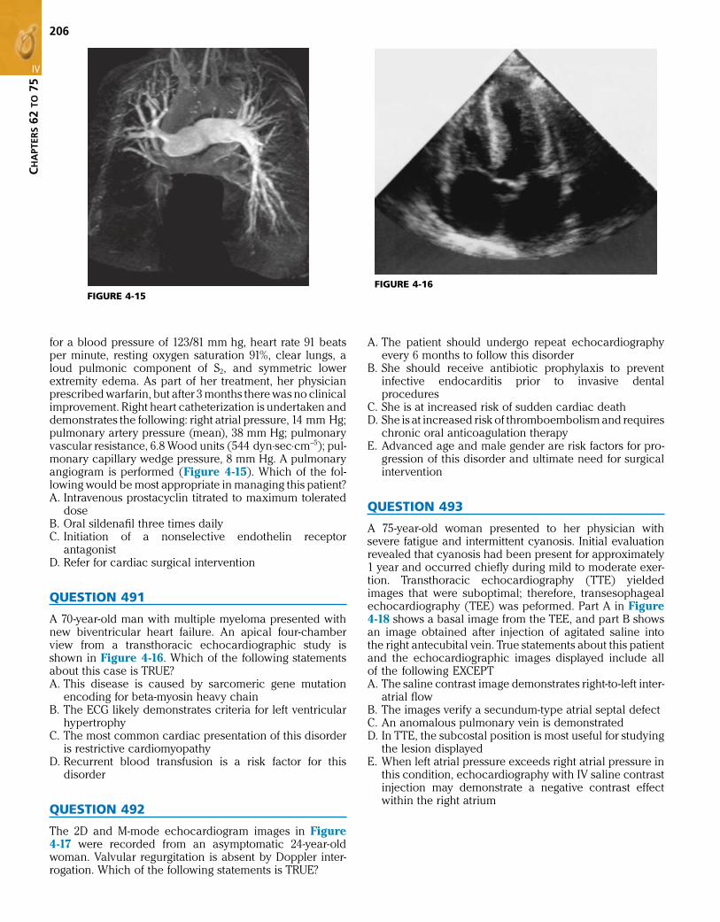
206
IV
CH
APT
ERS
62 T
O 7
5
FIGURE 4-15 FIGURE 4-16
for a blood pressure of 123/81 mm hg, heart rate 91 beats per minute, resting oxygen saturation 91%, clear lungs, a loud pulmonic component of S2, and symmetric lower extremity edema. As part of her treatment, her physician prescribed warfarin, but after 3 months there was no clinical improvement. Right heart catheterization is undertaken and demonstrates the following: right atrial pressure, 14 mm Hg; pulmonary artery pressure (mean), 38 mm Hg; pulmonary vascular resistance, 6.8 Wood units (544 dyn⋅sec⋅cm−5); pul-monary capillary wedge pressure, 8 mm Hg. A pulmonary angiogram is performed (Figure 4-15). Which of the fol-lowing would be most appropriate in managing this patient?A. Intravenous prostacyclin titrated to maximum tolerated
doseB. Oral sildenafil three times dailyC. Initiation of a nonselective endothelin receptor
antagonistD. Refer for cardiac surgical intervention
QUESTION 491
A 70-year-old man with multiple myeloma presented with new biventricular heart failure. An apical four-chamber view from a transthoracic echocardiographic study is shown in Figure 4-16. Which of the following statements about this case is TRUE?A. This disease is caused by sarcomeric gene mutation
encoding for beta-myosin heavy chainB. The ECG likely demonstrates criteria for left ventricular
hypertrophyC. The most common cardiac presentation of this disorder
is restrictive cardiomyopathyD. Recurrent blood transfusion is a risk factor for this
disorder
QUESTION 492
The 2D and M-mode echocardiogram images in Figure 4-17 were recorded from an asymptomatic 24-year-old woman. Valvular regurgitation is absent by Doppler inter-rogation. Which of the following statements is TRUE?
A. The patient should undergo repeat echocardiography every 6 months to follow this disorder
B. She should receive antibiotic prophylaxis to prevent infective endocarditis prior to invasive dental procedures
C. She is at increased risk of sudden cardiac deathD. She is at increased risk of thromboembolism and requires
chronic oral anticoagulation therapyE. Advanced age and male gender are risk factors for pro-
gression of this disorder and ultimate need for surgical intervention
QUESTION 493
A 75-year-old woman presented to her physician with severe fatigue and intermittent cyanosis. Initial evaluation revealed that cyanosis had been present for approximately 1 year and occurred chiefly during mild to moderate exer-tion. Transthoracic echocardiography (TTE) yielded images that were suboptimal; therefore, transesophageal echocardiography (TEE) was peformed. Part A in Figure 4-18 shows a basal image from the TEE, and part B shows an image obtained after injection of agitated saline into the right antecubital vein. True statements about this patient and the echocardiographic images displayed include all of the following EXCEPTA. The saline contrast image demonstrates right-to-left inter-
atrial flowB. The images verify a secundum-type atrial septal defectC. An anomalous pulmonary vein is demonstratedD. In TTE, the subcostal position is most useful for studying
the lesion displayedE. When left atrial pressure exceeds right atrial pressure in
this condition, echocardiography with IV saline contrast injection may demonstrate a negative contrast effect within the right atrium

Diseases o
f the H
eart, Pericardiu
m, an
d Pu
lmo
nary V
ascular B
ed207
4
FIGURE 4-17
AE
FC
D
LV
LA
RA
A
B
FIGURE 4-18
A B
QUESTION 494
A 29-year-old man is referred by his family practitioner for evaluation of a heart murmur that was first heard during childhood. He is asymptomatic. Part of his evalu-ation included an echocardiogram. An M-mode panel
from that study is displayed in Figure 4-19. True statements about this case include all of the following EXCEPTA. A vegetation is present on the anterior leaflet of the
mitral valveB. A bicuspid aortic valve is a cause of this finding
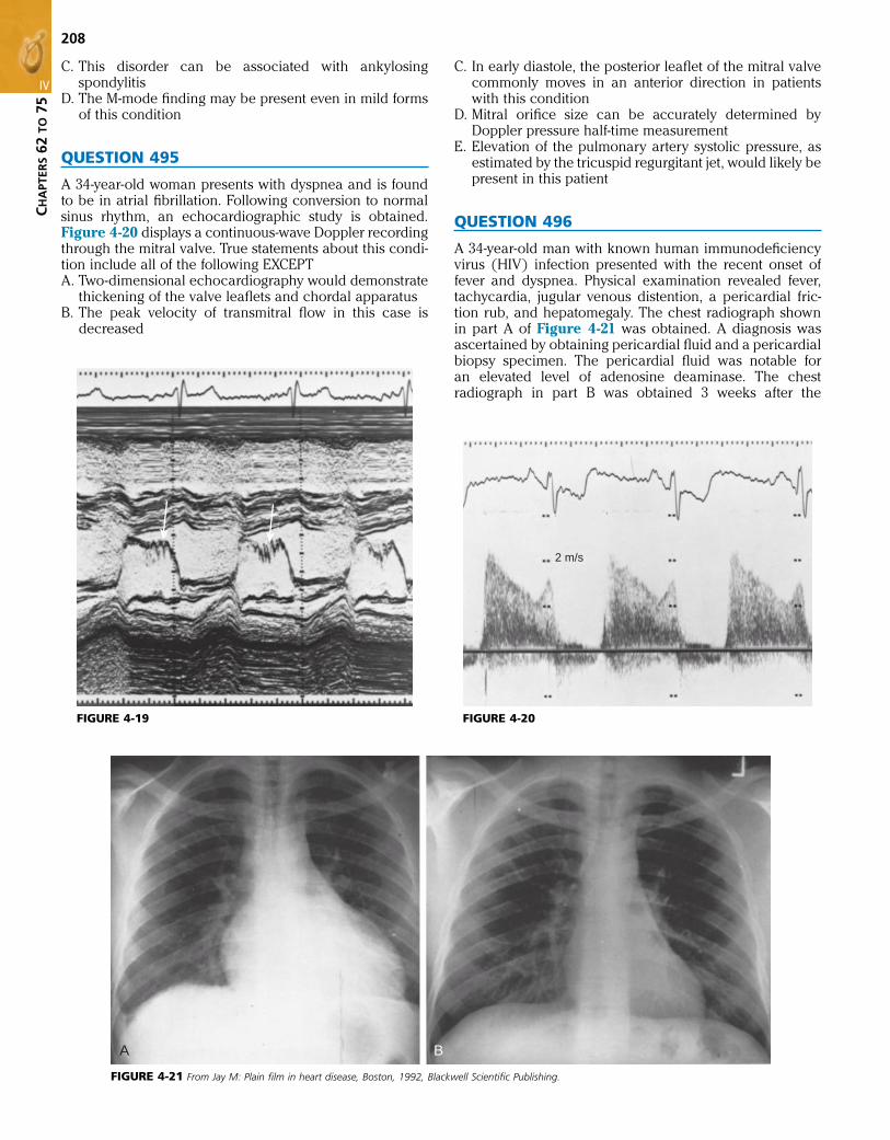
208
IV
CH
APT
ERS
62 T
O 7
5
FIGURE 4-19 FIGURE 4-20
2 m/s
FIGURE 4-21 From Jay M: Plain film in heart disease, Boston, 1992, Blackwell Scientific Publishing.
A B
C. This disorder can be associated with ankylosing spondylitis
D. The M-mode finding may be present even in mild forms of this condition
QUESTION 495
A 34-year-old woman presents with dyspnea and is found to be in atrial fibrillation. Following conversion to normal sinus rhythm, an echocardiographic study is obtained. Figure 4-20 displays a continuous-wave Doppler recording through the mitral valve. True statements about this condi-tion include all of the following EXCEPTA. Two-dimensional echocardiography would demonstrate
thickening of the valve leaflets and chordal apparatusB. The peak velocity of transmitral flow in this case is
decreased
C. In early diastole, the posterior leaflet of the mitral valve commonly moves in an anterior direction in patients with this condition
D. Mitral orifice size can be accurately determined by Doppler pressure half-time measurement
E. Elevation of the pulmonary artery systolic pressure, as estimated by the tricuspid regurgitant jet, would likely be present in this patient
QUESTION 496
A 34-year-old man with known human immunodeficiency virus (HIV) infection presented with the recent onset of fever and dyspnea. Physical examination revealed fever, tachycardia, jugular venous distention, a pericardial fric-tion rub, and hepatomegaly. The chest radiograph shown in part A of Figure 4-21 was obtained. A diagnosis was ascertained by obtaining pericardial fluid and a pericardial biopsy specimen. The pericardial fluid was notable for an elevated level of adenosine deaminase. The chest radiograph in part B was obtained 3 weeks after the

Diseases o
f the H
eart, Pericardiu
m, an
d Pu
lmo
nary V
ascular B
ed209
4
FIGURE 4-22 From Kang DH, Kim JH, Rim JH, et al: Circulation 119:797, 2009.
A B
initiation of appropriate therapy. True statements about this case include all of the following EXCEPTA. In industrialized nations, the incidence of this disorder
has decreased markedly in recent decadesB. This condition is the most common cause of pericardial
disease in African HIV-infected patientsC. Clinical detection of this disorder usually occurs either
in the effusive stage or after the development of constric-tive pericarditis
D. The acute onset of characteristic severe pericardial pain is common
E. This disorder is most likely to be diagnosed if both peri-cardial fluid and a pericardial biopsy specimen are obtained
QUESTION 497
Each of the following statements about ostium primum atrial septal defects (ASDs) is true EXCEPTA. Ostium primum ASDs often displace and cause a “cleft”
appearance of both the anterior and posterior leaflets of the mitral valve
B. The clinical features of ostium primum ASDs are similar to those of the ostium secundum type
C. Imaging usually reveals both right atrial and right ven-tricular enlargement
D. The presence of an ostium primum ASD accompanied by a ventricular septal defect comprises a complete atrio-ventricular canal malformation
E. Left ventriculography may demonstrate a “gooseneck” deformity
QUESTION 498
Surgical reconstruction (in distinction to replacement) of the mitral valve is likely to be successful in each of the fol-lowing patients EXCEPTA. A 33-year-old man with myxomatous elongation of the
mitral leaflets causing systolic prolapseB. A 62-year-old man with severe mitral regurgitation due
to annular dilatation after myocardial infarctionC. A 40-year-old woman with mitral regurgitation due
to ruptured chordae tendineae with active infective endocarditis
D. A 70-year-old woman with rheumatic heart disease, cal-cified mitral valve with deformed leaflets, and combined mitral stenosis and regurgitation
E. A 23-year-old man with a congenitally cleft mitral valve
QUESTION 499
A 53-year-old man has a grade III/VI apical holosystolic murmur. His echocardiographic apical three-chamber view with color Doppler imaging is shown in Figure 4-22. Mea-surements included a left ventricular (LV) end-diastolic chamber diameter of 56 mm, end-systolic dimension of 38 mm, and LV ejection fraction of 60%. He is asymptomatic and has not experienced inordinate dyspnea on exertion, orthopnea, or lower extremity edema. For this patient, which of the following statements is correct regarding the timing of mitral valve surgery?A. Surgical correction should be undertaken as soon as
possibleB. The risk of postoperative heart failure is low as long as
the preoperative ejection fraction is >50%C. His postoperative prognosis will not suffer if he under-
goes valve surgery before the LV end-systolic diameter exceeds 55 mm
D. Chronic administration of an angiotensin-converting enzyme inhibitor would delay the need for surgery
E. A resting pulmonary artery systolic pressure of 55 mm Hg is a class IIa indication for mitral valve surgery
QUESTION 500
A 23-year-old women delivers a baby boy at 36 weeks’ gesta-tion. Soon after delivery the neonate is noted to be cyanotic with a physical exam notable for a right ventricular impulse and a systolic thrill along the left sternal border. An echo-cardiogram reveals obstruction to right ventricular outflow, an outlet ventricular septal defect, overriding of the aorta, and right ventricular hypertrophy. With regard to the man-agement of this condition, each of the following statements is correct EXCEPTA. Early definitive repair is indicatedB. Postoperative increases in pulmonary venous return
often lead to right ventricular decompensation

210
IV
CH
APT
ERS
62 T
O 7
5
QUESTION 503
A 54-year-old man presented with increasing dyspnea on exertion. He was initially treated for an upper respira-tory tract infection; however, his symptoms persisted. A subsequent echocardiogram revealed an intracardiac mass suggestive of a malignant tumor. Regarding primary malig-nancies of the heart, which of the following statements is TRUE?A. The right atrium is the most common site of involvementB. The vast majority of patients already have metastases at
the time of diagnosisC. Malignant tumors account for half of primary cardiac
tumorsD. Lymphomas are the most common primary malignan-
cies of the heart
QUESTION 504
A 63-year-old man without known cardiac history pre-sented with a 4-month history of dyspnea on exertion and lower extremity edema. Echocardiography revealed a normal LV ejection fraction with no significant valvular disease. A chest CT showed a dense rim of calcium around the heart. Coronary angiography showed no sig-nificant coronary disease. Hemodynamic evaluation dem-onstrated elevation and equalization of right and left ventricular diastolic pressures with a “dip and plateau” configuration. Which of the following statements about this condition is correct?A. Evaluation for tuberculosis is appropriateB. The mortality rate associated with pericardiectomy in
the contemporary era is <1.0%C. Nearly all patients develop a high-output syndrome
immediately after pericardiectomy due to rapid expan-sion of the heart
D. Symptomatic improvement is reported in only 50% of surgical survivors
E. Pericardiectomy should be reserved only for patients with New York Heart Association functional class III or IV symptoms
C. The size of the pulmonary arteries is the single most important determinant in assessing candidacy for primary repair
D. If early corrective operation is not possible, a palliative procedure that leads to increased pulmonary blood flow is usually recommended
E. Bleeding complications are common in the postopera-tive period after repair
QUESTION 501
A 62-year-old man presents with the acute onset of short-ness of breath 2 weeks after prostate surgery. A high-resolution contrast chest computed tomographic (CT) scan is shown in Figure 4-23. Which of the following state-ments is correct?A. Echocardiography would be useful for further risk strati-
fication of this patientB. Pulmonary angiography is required to make a definitive
diagnosisC. This technique is insensitive for detecting thrombi in
subsegmental pulmonary arteriesD. The majority of patients with this disorder have evidence
of deep vein thrombosis in the systemic venous system
QUESTION 502
Each of the following conditions is associated with elevated right ventricular diastolic pressures with the pattern shown in Figure 4-24 EXCEPTA. Cardiac tamponadeB. Acute right ventricular infarctionC. Massive pulmonary embolismD. Constrictive pericarditisE. Restrictive cardiomyopathy
FIGURE 4-23 FIGURE 4-24 From Vaitkus PT, Cooper KA, Shuman WP, Hardin NJ: Circulation 93:834, 1996.
10
20
30
40
0RV LV

Diseases o
f the H
eart, Pericardiu
m, an
d Pu
lmo
nary V
ascular B
ed211
4C. Endocarditis frequently leads to this abnormalityD. Diabetes mellitus and hypercholesterolemia are risk
factors for its developmentE. A loud diastolic blowing murmur is likely
QUESTION 508
Which of the following statements about hemodynamic findings in constrictive pericarditis and restrictive cardio-myopathy is TRUE?A. A diastolic “dip-and-plateau” pattern is present in the
right ventricular (RV) waveform in constrictive pericar-ditis, but not restrictive cardiomyopathy
B. Concordance of left ventricular (LV) and RV systolic pressures during respiration is typical of constrictive pericarditis
C. In constrictive pericarditis, the ratio of RV systolic pres-sure to RV end-diastolic pressure is usually >3
D. An RV systolic pressure >50 mm Hg is more consistent with restrictive cardiomyopathy than with constrictive pericarditis
QUESTION 509
Riociguat is a newly available pharmacotherapy that results in improved exercise tolerance in patients with pulmonary arterial hypertension or chronic thromboembolic pulmo-nary hypertension. The mechanism of action of riociguat isA. Nitric oxide donorB. Soluble guanylyl cyclase stimulatorC. Heme oxygenase-1 inhibitorD. Kv1.5 channel stabilizer
QUESTION 510
A 54-year-old man presents to his physician with a 1-week history of dyspnea on exertion, cough, and pleuritic chest pain. His examination is notable for an elevated jugular venous pressure, distant heart sounds, and mild bilateral lower extremity edema. The chest radiograph reveals an enlarged cardiac silhouette. Echocardiography demon-strates a large circumferential pericardial effusion. An echocardiographic-guided pericardiocentesis is performed, removing most of the fluid; cytologic evaluation reveals adenocarcinoma. Each of the following statements is correct EXCEPTA. The most likely primary malignancy in this patient is in
the lungB. The prognosis is poor despite aggressive surgery or
chemotherapyC. Pericardial sclerotherapy would not significantly improve
the long-term prognosisD. Total surgical pericardiectomy should be performed
urgentlyE. Echocardiography should be repeated within 72 hours
QUESTION 511
A 34-year-old man presents to the emergency department with pleuritic chest pain after a recent upper respiratory tract infection. The pain is positional, relieved by sitting up.
QUESTION 505
A 25-year-old woman presents to her primary care doctor with fatigue and a rash. She recently returned from a New England vacation during which she hiked at least 6 miles daily. On physical examination she has a well-demarcated erythematous rash with central clearing on her chest. Lyme disease is suspected. Which of the following state-ments regarding cardiac involvement in Lyme disease is TRUE?A. Cardiac manifestations of Lyme disease typically occur
within days of the development of erythema chronicum migrans
B. Ten percent of patients with Lyme disease develop cardiac manifestations
C. Supraventricular and ventricular tachyarrhythmias are the most common cardiac manifestations of Lyme disease
D. Cardiomegaly and congestive heart failure are common among patients who develop Lyme carditis
E. Antibiotic therapy shortens the course of active Lyme carditis
QUESTION 506
Which of the following statements regarding the natural history of idiopathic pulmonary arterial hypertension is TRUE?A. The prevalence is equal in males and femalesB. Chest pain related to right ventricular ischemia is the
most common manifesting symptomC. Sudden cardiac death is a potential complication, but
only in patients with class IV symptomsD. Increased intensity of S1 is the most common physical
findingE. Electrocardiographic evidence of right ventricular hyper-
trophy is present in a minority of patients
QUESTION 507
Which of the following statements is TRUE about the heart valve abnormality shown in Figure 4-25?A. The abnormality is most likely congenitalB. Cardiac auscultation is almost certainly normal
FIGURE 4-25 From Salem DN, Isner JM: Chest 92:326, 1987.

212
IV
CH
APT
ERS
62 T
O 7
5
of the following statements about penetrating cardiac trauma is correct?A. The left ventricle is the cardiac chamber most commonly
injured by penetrating traumaB. Penetrating injuries to the atria are associated with better
survival than wounds to the ventriclesC. Rupture of the interventricular septum is a potential late
complicationD. In penetrating cardiac injury with suspected tamponade,
urgent pericardiocentesis is a mandatory intervention
QUESTION 515
Each of the following statements regarding persistent patent ductus arteriosus (PDA) in adults is correct EXCEPTA. Patients with small shunts and no audible murmur are at
negligible risk of endovascular infectionB. Patients with moderate-sized PDA typically present with
dyspnea or palpitationsC. Patients with moderate-sized PDA typically have a loud
continuous “machinery” murmur and a narrow pulse pressure
D. Patients with large PDA may develop enlarged central pulmonary arteries with peripheral pruning on chest radiograph
E. Transcatheter devices result in successful closure rates of >95%
QUESTION 516
A 46-year-old man is admitted to the hospital because of worsening shortness of breath. His history is notable for hypertension, treated with an angiotensin-converting enzyme inhibitor. Social history reveals that he has con-sumed one-half pint of liquor daily for the past 10 years. Physical examination shows an elevated jugular venous pressure (14 cm), bibasilar rales on pulmonary examina-tion, a laterally displaced cardiac apical impulse, a grade II/VI holosystolic murmur, an S3 gallop at the apex, and pitting edema of both lower extremities. Echocardiography dem-onstrates a dilated left ventricle with an ejection fraction of 25% and moderate mitral regurgitation. Which of the follow-ing statements regarding the cardiac effects of alcohol is true?A. Chronic heavy alcohol consumption is associated with
low systemic blood pressureB. The likelihood of developing dilated cardiomyopathy
correlates with the amount of alcohol consumed over a lifetime
C. Women are less susceptible than men to alcohol-associated cardiomyopathy
D. Moderate alcohol consumption is associated with an increased rate of sudden death
E. Dilated cardiomyopathy due to alcohol consumption is irreversible
QUESTION 517
Expected clinical manifestations of amyloidosis of the car-diovascular system include which of the following?A. Constrictive pericarditisB. High-output heart failureC. Orthostatic hypotension
Cardiac examination is notable for a three-component fric-tion rub. The ECG demonstrates diffuse ST-segment eleva-tion with PR-segment depression in several leads. Which of the following statements about the evolution of the ECG in this condition is correct?A. PR-segment deviation occurs in fewer than 25% of
patients with this conditionB. Initial ST-segment elevation is usually most prominent in
lead aVRC. The ratio of the height of ST-segment elevation to the
height of the T wave in this condition is typically <0.25 in lead V6
D. T wave inversion is expected to occur within days, while the ST segment is still elevated
E. T wave inversion may persist for months after the acute presentation
QUESTION 512
Which of the following statements regarding the natural history of untreated aortic stenosis (AS) is TRUE?A. Average survival from the onset of syncopal symptoms
is approximately 6 monthsB. Average survival from the onset of congestive heart
failure is approximately 2 yearsC. Syncope due to AS usually occurs at restD. Sudden death in patients with AS usually occurs in previ-
ously asymptomatic individualsE. Atrial fibrillation is usually well tolerated in patients with
advanced AS
QUESTION 513
A 46-year-old postmenopausal woman with a history of systemic sclerosis is admitted to the coronary care unit because of progressive dyspnea and chest pain. Initial data include blood pressure 88/55 mm Hg, heart rate 104 bpm, oxygen saturation 95%. General examination is notable for cool extremities and symmetric edema of the lower extrem-ities. The electrocardiogram demonstrates right ventricular hypertrophy with a strain pattern. The serum troponin T is 1.10 ng/mL (reference range <0.01 ng/mL), and contrast enhanced thoracic computed tomography demonstrates RV cavitary enlargement without pulmonary embolism. Coronary arteriography shows minimal coronary artery disease without coronary vasospasm. Right heart catheter-ization reveals the following: mean pulmonary artery pres-sure, 61 mm Hg; mean pulmonary capillary wedge pressure, 10 mm Hg; pulmonary vascular resistance, 16.2 Wood units (1296 dyn⋅sec⋅cm−5). Her body surface area is 1.6 m2. Which of the following is the most appropriate initial treatment for this patient?A. Tissue plasminogen activator 100 mg, administered as a
continuous IV infusion over 2 hoursB. Intravenous epoprostenol initiated at 2 ng/kg/minC. Oral macitentan 3 mg dailyD. Oral digoxin 0.125 mg daily
QUESTION 514
A 45-year-old man is transported to the emergency depart-ment, a victim of a stab wound to the chest that occurred during a robbery attempt at his convenience store. Which

Diseases o
f the H
eart, Pericardiu
m, an
d Pu
lmo
nary V
ascular B
ed213
4demonstrates midsystolic notching. This finding is most strongly associated with which of the following?A. Decreased pulmonary artery pressureB. Increased pulmonary vascular resistanceC. PneumothoraxD. Left-sided heart failure
QUESTION 522
A 55-year-old woman seeks medical attention because of progressive exertional dyspnea and rapid heart action. At age 12 she suffered from rheumatic fever, and a heart murmur has been subsequently noted. She has had inter-mittent episodes of atrial fibrillation over the past 2 years with good rate control on metoprolol succinate. Her vital signs include a heart rate of 80 beats/min, blood pressure 130/80 mm Hg, and respirations 16/min. She has inspira-tory rales at the lung bases. Her cardiac impulse is dis-placed laterally. There is a loud S1, a single S2, an apical opening snap, a holodiastolic rumbling murmur at the apex, and a soft diastolic blowing murmur along the left sternal border. Isometric handgrip augments the diastolic murmurs. She has mild peripheral edema. Her ECG is shown in Figure 4-27. The most likely valve lesions areA. Mitral regurgitation and tricuspid stenosisB. Mitral stenosis and mitral regurgitationC. Mitral stenosis and aortic regurgitationD. Mitral stenosis and pulmonic regurgitationE. Tricuspid stenosis and pulmonic regurgitation
QUESTION 523
Each of the following statements regarding the medical management of mitral stenosis (MS) is correct EXCEPTA. Antibiotic prophylaxis is not recommended for patients
with MS undergoing dental surgeryB. Diuretic therapy is an appropriate measure for relieving
symptoms of dyspneaC. In pure MS with normal left ventricular function, digoxin
is of benefit only if atrial fibrillation is presentD. The benefits of beta-blocker therapy in MS include heart
rate reduction and improved exercise toleranceE. Anticoagulation is indicated to prevent thromboembo-
lism in patients with MS regardless of the heart rhythm or history of previous thromboembolic events
QUESTION 524
Which of the following statements regarding infectious causes of pericarditis is correct?A. Tuberculosis is the leading cause of constrictive pericar-
ditis in developed nationsB. Haemophilus influenzae is the most common organism
responsible for bacterial pericarditisC. In the contemporary era, the prognosis of patients with
bacterial pericarditis is comparable to that of viral pericarditis
D. Enteroviruses are among the most frequent causes of viral pericarditis
E. Prolonged antifungal therapy is required to manage iso-lated pericardial disease caused by Histoplasma
D. ECG voltage criteria for left ventricular hypertrophyE. Electrical alternans
QUESTION 518
Which of the following statements regarding endocarditis caused by Staphylococcus aureus is correct?A. Central nervous system complications are rare, occur-
ring in fewer than 5% of patientsB. Staphylococcus aureus native valve endocarditis is an
absolute indication for surgical debridementC. The prognosis of right-sided S. aureus native valve endo-
carditis is similar to that of left-sided involvementD. Empirical initial therapy with oxacillin or cefazolin is
appropriate for suspected S. aureus endocarditisE. Prosthetic valve endocarditis with S. aureus is associ-
ated with a 50% mortality rate in patients treated medically
QUESTION 519
Each of the following is associated with Ebstein anomaly of the tricuspid valve EXCEPTA. Atrial septal defectB. Paradoxical splitting of S2
C. Ventricular preexcitationD. A widely split S1
E. Atrial flutter
QUESTION 520
A 42-year-old man from Uganda is diagnosed with Löffler endocarditis. Expected findings include all of the following EXCEPTA. EosinophiliaB. Signs and symptoms of heart failureC. Asthma and nasal polyposisD. Right ventricular pressure tracing showing a “dip-and-
plateau” patternE. Normal left ventricular ejection fraction
QUESTION 521
A 57-year-old man is referred for transthoracic echocardiog-raphy to investigate unexplained exertional dyspnea. The right ventricular outflow tract velocity time integral by Doppler interrogation is shown in Figure 4-26 and
FIGURE 4-26 From Opotowsky AR, Clair M, Afilalo J, et al: Am J Cardiol 112:873–882, 2013.
80
40
cm/s
–40
–80
–120
62bpm75mm/s

214
IV
CH
APT
ERS
62 T
O 7
5
parasternal long-axis view and a Doppler recording from the apical long-axis view are displayed in Figure 4-29. Which of the following statements is TRUE?A. This abnormality results from myxomatous valvular
degenerationB. The Doppler profile in part B in the figure is essentially
normalC. The severity of this abnormality correlates with the likeli-
hood of developing endocarditisD. The risk of systemic embolism in this condition corre-
lates with ageE. Chest pain accompanies this condition in the majority of
patients
QUESTION 528
A premature infant is found to have bounding peripheral pulses, a continuous murmur in the infraclavicular and
QUESTION 525
A 25-year-old graduate student presents to your office for physical examination. Several years ago, while growing up in Ecuador, he was diagnosed with Chagas disease when he had presented with fever, malaise, myalgias, and unilat-eral eyelid edema. Each of the following statements regard-ing Chagas disease is correct EXCEPTA. The level of parasitemia does not correspond to the
severity of chronic Chagas diseaseB. The disease is transmitted to humans by the reduviid bugC. An asymptomatic phase typically lasts for many years
between initial infection and chronic manifestations of the disease
D. The most common ECG abnormality in chronic Chagas disease is left bundle branch block
E. The classic echocardiographic findings of chronic cardiac Chagas disease are those of a dilated cardiomy-opathy with apical aneurysm formation
QUESTION 526
A 29-year-old woman was diagnosed with a congenital heart defect 6 years ago. At a routine office visit she describes occasional single palpitations during periods of emotional stress. An echocardiogram is obtained; the apical four-chamber view is shown in Figure 4-28. Which of the following is demonstrated?A. Bilateral atrial myxomasB. Lipomatous hypertrophy of the interatrial septumC. Normal position of an atrial septal closure deviceD. Infiltrative disease, most likely amyloidosisE. Normal echocardiogram
QUESTION 527
A 42-year-old woman, originally from Thailand, presents for evaluation because of exertional dyspnea. As part of her evaluation an echocardiographic study is performed; a
FIGURE 4-27
FIGURE 4-28
LV
LA
RV

Diseases o
f the H
eart, Pericardiu
m, an
d Pu
lmo
nary V
ascular B
ed215
4
B. The majority of ventricular emptying is less rapid than usual
C. Left ventricular end-systolic volume is usually increasedD. Left ventricular ejection fraction is typically reduced
QUESTION 530
A 46-year-old man is admitted with fever, dyspnea, hypoten-sion, and a new murmur of aortic regurgitation. Which of the following is typical of acute aortic regurgitation?A. A widened systemic pulse pressureB. A long, decrescendo diastolic murmurC. Delayed closure of the mitral valve on echocardiographyD. Diastolic mitral regurgitation on echocardiographyE. An enlarged left ventricle
QUESTION 531
Which of the following statements concerning tetralogy of Fallot is TRUE?A. Tetralogy of Fallot accounts for approximately 10% of all
forms of congenital heart diseaseB. It is an uncommon cause of cardiac cyanosis in children
after 1 year of ageC. Anomalous coronary circulation is present in 50% of
patients with tetralogy of FallotD. The degree of right-to-left shunting in tetralogy of Fallot is
independent of the severity of pulmonary obstructionE. The aortic root in this disorder is located to the left of
the origin of the pulmonary artery
QUESTION 532
A 68-year-old man presents for evaluation of progressive dyspnea. He has an extensive smoking history. Pulmonary function testing is consistent with severe airway obstruc-tion. An echocardiogram shows normal left ventricular sys-tolic function, right ventricular hypertrophy and dilation, and an elevated calculated pulmonary artery systolic pres-sure. Which of the following therapies improves survival in patients with chronic obstructive lung disease with pulmo-nary hypertension?A. DigoxinB. OxygenC. Beta-adrenergic agonists
interscapular regions, and precordial hyperactivity. An echocardiogram demonstrates a left-to-right shunt between the proximal descending aorta and the pulmonary artery. Which of the following statements about this condition is TRUE?A. Most preterm infants with a birth weight <1500 g have
this conditionB. Cardiopulmonary deterioration occurs in nearly all such
infantsC. Chest radiography likely shows a decreased size of the
cardiac silhouetteD. Surgical ligation is the only effective treatmentE. Noninvasive imaging typically shows right ventricular
enlargement with normal left ventricular size
QUESTION 529
A 17-year-old boy has a syncopal event while playing soccer. He is noted to have a systolic ejection murmur on examina-tion. An echocardiogram is obtained and a parasternal long-axis view is shown in Figure 4-30. Which of the fol-lowing statements about hemodynamic findings in patients with this condition is TRUE?A. Diastolic function is usually impaired to a greater extent
than systolic function
FIGURE 4-29
A B
FIGURE 4-30
LV
LA
Ao

216
IV
CH
APT
ERS
62 T
O 7
5
tachycardic woman. The serum creatinine is chronically elevated, and precludes performing a chest CT angiogram. Which of the following results would be most useful in EXCLUDING the diagnosis of pulmonary embolism?A. Normal PaO2 by arterial blood gas determinationB. Normal cardiopulmonary examinationC. Intermediate probability (ventilation/perfusion) lung
scanD. Normal plasma level of D-dimerE. Absence of right-sided heart strain or S1Q3T3 pattern on
ECG
QUESTION 536
True statements regarding primary tumors of the heart include each of the following EXCEPTA. Benign tumors are more common than malignant tumorsB. The most common malignant cardiac tumors are angio-
sarcomas and rhabdomyosarcomasC. The presence of a hemorrhagic pericardial effusion is
more consistent with a malignant tumorD. Malignant tumors are more likely to occur on the left side
of the heart
QUESTION 537
A 67-year-old man with adenocarcinoma of the lung pres-ents with dyspnea and weakness. Physical examination is notable for hypotension with pulsus paradoxus and jugular venous distention. An echocardiogram is ordered. Which of the following statements regarding echocardiography in cardiac tamponade is TRUE?A. On inspiration, there is augmentation of transmitral flow
velocities and reduction in transtricuspid flow velocitiesB. The absence of right ventricular diastolic collapse
ensures that the pericardial pressure is not elevatedC. Right ventricular diastolic collapse is more specific than
right atrial collapse in the diagnosis of tamponadeD. Cardiac tamponade is associated with normal inferior
vena cava collapse with inspirationE. A small-volume pericardial effusion observed on echo-
cardiography excludes the presence of tamponade
D. TheophyllineE. Bosentan
QUESTION 533
Which of the following statements regarding coarctation of the aorta is TRUE?A. It is more common in females than malesB. Most infants and children with coarctation are
asymptomaticC. Ten percent of adults with coarctation also have a bicus-
pid aortic valveD. Mean survival time in the presence of uncorrected
coarctation is 60 years
QUESTION 534
A 30-year-old woman presented to the hospital after a recent transient neurologic event consistent with a small cerebral embolism. During her evaluation, a transthoracic echocardiogram was performed. An apical four-chamber view from the study is displayed in Figure 4-31, with panels from both systole and diastole. Which of the following state-ments is TRUE?A. The echocardiographic appearance is most consistent
with an intracardiac thrombusB. Systemic symptoms associated with this condition
include fever, malaise, and an elevated erythrocyte sedi-mentation rate
C. Surgery is indicated only if there is hemodynamic compromise
D. The majority of such lesions are familialE. Unlike this patient, this condition most commonly arises
in the right atrium
QUESTION 535
A 54-year-old woman with chronic kidney disease presents to the emergency department because of sudden shortness of breath and right-sided pleuritic chest pain. She recently returned from vacation on a 12-hour flight. Physical exami-nation demonstrates a tachypneic, normotensive, and
FIGURE 4-31
RA
LVRV
SYSTOLE DIASTOLE

Diseases o
f the H
eart, Pericardiu
m, an
d Pu
lmo
nary V
ascular B
ed217
4progressive symptoms of exertional dyspnea, and occa-sional orthopnea. His echocardiogram shows severe aortic regurgitation, a left ventricular (LV) end-systolic diameter of 48 mm, and an LV ejection fraction of 45%. Each of the following statements regarding valve replacement for chronic aortic regurgitation (AR) is true EXCEPTA. Asymptomatic patients with advanced AR who have
normal LV function and end-systolic diameter <50 mm have an excellent prognosis such that surgery can be safely postponed
B. A left ventricular (LV) end-systolic diameter >55 mm before surgical intervention portends a worse postopera-tive prognosis than does a smaller ventricular diameter
C. Angiotensin-converting enzyme inhibitors delay the need for aortic valve replacement in chronic AR
D. Patients with chronic AR who have symptoms of conges-tive heart failure accompanied by reduced LV ejection fraction should be referred for aortic valve replacement
E. A preoperative ejection fraction <50% increases the risk of postoperative death from LV dysfunction
QUESTION 541
A 44-year-old man with human immunodeficiency virus (HIV) infection presents to your office for evaluation of shortness of breath. He reports 2 months of progressive fatigue and dyspnea on exertion. His CD4 count is 500 cells/mm3 (normal = 500 to 1500 cells/mm3), and he remains on highly active antiretroviral therapy. Transthoracic echocar-diography reveals a dilated heart with a left ventricular (LV) ejection fraction of 15% and a small pericardial effusion. Which of the following statements is TRUE?A. Fifty percent of HIV-infected individuals will ultimately
develop symptomatic LV dysfunctionB. Large asymptomatic pericardial effusions are the most
common cardiac manifestation of HIV infection, but the presence of effusion is not associated with increased mortality
C. In HIV-infected individuals, LV dysfunction has little bearing on 1-year mortality rates
D. Protease inhibitor therapy is associated with an increased risk of myocardial infarction
QUESTION 542
True statements regarding the cardiovascular consequences of Turner syndrome include all of the following EXCEPTA. Cardiovascular defects are seen in fewer than 5% of
patientsB. Coarctation of the aorta is the most commonly associ-
ated congenital cardiovascular abnormalityC. There is an increased risk of aortic dissectionD. Bicuspid aortic valve occurs at a higher frequency than
in the general populationE. Partial anomalous venous drainage occurs more fre-
quently than in the general population
QUESTION 543
A 20-year-old man with Duchenne muscular dystrophy presents for evaluation. Regarding cardiac involvement in this condition, which of the following statements is TRUE?
QUESTION 538
A 64-year-old woman with a remote history of chest irradia-tion presents with gradually progressive symptoms of fatigue, abdominal bloating, and lower extremity edema over the past 4 months. Her lateral chest radiograph is shown in Figure 4-32. Which of the following findings would be inconsistent with the likely diagnosis?A. Kussmaul signB. HepatomegalyC. Early diastolic pericardial knockD. Right and left ventricular systolic pressure concordance
during inspirationE. Elevation and equalization of intracardiac diastolic
pressures
QUESTION 539
Which of the following statements regarding the causes of aortic stenosis (AS) is TRUE?A. A congenitally bicuspid valve accounts for 20% of patients
requiring surgical treatment for AS in the United StatesB. In patients >age 70, rheumatic deformity is the most
common cause of AS that leads to surgical valve replacement
C. There are no established risk factors for the development of calcific AS of a trileaflet valve other than age
D. Age-related calcification of a congenitally bicuspid, or normal trileaflet valve, is the most common cause of AS in adults in the United States
E. Isolated rheumatic deformity of the aortic valve, without mitral valve involvement, is more common than rheu-matic involvement of both valves
QUESTION 540
A 68-year-old man with chronic aortic regurgitation pres-ents for office evaluation. He describes reduced stamina,
FIGURE 4-32

218
IV
CH
APT
ERS
62 T
O 7
5
QUESTION 547
A 35-year-old man presents with complaints of exertional dyspnea. His medical history is significant for chest radiation therapy as a teenager for treatment for a hema-tologic malignancy. Regarding cardiovascular effects of radiation therapy, which of the following statements is TRUE?A. Most complications develop within 5 years of radiation
exposureB. Constrictive pericarditis is typically an acute reaction to
radiation therapyC. The conduction system is typically spared from adverse
effects of radiationD. Dilated cardiomyopathy is the most common manifesta-
tion of mediastinal radiation therapyE. Cancer survivors who received head and neck radiation
are at a heightened risk of stroke
QUESTION 548
Which of the following statements is correct regarding familial forms of dilated cardiomyopathy (DCM)?A. Familial forms account for less than 3% of cases of
DCMB. Most inherited forms of dilated cardiomyopathy fit an
autosomal recessive patternC. Familial DCM most commonly results from mutations in
genes that encode sarcolemmal surface receptorsD. In symptomatic patients, histologic examination of the
heart typically demonstrates extensive areas of intersti-tial and perivascular fibrosis
E. Familial cardiomyopathy can be readily identified as the cause of DCM by specific immunologic markers
QUESTION 549
A 65-year-old man with a history of radiation therapy to the chest for lymphoma presents with worsening exertional dyspnea. On physical examination the heart rate is 120 beats/min, blood pressure 90/55 mm Hg, with pulsus para-doxus of 15 mm Hg. The jugular venous pressure is 10 cm H2O with a prominent x descent. He is found by echocar-diography to have a large pericardial effusion, which is drained by pericardiocentesis. After the procedure in the cardiac catheterization laboratory the intrapericardial pres-sure normalizes but the initially elevated right atrial pres-sure fails to decline and displays a prominent y descent. This scenario is most consistent withA. Persistence of pericardial tamponadeB. Cor pulmonaleC. Effusive-constrictive pericarditisD. Restrictive cardiomyopathyE. Uremic pericarditis
DirectionsEach group of questions below consists of lettered headings followed by a set of numbered questions. For each ques-tion, select the ONE lettered heading with which it is most closely associated. Each lettered heading may be used once, more than once, or not at all.
A. Fewer than 25% of patients with Duchenne muscular dystrophy >age 18 develop a dilated cardiomyopathy
B. The ECG typically shows tall R waves and increased R/S amplitude in V1 and deep narrow Q waves in the left precordial leads
C. There is a direct association between the presence of dilated cardiomyopathy and electrocardiographic abnormalities
D. Early cardiac involvement typically manifests as global left ventricular dysfunction
E. The most common rhythm disturbance is ventricular tachycardia
QUESTION 544
A 34-year-old man is receiving doxorubicin chemotherapy for lymphoma. Each of the following statements is true regarding the risk of doxorubicin-induced cardiotoxicity EXCEPTA. Previous or concurrent mediastinal irradiation increases
the risk of cardiotoxicityB. The age groups most at risk are the very young and the
very oldC. Cardiomyopathy does not develop unless the total cumu-
lative dose exceeds 700 mg/m2
D. Concurrent use of cyclophosphamide increases the risk of cardiotoxicity
E. A baseline LV ejection fraction of 45% increases the risk of cardiotoxicity
QUESTION 545
A 63-year-old man with metastatic colon cancer is pre-scribed therapy targeting vascular endothelial growth factor (VEGF). Which of the following statements is correct about the use of the monoclonal antibody/VEGF antagonist bevacizumab?A. The LV ejection fraction tends to increase with use of this
drugB. Hypotension is a common side effectC. The risk of arterial, but not venous, thromboembolic
events is increasedD. Hemorrhagic pericardial effusion is associated with con-
tinuous use of this agent
QUESTION 546
A 2-year-old boy with Down syndrome and a heart murmur is brought to the pediatrician’s office because of poor weight gain. The most likely finding on cardiac ausculta-tion isA. An early diastolic opening sound at the apex followed
by a diastolic rumbling murmurB. A midsystolic murmur at the upper left sternal border
and wide, fixed splitting of S2
C. A midsystolic click followed by a late systolic murmur at the apex
D. A blowing diastolic murmur at the right upper sternal border
E. A late-peaking systolic ejection murmur heard best at the upper right sternal border with a diminished S2

Diseases o
f the H
eart, Pericardiu
m, an
d Pu
lmo
nary V
ascular B
ed219
4565. Physical cause of abnormality easily visualized by
chest computed tomography566. Reduced medial mitral annular E′ velocity by
Doppler tissue imaging
QUESTIONS 567 TO 571
For each statement listed below, match the most appropri-ate condition:A. Cardiac myxomaB. Large papillary fibroelastomaC. BothD. Neither
567. Familial predilection568. Risk of distal embolism569. Surgical excision is management of choice570. May undergo malignant transformation571. Typically attaches to valves and subvalvular
structures
QUESTIONS 572 TO 575
Match the description with the associated form of therapy for pulmonary embolism:A. Unfractionated heparin or low-molecular-weight heparinB. Fibrinolytic therapyC. BothD. Neither
572. May be effective in pulmonary embolism even 1 to 2 weeks after the onset of symptoms
573. Dissolution of recently formed thrombus is a major action
574. Should be administered along with an antiplatelet agent
575. May suppress aldosterone secretion
QUESTIONS 576 TO 580
Match each description with the associated condition:A. HemochromatosisB. AmyloidosisC. BothD. Neither
576. Results in restrictive cardiomyopathy577. Early-stage disease is reversible with chelating
agents578. Ventricular tachyarrhythmia is the most common
initial presentation579. Autosomal recessive inheritance may be responsi-
ble580. Low-voltage QRS on ECG is typical
QUESTIONS 581 TO 585
Match each description with the associated condition:A. Tricuspid stenosisB. Pulmonic stenosisC. BothD. Neither
QUESTIONS 550 TO 553
For each statement below, match the appropriate lesion:A. Osler nodesB. Janeway lesionsC. Roth spotsD. Subungual hemorrhagesE. Bracht-Wächter bodies
550. Small (1- to 4-mm diameter), irregular, erythema-tous, nontender macules present on the thenar and hypothenar eminences of the hands
551. Small, raised red (or purple) tender lesions present in the pulp spaces of the terminal phalanges of the fingers
552. Collections of lymphocytes in the nerve layer of the retina
553. Linear or flame-shaped streaks
QUESTIONS 554 TO 558
For each cardiac condition, match the electrocardiographic finding that is most closely associated with it:A. Low QRS voltageB. Atrioventricular nodal blockC. Right bundle branch blockD. Diffuse ST-segment elevationE. Deeply inverted precordial T waves
554. Chronic Chagas disease555. Sarcoidosis556. Apical hypertrophic cardiomyopathy557. Amyloidosis558. Lyme carditis
QUESTIONS 559 TO 562
For each statement, match the appropriate prosthetic valve type:A. Starr-Edwards valveB. Carpentier-Edwards valveC. St. Jude valveD. Medtronic-Hall valve
559. Single tilting disc valve560. Lowest profile mechanical valve561. Least thrombogenic mechanical prosthesis for the
mitral position562. Porcine heterograft mounted on a Dacron cloth–
covered strut
QUESTIONS 563 TO 566
For each description, match the related cardiac condition(s):A. Constrictive pericarditisB. Restrictive cardiomyopathyC. BothD. Neither
563. Atrial fibrillation, low QRS voltage564. Right and left ventricular systolic pressure
discordance with respiration during cardiac catheterization

220
IV
CH
APT
ERS
62 T
O 7
5
QUESTIONS 598 TO 601
For each type of procedure below, match the appropriate prophylactic antibiotic regimen:A. No antibiotic prophylaxis requiredB. Amoxicillin, 2 g orally, taken 30 to 60 minutes before the
procedureC. Clindamycin 600 mg orally, taken 30 to 60 minutes before
the procedureD. Ampicillin, 2 g IM or IV 30 to 60 minutes before the
procedure
598. Elective colonoscopy in a patient with a mechani-cal mitral valve
599. Dental extraction in a patient with a bioprosthetic aortic valve
600. Dental extraction in penicillin-allergic patient with a mechanical aortic valve
601. Elective cholecystectomy in a patient with mitral stenosis
QUESTIONS 602 TO 606
Match the following descriptions with the appropriate condition:A. Complete atrioventricular septal defectB. Complete transposition (d-transposition) of the great
arteriesC. BothD. Neither
602. Presents with cyanosis603. Right-axis deviation on the ECG604. An interatrial communication is almost always
present605. On echocardiography, the AV valves appear abnor-
mally aligned at the same level606. A high-pitched, blowing, decrescendo diastolic
murmur is common at the left sternal border
QUESTIONS 607 TO 611
Match the following descriptions with the appropriate condition:A. Kartagener syndromeB. Holt-Oram syndromeC. LEOPARD syndromeD. Noonan syndrome
607. Webbed neck, pulmonic stenosis, left anterior fas-cicular block
608. Deafness, pulmonic stenosis, complete heart block609. Lentigines, pulmonic stenosis, PR prolongation610. Sinusitis, dextrocardia, bronchiectasis611. Abnormal scaphoid bone, atrial septal defect, right
bundle branch block
QUESTIONS 612 TO 615
Match each photograph in Figure 4-33 with the condition it represents:A. Part AB. Part BC. Part CD. Part D
581. Usually rheumatic in origin582. Typical of carcinoid heart disease583. Most adults are asymptomatic584. Ascites is common on physical examination585. Balloon valvuloplasty is the treatment of choice
QUESTIONS 586 TO 589
Match each description with the associated drug therapy in patients with idiopathic (group 1) pulmonary arterial hypertension (PAH):A. Calcium channel blockersB. Epoprostenol (prostacyclin)C. BothD. Neither
586. Studies have shown a survival benefit in patients with PAH
587. Should be started empirically in all patients with group 1 PAH
588. Not approved for oral administration589. Exert(s) an antithrombotic effect
QUESTIONS 590 TO 593
Match each hemodynamic scenario with the associated condition:A. Chronic constrictive pericarditisB. Cardiac amyloidosisC. BothD. Neither
590. Right ventricular (RV) pressure tracing shows a deep and rapid early decline at the onset of dias-tole, with a rapid rise to a plateau in early diastole (“dip and plateau” configuration)
591. Left ventricular end-diastolic pressure exceeds right ventricular end-diastolic pressure (RVEDP) by 10 mm Hg
592. Peak RV systolic pressure = 35 mm Hg, RVEDP = 18 mm Hg
593. Pulmonary artery systolic pressure = 68 mm Hg, RVEDP = 15 mm Hg
QUESTIONS 594 TO 597
Match the following descriptions with the appropriate symptom in aortic stenosis:A. PalpitationsB. AnginaC. SyncopeD. Exertional dyspnea as a manifestation of heart failure
594. Most significant clinical marker for adverse outcome in aortic stenosis, predictive of a 1- to 2-year survival if the valve lesion is not surgically corrected
595. Associated with reduced survival in untreated aortic stenosis of 3 to 5 years
596. Associated with reduced survival in untreated aortic stenosis of 2 to 3 years
597. Not a marker of reduced survival in patients with aortic stenosis

Diseases o
f the H
eart, Pericardiu
m, an
d Pu
lmo
nary V
ascular B
ed221
4
FIGURE 4-33 A, Courtesy Alan J. Lesse, MD; B, from duVivier A: Atlas of clinical dermatology, ed 3, Philadelphia, 2002, Elsevier; C, from Samiy N: Surv Ophthalmol 53:416–423, 2008; D, from Habif TP: Clinical dermatology, ed 5, Philadelphia, 2009, Mosby Elsevier.
A B
C D
612. Fabry disease613. Infective endocarditis614. Amyloidosis615. Scleroderma
QUESTIONS 616 TO 619
Match the following descriptions with the appropriate association:A. Turner syndromeB. Noonan syndromeC. BothD. Neither
616. Coarctation of the aorta617. Normal karyotype618. Pulmonic stenosis619. Short stature, webbing of the neck, skeletal anoma-
lies, and renal anomalies
QUESTIONS 620 TO 624
Match the following descriptions with the appropriate part of Figure 4-34:
A. Part AB. Part BC. BothD. Neither
620. A ventricular septal defect typically accompanies this anomaly
621. d-Transposition of the great arteries is known to accompany this anomaly
622. Coronary variations in this disorder include abnor-mal origin of the anterior descending artery from the right coronary artery
623. Cyanosis and an electrocardiographic finding of left ventricular hypertrophy are typical
624. The Fontan procedure may be a useful intervention for this condition
QUESTIONS 625 TO 629
Match the following descriptions with the associated anti-neoplastic therapy:A. Thoracic radiation therapyB. Anthracycline therapy

222
IV
CH
APT
ERS
62 T
O 7
5
C. BothD. Neither
625. Pericarditis is the most common cardiac complication
626. Premature coronary atherosclerosis627. Left ventricular systolic dysfunction628. Ventricular arrhythmias629. Aortic dissection
FIGURE 4-34
A B
A.I
P.SR
L
LV
RA
LA
LA
AO

223
SECTION IV(CHAPTERS 62 TO 75)
Diseases of the Heart, Pericardium, and Pulmonary Vascular Bed
ANSWER TO QUESTION 436
D (Braunwald, p. 1562; Fig. 65-7)This woman presents with features of an acute coronary syndrome, but coronary angiography demonstrates no sig-nificant coronary disease. Rather, LV angiography demon-strates a large region of apical akinesis and preserved contraction at the base. These features are consistent with takotsubo cardiomyopathy (also termed stress-induced car-diomyopathy or apical ballooning syndrome). This is a reversible condition that mimics acute myocardial infarc-tion and occurs primarily in postmenopausal women, usually precipitated by a profound emotional or physical event. Regional contractile abnormalities are typical, usually causing apical akinesis with compensatory hyper-contractility at the base. However, in some affected patients the akinetic or dyskinetic segment is located at the midven-tricle or base instead.
This syndrome is usually transient with rapid contractile recovery, within days to weeks. However, early complica-tions do occur rarely, including malignant arrhythmias, thromboembolism, LV rupture, and cardiogenic shock, such that acute in-hospital mortality is 1.2%. The mecha-nism by which takotsubo cardiomyopathy arises is specu-lative; it may be a response to a catecholamine surge resulting in regional microvascular dysfunction in suscep-tible patients. Recurrences occur in <5% of patients. Anti-coagulation is not routinely recommended despite the frequently present apical wall motion abnormality (unless thrombus is visualized) because of the usually rapid resolu-tion of the disorder.
BIBLIOGRAPHYDeshmukh A, Kumar G, Pant S, et al: Prevelance of takotsubo cardio-
myopathy in the United States. Am Heart J 164:66, 2012.
ANSWER TO QUESTION 437
C (Braunwald, pp. 1637-1640)The ECG demonstrates diffuse ST-segment elevations and PR-segment depressions that are most consistent with acute pericarditis.1 ST-segment elevations in all leads except aVR develop in the majority of patients with acute pericarditis, and PR-segment depression is found in up to 80%.
Although chest pain is the most common symptom in acute pericarditis, its location and quality are variable. The pain is often positional in that it improves by sitting upright or leaning forward and is aggravated with coughing, deep inspiration, or lying supine. Fever is common, but the car-dinal physical finding is a pericardial friction rub. The three components of the complete pericardial rub correspond to atrial systole, ventricular systole, and the early filling phase of ventricular diastole.
Cardiac serum biomarkers can be elevated in acute peri-carditis because of inflammation of the adjacent myocar-dium. As a result, an initial determination of such markers cannot reliably differentiate between acute pericarditis and myocardial infarction.2
Fibrinolytic therapy is of potential harm if errantly admin-istered to a patient with acute pericarditis, because hemor-rhagic tamponade could follow. Initial therapy for patients with acute viral or idiopathic pericarditis should include aspirin or other nonsteroidal anti-inflammatory drugs. For truly refractory symptoms, glucocorticoids may be benefi-cial, but they should not be used as initial therapy, as they are associated with increased recurrence rates of pericar-ditis. Randomized prospective clinical trials have shown that colchicine reduces the duration of acute symptomatol-ogy and recurrent episodes of pericarditis.3 Nitrates have no role in the management of pericardial symptoms.
REFERENCES1. Khandaker M, Espinoza R, Nishimura R, et al: Pericardial disease:
Diagnosis and management. Mayo Clin Proc 85:772, 2010.2. Imazio M, Demichelis B, Cecchi E, et al: Cardiac troponin I in acute
pericarditis. J Am Coll Cardiol 42:2144, 2003.3. Imazio M, Brucato A, Cemin R, et al: A randomized trial of colchicine
for acute pericarditis. N Engl J Med 369:1522, 2013.
ANSWER TO QUESTION 438
A (Braunwald, pp. 1432-1434)Localized coarctation of the aorta consists of a shelf in the posterolateral aortic wall opposite the ductus arteriosus. It is more common in males and there is a high degree of association with bicuspid aortic valve and Turner syndrome. Other associated congenital cardiac malformations include patent ductus arteriosus, ventricular septal defect, and mitral valve abnormalities. Young patients with coarctation
224
IV
CH
APT
ERS
62 T
O 7
5
REFERENCES1. Nishimura RA, Otto CM, Bonow RO, et al: 2014 AHA/ACC guideline
for the management of patients with valvular heart disease: execu-tive summary: a report of the American College of Cardiology/American Heart Association Task Force on Practice Guidelines. J Am Coll Cardiol 63:e57, 2014.
2. Rajappan K, Rimoldi OE, Dutka DP, et al: Mechanisms of coronary microcirculatory dysfunction in patients with aortic stenosis and angiographically normal coronary arteries. Circulation 105:470, 2002.
3. Vincentelli A, Susen S, Le Tourneau T, et al: Acquired von Willebrand syndrome in aortic stenosis. N Engl J Med 349:343, 2003.
ANSWER TO QUESTION 440
B (Braunwald, pp. 1606-1607)Cocaine is the most commonly used illicit drug among patients presenting to emergency departments, and associ-ated cardiovascular consequences include ischemia and infarction, myocardial diastolic and/or systolic dysfunction, and arrhythmias.1,2 In a survey of young adults aged 18 to 45 years old, approximately one in four nonfatal myocardial infarctions was attributable to cocaine use.3 The mecha-nisms of myocardial ischemia and infarction include (1) increased myocardial oxygen demand in the setting of limited oxygen supply, (2) intense coronary arterial vaso-constriction,4 and/or (3) enhanced platelet aggregation and thrombogenicity. In addition, the vascular injury caused by cocaine may lead to increased endothelial permeability and accelerated atherogenesis. The occurrence of myocar-dial infarction after cocaine use is not associated with the amount ingested, route of administration, or frequency of use.
Mechanisms of myocardial dysfunction after long-term cocaine use include (1) myocardial ischemia or infarction, (2) cardiomyopathy due to repeated sympathetic stimula-tion, and (3) altered myocardial and endothelial cytokine production. Myocardial dysfunction can also occur acutely after cocaine use, likely reflecting drug-associated meta-bolic disturbances or direct toxic effects of the drug. The co-ingestion of other toxins with cocaine can amplify the former’s deleterious effects. For example, the combination of cocaine and ethanol is associated with a higher rate of cardiovascular complications than the use of either agent alone.
REFERENCES1. Schwarts BG, Rezkalla S, Kloner RA: Cardiovascular effects of
cocaine. Circulation 122:2558, 2010.2. Maraj S, Figueredo VM, Lynn Morris D: Cocaine and the heart. Clin
Cardiol 33:264, 2010.3. Quereshi AI, Suri MF, Guterman LR, et al: Cocaine use and the likeli-
hood of nonfatal myocardial infarction and stroke: Data from the Third National Health and Nutrition Examination Survey. Circulation 103:502, 2001.
4. Gurudevan SV, Nelson MD, Rader F, et al: Cocaine-induced vasocon-striction in the human coronary microcirculation: new evidence from myocardial contrast echocardiography. Circulation 12:598,2013.
ANSWER TO QUESTION 441
D (Braunwald, pp. 1407-1412)The common anatomic types of ASD include the sinus venosus or “high” ASD, the ostium secundum ASD, and the
are usually asymptomatic. In older children or adults, symp-toms may include headache, leg fatigue, and intermittent claudication. Patients may also come to medical attention because of symptoms associated with left ventricular failure, infective endarteritis, or aortic rupture or dissection.
Simultaneous palpation of the upper and lower extremi-ties in patients with coarctation often reveals diminished and delayed femoral pulses compared with the radial pulse. Exercise typically accentuates this finding. A midsystolic murmur over the chest and back is often present. In addi-tion, auscultation may demonstrate the ejection click and systolic murmur of a coexistent bicuspid aortic valve.
Persistent systemic hypertension after repair of the coarc-tation occurs in up to one third of patients, even in the absence of recoarctation. Risk factors include older age at repair and higher blood pressure at the time of repair.1,2
REFERENCES1. De Divitiis M, Pilla C, Kattenhorn M, et al: Ambulatory blood pres-
sure, left ventricular mass, and conduit artery function late after successful repair of coarctation of the aorta. J Am Coll Cardiol 41:2259, 2003.
2. Vohra HA, Adamson L, Haw MP: Does surgical correction of coarcta-tion of the aorta in adults reduce established hypertension. Interact Cardiovasc Thorac Surg 8:127, 2009.
ANSWER TO QUESTION 439
C (Braunwald, pp. 1446-1458)The Doppler recording displays a symmetric systolic profile with an elevated transaortic valve systolic velocity of 4 m/sec, which corresponds to a peak instantaneous systolic gradient of 64 mm Hg. This patient thus presents with symptomatic aortic stenosis (AS). Angina pectoris, syncope, and heart failure are the cardinal symptoms of this condi-tion.1 The onset of symptoms in patients with AS is an ominous sign: natural history survival trends show that the time of death following symptom occurrence is approxi-mately 5 years for patients with angina, 3 years for those with syncope, and 2 years for patients with heart failure. Angina is present in approximately two thirds of patients with critical AS. It arises in the absence of significant coro-nary artery obstruction in up to half of patients, owing to both increased oxygen demand and impaired coronary vasodilatory reserve with microcirculatory dysfunction.2 Whereas the most common early symptom in patients with AS is a decrease in exercise tolerance or dyspnea on exer-tion (due to left ventricular diastolic dysfunction or the limited ability to increase cardiac output with exercise), symptoms of advanced heart failure (orthopnea, paroxys-mal nocturnal dyspnea and pulmonary edema) occur late in the course.
Syncope in AS typically occurs with exertion and is due to systemic vasodilatation with reduced vascular tone in the setting of a fixed cardiac output.
An association between calcific aortic stenosis and gas-trointestinal hemorrhage, particularly due to angiodyspla-sia, has been observed (termed the Heyde syndrome). Associated bleeding is thought to arise because of shear stress–induced platelet aggregation by the stenotic valve with reduction of high-molecular-weight multimers of von Willebrand factor and an increased number of proteolytic fragments.3 This abnormality is correctable by aortic valve replacement.

225
4Diseases o
f the H
eart, Pericardiu
m, an
d Pu
lmo
nary V
ascular B
ed
and bordered by fibrous continuity of the aortic and pulmonary valves.
The size of a VSD relates to the consequent pathophysiol-ogy and potential complications. A restrictive VSD is small (Qp/Qs < 1.4) and thus produces a significant pressure gradi-ent between the left and right ventricles. It typically does not cause hemodynamic impairment and may eventually spontaneously close, but the turbulence of the high-pressure jet across the defect presents a high risk of endocarditis. Because of the high gradient, infants with a restrictive VSD demonstrate a loud murmur and often present at an early age. Conversely, infants with large nonrestrictive defects present at a later age because the equalization of pressures across the defect attenuates the systolic murmur. A large, nonrestrictive VSD (Qp/Qs > 2.2) is accompanied by a large left-to-right shunt, with subsequent development of an ele-vated pulmonary artery systolic pressure. Such patients may eventually develop left ventricular volume overload, heart failure, and Eisenmenger syndrome (related to the progressive rise in pulmonary artery pressure). Closure of the defect, either surgically or by transcatheter device placement, is the treatment of choice for those with a sig-nificant VSD in the absence of contraindications (e.g., irre-versible pulmonary hypertension). After repair, the ECG is usually notable for right bundle branch block.
BIBLIOGRAPHYPenny DJ, Vick GW: Vemtricular septal defect. Lancet 377:1103, 2011.
ANSWER TO QUESTION 444
A (Braunwald, pp. 1407-1412)Although atrial septal defects (ASDs) are frequently asymp-tomatic, children may experience exertional dyspnea or frequent chest infections. Physical examination usually demonstrates wide fixed splitting of the second heart sound. Other common findings include a systolic murmur of increased flow across the pulmonic valve or a mid-diastolic rumble due to increased flow through the tricus-pid valve. The ECG may be helpful in determining the type of ASD. Secundum ASD patients often exhibit right-axis deviation on the ECG, whereas those with primum ASD characteristically exhibit left-axis deviation. Patients with sinus venosus ASD exhibit left-axis deviation of the P wave.
ASD repair is advised for patients with a Qp/Qs ≥ 1.5, particularly if the anatomy is suitable for percutaneous transcatheter device closure. Repair improves New York Heart Association functional class in symptomatic patients. For adults who are asymptomatic or mildly symptomatic, device closure improves exercise capacity.1,2,3 In patients who undergo successful repair of an isolated secundum defect (by surgery or by transcatheter device), lifelong endocarditis prophylaxis is not required. Prophylaxis is appropriate for the first 6 months after device closure, while the device endothelializes.
REFERENCES1. Rao PS: When and how should atrial septal defects be closed in
adults. J Invasive Cardiol 21:76, 2009.2. Brochu MC, Baril JF, Dore A, et al: Improvement in exercise capacity
in asymptomatic and mildly symptomatic adults after atrial septal defect percutaneous closure. Circulation 106:1821, 2002.
3. Vasquez A, Lasala J: Atiral septal defect closure. Cardiol Clin 31:385, 2013.
ostium primum ASD. The sinus venosus defect is often accompanied by anomalous pulmonary venous return. The ostium secundum type of ASD, which appears in the mid–interatrial septum, should be distinguished from patent foramen ovale, which occurs in up to 25% of adults. Ostium primum ASDs are a type of atrioventricular septal defect, discussed in a subsequent question.
Patients with ASDs are usually asymptomatic in early life, and only rare children with this disorder experience exer-tional dyspnea and easy fatigability. Atrial arrhythmias are uncommon. Children with ASDs do tend to be underdevel-oped physically and prone to respiratory tract infections. The diagnosis of ASD in the young is most often prompted by detection of a heart murmur on routine physical exami-nation. In adults, the most common manifesting symptoms are exercise intolerance and palpitations.
BIBLIOGRAPHYGeva T, Martins JD, Wald RM: Atrial septal defects. Lancet 383:145,
2014.
ANSWER TO QUESTION 442
B (Braunwald, p. 1400)The ECG is helpful in the assessment of congenital heart disease. In particular, evaluation for right-axis deviation and right ventricular hypertrophy, and rhythm and conduc-tion disturbances, is frequently important. For example, right ventricular hypertrophy may be a sign of pulmonary hypertension or right ventricular outflow tract obstruction or other disorders causing right-sided pressure or volume overload, including septal defects.
Atrial arrhythmias commonly accompany congenital heart disease. Atrial flutter is much more frequent in young patients than atrial fibrillation. It often arises in patients with a history of surgical repair and can be challenging to treat. Pharmacologic agents are generally ineffective, and catheter ablation is usually necessary; recurrence is common. Conduction disturbances, such as first-degree atrioventricular (AV) block, are often evident in patients with AV septal defects, congenitally corrected transposition of the great arteries, and Ebstein anomaly.
The electrocardiographic findings of myocardial infarc-tion in an infant suggest the presence of an anomalous origin of a coronary artery. Left ventricular volume overload due to aortic or mitral regurgitation can result in deep Q waves in the left precordial leads.
BIBLIOGRAPHYAbadair S, Khairy P: Electrophysiology and adult congenital heart
disease: advances and options. Prog Cardiovasc Dis 53:281, 2011.
ANSWER TO QUESTION 443
D (Braunwald, pp. 1412-1413)Ventricular septal defects (VSDs) may be classified by their locations and margins. Muscular VSDs are bordered entirely by myocardium and may be found in the tra-becular septum, inlet septum, or outlet septum. Membra-nous VSDs are bordered in part by fibrous continuity between an atrioventricular valve and an arterial valve. Doubly committed subarterial VSDs, which are more common in Asian patients, are situated in the outlet septum

226
IV
CH
APT
ERS
62 T
O 7
5
The transesophageal echocardiographic image dis-played demonstrates that the mitral valve prosthesis is well seated in the appropriate position. The prominent vegeta-tions seen on the atrial side of the valve are well delineated and provide an example of the increased sensitivity of this form of echocardiography compared with transthoracic studies (as described later in the Answer to Question 472).
REFERENCE1. Nataloni M, Pergolini M, Rescigno G, et al: Prosthetic valve endo-
carditis. J Cardiovasc Med 11:869, 2010.
ANSWER TO QUESTION 447
C (Braunwald, pp. 1272-1274, 1455)This patient with severe symptomatic aortic stenosis (AS) requires aortic valve replacement (AVR) but is at high risk for an open surgical procedure. Per the 2014 AHA/ACC Val-vular Guidelines, transcatheter aortic valve implantation (TAVI) is a class I indication for patients who warrant AVR but who have a prohibitive risk for surgery and who other-wise have a predicted survival >12 months. TAVI is a class IIa indication as an alternative to surgical AVR for patients at high surgical risk (e.g., Society of Thoracic Surgeons [STS] predicted risk of mortality >10%), as is this patient’s circumstance.1 Approved transcatheter valves in the U.S. include balloon-expandable (e.g., Edwards SAPIEN) and self-expanding devices (e.g., Medtronic CoreValve).
The PARTNER randomized, prospective trial evaluated TAVI in patients with severe aortic stenosis who were either inoperable (Cohort B) or were at high risk for surgical AVR (Cohort A), using the first-generation transcatheter Edwards SAPIEN valve. In the patients who were considered inoper-able, the outcome of TAVI was greatly superior to continued medical therapy, with absolute reductions in mortality of 20% at 1 year, 24% at 2 years, and 26.8% at 3 years.2 Com-plications of TAVI in this cohort included an increased stroke rate at 30 days and at 2 years following the proce-dure, and iliofemoral vascular complications associated with sheath placement and instrumentation. In patients who were candidates for surgical AVR but at high risk (Cohort A of the trial), TAVI was noninferior to surgical valve replacement, with similar mortality rates at 1 and 2 years of follow-up.3 Stroke and vascular complications were again more frequent with TAVI in this cohort, but bleeding occurred more often with surgical AVR.
The U.S. CoreValve High Risk Study compared TAVI (using the self-expanding Medtronic CoreValve) with surgi-cal AVR in patients at high surgical risk.4 Mortality at 1 year was better with TAVI (4.9% absolute reduction, p = 0.04). Vascular complications, and the need for a permanent pacemaker, were more common after TAVI, but bleeding and acute renal injury occurred more often with surgical AVR. There was no difference in the 1-year post-procedure stroke rates.
Conduction abnormalities, including complete heart block requiring permanent pacemaker placement may complicate TAVI because of device impingement on the LV outflow tract and underlying conduction tissue. This risk has been greater with the self-expanding CoreValve (≥19% of patients) than with balloon-inflated prostheses.4
Paravalvular leaks may occur following TAVI because of poor sealing or alignment of the prosthesis. Even mild-to-moderate leaks are associated with reduced late survival.3,5,6
ANSWER TO QUESTION 445
E (Braunwald, pp. 1865-1866)The echocardiogram demonstrates a pedunculated mass in the right atrium that is most suggestive of an atrial myxoma. Features that suggest myxoma include (1) attachment of the mass to the interatrial septum (the most common site of attachment is in the region of the fossa ovalis) and (2) a mass that is pedunculated and heterogeneous in appear-ance. Other cardiac tumors, including lipomas and rhabdo-myomas, would be within the differential diagnosis. However, these other tumors are rarely pedunculated and more often infiltrate into the myocardium itself. The echo-cardiographic appearance would also prompt consider-ation of an intracardiac thrombus. However, atrial thrombi tend to be located in the posterior portion of the atrium and have more of a layered appearance than the heterogeneous mottling apparent in this example.
Atrial myxoma is the most common primary tumor of the heart, and >80% of such tumors arise in the left atrium. The clinical presentation of a myxoma is related to intra-cardiac obstruction, embolization, and/or constitutional symptoms. Nearly 70% of patients with left atrial myxomas have cardiac symptoms, typically related to obstructive heart failure and syncope. Embolic events occur in approxi-mately 30% of patients, with left atrial myxomas causing strokes and transient ischemic attacks and right atrial myxomas leading to pulmonary emboli. Myxomas may secrete interleukin-6, an inflammatory cytokine associated with systemic symptoms such as fever, malaise, and weight loss. Surgical resection of myxomas is warranted to relieve systemic or intracardiac obstructive symptoms and to prevent embolic complications.
BIBLIOGRAPHYPercell R, Henning R, Siddique Patel M: Atrial myxoma: case report and
a review of the literature. Heart Dis 3;224, 2003.Ekmektzoglou KA, Samelis GF, Xanthos T: Heart and tumors: location,
metastasis, clinical manifestations, diagnostic approaches and therapeutic considerations. J Cardiovasc Med (Hagerstown) 9:769, 2008.
ANSWER TO QUESTION 446
B (Braunwald, pp. 1524-1542; Table 64-4)Prosthetic valve endocarditis (PVE) may occur “early,” within the first 60 days after placement of the valve, or “late,” in subsequent months or years.1 While this distinction is somewhat arbitrary, differences in both the clinical features and microbial patterns have been documented. Cases of early PVE usually are due to contamination in the immedi-ate operative or perioperative setting. Staphylococcus aureus is the most common organism isolated in this group, occurring in ~35% of cases, of which one quarter are methi-cillin resistant. Coagulase negative staphylococci account for ~28% of early PVE cases.
In late cases, as in this question, the source of infection is often difficult to identify but is presumed to be seeding of the valve by transient bacteremia. Staphylococci are also the most common organisms responsible for late PVE, but less so than for early PVE (~20% of late PVE results from coagulase negative staphylococci, and ~19% from S. aureus). Enterococci (~13%) and Streptococcus viridans (~11%) are less frequent causes of late PVE.

227
4Diseases o
f the H
eart, Pericardiu
m, an
d Pu
lmo
nary V
ascular B
ed
fourths of primary cardiac tumors are benign. The most common benign primary cardiac tumor in children is rhab-domyoma, and 80% of such tumors appear before the age of 1 year. Rhabdomyomas typically arise in the left ventri-cle. The majority of such patients have a history (or family history) or tuberous sclerosis.
The most common malignant cardiac tumor in children is rhabdomyosarcoma. In adults, the most common benign primary cardiac tumor is the atrial myxoma. Lipomas and papillary fibroelastomas are the next most common benign primary tumors. The most frequent malignant primary tumor of the heart in adults is angiosarcoma, accounting for 30% to 37% of cases.
BIBLIOGRAPHYLamba G and Frishman WH: Cardiac and pericardial tumors. Cardiol
Rev 20:237, 2012.
ANSWER TO QUESTION 449
D (Braunwald, p. 1570 )The patient described has the classic clinical signs and findings of type II glycogen storage disease.1 This disease is a consequence of the deficiency of alpha-1,4-glucosidase (acid maltase), a lysosomal enzyme that hydrolyzes glyco-gen into glucose. The condition commonly manifests in the neonatal period. Characteristic symptoms include failure to thrive, progressive hypotonia, lethargy, and a weak cry. Of all the glycogen storage diseases, type II (Pompe disease) is the most likely to cause cardiac symptoms. The ECG shows tall, broad QRS complexes with a short PR interval (commonly <0.09 second). The chest radiograph frequently shows cardiomegaly with pulmonary vascular redistribu-tion. The diagnosis is confirmed by demonstrating the enzy-matic deficiency in lymphocytes, skeletal muscle, or liver. The displayed myocardial biopsy (Figure 4-35B) shows prominent vacuoles within the myocardial fibers, which contain glycogen. Normal myocardial histology is shown in part A of the figure for comparison.
Cardiac glycogenosis may be confused with other enti-ties that cause cardiac failure, particularly in association with cardiomegaly in the early months of life. Endocardial fibroelastosis, a disease of unknown etiology, differs from
Incremental design improvements in newer transcatheter valves strive to reduce this complication.
Occasionally, TAVI may cause coronary orifice obstruc-tion. This is more likely to occur when there is a shortened distance between the aortic annulus and coronary ostia or if the sinuses of Valsalva are small. Preprocedure computed tomography, or other imaging techniques, can display these anatomic limitations, and such imaging is also used to assess suitability of the iliofemoral arteries for vascular access.
Since the long-term durability of TAVI prosthetic valves remains unknown, surgical AVR remains the procedure of choice for patients at low or intermediate risk for surgery.
REFERENCES1. Nishimura RA, Otto CM, Bonow RO, et al: 2014 AHA/ACC guideline
for the management of patients with valvular heart disease: execu-tive summary: a report of the American College of Cardiology/American Heart Association Task Force on Practice Guidelines. J Am Coll Cardiol 63:e57, 2014.
2. Kapadia SR, Tuzcu EM, Makkar RR, et al: Long-term outcomes of inoperable patients with aortic stenosis randomly assigned to trans-catheter aortic valve replacement or standard therapy. Circulation 130:1483, 2014.
3. Kodali S, Williams M, Smith CR, et al: Two year outcomes after transcatheter or surgical replacement in high risk patients with aortic stenosis. N Engl J Med 366:1686, 2012.
4. Adams DH, Popma JJ, Reardon MJ, et al: Transcatheter aortic-valve replacement with a self-expanding prosthesis. N Eng J Med 370:1790, 2014.
5. Popma JJ, Adams DH, Reardon MJ, et al: Transcatheter aortic valve replacement using a self-expanding bioprosthesis in patients with severe aortic stenosis at extreme risk for surgery. J Am Coll Cardiol 63:1972, 2014.
6. Généreux P, Head SJ, Hahn R, et al: Paravalvular leak after trans-catheter aortic valve replacement. J Am Coll Cardiol 61:1125, 2013.
ANSWER TO QUESTION 448
E (Braunwald, pp. 1865-1866, 1870-1871)Primary tumors of the heart are rare—autopsy series indi-cate a prevalence of 0.001% to 0.03%. Metastatic tumors to the heart are far more common. Approximately three
FIGURE 4-35 From Cotran RS, Kumar V, Collins T: Robbins Pathologic Basis of Disease, ed 5, Philadelphia, 1999, WB Saunders.
A B

228
IV
CH
APT
ERS
62 T
O 7
5
branches of the vagus nerve and sinoatrial node ischemia are believed to contribute to this phenomenon.
Normal inspiration causes increased right-sided cardiac filling and shifting of the interventricular septum toward the left, accompanied by reduced LV filling as a manifestation of ventricular interdependence. This effect is enhanced in tamponade since the ventricles are compressed and share a reduced total volume. Such proportionally greater reduc-tions in LV filling and stroke volume with inspiration con-tributes to pulsus paradoxus.
Echocardiographic features of tamponade include RA inversion and RV diastolic collapse. In the presence of concurrent pulmonary hypertension, the elevated right-sided pressures blunt the effect of right-sided chamber compression.
BIBLIOGRAPHYVeress G, Feng D, Oh JK: Echocardiography in pericardial diseases. New
developments. Heart Fail Rev 18:267, 2013.
ANSWER TO QUESTION 451
B (Braunwald, pp. 1499-1501, 1570-1571)This woman presents with physical findings and symptoms of tricuspid regurgitation (TR). By history, the course of her illness is relatively rapid and includes systemic symptoms of fatigue, weight loss, and episodes of rapid heartbeat, flushing, and diarrhea. The most likely diagnosis is the car-cinoid syndrome. Carcinoid is a slowly growing neuroendo-crine malignancy that leads to focal or diffuse deposits of fibrous tissue in the endocardium of the valvular cusps and cardiac chambers. The white fibrous carcinoid plaques are most extensive on the right side of the heart because vaso-active substances are released from hepatic metastases and drain through the inferior vena cava into the right atrium. This results in endocardial damage and fibrosis and causes the cusps of the tricuspid valve to adhere to the underlying right ventricle, producing TR (often accompanied by pul-monic valve disease on a similar basis, as described in a later question). Carcinoid syndrome is suggested by the coexistence of TR with flushing and diarrhea, which result from the release of vasoactive amines by the tumor cells.
Other causes of isolated TR may be divided into those involving an anatomically abnormal valve (primary TR) and those with an anatomically normal valve (functional TR). The latter, in which TR is due to dilatation of the right ventricle and tricuspid annulus, is more common. The most frequent cause of functional TR is right ventricular (RV) hypertension, especially as a result of mitral valve disease, congenital heart disease, pulmonary hypertension, or cor pulmonale. RV infarction may also cause functional TR.
Certain disease processes may affect the tricuspid valve directly, leading to primary TR. Among these are (1) con-genital abnormalities of the tricuspid valve (e.g., Ebstein anomaly or an atrioventricular canal defect); (2) rheumatic tricuspid valve disease (which is much less common than rheumatic mitral or aortic disease); (3) connective tissue disorders, including myxomatous redundancy of the leaf-lets; and (4) infective endocarditis. Other even less common causes include endomyocardial fibroelastosis, trauma, cardiac tumors (particularly right atrial myxomas), transve-nous pacemaker leads, repeated endomyocardial biopsies in a transplanted heart, or exposure to certain drugs, such as methysergide or the appetite suppressant dexfenfluramine.
Pompe disease in lacking the short PR interval and the fact that symptoms are limited to the cardiac system, whereas in Pompe disease skeletal muscle hypotonia is prominent. Furthermore, in endocardial fibroelastosis, mitral regurgita-tion and abnormalities of the cardiac valves, especially mitral and aortic, are frequent. Coarctation of the aorta, another cause of congestive heart failure in infants, can readily be distinguished by the presence of pulse and blood pressure discrepancies between the upper and lower extremities. Myocarditis, yet another cause of congestive heart failure, is usually of abrupt onset and is not associated with hypotonia. Anomalous pulmonary origin of the left coronary artery can cause cardiomegaly but usually has a distinctive electrocardiographic pattern of anterolateral myocardial infarction.
Shone syndrome is a developmental complex that con-sists of four obstructive anomalies: (1) a supravalvular ring of the left atrium; (2) a parachute mitral valve; (3) subaortic stenosis; and (4) coarctation of the aorta. It typically mani-fests with findings of mitral stenosis, because flow from the left atrium must pass through the abnormal intrachordal spaces of the mitral valve, which poses functional obstruc-tion. Pulmonary venous hypertension is a common finding in this condition because of left ventricular inflow and outflow obstruction. Shone syndrome manifests more often in early childhood than in infancy and lacks the skeletal muscle changes of Pompe disease.
Friedreich ataxia is a hereditary autosomal recessive disease that manifests during late childhood with progres-sive ataxia. The limbs, in addition to being ataxic, gener-ally show considerable weakness. About 50% of patients with Friedreich ataxia have cardiac involvement, typically hypertrophic cardiomyopathy.
REFERENCE1. Kishani PS, Hwu WL, Mandel H, et al: A retrospective, multinational,
multicenter study on the natural history of infantile-onset Pompe disease. J Pediatr 148:671, 2006.
ANSWER TO QUESTION 450
C (Braunwald, pp. 1640-1646)Cardiac tamponade occurs when the accumulation of peri-cardial fluid causes significant cardiac chamber compres-sion. It is characterized by an increase in intrapericardial pressure and impedance to diastolic filling of the ventricles, with a consequent fall in cardiac output. Normal intraperi-cardial pressure is several millimeters of mercury lower than right ventricular (RV) and left ventricular (LV) dia-stolic pressures. As fluid accumulates in the pericardial space, intrapericardial pressure rises to, or exceeds, the level of right atrial (RA) and RV diastolic pressures and tamponade physiology ensues.
As intrapericardial and RV diastolic pressures rise toward the level of LV diastolic pressure, all three pressures equal-ize, leading to a marked decrease in transmural distending pressures, decreased filling, and to a fall in stroke volume. Cardiac output and blood pressure in this setting may ini-tially be maintained by reflex tachycardia and increased vascular tone. However, as pericardial fluid further accumu-lates, compensatory mechanisms cannot maintain cardiac output and blood pressure falls. Whereas tachycardia is the usual response to falling stroke volume, bradycardia can be seen in severe tamponade. Both the cardiac depressor

229
4Diseases o
f the H
eart, Pericardiu
m, an
d Pu
lmo
nary V
ascular B
ed
vascular resistance (SVR) after starting alpha-blocker (tam-sulosin) therapy. Thus, initial treatment should include aug-menting the SVR (e.g., using the alpha agonist phenylephrine) and discontinuing the alpha-blocker.
Eisenmenger syndrome is characterized by irreversibly increased pulmonary vascular resistance with reversal of chronic left-to-right shunting to a bidirectional, or right-to-left, shunt direction, resulting in cyanosis. Shunts that may progress to Eisnmenger physiology include atrial septal defects, ventricular septal defects, patent ductus arteriosus, AV canal defects, and univentricular hearts. Such patients may survive into adulthood when the most common causes of mortality are sudden cardiac death, congestive heart failure, and pulmonary hemorrhage. The degree of right-to-left shunting in patients with Eisenmenger physiology is highly dependent on the systemic vascular resistance (SVR). For example, a decline in SVR (as a result of sepsis, hypovolemia, adrenal insufficiency, anesthesia, or vasodila-tors) as in this case augments the right-to-left shunt and further reduces systemic oxygen saturation.
BIBLIOGRAPHYDiller GP, Gatzoulis MA: Pulmonary vascular disease in adults with
congenital heart disease. Circulation. 115:1039, 2007.
ANSWER TO QUESTION 454
C (Braunwald, p. 1630)In autopsy studies, pericardial effusion is the most common cardiac manifestation of human immunodeficiency virus (HIV) infection. Such effusions tend to occur in patients with lower CD4 counts and more advanced disease and have been shown to be a marker for increased mortality. However, the effusions tend to be small, rarely cause symp-toms, and many spontaneously resolve without specific therapy. Cardiac tamponade is rare.
The causes of pericardial effusions in HIV infection include opportunistic infections and malignancy (e.g., Kaposi sarcoma or lymphoma); however, a definite etiology is often not found. In some cases, the development of effu-sions (pericardial and pleural) may occur as a result of a capillary leak phenomenon induced by elevated cytokine levels in patients with advanced HIV disease.
BIBLIOGRAPHYAlgag A, Suleiman A, Birnhak S, et al: Cardiac sequelae of human
immunodeficiency virus disesae. Am J Med Sci 348:82, 2014.
ANSWER TO QUESTION 455
B (Braunwald, pp. 1407-1412; see also Answer to Question 444)Common findings on physical examination of patients with an atrial septal defect (ASD) include a prominent right ven-tricular (RV) impulse, palpable pulmonary artery pulsa-tions, accentuation of the tricuspid valve closure sound, and a midsystolic ejection murmur due to increased flow across the pulmonic valve. If the shunt is large, a mid-diastolic rumbling murmur may be audible at the lower left sternal border. This murmur results from increased blood flow across the tricuspid valve.
The ECG may be helpful in the diagnostic evaluation of a patient with suspected ASD. In the common ostium
BIBLIOGRAPHYPatel C, Mathur M, Escarcega RO, et al: Carcinoid heart disease: current
understanding and future directions. Am Heart J 167:789, 2014.
ANSWER TO QUESTION 452
B (Braunwald, pp. 1524-1542)This patient has right-sided endocarditis in the setting of illicit IV drug use. The echocardiographic image shows a large vegetation on the atrial surface of the septal leaflet of the tricuspid valve (arrow in Figure 4-36). Injection drug users with endocarditis most frequently demonstrate right-sided valvular involvement (the tricuspid valve in 46% to 78%), with less common involvement of the left-sided valves (mitral valve involvement in 24% to 32% and aortic valve involvement in 8% to 19%); however, the latter group dis-plays greater mortality. Staphylococcus aureus infections account for more than 50% of IV drug user endocarditis (and 60% to 70% of those with tricuspid valve involvement), with the remainder caused by streptococcal species, gram-negative bacilli, and fungi. Pulmonary findings are frequent in those with right-sided endocarditis, with 65% to 75% displaying evidence of septic pulmonary emboli on chest radiograph. Other pulmonary complications include pul-monary infarction, abscess formation, and empyema. Serious cardiac complications include destruction of the tricuspid valve apparatus, with consequent severe regurgi-tation and right-sided heart failure.
BIBLIOGRAPHYAkinosoqlou K, Apostolakis E, Marangos M, et al: Native valve right
sided infective endocarditis. Eur J Intern Med 24:510, 2013.Kiefer TL, Bashore TM: Infective endocarditis: a comprehensive over-
view. Rev Cardiovasc Med 13:105, 2012.
ANSWER TO QUESTION 453
C (Braunwald, p. 1397)The patient has Eisenmenger syndrome, with currently exac-erbated right-to-left shunting due to decreased systemic
FIGURE 4-36
RA
RV

230
IV
CH
APT
ERS
62 T
O 7
5
more mild and the risk of sudden death is lower than in the traditional septal form of HCM.
BIBLIOGRAPHYFattori R, Biagini E, Lorenzini M et al: Significance of magnetic reso-
nance imaging in apical hypertrophic cardiomyopathy. Am J Cardiol 105:1592, 2010.
Kasirye Y, Manne JR, Epperla N, et al: Apical hypertrophic cardiomyopa-thy presenting as recurrent unexplained syncope. Clin Med Res 10:26, 2010.
ANSWER TO QUESTION 457
C (Braunwald, pp. 1492-1493)Physical findings in mitral regurgitation (MR) are influ-enced by the compliance of the left atrium. In patients with severe, acute mitral regurgitation (e.g., due to rupture of chordae tendineae), the regurgitant volume encounters an unprepared, relatively noncompliant left atrium. As a result, the left atrial pressure quickly rises, which serves to limit the pressure gradient between the left ventricle and left atrium late in systole, causing the systolic murmur to shorten and end before the second heart sound. However, the acutely elevated left atrial pressure also augments the pul-monary venous pressure and commonly results in acute pulmonary edema as well as findings of right-sided heart failure (e.g., jugular venous distention).
In contrast, the more gradual development of chronic MR allows the left atrium to gradually dilate and increase its compliance, such that the chamber is able to accommodate larger volumes without a substantial increase in pressure. As a result, in compensated chronic MR, substantial eleva-tion in pulmonary venous and capillary pressures is avoided. However, the mitral regurgitant volume returns to the left ventricle during diastole, resulting in gradual left ventricular enlargement. Thus, in distinction to acute MR, patients with chronic MR commonly demonstrate left ventricular and left atrial enlargement on cardiac imaging.1
REFERENCE1. Enriquez-Sarano M, Akins CW, Vahanian A: Mitral regurgitation.
Lancet 373:1382, 2009.
secundum ASD the tracing may show right-axis deviation and an rSR′ or rsR′ pattern in the right precordial leads with a normal QRS complex duration. The presence of negative P waves in the inferior leads (indicating a low atrial pace-maker) suggests the presence of a sinus venosus–type ASD. Left-axis deviation and superior orientation of the QRS complex in the frontal plane is consistent with an ostium primum defect. Tall R or R′ waves in V1 may indicate the presence of pulmonary hypertension and concomitant RV hypertrophy.
The chest radiograph may reveal enlargement of the right atrium and right ventricle, pulmonary arterial dilatation, and increased pulmonary vascular markings. Echocardio-graphic evaluation of ASD commonly shows pulmonary arterial and RV dilatation as well as anterior systolic (para-doxical) intraventricular septal motion, reflecting RV volume overload. The defect itself may sometimes be visual-ized by two-dimensional echocardiography, particularly on the subcostal view. Doppler interrogation reveals the pres-ence and direction of the transatrial shunt and allows cal-culation of the Qp/Qs ratio.
BIBLIOGRAPHYGeva T, Martins JD, Wald RM: Atrial septal defects. Lancet 383:145,
2014.
ANSWER TO QUESTION 456
E (Braunwald, p. 1575; Fig. 66-2)This patient displays the apical variant of hypertrophic car-diomyopathy (HCM), evidenced on this echocardiographic apical long-axis view. This variant is marked by predomi-nant hypertrophy of the left ventricular apex, which causes the chamber to display a “spade-like” deformity on cardiac imaging. This is even more clearly demonstrated on this patient’s subsequent cardiac magnetic resonance imaging study (see arrows in Figure 4-37). Apical HCM is rare in other parts of the world, but in Japan it accounts for 25% of patients with HCM. The ECG in this condition is frequently striking for deep, inverted T waves in the apical precordial leads. Intraventricular pressure gradients are usually absent. Symptoms associated with the apical variant tend to be
FIGURE 4-37
RV
LV
LA
LV
LA
DIASTOLE SYSTOLE

231
4Diseases o
f the H
eart, Pericardiu
m, an
d Pu
lmo
nary V
ascular B
ed
intervention or fibrinolytic therapy for ST elevation MI (STEMI), which limit infarct size, reduce the incidence of pericarditis. In addition, pericarditis is less common after non–ST-segment elevation infarction compared with STEMI. The use of heparin has not been associated with increased risk of pericarditis or tamponade. Clinical evidence of peri-carditis can be found as early as 12 hours after MI, and the earliest sign may be a pericardial friction rub. In about 70% of patients, the rub may be accompanied by pleuritic chest pain.
The appearance of symptoms and signs of pericarditis more than 10 days after MI is consistent with the second type of post-MI pericarditis, termed the Dressler syndrome. This delayed phenomenon is believed to be autoimmune in origin, and has become rare in the era of acute reperfusion therapies.
Typical diagnostic electrocardiographic changes of pericarditis are uncommon in post-MI pericarditis or Dressler syndrome. Instead, atypical T wave changes have been described, consisting of persistently upright T waves or premature reversal of initially inverted T waves after infarction.
BIBLIOGRAPHYImazio M, Negro A, Belli R, et al: Frequency and prognostic significance
of pericarditis following acute myocardial infarction treated by primary percutaneous coronary intervention. Am J Cardiol 103:1525, 2009.
ANSWER TO QUESTION 461
D (Braunwald, pp. 1578-1580)The tracing demonstrates simultaneous recordings of left ventricular (LV) and systemic pressure in a patient in normal sinus rhythm. The second electrocardiographic complex is a ventricular premature beat (VPB). The beat following the VPB demonstrates a rise in LV pressure due to post-extrasystolic potentiation. However, the aortic systolic pressure and the pulse pressure decline in the post-VPB beat. This is an example of the Brockenbrough-Braunwald phenomenon that is typical of hypertrophic cardiomyopathy (HCM) and is a reliable sign of dynamic LV outflow tract obstruction.
In normal subjects, and even in those with valvular abnormalities such as aortic stenosis or mitral regurgitation, a post-VPB contraction is associated with increased systolic aortic pressure and pulse pressure. Conversely, in HCM, the premature contraction increases the force of contrac-tion and the degree of outflow obstruction of the subse-quent beat, with a decline in the pulse pressure. While increased ventricular filling during the post-VPB compensa-tory pause would be expected to reduce the outflow gradi-ent in HCM, that effect is outweighed by the increased contractility associated with post-extrasystolic potentiation. In addition to the narrowed pulse pressure, the peripheral arterial waveform may show a “spike-and-dome” configura-tion, reflecting the attenuation of LV output due to dynamic outflow obstruction (see Section I, Fig. 1-18).
BIBLIOGRAPHYMaron BJ, Ommen ST, Semsarian C, et al: Hypertrophic
cardiomyopathy—present and future, with translation into con-temporary cardiovascular medicine. J Am Coll Cardiol 64:83, 2014.
ANSWER TO QUESTION 458
B (Braunwald, pp. 1471-1472, 1648; Figs. 63-25 and 71-12)The tracing shows the simultaneous recording of left atrial (LA) and left ventricular (LV) pressures and depicts the hemodynamic profile of mitral stenosis. The left atrial pressure is elevated (mean ~20 mm Hg) with a prominent atrial contraction (a) wave and an abnormally gradual pressure decline in diastole after mitral valve opening (y descent). There is a persistent gradient between left atrial and left ventricular pressure throughout diastole, in con-trast to the normal situation, in which there is rapid equilibration of left atrial and left ventricular pressures in diastole.
Mitral regurgitation would be associated with prominent v waves on the left atrial pressure tracing but not a persis-tent diastolic atrioventricular pressure gradient.
In constrictive pericarditis and restrictive cardiomyopa-thy, the atrial pressure decline in the earliest part of diastole is typically brisk (prominent y descent), the converse of this example.
ANSWER TO QUESTION 459
B (Braunwald, pp. 1637-1640)This patient has typical symptoms and signs of acute peri-carditis. The majority of patients with pericarditis manifest electrocardiographic abnormalities. The most common early finding is global ST-segment elevation, particularly in the inferior, lateral, and apical leads. The ST-segment elevations are believed to represent a current of injury caused by superficial inflammation of the adjacent myocar-dium. In contrast to the acute ST-segment abnormalities of ST-elevation myocardial infarction (STEMI), the elevations in pericarditis tend to be oriented in a concave upward direction and reciprocal ST-segment depressions in the opposite leads are not present. In later stages of pericarditis (days or weeks after presentation), the ST segment returns to baseline, and thereafter the T waves may deeply invert.
The PR segment is found to be depressed (or elevated in lead aVR) in 75% to 80% of patients with acute pericar-ditis. This finding may be the result of abnormal atrial repolarization due to atrial inflammation and is not typical of other conditions such as acute STEMI. PR-segment depression may occur even in the absence of ST-segment elevation and can be the initial, or only, electrocardio-graphic manifestation of acute pericarditis. Sinus tachycar-dia is also a common finding in acute pericarditis, related to pericardial inflammation, pain, or fever.
BIBLIOGRAPHYHerzog E, editor: Management of pericardial diseases. New York, 2014,
Springer International.
ANSWER TO QUESTION 460
E (Braunwald, p. 1653)Two forms of pericarditis can follow acute MI: early and delayed. When present, early acute fibrinous pericarditis develops within the first few days after MI. The incidence and degree of such pericardial inflammation are associated with the size of the infarct. Acute percutaneous coronary

232
IV
CH
APT
ERS
62 T
O 7
5
Approximately one third of infants with both cardiac and extracardiac anomalies have an established syndrome. For example, maternal rubella during pregnancy is associated with the rubella syndrome, which consists of cataracts, deafness, microcephaly, and some combination of patent ductus arteriosus, pulmonic valvular or arterial stenosis or both, and ASD. Maternal systemic lupus erythematosus during pregnancy has been linked to congenital complete heart block but not to any specific anatomic abnormality.
BIBLIOGRAPHYvan der Bom T, Bouma BJ, Meijboom FJ, et al: The prevalence of adult
congenital heart disease—results from a systematic review and evidence based calculation. Am Heart J 164:568, 2012.
ANSWER TO QUESTION 464
D (Braunwald, pp. 1526-1527, 1548; Table 64G-1)The American Heart Association 2007 updated guidelines for the prevention of infective endocarditis (IE) enumerate the type of patients for whom antibiotic prophylaxis is rec-ommended. The statement recognizes that (1) infective endocarditis is more likely to occur after random bactere-mias (e.g., after daily tooth brushing) than from dental and/or genitourinary or gastrointestinal procedures; and (2) because prophylaxis may prevent only a small number of cases of IE, the risk of antibiotic use in many cases may outweigh the benefit.
According to the revised guidelines, prophylaxis is only recommended for those patients with cardiac conditions associated with the highest risk of complications from endocarditis, including the presence of (1) prosthetic cardiac valves, (2) previous infective endocarditis, (3) cardiac transplantation recipients with valvular lesions, and (4) specific types of congenital heart disease (unre-paired cyanotic lesions, repaired congenital lesions with residual defects, or during the first 6 months after repair with prosthetic material or devices). Prophylaxis is not rec-ommended for other valvular abnormalities such as mitral valve prolapse with murmur.
BIBLIOGRAPHYWilson W, Taubert KA, Gewitz M, et al: Prevention of infective
endocarditis—recommendations by the American Heart Associa-tion. Circulation 115:1656, 2007.
ANSWER TO QUESTION 465
A (Braunwald, p. 1689; Table 74-3)The prognostic importance of elevated right atrial pressure on mortality in pulmonary arterial hypertension has been demonstrated in large patient registries. Although increased mean pulmonary artery pressure (mPAP) is also associated with increased mortality, mPAP may decrease as right ven-tricular failure progresses. Therefore, a normal or low mPAP may be present despite the presence of end-stage pulmo-nary vascular disease. Diastolic flattening on echocardiog-raphy indicates right heart volume overload, but is not specific for pulmonary arterial hypertension. A low trans-pulmonary gradient (calculated as the difference between mean pulmonary capillary wedge pressure and mean pul-monary artery pressure) is indicative of an elevated left
ANSWER TO QUESTION 462
D (Braunwald, p. 1441)The color Doppler profile in Part A of the figure demon-strates systolic turbulence due to valvular pulmonic steno-sis and as quantitated by the continuous Doppler profile in Part B, the peak velocity across the valve is ~4.0 m/sec, corresponding to a peak trans–pulmonic valve systolic gra-dient of 64 mm Hg (calculated from the simplified Bernoulli equation, P = 4v2). Thus in light of his chest discomfort, this patient likely has symptomatic pulmonic stenosis (PS).
PS is most commonly due to congenital deformity of the valve; much less common causes are carcinoid plaques or rheumatic inflammation. Congenital PS is characterized by fused leaflets and doming of the valve in systole with a narrowed opening. Typical features on physical exam include a prominent jugular venous a wave (due to second-ary right ventricular hypertrophy), a systolic ejection click, a crescendo-decrescendo ejection murmur at the upper left sternal border, and a widely split S2. Unlike other right-sided valvular lesions, the systolic click decreases in inten-sity with inspiration (likely because with inspiration, the augmented right-sided filling elevates the leaflets into the pulmonary artery prior to RV contraction, preempt-ing the rapid tensing in early systole that is thought to produce the sound).
Mechanical treatment of PS is indicated for symptomatic patients with a mean transvalvular gradient ≥30 mm Hg or a peak systolic gradient ≥50 mm Hg. In asymptomatic patients, intervention is usually recommended for a mean gradient >50 mm Hg. The intervention of choice is trans-catheter balloon valvuloplasty, which has shown excellent short- and medium-term results. For patients with promi-nently dysplastic valves balloon valvotomy may not offer sufficient gradient relief, such that pulmonary valve replace-ment becomes necessary.
BIBLIOGRAPHYGatzoulis MA et al: Diagnosis and management of adult congenital
heart disease. Edinburgh, 2011, Churchill Livingston.Warnes C, Williams RG, Bashore TM, et al: ACC/AHA 2008 guidelines
for the management of adults with congenital heart disease: execu-tive summary. Circulation 118:2395, 2008.
ANSWER TO QUESTION 463
D (Braunwald, pp. 1391-1393)Whereas the true incidence of congenital cardiovascular malformations is difficult to determine, it has been esti-mated that approximately 0.8% of live births are compli-cated by such a disorder. The most common significant malformation is ventricular septal defect, followed in fre-quency by atrial septal defect (ASD) and patent ductus arteriosus. These data do not include the common anoma-lies of a congenital, functionally normal bicuspid aortic valve and mitral valve prolapse.
Specific defects show a sex predilection. For example, patent ductus arteriosus and ASDs are more common in females, whereas valvular aortic stenosis, congenital aneu-rysms of the sinus of Valsalva, coarctation of the aorta, tetralogy of Fallot, and transposition of the great arteries are seen more frequently in males. Extracardiac anomalies occur in approximately 25% of infants born with significant cardiac disease and often are multiple.

233
4Diseases o
f the H
eart, Pericardiu
m, an
d Pu
lmo
nary V
ascular B
ed
stenotic mitral leaflets open. The more severe the mitral stenosis, the higher the left atrial pressure becomes, such that mitral opening occurs earlier and therefore the interval between A2 and the opening snap shortens. In patients with long-standing MS, pulmonary hypertension develops, in part because of the chronically elevated left atrial pres-sure. As a result, P2 becomes accentuated. The intensity of the diastolic murmur is not a good guide to the severity of mitral stenosis. A more reliable sign is the duration of the murmur: increasing severity of stenosis causes greater persistence of the gradient between the left atrium and left ventricle, and therefore the murmur lasts longer in diastole.
BIBLIOGRAPHYChandrashekhar Y, Westaby S, Narula J: Mitral stenosis. Lancet
374:1271, 2009.
ANSWER TO QUESTION 468
C (Braunwald, pp. 1461-1462)The murmur of aortic regurgitation (AR) is of high fre-quency and begins immediately after A2. It is best heard with the diaphragm of the stethoscope applied firmly while the patient is sitting up and leaning forward, with the breath held in expiration. The severity of regurgitation correlates better with the duration rather than severity of the murmur, and in cases of severe AR, a holodiastolic murmur with a “rough” quality may be heard. However, with the development of heart failure, equilibration of aortic and left ventricular diastolic pressures abolishes the late diastolic component and the murmur becomes shorter. When the murmur is musical (“cooing dove” murmur), it usually signifies eversion or perforation of a cusp. Regur-gitation due to primary aortic valve disease is usually loudest at the left sternal border at the 3rd and 4th inter-costal spaces. When the murmur is loudest at the right side of the sternum, it usually indicates the presence of a dilated ascending aorta as the cause of valvular insuf-ficiency. In general, maneuvers that increase afterload, such as a strenuous handgrip, augment aortic regurgitation and the associated murmur.
ANSWER TO QUESTION 469
A (Braunwald, p. 1651)Bacterial (purulent) pericarditis arises from one of several mechanisms, including hematogenous seeding during bac-teremia, contiguous spread of infection after thoracic surgery, or via rupture of a perivalvular abscess in endo-carditis. The majority of occurrences, however, are attribut-able to direct extension from an adjacent pulmonary infection. Several factors predispose to the development of purulent pericarditis, including a preexisting pericardial effusion in uremic pericarditis, or in immunosuppressed states such as hematologic malignancies, HIV infection, or following severe burns. Bacterial pericarditis is usually an acute fulminant illness of rapid onset that is character-ized by high spiking fevers, rigors, night sweats, and dyspnea. Typical symptoms of pericarditis may be absent, and the process may be heralded by new jugular venous distention and pulsus paradoxus as a result of cardiac tamponade.
ventricular end-diastolic pressure relative to pulmonary artery pressure, and excludes the diagnosis of pulmonary arterial hypertension.
BIBLIOGRAPHYMcLaughlin VV, et al: Treatment goals of pulmonary hypertension.
J Am Coll Cardiol 62(25 Suppl):D73, 2013.
ANSWER TO QUESTION 466
C (Braunwald, p. 1508)Homograft (also known as allograft) aortic valves are har-vested from human cadavers usually within 24 hours of death, sterilized, cryopreserved, and then placed directly into the recipient’s heart without the use of a prosthetic stent. Often the valve is implanted with a portion of attached donor aorta as a root replacement, with reimplantation of the coronaries into the graft. Aortic homografts provide potential advantages. The rate of thromboembolism is low, similar to that of animal-derived bioprosthetic valves, and therefore lifelong anticoagulation is not necessary. They provide a more physiologic hemodynamic profile than mechanical valves, and, because a prosthetic support stent is not used, they are also hemodynamically superior to stented porcine tissue valves. In addition, homografts have been considered to be more resistant to infection than other prostheses, so they are often chosen for patients with infective endocarditis of the aortic valve, particularly when complex root anatomy is present.
In experienced hands, the surgical mortality related to homograft placement is 1% to 2%, similar to that of other valve replacements. The rate of structural degeneration of cryopreserved homografts has been similar to that of porcine xenografts. In one recent retrospective review of 840 cryopreserved aortic allografts durability exceeded 15 years in middle-aged and older patients. The estimated median time until structural deterioration in this study was 20 years.1
REFERENCE1. Fukushima S, Tesar PJ, Pearse B, et al: Long-term clinical outcomes
after aortic valve replacement using cryopreserved aortic allograft. J Thorac Cardiovasc Surg 148:65, 2014.
ANSWER TO QUESTION 467
C (Braunwald, pp. 1472-1473)The echocardiographic image demonstrates diastolic doming of the mitral valve leaflets, typical of rheumatic mitral stenosis (MS). Patients with preserved flexibility of the leaflets in MS (i.e., fusion confined to the valve com-missures) demonstrate an accentuated S1. This is thought to occur because the persistent pressure gradient between the left atrium and left ventricle at the end of diastole keeps the flexible portion of the leaflets in a maximally separated position prior to ventricular contraction. Contrac-tion then forces the leaflets together from a relatively wide position, causing the intensity of S1 to be loud. If, however, the valve is markedly calcified, there is little movement of the leaflets during the cardiac cycle, such that S1 becomes abnormally soft.
The opening snap occurs when the left ventricular dia-stolic pressure falls below that of the left atrium and the

234
IV
CH
APT
ERS
62 T
O 7
5
ANSWER TO QUESTION 472
C (Braunwald, pp. 1524-1543)Although helpful, the observation of vegetations by echo-cardiography is not mandatory to establish the diagnosis of endocarditis. In addition, although the sensitivity of transesophageal echocardiography to detect vegetations in suspected endocarditis is 85% to 95%, the sensitivity of transthoracic echocardiography is substantially less. Staph-ylococcus aureus is an aggressive organism that results in rapid destruction of valves and perivalvular tissue. None-theless, antibiotic therapy alone is often curative for native valve endocarditis caused by this organism.
Vegetations >10 mm in diameter have a greater risk of thromboembolism than smaller vegetations, but it has not been definitively proved that early surgical intervention in patients with larger vegetations improves long-term outcome; early surgical intervention is an AHA/ACC class IIb indication for such patients. Although cerebral embo-lism can be a devastating complication of endocarditis, the rate of recurrence declines during the course of appropriate antibiotic treatment. Patients with endocarditis and intrac-table heart failure due to valve dysfunction have a 55% to 85% mortality rate when treated medically; the mortality falls to 10% to 30% with early surgical intervention.
BIBLIOGRAPHYThuny F, Grisoli D, Collart F, et al: Management of infective endocar-
ditis: challenges and perspectives. Lancet 379:965, 2012.Rossi M, Gallo A, De Silva RJ, Sayeed R: What is the optimal timing for
surgery in infective endocarditis with cerebrovascular complica-tions? Interact Cardiovasc Thorac Surg 14:72, 2012.
Kang DH, Kim YJ, Kim SH, et al: Early surgery versus conventional treat-ment for infective endocarditis. N Engl J Med 366:2466, 2012.
ANSWER TO QUESTION 473
E (Braunwald, p. 1652)Although tuberculous pericarditis is now uncommon in industrialized countries, the disease continues to be an important problem is developing nations and in immuno-suppressed patients, and is the most common cause of pericardial disease in HIV patients in Africa. Tuberculous pericarditis usually develops by retrograde spread from peritracheal, peribronchial, or mediastinal lymph nodes or by early hematogenous spread from the primary tubercu-lous infection. The process is usually chronic, with the gradual development of pericardial effusion. Symptoms may be systemic and nonspecific, and clinical detection of tuberculous pericarditis often does not occur until the effu-sive or late constrictive pericarditic stages are reached. The typical pericardial chest pain of acute viral or idiopathic pericarditis is uncommon in tuberculous pericarditis. In addition, typical signs or symptoms of cavitary pulmonary tuberculosis are usually absent.
Examination of patients with tuberculous pericarditis may reveal evidence of chronic cardiac compression, which can mimic heart failure. Common symptoms include cough, dyspnea, orthopnea, weight loss, and peripheral edema. Definitive diagnosis of tuberculous pericarditis is usually difficult because of the low yield of the bacillus in pericardial fluid, frequent failure to culture the organism, and/or the need to observe cultures for a minimum of 8 weeks. The probability of a definitive diagnosis is greatest
Despite the lower incidence of purulent pericarditis in the antibiotic era, overall survival is poor, with a mortality of approximately 30% in most series. The poor prognosis is in large part due to delayed diagnosis, as well as disease severity and comorbidities. Early complete drainage (via pericardiocentesis and usually surgical drainage) and par-enteral antibiotic therapy are critical. High concentrations of antibiotics are achieved in pericardial fluid with paren-teral therapy; therefore, instillation of antibiotics directly into the pericardial space is not warranted.
BIBLOGRAPHYImazio M, Brucato A, Mayosi BM, et al: Medical therapy of pericardial
diseases: I. Idiopathic and infectious pericarditis. J Cardiovasc Med (Hagerstown) 11:712, 2010.
ANSWER TO QUESTION 470
A (Braunwald, pp. 1582-1583)This 16-year-old youth has an examination and ECG consis-tent with hypertrophic cardiomyopathy, which should be confirmed by echocardiography. Current recommenda-tions are that competitive sports not be allowed in this condition if high-risk clinical features are present, including marked ventricular hypertrophy, evidence of significant outflow gradient, supraventricular or ventricular arrhyth-mias, history of exertional syncope or hypotension, or history of sudden death in close relatives. Low-intensity sports may be allowed if none of these conditions is present. This teen has evidence of left ventricular hypertrophy on the ECG and a history of sudden death in a first cousin. Thus, competitive sports should be avoided.
BIBLIOGRAPHYMaron BJ, Haas TS, Doerer JJ, et al: Comparison of U.S. and Italian
experiences with sudden cardiac deaths in young competitive ath-letes and implications for preparticipation screening strategies. Am J Cardiol 104:276, 2009.
Maron BJ, Ommen ST, Semsarian C, et al: Hypertrophic cardiomyopathy—present and future, with translation into contemporary cardiovas-cular medicine. J Am Coll Cardiol 64:83, 2014.
ANSWER TO QUESTION 471
C (Braunwald, pp. 1449-1450)The left ventricle responds to sudden obstruction to outflow by dilatation and reduction of stroke volume. However, in most adults with aortic stenosis the obstruction develops slowly over a long period of time, resulting in significant compensatory measures. Fundamentally, left ventricular (LV) output is maintained by development of LV hypertro-phy with generation of increased contractile force. This enables the LV to sustain a large pressure gradient across the aortic valve without reduction in cardiac output, chamber dilatation, or symptoms. The compensation is contributed to by coexisting left atrial (LA) hypertrophy; the more force-ful atrial contraction helps to maintain LV filling, but also contributes to a large a wave in the LA pressure curve and an elevated LV end-diastolic pressure. The latter is also aug-mented by reduced LV diastolic compliance due to ventricu-lar hypertrophy. However, it is only the patient with LV contractile dysfunction in whom dilatation and an increase in LV end-diastolic volume occur.

235
4Diseases o
f the H
eart, Pericardiu
m, an
d Pu
lmo
nary V
ascular B
ed
have a right-to-left intracardiac shunt or a primary pulmo-nary carcinoid tumor. It is rare for a carcinoid tumor to actually metastasize to the heart.
REFERENCE1. Patel C, Mathur M, Escarcega RO, et al: Carcinoid heart disease:
current understanding and future directions. Am Heart J 167:789, 2014.
ANSWER TO QUESTION 475
C (Braunwald, pp. 1419-1420)Percutaneous balloon mitral valvuloplasty (BMV) is the treatment of choice for patients with mitral stenosis who require mechanical intervention. Surgical valve repair or replacement is now reserved for patients who are not can-didates for the percutaneous procedure. In the most com-monly practiced form of BMV, a catheter is maneuvered across the interatrial septum through a small septal punc-ture and a balloon device is advanced across the mitral valve. Balloon inflation separates and fractures the calci-fied valve commissures, improving the transmitral gradient, valve area, and cardiac output. Selection of patients for this approach is usually determined by echocardiographic features including (1) mitral valve rigidity, (2) leaflet thick-ening, (3) valve calcification, and (4) subvalvular apparatus thickening and calcification. In a commonly used scoring system, each of these factors is assigned a score from 0 to 4, with 0 representing the absence of each abnormality and 4 representing the most severe form (Table 4-1). A total score ≤8 is associated with excellent results after balloon valvuloplasty. Transesophageal echocardiography is usually performed before the procedure to exclude the presence of left atrial thrombus, a potential source of embo-lism during catheter manipulation.
The major complications of BMV include death (1% to 2%), thromboembolism (1% to 2%), cardiac perforation (1%), and severe mitral regurgitation requiring surgical repair (approximately 2%). A small residual iatrogenic atrial septal defect persists in approximately 5% of patients and is rarely hemodynamically significant.
BIBLIOGRAPHYNobuyoshi M, Arita T, Shirai S, et al: Percutaneous balloon mitral
valvuloplasty: A review. Circulation 119:211, 2009.
if both pericardial fluid and a pericardial biopsy are obtained in the effusive stage of the disease. Polymerase chain reaction amplification of pericardial specimens offers the potential to obtain results more rapidly than standard cultures, but its sensitivity is low. Measurement of pericar-dial fluid adenosine deaminase is diagnostically useful, with reported sensitivity and specificity for tuberculous pericarditis of 88% and 83%, respectively.
Initial therapy for tuberculous pericarditis includes a three-drug regimen ordinarily consisting of isoniazid, rifampin, and streptomycin or ethambutol. The role of cor-ticosteroids remains unclear. Studies have failed to show that the early use of corticosteroids affects mortality or progression to constriction, although such therapy may shorten the duration of symptoms.
BIBLIOGRAPHYNtsekhe M, Mayosi BM: Tuberculous pericarditis with and without HIV.
Heart Fail Rev 18:367, 2013.
ANSWER TO QUESTION 474
A (Braunwald, pp. 1570-1571; see also Answer to Question 451)This patient has findings of carcinoid syndrome, character-ized by episodic flushing, diarrhea, and bronchoconstric-tion. The vast majority of carcinoid tumors originate in the appendix and other areas of the gastrointestinal tract, with the remainder arising in the respiratory tract. Carcinoid tumors secrete large amounts of vasoactive substances, including serotonin and bradykinin, which are usually inactivated by the liver, lungs, and brain. However, in the presence of large hepatic metastases, these vasoactive sub-stances reach the systemic circulation and heart. Cardiovas-cular involvement occurs in up to two thirds of patients with carcinoid syndrome and includes the formation of fibrous plaques in the right heart endocardium, which can lead to fixation and retraction of the tricuspid and pulmonic valves. This results in tricuspid and/or pulmonic valve dysfunction and eventual right-sided heart failure. Higher circulating serotonin levels are associated with greater severity and progression of cardiac disease. Left-sided heart involvement is not common (occurring in <10% of patients) presumably because of pulmonary inactivation of the vasoactive hor-mones.1 Patients who do develop left-sided disease may
Data from Oh JK, Seward JB, Tajik AJ: The echo manual. ed 3, Philadelphia, 2006, Lippincott Williams & Wilkins.
TABLE 4-1 Echocardiographic Score Used to Predict Outcome of Mitral Balloon Valvuloplasty*GRADE MOBILITY SUBVALVULAR THICKENING THICKENING CALCIFICATION
1 Highly mobile valve with only leaflet tips restricted
Minimal thickening just below the mitral leaflets
Leaflets nearly normal in thickness (4-5 mm)
A single area of increased echo brightness
2 Leaflet mid and base portions have normal mobility
Thickening of chordal structures extending up to one third of the chordal length
Midleaflets normal, considerable thickening of margins (5-8 mm)
Scattered areas of brightness confined to leaflet margins
3 Valve continues to move forward in diastole, mainly from the base
Thickening extending to the distal third of the chords
Thickening extending through the entire leaflet (5-8 mm)
Brightness extending into the midportion of the leaflets
4 No or minimal forward movement of the leaflets in diastole
Extensive thickening and shortening of all chordal structures extending down to the papillary muscles
Considerable thickening of all leaflet tissue (>8-10 mm)
Extensive brightness throughout much of the leaflet tissue
*The total echocardiographic score was derived from an analysis of mitral leaflet mobility, valvular and subvalvular thickening, and calcification, which were graded from 0 (normal) to 4 according to the above criteria. This gave a total score of 0 to 16.

236
IV
CH
APT
ERS
62 T
O 7
5
biopsy may be useful in establishing the diagnosis; however, cardiac involvement tends to be patchy such that biopsy is often low-yield and a negative biopsy does not exclude the diagnosis. Magnetic resonance imaging (MRI) has emerged as an even more sensitive modality for diagnosis of cardiac sarcoid. Delayed gadolinium enhancement in this condi-tion is usually nontransmural, typically appearing at the basal and/or midventricular septum. 18FDG PET scanning is another useful imaging modality that reveals regions of inflammation in active sarcoid, and can be used to monitor the response to anti-inflammatory therapies.
Sudden death is the most common cause of mortality in sarcoid heart disease, likely resulting from paroxysmal ven-tricular arrhythmias or high-grade atrioventricular block. Because the risk of sudden death appears to be greatest in those with extensive myocardial involvement, it is reason-able to administer glucocorticosteroid therapy to such patients, although the benefit of this approach has not been confirmed in randomized trials. Insertion of an implantable cardiac defibrillator should be considered in those deemed at high risk of sudden death. In other cases, permanent pacing may be appropriate for those with advanced heart block.
BIBLIOGRAPHYMantini N, Williams B, Stewart J: Cardiac sarcoid: a clinician’s review
on how to approach the patient with cardiac sarcoid. Clin Cardiol 35:410, 2012.
ANSWER TO QUESTION 478
C (Braunwald, pp. 1412-1413)The size of a VSD and the degree to which pulmonary vas-cular resistance is altered determine the clinical presenta-tion of adults with this lesion. In general, patients with small defects are asymptomatic and are not at risk for the devel-opment of pulmonary vascular obstructive disease. However, all individuals with ventricular septal defects are at increased risk for infective endocarditis, which arises in up to 4% of patients with VSDs.1 This complication usually occurs by the third or fourth decade of life. The infection usually develops in the right ventricle at the site where shunted blood impacts the ventricular wall.
In an asymptomatic individual, a VSD with a normal pulmonary artery pressure and a pulmonary:systemic flow ratio <1.5:1 generally does not require surgical closure. Women with VSDs and pulmonary:systemic flow ratios <2:1 with only modest pulmonary hypertension generally toler-ate pregnancy well. In women with larger left-to-right shunts, however, LV failure may occur during pregnancy. In those who have Eisenmenger complex, pregnancy is poorly toler-ated, with a maternal mortality between 40% and 45%.2 The development of Eisenmenger complex due to progressive pulmonary vascular disease is one of the most serious com-plications of VSDs. This usually develops in early adult-hood, in the second or third decade of life,3 and may be complicated by heart failure, hemoptysis, chest pain, cere-bral abscess, thromboembolism, and sudden death.
REFERENCES1. Gabriel HM, Heger M, Innerhofer P, et al: Long-term outcome
of patients with ventricular septal defect considered not to require surgical closure during childhood. J Am Coll Cardiol 39:1066, 2002.
ANSWER TO QUESTION 476
B (Braunwald, pp. 1660-1661)The incidence of cardiac injury after blunt chest trauma varies by mode of injury and criteria for diagnosis but ranges between 10% and 50%. Potential cardiac complica-tions include septal rupture, free wall rupture, coronary artery thrombosis, rupture of chordae tendineae or papil-lary muscles, and dysrhythmias. Immediate evaluation should include assessment for cardiac tamponade (e.g., hypotension, pulsus paradoxus, distended jugular veins, and muffled heart sounds). Chest radiography is an impor-tant part of the evaluation that can identify pneumothorax, bony fractures, or an enlarged cardiac silhouette suggesting pericardial effusion. Echocardiography can confirm the presence of traumatic blood in the pericardial space, but a transthoracic study has limited value in identifying other consequences of blunt trauma as accompanying chest wall injuries typically render the study technically limited. Con-versely, transesophageal echocardiography is sensitive in identifying the effects of blunt injury, including traumatic ventricular septal defects, rupture of chordae tendineae or portions of papillary muscle with valvular insufficiency, and wall motion abnormalities.
The most common electrocardiographic abnormalities in blunt cardiac trauma are sinus tachycardia and ven-tricular premature beats. Other ECG findings can include ST-segment abnormalities, atrioventricular blocks, right bundle branch block (with or without left anterior fas-cicular block), atrial fibrillation, and ventricular tachycar-dia. Although debate exists over the role of cardiac-specific biomarker elevations after chest trauma, studies have not shown a consistent correlation between cardiac troponin I or T levels and the presence and prognosis of blunt cardiac injury.
BIBLIOGRAPHYYousef R, Carr JA: Blunt cardiac trauma: a review of the current knowl-
edge and management. Ann Thorac Surg 98:1134, 2014.
ANSWER TO QUESTION 477
D (Braunwald, pp. 1566-1568)Clinical cardiac involvement occurs in ~ 5% of patients with pulmonary sarcoidosis, although at autopsy granulomas are found in the myocardium of 20% to 25% (or more) of patients with this condition.
The typical pathologic feature of cardiac sarcoidosis is the presence of noncaseating granulomas that infiltrate the myocardium and eventually become fibrotic scars. The granulomas may involve any region of the heart, but the left ventricular (LV) free wall and interventricular septum are the most common sites. The extensive granular scar tissue in the septum leads to conduction system abnormalities. As a result, high-grade AV block may occur and ventricular arrhythmias are common. Involvement of cardiac valves is unusual. The murmur of mitral regurgitation is common; however, it appears to be caused by LV dilatation rather than by direct sarcoid involvement of either papillary muscles or the valve.
The diagnosis of sarcoid heart disease may be suspected in patients with bilateral hilar adenopathy on chest radiog-raphy, in whom there is clinical or echocardiographic evi-dence of myocardial dysfunction. Percutaneous myocardial

237
4Diseases o
f the H
eart, Pericardiu
m, an
d Pu
lmo
nary V
ascular B
ed
ANSWER TO QUESTION 481
C (Braunwald, p. 1415)Classic tetralogy of Fallot is marked by a large ventricular septal defect (VSD) in association with infundibular or valvular pulmonic stenosis or both, right ventricular (RV) hypertrophy, and an overriding aorta. In all cases, the VSD is located proximal to the level of the RV outflow tract obstruction and is therefore associated with elevated RV systolic pressure and right-to-left shunting. This is in con-trast to the situation in patients who have a double-chambered right ventricle and a membranous VSD, in whom the septal defect communicates with the low-pressure distal portion of the right ventricle and leads to simple VSD physiology rather than the right-to-left shunting and cyanosis that mark tetralogy of Fallot.
Symptoms and clinical findings of tetralogy of Fallot depend on the severity of RV outflow tract obstruction. With only mild obstruction, a left-to-right shunt through the VSD is predominant and the patient may remain acya-notic. The condition in patients with RV obstruction severe enough to cause cyanosis usually is recognized during infancy or early childhood; such patients may then undergo palliative or total surgical repair. Patients with uncorrected tetralogy of Fallot who survive into adult life usually have at most moderate obstruction to RV outflow, in associa-tion with relatively well-preserved pulmonary blood flow. At the other end of the spectrum, if obstruction to RV outflow is complete, a large obligatory right-to-left shunt will be present, and the lungs are then perfused through collateral vessels that arise from systemic vessels. In these patients there is severe cyanosis, absence of the typical systolic heart murmur, and the presence of a continuous murmur that originates from bronchial collateral vessels. This variant of tetralogy of Fallot is termed pseudotruncus arteriosus.
Because the large VSD allows for decompression of the right ventricle in tetralogy of Fallot, the RV systolic pressure does not exceed that in the aorta, even in the presence of severe obstruction to RV outflow. Congestive heart failure is unusual in patients with tetralogy of Fallot. Instead, a decrease in cardiac reserve in adults with this condition is more typical, with symptoms of dyspnea and limited exer-cise tolerance.
BIBLIOGRAPHYKalra N, Klewer SE, Raasch H, Sorrell VL: Update on tetralogy of Fallot
for the adult cardiologist including a brief historical and surgical perspective. Congenit Heart Dis 5:208, 2010.
ANSWER TO QUESTION 482
D (Braunwald, p. 1526)The HACEK group of organisms refers to a number of gram-negative bacilli that are among the less common causes of endocarditis. The E in HACEK stands for Eikenella corrodens, not Escherichia coli. Although they are fastidi-ous and slow growing, HACEK organisms can usually be detected in blood cultures within 5 days. Native valve endocarditis with HACEK organisms have been associated with large vegetations and a high frequency of systemic emboli. All of these organisms share common antibiotic sensitivities.
2. Minette MS, Sahn DJ: Ventricular septal defects. Circulation 114:2190, 2006.
3. Brickner ME, Hillis LD, Lange RA: Congenital heart disease in adults. N Engl J Med 342:334, 2000.
ANSWER TO QUESTION 479
C (Braunwald, p. 1570)Endomyocardial fibrosis (EMF) is found primarily in tropi-cal and subtropical Africa, although it has been increas-ingly recognized in tropical regions of South America, Asia, and the Middle East. It is most common in children and young adults and only occasionally manifests in older indi-viduals. It results in fibrous obliteration of the apex of the affected ventricle(s) and typically involves the papillary muscles and chordae tendineae. EMF affects both ventri-cles in approximately 50% of patients, purely the left ven-tricle in 40%, and solely the right ventricle in 10%.
Echocardiographic features include increased endocar-dial reflectivity, fibrotic obliteration of the affected ventricu-lar apex, atrial enlargement, and pericardial effusion. Fibrotic lesions of the papillary muscles and chordae ten-dineae distort the valve apparatus and, in the case of the left ventricle, lead to mitral regurgitation.
Clinical features of EMF depend on which of the ventri-cles is involved: left-sided disease manifests as pulmonary congestion, and right-sided involvement may manifest as symptoms and signs similar to restrictive cardiomyopathy or constrictive pericarditis. The clinical course of EMF tends to be progressive and complications include heart failure and thromboemboli, because the fibrotic tissue pro-vides a site for thrombus formation. Up to 50% of patients with advanced disease die within 2 years.
BIBLIOGRAPHYHassan WM, Fawzy ME, Al Helaly S, et al: Pitfalls in diagnosis and
clinical, echocardiographic, and hemodynamic findings in endo-myocardial fibrosis: A 25-year experience. Chest 128:3985, 2005.
Qureshi N, Amin F, Chatterjee D, et al: MR imaging of endomyocardial fibrosis (EMF). Int J Cardiol 149:e36, 2011.
ANSWER TO QUESTION 480
A (Braunwald, pp. 1581-1582)Clinical features that have been most consistently associ-ated with a high risk of sudden death in HCM include prior cardiac arrest or sustained ventricular tachycardia, the presence of nonsustained ventricular tachycardia (NSVT) on ambulatory Holter monitoring, unexplained syncope, young age (<30 years) at diagnosis, family history of HCM with sudden death, an abnormal hypotensive blood pressure response to exercise, and an extreme degree of hypertrophy (left ventricular wall thickness ≥3.0 cm). While long-standing dynamic left ventricular outflow tract obstruc-tion in patients with HCM is a predictor of progression to heart failure and reduced survival, there is only a weak relationship between outflow tract obstruction and the risk of sudden death.
BIBLIOGRAPHYMaron BJ, Ommen SR, Semsarian C, et al: Hypertrophic cardiomyopa-
thy: present and future, with translation into contemporary cardio-vascular medicine. J Am Coll Cardiol 64:83, 2014.

238
IV
CH
APT
ERS
62 T
O 7
5
and protein leakage has been implicated in the pathogen-esis of PLE.
BIBLIOGRAPHYDriscoll DJ: Long-term results of the Fontan operation. Pediatr Cardiol
28:438, 2007.Lastinger L, Zaidi AN: The adult with a Fontan: a panacea without a
cure? Review of long-term complications. Circ J 77:2672-2681, 2013.
ANSWER TO QUESTION 485
B (Braunwald, p. 1668)A primary hypercoagulable state (inherited thrombophilia) should be suspected when a patient presents with deep vein thrombosis (DVT) in the absence of a predisposing condition such as trauma, prolonged bed rest, or malig-nancy. Activated protein C (aPC) resistance is a form of thrombophilia in which aPC cannot appropriately cleave and inactivate coagulation factor V. This is caused by a single point mutation in the factor V gene, known as the factor V Leiden mutation, that abolishes a protein C cleavage site. Because factor V is critical in the conver-sion of prothrombin to thrombin and subsequent clot formation, the inability to inactivate it leads to a hyper-coagulable state and triples the risk of DVT. This mutation has been found in nearly 20% of unselected patients with DVT.
The prothrombin gene 20210 mutation (a single G-to- A substitution at nucleotide position 20210) results in increased levels of prothrombin and doubles the risk of DVT. It has been identified in 5% to 8% of patients with venous thrombosis, whereas protein C deficiency has been found in 2% to 4%, protein S deficiency in 2% to 5%, and antithrombin deficiency in 1% to 3%.
BIBLIOGRAPHYCohoon KP, Heit JA: Inherited and secondary thrombophilia. Circula-
tion 129:254, 2014.
ANSWER TO QUESTION 486
C (Braunwald, p. 1707)This man has classic findings of sleep apnea. Such patients often present with daytime sleepiness, nighttime snoring with apnea, nocturnal awakenings, difficulties in their jobs, and automobile accidents due to falling asleep at the wheel. Specific symptoms include paroxysmal nocturnal dyspnea, morning headaches, cardiac arrhythmias, truncal obesity, pulmonary hypertension, nocturnal enuresis, peripheral edema, hypertension, and an elevated hematocrit on laboratory examination.
Three patterns of sleep apnea have been described: central apnea, obstructive apnea, and mixed apnea. In central apnea, there is impaired central nervous system control of respiratory effort. Conversely, in obstructive apnea, the upper airway becomes transiently obstructed, causing airflow to stop despite continuing efforts of the respiratory muscles. Affected patients experience apneic periods, which can occur between 40 and 100 times per hour. During prolonged periods of apnea, the Po2 can fall to values as low as 20 to 25 mm Hg with oxygen satura-tion <50%. Sustained hypoxemia of this type leads to
BIBLIOGRAPHYNishimura RA, Otto CM, Bonow RO, et al: 2014 AHA/ACC guideline
for the management of patients with valvular heart disease: execu-tive summary: a report of the American College of Cardiology/American Heart Association Task Force on Practice Guidelines. J Am Coll Cardiol 63:2489, 2014.
ANSWER TO QUESTION 483
E (Braunwald, p. 1570)Endocardial fibroelastosis (EFE) is a rare disorder of fetuses and infants in which there is thickening of the endocardium due to deposition of collagen and elastin. This entity is dis-tinct from endomyocardial fibrosis (see Answer to Question 479), a tropical and subtropical condition in which there is prominent myocardial involvement and, commonly, eosino-philia. EFE does not appear to be a specific disease but rather a reaction in the first 1 to 2 years of life to stressors that include viral infections (including mumps exposure during fetal life), metabolic disorders, and congenital left-sided obstructive lesions. Recent reports also implicate mitochon-drial disorders and placental insufficiency as causative. EFE predominantly involves the left ventricle and usually pro-gresses to severe congestive heart failure and death.
The echocardiographic finding of a highly reflective endocardial surface of the ventricle suggests the presence of endocardial fibroelastosis.
BIBLIOGRAPHYLurie PR: Changing concepts of endocardial fibroelastosis. Cardiol
Young 20:115, 2010.Perez MH, Boulos T, Stucki P, et al: Placental immaturity, endocardial
fibroelastosis and fetal hypoxia. Fetal Diagn Ther 26:107, 2009.
ANSWER TO QUESTION 484
E (Braunwald, p. 1418)The Fontan procedure, developed originally for tricuspid atresia, involves surgical diversion of systemic venous return directly to the pulmonary arteries without passing through a subpulmonary ventricle. It is a palliative, not curative, operation. The majority of patients (~90%) are in functional class I or II 5 years after the operation, but pro-gressive deterioration over time is typical. The most common causes of death are congestive heart failure and arrhythmias.
Physical findings after the Fontan procedure include an elevated nonpulsatile jugular venous pulse and a single S2 (the pulmonary artery is typically tied off).
Supraventricular arrhythmias are common (especially atrial fibrillation and flutter), observed in 15% to 20% of patients by 5 years after the operation, and the development of such arrhythmias can lead to hemodynamic deteriora-tion and heart failure. Thromboemboli occur in 6% to 25% of patients, sources of which include atrial arrhythmias, right atrial dilatation with blood stasis, and the potentially thrombogenic material used to construct the circuit.
Protein-losing enteropathy (PLE) develops in 4% to 13% of patients, and 5-year survival in this subgroup is approxi-mately 50%. This complication is diagnosed by the finding of low plasma alpha1-antitrypsin levels and high alpha1-antitrypsin stool clearance. Chronically elevated systemic venous pressure with resultant intestinal lymphangiectasia

239
4Diseases o
f the H
eart, Pericardiu
m, an
d Pu
lmo
nary V
ascular B
ed
Modified from Li JS, Sexton DJ, Mick N, et al: Proposed modifications to the Duke criteria for the diagnosis of infective endocarditis. Clin Infect Dis 30:633, 2000.
TABLE 4-2 Definition of Infective Endocarditis: Modified Duke Criteria
Definite Infective EndocarditisPathologic Criteria
• Microorganisms demonstrated by results of cultures or histologic examination of a vegetation, a vegetation that has embolized, or an intracardiac abscess specimen, or
• Pathologic lesions, vegetation, or intracardiac abscess confirmed by results of histologic examination showing active endocarditis
Clinical Criteria
• 2 major criteria, or• 1 major criterion and 3 minor criteria, or• 5 minor criteria
Possible Infective Endocarditis
• 1 major criterion and 1 minor criterion, or• 3 minor criteria
Rejected Diagnosis of Infective Endocarditis
• Firm alternate diagnosis explaining evidence of suspected IE, or• Resolution of IE syndrome with antibiotic therapy for ≤4
days, or• No evidence of IE at surgery or autopsy, on antibiotic therapy for ≤4
days, or• Does not meet criteria for possible IE
Definition of Terms Used in the Modified Duke Criteria for Diagnosis of Infective Endocarditis
Major Criteria
• Blood culture findings positive for IETypical microorganisms consistent with IE from two separate blood
cultures:Viridans streptococci, Streptococcus gallolyticus (formerly known as
S. bovis), Staphylococcus aureus, HACEK group, orCommunity-acquired enterococci, in the absence of a primary
focus, orMicroorganisms consistent with IE from persistently positive blood
culture findings, defined as≥2 positive culture findings of blood samples drawn >12 hr
apart, or3 or most of ≥4 separate culture findings of blood (with first and last
sample drawn ≥1 hr apart)Single positive blood culture for Coxiella burnetii or anti–phase I IgG
titer ≥1:800• Evidence of endocardial involvement
Echocardiographic findings positive for IE (TEE recommended in patients with prosthetic valves, rated at least possible IE by clinical criteria or complicated IE [paravalvular abscess]; TTE as first test in other patients), defined as follows:Oscillating intracardiac mass on valve or supporting structures, in the
path of regurgitant jets, or on implanted material in the absence of an alternative anatomic explanation, or
Abscess, orNew partial dehiscence of prosthetic valve, orNew valvular regurgitation; worsening or changing of preexisting
murmur not sufficient
Minor Criteria
• Predisposition, predisposing heart condition, or intravenous drug use
• Fever—temperature >38° C• Vascular phenomena, major arterial emboli, septic pulmonary infarcts,
mycotic aneurysm, intracranial hemorrhage, conjunctival hemorrhages, and Janeway lesions
• Immunologic phenomena: glomerulonephritis, Osler nodes, Roth spots, and rheumatoid factor
• Microbiologic evidence: positive blood culture finding but does not meet a major criterion as noted above (excludes single positive culture findings for coagulase-negative staphylococci and organisms that do not cause endocarditis) or serologic evidence of active infection with organism consistent with IE
arrhythmias, including sinus bradycardia, sinus arrest, long asystoles, frequent atrial premature beats, and ventricular arrhythmias. Pulmonary hypertension may develop with secondary right ventricular hypertrophy.
An effective therapy for many patients with sleep apnea is continuous positive airway pressure (CPAP), applied during sleep via mask or nasal prongs. Patients treated with CPAP demonstrate improved neuropsychiatric function and reduced daytime somnolence. Nocturnal desaturation, pulmonary hypertension, and right-sided heart failure find-ings can all improve with this technique.
More severe forms of sleep apnea may require palatal cor-rective surgery or, in the most difficult cases, tracheostomy.
BIBLIOGRAPHYBasner RC: Cardiovascular morbidity and obstructive sleep apnea. New
Eng J Med 370:2339, 2014.Kasai T, Floras JS, Bradley TD: Sleep apnea and cardiovascular disease:
a bidirectional relationship. Circulation 126:1495, 2012.
ANSWER TO QUESTION 487
D (Braunwald, pp. 1528-1529)The modified Duke criteria provide a framework for the systematic diagnostic evaluation of patients with suspected infective endocarditis, integrating clinical, laboratory, and imaging data. The clinical criteria are divided into major and minor, and the details of each criterion are listed in Table 4-2. The major criteria include (1) positive blood cultures of typical microorganisms and (2) evidence of endocardial involvement, by echocardiographic findings of vegetation, abscess, new dehiscence of prosthetic valve, or new clinical valvular regurgitation.
Minor criteria include predisposing factors (e.g., predis-posing structural cardiac condition or intravenous drug use), fever, vascular phenomena (including arterial or pul-monary septic emboli, mycotic aneurysm, and Janeway lesions), and immunologic phenomena (e.g., Osler nodes and Roth spots). The definitive clinical diagnosis of infec-tive endocarditis is established by identifying two major criteria, one major criterion and three minor criteria, or five minor criteria.
BIBLIOGRAPHYMurdoch DR, Corey GR, Hoen B, et al: Clinical presentation, etiology,
and outcome of infective endocarditis in the 21st century. Arch Intern Med 169:463, 2009.
ANSWER TO QUESTION 488
C (Braunwald, p. 1551; Table 65-2)This patient presents with symptoms of a dilated cardiomy-opathy (DCM) of recent onset. In North America, the major-ity of new DCMs are not found to have a specific cause and the cause is often presumed to be related to viral myocar-ditis, which is particularly relevant in this patient’s case given her antecedent repiratory tract infection. The most common viruses that have been implicated are the entero-viruses (including coxsackieviruses) and adenovirus. Con-sideration could also be given to the possibility of a genetic form of cardiomyopathy, as familial forms are believed to be responsible for 20% to 30% of what were once classified as idiopathic DCM.

240
IV
CH
APT
ERS
62 T
O 7
5
BIBLIOGRAPHYMaron BJ, Ommen SR, Semsarian C, et al: Hypertrophic cardiomy-
opathy: Present and future, with translation into comtemporary cardiovascular medicine. J AM Coll Cardiol 64:83-99, 2014.
ANSWER TO QUESTION 490
D (Braunwald, p. 1689)This patient’s clinical history and hemodynamic data are consistent with pulmonary vascular disease, with increased mean pulmonary artery pressure (normal <25 mm Hg) and pulmonary vascular resistance (normal <2.5 Wood units) but a normal pulmonary capillary wedge pressure. The pulmonary angiogram in the figure demonstrates abrupt tapering and cutoff of vessels with decreased opacification of the medium and small pulmonary arterioles, consistent with chronic thromboembolic pulmonary hypertension (CTEPH). Unlike other forms of pulmonary hypertension, surgical pulmonary endarterectomy is the definitive treat-ment for CTEPH, typically resulting in improved symptoms and hemodynamics.
BIBLIOGRAPHYKim NH, Delcroix M, Jenkins DP, et al: Chronic thromboembolic pul-
monary hypertension. J Am Coll Cardiol 62:D92, 2013.Pepke-Zaba J, Delcroix M, Lang I, et al: Chronic thromboembolic
pulmonary hypertension (CTEPH): results from an international prospective registry. Circulation 124:1973, 2011.
ANSWER TO QUESTION 491
C (Braunwald, p. 1563)The provided apical four-chamber view demonstrates biventricular increased wall thickness, enlarged atria, and a small pericardial effusion, which are typical findings of cardiac amyloidosis. Systemic amyloidosis represents a col-lection of pathologic disorders that result in the extracel-lular deposition of insoluble fibrillar proteins in organs and tissues. The most common form is primary (AL) amyloido-sis, which is associated with plasma cell dyscrasias such as multiple myeloma. Cardiac involvement occurs in approxi-mately 50% of patients with AL amyloidosis, and the most common presentation is that of restrictive cardiomyopathy. In such cases, right-sided heart failure findings predomi-nate on physical examination.
Another presentation of cardiac amyloidosis is conges-tive heart failure due to systolic dysfunction, generally a later finding. In approximately 10% of patients with amy-loidosis, orthostatic hypotension occurs and is presumed to be a result of amyloid infiltration of the autonomic nervous system. A fourth, rare presentation of cardiac amyloidosis infiltration is impaired cardiac impulse forma-tion and conduction. Such patients present with arrhyth-mias and conduction disturbances and sudden death may occur.
Although not obvious in this example, the echocardio-graphic findings in cardiac amyloidosis may include a dis-tinctive granular, sparkling appearance of the thickened cardiac walls, representative of the infiltrative process. Despite the thickened ventricular walls, the ECG in cardiac amyloidosis typically demonstrates low limb lead voltage, often accompanied by ventricular conduction delay and pseudoinfarction patterns.
Endomyocardial biopsy (EMB) is rarely helpful in iden-tifying the etiology of DCM because the findings are often nonspecific. Furthermore, a negative biopsy does not rule out active viral myocarditis, because the myo-cardial damage may be focal and not included in the biopsy specimen. EMB is more helpful in patients with fulminant new-onset heart failure when giant cell myo-carditis or lymphocytic myocarditis is suspected, to estab-lish the diagnosis and direct therapy. In contrast to EMB, cardiac magnetic resonance imaging has high diagnostic accuracy for the presence of global or focal myocarditis and further can identify infiltrative or ischemic contribu-tions to DCM.
When viral myocarditis is suspected, acute and conva-lescent serologic testing can be obtained to confirm a viral cause, but the findings are not available sufficiently rapidly to affect management. Myocardial damage in viral myocarditis is believed to be due to cell-mediated immu-nologic recognition of new antigens expressed on myo-cardial cells by the infecting virus, although direct invasion of the myocardium and production of a myocardial toxin may play a role. As a result of myocardial cell destruction, serum myocardial markers, including the cardiac tropo-nins, can be elevated in active myocarditis even in the absence of coronary artery disease. Because of the immu-nologic nature of cell death, it stands to reason that cor-ticosteroid therapy would be beneficial in the treatment of acute myocarditis. However, randomized trial data to date indicate that immunosuppressive therapy for acute viral myocarditis does not significantly affect left ventricu-lar function or survival.
BIBLIOGRAPHYBlauwet LA, Cooper LT: Myocarditis. Prog Cardiovasc Dis 52:274,
2010.Lakdawala NK, Givertz MM: Dilated cardiomyopathy with conduction
disease and arrhythmia. Circulation 122:527, 2010.
ANSWER TO QUESTION 489
C (Braunwald, p. 1581)The M-mode and cardiac magnetic resonance images demonstrate marked asymmetric septal hypertrophy in this patient with hypertrophic cardiomyopathy. Furthermore, on the M-mode tracing, the motion of the mitral valve is abnormal: the anterior leaflet moves anteriorly during systole, toward the interventricular septum, consistent with dynamic outflow tract obstruction. The patient should be advised to refrain from competitive sports, because such activity may increase the likelihood of syncope and sudden death. Initial medical therapy for this disorder includes beta-blocker therapy, to reduce myocardial oxygen con-sumption and lower the outflow gradient in patients with dynamic obstruction.
Diuretics should generally be avoided, or administered carefully, in patients with HCM to prevent intravascular volume depletion and intensification of the outflow gradi-ent. Digoxin should be avoided in HCM in the absence of systolic dysfunction; the increased inotropic effect could exacerbate the LV outflow gradient. Per the 2007 updated American Heart Association guidelines for the prevention of endocarditis, HCM is not an indication for routine antibi-otic prophylaxis before dental procedures.

241
4Diseases o
f the H
eart, Pericardiu
m, an
d Pu
lmo
nary V
ascular B
ed
directed predominantly from the left to the right atrium, left atrial (noncontrast) blood passes through the ASD and pro-duces a negative contrast effect within the right atrium. Conversely, when Eisenmenger syndrome has developed, as in the case described, the shunt reverses (i.e., right-to-left direction) and contrast medium passes from the right atrium into the left-sided chambers after a peripheral vein saline injection. Anomalous pulmonary venous return (not demonstrated in this case) is most commonly associated with the sinus venosus type of ASD and is best visualized by transesophageal esophagography.
BIBLIOGRAPHYRigatelli G, Cardaioli P, Hijazi ZM: Contemporary clinical management
of atrial septal defects in the adult. Expert Rev Cardiovasc Ther 5:1135, 2007.
ANSWER TO QUESTION 494
A (Braunwald, p. 1463)This M-mode panel displays high-frequency vibration of the anterior leaflet of the mitral valve in diastole, which is char-acteristic of aortic regurgitation (AR). This sign is an echo-cardiographic correlate of the Austin Flint murmur, although unlike the auscultatory finding it may occur even in cases of mild AR.
AR develops due to abnormalities of the aortic valve itself and/or because of dilatation of the aortic root. In recent decades, primary aortic root pathology has become the most common cause of pure AR in the United States, reflect-ing the decline of rheumatic heart disease. AR due to dilata-tion of the ascending aorta is typically degenerative in origin (annuloaortic ectasia), and it may also be associated with specific disorders, including systemic hypertension, cystic medial necrosis of the aorta (isolated or in associa-tion with Marfan syndrome), giant cell arteritis, ankylosing spondylitis, rheumatoid arthritis, and syphilis. Causes of valvular AR include a bicuspid aortic valve, endocarditis, traumatic injury, and rheumatic aortic valve disease.
BIBLIOGRAPHYMaurer G: Aortic regurgitation. Heart 92:994, 2006.Stout KK, Verrier ED: Acute valvular regurgitation. Circulation 119:3232,
2009.
ANSWER TO QUESTION 495
B (Braunwald, p. 1473; Table 63-7)The transmitral Doppler pattern is typical of mitral stenosis (MS). Echocardiographic study in a patient with MS pro-vides a wealth of information about the presence and severity of the lesion. Typical findings include leaflet thick-ening, decreased leaflet separation in diastole, and anterior movement of the posterior mitral valve leaflet during early diastole. The chordae are variably thickened, fused, and shortened.
Accurate determination of mitral orifice size can be obtained from two-dimensional and Doppler echocardiog-raphy (see Answer to Question 26). Planimetry of the mitral orifice in the parasternal short-axis view permits an accu-rate determination of the valve area. Doppler echocardiog-raphy is especially useful in quantifying the severity of MS. The peak velocity of transmitral flow is increased in MS,
BIBLIOGRAPHYQuarta CC, Kruger JL, Falk RH: Cardiac amyloidosis. Circulation
126:e178, 2012.
ANSWER TO QUESTION 492
E (Braunwald, pp. 1495-1497)The figure shows two-dimensional (apical four-chamber) and M-mode (mid-ventricular level) echocardiographic views demonstrating prominent late-systolic mitral valve prolapse (MVP). In Part B of the figure, Point C represents coaptation of the anterior and posterior mitral valve leaflets at the onset of systole. The posterior displacement (the U-shaped deformity) of the C-D segment represents pro-lapse of the leaflets into the left atrium in late systole.
By using standardized echocardiographic criteria the prevalence of MVP in the population is 2.4%. MVP is twice as common in women as in men, and there appears to be a strong hereditary component. The majority of affected people are asymptomatic, and moderate or severe mitral regurgitation (MR) is found in only 10%. For asymptomatic patients who do not have significant MR, reassurance is appropriate, with follow-up every few years, including repeat echocardiographic evaluation if there is a clinical change. For patients with advanced MR, more frequent (e.g., yearly) clinical and echocardiographic evaluations are recommended. Patients with MVP do not routinely require predental antibiotic prophylaxis to prevent endo-carditis (see Answer to Question 464).
The risk of sudden death in patients with MVP in the absence of significant MR is very low. Extensive evaluation for arrhythmias is not warranted for asymptomatic patients but should be pursued in those with a history of sustained palpitations, lightheadedness, or syncope. Cerebral embolic events have been reported to be more common in patients with MVP. However, a large case-controlled study showed no association between this disorder and ischemic neuro-logic events in patients <age 45 years. The most common serious complication of MVP is progressive MR. Those at the highest risk are men and individuals >50 years of age.
BIBLIOGRAPHYShah PM: Current concepts in mitral valve prolapse: diagnosis and
management. J Cardiol 56:125, 2010.Nishimura RA, Otto CM, Bonow RO, et al: 2014 AHA/ACC guideline for
the management of patients with valvular heart disease: executive summary: a report of the American College of Cardiology/American Heart Association Task Force on Practice Guidelines. J Am Coll Cardiol.63:2489, 2014.
ANSWER TO QUESTION 493
C (Braunwald, p. 1407)These images reveal an ostium secundum atrial septal defect (ASD). Two-dimensional echocardiography, espe-cially from the subcostal position, allows direct examina-tion of the interatrial septum. Some types of ASDs, such as the sinus venosus defect, may be difficult to identify by transthoracic esophagography and can be more easily resolved by a transesophageal study. Both color flow Doppler and saline contrast echocardiography (part B) may be used to evaluate the direction of flow across an ASD. In the uncomplicated situation, in which flow is

242
IV
CH
APT
ERS
62 T
O 7
5
therapy to standard antituberculous agents may shorten the time to resolution of symptoms.
BIBLIOGRAPHYNtsekhe M, Mayosi BM: Tuberculous pericarditis with and without HIV.
Heart Fail Rev 18:367, 2013.
ANSWER TO QUESTION 497
A (Braunwald, p. 1411)Atrioventricular (AV) septal defects include malformations characterized by varying degrees of incomplete develop-ment of the atrial septum, the inflow portion of the ventricu-lar septum, and the AV valves. These anomalies are also known as endocardial cushion defects or AV canal defects.
An ostium primum atrial septal defect (ASD), a type of AV septal defect, occurs immediately adjacent to the AV valves, either of which may be deformed or incompetent. Most commonly it is only the anterior leaflet of the mitral valve that is displaced and “cleft”; the posterior leaflet of the mitral valve and the tricuspid valve are generally not involved. Ostium primum ASDs lead to prominent left-to-right transatrial shunting and have clinical features that resemble those of ostium secundum defects. In addition to similar findings on physical examination, imaging typically reveals right atrial and right ventricular prominence and increased pulmonary vascular markings. Transthoracic echocardiography successfully displays the features of this condition. If cardiac catheterization is undertaken, left ven-triculography may show a pathognomonic “gooseneck” deformity that results from a narrowed and elongated left ventricular outflow tract.
When a ventricular septal defect accompanies an ostium primum septal defect (and a common AV orifice is therefore present) the malformation is known as a complete AV canal defect. Approximately 35% of patients with common AV canal have accompanying cardiovascular abnormalities, including tetralogy of Fallot, double-outlet right ventricle, transposition of the great arteries, total anomalous pulmo-nary venous connections, left ventricular outflow tract obstruction, pulmonic stenosis, and a persistent left supe-rior vena cava. In addition, common AV canal is often present in patients with trisomy 21 (Down syndrome).
BIBLIOGRAPHYWarnes CA, Williams, RG, Bashore TM, et al: ACC/AHA 2008 guidelines
for the management of adults with congenital heart disease: a report of the American College of Cardiology/American Heart Asso-ciation Task Force on Practice Guidelines (Writing Committee to Develop Guidelines on the Management of Adults With Congenital Heart Disease) developed in collaboration with the American Society of Echocardiography, Heart Rhythm Society, International Society for Adult Congenital Heart Disease, Society for Cardiovascular Angi-ography and Interventions, and Society of Thoracic Surgeons. J Am Coll Cardiol 52:143, 2008.
ANSWER TO QUESTION 498
D (Braunwald, pp. 1487-1489)When the decision is made to operate on a patient with mitral regurgitation, mitral valve repair is preferred to valve replacement. Successful valve repair avoids the long-term hazards of chronic anticoagulation and thromboembolism
as in this case, and the rate of decline is reduced during early diastole. In the continuous-wave Doppler flow velocity signal illustrated, the peak velocity exceeds 2 m/sec, yield-ing a peak transmitral gradient of >16 mm Hg (per the modified Bernoulli equation, P = 4v2), consistent with advanced MS. In addition, the valve area may be estimated using the pressure half-time, which is based on the cor-relation between the size of the mitral orifice and the time required for peak pressure to reach half its initial level. Finally, Doppler interrogation of the tricuspid regurgitant jet can help estimate the degree of elevated pulmonary artery pressure.
BIBLIOGRAPHYChandrashekhar Y, Westaby S, Narula J: Mitral stenosis. Lancet
374:1271, 2009.
ANSWER TO QUESTION 496
D (Braunwald, pp. 1640, 1652; see also Answer to Question 473)This case illustrates a patient infected with human immu-nodeficiency virus (HIV) presenting with tuberculous peri-carditis during the effusive stage of the illness. Whereas the incidence of tuberculous pericarditis has decreased in industrialized nations in recent decades, the disorder remains an important problem in immunosuppressed patients, including those with HIV disease. It is also a major cause of pericarditis among populations in developing regions, especially in sub-Saharan Africa. Among Africans with HIV infection, tuberculosis is the most common cause of pericardial disease.
Tuberculous pericarditis usually manifests in the effusive stage as in this case or late in its course, following the development of constrictive pericarditis. The disease usually arises slowly and is marked by nonspecific systemic findings, including fever, night sweats, dyspnea, and fatigue. The acute onset of severe pericardial pain, which is seen frequently in viral or idiopathic pericarditis, is uncommon in tuberculous pericarditis. Abnormalities on physical examination may include fever, tachycardia, and a pericar-dial friction rub, as well as jugular venous distention, ascites, and hepatomegaly.
The chest radiograph in this case displays common fea-tures of effusive tuberculous pericarditis. These include an enlarged cardiac silhouette with accompanying mediasti-nal widening suggestive of a pericardial effusion, normal lung hila and apices, and a small pleural effusion. Patients may also present with chronic constrictive pericarditis with symptoms and signs consistent with severe systemic venous congestion. The diagnosis of tuberculous pericarditis requires a high index of suspicion and is best confirmed by obtaining both pericardial fluid and a pericardial biopsy specimen during the early effusive stage. The measurement of a high level of adenosine deaminase activity (>40 U/L) is also supportive of the diagnosis with a sensitivity of 88% and a specificity of 83%. In addition, polymerase chain reaction to detect Mycobacterium tuberculosis from small amounts of fluid or pericardial tissue can sometimes accel-erate the diagnosis. Without antituberculous therapy, the disease is fatal, with an early mortality >80%. The effective-ness of corticosteroids in tuberculous pericarditis remains unclear, but some trials suggest that the addition of steroid

243
4Diseases o
f the H
eart, Pericardiu
m, an
d Pu
lmo
nary V
ascular B
ed
TABLE 4-3 ACC/AHA Guidelines for Intervention for Chronic Primary Mitral RegurgitationCLASS INDICATION LOE
I MV surgery is recommended for symptomatic patients with severe primary MR (stage D) and LVEF >30%
B
MV surgery is recommended for asymptomatic patients with severe primary MR and LV dysfunction (LVEF 30-60% and/or LVESD ≥40 mm, stage C2)
B
MV repair is recommended in preference to MVR when surgical treatment for patients with severe primary MR limited to the posterior leaflet
B
MV repair is recommended in preference to MVR when surgical treatment for patients with severe primary MR involving the anterior leaflet or both leaflets when a successful and durable repair can be accomplished
B
Concomitant MV repair or replacement is indicated in patients with severe primary MR undergoing cardiac surgery for other indications
B
IIa MV repair is reasonable in asymptomatic patients with severe primary MR (stage C1) with preserved LV function (LVEF >60% and LVESD <40 mm) in whom the likelihood of a successful and durable repair without residual MR is >95% with an expected mortality <1% when performed at a heart valve center of excellence
B
MV repair is reasonable for asymptomatic patients with severe nonrheumatic primary MR (stage C1) and preserved LV function in whom there is a high likelihood of a successful and durable repair with (1) new-onset AF or (2) resting pulmonary hypertension (PA systolic arterial pressure >50 mm Hg)
B
Concomitant MV repair is reasonable in patients with moderate primary MR (stage B) undergoing cardiac surgery for other indications
C
IIb MV surgery may be considered in symptomatic patients with severe primary MR and LVEF ≤30% (stage D)
C
MV repair may be considered in patients with rheumatic primary MR when surgical treatment is indicated if a durable and successful repair is likely or if the reliability of long-term anticoagulation management is questionable
B
Transcatheter MV repair may be considered for severely symptomatic patients (NYHA class III-IV) with severe primary MR (stage D) who have a reasonable life expectancy, but a prohibitive surgical risk because of severe comorbidity
B
III MVR should not be performed for the treatment of isolated severe primary MR limited to less than one half of the posterior leaflet unless MV repair has been attempted and was unsuccessful
B
LOE, Level of evidence; LVEF, left ventricular ejection fraction; LVESD, left ventricular end-systolic dimension; MV, mitral valve; MVR, mitral valve replacement; PA, pulmonary artery.
that accompany the use of a mechanical prosthesis, and the concern of late structural failure of a bioprosthesis. Repair procedures can be performed in a wide range of patients, including those with ruptured chordae tendineae, annular dilatation, and even active endocarditis. Excellent results can be obtained with reconstructive procedures that employ a rigid or semirigid prosthetic annulus such as the Carpentier ring. Direct suture repair of the valve and replacement, reimplantation, elongation, or shortening of the chordae tendineae to make the valve competent are often performed. Mitral valve repair is less likely to be successful in elderly patients with advanced calcification and/or scarred, deformed rheumatic leaflets. Mitral valve replacement is often the superior option in these patients.
BIBLIOGRAPHYAmirak E, Chan KM, Zakkar M, et al: Current status of surgery for
degenerative mitral valve disease. Prog Cardiovasc Dis 51:454, 2009.
Nishimura RA, Otto CM, Bonow RO, et al: 2014 AHA/ACC guideline for the management of patients with valvular heart disease: executive summary: a report of the American College of Cardiology/American Heart Association Task Force on Practice Guidelines. J Am Coll Cardiol 63:2489, 2014.
ANSWER TO QUESTION 499
E (pp. 1486-1488)The figure demonstrates mitral valve prolapse with posteriorly-directed severe mitral regurgitation by color Doppler (the blue/yellow jet, representing a high-velocity aliasing signal, extending to the posterior left atrium and pulmonary veins). The optimal timing of mitral valve repair or replacement in patients with chronic asymptomatic severe primary mitral regurgitation (MR) is often a difficult clinical decision. The goal is to operate, even in asymptom-atic individuals, before irreversible left ventricular (LV) dila-tation and dysfunction have developed. If congestive heart failure or ventricular dysfunction supervenes, surgery should be performed as soon as possible to prevent further dysfunction. Thus, serial evaluation of the LV ejection frac-tion can be helpful in deciding when to intervene. The ejection fraction should be supranormal in patients with advanced MR. Once it falls below 60%, early systolic dys-function is likely and the risk of postoperative LV dysfunc-tion increases.1 The LV end-systolic diameter (LVESD) is also helpful in timing surgical intervention. Patients should be referred for surgery before the LVESD exceeds 40 mm, to minimize the risk of postoperative LV dysfunction. Early pulmonary hypertension is considered a relative indication for mitral valve surgery, because patients with this compli-cation, especially if it is accompanied by right ventricular dysfunction, have a worse prognosis.2 Although they may reduce the regurgitant fraction and improve forward cardiac output, neither angiotensin-converting enzyme inhibitors nor other vasodilators have been shown to delay the need for surgery or improve long-term outcomes in patients with chronic MR.3
Table 4-3 lists the ACC/AHA Guidelines for intervention in patients with chronic primary mitral regurgitation.3 Class I indications include symptomatic patients with LVEF >30%, and asymptomatic patients with evidence of LV dysfunction (LVEF 30-60% and/or LV end-systolic diameter ≥40 mm). Class IIa indications include asymptomatic patients with
preserved LV function in whom there is >95% likelihood of successful repair, or if there is high likelihood of repair and the development of either atrial fibrillation or pulmonary hypertension.
REFERENCES1. Rosenhek R, Maurer G: Management of valvular mitral regurgita-
tion: The importance of risk stratification. J Cardiol 56:255, 2010.2. Adams DH, Rosenhek R, Falk V: Degenerative mitral valve regurgita-
tion: Best practice revolution. Eur Heart J 31:1958, 2010.3. Nishimura RA, Otto CM, Bonow RO, et al: 2014 AHA/ACC guideline
for the management of patients with valvular heart disease: execu-tive summary: a report of the American College of Cardiology/American Heart Association Task Force on Practice Guidelines. J Am Coll Cardiol 63:2489, 2014.

244
IV
CH
APT
ERS
62 T
O 7
5
direct visualization of thrombus within the pulmonary arter-ies, the ability to concurrently identify thrombi in the proxi-mal veins in the legs, and imaging of the lung parenchyma to identify alternative diagnoses. Whereas early CT scanners could image the proximal pulmonary arteries, current gen-eration multidetector devices allow high-resolution imaging of even subsegmental pulmonary arteries.1 Although inva-sive pulmonary angiography was historically the gold standard for the diagnosis of PE, contrast CT has assumed that role.
Echocardiography is very helpful in risk stratification in patients with large PE. The presence of right ventricular dysfunction portends a higher risk of complications and may warrant consideration of more aggressive treatment such as fibrinolysis or suction catheter embolectomy.2
In patients with suspected PE, the identification of deep vein thrombosis (DVT) of the lower extremities by venous ultrasonography can provide circumstantial evidence of PE disease. The absence of DVT in such patients, however, does not exclude pulmonary embolism because the major-ity of patients with PE do not have imaging evidence of DVT elsewhere.3
REFERENCES1. Hunsaker AR, Lu MT, Goldhaber SZ, et al: Imaging in acute pulmo-
nary embolism with special clinical scenarios. Circ Cardiovasc Imaging 3:491, 2010.
2. Meyer G, et al: Fibrinolysis for patients with intermediate-risk pul-monary embolism. N Eng J Med 370:1402–1411, 2014.
3. Righini M, Le Gal G, Aujesky D, et al: Complete venous ultrasound in outpatients with suspected pulmonary embolism. J Thromb Haemost 7:406, 2009.
ANSWER TO QUESTION 502
A (Braunwald, p. 1648)Each of the listed conditions results in elevated right-sided heart pressures. The diastolic “dip-and-plateau” pattern shown in the figure, also known as a “square root sign,” is produced when early rapid diastolic inflow into the ventri-cle commences but is then abruptly halted by an opposing force. In the case of constrictive pericarditis, early diastolic inflow is terminated as ventricular filling reaches the volume limit imposed by the surrounding rigid pericardium. In restrictive cardiomyopathy, the myocardium is abnormally “stiff” and impaired relaxation accounts for a ventricular filling pattern that mimics pericardial constriction. Other conditions with similar right-sided hemodynamics as peri-cardial constriction include acute right ventricular infarc-tion and massive pulmonary embolism. In these entities, filling of the acutely strained and dilated right ventricle is limited by an unprepared and relatively noncompliant pericardium.
In cardiac tamponade, the surrounding elevated pericar-dial pressure equalizes the diastolic pressures of the cardiac chambers. Because even early diastolic ventricular filling is impaired (manifest by blunting of the y descent on right atrial tracing), there is no dip-and-plateau configuration.
BIBLIOGRAPHYSorajja P: Invasive hemodynamics of constrictive pericarditis, restric-
tive cardiomyopathy, and cardiac tamponade. Cardiol Clin 29:191, 2011.
ANSWER TO QUESTION 500
B (Braunwald, p. 1415; see also answer to Question 481)This patient has tetralogy of Fallot, the components of which are (1) an outlet VSD, (2) obstruction to right ven-tricular (RV) outflow, (3) overriding of the aorta, and (4) RV hypertrophy. In general, total correction of tetralogy of Fallot is advised for almost all patients, even in infancy.1 Successful early correction appears to prevent the conse-quences of progressive infundibular obstruction and acquired pulmonary atresia, delayed growth and develop-ment, and the complications secondary to hypoxemia and polycythemia. The size of the pulmonary arteries is most important in determining candidacy for primary repair of tetralogy of Fallot, as opposed to the age or size of the infant or child. Marked hypoplasia of the pulmonary arter-ies is a relative contraindication to a total corrective opera-tion. If this is present, a palliative procedure designed to increase pulmonary blood flow is generally recommended, such as balloon valvuloplasty of the right ventricular outflow tract and pulmonary arteries. Total correction of the tetralogy may then be carried out later in childhood or adolescence at lower risk.
The postoperative period after palliative or corrective surgery is susceptible to several common complications. A sudden increase in pulmonary venous return may lead to left ventricular decompensation, whereas varying degrees of pulmonic valvular regurgitation may increase right ven-tricular cavity size. In addition, bleeding difficulties may be seen, especially in older polycythemic patients. Complete right bundle branch block or left anterior hemiblock is often observed postoperatively.2 The greatest cause of early and late mortality and poor surgical results is restriction of pulmonary arterial flow owing to persistent right-sided outflow tract obstruction.3 Most often, surgical repair leads to relief of symptoms of hypoxemia and severe exercise intolerance that mark the preoperative period.
REFERENCES1. Apitz C, Webb GD, Redington AN: Tetralogy of Fallot. Lancet
374:1462, 2009.2. Warnes CA, et al: ACC/AHA 2008 guidelines for the management of
adults with congenital heart disease: a report of the American College of Cardiology/American Heart Association Task Force on Practice Guidelines (Writing Committee to Develop Guidelines on the Management of Adults With Congenital Heart Disease). Devel-oped in Collaboration With the American Society of Echocardiogra-phy, Heart Rhythm Society, International Society for Adult Congenital Heart Disease, Society for Cardiovascular Angiography and Inter-ventions, and Society of Thoracic Surgeons. J Am Coll Cardiol 52(23):e143-263 2008.
3. Therrien J, Marx GR, Gatzoulis MA: Late problems in tetralogy of Fallot: Recognition, management, and prevention. Cardiol Clin 20:395, 2002.
ANSWER TO QUESTION 501
A (Braunwald, p. 1669)The high-resolution contrast-enhanced chest CT shows large thromboemboli in the right and left main pulmonary arteries (arrows in figure). High-resolution chest CT is the primary imaging test in patients with suspected PE as it offers a number of advantages over other modalities, including

245
4Diseases o
f the H
eart, Pericardiu
m, an
d Pu
lmo
nary V
ascular B
ed
and abnormal filling pressures. The 5-year postoperative survival is approximately 75%. Outcomes are worst in patients with radiation-induced constriction.
BIBLIOGRAPHYImazio M, Brucato A, Mayosi BM, et al: Medical therapy of pericardial
diseases: II. Noninfectious pericarditis, pericardial effusion and con-strictive pericarditis. J Cardiovasc Med (Hagerstown) 11:785, 2010.
Schwefer M, Aschenbach R, Heidemann J, et al: Constrictive pericardi-tis, still a diagnostic challenge: comprehensive review of clinical management. Eur J Cardiothorac Surg 36:502, 2009.
ANSWER TO QUESTION 505
B (Braunwald, p. 1539)Lyme disease is a tickborne illness caused by the spiro-chete Borrelia burgdorferi. Patients who contract this condi-tion typically develop a rash (erythema chronicum migrans) at the site of the tick bite. Weeks to months later, if the condition is untreated, patients may develop complications involving the joints, central nervous system, and cardiovas-cular system.1 Approximately 10% of such patients develop cardiac complications, the most frequent manifestation of which is transient atrioventricular (AV) nodal block, includ-ing complete heart block. Although some patients require temporary pacing, AV block usually improves spontane-ously and a permanent pacemaker is rarely needed. Lyme disease can also result in myocarditis, which is usually mild and self-limited. Manifestations include nonspecific ST-segment and T wave abnormalities on the ECG; only rarely do such patients develop symptoms of heart failure. The efficacy of antibiotics for Lyme carditis has not been established,2 although they are routinely prescribed.
REFERENCES1. Bratton RL, Whiteside JW, Hovan MJ, et al: Diagnosis and treatment
of Lyme disease. Mayo Clin Proc 83:566, 2008.2. Fish AE, Pride YB, Pinto DS: Lyme carditis. Infect Dis Clin North Am
22:275, 2008.
ANSWER TO QUESTION 506
C (Braunwald, p. 1684)The natural history of idiopathic pulmonary artery hyperten-sion (IPAH) was defined by a National Institutes of Health (NIH) Registry of patients studied in the 1980s. In this regis-try, 63% of the patients were female with a mean age of 36 years at the time of diagnosis. The most common initial symptoms were dyspnea (80%), fatigue (19%), syncope (13%), and Raynaud phenomenon (10%). Patients may describe chest pain, but it is not a very common symptom. In the NIH Registry the most common physical finding was a loud pulmonic component of the second heart sound (P2). Tricuspid regurgitation was found in 40% and peripheral edema in 32%. Eighty-seven percent of patients had electro-cardiographic evidence of right ventricular hypertrophy.
The most common cause of death of patients in the NIH Registry was progressive right-sided heart failure (47%). Sudden cardiac death occurred in 26% and was limited to patients with severe (Class IV) symptoms.
BIBLIOGRAPHYMcLaughlin VV, McGoon MD: Pulmonary arterial hypertension. Circula-
tion 114:1417, 2006.
ANSWER TO QUESTION 503
A (Braunwald, p. 1865)Primary cardiac tumors are much less common than meta-static cardiac lesions. Approximately 25% of primary cardiac tumors display malignant characteristics, and up to 75% of these are sarcomas. Primary cardiac lymphomas are the next most common group. The sites of cardiac malignan-cies, in decreasing order of frequency, are the right atrium, the left atrium, the right ventricle, and the left ventricle.
Tumors limited to the myocardium without intracavitary involvement may be asymptomatic or cause arrhythmias or conduction disturbances. Other typical presentations of cardiac malignancies include precordial pain, heart failure, pericardial effusion, tamponade, conduction system disor-ders, and/or vena caval obstruction. Tumors that involve the right-sided heart chambers may predominantly cause right-sided heart failure. The prognosis of cardiac malignancies is poor, with common survival times of a few weeks to 2 years after diagnosis. Historically, 75% of patients with cardiac sarcoma had evidence of distant metastases at the time of death, but more recent series have shown that only 25% to 50% of patients have metastatic disease at the time of diagnosis, likely owing to earlier detection with improvements in noninvasive diagnostic capabilities.
BIBLIOGRAPHYBruce CJ: Cardiac tumours: Diagnosis and management. Heart 97:151,
2011.
ANSWER TO QUESTION 504
A (Braunwald, pp. 1646, 1650)This patient has constrictive pericarditis, etiologies of which include prior idiopathic or post–cardiac surgical pericardi-tis, previous mediastinal radiation therapy, and tuberculo-sis. Because of the progressive nature of this condition, the majority of patients become symptomatic and come to medical attention because of weakness, peripheral edema, or ascites. The treatment of constrictive pericarditis is com-plete resection of the pericardium, including excision from the anterior and inferior surfaces of the right ventricle and the diaphragmatic and anterolateral surfaces of the left ven-tricle. This procedure has been performed more success-fully via median sternotomy than by left thoracotomy because the former allows greater mobility of the heart. In recent series, the average operative mortality has ranged between 2.2% and 15%, with a correlation between the degree of the functional disability before the operation and survival after repair. Thus, it is generally recommended that patients undergo pericardiectomy soon after the develop-ment of symptoms. Between 14% and 28% of patients display a low-output syndrome in the immediate postoperative period, possibly related to rapid dilatation of the heart after release of the restraining pericardium.
Predictors of late survival include preoperative New York Heart Association class, age, renal function, pulmonary artery pressure, and history of radiotherapy exposure to the heart. Symptomatic improvement occurs in approximately 80% of survivors of pericardiectomy, although the time course for such recovery varies. Some patients experience an immediate decrease in symptoms, whereas others may have a delayed or partial response that requires weeks or months for resolution of elevated jugular venous pressure

246
IV
CH
APT
ERS
62 T
O 7
5
cardiac catheterization laboratory can help accentuate this difference.
Other techniques are also used to distinguish constric-tion from restrictive cardiomyopathy. For example, chest computed tomography or magnetic resonance imaging most often (>80% of patients) demonstrate a thickened peri-cardium in constriction but not in restrictive disease. Also, if performed, a transvenous endomyocardial biopsy is usually normal in constriction but abnormal in myocardial restrictive disease.
BIBLIOGRAPHYSorajja P: Invasive hemodynamics of constrictive pericarditis, restric-
tive cardiomyopathy, and cardiac tamponade. Cardiol Clin 29:191, 2011.
ANSWER TO QUESTION 509
Answer: B (Braunwald, p. 1693)Riociguat is a first-in-class therapy approved to improve exercise capacity and functional class in patients with chronic thromboembolic pulmonary hypertension (CTEPH; WHO Group 4) who are inoperable, or who have persistent or recurrent symptoms after surgical treatment. It is also approved for use in patients with WHO Group 1 pulmonary arterial hypertension. Riociguat stimulates soluble guanylyl cyclase, which catalyzes the conversion of GTP to cGMP. The latter is a potent second messenger that results in vaso-relaxation. Although nitric oxide is also a critical biological stimulator of soluble guanylyl cyclase, riociguat does not influence nitric oxide bioactivity.
BIBLIOGRAPHYGhofrani HA, Galié N, Grimminger F, et al: Riociguat for the treatment
of pulmonary arterial hypertension. N Engl J Med 369:330, 2013.
ANSWER TO QUESTION 510
D (Braunwald, p. 1654)This patient has a malignant pericardial effusion. The most common tumors that invade the pericardium are, in order of decreasing frequency, lung, breast, and lymphoma.1 Malignant pericardial effusions may result from direct extension of tumor into the pericardium or from hematog-enous or lymphatic spread. In patients who develop malig-nant pericardial effusions, the prognosis is poor; in a study of such patients the mean survival was 15.1 weeks.2 After initial drainage of a large malignant pericardial effusion, a repeat echocardiographic study should be obtained within 72 hours to assess the rate of reaccumulation and to ensure the absence of tamponade physiology. If the fluid reaccu-mulates at a rapid rate, repeat pericardiocentesis followed by more aggressive therapy is warranted. Intrapericardial instillation of a sclerosing agent, such as tetracycline, during catheter drainage has been advocated as a means of controlling recurrences, although complications such as fever, chest pain, and constriction may be troublesome and prognosis of the underlying con dition is not significantly improved by this technique.3 Surgical pericardiectomy is not usually advocated in this situation because of the high operative mortality and the poor prognosis related to the underlying condition. Rather, more limited subxiphoid pericardiostomy (“pericardial window”) or video-assisted
ANSWER TO QUESTION 507
D (Braunwald, p. 1446)The valve depicted is a calcified, stenotic aortic valve with three cusps. This is an acquired type of aortic ste-nosis (AS), and the thickened leaflets and nodular cal-cification are typical of the degenerative form seen in elderly patients. If the valve were bicuspid, it would reflect congenital AS. Historically, degenerative calcific AS was assumed to be due to long-standing mechanical stress on the valve, but more recent studies suggest that chronic inflammation with lipid accumulation is evident, a process that may be similar to the pathogenesis of coronary ath-erosclerosis. It is therefore salient that diabetes mellitus, hypercholesterolemia (elevated low-density lipoprotein), smoking, and hypertension increase the risk of aortic valve calcification.
The most likely murmur in this case would be crescendo-decrescendo during systole, loudest at the upper right sternal border and radiating toward the neck. Because apposition of the valve leaflets otherwise appears intact, it is unlikely that this individual has significant aortic regurgitation.
BIBLIOGRAPHYVahanian A, Otto CM: Risk stratification of patients with aortic steno-
sis. Eur Heart J 31:416, 2010.
ANSWER TO QUESTION 508
D (Braunwald, p. 1563)It is clinically important to differentiate constrictive pericar-ditis from restrictive cardiomyopathies, because the former is typically a treatable condition, whereas therapeutic options for the latter are very limited. Both conditions have similar hemodynamic findings during heart catheterization: all of the intracardiac diastolic pressures are elevated and the left and right ventricular pressure tracings usually demonstrate a “dip-and-plateau” configuration. There are, however, several hemodynamic features that can help dif-ferentiate these two entities. Because restrictive cardiomy-opathy tends to cause some left ventricular (LV) systolic dysfunction as well as the diastolic abnormality, pulmonary hypertension and elevated right ventricular (RV) systolic pressure are frequently also present. Therefore, the ratio of RV systolic pressure to RV end-diastolic pressure (RVEDP) is usually >3, whereas in constrictive pericarditis it is typi-cally <3. Similarly, an RV systolic pressure >50 mm Hg is more consistent with restrictive cardiomyopathy than with pericardial constriction.
Close inspection of simultaneous RV and LV pressure tracings can also help discern which of these conditions is present. The pathophysiology of ventricular interdepen-dence imposed by constrictive pericarditis leads to a dis-cordance of LV and RV systolic pressures during respiration. That is, in contrast to the normal situation or in patients with restrictive cardiomyopathy, simultaneous LV and RV pres-sure measurements during inspiration in constriction show that RV systolic pressure rises as LV pressure falls. Further-more, in constriction, the LV end-diastolic pressure (LVEDP) and RVEDP are equal, whereas in restriction, disproportion-ate LV involvement often causes the LVEDP to be more than 5 mm Hg greater than the RVEDP. In restriction (but not constriction), an intravenous volume challenge in the

247
4Diseases o
f the H
eart, Pericardiu
m, an
d Pu
lmo
nary V
ascular B
ed
artery disease, resulting from the combination of increased oxygen demand and the reduction of oxygen delivery.
Syncope in AS occurs most commonly during or after exertion. It is related to reduced cerebral perfusion caused by systemic vasodilatation during exercise in the presence of a fixed cardiac output. Sudden death occurs with increased frequency in patients with critical AS but almost invariably in those who have had previous symptoms.
Symptoms of AS may become exacerbated by the devel-opment of atrial fibrillation. Because of the decreased dia-stolic compliance of the hypertrophied ventricle, patients with AS are highly dependent on atrial contraction to deliver preload and maintain cardiac output. Thus when atrial fibrillation develops, significant hemodynamic dete-rioration may follow.
BIBLIOGRAPHYNishimura RA, Otto CM, Bonow RO, et al: 2014 AHA/ACC guideline
for the management of patients with valvular heart disease: execu-tive summary: a report of the American College of Cardiology/American Heart Association Task Force on Practice Guidelines. J Am Coll Cardiol 63:2489, 2014.
ANSWER TO QUESTION 513
B (Braunwald, Fig. 74-5)This patient has severe pulmonary hypertension with sys-temic hypotension. Using the provided data, the patient’s cardiac output ([mean PA pressure—mean PCW pressure]/PVR) is 3.1 L/min and the cardiac index (=CO/BSA) is reduced at 2.0 L/min, consistent with cardiogenic shock (see answer to Question 384). Current guidelines recom-mend initiation of intravenous prostacyclin replacement therapy in patients with severe pulmonary hypertension and cardiogenic shock. Although macitentan, a nonselec-tive endothelin receptor antagonist, is approved for the treatment of pulmonary arterial hypertension, no data are currently available for use of this therapy in patients with cardiogenic shock. The other two options (tPA and digoxin) would not be appropriate for this clinical scenario.
BIBLIOGRAPHYGalié N, et al: Updated treatment algorithm of pulmonary arterial
hypertension. J Am Coll Cardiol 62:D60, 2013.
thoracoscopic pericardiectomy is appropriate for symptom-atic palliation.
REFERENCES1. Khandaker MH, et al: Pericardiectomy vs medical management in
patients with relapsing pericarditis. Mayo Clin Proc 87:1062, 2012.2. Gornik HL, Gerhard-Herman M, Beckman JA: Abnormal cytology
predicts poor prognosis in cancer patients with pericardial effusion. J Clin Oncol 23:5211, 2005.
3. Maisch B, Seferovic PM, Ristic AD, et al: Guidelines on the diagnosis and management of pericardial diseases: executive summary. The task force on the diagnosis and management of pericardial diseases of the European Society of Cardiology. Eur Heart J 25:587, 2004.
ANSWER TO QUESTION 511
E (Braunwald, p. 1638; Fig. 71-2)This patient has acute pericarditis, a condition with electro-cardiographic alterations that are diagnostically useful. Four stages of abnormalities of the ST segments and T waves may be distinguished. Stage I includes ST-segment elevation, in which the segment is concave upward; this elevation typically occurs in all leads except aVR and V1. The T waves during this stage are usually upright. PR-segment depression (or PR elevation in aVR) occurs in this stage in approximately 80% of patients. The return of ST segments to baseline, accompanied by T wave flattening, comprises stage II of this process. The third stage, which occurs in some but not all patients, is characterized by inversion of the T waves, such that the T wave vector is directed oppo-site to that of the ST segment. This should be contrasted to early inversion of the T wave in acute ST-segment elevation myocardial infarction (in the absence of successful early reperfusion), which occurs before the return of ST segments to baseline. In those who develop T wave inversions, Stage IV represents reversion of the T wave to normal and may not occur for weeks to months after the acute event.
Whereas all four stages are detected in approximately half of patients with acute pericarditis, about 90% of patients will demonstrate some of these electrocardiographic abnor-malities that allow characterization of an acute chest pain episode as pericarditis.
Stage I changes of pericarditis can be differentiated from the electrocardiographic presentation of early repolariza-tion. An ST-segment/T wave ratio >0.25 in lead V6 is more consistent with acute pericarditis, whereas a ratio <0.25 is more suggestive of early repolarization.
BIBLIOGRAPHYKhandaker M, Espinoza R, Nishimura R, et al: Pericardial disease: diag-
nosis and management. Mayo Clin Proc 85:772, 2010.
ANSWER TO QUESTION 512
B (Braunwald, p. 1454)The most serious symptoms of aortic stenosis (AS) in adults are angina pectoris, syncope, and heart failure. When symp-toms become manifest, the prognosis for untreated AS is poor. Natural history survival studies show that the interval from the onset of symptoms to the time of death is approxi-mately 5 years in patients with angina, 3 years in those with syncope, and 2 years in patients with heart failure (Figure 4-38). Angina is present in approximately two thirds of patients with critical AS, even in the absence of coronary
FIGURE 4-38 From Ross J, Jr, Braunwald E: Aortic stenosis. Circulation 38(Suppl V):61, 1968.
20 4 6
AnginaSyncope
Failure
Age (yr)
100
80
60
40
20
0
40 50 60 70 80
Latent period(increasing obstruction,myocardial overload)
Onset of severe symptoms
Average agedeath (male)
Average survival (yr)
Sur
viva
l (%
)

248
IV
CH
APT
ERS
62 T
O 7
5
closure can achieve a success rate of 99% for complete ductal closure at 1 year.1
REFERENCE1. Fortescue EB, Lock JE, Galvin T, et al: To close or not to close: the
very small patent ductus arteriosus. Congenit Heart Dis 5:354, 2010.
ANSWER TO QUESTION 516
B (Braunwald, p. 1557)Chronic heavy consumption of alcohol is associated with systolic and diastolic ventricular dysfunction, systemic hypertension, arrhythmias, and sudden death. Alcohol abuse is a leading cause of nonischemic dilated cardiomy-opathy in industrialized countries. The likelihood of devel-oping alcohol-induced dilated cardiomyopathy correlates with the amount of alcohol consumed over a lifetime. Most men with this condition have consumed more than 90 grams of ethanol daily for over 5 years. Women appear to be more susceptible to the development of dilated cardio-myopathy at the same level of alcohol consumption as men. With abstinence from alcohol, LV systolic and diastolic function typically improve, often dramatically. Most of the improvement occurs in the first 6 months of abstinence, but systolic function may continue to normalize over a more prolonged time. The prognosis of patients who continue to drink heavily is poor.
Alcohol consumption at moderate levels in those without known coronary artery disease is associated with a reduc-tion in cardiovascular mortality, including a lower inci-dence of sudden death.
BIBLIOGRAPHYLaonigro I, Coreeale M, DiBiase M, et al: Alcohol abuse and heart
failure. Eur J Heart Fail 11:453, 2009.
ANSWER TO QUESTION 517
C (Braunwald, p. 1563)Amyloidosis can afflict the cardiovascular system in several different ways. Restrictive cardiomyopathy with diastolic dysfunction is the most common presentation and is due to myocardial infiltration by amyloid protein.1 In this situa-tion, right-sided heart failure symptoms often predominate, with peripheral edema, hepatic congestion, and elevated jugular venous pressure. A second common presentation is biventricular heart failure due to systolic dysfunction. In some patients, congestive symptoms may be exacerbated by amyloid deposition in the atria with loss of effective atrial transport, despite the presence of sinus rhythm. Orthostatic hypotension occurs in 10% of patients with amyloidosis and results from infiltration of the autonomic nervous system. Other cardiovascular presentations include rhythm distur-bances (especially atrial fibrillation) and conduction system disease. The ECG typically demonstrates low limb lead voltage. Small pericardial effusions are often observed by echocardiography in cardiac amyloidosis; however, signifi-cant pericardial disease is rare and cardiac tamponade and pericardial constriction are not typical complications.
REFERENCE1. Guan J, Mishra S, Falk RH, et al: Current perspectives on cardiac
amyloidosis. Am J Physiol Heart Circ Physiol 302:H544, 2012.
ANSWER TO QUESTION 514
C (Braunwald, p. 1658; Table 72-1)Penetrating cardiac trauma is most commonly due to stab wounds and gunshot injuries. The consequences of such injury depend on the nature of the penetrating object, the size of the wound, the entering location, and the structures that are impacted. The site of cardiac injury can be pre-dicted by the location of entry on the chest wall. Because of its anterior location, the right ventricle is at greatest risk of penetrating chest trauma. Of note, survival is higher in patients who suffer penetrating injuries to the ventricles than the atria, because the former are thicker walled struc-tures more likely to seal the laceration site.
Patients who suffer penetrating injuries to the chest wall are at high risk of cardiac tamponade because of bleeding into the pericardial sac. If this complication develops, patients usually have muffled heart sounds, jugular venous distention, and hypotension with a nar-rowed pulse pressure; pulsus paradoxus is often present. Many trauma surgeons discourage pericardiocentesis for suspected tamponade after acute trauma because clots form quickly and often cannot be drained successfully through a needle and a negative pericardiocentesis does not rule out tamponade. Furthermore, if there is continued bleeding from cardiac perforation, any beneficial effect of pericardiocentesis would be short-lived. Therefore, patients with penetrating chest injuries, significant bleed-ing, and a clinical picture that raises concern of tam-ponade should proceed directly to thoracotomy. Rupture of the interventricular septum has been described as a potential late complication in patients who have experi-enced penetrating chest trauma.
BIBLIOGRAPHYKang N, Hsee L, Rizoli S, Alison P: Penetrating cardiac injury: overcom-
ing the limits set by nature. Injury 40:919, 2009.
ANSWER TO QUESTION 515
C (Braunwald, p. 1414)The presentation of persistent PDA in adults depends on the degree of left-to-right shunting. Small silent shunts (i.e., those without murmurs) are almost always asymptomatic and are incidental findings during echocardiography. The risk of endarteritis is negligible in such patients. However, small audible shunts may occasionally present because of a superimposed endovascular infection.
Moderate shunts impose a volume load on the left ven-tricle and left atrium, resulting in left ventricular dysfunc-tion and atrial fibrillation. Such patients typically present with either dyspnea or palpitations. On physical examina-tion, a loud continuous “machinery” murmur is best appre-ciated in the first or second left intercostal space. The pulse pressure is usually wide owing to aortic diastolic runoff into the pulmonary trunk.
Large shunts initially cause left-sided volume overload but subsequently lead to irreversible pulmonary hyperten-sion and Eisenmenger physiology. Chest radiology at that stage reveals enlarged central pulmonary arteries with peripheral pruning. Closure of a clinically detectable PDA is recommended except when it is accompanied by irre-versible pulmonary hypertension. A multicenter trial of the Amplatzer occluder device indicates that transcatheter

249
4Diseases o
f the H
eart, Pericardiu
m, an
d Pu
lmo
nary V
ascular B
ed
may show preexcitation with a shortened PR interval and a delta wave.
The diagnosis of Ebstein anomaly can be confirmed by echocardiography, with the finding of apical displacement of the septal leaflet of the tricuspid valve, combined with an elongated anterior leaflet.
BIBLIOGRAPHYAttenhofer Jost CH, Connolly HM, Dearani JA, et al: Ebstein’s anomaly.
Circulation 115:277, 2007.Badiu CC, Schreiber C, Horer J, et al: Early timing of surgical
intervention in patients with Ebstein’s abnormality predicts superior long-term outcome. Eur J Cardiothoracic Surg 37:186, 2010.
ANSWER TO QUESTION 520
C (Braunwald, p. 1571)Löffler endocarditis is a syndrome associated with eosino-philia that occurs in temperate climates. The typical patient with this condition is a male in his fourth decade with a persistent eosinophilia count >1500/mm3 for at least 6 months, and evidence of organ involvement.1 Cardiac mani-festations are present in approximately 75% of patients. The eosinophilia may be reactive (e.g., associated with an aller-gic or parasitic disorder), associated with leukemia, or there may be no apparent cause. The combination of hype-reosinophilia and cardiac involvement is also part of the Churg-Strauss syndrome, which can be differentiated from Löffler endocarditis by the coexisting presence of asthma, nasal polyposis, and necrotizing vasculitis.2
The pathology of Löffler endocarditis involves mural endocardial thickening of the inflow and apical portions of both ventricles. Histologic findings include (1) an acute inflammatory eosinophilic myocarditis, (2) thrombosis and inflammation of intramural coronary vessels, (3) mural thrombosis, and (4) fibrotic thickening of the ventricular wall.
Clinically, patients present with weight loss, fever, cough, rash, and heart failure. Cardiomegaly is present early in the course and the murmur of mitral regurgitation is common. Systemic embolism occurs frequently and may lead to neurologic and renal dysfunction.
Laboratory findings include an elevated erythrocyte sedi-mentation rate and an increased eosinophil count. Imaging frequently demonstrates localized thickening of the pos-terobasal left ventricular wall with absent or reduced motion of the mitral posterior leaflet by echocardiography. The apex may contain thrombus. Systolic ventricular func-tion is usually normal. The hemodynamic consequences of the dense endocardial scarring are those of a restrictive cardiomyopathy (RCM) with abnormal diastolic filling. Findings at cardiac catheterization are consistent with RCM, including elevated ventricular filling pressures with an early diastolic “dip-and-plateau” configuration.
Medical treatment of Löffler endocarditis is moderately effective. Administration of steroids and hydroxyurea can improve survival.
REFERENCES1. Horenstein MS, Humes R, Epstein ML, Draper D: Loffler’s endocar-
ditis presenting in 2 children as fever with eosinophilia. Pediatrics 110:1014, 2002.
ANSWER TO QUESTION 518
E (Braunwald, p. 1525)Staphylococcus aureus is one of the most aggressive organ-isms in infective endocarditis, and infection with this bac-terium results in rapid destruction of heart valves and surrounding tissues. S. aureus has a propensity to cause metastatic infections, including in the central nervous system, which is affected in 30% to 50% of patients.1 Central nervous complications include cerebral embolism, menin-gitis, and cerebral and subarachnoid hemorrhage due to rupture of mycotic aneurysms. Patients with right-sided endocarditis (i.e., tricuspid valve involvement) due to S. aureus have a better prognosis than those with left-sided infections.
Because S. aureus endocarditis is so destructive, it requires prompt and aggressive therapy. Although some experts have advocated early surgical intervention for native valve endocarditis due to this organism, there is a risk of infecting the new prosthetic valve if surgery is per-formed before adequate sterilization of the blood. There-fore, initial aggressive antibiotic therapy should be undertaken, with surgery reserved for those who fail to respond or for those who develop complications such as intractable heart failure. In contrast, prosthetic valve endo-carditis caused by S. aureus carries an extremely high mor-tality rate, approaching 50% in patients treated medically,2 and early surgical therapy should be undertaken.
Methicillin-resistant S. aureus (MRSA) is a common cause of both nosocomial and community-acquired infections, such that empirical therapy for suspected S. aureus endo-carditis should begin with an agent that is effective against MRSA, such as vancomycin.
REFERENCES1. Johnson MD, Johnson CD: Neurologic presentations of infective
endocarditis. Neurol Clin 28:311, 2010.2. Nataloni M, Pergolini M, Rescigno G, et al: Prosthetic valve endo-
carditis. J Cardiovasc Med 11:869, 2010.
ANSWER TO QUESTION 519
B (Braunwald, p. 1431)In Ebstein abnormality, there is apical displacement of the septal leaflet of the tricuspid valve. Associated anomalies include atrial septal defects (or patent foramen ovale) in 50% of patients, accessory conduction pathways (usually right-sided) in 25%, and less commonly pulmonic stenosis or atresia, ventricular septal defects, aortic coarctation, and patent ductus arteriosus.
The usual clinical manifestations of Ebstein anomaly with severe tricuspid deformity in infancy are cyanosis, failure to thrive, and congestive heart failure. However, patients with less advanced disease may be asymptomatic until early adulthood, when symptoms include exertional dyspnea and fatigue, palpitations, and cyanosis due to right-to-left shunting. Cardiac examination typically shows wide splitting of S1, a widely split S2 (due to accompanying right bundle branch block [RBBB]), a right-sided S3, and the murmur of tricuspid regurgitation.
The ECG may be normal, but common findings include right atrial enlargement, a prolonged PR interval, and RBBB. When an accessory conduction pathway is present the ECG

250
IV
CH
APT
ERS
62 T
O 7
5
ANSWER TO QUESTION 523
E (Braunwald, p. 1475)Diuretic therapy and negative chronotropic agents are the mainstays of medical therapy for patients with symptomatic mitral stenosis (MS). The high left atrial pressure in this condition elevates pulmonary venous pressure, resulting in dyspnea due to transudation of fluid into the alveolar spaces and stimulation of pulmonary J fibers. Diuretics reduce intravascular volume and left atrial pressure and therefore improve dyspnea. Maintaining a controlled, relatively slow heart rate prolongs diastole, maximizes the time for left atrial emptying, and reduces left atrial pressure. Thus, nega-tive chronotropic agents such as beta-blockers improve exercise capacity in MS patients, even in individuals in sinus rhythm.
Since left ventricular contractile function is normal in pure MS, digitalis glycosides have no beneficial hemody-namic effect and are useful only as agents for ventricular rate control if atrial fibrillation supervenes, though beta-blockers are preferred for this purpose. There is insufficient evidence to support the routine use of anticoagulants to reduce the risk of thromboembolic events in patients with MS in the absence of atrial fibrillation, a history of throm-boembolism, or extreme left atrial enlargement.
As reviewed in the Answer to Question 464, endocarditis prophylaxis before dental procedures is not recommended for native valve abnormalities such as MS.
BIBLIOGRAPHYNishimura RA, Otto CM, Bonow RO, et al: 2014 AHA/ACC guideline
for the management of patients with valvular heart disease: execu-tive summary: a report of the American College of Cardiology/American Heart Association Task Force on Practice Guidelines. J Am Coll Cardiol 63:2489, 2014.
ANSWER TO QUESTION 524
D (Braunwald, pp. 1651-1652; Table 71-1)Most cases of infectious acute pericarditis are caused by a virus. The most commonly implicated viruses are the enteroviruses and adenovirus. In contrast, the incidence of nontuberculous bacterial pericarditis is low, approximately 5% of cases, and it develops most often as a complication of pneumonia, mediastinitis, or infective endocarditis. The most commonly involved bacterial organisms are strepto-coccal and staphylococcal species. The overall survival in bacterial pericarditis is poor, averaging 30%.
Tuberculosis was once the leading cause of constrictive pericarditis worldwide. However, with effective screening and treatment programs, the incidence of this condition is now rare in developed nations, and it is encountered mostly in immunocompromised hosts and in developing societies.
Pericarditis associated with fungal disease is most often caused by Histoplasma. It occurs in otherwise healthy young individuals and is thought to be a noninfectious inflamma-tory effusion secondary to fungal infection in nearby medi-astinal lymph nodes. This process should be considered in patients with suspected pericarditis who live where the fungus is endemic—the Ohio and Mississippi River valleys. It is generally a self-limited disease, and treatment typically consists of nonsteroidal anti-inflammatory agents without antifungal therapy. The latter is necessary only when there is evidence of disseminated histoplasmosis.
2. Cocker MS, et al: Images in cardiology. Cardiac magnetic resonance imaging in Loffler’s endocarditis. Can J Cardiol 24:e89, 2008.
ANSWER TO QUESTION 521
B (Braunwald, p. 1688)The figure demonstrates a midsystolic notch in the right ventricular outflow tract velocity time integral, which is indicative of elevated pulmonary vascular resistance. Notching occurs due to a reflective fluid wave from the pulmonary vascular bed toward the right ventricle, and is a consequence of decreased pulmonary vascular compli-ance. It occurs more commonly in patients with pulmonary hypertension due to pulmonary vascular disease (e.g., group I pulmonary hypertension) than in patients with pulmonary hypertension on the basis of left heart failure.
BIBLIOGRAPHYOpotowsky AR, Clair M, Afilalo J, et al: A simple echocardiographic
method to estimate pulmonary vascular resistance. Am J Cardiol 112:873, 2013.
Opotowsky AR, Ojeda J, Rogers F, et al: A simple echocardiographic prediction rule for hemodynamics in pulmonary hypertension. Circ Cardiovasc Imaging 6:765, 2012.
ANSWER TO QUESTION 522
C (Braunwald, p. 1469)This woman has rheumatic heart disease. She has mitral stenosis (MS) based on the loud S1 and apical opening snap. Many patients with advanced MS have an early blowing diastolic murmur along the left sternal border and a normal systemic pulse pressure. In the majority of such patients the murmur is due to mild aortic regurgitation (AR) and it is usually of little clinical importance. However, approximately 10% of patients with MS have severe rheu-matic AR. This can usually be recognized by peripheral signs of AR, such as a widened pulse pressure and signs of left ventricular (LV) enlargement by ECG and chest radio-graph, with confirmation by echocardiography.
In patients with multivalvular disease, a proximal valve lesion may mask the presence of a more distal abnormality. Thus significant AR may not be easily auscultated in patients with severe MS. The widened pulse pressure, in particular, may be absent in the presence of severe MS. Furthermore, the apical diastolic Austin Flint murmur associated with AR may be mistaken for the rumbling murmur of MS. These two murmurs may be distinguished during auscultation by means of bedside maneuvers. Isometric handgrip and squatting augment the diastolic murmur of AR (and the associated Austin Flint murmur) but have little effect on the murmur of MS. In this patient, the response to handgrip is consistent with the presence of an Austin Flint murmur.
The fact that the ECG of this patient shows LV hypertro-phy in addition to left atrial enlargement is inconsistent with simple MS and suggests that the degree of superimposed AR is significant. There is no evidence on examination of tricuspid stenosis or mitral regurgitation. Pulmonic regurgi-tation due to pulmonary hypertension in patients with MS can also cause an early diastolic murmur along the left sternal border. However, such a murmur would be associ-ated with a loud pulmonic component of S2 and would not intensify with handgrip.

251
4Diseases o
f the H
eart, Pericardiu
m, an
d Pu
lmo
nary V
ascular B
ed
Atrial myxomas are the most common primary heart tumor. When attached to the interatrial septum, echocar-diography most commonly demonstrates a mobile mass attached to the limbus of the fossa ovalis by a stalk. Lipo-matous hypertrophy of the interatrial septum occurs most often in obese, elderly female patients. It represents a prom-inent fatty deposition at the interatrial septum that spares the fossa ovalis, creating a thick “dumbbell” appearance on echocardiography. This condition is associated with a high incidence of atrial arrhythmias. In cardiac amyloido-sis, infiltration causes increased thickness of all cardiac chamber walls, not just the interatrial septum.
BIBLIOGRAPHYKutty S, Hazeem AA, Brown K, et al: Long-term (5- to 20-year) out-
comes after transcatheter or surgical treatment of hemodynami-cally significant isolated secundum atrial septal defect. Am J Cardiol 109:1348, 2012.
ANSWER TO QUESTION 527
D (Braunwald, pp. 1474-1477; see also Answer to Question 522)The parasternal long-axis view displays typical rheumatic deformity of the mitral valve with diastolic doming of the leaflets, with a “hockey-stick” appearance of the anterior leaflet (see arrow in Figure 4-40). There is accompanying
BIBLIOGRAPHYImazio M, Brucato A, Mayosi BM, et al: Medical therapy of pericardial
diseases: I. Idiopathic and infectious pericarditis. J Cardiovasc Med 11:712, 2010.
ANSWER TO QUESTION 525
D (Braunwald, p. 1593)Chagas disease is caused by the protozoan Trypanosoma cruzi. It is prevalent in Central and South America, where 8 to10 million people are infected. The disease is character-ized by three phases: acute, latent, and chronic. During the acute phase, the disease is transmitted to humans by the bite of a reduviid bug, commonly called the kissing bug. After inoculation, protozoa multiply and migrate widely through the body. Clinical manifestations during the acute phase may include fever, muscle pains, hepatosplenomeg-aly, myocarditis, and meningoencephalitis. The disease then enters an asymptomatic latent phase for many years. Typically, chronic cardiac manifestations become evident 5 to 15 years after the initial infection. Interestingly, only 30% of infected individuals develop symptoms of chronic Chagas disease, and many individuals with high parasite burdens do not. There is poor correlation between the level of parasitemia and the severity of disease later.
The major cardiovascular findings of the chronic phase of Chagas disease include myocardial fibrosis, conduction system abnormalities, ventricular dilation (dilated cardio-myopathy), and apical aneurysm formation. Associated clinical manifestations include progressive heart failure (predominantly right-sided), arrhythmias, conduction dis-turbances, and possible thromoboembolism. There is usually severe cardiomegaly, with the most common elec-trocardiographic abnormalities being right bundle branch block and left anterior fascicular block; atrioventricular block occurs less frequently. Ventricular arrhythmias are common.
The diagnosis of Chagas disease is confirmed using a complement-fixation test (Machado-Guerreiro test). Antitry-panosomal therapy should be administered to patients with acute disease, those with positive serolgies without symp-toms of chronic disease, and those with early cardiac involvement. Such therapy is not effective in reversing patho-logic changes and complications in those with advanced chronic Chagas cardiac disease; standard heart failure and antiarrhythmic treatments should be undertaken instead.
BIBLIOGRAPHYRassi A, Jr, Rassi A, Marin-Neto JA: Chagas disease. Lancet 375:1388,
2010.
ANSWER TO QUESTION 526
C (Braunwald, p. 1409)The echocardiogram demonstrates prominent echogenicity along both surfaces of the interatrial septum (see arrow in Figure 4-39), consistent with normal deployment of a transcatheter atrial septal defect (ASD) closure device. Indi-cations for this method are the same as for surgical closure of an ASD, but strict structural criteria must be met. Suc-cessful closure can be accomplished in the majority of patients, and complications, such as device embolization or atrial perforation, are rare.
FIGURE 4-39
LV
LA
RV
FIGURE 4-40

252
IV
CH
APT
ERS
62 T
O 7
5
synthesis and achieve constriction and closure. Approxi-mately 10% of infants are unresponsive to indomethacin and require surgical ligation.
BIBLIOGRAPHYCuaso CC, Tan RB, Del Rosario JD, et al: Update on the Amplatzer duct
occluder: a 10-year experience in Asia. Pediatr Cardiol 33:533, 2012.Fortescue EB, Lock JE, Galvin T, McElhinney DB: To close or not to close:
The very small patent ductus arteriosus. Congenit Heart Dis 5:354, 2010.
ANSWER TO QUESTION 529
A (Braunwald, pp. 1574-1581)The echocardiogram demonstrates hypertrophic cardiomy-opathy (HCM) with asymmetric septal hypertrophy. The most common physiologic abnormality in HCM is not sys-tolic but rather diastolic dysfunction. HCM is characterized by abnormal stiffness of the left ventricle, which results in impaired diastolic filling. The abnormal diastolic relaxation increases the left ventricular (LV) end-diastolic pressure with subsequent elevations of left atrial, pulmonary venous, and pulmonary capillary pressures.
Although the generation of a pressure gradient due to associated subaortic obstruction would imply that LV ejec-tion is slowed or impeded during systole, there is actually rapid ventricular emptying and a normal, or even aug-mented, ejection fraction in such patients. Hemodynamic studies have confirmed that the majority of LV output is unusually rapid in patients with HCM and is completed earlier in systole than normal, regardless of whether an outflow gradient is present. Thus, the common symptom of dyspnea in this condition is largely due to impaired dia-stolic relaxation rather than compromised systolic ejection. Although there is a strong temporal and quantitative rela-tionship between mitral valve systolic anterior motion and the development of subaortic obstruction, symptoms do not necessarily correlate with the magnitude of the generated gradient. Furthermore, there can be significant variations on a daily basis in both the extent of the obstruction (and associated murmur) and symptomatology.
Exertional syncope and angina occur in some patients with HCM, and these symptoms are likely related, at least in part, to systolic obstruction.
The echolucent space posterior to the left ventricle in the echocardiogram represents a tiny pericardial effusion.
BIBLIOGRAPHYCarasso S, Yang H, Woo A, et al: Diastolic myocardial mechanics in
hypertrophic cardiomyopathy. J Am Soc Echocardiogr 23:164, 2010.Maron BJ, Ommen SR, Semsarian C, et al: Hypertrophic cardiomy-
opathy: Present and future, with translation into contemporary cardiovascular medicine. J AM Coll Cardiol 64:83–99, 2014.
ANSWER TO QUESTION 530
D (Braunwald, pp. 1458-1465)Acute aortic regurgitation (AR) is caused most often by infective endocarditis, aortic dissection, or trauma. Many of the physical findings typical of chronic AR are not present in patients with acute AR. In acute AR the left ventricle has not yet dilated and is relatively noncompliant, such that its early diastolic pressure rises rapidly. If the resulting left
left atrial enlargement. The transmitral spectral Doppler in part B in Figure 4-29 shows an increased early diastolic velocity with a slowed diastolic descent, indicative of a diastolic pressure gradient across the valve due to mitral stenosis (MS).
Atrial fibrillation (AF) is common in patients with MS, with an increasing prevalence with age. It tends to be poorly tolerated because of the lack of effective atrial con-tribution to filling and the shortened diastolic left ventricu-lar filling time in patients with rapid ventricular rates. AF also predisposes to atrial thrombus formation and emboli-zation. The risk of systemic embolism in MS correlates directly with patient age and left atrial size and inversely with the cardiac output.
Infective endocarditis in MS tends to be more common in patients with milder forms of the disease, occurring less frequently on very thickened and calcified mitral valves.
Only about 15% of patients with isolated MS experience anginal-type chest discomfort. This symptom may arise from right ventricular hypertension, concomitant athero-sclerosis, or coronary obstruction due to embolization of left atrial thrombus.
BIBLIOGRAPHYChambers JB, Bridgewater B: Epidemiology of valvular heart disease.
In Otto CM, Bonow RO editors: Valvular heart disease: a companion to Braunwald’s heart disease, ed 4, Philadelphia, 2013, Saunders, pp 1–13.
Iung B, Vahanian A: Rheumatic mitral valve disease. In Otto CM, Bonow RO, editors: Valvular heart disease: a companion to Braunwald’s heart disease, ed 4, Philadelphia, 2013, Saunders, pp 255–277.
ANSWER TO QUESTION 528
A (Braunwald, p. 1414)These findings are consistent with patent ductus arteriosus (PDA). In the majority of preterm infants weighing <1500 g, a patent ductus persists for a prolonged period, and in approximately one third of these infants a large shunt leads to significant cardiopulmonary deterioration. Noninvasive evaluation may reveal evidence of significant shunting before the appearance of physical findings of ductal patency. Physical examination may reveal bounding periph-eral pulses, an infraclavicular and interscapular systolic murmur (occasionally heard as a continuous murmur), a hyperactive precordium, hepatomegaly, and recurrent epi-sodes of apnea and bradycardia. An increase in the cardio-thoracic ratio is seen on sequential chest radiographs and may be accompanied by increased pulmonary arterial markings, perihilar edema, and ultimately generalized pul-monary edema. Echocardiography may demonstrate increased left ventricular end-diastolic and left atrial dimen-sions, and color Doppler confirms the presence of left-to-right shunting. Cardiac catheterization is rarely necessary.
Management of the premature infant with a PDA depends on the clinical presentation of the disorder. In an asymp-tomatic infant, intervention is usually unnecessary, because the PDA will almost always undergo spontaneous closure and will not require surgical ligation and division. Infants with respiratory distress syndrome and signs of a significant ductal shunt usually are unresponsive to medical measures to control congestive heart failure and require closure of the PDA for survival. This usually is accomplished pharma-cologically, using indomethacin to inhibit prostaglandin

253
4Diseases o
f the H
eart, Pericardiu
m, an
d Pu
lmo
nary V
ascular B
ed
The only effective treatment for patients with COPD and pulmonary hypertension is supplemental oxygen, with several studies showing an improvement in morbidity and mortality.2 In a series of trials in the 1980s, continuous oxygen supplementation was more effective than nocturnal therapy alone. Thus, long-term oxygen therapy is recom-mended if the resting Pao2 is <55 mm Hg.
Although digoxin may improve the RV ejection fraction in some patients with RV dilation and dysfunction, it can also contribute to adverse pulmonary vasoconstriction and has not been shown to have a beneficial effect on survival. Furthermore, digitalis toxicity may be precipitated by the hypoxemia and acidemia often associated with COPD. The-ophylline, beta agonists, hydralazine, and other vasodila-tors (e.g., endothelin receptor antagonists such as bosentan) have not been shown to improve survival of patients with pulmonary hypertension due to COPD.3
REFERENCES1. Cottin V, Le Pavec J, Prevot G, et al: Pulmonary hypertension in
patients with combined pulmonary fibrosis and emphysema syn-drome. Eur Respir J 35:105, 2010.
2. Chaouat A, Naeije R, Weitzenblum E: Pulmonary hypertension in COPD. Eur Respir J 32:1371, 2008.
3. Stolz D, Rasch H, Linka A, et al: A randomised, controlled trial of bosentan in severe COPD. Eur Respir J 32:619, 2008.
ANSWER TO QUESTION 533
B (Braunwald, pp. 1432-1435)Coarctation of the aorta (Figure 4-42) occurs two to five times more commonly in males.1 Most infants and children with coarctation are asymptomatic. In contrast, neonates with severe coarctation often develop overt heart failure due to the sudden rise in left ventricular (LV) afterload when the ductus arteriosus closes at birth. Coarctations discovered in the adult are frequently associated with addi-tional abnormalities, including bicuspid aortic valve (50% to 85%) and intracranial aneurysms.
Pathophysiologically, significant coarctation places a pressure load on the left ventricle, which leads to LV
ventricular (LV) diastolic pressure exceeds the left atrial pressure, the mitral valve may close prematurely and can produce diastolic mitral regurgitation. Because the elevated LV diastolic pressure blunts runoff of blood from the aorta into the left ventricle, the aortic diastolic pressure does not decline as substantially as in chronic AR. Therefore, the pulse pressure does not widen significantly, and physical findings typical of an increased pulse pressure, such as Corrigan pulse (abrupt upstroke, then quick collapse of the arterial pulse), are absent. Similarly, the duration of the diastolic murmur is shorter than in chronic AR.
BIBLIOGRAPHYOtto CM: Echocardiographic evaluation of valvular heart disease.
In Otto CM, Bonow RO, editors: Valvular heart disease: a companion to Braunwald’s heart disease, ed 4, Philadelphia, 2013, Saunders, pp 62–85.
Stout KK, Verrier ED: Acute valvular regurgitation. Circulation 119:3232, 2009.
ANSWER TO QUESTION 531
A (Braunwald, pp.1415-1418)Tetralogy of Fallot accounts for about 10% of all congenital heart disease and is one of the most common congenital cardiac defects associated with cyanosis after the first year of age. As summarized in the Answer to Question 481, there are four anomalies that constitute this malformation: (1) ventricular septal defect (VSD), (2) right ventricular (RV) outflow obstruction, (3) overriding of the aorta, and (4) RV hypertrophy. The VSD is usually located high in the septum below the right aortic valve cusp. The aortic root may be displaced anteriorly and override the septal defect but, as in the normal heart, the aortic root is to the right of the origin of the pulmonary artery.
The clinical picture of tetralogy of Fallot depends on the degree of RV outflow obstruction. The obstruction can be infundibular, or it may coexist with valvular stenosis. When the obstruction is severe, pulmonary blood flow is markedly reduced and the degree of right-to-left shunting across the VSD is increased, resulting in cyanosis. Supra-valvular and peripheral pulmonary artery stenosis can also be present in patients with tetralogy and may occur at single or multiple sites. Coronary anomalies occur in approximately 5% of patients. The most common consists of an anterior descending artery that originates from the right coronary artery.
BIBLIOGRAPHYMawson JB: Congenital heart defects and coronary anatomy. Tex Heart
Inst J 29:271, 2002.
ANSWER TO QUESTION 532
B (Braunwald, pp. 1696-1698)The development of pulmonary hypertension in patients with chronic obstructive lung disease (COPD) arises from several factors including pulmonary vasoconstriction induced by alveolar hypoxemia, acidemia, the mechanical effects of the high lung volumes on the pulmonary vessels, and small-vessel loss in regions of emphysema.1 Right ven-tricular (RV) hypertrophy and dilatation may develop over time (Figure 4-41).
FIGURE 4-41 From Kumar V, Abbas AK, Fausto N editors: Robbins and Cotran Pathologic Basis of Disease, ed 7, Philadelphia, 2005, Elsevier, p 588.
RV
LV

254
IV
CH
APT
ERS
62 T
O 7
5
observational study by the CCISC (Congenital Cardiovascular Inter-ventional Study Consortium). J Am Coll Cardiol 58:2664, 2011.
ANSWER TO QUESTION 534
B (Braunwald, pp. 1865-1866)The apical four-chamber views demonstrate a large mass attached to the interatrial septum, which in diastole pro-lapses across the orifice of the mitral valve, and is most consistent with a left atrial myxoma. Echocardiography is helpful in differentiating between left atrial thrombus and myxoma. Thrombus usually produces a layered appear-ance and tends to localize to the more posterior segments of the atrium. In contrast, left atrial myxoma is often mottled in appearance and typically attaches along the limbus of the fossa ovalis of the interatrial septum. In some atrial myxomas, including the one demonstrated here, echolu-cent areas are seen within the tumor mass that correspond pathologically to regions of hemorrhage.
Eighty-three percent of cardiac myxomas appear in the left atrium and 12.7% in the right atrium; the remainder are biatrial or located in one of the ventricles. Signs and
hypertrophy and ultimately heart failure. On physical exam-ination, findings include a differential systolic blood pres-sure (brachial artery pressure greater than popliteal artery pressure by >10 mm Hg) and radial-femoral pulse delay. The mean survival time of patients with uncorrected coarc-tation is 35 years. Heart failure is the most common cause of death in adults, followed by bacterial endocarditis, intra-cranial hemorrhage, and aortic dissection. Surgical repair is associated with low mortality. Outcomes are most influ-enced by the presence of other congenital anomalies or more complex variants of aortic coarctation involving the aortic arch, rather than the type of surgery performed.
Recoarctation and true aneurysm formation at the site of repair can occur.2 The reported incidence of these compli-cations varies widely in the surgical literature, ranging from 6% to 60%. Prior hypertension resolves in up to 50% of patients but it may recur later in life.2
REFERENCES1. Abbruzzese PA, Aidala E: Aortic coarctation: an overview. J Cardio-
vasc Med 8:123–128, 2007.2. Forbes TJ, Kim DW, Du W, et al: Comparison of surgical, stent, and
balloon angioplasty treatment of native coarctation of the aorta: An
FIGURE 4-42 A, Montage of coarctation of the aorta. The left image is a specimen that shows the site of the posterior shelf. The right image is from MRI and shows the posterior shelf and some associated transverse arch hypoplasia. B, Angiogram of coarctation of the aorta before and after stenting (arrows). AO, Aorta.
A
B
255
4Diseases o
f the H
eart, Pericardiu
m, an
d Pu
lmo
nary V
ascular B
edand an S1Q3T3 pattern) but such findings are generally absent when small PEs are present. The most common electrocardiographic abnormality in PE is simply sinus tachycardia, and a normal tracing does not exclude PE.
Ventilation-perfusion lung scanning results are often ambiguous. Although PE is very unlikely to be present with a completely normal scan, and very likely when the test is “high probability,” scans frequently fall between these two extremes (“intermediate probability”) and do not provide definitive results. This test is now useful for diagnosis of acute PE only in occasional circumstances (e.g., renal insuf-ficiency or a history of anaphylaxis to IV contrast agents precluding a CT angiogram, or pregnancy).
REFERENCES1. Becattini C, Agnelli G, Vedovati MC, et al: Multidetector computed
tomography for acute pulmonary embolism: Diagnosis and risk stratification in a single test. Eur Heart J 32:1657, 2011.
2. Goldhaber SZ and Bournameaux H.Pulmonary embolism and deep venous thrombosis. Lancet 379:9828, 2012.
ANSWER TO QUESTION 536
D (Braunwald, pp. 1863-1973; see also Answer to Question 503)Primary tumors of the heart are rare (0.2% of all cardiac tumors), with benign tumors representing approximately 75%. The majority of benign cardiac tumors are myxomas, followed in frequency by lipomas, papillary fibroelastomas, rhabdomyomas, and fibromas. Among the malignant tumors, the most common are sarcomas, and of these, angiosarcoma and rhabdomyosarcoma are the most fre-quent forms.1
Although it can be difficult to differentiate benign from malignant tumors clinically, certain findings may be helpful.
symptoms of left atrial myxoma may be similar to those of mitral valve disease owing to interference of the mass with normal mitral valvular function. Physical examination may demonstrate pulmonary congestion and an intensified S1. It is believed that the loud S1 occurs when there is late onset of mitral valve closure as a consequence of tumor prolapse through the valvular orifice. An early diastolic sound (tumor “plop”) may be present, although this is often positional and may be confused with an S3. Cardiac myxomas may also produce a variety of constitutional symptoms and extracardiac findings, including fever, weight loss, and arthralgias. These symptoms have been attributed to the tumor’s production of interleukin-6. Several abnormal laboratory findings may be present, including an elevated erythrocyte sedimentation rate, poly-cythemia, leukocytosis, and anemia.
Approximately 10% of cardiac myxomas are familial. In such cases, patients typically present at a younger age and are more likely to have multiple additional myxomas in noncardiac locations. The Carney complex is inherited as an autosomal dominant syndrome consisting of cardiac myxomas, along with dermatologic (hyperpigmented skin lesions) and endocrine abnormalities (adrenal, pituitary, testicular).
The treatment of symptomatic cardiac myxoma is prompt surgical resection of the tumor. Recurrences appear in approximately 3% of patients, with a higher incidence in those with familial myxomas.
BIBLIOGRAPHYIpek G, Erentug V, Bozbuga N, et al: Surgical management of cardiac
myxoma. J Card Surg 20:300, 2005.
ANSWER TO QUESTION 535
D (Braunwald, pp. 1669-1672; Fig. 73-11)The diagnostic approach for possible pulmonary embolism (PE) involves careful clinical assessment in conjunction with judicious diagnostic testing (Figure 4-43). The most common symptoms of pulmonary embolism (PE) are dyspnea and pleuritic chest pain. Physical examination may be unremarkable, or findings may include tachycardia, tachypnea, and increased intensity of the pulmonic com-ponent of S2.
Arterial blood gas determinations are generally not helpful in the diagnosis of PE. In the Prospective Investiga-tion of Pulmonary Embolism Diagnosis (PIOPED) study, the Pao2 did not differentiate between individuals with and without PE. Thus, normal values of the alveolar-arterial oxygen gradient do not exclude PE.
When PE is highly suspected, chest CT angiography is the superior imaging test.1 This technique has the capability to visualize even subsegmental pulmonary emboli. When the clinical probability of PE is not high, a normal plasma D-dimer assay usually suffices to rule out this condition.2 The D-dimer assay is >90% sensitive for the presence of PE but has low specificity because elevations also occur in sepsis, recent surgery or trauma, malignancies, and other systemic illnesses. The negative predictive value of a normal D-dimer assay is excellent at 95% (i.e., a patient with a normal D-dimer is 95% likely to not have PE).
In patients with large PE, electrocardiographic signs of right-sided heart strain may be present (including right bundle branch block, right-axis deviation, P pulmonale,
FIGURE 4-43
Suspect pulmonary embolism
History, exam, oximetry, CXR, ECG
Assess clinical likelihood of PE
Non-highclinical probability
High clinicalprobability
D-dimer
Normal High
No pulmonaryembolism
Chest CT
Normal Positive
No pulmonaryembolism
Treat forpulmonaryembolism

256
IV
CH
APT
ERS
62 T
O 7
5
The clinical presentation of patients with chronic con-strictive pericarditis (CP) is predominantly that of right-sided heart failure. In early stages, signs include jugular venous distention (with a rapid y descent) and Kussmaul sign (inspiratory augmentation of the jugular venous pres-sure, instead of the normal decline), peripheral edema, and vague abdominal discomfort due to passive hepatic con-gestion. As the disease progresses, ascites, jaundice, and anasarca may ensue.
The most notable auscultatory finding observed in some patients with advanced CP is the pericardial knock, an early diastolic sound heard best at the left sternal border or apex that corresponds to the sudden cessation of ventricu-lar filling imposed by the rigid, constricting pericardium. The pericardial knock occurs earlier and is of higher fre-quency than an S3 gallop sound and may be confused with the opening snap of mitral stenosis (MS), but is not followed by the diastolic rumbling murmur of MS.
Heart catheterization in patients with CP is notable for elevation and equalization of the intracardiac diastolic pressures and a diastolic “dip-and-plateau” configuration of the ventricular pressure tracings. Intravascular volume depletion can mask these typical hemodynamic findings, but they can be uncovered with an intravenous fluid chal-lenge. One important way to distinguish CP from restrictive cardiomyopathy (CMP) during catheterization is to observe the variation in simultaneously recorded right and left ven-tricular systolic pressures. In contrast to normal individuals or those with restrictive CMP, in CP there is usually a striking discordance between these pressures during respiration: with inspiration, the left ventricular systolic pressure falls while that of the right ventricle increases. This finding reflects the exaggerated ventricular interdependence that results from pericardial constraint.
BIBLIOGRAPHYSchwefer M, Aschenbach R, Heidemann J, et al: Constrictive pericardi-
tis, still a diagnostic challenge: Comprehensive review of clinical management. Eur J Cardiothorac Surg 36:502, 2009.
Talreja DR, Nishimura RA, Oh JK, Holmes DR: Constrictive pericarditis in the modern era: Novel criteria for diagnosis in the cardiac cath-eterization laboratory. J Am Coll Cardiol 22:315, 2008.
ANSWER TO QUESTION 539
D (Braunwald, pp. 1446-1458; see also Answer to Question 507)The three primary causes of valvular aortic stenosis (AS) are (1) a congenitally bicuspid valve that becomes calci-fied, (2) calcification of a structurally normal trileaflet valve, and (3) rheumatic disease (Figure 4-44). Calcifica-tion of a congenitally bicuspid or of a normal trileaflet valve is the most common etiology of AS in adults.1 In one U.S. series of 933 patients who underwent surgical valve replace-ment for AS, a bicuspid valve was found in >50%, including two thirds of those <70 years and 40% of those >70.2
Although calcific aortic valve disease was once consid-ered to result from years of mechanical stress on the valve, it actually appears to represent proliferative and inflam-matory changes, with lipid accumulation and infiltration of macrophages and T lymphocytes, in a manner similar to vascular calcification. As in atherosclerosis, cigarette smoking, hyperlipidemia, and diabetes are risk factors for the development of this valvular process.
The presence of distant metastases, local mediastinal inva-sion, evidence of rapid growth in tumor size, hemorrhagic pericardial effusion, precordial pain, location of the tumor on the right as opposed to the left side of the heart, and extension into the pulmonary veins are all suggestive of a malignant rather than a benign tumor.1 Furthermore, infiltration of the myocardium is more indicative of a malig-nant process. Benign tumors are more likely to occur on the left side of the interatrial septum and to grow slowly.
Cardiac symptoms caused by tumors are primarily deter-mined by mechanical interferences. Myxomas that are located on the left side may produce mitral valve symp-toms, whereas right-sided malignant tumors may produce signs of right-sided failure.2
REFERENCES1. Goldberg AD, Blankstein R, Padera RF: Tumors metastatic to the
heart. Circulation 128:16 2013.2. Ekmektzoglou KA, Samelis GF, Xanthos T: Heart and tumors: loca-
tion, metastasis, clinical manifestations, diagnostic approaches and therapeutic considerations. J Cardiovasc Med 9:769, 2008.
ANSWER TO QUESTION 537
C (Braunwald, pp. 1640-1644)Echocardiography is the most useful noninvasive test to evaluate for cardiac tamponade. Findings most suggestive of tamponade physiology include (1) a large pericardial effusion, (2) right atrial and right ventricular (RV) diastolic compression, (3) an exaggerated inspiratory increase in transtricuspid flow velocities with concurrent reduction of transmitral flow velocities, and (4) inferior vena caval dila-tation with absence of normal inspiratory collapse.1
RV diastolic collapse is the most predictive sign of tam-ponade.2 It is more specific than right atrial compression and more sensitive and specific than pulsus paradoxus in detecting increased pericardial pressure. However, dia-stolic RV collapse may not be evident in patients with ele-vated intrapericardial pressure who also have pulmonary hypertension, because RV pressures are higher than normal in that case.
A small pericardial effusion makes tamponade less likely but does not exclude the diagnosis, especially if the peri-cardial fluid has accumulated rapidly. In addition, small pericardial effusions can be associated with “low-pressure” tamponade in patients with intravascular volume depletion. Small loculated effusions (e.g., after cardiac surgery) can also result in tamponade physiology because of localized chamber compression.
REFERENCES1. Spodick DH: Acute cardiac tamponade. N Engl J Med 349:684, 2003.2. Little WC, Freeman GL: Pericardial disease. Circulation 113:1622,
2006.
ANSWER TO QUESTION 538
D (Braunwald, pp. 1646, 1647, 1648)The lateral chest radiograph shows calcification of the peri-cardium, suggestive of constrictive pericarditis. Pericardial calcification is now seen only in the minority of patients with chronic constriction and appears primarily in those with tuberculosis. In this patient’s case, prior radiation therapy is the likely cause of constriction.

257
4Diseases o
f the H
eart, Pericardiu
m, an
d Pu
lmo
nary V
ascular B
ed
symptomatic and have reduced LV systolic function should be referred for surgery, unless an absolute contraindica-tion exists.1 Asymptomatic patients with advanced AR who have normal LV systolic function without prominent ven-tricular dilatation (i.e., an end-systolic diameter <50 mm) have an excellent prognosis and typically can be observed clinically and by echocardiography to assess for changes in LV size and function. In such individuals, the end-systolic diameter, serially measured by echocardiography, is valuable in predicting the postoperative outcome. An end-systolic diameter <40 mm predicts a low likelihood of postoperative instability or heart failure. However, an end-systolic diameter >55 mm, or a preoperative ejec-tion fraction <50%, increases the risk of postoperative death from LV dysfunction.1,2 Several vasodilators, includ-ing angiotensin-converting enzyme inhibitors, have been shown to provide beneficial hemodynamic effects in chronic AR. However, a 7-year randomized trial compared nifedipine, enalapril, and placebo in patients with chronic AR and did not demonstrate a benefit of either vasodilator regimen in reducing symptoms or LV dysfunction war-ranting valve replacement.3
Rheumatic valvular disease has declined in frequency in industrialized countries but remains an important condi-tion in developing nations. Rheumatic AS results from postinflammatory fusion and adhesion of the valve com-missures and rarely develops in the absence of rheumatic mitral involvement.
REFERENCES1. Lindman BR, Bonow RO, Otto CM: Current management of calcific
aortic stenosis. Circ Res 113:223, 2013.2. Roberts WC, Ko JM: Frequency by decades of unicuspid, bicuspid,
and tricuspid aortic valves in adults having isolated aortic valve replacement for aortic stenosis, with or without associated aortic regurgitation. Circulation 111:920, 2005.
ANSWER TO QUESTION 540
C (Braunwald, pp. 1458-1468)Appropriate timing for surgical intervention in chronic aortic regurgitation (AR) depends on the patient’s symp-toms and state of left ventricular (LV) contractile function. Regardless of the severity of AR, patients who are
FIGURE 4-44 Major types of aortic valve stenosis. A, Normal aortic valve. B, Congenital bicuspid aortic stenosis. A false raphe is present at the 6-o’clock position. C, Rheumatic aortic stenosis. The commissures are fused with a fixed central orifice. D, Calcific degenerative aortic stenosis. A, from Manabe H, Yutani C editors: Atlas of Valvular Heart Disease. Singapore, 1998, Churchill Livingstone, pp 6, 131; B-D, Courtesy Dr. William C. Roberts, Baylor University Medical Center, Dallas, TX.
A B
C D

258
IV
CH
APT
ERS
62 T
O 7
5
Pericardial effusion historically has been common in HIV disease, occurring in 7% to 10% of patients3 but the inci-dence has fallen greatly since the introduction of highly active antiretroviral therapies.4 Typically such effusions are small and a large number resolve spontaneously. Most are of “idiopathic” origin but can also be related to opportunis-tic infections, drug toxicities, uremia, or malignancy. The presence of a pericardial effusion increases the mortality risk in HIV-infected patients.
HIV-infected patients are at increased risk of accelerated coronary artery disease, even in the absence of traditional risk factors. This may be related to direct HIV vasculopathy with endothelial dysfunction or co-infection with other viruses such as cytomegalovirus. Moreover, protease inhibi-tor therapy can contribute to dyslipidemia and insulin resis-tance and is associated with an increased risk of myocardial infarction.5
REFERENCES1. Ho JE, Hsue PY: Cardiovascular manifestations of HIV infection.
Heart 95:1193, 2009.2. Mondy KE, Gottdiener J, Overton ET, et al: High prevalence of echo-
cardiographic abnormalities among HIV-infected persons in the era of highly active antiretroviral therapy. Clin Infect Dis 52:378, 2011.
3. Barbaro G: Cardiovascular manifestations of HIV infection. Circula-tion 106:1420, 2002.
4. Lind A, Reinsch N, Neuhaus K, et al: Pericardial effusion of HIV-infected patients—results of a prospective multicenter cohort study in the era of antiretroviral therapy. Eur J Med Res 16:480, 2011.
5. DAD Study Group: Class of antiretroviral drugs and the risk of myocardial infarction. N Engl J Med 356:1723, 2007.
Figure 4-45 summarizes the current American Heart Association/American College of Cardiology Foundation management strategy for patients with chronic severe AR.2
REFERENCES1. Enriquez-Sarano M, Tajik AJ: Aortic regurgitation. N Engl J Med
351:1539, 2004.2. Nishimura RA, Otto CM, Bonow RO, et al: 2014 AHA/ACC guideline
for the management of patients with valvular heart disease: execu-tive summary: a report of the American College of Cardiology/American Heart Association Task Force on Practice Guidelines. J Am Coll Cardiol 63:e57, 2014.
3. Evangelista A, Tornos P, Sambola A, et al: Long-term vasodilator therapy in patients with severe aortic regurgitation. N Engl J Med 353:1342, 2005.
ANSWER TO QUESTION 541
D (Braunwald, pp. 1624-1628)Identification of cardiac complications of human immuno-deficiency virus (HIV) infection has increased as greater longevity has been achieved by highly effective antiretrovi-ral therapies. Four percent to 28 percent of patients with HIV infection develop symptomatic heart failure over a 5-year period of observation.1 Specifically, dilated cardio-myopathy afflicts up to 18% of patients, including 8% of asymptomatic HIV-infected patients.2 Putative mechanisms for LV dysfunction include direct viral infection of myo-cytes, immune-mediated myocarditis, nutritional deficien-cies, and direct toxicities of antiretroviral therapies.
FIGURE 4-45 From Nishimura RA, Otto CM, Bonow RO, et al: 2014 AHA/ACCF guideline for the management of patients with valvular heart disease: a report of the American College of Cardiology Foundation/American Heart Association Task Force on Practice Guidelines. J Am Coll Cardiol 63:e57: 2014.
AVR(I)
AVR(IIa)
AVR(IIa)
AVR(IIb)
Class I
Class IIa
Class IIb
Aortic regurgitation
Severe AR(stages C and D)
Vena contracta >0.6 cmHolodiastolic aortic flow reversal
Rvol ≥ 60 mL/beatRF ≥ 50%
ERO ≥0.3 cm2
LV dilation
Symptomatic(stage D)
Asymptomatic(stage C)
Progressive AR(stage B)
Vena contracta ≤ 0.6 cmRvol <60 mL/beat
RF <50%ERO <0.3 cm2
Other cardiac surgery
Other cardiacsurgery
No Yes
Periodic monitoring
LVEF <50%(stage C2)
LVEF ≥50%LVESD >50 mm
(stage C2)
LVEF ≥50%LVEDD >65 mmLow surgical risk
LVEF ≥50%LVESD ≤50 mmLVEDD ≤65 mm

259
4Diseases o
f the H
eart, Pericardiu
m, an
d Pu
lmo
nary V
ascular B
ed
Persistent or labile sinus tachycardia is the most common arrhythmia in DMD. Other atrial arrhythmias, including atrial fibrillation and atrial flutter, occur primarily as a pre-terminal rhythm. Ventricular arrhythmias occur on monitor-ing in 30%, primarily ventricular premature beats.
REFERENCES1. Bushby K, Finkel R, Birnkrant DJ, et al: Diagnosis and manage-
ment of Duchenne muscular dystrophy, part 1: Diagnosis, and pharmacological and psychosocial management. Lancet Neurol 9:77, 2010.
2. Jefferies JL, Eidem BW, Belmont JW, et al: Genetic predictors and remodeling of dilated cardiomyopathy in muscular dystrophy. Circu-lation 112:2799, 2005.
3. Thrush PT, Allen HD, Viollet L, et al: Re-examination of the electro-cardiogram in boys with Duchenne muscular dystrophy and correla-tion with its dilated cardiomyopathy. Am J Cardiol 103:262, 2009.
ANSWER TO QUESTION 544
C (Braunwald, pp. 1613-1615)The anthracycline antineoplastic agents (doxorubicin, dau-norubicin, and idarubicin) are widely recognized for their potential cardiac toxicity. Rarely, a single high dose of an anthracycline causes an acute cardiotoxic reaction mani-fested by atrial and ventricular arrhythmias and conduction disturbances. The more common complication is chronic cardiomyopathy after long-term exposure.1 The major risk factor for this complication is the cumulative dose. Retro-spective analyses have indicated that the incidence of heart failure is 2.2% overall, but 7.5% in patients receiving a cumu-lative dose >550 mg/m2 of doxorubicin. Thus, oncologists typically limit the total dose to no more than 450 to 500 mg/m2. Age at the time of exposure is another important risk factor, because the very young and the very old are particu-larly susceptible.1 The incidence of anthracycline cardio-toxicity in children approaches 10% for cumulative doses of 550 mg/m2.
Additional risk factors for anthracycline cardiotoxicity include prior mediastinal radiation, the concurrent admin-istration of other cardiotoxic agents (e.g., cyclophospha-mide or trastuzumab), and a history of cardiac disease.2 For example, cardiotoxicity is more likely to arise when the baseline left ventricular ejection fraction is <50%.
In randomized trials, the iron chelator dexrazoxane has demonstrated a protective effect against anthracycline cardiotoxicity. Because there has been concern that
ANSWER TO QUESTION 542
A (Braunwald, pp. 1399, 1432)Turner syndrome, also termed gonadal dysgenesis, occurs in 1 of every 2500 females. Clinical features include primary amenorrhea, short stature, and immature genital and breast development. The condition arises from defects of the X chromosome, including the 45,XO karyotype in about 50% of cases. Cardiovascular abnormalities are common, occur-ring in 20% to 50% of patients.1 The most frequent manifesta-tion is coarctation of the aorta, found in 50% to 70% of patients with Turner syndrome who have cardiovascular defects. Coarctation can occur alone or in combination with other aortic abnormalities, including bicuspid aortic valve and a dilated aortic root.
Patients with Turner syndrome are also at increased risk for aortic dissection independent of other aortic abnormali-ties. Histopathologic studies have shown abnormalities of the elastic media similar to those in patients with diseases of collagen formation. Partial anomalous pulmonary venous drainage is also found frequently in patients with this condition.
REFERENCE1. Lin AE, Silberbach M: Focus on the heart and aorta in Turner syn-
drome. J Pediatr 150:572–574, 2007.
ANSWER TO QUESTION 543
B (Braunwald, pp. 1890-1893)Duchenne muscular dystrophy (DMD) is an X-linked reces-sive disorder that arises from a mutation in the gene that encodes dystrophin.1 Most patients with DMD develop dilated cardiomyopathy, and specific mutations in the dys-trophin gene are associated with the prevalence of left ven-tricular dysfunction.2 Preclinical cardiac involvement is present in 25% by age 6. Up to 90% of patients with Duch-enne muscular dystrophy who are 18 years of age or older will have developed echocardiographic evidence of dilated cardiomyopathy. There is a predilection for involvement of the inferobasal and lateral left ventricle, which likely accounts for the classic electrocardiographic abnormali-ties: tall R waves and increased R/S amplitude in V1 and deep narrow Q waves in the left precordial leads (Figure 4-46). There does not appear to be an association between the presence of dilated cardiomyopathy and electrocardio-graphic abnormalities.3
FIGURE 4-46 Dilated cardiomyopathy in a 19-year-old man with Duchenne muscular dystrophy. ECG shows a QRS complex that is typical of a Duchenne muscular dystrophy, with tall R waves in V1 and deep narrow Q waves in leads I and aVL.
III
II
I
aVF
aVL
aVR
V3
V2
V1
V6
V5
V4

260
IV
CH
APT
ERS
62 T
O 7
5
wide, fixed splitting of S2. A mid-diastolic murmur is also common owing to increased flow through the tricuspid valve. When a cleft mitral valve is part of the partial endo-cardial cushion defect, a holosystolic murmur at the apex is present that frequently radiates toward the sternum, because the right atrium receives regurgitant flow in this condition. When an interventricular communication exists as part of the defect, a holosystolic murmur is usually present at the mid to lower left sternal edge.
A midsystolic click followed by a late systolic murmur at the apex describes the auscultatory findings of mitral valve prolapse. Mitral valve prolapse occurs more fre-quently in patients with Down syndrome than in typical individuals, but it generally does not become evident until later in life. Choice A describes rheumatic mitral stenosis, choice D is the murmur of aortic regurgitation, and choice E represents aortic stenosis. These entities do not occur at an increased frequency in patients with Down syndrome.
BIBLIOGRAPHYBull MJ: Health supervision for children with Down syndrome.
Pediatrics 128:393, 2011.
ANSWER TO QUESTION 547
E (Braunwald, pp. 1620-1621)Radiation therapy is used to treat several types of malignan-cies. Historically, the heart and vasculature were subjected to high doses of radiation in patients with lymphoma and in those with cancers of the breast, lung, or esophagus. As a consequence, cardiovascular complications are leading causes of mortality among long-term cancer survivors treated with older radiation techniques. Modern approaches since the 1980s have limited and focused radiation delivery with less heart exposure, and there has been a subsequent decline in cardiovascular complications.
Radiation therapy may cause myocardial damage and fibrosis, endothelial dysfunction, and arterial intimal hyper-plasia. The resulting clinical effects on the cardiovascular system usually do not manifest themselves until long after the irradiation has occurred, often arising 10 to 20 years (or longer) after exposure. Late cardiovascular complications include constrictive pericarditis, proximal coronary artery disease, conduction system disease, valvular disease (espe-cially of the aortic valve), and heart failure due to myocar-dial fibrosis and restrictive cardiomyopathy.1 In addition, long-term cancer survivors who have received head and neck irradiation have a two- to threefold increased risk of stroke and transient ischemic attacks.2
REFERENCES1. Heidenreich PA, Kapoor JR: Radiation induced heart disease:
Systemic disorders in heart disease. Heart 95:252, 2009.2. De Bruin ML, Dorresteijn LD, van’t Veer MB, et al: Increased risk of
stroke and transient ischemic attack in 5-year survivors of Hodgkin lymphoma. J Natl Cancer Inst 101:928, 2009.
ANSWER TO QUESTION 548
D (Braunwald, pp. 1551-1556)Approximately 30% of patients with dilated cardiomyopa-thy (DCM) have an inherited form of the disease. Most
dexrazoxane may reduce anthracycline efficacy, its use is generally limited to patients who have received >300 mg/m2 of doxorubicin or its equivalent.
REFERENCES1. Lipshultz SE, Adams MJ, Colan SD, et al: Long-term cardiovascular
toxicity in children, adolescents, and young adults who receive cancer therapy: pathophysiology, course, monitoring, management, prevention, and research directions: a scientific statement from the American Heart Association. Circulation 128:17, 2013.
2. Yeh ET, Bickford CL: Cardiovascular complications of cancer therapy: incidence, pathogenesis, diagnosis, and management. J Am Coll Cardio 53:2231, 2009.
ANSWER TO QUESTION 545
C (Braunwald, p. 1620)Vascular endothelial growth factor (VEGF) antagonists interfere with the vascular supply of tumors. The mono-clonal antibody bevacizumab targets VEGF-A and, com-bined with chemotherapy, improves survival in patients with metastatic colorectal cancer and metastatic non-squamous non–small cell lung cancer. Several adverse reactions may occur with this agent. Hypertension (not hypotension) is a common side effect and can be severe in 8% to 20% of patients. Bevacizumab is also associated with heart failure, and although the risk of precipitating left ventricular dysfunction is low, it is more likely to develop in patients who have also received anthracyclines or irradiation. There is an approximately twofold increase in arterial (but not venous) thromboembolic events with bevacizumab.1,2
Hemorrhagic pericardial effusion is not a common side effect of bevacizumab therapy.
REFERENCES1. Scappaticci FA, Skillings JR, Holden SN, et al: Arterial thromboem-
bolic events in patients with metastatic carcinoma treated with chemotherapy and bevacizumab. J Natl Cancer Inst 99:1232, 2007.
2. Lipshultz SE, Adams MJ, Colan SD, et al: Long-term cardiovascular toxicity in children, adolescents, and young adults who receive cancer therapy: pathophysiology, course, monitoring, management, prevention, and research directions: a scientific statement from the American Heart Association. Circulation 128:17, 2013.
ANSWER TO QUESTION 546
B (Braunwald, pp. 1399, 1411)Congenital heart defects are the most common cause of morbidity and mortality in individuals with Down syn-drome. The most frequent abnormalities are endocardial cushion defects, including partial or complete atrioven-tricular septal defects. The typical presentation includes a history of poor weight gain and frequent respiratory infections in the first year of life. Cardiac auscultation find-ings depend on the degree of atrial and ventricular septal communications and whether there is atrioventricular valve involvement. In patients with the more common partial endocardial cushion defect, the examination findings are similar to those of an ostium primum atrial septal defect. There is a prominent midsystolic flow murmur (because of increased flow across the pulmonic valve) followed by

261
4Diseases o
f the H
eart, Pericardiu
m, an
d Pu
lmo
nary V
ascular B
ed
ANSWERS TO QUESTIONS 550 TO 553
550–B, 551–A, 552–C, 553–D (Braunwald, p. 1528)The classic peripheral manifestations of infective endocar-ditis (IE) are encountered only infrequently today. Janeway lesions are small, painless, nontender macular lesions on the thenar and hypothenar eminences of the palms and soles. They appear less often on the tips of the fingers or plantar surfaces of the toes. Lesions on the hands and feet blanch with pressure and with elevation of the extremities. In cases of acute valvular infections, the lesions tend to be purple and hemorrhagic.
In contrast, Osler nodes are small raised, nodular, and tender lesions present most often in the pulp spaces of the terminal phalanges of the fingers. They may also be present on the backs of the toes, the soles, and the thenar and hypothenar eminences. The most characteristic feature of these lesions is their tenderness. Lesions may be fleeting in some cases, disappearing within a few hours after they have developed; however, they usually persist for 4 to 5 days. Although almost completely restricted to the subacute form of endocarditis, Osler nodes are occasionally present in acute valvular infection.
Subungual or splinter hemorrhages in IE are linear (or sometimes flame-shaped) dark red streaks in the proximal nailbed. Distal splinter hemorrhages at the nail tip are non-diagnostic and are frequently caused by trauma.
The ocular signs of endocarditis include retinal pete-chiae and Roth spots. The latter are located in the nerve layer of the retina and appear as hemorrhagic exudates. Histologically they consist of aggregations of cytoid bodies (perivascular accumulation of lymphocytes with or without surrounding hemorrhage).
Bracht-Wächter bodies represent embolic events to the myocardium in IE that cause a localized inflammatory reac-tion consisting of collections of lymphocytes and mono-nuclear cells.
BIBLIOGRPAHYMurdoch DR, Corey GR, Hoen B, et al: Clinical presentation, etiology,
and outcome of infective endocarditis in the 21st century: The international Collaboration on Endocarditis Prospective Cohort Study. Arch Intern Med 169:463, 2009.
ANSWERS TO QUESTIONS 554 TO 558
554–C, 555–B, 556–E, 557–A, 558–B (Braunwald, pp. 1559, 1566, 1589, 1593)The ECG is often nonspecific in cardiomyopathic disor-ders, but certain features are common in each of the listed conditions. In chronic Chagas disease, the most common electrocardiographic findings are right bundle branch block, left anterior hemiblock, atrial fibrillation, and premature ventricular depolarizations. Sarcoidosis has a propensity to infiltrate the atrioventricular (AV) node and bundle of His, producing heart block. Ventricular tachyarrhythmias are also common in this disorder. Apical hypertrophic cardiomyopathy is associated with deeply inverted T waves in the precordial leads. Amyloidosis results in low QRS voltage due to myocardial infiltration by amyloid protein. Other electrocardiographic features in amyloidosis include a pseudoinfarct pattern in the inferior leads and the frequent presence of atrial
familial examples of DCM fit an autosomal dominant pattern of inheritance, although some autosomal recessive and X-linked examples have been identified. Familial DCM appears to result primarily from mutations in genes that code for cytoskeletal, nuclear membrane, or contractile proteins; mitochondrial DNA mutations have also been reported.
Like other forms of DCM, cardiac histologic examina-tion typically demonstrates extensive areas of interstitial and perivascular fibrosis, particularly in the left ventricular subendocardium. There are currently no specific immu-nologic, histochemical, or ultrastructural markers that can differentiate a genetic cause from other common forms of DCM.
BIBLIOGRAPHYHershberger RE, LIndenfeld J, Mestroni L, et al: Genetic evaluation of
cardiomyopathy—a Heart Failure Society of America practice guideline. J Card Fail 15:83, 2009.
Maron BJ, Towbin JA, Thiene G, et al: Contemporary definitions and classification of the cardiomyopathies: An American Heart Association scientific statement from the Council of Clinical Car-diology, Heart Failure, and Transplantation Committee. Circulation 113:1807, 2006.
ANSWER TO QUESTION 549
C (Braunwald, pp. 1646-1649; Figs. 71-10 and 71-12)The clinical scenario is most consistent with effusive-constrictive pericarditis, which indicates the simultaneous presence of a hemodynamically significant pericardial effusion and visceral pericardial constriction. This patho-physiology leads to the hemodynamic hallmark of the condition, which is continued elevation of the right atrial pressure following aspiration of the pericardial fluid despite the return of intrapericardial pressure to normal. The causes of this entity are the same as those for chronic constrictive pericarditis. The most common include idio-pathic or postviral pericarditis, neoplastic infiltration of the pericardium, mediastinal radiation therapy, and tuber-culosis. The physical findings on initial presentation are most consistent with cardiac tamponade, including pulsus paradoxus and jugular venous distention with a prominent x descent.
The diagnosis of effusive-constrictive pericarditis is made at cardiac catheterization, with careful hemody-namic monitoring of intrapericardial and right atrial pres-sures before and after pericardiocentesis. Although intrapericardial pressure returns to baseline (i.e., ~0 mm Hg) after pericardiocentesis, intracardiac pres-sures do not normalize. Rather, hemodynamic findings convert to a form more consistent with constrictive peri-carditis, with a prominent y descent in the right atrial pressure tracing and a “dip-and-plateau” pattern in the right ventricular pressure tracing. As might be expected from the pathophysiology, pericardiocentesis provides only partial and transient symptomatic relief; definitive therapy for this condition requires total pericardiectomy.
BIBLIOGRAPHYSagrist-Sauleda J, Angel J, Sanchez A, et al: Effusive-constrictive
pericarditis. N Engl J Med 350:469, 2004.

262
IV
CH
APT
ERS
62 T
O 7
5
All of the mechanical prosthetic valves are durable, but they are associated with a risk of thromboembolism, which is greatest in the first postoperative year. Without anticoagu-lation, the incidence of thromboembolism is three- to sixfold higher than in properly anticoagulated patients. Despite treatment with anticoagulants, the incidence of thromboembolic complications is still one to two nonfatal events per 100 patient-years. The incidence is significantly higher for prostheses in the mitral than in the aortic posi-tion. The thrombosis rate of mechanical prostheses in the tricuspid position is extremely high, and for this reason bioprostheses are preferred. Administration of warfarin also carries its own morbidity and mortality, estimated at 2.2 and 0.2 per 100 patient-years, respectively.
The high risk of thromboembolism inherent with the use of mechanical valves was the impetus for the development of bioprosthetic prostheses. Among the tissue valves are the Hancock and Medtronic Mosaic porcine heterografts and the Carpentier-Edwards bovine pericardial valve. Anti-coagulation for such valves is generally desirable for the first 3 months postoperatively (especially in the mitral posi-tion) while the sewing ring endothelializes. Thereafter, anti-coagulants are generally not required. The main limitation of bioprosthetic valves is limited durability over time, and such deterioration occurs more frequently in the mitral than the aortic position, and more rapidly in younger patients than in the elderly.
Selection of an artificial valve requires consideration of durability, hemodynamic properties, and thromboembo-lism risk. In general, tissue valves are preferred over
fibrillation. Lyme carditis results in variable degrees of AV block. Diffuse ST-segment elevation is typical of acute myopericarditis.
ANSWERS TO QUESTIONS 559 TO 562
559–D, 560–C, 561–C, 562–B (Braunwald, pp. 1504-1506)Artificial cardiac valves consist of mechanical prostheses and bioprosthetic (tissue) valves (Figure 4-47). Mechani-cal prostheses are divided into three structural groups: caged-ball, bileaflet, and tilting-disc valves. The Starr-Edwards caged-ball valve was one of the earliest prostheses used and has a long record of predictable performance. However, it has a bulky design, which makes it unsuitable for patients with a small left ventricular cavity or a narrow aortic annulus. Furthermore, the flow characteristics and action of the ball in the cage can generate low-level hemo-lysis. Tilting-disc valves have replaced the caged-ball pros-theses, primarily because they are less bulky and have a lower profile. The Medtronic-Hall valve is a type of pivoting disc prosthesis. The bileaflet St. Jude valve has two semicir-cular discs that pivot between open and closed positions without need for supporting struts. It has a lower transval-vular gradient than either the caged-ball or tilting-disc types and has particularly favorable hemodynamic characteris-tics in the smaller sizes. Thrombogenicity of the St. Jude prosthesis is the lowest of the mechanical valves listed, which makes it an excellent candidate for use in mitral valve replacement in adults.
FIGURE 4-47 From Pibarot P, Dumesnil JG: Prosthetic heart valves: Selection of the optimal prosthesis and long-term management. Circulation 119:1034, 2009.
A B C
D E

263
4Diseases o
f the H
eart, Pericardiu
m, an
d Pu
lmo
nary V
ascular B
ed
2. Sengupta PP, Krishnamoorthy VK, Abhayaratna WP, et al: Disparate patterns of left ventricular mechanics differentiate constrictive pericarditis from restrictive cardiomyopathy. J Am Coll Cardiol Cardiovasc Imaging 1:29, 2008.
3. Talreja DR, Edwards WD, Danielson GK, et al: Constrictive pericardi-tis in 26 patients with histologically normal pericardial thickness. Circulation 108:1852, 2003.
ANSWERS TO QUESTIONS 567 TO 571
567–A, 568–C, 569–C, 570–D, 571–B (Braunwald, pp. 1865-1869)Cardiac myxomas and papillary fibroelastomas are two of the most common primary tumors of the heart. They are benign and do not undergo malignant transformation. Pap-illary fibroelastomas are the most common primary tumors found on cardiac valves. These lesions are usually small, frond-like structures that consist of a collagen core sur-rounded by elastic fibers and loose connective tissue. They are most often detected as an incidental finding on echo-cardiography or at postmortem examination. Most papil-lary fibroelastomas are clinically insignificant, although there is the potential for embolization or valve orifice obstruction if the tumor is large. Anticoagulation does not appear to protect against embolic risk. Surgical resection of these tumors is generally recommended, especially those in left-sided locations, to prevent thromboemboli. Resection can usually be performed with valve-sparing techniques. There is no known familial syndrome associ-ated with papillary fibroelastomas.1
Conversely, approximately 10% of myxomas are famil-ial, transmitted in an autosomal dominant pattern. Spo-radic myxomas most commonly attach to the interatrial septum; valvular origins for myxoma are rare. Myxomas have a high risk of distal embolization due to the friable nature of the tumor. Surgical resection is recommended to avoid new or recurrent complications, including pro-gressive constitutional symptoms, embolism, and valvu-lar obstruction.2
REFERENCES1. Sydow K, Willems S, Reichenspurner H, et al: Papillary fibroelasto-
mas of the heart. Thorac Cardiovasc Surg 56:9, 2008.2. Ipek G, Erentug V, Bozbuga N, et al: Surgical management of cardiac
myxoma. J Card Surg 20:300, 2005.
ANSWERS TO QUESTIONS 572 TO 575
572–C, 573–B, 574–D, 575–A (Braunwald, pp. 1822-1823, 1825)Unfractionated heparin (UFH) has long been the traditional acute therapy for pulmonary embolism. UFH is a porcine- or bovine-derived glycosaminoglycan that acts by binding to antithrombin with subsequent inhibition of the procoagu-lant factors thrombin (IIa), Xa, IXa, XIa, and XIIa. Heparin prevents additional thrombus formation and promotes endogenous fibrinolysis; however, it does not directly dis-solve formed clots. For patients at average bleeding risk, a weight-based IV bolus of 80 units/kg is begun, followed by 18 units/kg/hr by continuous infusion. The activated partial thromboplastin time should be maintained between 1.5 to 2.5 times the control value (typical therapeutic range is 60-80 sec) for effective treatment. The major risk of heparin use is hemorrhage. Thrombocytopenia, osteopenia, and
mechanical prostheses for patients in whom anticoagula-tion is difficult or especially hazardous. The common rec-ommendation is to employ mechanical prosthetic valves in patients <65 to 70 years old who have no contraindications to anticoagulants. Bioprostheses are recommended for patients with coexisting disease that is likely to make them prone to hemorrhage, for those who are noncompliant, for those unable to take anticoagulants for other reasons, and for those of advanced age (age >65).
BIBLIOGRAPHYO’Gara PT: Prosthetic heart valves. In Otto CM, Bonow RO editors:
Valvular heart disease: a companion to Braunwald’s heart disease, ed 4, Philadelphia, 2013, Saunders, pp 420–438.
Pibarot P, Dumesnil JG: Prosthetic heart valves: Selection of the optimal prosthesis and long-term management. Circulation 119:1034, 2009.
ANSWERS TO QUESTIONS 563 TO 566
563–C, 564–A, 565–A, 566–B (Braunwald, pp. 1649-1650)The clinical distinction between constrictive pericarditis (CP) and restrictive cardiomyopathy (RCM) may be diffi-cult. Both RCM and CP may show electrocardiographic changes of left atrial abnormality, diffuse low voltage, and T wave flattening. The presence of atrioventricular block and conduction disturbances favors the diagnosis of RCM, especially infiltrative diseases such as amyloidosis.
In both conditions, right ventricular (RV) and left ven-tricular (LV) diastolic pressures are elevated, stroke volume and cardiac output are decreased, and LV end-diastolic volume is normal or decreased, with impaired diastolic filling. A diagnosis of RCM is more likely when marked RV systolic hypertension is present (pressure >60 mm Hg) and when LV and RV diastolic pressures at rest, or during exer-cise, differ by more than 5 mm Hg. Some patients with RCM may display hemodynamics that are indistinguishable from those seen with CP, including sustained and complete equilibration of RV and LV diastolic pressures, as well as the presence of a “dip-and-plateau” pattern in the ventricular waveform. Simultaneous RV and LV systolic pressure measurements also have predictive value in identification of CP. Owing to exaggerated interventricular dependence, patients with CP often demonstrate marked discordance of RV and LV systolic pressure during inspiration.1,2 Specifi-cally, with the onset of inspiration, LV systolic pressure declines with a reciprocal increase in RV systolic pressure.
Echocardiography in restrictive cardiomyopathy may reveal abnormally thickened ventricular myocardium, often with a “sparkling” appearance if amyloidosis is present. Other useful differentiating features on echocardiography include a diastolic septal “bounce” and a normal or in-creased tissue Doppler mitral annular E′ velocity in con-striction, in distinction to a blunted E′ value (<8 cm/sec) in RCM. Chest computed tomography and magnetic reso-nance imaging are also helpful distinguishing these enti-ties, demonstrating a thickened pericardium in >80% of patients with constrictive pericarditis3 but not in patients with restrictive cardiomyopathy.
REFERENCES1. Talreja DR, Nishimura RA, Oh JK, Holmes DR: Constrictive pericar-
ditis in the modern era: Novel criteria for diagnosis in the cardiac catheterization laboratory. J Am Coll Cardiol 22:315, 2008.

264
IV
CH
APT
ERS
62 T
O 7
5
less likely to cause myocardial dysfunction. A familial form of amyloidosis is inherited as an autosomal dominant trait with the abnormal production of the protein transthyretin. The senile form of amyloidosis is believed to be due to the production of an atrial natriuretic–like protein or trans-thyretin by older individuals.
Because of the infiltrative nature of these conditions, each initially leads to a “stiffened” myocardium and usually presents as a restrictive cardiomyopathy. In later stages, however, dilatation of the ventricles may ensue. Although arrhythmias are common in both conditions, rhythm dis-orders are not usually prominent initial symptoms. The ECG in hemochromatosis usually shows diffuse ST-segment and T wave abnormalities as well as supraventricular arrhythmias. Abnormal rhythms, especially atrial fibrilla-tion, are also common in amyloidosis, in addition to electrocardiographic findings of low-voltage QRS com-plexes and pseudoinfarction patterns.
If diagnosed early, the cardiomyopathy of hemochroma-tosis may be reversed with phlebotomy and chelating agents. Both computed tomography and cardiovascular magnetic resonance imaging have been used to detect early subclinical myocardial involvement at a time when therapy is most effective.1 Cardiac involvement with amyloi-dosis carries a grim prognosis, and no therapy to date has been shown to consistently improve cardiac dysfunction. Cardiac transplantation has been performed for this condi-tion; however, outcomes tend to be poor due to progression of amyloidosis in other organs or recurrence in the trans-planted heart. Select patients with primary (AL) amyloido-sis have been shown to benefit from chemotherapy with alkylating agents alone or in combination with autologous bone marrow stem cell transplantation.2
REFERENCES1. Fleming RE, Ponka P: Iron overload in human disease. N Engl J Med
366:348, 2012.2. Guan J, Mishra S, Falk RH, Liao R: Current perspectives on cardiac
amyloidosis. Am J Physiol Heart Circ Physiol 302:H544, 2012.
ANSWERS TO QUESTIONS 581 TO 585
581–A, 582–B, 583–B, 584–A, 585–B (Braunwald, pp. 1497-1499, 1502)Tricuspid stenosis (TS) is most commonly rheumatic in origin. It almost never occurs as an isolated lesion but rather usually accompanies rheumatic mitral valve disease. Typical findings include fatigue, right-sided heart failure with hepatomegaly, abdominal swelling due to ascites, and anasarca. Auscultation usually reveals signs of mitral steno-sis (MS), and these often overshadow those of TS. The diastolic murmur of TS is heard best along the lower left sternal border at the fourth intercostal space and is usually softer, higher pitched, and shorter in duration than the murmur of MS. The management of severe TS is surgical and requires valvulotomy or valve replacement when the orifice is less than ~2 cm2. In general, tissue valves are used preferentially in the tricuspid position because of the high risk of thrombosis with mechanical prostheses.
The most common form of pulmonic stenosis (PS) is congenital. A rarer form results from carcinoid plaques. The latter cause constriction of the pulmonic valve ring, retraction and fusion of the cusps, and PS with or without pulmonic regurgitation. Carcinoid heart disease can also
elevated liver enzymes may all complicate heparin therapy. Protamine may be used to reverse the anticoagulant effects of unfractionated heparin.
Low-molecular-weight heparin (LMWH) formulations are the preferred parenteral therapy for acute management of PE in hemodynamically stable patients.1 LMWH has greater bioavailability than unfractionated heparin, has a more pre-dictable dose response, and does not require serial mea-surements of the aPTT. Compared with UFH, LMWH results in reduced mortality, less bleeding, and fewer recurrent thromboemboli. If needed, the effectiveness of LMWH can be monitored by measuring the plasma heparin (anti–factor Xa) level. Both unfractionated heparin and LMWHs can inhibit the secretion of aldosterone, particularly with pro-longed administration. This effect can result in hyperkale-mia, especially in patients with renal failure or diabetes. An alternative to LMWH is subcutaneous fondaparinux, a factor Xa inhibitor, which demonstrates similar benefits and risks. Oral factor Xa inhibitors that are approved for the acute treatment of stable patients with PE include rivaroxa-ban and apixaban.
Fibrinolytic therapy (alteplase) physically dissolves ana-tomically obstructing pulmonary arterial thrombus and improves pulmonary capillary blood flow but is associated with a substantial risk of bleeding complications. High- risk subgroups who may benefit from fibrinolytic therapy include patients with massive pulmonary embolism who have moderate-to-severe right ventricular dysfunction and elevation of cardiac biomarkers. Unlike in acute ST-segment elevation myocardial infarction, fibrinolytic therapy for PE may be efficacious for up to 2 weeks after the onset of symptoms.2,3
REFERENCES1. Kearon C, Akl EA, Comerota AJ, et al: Antithrombotic therapy for
VTE disease: Antithrombotic Therapy and Prevention of Thrombosis, 9th ed: American College of Chest Physicians Evidence-Based Clini-cal Practice Guidelines. Chest 141:e419S, 2012.
2. Jaff MR, McMurtry MS, Archer SL, et al: Management of massive and submassive pulmonary embolism, iliofemoral deep vein throm-bosis, and chronic thromboembolic pulmonary hypertension: A sci-entific statement from the American Heart Association. Circulation 123:1788, 2011.
3. Todd JL, Tapson VF: Thrombolytic therapy for acute pulmonary embolism: A critical appraisal. Chest 135:1321, 2009.
ANSWERS TO QUESTIONS 576 TO 580
576–C, 577–A, 578–D, 579–A, 580–B (Braunwald, pp. 1563-1566, 1570)Hemochromatosis and amyloidosis are both infiltrative dis-eases. Hemochromatosis results in deposition of excessive iron within the myocardium and other organs, including the liver, pancreas, and gonads. It can occur in a familial pattern (transmitted in an autosomal recessive pattern), as an “idiopathic” disorder, or as a result of multiple blood transfusions. Certain forms of cardiac amyloidosis result in deposition of twisted beta-pleated sheet fibrils in the myo-cardium, formed from various proteins. Primary (AL) amy-loidosis results from production of a protein derived from immunoglobulin light chains by plasma cells, usually in the setting of multiple myeloma. Myocardial involvement is less common in secondary (reactive) amyloidosis, in which nonimmunoglobulin AA protein deposits are smaller and

265
4Diseases o
f the H
eart, Pericardiu
m, an
d Pu
lmo
nary V
ascular B
ed
improve exertional capacity, and are well-tolerated. Addi-tionally, a group of endothelin receptor blockers (bosentan, ambrisentan, macitentan) improve hemodynamics, symp-toms, and stamina in this condition.4 Side effects of this group include peripheral edema and hepatic toxicity. Most recently, riociguat, a soluable guanylate cyclase stimulator, has also been shown to improve exercise capacity and functional class in patients with PAH.5,6
REFERENCES1. Sitbon O, Humbert M, Jais X, et al: Long term response to calcium
channel blockers in idiopathic pulmonary arterial hypertension. Circulation 111:3105, 2005.
2. Badesch DB, Abman SH, Simonneau G, et al: Medical therapy for pulmonary arterial hypertension. Chest 131:1917, 2007.
3. McLaughlin VV, Benza RL, Rubin LJ, et al: Addition of inhaled trepro-stinil to oral therapy for pulmonary arterial hypertension: A random-ized controlled clinical trial. J Am Coll Cardiol 55:1915, 2010.
4. Dupuis J, Hoeper MM: Endothelin receptor antagonists in pulmonary arterial hypertension. Eur Respir J 31:407, 2008.
5. Ghofrani HA, Galiè N, Grimminger F, et al: Riociguat for the treat-ment of pulmonary arterial hypertension. N Engl J Med 369:330, 2013.
6. Galiè N: A treatment algorithm for pulmonary arterial hypertension. Adv Pulm Hypertension 13:31, 2014.
ANSWERS TO QUESTIONS 590 TO 593
590–C, 591–B, 592–A, 593–B (Braunwald, pp. 1649-1650; Table 71-6; see also Answers to Questions 563 to 566)The clinical and hemodynamic features of restrictive heart disease such as those caused by cardiac amyloidosis may be very similar to those of chronic constrictive pericarditis. Endomyocardial biopsy, computed tomography, and mag-netic resonance imaging may be useful in differentiating the two diseases. The typical hemodynamic feature present in both conditions is the deep and rapid early decline in ventricular pressure at the onset of diastole, with a rapid rise to a plateau in early diastole. Patients with restrictive cardiomyopathy may have left ventricular filling pressures that exceed RV filling pressures by >5 mm Hg (whereas there is <5 mm Hg difference between them in pericardial constriction), and this difference can be accentuated by exercise. Furthermore, the pulmonary artery systolic pres-sure may be >60 mm Hg in patients with restrictive car-diomyopathy but is usually lower in constrictive pericarditis. As a result, the plateau of the RV diastolic pressure is usually greater than one third of the peak RV systolic pressure in patients with constrictive pericarditis while it is more commonly less than one third in patients with restrictive cardiomyopathy.
In the atrial tracings, the dip-and-plateau sign manifests as a prominent y descent followed by a rapid rise in pres-sure. The x descent may also be prominent, and the com-bination results in the characteristic M-shaped waveform in the atrial pressure tracing. Both systemic and pulmonary venous pressures are usually elevated.
BIBLIOGRAPHYTalreja DR, Nishimura RA, Oh JK, Holmes DR: Constrictive pericarditis
in the modern era: Novel criteria for diagnosis in the cardiac cath-eterization laboratory. J Am Coll Cardiol 22:315, 2008.
involve the tricuspid valve but most commonly leads to tricuspid regurgitation.
Most adults with mild to moderate PS are asymptomatic, and it is only the more severe forms that result in dyspnea and fatigue. Patients with advanced PS may develop sec-ondary tricuspid regurgitation and frank right ventricular failure. The severity of PS is graded according to the peak systolic pressure gradient between the right ventricle and the pulmonary artery. Gradients of 50 to 79 mm Hg are considered “moderate,” whereas those >80 mm Hg are “severe.”
In the majority of patients, valvular PS is a stable or slowly progressive disease. Physical examination is notable for a systolic thrill along the left upper sternal border, a promi-nent systolic ejection murmur heard at the location of the thrill, and often an associated ejection click. In contrast to the inspiratory increase in most right-sided cardiac sounds, the intensity of the pulmonic click in PS decreases with inspiration. Echocardiography can define the lesion, and Doppler imaging allows accurate estimation of the gradient across the valve. In adults, percutaneous balloon valvulo-plasty for typical valvular PS is safe and effective and is the treatment of choice.
BIBLIOGRAPHYBruce CJ, Connolly HM: Right-sided valve disease deserves a little more
respect. Circulation 119:2726, 2009.
ANSWERS TO QUESTIONS 586 TO 589
586–C, 587–D, 588–B, 589–B (Braunwald, pp. 1690-1692)Calcium channel blockers and prostacyclin are two forms of vasodilator therapy used in the chronic management of patients with idiopathic (group 1) PAH. They are prescribed to carefully selected patients with hemodynamic guidance. Up to 20% of patients with idiopathic PAH show a vasoreac-tive response and demonstrate a dramatic drop in pulmo-nary artery pressure and resistance with high-dose calcium channel blockers.1 Such patients have improved quality of life and survival compared with control subjects and with those who do not demonstrate a dramatic hemodynamic response. Potential adverse effects of calcium channel blockers in patients with PAH include a right ventricular negative inotropic effect and reflex sympathetic stimulation with tachycardia.
Chronic intravenous epoprostenol (prostacyclin) admin-istration has been shown to improve symptoms, quality of life, and survival in patients with idiopathic PAH.2 It also demonstrates antithrombotic effects that may be of benefit in the management of the thrombo-occlusive component of PAH and may help to restore the integrity of the pulmo-nary vascular endothelium. Epoprostenol therapy is, however, cumbersome because it requires a surgically implanted central venous catheter and an infusion pump system. The prostacyclin analogs iloprost (by inhalation) and treprostinil (subcutaneously or by inhalation) improve symptoms and are more easily administered, but evidence of survival benefit is lacking.3
Additional agents are available that improve the quality of life in patients with group 1 pulmonary arterial hyper-tension. The phosphodiesterase type 5 inhibitors sildenafil, tadalafil, and vardenafil reduce pulmonary arterial pressure,

266
IV
CH
APT
ERS
62 T
O 7
5
2. Wilson W, Taubert KA, Gewitz M, et al: Prevention of infective endo-carditis. Guidelines from the American Heart Association: A Guide-line from the American Heart Association Rheumatic Fever, Endocarditis, and Kawasaki Disease Committee, Council on Cardio-vascular Disease in the Young, and the Council on Clinical Cardiol-ogy, Council on Cardiovascular Surgery and Anesthesia, and the Quality of Care and Outcomes Research Interdisciplinary Working Group. Circulation 115:1656, 2007.
ANSWERS TO QUESTIONS 602 TO 606
602–B, 603–B, 604–C, 605–A, 606–D (Braunwald, pp. 1410-1411, 1424-1425)Complete atrioventricular canal defects (also known as endocardial cushion defects) comprise a group of abnor-malities in which the common features are (1) absence of the membranous and muscular atrioventricular septum (causing the atrioventricular (AV) valves to appear at the same level on echocardiography), (2) inlet/outlet dispro-portion resulting in an elongated left ventricular outflow tract (producing a “gooseneck” deformity on angiography), (3) lateral rotation of the posteromedial papillary muscle, and (4) abnormal configuration of the AV valves (e.g., the left AV valve is trileaflet with a “cleft” appearance). Infants usually present in the first year of life with a history of poor weight gain and frequent respiratory infections, and heart failure is common. On physical examination, several murmurs may be present, including a holosystolic murmur along the lower left sternal border due to the VSD, a holo-systolic murmur at the apex due to mitral regurgitation, and a right ventricular (RV) outflow tract systolic murmur. Because the direction of intracardiac shunting is left to right, cyanosis is not typical. Typical electrocardiographic findings include left-axis deviation due to left anterior hemi-block and incomplete right bundle branch block. Echocar-diography is usually diagnostic for this condition.
In complete transposition (d-transposition) of the great arteries (TGA), the aorta arises from the morphologic right ventricle and the pulmonary artery arises from the morphologic left ventricle. This anatomic arrangement results in two separate and parallel circulations. Some communication between the pulmonary and systemic circulations must exist after birth to sustain life. Almost all patients have an interatrial communication, two thirds have a patent ductus arteriosus (PDA), and one third have an associated VSD. The typical clinical manifesta-tions are dyspnea and cyanosis, progressive hypoxemia, and congestive heart failure. Cardiac murmurs are absent or insignificant in 30% to 50% of patients with TGA and an intact ventricular septum. In infants with TGA and a large PDA, fewer than half have the typical continuous murmur of the aortopulmonary connection. Electrocar-diographic findings include right-axis deviation, right atrial enlargement, and RV hypertrophy. Echocardiography is usually diagnostic for identifying the abnormal great-vessel relationship and accompanying abnormalities. An arterial switch operation is the treatment of choice.
BIBLIOGRAPHYDunlop KA, Mulholland HC, Casey F, et al: A ten year review of atrio-
ventricular defects. Cardiol Young 14:15–23, 2004.Villafañe J, Lantin-Hermoso MR, Bhatt AB, et al: D-transposition of the
great arteries. J Am Coll Cardiol 64:498, 2014.
ANSWERS TO QUESTIONS 594 TO 597
594–D, 595–B, 596–C, 597–A (Braunwald, p. 1452)The natural history of aortic stenosis (AS) has been described in a cohort of patients who did not undergo aortic valve surgery. Patients whose only symptom of AS was angina had an average survival of 3 to 5 years. Those with syncope had a shorter average survival of 2 to 3 years. Those with signs and symptoms of congestive heart failure had the worst prognosis—an average survival of only 1.5 to 2 years. Because of such reduced longevity, patients with AS and any of these symptoms should be referred for surgi-cal correction, especially if symptoms of heart failure are present. Palpitations are not a marker for adverse survival in patients with AS.
BIBLIOGRAPHYDal-Bianco JP, Khandheria BK, Mookadam F, et al: Management of
asymptomatic severe aortic stenosis. J Am Coll Cardiol 52:1279, 2008.
Carabello BA, Paulus WJ: Aortic stenosis. Lancet 373:956, 2009.
ANSWERS TO QUESTIONS 598 TO 601
598–A, 599–B, 600–C, 601–A (Braunwald, p. 1548)In 2007, the American Heart Association published an updated set of consensus recommendations for antibiotic prophylaxis for the prevention of infective endocarditis (IE).1,2 This statement simplifies older guidelines and focuses on prophylaxis for patients at the highest risk of complications from IE. High-risk patients are considered to be those with (1) prosthetic heart valves, (2) prior endo-carditis, (3) specific types of congenital heart disease (unrepaired cyanotic lesions, repaired congenital lesions with residual defects, or during the first 6 months after repair with prosthetic material or device), and (4) cardiac transplant recipients with valvulopathy. Routine gastroin-testinal or genitourinary tract procedures are not indica-tions for antibiotic prophylaxis unless there is an active gastrointestinal or genitourinary tract infection in a patient with one of these high-risk cardiac conditions.
The first patient in this question is at high risk owing to the prosthetic mitral valve, but prophylaxis is not recom-mended for routine gastrointestinal procedures such as colonoscopy. The second and third patients are at high risk owing to the prosthetic valves and prophylaxis is recom-mended. The third patient is penicillin-allergic so an alter-native regimen such as clindamycin should be administered instead. The fourth patient is not high risk, and prophylaxis for gastrointestinal procedures is not recommended unless there is active infection.
REFERENCES1. Nishmura RA, Carabello BA, Faxon DP, et al: ACC/AHA 2008
Guideline Update on Valvular Heart Disease: Focused Update on Infective Endocarditis: A Report of the American College of Cardiology/American Heart Association Task Force on Practice Guidelines: Endorsed by the Society of Cardiovascular Anesthe-siologists, Society for Cardiovascular Angiography and Interven-tions, and Society of Thoracic Surgeons. Circulation 118:887, 2008.

267
4Diseases o
f the H
eart, Pericardiu
m, an
d Pu
lmo
nary V
ascular B
ed
ventricular septal defect being next most common. Elec-trocardiographic abnormalities are frequently present as well and may include first-degree atrioventricular block, right bundle branch block, and bradycardia. Deformities of the forearm and hand are the most apparent features of the Holt-Oram syndrome (e.g., hypoplasia or triphalan-geal abnormalities of the thumbs). The most specific upper extremity finding, an abnormal scaphoid bone or acces-sory carpal bones or both, may be detected by wrist radiography. One form of Holt-Oram syndrome has been linked to mutations in the TBX5 gene located on chromo-some 12. TBX5 is a member of the T-box family of tran-scription factors that critically regulate morphogenesis in the developing embryo.2
REFERENCES1. Jongmans M, Otten B, Noordam K, van der Burgt I: Genetics and
variation in phenotype in Noonan syndrome. Horm Res 62(Suppl 3):56, 2004.
2. Heinritz W, Moschik A, Kujat A, et al: Identification of new mutations in the TBX5 gene in patients with Holt-Oram syndrome. Heart 91:383–384, 2005.
ANSWERS TO QUESTIONS 612 TO 615
612–C, 613–A, 614–B, 615–D (Braunwald, pp. 1528, 1564, 1568, 1853)Extracardiac manifestations of systemic disorders that also have major cardiac involvement may provide important clues to the underlying diagnosis. One example is infective endocarditis, in which specific lesions may appear in the skin and the eyes. In the skin, these include petechiae, Osler nodes, Janeway lesions, Roth spots, subungual (“splin-ter”) hemorrhages, and embolic infarcts of the digits, as may occur with Staphylococcus aureus infection of the left-sided valves; the last are illustrated in part A in the figure. Osler nodes are small, raised, nodular, and painful red to purple lesions that appear in the pulp spaces of the terminal phalanges of the fingers. Janeway lesions are small, irregu-lar, flat, and nontender macules occurring most often on the thenar and hypothenar eminences of the hands and soles. Roth spots on the retina have the appearance on funduscopic examination of a “cotton wool” exudate and consist of aggregations of cytoid bodies. All of these periph-eral stigmata of infective endocarditis have become rare in the current era.
Amyloidosis is a systemic illness in which unique protein-derived fibrils are deposited in a variety of organs. Cardiac involvement is common in primary (AL) amyloidosis (often a consequence of multiple myeloma) and in familial amy-loidosis (an autosomal dominant condition that typically results from a mutated form of the protein transthyretin).1 Cardiac sequelae include restrictive cardiomyopathy with diastolic dysfunction, conduction abnormalities, systolic dysfunction, and orthostatic hypotension. Dermatologic findings include small papules on the face, scalp, neck fold, or intertriginous folds. In addition, small, smooth, and yel-lowish papules may be seen in the area around the eyes and be mistaken for xanthomas. Amyloid infiltration can result in macroglossia, as shown in part B.
Fabry disease is a sex-linked disorder that is due to a deficiency of the enzyme alpha-galactosidase A. The disor-der is characterized by the accumulation of glycosphingo-lipid within the myocardium, skin, and kidneys. Systemic
ANSWERS TO QUESTIONS 607 TO 611
607–D, 608–C, 609–C, 610–A, 611–B (Braunwald, pp. 1392, 1399, 1439)Noonan syndrome is a relatively common (1 per 1000) genetic disorder with autosomal dominant inheritance.1 About one half of patients have mutations in the PTPN11 gene on chromosome 12. This syndrome is characterized phenotypically by short stature, a unique facial appear-ance, mild mental retardation, webbing of the neck, crypt-orchidism, and renal anomalies. Approximately half of all patients with Noonan syndrome have congenital heart disease, the most common lesion of which is valvular pul-monic stenosis. The ECG commonly displays left anterior fascicular block. Approximately 20% of patients with Noonan syndrome have an accompanying atrial septal defect or hypertrophic cardiomyopathy.
The LEOPARD syndrome is a rare, single-gene complex of congenital malformations. The acronym LEOPARD sym-bolizes the main components of the syndrome: L, lentigi-nes; E, electrocardiographic conduction defects; O, ocular hypertelorism; P, pulmonic valve stenosis; A, abnormalities of the genitals; R, retardation of growth; and D, deafness. Eighty percent of patients display lentigines, whereas deaf-ness and abnormalities of the genitals occur in about 20% of patients. The most common structural cardiac feature is pulmonic stenosis, which may exist as an isolated anomaly or in combination with aortic stenosis. The most common electrocardiographic conduction defects are PR-segment prolongation, QRS complex widening, left anterior fascicular block, and complete heart block. The most striking physical feature of the syndrome is the pres-ence of small, dark lentigines, which are concentrated over the neck and upper extremities. LEOPARD syndrome is transmitted in an autosomal dominant fashion, with cardiovascular abnormalities in at least 95% of affected patients.
Kartagener syndrome is an autosomal recessive disorder, the primary defect of which has been elucidated by elec-tron microscopic investigation of cilia from affected indi-viduals’ bronchial mucosa or sperm. Dynein arms, which are protein structures that normally form cross-bridges between adjacent microtubules in cilia and sperm tails, are abnormal in this disorder. Several different mutations capable of producing the syndrome are recognized, and in each case, the mutant gene disrupts the synthesis either of the dynein protein itself or of a protein that binds dynein to the microtubules. Clinically, the syndrome consists of the triad of sinusitis, bronchiectasis, and situs inversus with dextrocardia. Cases of Kartagener syndrome usually come to attention in infancy due to recurrent upper respiratory infections or pneumonia, and development of classic sinus-itis and chronic bronchiectasis occurs as childhood pro-gresses. The majority of individuals with Kartagener syndrome have dextrocardia as the only cardiac manifesta-tion, which leads to an abnormal 12-lead ECG but no other clinical consequences. On rare occasions associated cardiac anomalies may be present, including transposition of the great vessels.
Holt-Oram syndrome is a rare autosomal dominant dis-order. The classic clinical manifestation is the simultaneous occurrence of congenital heart disease and an upper limb deformity. The most common cardiovascular abnormality is an atrial septal defect of the secundum type, with

268
IV
CH
APT
ERS
62 T
O 7
5
dextrocardia. Stenosis of the pulmonic valve is rarely seen in Turner syndrome, in contrast to Noonan syndrome.
Noonan syndrome appears physically similar to Turner syndrome; however, as indicated in Answers to Questions 607 to 611, affected patients have a unique facial appear-ance, with hypertelorism, strabismus, small chin, and low-set ears. It is inherited as an autosomal dominant trait and, in contrast to Turner syndrome, both males and females are susceptible to this condition; the karyotype in both sexes is normal. In many families, the genetic abnor-mality maps to chromosome 12q24; about half have a muta-tion in the PTPN11 gene in that region. Approximately 50% of patients with Noonan syndrome have congenital heart disease; the most common lesion is valvular pulmonary stenosis, occurring in about 60% of patients. Characteristi-cally, the annulus of the pulmonary valve is normal, but the leaflets are thickened and immobile. Other findings include atrial septal defect and hypertrophic cardiomyopathy.
ANSWERS TO QUESTIONS 620 TO 624
620–C, 621–A, 622–B, 623–A, 624–A (Braunwald, pp. 1415-1418)The two-dimensional echocardiogram in part A of the figure displays an apical, four-chamber view of tricuspid atresia, demonstrating a dense band in the tricuspid annulus and an enlarged left ventricle. This anomaly is marked by the absence of the tricuspid orifice with an atrial septal defect, typically with hypoplasia of the right ventricle, and communication between the ventricles, typically via a ven-tricular septal defect. In 60% to 70% of patients with this condition, the great arteries have normal relationships; the remainder have d-transposition of these vessels. In addi-tion, pulmonic stenosis or atresia may be present. Clinically, the marked diminution in pulmonary blood flow usually leads to severe cyanosis. In those infants in whom transpo-sition coexists with a ventricular septal defect and an unob-structed pulmonary outflow tract, torrential pulmonary blood flow will occur, leading to heart failure rather than cyanosis as the predominant problem. The majority of infants with tricuspid atresia have pulmonary hypoperfu-sion and the clinical picture of cyanosis, with electrocar-diographic findings of left ventricular hypertrophy, left-axis deviation, and right atrial enlargement.1
Echocardiographic examination in this disorder is usually diagnostic. At cardiac catheterization, the right ventricle cannot be entered from the right atrium. When the great arteries are normally related, pulmonary blood flow is maintained via a ventricular septal defect or patent ductus arteriosus. However, in complete transposition, the pulmonary artery blood flow is derived directly from the left ventricle. Functional correction of tricuspid atresia has been accomplished by the Fontan procedure, which con-sists of construction of a prosthetic conduit between the right atrium and pulmonary artery and closure of the intra-atrial communication.
The two-dimensional echocardiogram in part B is a para-sternal long-axis view of tetralogy of Fallot, which demon-strates (1) the aorta overriding the interventricular septum and (2) a ventricular septal defect. The two other compo-nents that comprise this malformation are (1) right ventricu-lar (RV) outflow obstruction and (2) RV hypertrophy.2 The clinical presentation in tetralogy of Fallot is determined
hypertension, mitral valve prolapse, renovascular hypoten-sion, and congestive heart failure are all common clinical manifestations of the disease. Fabry disease may also cause restrictive cardiomyopathy. Ocular signs in the disorder are common. Approximately 90% of patients have corneal opacities, whereas two thirds of patients have conjunctival vessel tortuosity as illustrated in part C of the figure.2 Hyper-tensive cardiovascular disease, renal failure, and cerebro-vascular disease are the major causes of death in this disorder. Purified human alpha-galactosidase A is available as an intravenous therapeutic option, which reduces tissue storage of the offending glycosphingolipid.
Progressive systemic sclerosis, or scleroderma, presents as tightening and thickening of the skin, with Raynaud’s phenomenon occurring in almost all patients. Cardiac involvement is common and is a frequent cause of death, second only to involvement of the kidneys as a factor shortening survival.3 Scleroderma heart disease is primarily a myocardial process, leading to vascular insufficiency and fibrosis in the small vessels of the heart, which produces cardiomyopathy with congestive heart failure, conduction system abnormalities, and/or ventricular dysrhythmias. Pericardial involvement with fibrinous pericarditis is common, as is the development of pulmonary hypertension due to intrinsic pulmonary vascular disease or interstitial fibrosis. The CREST variant of this syndrome includes patients with calcinosis, Raynaud phenomenon, esopha-geal dysmotility, sclerodactyly, and telangiectasia. Recur-rent painful ulcerations of the fingertips, which may become infected, are a common problem in this disorder and are illustrated in part D.
REFERENCES1. Rapezzi C, Merlini G, Quarta CC, et al: Systemic cardiac amyloido-
ses: Disease profiles and clinical courses of the 3 main types. Circulation 120:1203, 2009.
2. Samiy N: Ocular features of Fabry disease: Diagnosis of a treatable life-threatening disorder. Surv Ophthalmol 53:416-423, 2008.
3. Kahan A, Coghlan G, McLaughlin V: Cardiac complications of sys-temic sclerosis. Rheumatology 48(Suppl 3):iii45, 2009.
ANSWERS TO QUESTIONS 616 TO 619
616–A, 617–B, 618–B, 619–C (Braunwald, p. 1399; see also Answers to Questions 607 to 611)The Turner and Noonan syndromes share several superfi-cial features, including short stature, webbing of the neck, skeletal anomalies, renal abnormalities, and congenital heart disease. Because of these clinical similarities, Noonan syndrome is frequently referred to as male Turner syndrome or as Turner phenotype with normal chromosomes. However, there are several striking genetic and clinical dif-ferences that can readily distinguish these two disorders. The Turner syndrome occurs exclusively in females. In about 50% of patients, the karyotype is 45,XO. The remain-ing patients are mosaics with various other X chromosome abnormalities. Most fetuses with the 45,XO form of Turner syndrome die in utero. Of those that survive, cardiovascular abnormalities occur in 35% to 50%. Coarctation of the aorta is the most common cardiovascular lesion. Other abnor-malities include bicuspid aortic valve, hypertrophic cardio-myopathy, atrial septal defect, mitral valve prolapse, and

269
4Diseases o
f the H
eart, Pericardiu
m, an
d Pu
lmo
nary V
ascular B
ed
ANSWERS TO QUESTIONS 625 TO 629
625–A, 626–A, 627–B, 628–B, 629–D (Braunwald, pp. 1613-1615, 1620-1621)Radiation therapy and chemotherapeutic agents have potentially serious cardiac adverse effects.1,2 Cardiac expo-sure may occur as a result of therapeutic irradiation of lung, breast, or esophageal cancers or mediastinal radiation for lymphomas. All cardiac structures are susceptible to radia-tion damage, especially the pericardium, but also the myo-cardium, coronary arteries, conduction system, and valves. The effects of radiation on the pericardium may be acute (e.g., hemorrhagic effusions) or delayed for several years (fibrosis with constrictive pericarditis). Radiation therapy is also a cause of premature coronary artery disease (CAD). Retrospective studies have demonstrated that children treated with radiation therapy for Hodgkin disease have a significantly increased risk of developing symptomatic CAD at an early age. Radiation-induced myocardial fibrosis is common and may lead to restrictive cardiomyopathy. Radi-ation effects on the conduction system may cause sinus node disease and atrioventricular block. Radiation expo-sure of the aortic valve and papillary muscles has been implicated as a cause of regurgitation of the aortic and mitral valves.
As described in the Answer to Question 544, anthracy-cline chemotherapeutic agents can have an acute direct toxic effect on the myocardium, resulting in ventricular and supraventricular dysrhythmias as well as conduction system blocks. More commonly, cumulative exposure to anthracy-clines results in a chronic cardiomyopathy, especially at a total cumulative dose >400 mg/m2. Rare cases of acute myocarditis-pericarditis within 2 weeks of anthracycline therapy have been described.
Aortic dissection is not associated with these antineo-plastic therapies.
REFERENCES1. Ng R, Better N, Green MD: Anticancer agents and cardiotoxicity.
Semin Oncol 33:2, 2006.2. Heidenreich PA, Kapoor JR: Radiation induced heart disease: Sys-
temic disorders in heart disease. Heart 95:252, 2009.
principally by the degree of obstruction to pulmonary blood flow. Infants with tetralogy of Fallot become symp-tomatic and cyanotic before the age of 1 year. There is a direct correlation between the time of onset of symptoms and the severity of pulmonary outflow tract obstruction. Intense cyanotic spells related to sudden increases in veno-arterial shunting and simultaneous decreases in pulmonary blood flow occur between 2 and 9 months of age and may be life threatening.
Physical examination of infants with tetralogy of Fallot usually reveals varying degrees of underdevelopment and cyanosis, commonly with clubbing of the terminal digits within the first year of life. An RV impulse and systolic thrill may often be appreciated along the left sternal border. A systolic flow murmur across the pulmonic valve is often present, and the intensity and duration of this murmur vary inversely with the severity of the pulmonic outflow tract obstruction. The ECG usually shows RV hypertrophy. On chest radiography, a normal-sized boot-shaped heart with prominence of the right ventricle and a concavity in the region of the underdeveloped RV outflow tract is typical. Echocardiographic findings are diagnostic. Cardiac cathe-terization is used to delineate the course of the pulmonary arteries and collateral channels when patients have pulmo-nary atresia as part of the syndrome and to document the coronary artery anatomy before surgical repair. Coronary variations in this disorder include the abnormal origin of the anterior descending artery from the right coronary artery or a single right or left coronary artery giving rise to the remaining coronary vessels.
The management of tetralogy of Fallot consists of total correction of the anomaly, with early definitive repair being advocated in most centers, often during the first 6 months. Pulmonary arterial size is the single most important deter-minant in evaluating a patient for primary repair. For those in whom marked hypoplasia of the pulmonary arteries is present, balloon dilatation may be used in a palliative manner to allow the infant to survive until an older age, at which time total correction may be carried out at lower risk.
REFERENCES1. Sittiwangkul R, Azakie A, Van Arsdell GS, et al: Outcomes of the
tricuspid atresia in the Fontan era. Ann Thorac Surg 77:889, 2004.2. Bashore TM: Adult congenital heart disease: Right ventricular
outflow tract lesions. Circulation 115:1933–1947, 2007.

This page intentionally left blank

271
SECTION V(CHAPTERS 76 TO 89)
Cardiovascular Disease in Special Populations; Cardiovascular Disease and Disorders of Other Organs
Marc P. Bonaca and Leonard S. Lilly
Directions:For each question below, select the ONE BEST response.
QUESTION 630
A 24-year-old man who is training for the Olympic decathlon team experiences a presyncopal event and is referred for evaluation. He has been training aggressively for the past 2 years and aside from occasional single palpitations has not noticed any prior lightheadedness, other cardiac symptoms, or physical limitations. He has no history of hypertension and his family history is free of premature coronary disease or sudden cardiac death. On physical examination his blood pressure and heart rate are normal. A grade II/VI rough crescendo-decrescendo systolic murmur is auscultated along the left sternal border, which becomes louder when the patient stands. The patient is anxious to return to his training regimen. Which of the following statements is TRUE?A. Voltage criteria for left ventricular hypertrophy on this
patient’s ECG is sufficient to establish the diagnosis of hypertrophic cardiomyopathy and should prohibit him from resuming competitive athletics
B. An echocardiographic end-diastolic septal wall thick-ness of 14 mm would be diagnostic of hypertrophic cardiomyopathy
C. Persistent left ventricular hypertrophy by echocardiogra-phy 6 months after cessation of exercise is consistent with genetic hypertrophic cardiomyopathy
D. A maximum oxygen uptake of 50 mL/kg/min on cardio-pulmonary exercise testing is more consistent with hypertrophic cardiomyopathy than “Athlete’s Heart”
E. Anomalous origin of the left coronary artery is a more common cause of sudden cardiac death in young ath-letes than hypertrophic cardiomyopathy
QUESTION 631
A 66-year-old man with chronic kidney disease (CKD) is referred for office evaluation after a recent admission for an acute coronary syndrome. Which of the following
statements is TRUE concerning chronic kidney disease (CKD) and cardiovascular disease?A. Patients with CKD are at increased risk of bleeding but
decreased risk of thrombotic events when compared with normal individuals
B. The outcomes of patients with CKD who present with acute coronary syndromes are similar to those of patients with normal renal function
C. Patients with CKD who present to the hospital with chest pain comprise a relatively low-risk group of acute coronary syndromes, with a cardiac event rate of <5% at 30 days
D. Renal dysfunction is the most significant independent predictor of mortality of patients in coronary care units
E. Uremia is associated with enhanced platelet aggregation
QUESTION 632
A 20-year-old female student presents to the emergency department with recent malaise, myalgias, fevers, sweats, and claudication of the right lower extremity and left arm. On physical examination the blood pressure is 160/90 mm Hg in the right arm and 120/85 mm Hg in the left arm. The left radial and right femoral pulses are diminished and a left-sided subclavian bruit is auscultated. The erythrocyte sedi-mentation rate is markedly elevated. Which of the following statements about this condition is TRUE?A. Arterial biopsy would reveal a polymorphonuclear
infiltrateB. Aortic aneurysm formation is more common than arte-
rial stenosesC. This condition is 10 times more common in women than
menD. Claudication occurs more commonly in the lower
extremities than the upper extremitiesE. Coronary vasculitis is not typical in this syndrome
QUESTION 633
A 58-year-old woman complains of a new-onset severe headache, jaw pain with chewing, and temporal artery
5

272
V
CH
APT
ERS
76 T
O 8
9
D. Approximately 10% of PPCM patients show recovery within the first 6 months after delivery
E. Younger maternal age is a risk factor for PPCM
QUESTION 637
A 67-year-old man with multivessel coronary disease is scheduled for coronary artery bypass graft surgery (CABG). Which of the following statements regarding CABG and perioperative complications is TRUE?A. In-hospital mortality after isolated CABG has increased
over the past 2 decades, because of the increased com-plexity of patients who undergo the procedure
B. Early postoperative cognitive decline occurs in <10% of patients after CABG surgery
C. Atrial fibrillation appears in approximately 40% of patients after CABG surgery
D. Of patients who develop postoperative atrial fibrillation, only 20% will spontaneously revert to sinus rhythm within 24 hours
E. N-Acetylcysteine prevents renal dysfunction after CABG
QUESTION 638
The patient in Question 637 undergoes CABG without com-plication. At his first postoperative office visit he reports resolution of his anginal symptoms and he asks how long the benefit will last. Which of the following statements regarding coronary artery bypass grafts is TRUE?A. Fewer than 2% of saphenous vein grafts become occluded
in the early perioperative periodB. Internal mammary artery grafts typically develop intimal
hyperplasia over timeC. Saphenous vein grafts have a 10-year patency rate of
>75%D. Radial artery grafts are less likely to develop vasospasm
than internal mammary graftsE. Patients who receive internal mammary artery grafts
suffer fewer late deaths and myocardial infarctions than those who receive only saphenous vein grafts
QUESTION 639
A previously healthy 17-year-old high school athlete col-lapses during a basketball game. He is noted to have brief seizure-like activity and has no initial pulse but is immedi-ately resuscitated by a brisk precordial thump. Assuming that this event represents sudden cardiac death (SCD), each of the following statements is true EXCEPTA. Arrhythmogenic right ventricular cardiomyopathy is
responsible for approximately 30% of SCD in young athletes
B. Anomalous origin of the left main coronary artery from the right coronary cusp is the most common of the congenital coronary abnormalities that result in SCD in young athletes
C. His normal pretraining history and physical examination do not exclude the possibility of hypertrophic cardiomy-opathy (HCM)
D. Patients who experience SCD due to HCM often have no history of prior cardiac symptoms
E. Congenital aortic stenosis is a cause of SCD in young athletes
tenderness. Laboratory examination shows an erythrocyte sedimentation rate (ESR) of 80 mm/hr. Which of the follow-ing is TRUE regarding treatment of this condition?A. Corticosteroids should not be administered until biopsy
evidence confirms the diagnosisB. Clinical improvement with appropriate treatment gener-
ally occurs over several weeksC. Anti-TNF therapies have been shown to be beneficial in
this conditionD. In the absence of contraindications, low-dose aspirin
should be prescribed to patients with this conditionE. Normalization of the ESR is a reliable indicator of treat-
ment response
QUESTION 634
A 35-year-old woman with hypertension is considering pregnancy. She is currently taking lisinopril 10 mg daily. Which of the following statements is TRUE?A. She should remain on her current antihypertensive
regimen before and during pregnancyB. An angiotensin receptor blocker should be substitutedC. Labetalol should be avoided during pregnancyD. Women with preexisting hypertension have a higher inci-
dence of preeclampsia compared with those with new-onset hypertension during pregnancy
E. Antihypertensive therapy is effective in preventing pre-eclampsia during pregnancy
QUESTION 635
A 58-year-old woman in good health presents for evalua-tion. She is concerned about cardiovascular risk because her father sustained a myocardial infarction at age 70. All of the following statements about heart disease in women are true EXCEPTA. Cardiovascular disease is the leading cause of death in
womenB. In recent decades, age-adjusted cardiovascular mortality
in the United States has increased in women while it has declined in men
C. Coronary heart disease presents approximately 10 years later in women than in men
D. Cardiovascular disease is twice as common in women with diabetes compared with nondiabetics
E. Hormone replacement therapy with estrogen does not reduce the risk of cardiac events in postmenopausal women
QUESTION 636
A 36-year-old woman with no prior history of cardiac disease develops exertional dyspnea and orthopnea 1 month after delivering a healthy full-term infant. Echocar-diography demonstrates a dilated left ventricle with glob-ally reduced contractile function. Which of the following statements regarding peripartum cardiomyopathy (PPCM) is correct?A. Symptoms of PPCM always arise during the last month
of pregnancy or within 1 week after deliveryB. Clinical and hemodynamic findings in PPCM are indis-
tinguishable from those of other forms of dilated cardiomyopathy
C. The incidence of PPCM is greatest in first pregnancies

Card
iovascu
lar Disease in
Special Po
pu
lation
s; Card
iovascu
lar Disease an
d D
isord
ers of O
ther O
rgan
s273
5C. Diabetes mellitusD. Atrial fibrillationE. Obesity
QUESTION 644
A 38-year-old woman presents at the 37th week of preg-nancy because of severe substernal chest pain over the past 30 minutes. An ECG demonstrates 4-mm ST-segment eleva-tions in leads V1 to V4. Which of the following statements is true?A. Coronary artery spasm is the most common cause of this
finding during pregnancyB. Coronary artery dissection is the most likely cause in the
peripartum periodC. Pregnancy does not alter the risk of sustaining a myocar-
dial infarctionD. Inferior wall myocardial infarction is more common than
anterior wall myocardial infarction during pregnancyE. Pregnancy is an absolute contraindication to angiogra-
phy
QUESTION 645
An 83-year-old man with hypertension and diabetes under-went a coronary artery bypass graft (CABG) operation 14 hours ago on an emergency basis. Preoperative angiogra-phy revealed severe stenoses of the left main and right coronary arteries. During the operation, the left internal mammary artery was grafted to the native left anterior descending artery and saphenous vein grafts were anasto-mosed to the first obtuse marginal branch of the circumflex coronary artery and to the posterior descending artery. The surgeon consults you because he is concerned that the patient may have suffered a perioperative myocardial infarc-tion (MI). Each of the following statements is true EXCEPTA. His age, the emergency nature of his procedure, and the
presence of left main coronary artery disease put this patient at increased risk for perioperative MI
B. Chest pain is not a reliable sign of MI in the post-CABG patient
C. An elevation of cardiac-specific troponin T >three times the 99% upper reference limit is diagnostic of MI in this setting
D. The finding of new Q waves on the ECG is a reliable sign of perioperative MI
E. Paradoxical motion of the interventricular septum on echocardiography is a common finding after cardiac sur-gical procedures and does not necessarily indicate MI
QUESTION 646
A 78-year-old man presents with chest pain and new 2-mm ST-segment elevations in leads V1 to V3. Which of the follow-ing statements is TRUE regarding the management of acute myocardial infarction (MI) in elderly patients?A. Fibrin-specific fibrinolytic agents are not associated with
heightened intracerebral bleeding rates in patients >75 years
B. Antiplatelet therapy with prasugrel leads to superior out-comes compared with clopidogrel in patients >75 years of age who undergo percutaneous intervention
QUESTION 640
A 68-year-old woman with a history of hypertension pres-ents to the emergency department with the new onset of dyspnea and nausea. The ECG shows anterolateral ST-segment depressions, and the initial serum measure-ment of cardiac troponin T is elevated. Which of the follow-ing statements is TRUE regarding women who present with acute coronary syndromes (ACS)?A. Most women presenting with an acute myocardial infarc-
tion do not describe chest painB. Women presenting with myocardial infarction are less
likely than men to have accompanying cardiovascular comorbidities
C. Women with ACS are more likely to present earlier in the course of symptoms than men
D. Women who present with chest discomfort are more likely than men to have nonatherosclerotic causes of ischemia, such as vasospasm
E. Women are admitted to the hospital for the evaluation of chest pain less often than men
QUESTION 641
A 65-year-old man with end-stage renal disease, on stable hemodialysis, is diagnosed with atrial fibrillation. It is deter-mined that he warrants anticoagulation for stroke preven-tion. Which of the following agents is labeled for use in patients on hemodialysis?A. RivaroxabanB. ApixabanC. DabigatranD. None of the above
QUESTION 642
A 58-year-old postmenopausal women presents for a routine office visit. She inquires about oral hormone replacement therapy recommended by her gynecologist. Which of the following statements is TRUE regarding the use of hormone replacement therapy and cardiovascular risk?A. Current guidelines recommend hormone replacement
therapy for postmenopausal women who do not have a history of coronary artery disease
B. The prospective Women’s Health Initiative trial showed a reduced rate of coronary events in postmenopausal women randomized to estrogen-only treatment
C. The Women’s Health Initiative showed no difference in the rate of stroke or pulmonary embolism in patients randomized to estrogen therapy
D. The American Heart Association guidelines assign a Class III recommendation to starting or continuing estro-gen plus progestin therapy for primary or secondary pre-vention of cardiovascular disease
QUESTION 643
A 55-year-old overweight man with type 2 diabetes mellitus, coronary artery disease, and atrial fibrillation is scheduled for coronary artery bypass graft (CABG) surgery in 1 week. Which of the following conditions is associated with increased perioperative mortality in this specific patient?A. His ageB. Timing of surgery

274
V
CH
APT
ERS
76 T
O 8
9
QUESTION 650
A 64-year-old woman presents to the emergency depart-ment with nausea and vomiting. Her ECG shows ST- segment depressions in the inferior leads and the serum troponin T is elevated. Each of the following statements regarding acute coronary syndromes (ACS) in women is true EXCEPTA. Women are more likely to develop symptoms of angina
than to present initially with a myocardial infarction (MI)
B. Women tend to be older than men at the time of presentation
C. In the setting of an MI, women are more likely than men are to present with nausea, palpitations, shortness of breath, and neck, jaw, and back pain
D. Women have a higher prevalence of vasospastic and microvascular angina than men
E. Women <50 years of age have lower mortality rates during ACS than men of the same age
QUESTION 651
A 60-year-old woman with a history of hypertension and diabetes mellitus is admitted to the hospital because of an acute severe headache, nausea, and vomiting. Physical examination demonstrates a blood pressure of 180/90 mm Hg, normal jugular venous pressure, bibasilar rales, no cardiac gallops or murmurs, and no focal neuro-logic signs. The troponin I is elevated at 0.62 ng/dL (normal: <0.10 ng/dL). Computed tomography of the brain demon-strates an acute subarachnoid hemorrhage. The patient’s ECG is shown in Figure 5-1. Which of the following state-ments is TRUE?A. A ruptured coronary plaque with partially occlusive
thrombus is likely presentB. QT interval prolongation is uncommon in this settingC. All of the cardiac abnormalities can be attributed to
autonomic dysfunction in the setting of acute brain injury
D. Beta-blockers are not effective at controlling ventricular tachycardia and fibrillation in this situation
E. The magnitude of electrocardiographic abnormalities in such a patient correlates with a poor cardiovascular outcome
C. Angiotensin-converting enzyme inhibitors have been shown to reduce fatal and nonfatal events after MI in elderly patients
D. Elderly patients are less likely than younger patients to benefit from beta-blockade for secondary prevention
QUESTION 647
Preoperative factors that portend an increased risk of cardiac complications after major noncardiac surgery in patients >40 years of age include which of the following?A. Presence of an S3 gallopB. Active cigarette smokingC. Serum creatinine = 1.5 mg/dLD. Mitral stenosis with calculated valve area of 2.0 cm2
E. Hypertension controlled on drug therapy
QUESTION 648
A 69-year-old woman presents for a routine office visit. She has a history of hypertension and her blood pressure today is 165/90 mm Hg. The only medication is metoprolol suc-cinate 25 mg daily. Which of the following statements regarding hypertension in the elderly is TRUE?A. Therapy for isolated systolic hypertension in the elderly
does not reduce the incidence of future cardiac events
B. Clinical trials have shown that beta-blockers offer less cardiovascular protection than diuretic therapy
C. The presence of left ventricular hypertrophy in hyperten-sive patients >age 65 is not an independent risk factor for adverse cardiovascular outcomes
D. Hypertensive hypertrophic cardiomyopathy of the elderly is more common in men
QUESTION 649
Each of the following statements regarding hemodynamic changes during normal pregnancy is correct EXCEPTA. Total blood volume increasesB. Cardiac output increasesC. Stroke volume increasesD. Heart rate increasesE. Systemic vascular resistance increases
FIGURE 5-1 Courtesy Dr. Charles Fisch, Indiana University of Medicine, Indianapolis.
I
II
III
aVR
aVL
aVF
V1
V2
V3
V4
V5
V6

Card
iovascu
lar Disease in
Special Po
pu
lation
s; Card
iovascu
lar Disease an
d D
isord
ers of O
ther O
rgan
s275
5D. Very high doses of chronic calcium channel blocker
therapy would likely be necessary to realize full clinical benefit
E. A rise in PCWP in response to vasodilator therapy would be of concern for impending left ventricular failure
QUESTION 656
Which of the following interventions has been shown to reduce the incidence of contrast-induced acute kidney injury after coronary angiography in patients with chronic renal insufficiency?A. Infusion of 20% mannitol before angiographyB. Administration of atrial natriuretic peptideC. Normal saline administration before and after angiogra-
phyD. Intravenous furosemide administration before angiogra-
phyE. Low-dose dopamine infusion
QUESTION 657
A 42-year-old woman presents to her primary care physi-cian with recent fatigue and weight gain. Laboratory evalu-ation reveals a markedly elevated thyroid-stimulating hormone level. Common cardiac findings in patients with hypothyroidism include all of the following EXCEPTA. HypotensionB. Decreased heart rateC. Pericardial effusionD. Decreased cardiac outputE. Prolonged QT interval on the ECG
QUESTION 658
A 28-year-old woman with Marfan syndrome presents for an office visit. Her echocardiogram shows mitral valve pro-lapse, an aortic root diameter of 4.5 cm, and mild aortic regurgitation. Each of the following statements regarding cardiovascular disease in patients with Marfan syndrome is true EXCEPTA. Sixty percent to 80 percent of patients with Marfan syn-
drome have mitral valve prolapse on echocardiographyB. The development of aortic regurgitation correlates with
the aortic root diameterC. Patients with Marfan syndrome should be considered for
elective aortic root replacement once the aortic root diameter exceeds 5.5 cm
D. Beta-blockers should be administered to all patients with Marfan syndrome unless a contraindication exists
E. The risk of aortic dissection during pregnancy in patients with Marfan syndrome is increased if the diameter of the aortic root exceeds 4 cm
QUESTION 659
A 54-year-old man was admitted to the hospital because of acute pulmonary embolism. Intravenous administration of heparin was begun. On admission, the platelet count was 223,000/µL. Four days later it fell to 16,000/µL, although the patient remained asymptomatic, without overt bleeding. Which of the following statements regarding this patient’s condition is TRUE?
QUESTION 652
A 68-year-old man with a history of myocardial infarction is scheduled for elective hernia repair. Which of the follow-ing statements regarding perioperative medication use in coronary disease patients undergoing noncardiac surgery is TRUE?A. Cardiac adverse event rates are reduced when high-dose
beta-blocker therapy is initiated immediately before surgery
B. Nitrates decrease intraoperative myocardial ischemia and reduce rates of adverse cardiac outcomes
C. Most cardiac medications should be discontinued 2 to 3 days before surgery and resumed as soon as the patient can tolerate oral intake
D. Statin therapy has been associated with reduced periop-erative cardiovascular event rates in high-risk patients
QUESTION 653
True statements regarding alterations in cardiovascular function with aging include each of the following EXCEPTA. Endothelial production of nitric oxide decreases with
ageB. Left ventricular myocardial cells typically develop mod-
erate hypertrophyC. There is a fall in stroke volume and ejection fraction due
to a decrease in peak contractile forceD. Heart rate during exercise increases less in older, com-
pared with younger, individualsE. The resting ejection fraction tends to remain constant
with age in the absence of cardiac disease
QUESTION 654
A 58-year-old diabetic man develops fever and tenderness at the sternal wound site 12 days after coronary artery bypass graft surgery. All of the following have been associ-ated with the development of deep sternal wound infection after cardiac surgery EXCEPTA. Prolonged cardiopulmonary bypass timeB. Use of both internal mammary arteries as bypass
vesselsC. DiabetesD. History of cigarette smoking prior to surgeryE. Preoperative atrial fibrillation
QUESTION 655
A 27-year-old woman undergoes right-sided heart catheter-ization for the assessment of suspected pulmonary arterial hypertension. The mean pulmonary artery pressure (PAP) is 44 mm Hg, and the pulmonary capillary wedge pressure (PCWP) is 11 mm Hg. Infusion of epoprostenol reduces the mean PAP to 31 mm Hg with no significant change in sys-temic blood pressure or PCWP. Each of the following state-ments is true EXCEPTA. Both intravenous adenosine and inhaled nitric oxide are
alternative useful agents to assess vasoreactivityB. The observed drop in mean PAP is predictive of a favor-
able response to oral calcium channel blockersC. The failure of the systemic blood pressure to decline
suggests that the vasodilator challenge was ineffective

276
V
CH
APT
ERS
76 T
O 8
9
QUESTION 663
A 42-year-old woman with rheumatoid arthritis (RA) pres-ents with exertional dyspnea. An echocardiogram shows a small circumferential pericardial effusion without evidence of hemodynamic compromise. Which of the following state-ments regarding cardiovascular involvement in rheumatoid arthritis is TRUE?A. Acute pericarditis related to RA occurs in 10% to 15% of
patientsB. Patients with RA have a reduced incidence of clinical
CAD compared to the general populationC. Conduction system disease in RA is most commonly due
to inflammation of the conduction system.D. Women with RA are twice as likely as age-matched con-
trols to suffer myocardial infarctionE. At autopsy, epicardial coronary arteritis is found in >50%
of patients with RA
QUESTION 664
A 76-year-old man with hypertension and diabetes is found to have atrial fibrillation for the first time on a routine office ECG. He has no history of cardiac symptoms, and an echo-cardiogram shows normal left ventricular contractile func-tion and no valvular disease or pericardial effusion. His physician initiates dabigatran etexilate for long-term antico-agulation. Which of the following statements is TRUE?A. Anticoagulation for this patient is not warranted because
his risk of thromboembolism is lowB. Dabigatran etexilate is an oral factor Xa inhibitorC. The risk of stroke or systemic embolism with dabigatran
150 mg twice daily is lower than that with warfarin anti-coagulation and a target international normalized ratio (INR) of 2.0 to 3.0
D. Dabigatran 150 mg twice daily is associated with a higher rate of hemorrhagic stroke than warfarin anticoagulation
E. Hepatotoxicity is the major gastrointestinal side effect of dabigatran
QUESTION 665
A 66-year-old man with a history of atrial fibrillation suc-cessfully suppressed by amiodarone presents with tremors and recent weight loss. His physician suspects amiodarone-induced thyroid dysfunction. Each of the following state-ments about amiodarone and thyroid function is correct EXCEPTA. Amiodarone inhibits the peripheral conversion of thy-
roxine (T4) to triiodothyronine (T3)B. During initial therapy, amiodarone decreases thyroid-
stimulating hormone (TSH) levelsC. Amiodarone contains 30% iodine by weightD. Amiodarone’s iodine content inhibits synthesis and
release of T4 from the thyroid glandE. Glucocorticoid therapy is beneficial for patients with
amiodarone-induced hyperthyroidism who have ele-vated circulating levels of interleukin-6
QUESTION 666
A 54-year-old man presents for evaluation of excessive daytime sleepiness. His wife has observed his prominent
A. Low-molecular-weight heparin can be safely substituted for intravenous unfractionated heparin
B. A direct thrombin inhibitor, such as lepirudin, should be substituted for heparin
C. Intravenous heparin should be continued, because the low platelet count represents a laboratory artifact without clinical significance
D. Antibodies directed against the platelet glycoprotein IIb/IIIa receptor participate in this disorder
E. This is a transient reaction to heparin and does not pre-clude future heparin treatment for this patient
QUESTION 660
A 51-year-old woman presents to discuss anticoagulation options for atrial fibrillation. Her history is notable for rheu-matic mitral stenosis, which led to mitral valve replacement with a St. Jude mechanical prosthesis 6 years ago. She had atrial fibrillation prior to cardiac surgery and it has persisted thereafter. She has been maintained successfully on warfa-rin anticoagulation, but she wishes to free herself of the required frequent testing of the prothrombin time and dose adjustments. She asks if one of the newer anticoagulants would be an option for her. She has normal renal function. Which of the agents below would be appropriate for this patient?A. RivaroxabanB. ApixabanC. DabigatranD. None of the above
QUESTION 661
Which of the following statements regarding metastatic disease involving the heart is TRUE?A. Tumor metastases frequently involve the cardiac
valvesB. A chylous pericardial effusion is characteristic of meta-
static breast carcinomaC. The most common cause of cause of cardiac metastasis
in men and women is lung cancer.D. A solitary cardiac mass is more likely to be malignant
than benign
QUESTION 662
While volunteering at a medical clinic in rural India you are asked to examine a 12-year-old girl who presents with fever, migratory polyarthritis, and an erythematous rash on her trunk that forms a snakelike ring with central clearing. Her cardiac examination is notable for a soft, high-pitched pan-systolic murmur at the apex. She recalls a prolonged painful pharyngitis a few weeks ago. Two older family members are known to have mitral stenosis. In addition to the “major manifestations” considered diagnostic of this patient’s likely condition, other supporting evidence would include all of the following EXCEPTA. Prolonged QT intervalB. FeverC. Elevated C-reactive proteinD. Normochromic, normocytic anemiaE. Rising streptococcal antibody titers

Card
iovascu
lar Disease in
Special Po
pu
lation
s; Card
iovascu
lar Disease an
d D
isord
ers of O
ther O
rgan
s277
5the following statements regarding this patient’s condition is true EXCEPTA. The incidence of this disorder is greater with twin
pregnanciesB. Approximately half of the patients with this disorder will
completely recover normal cardiac functionC. Subsequent pregnancies are usually well toleratedD. This condition becomes symptomatic in most patients
during the last trimester of gestation or in the early post-partum period
E. The incidence of this disorder is higher among African-American women than among whites
DirectionsEach group of questions below consists of lettered headings followed by a set of numbered questions. For each ques-tion, select the ONE lettered heading with which it is most closely associated. Each lettered heading may be used once, more than once, or not at all.
QUESTIONS 670 TO 674
For each description listed below, match the appropriate diagnosis:A. Behçet syndromeB. Systemic sclerosisC. Ankylosing spondylitisD. Reiter syndromeE. Giant cell arteritis
670. Granuloma formation in the coronary arteries671. Aortitis, uveitis, and urethritis672. Myocardial fibrosis and contraction band necrosis673. Histologically similar to syphilitic aortitis674. Occlusion of the subclavian artery and aneurysms
of the common carotid artery
QUESTIONS 675 TO 678
Match the cardiac medication with the potential adverse effect:A. HeparinB. Alpha-methyldopaC. ProcainamideD. Ticagrelor
675. Erythematous rash, leukopenia676. Coombs-positive hemolysis677. Dyspnea678. Thrombocytopenia
QUESTIONS 679 TO 683
Match the description with the associated disease:A. Takayasu arteritisB. Giant cell arteritisC. BothD. Neither
679. Occurrence is predominantly in women680. Onset is typically during teenage years681. Jaw muscle claudication suggests the diagnosis
snoring and gasping at night. Each of the following state-ments regarding sleep-related breathing disorders and car-diovascular disease is true EXCEPTA. Individuals with obstructive sleep apnea exhibit persis-
tently increased sympathetic activity, even during daytime wakefulness
B. Obstructive sleep apnea is associated with drug-resistant hypertension, automatic tachycardias, and nocturnal bradycardias
C. Positive airway pressure therapy in obstructive sleep apnea is associated with reduced cardiovascular morbidity
D. Unlike obstructive sleep apnea, central sleep apnea is not associated with cardiovascular disease
E. Positive airway pressure benefits central sleep apnea as well as obstructive sleep apnea
QUESTION 667
A 38-year-old man with a recent history of unprovoked deep vein thrombosis presents to discuss long-term therapy for recurrent venous thromboembolism (VTE) prevention. He has just completed 6 months of warfarin anticoagulation without complication. However, he has a high-risk occupa-tion and does not wish to restrict his work based on the need for extended-duration anticoagulation therapy. Which of the following is an evidenced-based regimen that has been shown to reduce the risk of recurrent VTE?A. Aspirin 100 mg dailyB. Warfarin 2 mg every other dayC. Apixaban 2.5 mg once dailyD. None of the above
QUESTION 668
Which of the following statements regarding perioperative cardiac risk assessment in patients undergoing noncardiac surgery is TRUE?A. Preoperative serum creatinine concentration >1.5 mg/dL
is an independent predictor of cardiac complicationsB. Controlled insulin-dependent diabetes mellitus is a pre-
dictor of postoperative adverse cardiac outcomesC. Transurethral prostate surgery is considered a high-risk
procedure with respect to cardiac complicationsD. In the current era, there is a 20% risk of reinfarction if
surgery is performed 3 to 6 months after an acute MIE. A history of exertional angina indicates high periopera-
tive risk, even if the patient can carry a bag of groceries up one flight of stairs without stopping
QUESTION 669
A previously healthy 36-year-old woman presents to her primary care physician 3 weeks after the delivery of her second child because of new dyspnea, orthopnea, and peripheral edema. On examination, she appears fatigued. The blood pressure is 100/70 mm Hg and the heart rate is 120 beats/min. The jugular venous pressure is 12 cm H2O. There are basilar rales, a prominent apical S3 gallop, hepa-tomegaly, and bilateral lower extremity edema. An echocar-diogram shows four-chamber cardiac enlargement and severe, global reduction of biventricular systolic function, compared with a normal study before pregnancy. Each of

278
V
CH
APT
ERS
76 T
O 8
9
C. UrokinaseD. Tenecteplase
694. Both single- and two-chain forms demonstrate pro-teolytic activity
695. Synthesized in renal tubular epithelial cells and endothelial cells
696. Prolonged half-life allows administration as a single bolus
697. Must form complex with plasminogen to exhibit enzymatic activity
QUESTIONS 698 TO 702
Match the finding to the associated anticoagulant:A. Unfractionated heparinB. Low-molecular-weight heparinC. BothD. Neither
698. Hyperkalemia699. Bioavailability after subcutaneous injection is
>90%700. Inactivates clot-bound thrombus701. Thrombocytopenia702. Increases vascular permeability
QUESTIONS 703 TO 706
Match the following complications with the associated rheumatologic disorder:A. Aortitis and headachesB. Libman-Sacks endocarditisC. Pulmonary hypertensionD. Aneurysmal dilatation of the subclavian and carotid
arteries
703. Behçet syndrome704. Systemic lupus erythematosus705. Giant cell arteritis706. Scleroderma
682. Fever is almost always present683. Steroid therapy is a cornerstone of management
QUESTIONS 684 TO 688
Match the chemotherapeutic agent with the likely cardiac complication:A. Myocardial infarctionB. Acute myopericarditisC. Capillary leak syndromeD. Arrhythmias (acutely) and dilated cardiomyopathy
(chronically)E. Hypertension
684. Interleukin-2685. 5-Fluorouracil686. Cyclophosphamide687. Doxorubicin688. Sunitinib
QUESTIONS 689 TO 693
Match the cardiac finding with the most likely endocrine abnormality:A. HyperparathyroidismB. HypothyroidismC. HyperaldosteronismD. Cushing syndromeE. Hyperthyroidism
689. U waves on the ECG690. Cardiac myxoma691. Means-Lerman scratch692. Shortened QT interval693. Pericardial effusion
QUESTIONS 694 TO 697
Match the description to the associated fibrinolytic agent:A. StreptokinaseB. Tissue-type plasminogen activator

279
SECTION V(CHAPTERS 76 TO 89)
Cardiovascular Disease in Special Populations; Cardiovascular Disease and Disorders of Other Organs
ANSWER TO QUESTION 630
C (Braunwald, pp. 1582-1583, 1771-1772, 1774-1775)In the United States, standard screening of young athletes typically consists only of history-taking and physical exami-nation. This approach has a limited capability of detecting serious forms of cardiac disease that could lead to sudden cardiac death (SCD) during training, such as hypertrophic cardiomyopathy, anomalous origin of the coronary arteries, arrhythmogenic right ventricular cardiomyopathy, and inherited arrhythmia syndromes (e.g., long QT syndromes or Brugada syndrome). Patients with a family history of SCD or premature heart disease, and those with cardiac symp-toms (including inordinate exertional dyspnea, chest pain, syncope, or near-syncope) or a heart murmur that aug-ments with standing or Valsalva maneuver warrant a more complete evaluation, usually including electrocardiogra-phy and echocardiography. This issue is particularly rele-vant since implantation of automatic defibrillators can prevent SCD in patients with predisposing conditions.
The most common cause of SCD in athletes in the United States is hypertrophic cardiomyopathy (HCM), accounting for 26.4% of episodes. This diagnosis can be difficult to distinguish from the physiologic hypertrophy observed in individuals who participate in chronic endur-ance or isometric activities (“athlete’s heart”), in whom left ventricular (LV) wall thicknesses of 13 to 15 mm can be observed (Figure 5-2). Thus, voltage criteria for LV hypertrophy do not establish the diagnosis of hypertrophic or hypertensive cardiomyopathy in trained athletes. Echo-cardiographic findings more suggestive of pathologic hypertrophy include (1) LV wall thicknesses >15 mm, (2) prominent asymmetric LV hypertrophy, (3) LV end-diastolic cavity diameter <45 mm, (4) marked left atrial enlarge-ment, (5) abnormal LV Doppler filling patterns, and (6) lack of regression of hypertrophy after a period of decon-ditioning. A distinguishing feature during cardiopulmonary exercise testing is that conditioned athletes with physio-logic hypertrophy can achieve maximum oxygen uptakes >45 mL/kg/min (>110% predicted), whereas those with HCM typically cannot. Patients in whom cardiac assess-ment reveals hypertrophic cardiomyopathy should be restricted from competitive athletics.
Anomalous origin of the coronary arteries is the second most common cause of SCD in athletes in the United States, accounting for 13.7% of episodes. Evaluation for this diag-nosis includes coronary angiography or CT angiography.
BIBLIOGRAPHYBaggish AL, Wood MJ: Athlete’s heart and cardiovascular care of the
athlete: scientific and clinical update. Circulation 123:2723, 2011.Chandra N, Bastiaenen R, Papadakis M, Sharma S: Sudden cardiac
death in young athletes: practical challenges and diagnostic dilem-mas. J Am Coll Cardiol 61:1027, 2013.
Maron BJ, Zipes DP: 36th Bethesda Conference eligibility recommenda-tions for competitive athletes with cardiovascular abnormalities. J Am Coll Cardiol 45:1312, 2013.
ANSWER TO QUESTION 631
D (Braunwald, pp. 1909, 1918)Chronic kidney disease (CKD) identifies a patient popula-tion at high risk for cardiovascular events. Up to 40% of patients with CKD who present to the hospital with chest pain have a cardiac event within 30 days. Moreover, retro-spective studies of patients in coronary care units have identified renal dysfunction as the most significant prognos-tic factor for long-term adverse outcomes: patients with end-stage renal disease have the highest mortality after acute myocardial infarction of any population studied. Con-tributors to poor outcomes after acute coronary syndromes in patients with CKD include (1) frequently occurring comorbidities, including diabetes mellitus and heart failure, (2) reduced use of effective therapeutics due to fear of worsening renal dysfunction, (3) therapeutic toxicities, and (4) vascular dysfunction that is exacerbated by renal failure including a procoagulant and proinflammatory state. Whereas the latter contributes to increased rates of coro-nary thrombosis, uremia is also associated with impaired platelet aggregation, so patients with CKD can display increased bleeding risk at the same time.
BIBLIOGRAPHYMcCullough PA, Jurkovitz CT, Pergola PE, et al: Independent compo-
nents of chronic kidney disease as a cardiovascular risk state: results from the Kidney Early Evaluation Program (KEEP). Arch Intern Med 167:1122, 2007.

280
V
CH
APT
ERS
76 T
O 8
9
Mehran R, Nikolsky E, Lansky AJ, et al: Impact of chronic kidney disease on early (30-day) and late (1-year) outcomes of patients with acute coronary syndromes treated with alternative antithrombotic treat-ment strategies: an ACUITY (Acute Catheterization and Urgent Intervention Triage strategY) substudy. JACC Cardiovasc Interv 2:8, 2009.
ANSWER TO QUESTION 632
C (Braunwald, pp. 1846-1850)This patient has signs and symptoms of Takayasu arteritis (TA), an idiopathic large-vessel vasculitis that usually affects the aorta and its branches. TA is 10 times more likely to affect women than men, and the median age at onset is 25 years old. This disease occurs worldwide with a preva-lence of 2.6 per million in the United States and 1.26 per million in northern Europe. Features that should prompt consideration of TA are listed in Table 5-1.
Clinically, arterial stenoses occur three to four times more often than aneurysms. Thus, claudication (more common in the upper than lower extremities) is the major
FIGURE 5-2 Modified from Maron BJ, Pelliccia A: The heart trained athletes: Cardiac remodeling and the risks of sports includ-ing sudden death. Circulation 114:1633, 2006.
Unusual patterns of LV hypertrophy
LV cavity <45 mm
LV cavity >55 mm
Left atrial enlargement
Bizarre ECG patterns
Abnormal LV filling
Female gender
↓ Thickness with deconditioning
Family history of HCM
Max. VO2 >45 mL/kg/min >110% predicted
HCM Athlete’sheart
“Gray zone” of LVwall thickness(13–15 mm)
TABLE 5-1 “Red Flags” for Takayasu Arteritis
In patients younger than 40 years, the following may be indicative of TA:Unexplained acute-phase response (raised ESR and/or CRP)CarotidyniaHypertensionDiscrepant blood pressure between the arms (>10 mm Hg)Absent/weak peripheral pulse or pulsesLimb claudicationArterial bruitAngina
TABLE 5-2 Clinical Profile of Giant Cell ArteritisABNORMALITY FREQUENCY (%)
Atypical headache 60-90
Tender temporal artery 40-70
Systemic symptoms not attributable to other diseases
20-50
Fever 20-50
Polymyalgia rheumatica 30-50
Acute visual abnormalities 12-40
Transient ischemic attack or stroke 5-10
Claudication Jaw 30-70 Extremity 5-15
Aortic aneurysm 15-20
Dramatic response to corticosteroid therapy ~100
Positive temporal artery biopsy ~50-80
symptom; on physical examination, bruits and pulse asym-metries are the most frequent findings. When aneurysms occur, the aortic root is the most common location, which can lead to aortic regurgitation. Hypertension is common and is often caused by renal artery stenosis. Coronary arte-rial vasculitis most often affects the ostia of the vessels, leading to myocardial ischemia. In addition, myocarditis occurs in about 18% of patients.
Treatment involves high-dose corticosteroids, but relapses are common. Patients with resistant or relapsing symptoms may respond to azathioprine, methotrexate, or intravenous cyclophosphamide. Recent data suggest pharmacologic blockade of tumor necrosis factor may decrease disease activity in patients who have experienced relapse on stan-dard regimens. Anatomic bypass of clinically significant stenosis may be necessary. Involvement of the aortic root may necessitate aortic repair, with or without valve replacement.
BIBLIOGRAPHYComarmond C, Plaisier E, Dahan K, et al: Anti TNF-alpha in refractory
Takayasu’s arteritis: cases series and review of the literature. Auto-immun Rev 11:678, 2012.
Maksimowicz-McKinnon K, Clark TM, Hoffman GS: Takayasu’s arteri-tis: limitations of therapy and guarded prognosis in an American cohort. Arthritis Rheum 56:1000, 2007.
Soto ME, Melendez-Ramirez G, Kimura-Hayama E, et al: Coronary CT angiography in Takayasu arteritis. JACC Cardiovasc Imaging 4:958, 2011.
ANSWER TO QUESTION 633
D (Braunwald, pp. 1846-1847)This patient has characteristic features of giant cell arteritis (GCA), which typically include the new onset of headache, scalp and temporal artery tenderness, polymyalgia rheu-matica, acute visual abnormalities, and claudication of the jaw (Table 5-2). Such symptoms, in the presence of an increased erythrocyte sedimentation rate (ESR), support the diagnosis of GCA and mandate treatment, even without proof of diagnosis from a temporal artery biopsy. Cortico-steroid treatment is the most effective therapy for GCA, and dramatic clinical improvement typically follows within 24

281
5Card
iovascu
lar Disease in
Special Po
pu
lation
s; Card
iovascu
lar Disease an
d D
isord
ers of O
ther O
rgan
s
reversible and is treated with bed rest, salt restriction, and often magnesium sulfate to prevent eclamptic sei-zures; urgently delivering the fetus is usually necessary, after which blood pressure quickly normalizes. Eclampsia is present when findings of preeclampsia are accompa-nied by neurologic complications, including seizures.
Pharmacologic therapies that have been used success-fully to lower blood pressure during pregnancy include methyldopa, beta-blockers (particularly labetalol), calcium channel blockers, hydralazine, and hydrochlorothiazide. Although these medications are effective in treating chronic hypertension that has worsened during pregnancy, they are not effective in preventing preeclampsia.
Angiotensin-converting enzyme inhibitors and angioten-sin receptor blockers can induce congenital malformations and neonatal renal failure and should not be used in pregnancy.
BIBLIOGRAPHYGifford RW, August PA, Cunningham G, et al: Report of the National
High Blood Pressure Education Program Working Group on high blood pressure in pregnancy. Am J Obstet Gynecol 183:S1, 2000.
ANSWER TO QUESTION 635
B (Braunwald, pp. 1744, 1747, 1752-1753)Cardiovascular disease is the number one cause of mortal-ity for women in the United States, accounting for 1 in 3 deaths. Fortunately, statistical trends between 1980 and 2002 reveal that age-adjusted heart disease mortality has declined in both men (52%) and women (49%), attributed to beneficial risk factor modifications and the influence of evidence-based therapies in the treatment of coronary artery disease, acute coronary syndromes, and heart failure.1,2
Coronary heart disease first presents approximately 10 years later in women than in men, most commonly after menopause, and the INTERHEART study demonstrated that this pattern holds widely around the globe. The age difference is likely contributed to by a protective effect of circulating estrogen before menopause. Nonetheless, phar-macologic replacement of estrogen after menopause does not prevent clinical cardiovascular events.3
As in men, hyperlipidemia, hypertension, tobacco use, diabetes mellitus, obesity, and a sedentary lifestyle are all important modifiable risk factors for the development of heart disease in women. In the United States, nearly 32% of women >age 20 have a low-density lipoprotein cholesterol level ≥130 mg/dL and more than 35 million women are hypertensive, a 15% higher prevalence than men. There are >9.5 million women with diabetes mellitus, a population in which cardiovascular disease is twice as common com-pared with nondiabetics.
REFERENCES1. Go AS, Mozaffarian D, Roger VL, et al: Heart disease and stroke
statistics—2013 update: a report from the American Heart Associa-tion. Circulation 127:e6, 2013.
2. Ford ES, Ajani UA, Croft JB, et al: Explaining the decrease in U.S. deaths from coronary disease, 1980-2000. N Engl J Med 356:2388, 2007.
3. Ouyang P, Michos ED, Karas RH: Hormone replacement therapy and the cardiovascular system: lessons learned and unanswered questions. J Am Coll Cardiol 47:1741, 2006.
TABLE 5-3 Classification of Hypertension in Pregnancy
HYPERTENSION TYPE DEFINITION/DESCRIPTION
Chronic hypertension Hypertension (blood pressure ≥140 mm Hg systolic or ≥90 mm Hg diastolic) present before pregnancy or that is diagnosed before the 20th week of gestation
Gestational hypertension • New hypertension with a blood pressure of 140/90 mm Hg on two separate occasions, without proteinuria, arising de novo after the 20th week of pregnancy
• Blood pressure normalizes by 12 weeks post partum
Preeclampsia superimposed on chronic hypertension
Increased blood pressure above the patient’s baseline, a change in proteinuria, or evidence of end-organ dysfunction
Preeclampsia-eclampsia • Proteinuria (protein excretion >0.3 g during 24 hours or grade ++ in two urine samples) in addition to new hypertension
• Edema no longer included as diagnostic criterion because of poor specificity
• In the absence of proteinuria, the disease should nevertheless be suspected when increased blood pressure is associated with headache, blurred vision, abdominal pain, low platelets, or abnormal liver enzymes
to 72 hours. The ESR itself does not always normalize with effective therapy, and it should not be relied on as the main measure of disease activity.
Cytotoxic and other immunosuppressive agents, includ-ing anti-TNF agents, have not proved effective in GCA in controlled trials.1 Two retrospective studies have demon-strated that the use of low-dose aspirin reduces cerebral ischemic events three- to fourfold compared with patients who had not received such therapy. Therefore, most patients with GCA should receive low-dose daily aspirin.2
REFERENCES1. Hoffman GS, Cid MC, Rendt-Zagar KE, et al: Infliximab-GCA Study
Group: Infliximab for maintenance of glucocorticosteroid-induced remission of giant cell arteritis: a randomized trial. Ann Intern Med 146:621, 2007.
2. Lee MS, Smith SD, Galor A, et al: Antiplatelet and anticoagulant therapy in patients with giant cell arteritis. Arthritis Rheum 54:3306, 2006.
ANSWER TO QUESTION 634
D (Braunwald, pp. 1763-1764)Hypertension during pregnancy is associated with increased maternal mortality and morbidity and consists of four forms (Table 5-3): (1) chronic hypertension (hypertension that precedes pregnancy), (2) gestational hypertension (hypertension that develops after 20 weeks of gestation and resolves by the 12th postpartum week), (3) preeclampsia superimposed on chronic hypertension, and (4) preeclampsia/eclampsia. Preeclampsia is identified by the new onset of hypertension after the 20th week of gestation with proteinuria. It is more likely to occur in primigravid patients, in twin pregnancies, and in those with preexisting hypertension. It can be accompanied by headache, blurred vision, a low platelet count, and/or abnormal liver enzyme levels. Preeclampsia is

282
V
CH
APT
ERS
76 T
O 8
9
40% of patients within 3 days after surgery, and it is associated with a two- to threefold increase in postopera-tive stroke. Up to 80% of patients spontaneously revert to sinus rhythm within 24 hours without treatment other than rate control agents. Prophylactic beta-blocker or amioda-rone therapy each reduce the occurrence of postoperative AF.3
The incidence of renal failure requiring dialysis after CABG is low (0.5% to 1.0%) but is associated with greater morbidity and mortality when it occurs. Predictors of post-operative renal dysfunction include advanced age, diabe-tes, preexisting renal dysfunction, and heart failure. In a randomized trial, N-acetylcysteine did not prevent renal dysfunction after CABG.4
REFERENCES1. Ferguson TB, Jr, Hammill BG, Peterson ED, et al for the STS National
Database Committee: A decade of change—risk profiles and out-comes for isolated coronary artery bypass grafting procedures, 1990-1999: a report from the STS National Database Committee and the Duke Clinical Research Institute. Society of Thoracic Sur-geons. Ann Thorac Surg 73:480, 2002.
2. Dacey LJ, Likosky DS, Leavitt BJ, et al: Perioperative stroke and long-term survival after coronary bypass graft surgery. Ann Thorac Surg 79:532, 2005.
3. Mitchell LB: Canadian Cardiovascular Society atrial fibrillation guidelines 2010: prevention and treatment of atrial fibrillation fol-lowing cardiac surgery. Can J Cardiol 27:91, 2011.
4. Burns KE, Chu MW, Novick RJ, et al: Perioperative N-acetylcysteine to prevent renal dysfunction in high-risk patients undergoing CABG surgery: a randomized controlled trial. JAMA 294:342, 2005.
ANSWER TO QUESTION 638
E (Braunwald, pp. 1213-1214)A variety of conduit options are available for coronary artery bypass graft surgery. Saphenous vein grafts are relatively easy to harvest, but they suffer from several drawbacks. Approximately 8% to 12% of saphenous vein grafts become occluded during the early perioperative period. By 1 year, 15% to 30% of vein grafts occlude. Some of these occlusions may be due to endothelial denuding during surgical preparation, thereby predispos-ing the graft to early thrombosis. Intimal hyperplasia and accelerated atherosclerosis in vein grafts are common, and by 10 years after surgery, the patency rate of saphe-nous vein conduits is <50%.
In contrast, internal mammary artery (IMA) grafts, although more difficult to harvest, do not develop intimal hyperplasia and have 10-year patency rates of >80%. Pos-sible explanations for the superiority of IMA grafts include (1) the medial layer of arterial grafts may derive additional nourishment from the vasa vasorum, (2) the endothelium of the IMA produces high levels of endogenous vasodila-tors, and (3) the diameter of the IMA is closer to that of the recipient coronary artery than is the diameter of the saphe-nous vein. Compared with patients with saphenous vein grafts, patients who receive IMA conduits have a decreased risk of death, myocardial infarction, and reoperation. Other arterial conduits, such as the radial artery, are used less often. Although more likely to develop vasospasm than IMA grafts, radial artery grafts are also effective conduits with high long-term patency rates.
ANSWER TO QUESTION 636
B (Braunwald, pp. 1762-1763)Peripartum cardiomyopathy (PPCM) is a form of dilated cardiomyopathy that occurs for the first time in the antepar-tum or postpartum period and is clinically indistinguish-able from other types of dilated cardiomyopathy.1 It is generally defined by four criteria: (1) the development of cardiac failure in the last month of pregnancy or within 5 months of delivery, (2) absence of an identifiable cause for the cardiac failure, (3) absence of recognizable heart disease before the last month of pregnancy, and (4) left ventricular systolic dysfunction demonstrated by classic imaging criteria.
The incidence of PPCM is approximately 1 in 3000 pregnancies, and risk factors for its occurrence include multiparity, being black, older maternal age, and pre-eclampsia. In one retrospective study, 43% of patients had a history of hypertension, and twin pregnancies were present in 13%.1
Approximately 50% of patients with PPCM show complete or near-complete recovery during the first 6 months after delivery, whereas the remainder demonstrates either con-tinued deterioration or persistent left ventricular dysfunc-tion and chronic heart failure symptomatology. Subsequent pregnancies in patients with PPCM with persistent cardiac dysfunction should be discouraged because of the high likelihood of relapse. Even patients who have recovered from an episode of PPCM have a 30% risk of experiencing relapse during subsequent pregnancies.2
REFERENCES1. Sliwa K, Hilfiker-Kleiner D, Petrie MC, et al: Current state of knowl-
edge on aetiology, diagnosis, management, and therapy of peripar-tum cardiomyopathy: a position statement from the Heart Failure Association of the European Society of Cardiology Working Group on peripartum cardiomyopathy. Eur J Heart Fail 12:767, 2010.
2. Blauwet LA, Cooper LT: Diagnosis and management of peripartum cardiomyopathy. Heart 97:1070, 2011.
ANSWER TO QUESTION 637
C (Braunwald, pp. 1214-1215, 1785-1787)The frequency of perioperative complications in coronary artery bypass graft surgery (CABG) has increased because of the greater percentage of high-risk patients who undergo the operation. The rate of major morbidity (e.g., stroke, renal failure, reoperation, prolonged ventilation, sternal infection) within 30 days was 13.4% in the Society of Tho-racic Surgeons (STS) database of CABG-only procedures between 1997 and 1999.1 However, the in-hospital mortality after isolated CABG has actually declined, from 3.05% in 1997-1999 to ~2.0% in 2013. The reported incidence of peri-operative myocardial infarction (MI) varies widely, with an average of ~3.9%.
Neurologic complications can occur after cardiac surgery by mechanisms that include atherosclerotic cerebral emboli from the aorta, emboli related to the cardiopulmonary bypass machine, and intraoperative hypotension. Prospec-tive studies reveal a post–cardiac surgical incidence of stroke ranging from 1.5% to 5.0%.2 Short-term cognitive decline has been reported in 33% to 83% of patients.
Atrial fibrillation (AF) is one of the most frequent com-plications of bypass surgery, developing in approximately

283
5Card
iovascu
lar Disease in
Special Po
pu
lation
s; Card
iovascu
lar Disease an
d D
isord
ers of O
ther O
rgan
s
aorta and pulmonary trunk during exercise. Like HCM, coronary anomalies are very difficult to identify by routine screening.
Much less common causes of SCD in young athletes include atherosclerotic coronary disease, myocarditis, aortic dissection, congenital valvular aortic stenosis, and arrhythmogenic right ventricular cardiomyopathy.
BIBLIOGRAPHYKim JH, Malhotra R, Chiampas G, et al: Cardiac arrest during long-
distance running races. N Engl J Med 366:130, 2012.Maron BJ, Doerer JJ, Haas TS, et al: Sudden death in young competitive
athletes: analysis of 1866 deaths in the United States, 1980-2006. Circulation 119:1085, 2009.
ANSWER TO QUESTION 640
D (Braunwald, p. 1747)Compared with men, women experiencing an acute coro-nary syndrome more frequently describe milder symptoms and are more likely to express nonspecific prodromal symptoms such as fatigue.1 Among all patients in emer-gency departments with symptoms of CAD other than chest pain, women more often than men describe dyspnea, nausea, indigestion, and arm or shoulder discomforts on presentation. However, the Myocardial Infarction Triage and Intervention (MITI) Project investigators demonstrated that chest pain was present in almost all women (99.6%) and men (99%) who experienced a documented acute
BIBLIOGRAPHYPuskas JD, Sadiq A, Vassiliades TA, et al: Bilateral internal thoracic
artery grafting is associated with significantly improved long-term survival, even among diabetic patients. Ann Thorac Surg 94:710, 2012.
ANSWER TO QUESTION 639
A (Braunwald, pp. 1774-1775; see also Answer to Question 630)Sudden cardiac death (SCD) in young athletes is a rare, tragic event. The most commonly identified abnormality in this situation is hypertrophic cardiomyopathy (HCM; Table 5-4). A recent review confirmed that HCM accounts for one third of cases of SCD in which a cause is identified. It is not uncommon for such persons to have been asymptomatic throughout their lives, and even routine history and physi-cal examination before undertaking competitive sports will miss many cases.
The second most common etiology of SCD identified in young athletes is anomalous origin of a coronary artery, accounting for approximately 20% of cases. The most frequent form is anomalous origin of the left main coro-nary artery from the right coronary cusp. The opposite configuration, anomalous origin of the right coronary artery from the left aortic sinus, has also been identified as a cause of SCD. Myocardial ischemia in individuals with anomalous coronaries may relate to a kinked takeoff of the artery or compression of the vessel between the
From Thompson PD, Franklin BA, Balady GJ, et al: Exercise and acute cardiovascular events placing the risks into perspective: a scientific statement from the American Heart Association Council on Nutrition, Physical Activity, and Metabolism and the Council on Clinical Cardiology. Circulation 115:2358, 2007.
TABLE 5-4 Cardiovascular Causes (%) of Exercise-Related Sudden Cardiac Death in Young Athletesa
VAN CAMP (N = 100)b,c MARON (N = 134)d CORRADO (N = 55)e,f
Hypertrophic CM 51 36 1
Probable hypertrophic CM 5 10 —
Coronary anomalies 18 23 9
Valvular and subvalvular aortic stenosis 8 4 —
Possible myocarditis 7 3 5
Dilated and nonspecific CM 7 3 1
Atherosclerotic CAD 3 2 10
Aortic dissection/rupture 2 5 1
Arrhythmogenic right ventricular CM 1 3 11
Myocardial scarring — 3 —
Mitral valve prolapse 1 2 6
Other congenital abnormalities — 1.5 —
Long-QT syndrome — 0.5 1
Wolff-Parkinson-White syndrome 1 — 1
Cardiac conduction disease — — 3
Cardiac sarcoidosis — 0.5 —
Coronary artery aneurysm 1 — —
Normal heart at necropsy 7 2 1
Pulmonary thromboembolism — — 1
CAD, Coronary artery disease; CM, cardiomyopathy.aAges ranged from 13 to 24 years, 12 to 40 years, and 12 to 35 years.bThe total exceeds 100% because several athletes had multiple abnormalities.cVan Camp SP, Bloor CM, Mueller FO, et al: Nontraumatic sports death in high school and college athletes. Med Sci Sports Exerc 27:641, 1995.dMaron BJ, Shirani J, Poliac LC, et al: Sudden death in young competitive athletes. Clinical, demographic, and pathological profiles. JAMA 276:199, 1996.eIncludes some athletes whose deaths were not associated with recent exertion. Aberrant artery origin and course, tunneled arteries, and other abnormalities are included.fCorrado D, Basso C, Rizzoli G, et al: Does sports activity enhance the risk of sudden death in adolescents and young adults? J Am Coll Cardiol 42:1959, 2003.

284
V
CH
APT
ERS
76 T
O 8
9
However, more recent large randomized prospective trials have shown that hormone replacement therapy fails to reduce coronary heart disease events. In the Women’s Health Initiative estrogen-only trial, 10,739 postmenopausal women were randomized to placebo or 0.625 mg of oral conjugated equine estrogens daily. The trial was stopped early because there was no significant benefit of estrogen on the primary composite endpoint of death or nonfatal MI, but the risk of stroke and pulmonary embolism increased in the estrogen arm of the study. Current American Heart Association guidelines assign a class III recommendation to the use of estrogen plus progestin for primary and sec-ondary prevention (i.e., risk is greater than benefit and should not be used).
BIBLIOGRAPHYShuelt CL, Bairey Merz CN: Contraceptive hormone use and cardiovas-
cular disease. J Am Coll Cardiol 53:221, 2009.
ANSWER TO QUESTION 643
D (Braunwald, pp. 1214-1215)The factors that most significantly increase mortality with CABG include increasing age, emergency or urgent surgery, prior cardiac surgery, dialysis dependency or serum creati-nine >2 mg/dL, female gender, left ventricular ejection fraction <40%, peripheral vascular disease, and chronic obstructive lung disease. With the exception of chronic obstructive lung disease and female gender, these variables are also predictors of cerebrovascular accidents in the peri-operative period. A simple scoring system (EuroSCORE) can be used to assess the risk of adverse outcomes in indi-vidual patients.1 Interestingly, diabetes and obesity have not been shown to be independent predictors of mortality or cerebrovascular accidents after CABG. These characteris-tics are, however, significant independent predictors for the development of postoperative mediastinitis.
Although not included in the EuroSCORE, preoperative atrial fibrillation is also associated with increased periop-erative mortality and morbidity in patients undergoing cardiac surgery.2
REFERENCES1. Ranucci M, Castelvecchio S, Menicanti L, et al: An adjusted EuroS-
CORE model for high-risk cardiac patients. Eur J Cardiothorac Surg 36:791, 2009.
2. Ad N, Barnett SD, Haan CK, et al: Does preoperative atrial fibrillation increase the risk for mortality and morbidity after coronary artery bypass grafting? J Thorac Cardiovasc Surg 137:901, 2009.
ANSWER TO QUESTION 644
B (Braunwald, p. 1763)Acute myocardial infarction (AMI) is rare in women of childbearing age, but pregnancy increases the risk three- to fourfold.1 AMI can occur during any stage of pregnancy and is more frequent in multigravida women. Most events occur in women >age 30 and the majority of the time the location is the anterior wall. Although atherosclerotic disease is typically uncommon in young women, there is a relatively high incidence of traditional risk factors among those who experience AMI during pregnancy, including cigarette smoking, hyperlipidemia, hypertension,
myocardial infarction. In addition, women with myocardial infarction typically have more comorbidities, including hypertension, and present later in the course of symptoms and more frequently with high-risk clinical findings such as heart failure.2
Women are hospitalized more frequently than men each year for the evaluation of chest pain, but women who present with chest discomfort are more likely than men to have a noncardiac cause or other nonatherosclerotic causes, such as vasospasm.
REFERENCES1. Canto JG, Rogers WJ, Goldberg RJ, et al: Association of age and sex
with myocardioal infarction symptom presentation and in-hospital mortality. JAMA 307:813, 2012.
2. Shaw LJ, Bairey Merz CN, Pepine CJ, et al: Insights from the NHLBI-sponsored Women’s Ischemia Syndrome Evaluation (WISE) Study: I. Gender differences in traditional and novel risk factors, symptom evaluation, and gender-optimized diagnostic strategies. J Am Coll Cardiol 47:4S, 2006.
ANSWER TO QUESTION 641
B (Braunwald, pp. 803-804, 1829-1830; Table 82-10; see also Answer to Question 74)The direct thrombin inhibitor dabigatran, and the factor Xa inhibitors rivaroxaban and apixaban, are non–vitamin K antagonist oral anticoagulants (NOACs) approved by the FDA for the prevention of stroke in patients with nonvalvu-lar atrial fibrillation, and for the treatment of, and preven-tion of recurrences of, deep venous thrombosis and pulmonary embolism. Each of these agents is in part cleared through renal excretion (see Braunwald, Table 82-10) and the doses of all 3 must be lowered in patients with reduced creatinine clearance (CCr). Additionally, riva-roxaban and dabigatran should not be prescribed at all in patients with CCr <15 mL/min.
Apixaban has the lowest proportion of renal excretion (25%) among the available NOACs. Its FDA approved label-ing recommends reduced dosage for patients with atrial fibrillation with serum creatinine >1.5 mg/dL (2.5 mg twice daily, instead of 5 mg twice daily). It is approved for use in hemodialysis (5 mg twice daily, or 2.5 mg twice daily for age ≥80 years or body weight ≤60 kg). Nonetheless, for patients with end-stage renal disease, and for those on dialysis, there is a long track record of acceptable safety with warfarin, which remains the treatment of choice in these conditions.
BIBLIOGRAPHYConnolly SJ, Eikelboom J, Joyner C, et al: Apixaban in patients with
atrial fibrillation. N Engl J Med 364:806, 2011.January CT, Wann LS, Alpert JS, et al: 2014 AHA/ACC/HRS Guideline for
the management of patients with atrial fibrillation: executive summary. J Am Coll Cardiol 64:2246, 2014. doi: 10.1016/j.jacc. 2014.03.021.
ANSWER TO QUESTION 642
D (Braunwald, pp. 1747, 1752-1753)Past observational studies had suggested a benefit of post-menopausal hormone replacement therapy in primary and secondary prevention of coronary artery disease events.

285
5Card
iovascu
lar Disease in
Special Po
pu
lation
s; Card
iovascu
lar Disease an
d D
isord
ers of O
ther O
rgan
s
REFERENCES1. Landesberg G, Beattie WS, Mosseri M, et al: Perioperative myocar-
dial infarction. Circulation 119:2936, 2009.2. Thygesen K, Alpert JS, Jaffe AS, et al: Writing Group on behalf of the
Joint ESC/ACCF/AHA/WHF Task Force for the Universal Definition of Myocardial Infarction. Third universal definition of myocardial infarction. Circulation 126:2020, 2012.
ANSWER TO QUESTION 646
C (Braunwald, pp. 1726-1727)Older patients (>65 years of age) comprise 60% of hospital admissions for acute myocardial infarction (MI). According to a review of Medicare beneficiaries, elderly patients who present with an acute MI are more likely to have comor-bid illnesses and exhibit higher rates of congestive heart failure, ventricular rupture, and mortality compared with younger individuals. Like younger age groups, there is a significant survival benefit from reperfusion therapies (per-cutaneous coronary intervention [PCI] or fibrinolysis) in acute ST-segment elevation MI in older patients, although few individuals >age 75 years have been included in perti-nent clinical trials. For persons up to the age of 75 years, most trials show that fibrinolytic, antiplatelet, and antithrom-bin therapy is associated with a survival advantage; however, bleeding and transfusion rates are higher in older patients. Even fibrin-specific fibrinolytic agents are associated with increased stroke risk due to intracerebral hemorrhage in those >age 75 years. In patients who undergo PCI, antiplate-let therapy with prasugrel is associated with an increased risk of fatal bleeding events compared with clopidogrel in patients >75 years and should be avoided in that population.
Ventricular remodeling after infarction may differ in the elderly because of alterations in the inflammatory response, decreased ability of the myocardium to hypertrophy, and increased collagen content of cardiac tissue. However, elderly patients benefit as much as younger individuals from beta-blocker therapy for secondary prevention. In addition, in patients >65 years of age who have sustained an MI with residual left ventricular ejection fraction <40%, angiotensin-converting enzyme inhibitors reduce fatal and nonfatal events just as they do for younger individuals.
BIBLIOGRAPHYForman DE, Chen AY, Wiviott SD, et al: Comparison of outcomes in
patients aged <75, 75 to 84, and ≥85 years with ST-elevation myocardial infarction (from the ACTION Registry-GWTG). Am J Cardiol 106:1382, 2010.
O’Gara PT, Kushner FG, Ascheim DD, et al: 2013 ACCF/AHA guideline for the management of ST-elevation myocardial infarction: execu-tive summary: a report of the American College of Cardiology Foundation/American Heart Association Task Force on Practice Guidelines. Circulation 127:529, 2013.
ANSWER TO QUESTION 647
A (Braunwald, p. 1779)Many patients >age 40 are likely to have coronary artery disease or other cardiac conditions that may influence the safety of noncardiac surgery. By accepted criteria, six independent predictors of complications have been identi-fied: high-risk type of surgery, history of ischemic heart disease, history of congestive heart failure (e.g., the S3
and diabetes.1 The most common findings at coronary angiography in pregnant women with AMI are atheroscle-rotic disease (with or without intracoronary thrombus) or coronary artery dissection, the latter being the most common cause of AMI in the peripartum period.1 Coronary spasm or embolism are much rarer. Even during pregnancy, acute management of ST-segment elevation AMI warrants urgent coronary angiography, with percutaneous coronary intervention and stenting if appropriate. The safety of drug-eluting stents and their required prolonged dual antiplatelet therapy is unknown in pregnancy, and most experience to date has been with bare metal stents.
REFERENCE1. Roth A, Elkayam U: Acute myocardial infarction associated with
pregnancy. J Am Coll Cardiol 52:171, 2008.
ANSWER TO QUESTION 645
C (Braunwald, p. 1787)Perioperative myocardial infarction (MI) in association with CABG has an adverse effect on early and late progno-sis.1 The reported incidence of this complication is variable (0% to >10%) because of heterogeneous diagnostic criteria, with an average of ~3.9%. The diagnosis of MI immediately after CABG can be difficult because usual criteria are not often applicable. For example, symptoms are not reliable because most patients are sedated and may not sense ischemic pain. Conversely, any chest discomfort described by the patient may be difficult to distinguish from sternal or pericardial sensations. The ECG may not be diagnostic because ST-segment abnormalities are very common after CABG. If new ST-segment elevations are observed, however, there should be concern for acute graft failure or spasm, which must be differentiated from the common diffuse ST elevations representative of postoperative pericardial inflammation. The most reliable electrocardiographic finding of a post-CABG MI is new and persistent Q waves. Echocardiography may aid in the diagnosis of postopera-tive MI. If a preoperative study is available, new wall motion abnormalities support the diagnosis of myocardial injury. Abnormal septal motion, however, is not specific because most patients who undergo cardiac surgery have paradoxi-cal septal movement for at least several months after cardiac surgery. Serum markers of myocardial necrosis are also difficult to interpret in the post-CABG setting. Total creatine kinase elevations are almost universally observed because of damage to skeletal muscle in the chest wall. Myocardial-specific CK-MB isoenzyme and cardiac troponins are fre-quently detected in the serum as a result of myocardial incisions made during the institution of cardiopulmonary bypass. As a result, the third Universal Definition of Myo-cardial Infarction defines MI after CABG as elevation of cardiac biomarker values >10× the 99th percentile upper reference limit (in patients with normal baseline troponin values) in the presence of other clinical findings supportive of myocardial infarction, including (1) new pathologic Q waves or new LBBB, (2) angiographic evidence of new coronary or bypass graft occlusion, and/or (3) imaging evi-dence of new infarcted territory.2
Clinical risk factors for peri-CABG MI include older age, longer pump time, elevated left ventricular end-diastolic pressure, preoperative unstable angina, and significant left main coronary artery disease.

286
V
CH
APT
ERS
76 T
O 8
9
ANSWER TO QUESTION 649
E (Braunwald, pp. 1755-1756)Several hemodynamic alterations occur during normal pregnancy (Figure 5-3). Blood volume increases substan-tially, beginning during the second month, then rises to an average volume expansion of 50% by late pregnancy. The increase in blood volume occurs more quickly than the increase in hemoglobin, so anemia is common. The increased blood volume augments ventricular preload and stroke volume. The heart rate also rises steadily, usually by 10 to 20 beats/min by the third trimester. The augmented heart rate and stroke volume lead to a rise in cardiac output throughout pregnancy. Systemic vascular resistance begins to fall during the first trimester, reaches its lowest level in midpregnancy, then returns to the prepregnancy level before delivery. The drop in systemic vascular resistance likely reflects a combination of circulating gestational hor-mones, vasodilating prostaglandins, atrial natriuretic pep-tides, and nitric oxide, as well as the low-resistance circulation of the gravid uterus.
ANSWER TO QUESTION 650
E (Braunwald, pp. 1747-1750; see also Answer to Question 640)It had long been thought that men and women had similar clinical presentations of coronary artery disease, but evi-dence now shows otherwise. Women are more likely to develop angina as the first manifestation of heart disease and less likely to present with an acute myocardial infarc-tion (MI) than men. Women also tend to be, on average, 5 to 10 years older than men at the time of symptom onset. Compared with men, women presenting with an MI are more likely to experience nausea, shortness of breath, pal-pitations, and neck, jaw, and back pain and are less likely to experience diaphoresis.
It is not clear what accounts for these differences. Perhaps the age at presentation results in a higher incidence of comorbid conditions such as diabetes, hypertension, and congestive heart failure. Angiographic findings are similar, but women have a higher prevalence of vasospastic and microvascular angina.
Mortality rates appear to differ between men and women as well, with women <50 years of age having a twofold greater mortality in association with acute MI. The gender differences in mortality diminish with age.
gallop described in the vignette), history of cerebrovascular disease, preoperative treatment with insulin, and a preop-erative serum creatinine >2 mg/dL. The greater the number of these factors, the more likely is a perioperative cardio-vascular complication.
Notably, statistically nonsignificant risk factors include smoking, hyperlipidemia, and mild to moderate hyperten-sion. And while preoperative unstable symptoms of angina portend a complicated postoperative course, that is not the case for patients with stable class I to II angina or remote MI without active angina. A history of supraven-tricular arrhythmias such as atrial fibrillation should alert the clinician to the possible development of similar rhythm disturbances postoperatively. Nonsustained ven-tricular tachycardia has not been associated with poor postoperative outcomes.
BIBLIOGRAPHYFleisher LA, Fleischmann KE, Auerbach AD, et al: 2014 ACC/AHA
guideline on perioperative cardiovascular evaluation and manage-ment of patients undergoing noncardiac surgery: a report of the American College of Cardiology/American Heart Association Task Force on Practice Guidelines. J Am Coll Cardiol 130:e278, 2014.
ANSWER TO QUESTION 648
B (Braunwald, pp. 1718-1720)Data from several major randomized placebo-controlled trials indicate that therapy for systolic and/or diastolic hypertension in elderly patients reduces cardiovascular events. For example, in the SHEP study in the elderly, treatment of isolated systolic hypertension reduced the incidence of both stroke and major cardiovascular events. According to the 2014 blood pressure (BP) guidelines of the 8th Joint National Committee panel members, the goal blood pressures in patients ≥ age 60 are diastolic target <90 mm Hg and systolic target <150 mm Hg (<140 mm Hg for patients with diabetes or chronic kidney disease).
Several studies, including the ALLHAT trial, have demon-strated that thiazide diuretics are efficacious and a pre-ferred first-line antihypertensive agent in the elderly. In large clinical trials, beta-blockers have resulted in less car-diovascular protection than diuretics as first-line therapy. Other first-line agents include calcium channel blockers, ACE inhibitors, and angiotensin receptor blockers.
Data from the Framingham Study has confirmed the importance of left ventricular hypertrophy as an inde-pendent risk factor for adverse cardiac outcomes in older, hypertensive subjects. Hypertensive hypertrophic cardio-myopathy in the elderly tends to be more common in women, and the presenting symptom is usually dyspnea.
BIBLIOGRAPHYAronow WS, Fleg JL, Pepine CJ, et al: ACCF/AHA 2011 expert consen-
sus document on hypertension in the elderly: a report of the Ameri-can College of Cardiology Foundation Task Force on Clinical Expert Consensus Documents. J Am Coll Cardiol 57:2037, 2011.
James PA, Oparil S, Carter BL, et al: 2014 Evidence-based guideline for the management of high blood pressure in adults: Report from the panel members appointed to the Eighth Joint National Committee (JNC 8). JAMA 311:507, 2014.
FIGURE 5-3
PREGNANCY AND THE HEARTHemodynamics During Pregnancy
Peripheral resistance ↓↑ uterine blood flow
Blood volume ↑ 40%-45%
Heart rate ↑ 10%-20%
Blood pressure → or ↓
Pulmonary vascular resistance ↓
Venous pressure in lower extremities ↑
Cardiacoutput ↑ 30%

287
5Card
iovascu
lar Disease in
Special Po
pu
lation
s; Card
iovascu
lar Disease an
d D
isord
ers of O
ther O
rgan
s
Although nitrates reduce intraoperative ischemia, cardiac outcomes are not affected.
Statins have anti-inflammatory and plaque-stabilizing properties, and studies in patients undergoing vascular surgery have demonstrated reduced cardiac event rates in patients on such therapy perioperatively.4
REFERENCES1. Fleisher LA, Fleischmann KE, Auerbach AD, et al: 2014 ACC/AHA
guideline on perioperative cardiovascular evaluation and manage-ment of patients undergoing noncardiac surgery: a report of the American College of Cardiology/American Heart Association Task Force on Practice Guidelines. Circulation 130:e278, 2014.
2. Wijeysundera DN, Duncan D, Nkonde-Price C, et al: Perioperative beta blockade in noncardiac surgery: a systematic review for the 2014 ACC/AHA guideline on perioperative cardiovascular evaluation and management of patients undergoing noncardiac surgery: a report of the American College of Cardiology/American Heart Asso-ciation Task Force on Practice Guidelines. J Am Coll Cardiol 64:2406, 2014.
3. Devereaux PJ, Yang H, Yusuf S, et al: Effects of extended-release metoprolol succinate in patients undergoing non-cardiac surgery (POISE trial): a randomised controlled trial. Lancet 371:1839, 2008.
4. Durazzo AE, Machado FS, Ikeoka DT, et al: Reduction in cardiovas-cular events after vascular surgery with atorvastatin: a randomized trial. J Vasc Surg 39:967, 2004.
ANSWER TO QUESTION 653
C (Braunwald, pp. 1712-1713)Assessment of the heart’s normal aging process is difficult because of the high prevalence of cardiovascular disease in older individuals. Studies in which coronary artery disease and other common cardiovascular conditions have been carefully excluded have revealed several pertinent findings. First, there is moderate hypertrophy of left ven-tricular (LV) myocardial cells, probably in response to increased arterial stiffness and loss of cardiac myocyte number with age. Although myocardial cells are unable to proliferate, they can increase in size as an adaptive response. Despite alterations in contractile proteins leading to reductions in the velocity of contraction and lengthening of contraction and relaxation times, peak contractile force production is maintained at normal levels, and there appear to be no changes in cardiac output, stroke volume, or ejection fraction at rest with normal aging. However, there are changes in beta-adrenoceptor–mediated inotro-pic and chronotropic cardiovascular responses with aging that result from generalized desensitization. Thus, the maximal heart rate during exercise and other cardiovas-cular responses to exercises are blunted.
Among the cellular and molecular changes that occur with aging, endothelial production of nitric oxide (NO) decreases, likely reflecting a combination of decreased endothelial cell mass (increased cell senescence and apop-tosis) and increased NO utilization because of elevated vascular superoxide anion production in older subjects.
BIBLIOGRAPHYLakatta E, Wang M, Najjar S: Arterial aging and subclinical arterial
disease are fundamentally intertwined at macroscopic and molecu-lar levels. Med Clin North Am 93:583, 2009.
O’Rourke M, Hashimoto J: Mechanical factors in arterial aging: a clinical perspective. J Am Coll Cardiol 50:1, 2007.
BIBLIOGRAPHYCanto JG, Rogers WJ, Goldberg RJ, et al: Association of age and sex
with myocardial infarction symptom presentation and in-hospital mortality. JAMA 307:813, 2012.
ANSWER TO QUESTION 651
C (Braunwald, pp. 1905-1906; Fig. 87-19)This patient presents with an acute subarachnoid hemor-rhage and a markedly abnormal ECG. Electrocardiographic abnormalities are present in approximately 70% of patients with subarachnoid hemorrhage and can include ST-segment elevation or depression, deep symmetric T wave inversions as in this patient, and a prolonged QT interval that can lead to torsades de pointes.1 The mechanism of cardiac and electrocardiographic abnormalities in acute brain injury likely relates to autonomic nervous system dysfunc-tion and excessive myocardial catecholamine release.2 In this setting, myocardial damage can occur with release of cardiac biomarkers, without primary acute coronary plaque rupture or thrombus formation. The magnitude of peak troponin elevation, but not the degree of electrocar-diographic abnormality, is predictive of an adverse cardiac outcome. Beta-blockers appear useful in minimizing myo-cardial damage and controlling arrhythmias in patients with subarachnoid hemorrhage.
REFERENCES1. Naidech AM, Kreiter KT, Janjua N, et al: Cardiac troponin elevation,
cardiovascular morbidity, and outcome after subarachnoid hemor-rhage. Circulation 112:2851, 2005.
2. Samuels MA: The brain-heart connection. Circulation 116:77, 2007.
ANSWER TO QUESTION 652
D (Braunwald, pp. 1789-1791)It is generally safe and appropriate to continue most chronically administered cardiac medications up to the day of surgery and to resume them as soon as possible after the operation. This is true of beta-blockers in patients with underlying coronary artery disease (or other indica-tions for chronic beta-blocker use) and continuing such therapy perioperatively is a class I American College of Cardiology/American Heart Association guideline recom-mendation.1 However, there has been controversy about the role of initiating preoperative beta-blocker therapy for the purpose of risk reduction, as summarized in a recent systemic review.2 Conclusions from randomized clinical trials have varied, in part related to methodologic differ-ences in the studies. The evidence to date suggests that beta-blockers reduce the perioperative risk of cardiac events (ischemia, atrial fibrillation, need for coronary interventions), but can be associated with bradycardia, hypotension, and stroke, particularly if high doses or long-acting preparations are initiated shortly before surgery.3 If initiation of preoperative beta-blocker therapy is planned, it should be started at least 2-7 days before surgery, to assess tolerability and safety, and to allow titration of the dosage if appropriate. Thus, current guidelines consider the initiation of a beta-blocker without dose titration imme-diately before surgery (especially high-dose or long-acting forms) as potentially harmful, and should be avoided (class III indication).1

288
V
CH
APT
ERS
76 T
O 8
9
pulmonary veno-occlusive disease or impending left ven-tricular failure.
When oral calcium channel blockers are used to treat PAH, high doses are required to achieve full clinical benefit (e.g., amlodipine 20 to 30 mg/day, nifedipine 180 to 240 mg/day, or diltiazem 720 to 960 mg/day).
BIBLIOGRAPHYGaliè N, Corris PA, Frost A, et al: Updated treatment algorithm of
pulmonary arterial hypertension. J Am Coll Cardiol 62:D60, 2013.
ANSWER TO QUESTION 656
C (Braunwald, pp. 1912-1914, Fig. 88-8)Risk factors for contrast-induced acute kidney injury (CI-AKI) include chronic renal insufficiency, diabetic nephrop-athy, intravascular volume depletion, renal artery stenosis, and concurrent use of agents that alter renal hemodynam-ics (e.g., angiotensin-converting enzyme inhibitors). The smallest possible volume of contrast agent should be used in patients with renal insufficiency, because the risk of nephrotoxicity is related to the amount injected.
At present, the intervention that has been demonstrated to consistently reduce the incidence of this complication in patients at risk is intravenous normal saline hydration before and after the procedure. A randomized trial of IV isotonic sodium bicarbonate in elective coronary proce-dures showed no difference in post-procedure CI-AKI com-pared with IV saline, such that either could be used for hydration. Several other agents have been evaluated for prevention of CI-AKI, including mannitol, calcium channel antagonists, dopamine, and atrial natriuretic peptide; however, none has been shown to reduce the risk of renal complications.1
It has been hypothesized that lower ionic strength con-trast agents should reduce the incidence of contrast nephropathy. Although that has not been demonstrated in patients with normal baseline renal function, the risk of contrast-induced nephropathy is reduced in patients with baseline renal insufficiency (with or without diabetes) if nonionic low-osmolar contrast medium is used.
Several small studies had suggested that oral administra-tion of N-acetylcysteine, an antioxidant, can reduce the risk of CI-AKI; however, a large randomized trial did not confirm this benefit.2
REFERENCES1. James MT, Samuel SM, Manning MA, et al: Contrast-induced acute
kidney injury and risk of adverse clinical outcomes after coronary angiography: a systematic review and meta-analysis. Circ Cardio-vasc Interv 6:37, 2013.
2. ACT Investigators: Acetylcysteine for prevention of renal outcomes in patients undergoing coronary and peripheral vascular angiogra-phy: main results from the randomized Acetylcysteine for Contrast-induced nephropathy Trial (ACT). Circulation 124:11, 2011.
ANSWER TO QUESTION 657
A (Braunwald, pp. 1802-1803; Table 81-4)The major cardiovascular changes that occur in hypothy-roidism include a reduction in cardiac contractility, an increase in systemic vascular resistance, and a slowing of the heart rate. The decreased cardiac contractility and
ANSWER TO QUESTION 654
D (Braunwald, p. 1213)Deep sternal wound infection is among the most serious complications of cardiac surgery. Approximately 1.4% of patients undergoing median sternotomy experience this adverse outcome. Such patients present approximately 2 weeks after surgery with fever, leukocytosis, bacteremia, discharge, and erythema at the wound site. Risk factors for the development of mediastinal infection include a pro-longed cardiopulmonary bypass time, excessive bleeding necessitating reexploration for hemostatic control, the use of both internal mammary arteries, and older age. Atrial fibrillation is also a predictor of mediastinitis in patients undergoing coronary artery bypass grafting.1 Obesity is the most important risk factor for sternal dehiscence, whether or not infection is present.
The incidence of postoperative deep sternal wound infec-tion appears to be decreasing.2 A significant contribution to this reduction is that the rate among diabetics has fallen from about 3.2% to about 1.0% over the past decade, pos-sibly related to the introduction of perioperative intrave-nous insulin.
About half of deep sternal wound infections are caused by Staphylococcus species, whereas gram-negative organ-isms account for about 40%. Confirmation of a sternal wound infection often requires surgical exploration and removal of material for Gram stain and culture. Imaging techniques, including computed tomography or magnetic resonance imaging are helpful. Intravenous antibiotics, with possible débridement and irrigation, may be required for prolonged periods. Early diagnosis and initiation of treatment enhance the prognosis. Mediastinal infections do not seem to change patency rates of the bypass grafts themselves.
REFERENCES1. Elenbaas TW, Soliman Hamad MA, Schönberger JP, et al: Preopera-
tive atrial fibrillation and elevated C-reactive protein levels as pre-dictors of mediastinitis after coronary artery bypass grafting. Ann Thorac Surg 89:704, 2010.
2. Matros E, Aranki SF, Bayer LR, et al: Reduction in incidence of deep sternal wound infections: random or real? J Thorac Cardiovasc Surg 139:680, 2010.
ANSWER TO QUESTION 655
C (Braunwald, p. 1689)Cardiac catheterization plays an important role in patients with suspected pulmonary arterial hypertension (PAH), to confirm the diagnosis, establish the severity of disease, and determine prognosis. Patients with PAH demonstrate a normal or low pulmonary capillary wedge pressure (PCWP), distinguishing PAH from pulmonary venous hypertension. A vasodilator challenge during catheterization allows assessment of pulmonary vasoreactivity and helps to guide therapy. Such a challenge can be accomplished with intra-venous adenosine, intravenous epoprostenol, or inhaled nitric oxide. A favorable acute effect of these vasodilators (i.e., >10 mm Hg decrease in mean PAP and/or >33% decrease in pulmonary vascular resistance) without adverse effects (e.g., a decline in cardiac output or systemic blood pressure, or a rise in PCWP) is predictive of a favorable response to oral calcium channel blockers. An increase in PCWP during vasodilator testing would be consistent with

289
5Card
iovascu
lar Disease in
Special Po
pu
lation
s; Card
iovascu
lar Disease an
d D
isord
ers of O
ther O
rgan
s
REFERENCES1. Hiratzka LF, Bakris GL, Beckman JA, et al: 2010 guidelines for the
diagnosis and management of patients with thoracic aortic disease: a report of the American College of Cardiology Foundation/American Heart Association Task Force on Practice Guidelines, American Asso-ciation for Thoracic Surgery, American College of Radiology, Ameri-can Stroke Association, Society of Cardiovascular Anesthesiologists, Society for Cardiovascular Angiography and Interventions, Society of Interventional Radiology, Society of Thoracic Surgeons, and Society for Vascular Medicine. Circulation 121:e266, 2010.
2. Goland S, Elkayam U: Cardiovascular problems in pregnant women with Marfan syndrome. Circulation 119:619, 2009.
3. Weyman AE, Scherrer-Crosbie M: Marfan syndrome and mitral valve prolapse. J Clin Invest 114:1543, 2004.
ANSWER TO QUESTION 659
B (Braunwald, p. 1824)This patient has heparin-induced thrombocytopenia (HIT), of which there are two forms. Type I HIT is the common, milder form that likely results from non–immune-mediated heparin-induced aggregation of platelets. Platelet counts usually drop within 2 days of therapy but rarely fall below 100,000/µL, and patients do not often develop bleeding complications. In the majority of such cases, heparin can be continued and the platelet count will improve.
Type II HIT, which has developed in the patient presented in this question, is the more dangerous form. It produces more severe thrombocytopenia, with levels often <50,000/µL. It develops when antibodies form against the heparin–platelet factor 4 (PF4) complex on the surface of platelets. These antibodies bind simultaneously to the heparin-PF4 complex and to platelet Fc receptors, an action that stimu-lates platelet activation and thrombosis. The diagnosis can be confirmed by the measurement of anti–heparin/PF4 antibodies.
Type II HIT usually becomes manifest for the first time within 4 to 14 days of the initiation of heparin therapy. However, on subsequent exposure, it can present quickly after even small doses of heparin. Therefore, patients with a history of type II HIT should never receive any form of heparin.1
When Type II HIT develops, heparin should be stopped immediately (Table 5-5). If further anticoagulation is needed, a direct thrombin inhibitor can be substituted.2 For example, lepirudin (a recombinant form of the direct throm-bin inhibitor hirudin) and argatroban (a small-molecule direct thrombin inhibitor) have been shown to be effective treatment strategies in such patients. Low-molecular-weight heparins (LMWHs) are associated with a lower incidence of HIT than is intravenous unfractionated heparin (UFH). However, antibodies to UFH cross-react with LMWHs, so
relative bradycardia result in a lower cardiac output. Thyroid hormone normally reduces smooth muscle tone, resulting in a decrease in peripheral vascular resistance. In the relative absence of thyroid hormone, peripheral vascular tone increases and contributes to hypertension, which is common in hypothyroid patients. Thyroid hormone deficiency results in alterations in myocardial depolarization that can prolong the QT interval. Hypothy-roidism is also associated with increased vascular perme-ability. Thus, pericardial effusions are common, developing in approximately one third of patients, although progres-sion to cardiac tamponade is rare.
BIBLIOGRAPHYKlein I: The cardiovascular system in hypothyroidism. In Braverman LE,
Cooper DS, editors: Werner & Ingbar’s the thyroid: a fundamental and clinical text, ed 10, Philadelphia, 2012, Lippincott, Williams & Wilkins, pp 575.
Rodondi N, Bauer DC, Cappola AR: Subclinical thyroid dysfunction, cardiac function, and the risk of heart failure. The Cardiovascular Health study. J Am Coll Cardiol 52:1152, 2008.
ANSWER TO QUESTION 658
C (Braunwald, pp. 1282-1287, 1308; see also Answer to Question 67)Marfan syndrome, caused by mutations in the fibrillin gene (FBN1), is associated with significant morbidity and mortal-ity from cardiovascular causes. The most life-threatening complication is aortic dissection. Patients with Marfan syn-drome are predisposed to this complication because of aortic cystic medial degeneration, and such dissections usually commence just above the coronary ostia and may extend into the entire length of the aorta. Beta-blockers limit aortic shear stress and are an important component of prevention. Prospective studies have confirmed a slowing of aortic dilatation and reduced risk of dissection in patients treated with atenolol or propranolol. For hypertensive patients, an angiotensin receptor blocker should also be considered, based on animal studies showing a beneficial effect at slowing progression of aortic enlargement, presum-ably through effects on TGF-β signaling.
The dimension of the proximal aorta can be followed serially by transthoracic echocardiography, computed tomography, or magnetic resonance imaging. Prophylactic aortic root replacement is recommended in Marfan syn-drome patients once the diameter approaches 5 cm to prevent dissection and progressive aortic regurgitation.1 Some groups recommend replacement even earlier, when the diameter is in the 4.5- to 5.0-cm range.
Aortic dissection is an unfortunate potential complica-tion of pregnancy in Marfan syndrome, occurring most commonly between the third trimester and the first month postpartum. The risk of dissection in this setting is related to the size of the aortic root and appears to be low in patients with root diameters of ≤4 cm.2
Progressive valvular impairments are also common in patients with Marfan syndrome. The risk of severe aortic regurgitation increases as the diameter of the aortic root enlarges. Mitral valve prolapse, associated with elongated and redundant leaflets, is detected in 60% to 80% of patients by echocardiography.3 Progression to severe mitral regurgi-tation occurs in up to 25% of patients.
TABLE 5-5 Management of Heparin-Induced Thrombocytopenia
Stop all heparin.Give an alternative anticoagulant, such as lepirudin, argatroban,
bivalirudin, or fondaparinux.Do not give platelet transfusions.Do not give warfarin until the platelet count returns to baseline levels; if
warfarin was administered, give vitamin K to restore the INR to normal.Evaluate for thrombosis, particularly deep vein thrombosis.

290
V
CH
APT
ERS
76 T
O 8
9
ANSWER TO QUESTION 662
A (Braunwald, pp. 1836-1838)This patient presents with acute rheumatic fever after prob-able streptococcal pharyngitis. The diagnosis of acute rheu-matic fever is based on five major manifestations (the “Jones criteria”): pancarditis, polyarthritis, chorea, subcuta-neous nodules, and erythema marginatum. In addition, several other symptoms, laboratory findings, and data are commonly observed in this condition and are considered “minor manifestations.” Nonspecific arthralgias, especially of large joints, and fever may occur. Elevated acute-phase reactants are a result of tissue inflammation. In the acute phase of the disease, the erythrocyte sedimentation rate and C-reactive protein are commonly elevated. Electrocar-diographic findings in acute rheumatic fever include pro-longation of the PR interval (not the QT interval), tachycardia, and ST-segment and T wave abnormalities consistent with myocarditis. Patients often develop leukocytosis and a nor-mochromic, normocytic anemia.
Only about 11% of patients have positive throat cultures for group A streptococci at the time of diagnosis of acute rheumatic fever. A positive throat culture does not distinguish between a recent infection and chronic oropha-ryngeal colonization. Therefore, rising antistreptococcal antibody levels may provide a more reliable method to confirm infection.
BIBLIOGRAPHYMody GM, Mayosi BM: Acute rheumatic fever. In Hochberg MC, Silman
AJ, Smolen JS, et al, editors: Rheumatology, ed 5, St. Louis, 2010, Elsevier, pp 1093–1102.
ANSWER TO QUESTION 663
D (Braunwald, pp. 1844, 1856, 1857; Table 84-1)Rheumatoid arthritis (RA) is the most common systemic rheumatic disorder. Potential cardiac complications include pericardial, myocardial, coronary, or conduction system disease. The incidence of symptomatic pericarditis has declined (<2% of patients) with current aggressive manage-ment and increasing use of biologic therapies in RA, and asymptomatic pericardial effusions are more common than acute pericarditis. Very rarely, pericardial constriction develops.
Arrhythmias or conduction abnormalities (first degree AV block most commonly) are not unusual in RA, occurring in up to 50% of patients, but are rarely symptomatic. Conduc-tion disease may result from impingement on the conduc-tion fibers by rheumatoid nodules, or in the case of AV block, by rare rheumatic myocarditis or amyloid deposition.
Patients with RA have an increased incidence of athero-sclerotic coronary artery disease (CAD), likely related to both traditional risk factors and disease-related inflamma-tion. Symptoms of CAD may be masked in RA patients with limited physical mobility, but the risk for MI is similar to that of diabetics. Women with RA are twice as likely as age-matched controls to suffer myocardial infarction. In the past, coronary arteritis has been reported in up to 20% of patients in autopsy series; however, in the current antiin-flammatory era, it is only rarely of clinical significance. The epicardial arteries are generally spared, because arteritis is usually confined to smaller intramyocardial vessels.
once a diagnosis of type II HIT is made, both types of heparin should be avoided.
Fondaparinux, a synthetic pentasaccharide with anti–factor Xa activity, is another alternate anticoagulant that has been used successfully in patients who have experienced HIT.3
REFERENCES1. Arepally GM, Ortel TL: Clinical practice: heparin-induced thrombo-
cytopenia. N Engl J Med 355:809, 2006.2. Jaax ME, Greinacher A: Management of heparin-induced thrombo-
cytopenia. Expert Opin Pharmacother 13:987, 2012.3. Dager WE, Andersen J, Nutescu E: Special considerations with
fondaparinux therapy: heparin-induced thrombocytopenia and wound healing. Pharmacotherapy 24:88S, 2004.
ANSWER TO QUESTION 660
D (Braunwald, pp. 803-804, 1829-1830)The non–vitamin K antagonist oral anticoagulants are approved for use in nonvalvular atrial fibrillation, among other indications. However, none of these agents is approved for use in patients with mechanical heart valves. Dabigatran was compared to warfarin in the randomized, prospective RE-ALIGN trial of patients with bileaflet mechanical heart valves. The trial was terminated early because dabigatran was associated with increased rates of thromboembolic and bleeding complications compared with warfarin.
BIBLIOGRAPHYEikelboom JW, Connolly SJ, Brueckmann M, et al: Dabigatran versus
warfarin in patients with mechanical heart valves. N Engl J Med 369:1206, 2013.
ANSWER TO QUESTION 661
C (Braunwald, pp. 1871-1872)Metastatic tumors to the heart or pericardium are much more common than are primary cardiac malignancies. Metastases within cardiac structures are present at autopsy in approximately 9.1% of patients with malignant disease, whereas primary cardiac tumors are found in fewer than 0.1% of individuals postmortem. The most common malig-nancy that metastasizes to the heart is carcinoma of the lung (36-39% of metastases), followed in frequency by car-cinoma of the breast and hematologic malignancies. Single metastases to the heart are rare, such that the finding of a solitary cardiac tumor is more likely indicative of a benign process.
Cardiac metastases typically involve the pericardium and myocardium, with the valves and endocardium rarely affected. Many cardiac metastases are clinically silent. For example, in malignant melanoma, 28% to 56% of patients have metastases to the myocardium or pericardium, yet cardiac symptoms are rare. The most common clinical manifestations of metastatic disease are due to pericardial effusion (i.e., tamponade), tachyarrhythmias, conduction blocks, and congestive heart failure.
A chylous pericardial effusion is characteristic of lym-phoma.
BIBLIOGRAPHYGoldberg AD, Blankstein R, Padera RF. Tumors metastatic to the heart.
Circulation 128:1790, 2013.

291
5Card
iovascu
lar Disease in
Special Po
pu
lation
s; Card
iovascu
lar Disease an
d D
isord
ers of O
ther O
rgan
s
rise in thyroid-stimulating hormone (TSH). Within a short time, however, a compensatory increase in serum T4 levels occurs and TSH returns to normal. Clinically, such patients are euthyroid, even though T4 levels are elevated. Amio-darone’s second effect relates to its large content of iodine (30% by weight), which inhibits synthesis and release of T4 from the thyroid gland, causing a more sustained rise in TSH. In some patients, especially those with underlying thyroid disease, clinical hypothyroidism and a marked rise in TSH result.
Amiodarone-induced hyperthyroidism is less common and may arise from two distinct mechanisms. Type I devel-ops primarily in individuals with underlying thyroid disease, typically in iodine-deficient environments. Such patients display evidence of thyroid autoimmunity, including anti-thyroid antibodies. In contrast, type II is a form of thyroiditis that develops in a previously normal gland, presumably mediated by proinflammatory cytokines, marked by an elevation of circulating interleukin-6. This condition is a thyroid-destructive process, and hyperthyroidism results from release of preformed thyroid hormone, a situation that can persist for months. Unlike type I, this form of hyperthy-roidism tends to respond to glucocorticoid therapy.
BIBLIOGRAPHYCohen-Lehman J, Dahl P, Danzi S, Klein I: Effects of amiodarone on
thyroid function. Nat Rev Endocrinol 6:34, 2010.
ANSWER TO QUESTION 666
D (Braunwald, pp. 1703-1704; see also Answer to Question 486)The two principal sleep disorders associated with cardio-vascular disease are obstructive sleep apnea (OSA) and central sleep apnea (CSA). OSA is characterized by tran-sient upper airway occlusion that results in partial or com-plete cessation in airflow leading to hypoxia, sympathetic acti vation, repeated arousal and wakefulness, and sleep fragmentation. Individuals with OSA demonstrate persis-tently heightened sympathetic activity, even during daytime wakefulness. This in turn leads to peripheral vasoconstric-tion and hypertension, as well as automatic tachycardias driven by the augmented sympathetic tone. In addition, reflex parasympathetic activity can lead to profound noc-turnal bradycardias.
Positive airway pressure (PAP) is the cornerstone of OSA management. This therapy effectively splints open the airway, thus preventing airway collapse. PAP improves noc-turnal hypoxia and reduces sympathetic activity, and long-term observational studies show that OSA patients who utilize PAP are at decreased risk of major cardiovascular events including myocardial infarction, stroke, and death.1,2
In contrast to OSA, CSA represents an instability of venti-latory control, resulting in oscillations in ventilation with periodic hyperpnea and apnea. When manifest as Cheyne-Stokes respirations, it is associated with advanced heart failure. PAP is effective in the management of CSA by improving the hemodynamics of heart failure. It is associ-ated with decreased sympathetic drive, reduced ventricular afterload, and improvement in left ventricular ejection frac-tion. PAP has been shown to improve nocturnal oxygen saturation and 6-minute walk distance in patients with class II to IV heart failure and CSA.3
Autopsy studies have demonstrated rheumatoid involve-ment of the cardiac valves, but such lesions are only occa-sionally identified clinically. Valvular thickening may be evident echocardiographically, more commonly in patients with seropositive RA and prominent extra-articular nodular disease.
BIBLIOGRAPHYSymmons DP, Gabriel SE: Epidemiology of CVD in rheumatic disease,
with a focus on RA and SLE. Nat Rev Rheumatol 7:399, 2011.
ANSWER TO QUESTION 664
C (Braunwald, pp. 1829-1830)A major goal of therapy in patients with atrial fibrillation (AF) is to prevent thromboembolic complications such as stroke. This patient is at increased risk for thromboembo-lism in the setting of AF because of his age, hypertension, and diabetes (CHA2DS2-VASc score = 4) and therefore he warrants chronic anticoagulation therapy.
Dabigatran etexilate is an oral direct thrombin inhibitor that reduces the risk of stroke and systemic embolism in patients with nonvalvular atrial fibrillation. In the RE-LY trial, dabigatran (at dosages of 110 and 150 mg twice daily) was compared prospectively with warfarin (dose adjusted to achieve an international normalized ratio [INR] between 2.0 and 3.0) for stroke and systemic embolism prevention in 18,113 patients with nonvalvular atrial fibrillation, fol-lowed for a median of 2 years. The annual rate of stroke or systemic embolism was 1.7% with warfarin, 1.5% with the lower-dose dabigatran regimen (p < 0.001), and 1.1% with the higher-dose regimen (p < 0.001). The lower-dose dabiga-tran regimen was noninferior to warfarin, whereas the higher dose regimen was actually superior for stroke and embolism prevention. The annual rates of major bleeding were 3.4% with warfarin compared with 2.7% and 3.1% with the lower-dose and higher-dose dabigatran regimens, respectively. That is, the lower-dose dabigatran regimen was associated with less major bleeding than warfarin, whereas the rates were similar in the higher-dose dabiga-tran and warfarin cohorts. Episodes of hemorrhagic stroke were significantly fewer with either dose of dabigatran com-pared with warfarin.
The most common gastrointestinal side effects of dabiga-tran are dyspepsia and gastritis; hepatotoxicity was not observed with dabigatran in the RE-LY trial.
The current labeling for dabigatran recommends a dose adjustment to 75 mg twice daily for patients with a reduced creatinine clearance between 15 and 30 mL/min. There is insufficient experience to recommend a dose for a creati-nine clearance <15 mL/min.
BIBLIOGRAPHYConnolly SJ, Ezekowitz MD, Yusuf S, et al: Dabigatran versus warfarin
in patients with atrial fibrillation. N Engl J Med 361:1139, 2009.
ANSWER TO QUESTION 665
B (Braunwald, pp. 1804-1805)Amiodarone, a potent class III antiarrhythmic agent, has two primary effects on thyroid function. First, it inhibits the peripheral conversion of thyroxine (T4) to triiodothy-ronine (T3), causing a reduction of serum T3 and a transient

292
V
CH
APT
ERS
76 T
O 8
9
preoperative cardiac testing is typically necessary. Con-versely, a patient who develops shortness of breath or chest discomfort with only minor exertion is at high risk for postoperative cardiac events and warrants additional cardiac testing, especially if more than a minor surgical procedure is planned.
Historically, the cardiac risk of noncardiac surgery in patients with prior myocardial infarction (MI) has related inversely to the length of time that has passed since the acute coronary event. Studies from the 1970s concluded that purely elective surgery should be delayed for 6 months after an MI to ensure that cardiovascular risk had returned to baseline. In those studies, the risk of reinfarction or death was approximately 30% when patients were operated on within 3 months of an MI, but only 5% when 6 months had elapsed before the operation. Later studies, in the era of careful perioperative monitoring, demonstrated much lower cardiac complication rates: about a 6% risk of rein-farction for operations performed within 3 months of an MI and a 2% risk for operations performed within 3 to 6 months. These historic risks are less relevant today in the era of early revascularization (mechanical or fibrinolytic) and aggres-sive postevent pharmacologic therapy for acute coronary syndromes (ACS). Current guidelines suggest that the highest risk of elective surgery for such patients is limited to the first 30 days after an ACS, as disrupted coronary plaque and myocardium heal.
BIBLIOGRAPHYFleisher LA, Fleischmann KE, Auerbach AD, et al: 2014 ACC/AHA
guideline on perioperative cardiovascular evaluation and manage-ment of patients undergoing noncardiac surgery: a report of the American College of Cardiology/American Heart Associa-tion Task Force on Practice Guidelines. Circulation 130:e278, 2014.
Gupta PK, Gupta H, Sundaram A, et al: Development and validation of a risk calculator for prediction of cardiac risk after surgery. Circulation 124:381, 2011.
ANSWER TO QUESTION 669
C (Braunwald, pp. 1762-1763; see also Answer to Question 636)The presentation of this patient is most consistent with peri-partum cardiomyopathy. This disorder is a form of dilated cardiomyopathy that manifests in the last trimester of preg-nancy or in the early postpartum period.1 The etiology is unknown, and there are no diagnostic tests to confirm the diagnosis. Its incidence is higher in women >the age of 30, in twin pregnancies, in multiparous women, and in African Americans.
The prognosis of this disorder is favorable compared with other forms of dilated cardiomyopathy, with 50% to 60% of patients showing marked improvement or complete recovery within 6 months postpartum. The remainder either stabilizes with reduced cardiac function or declines progressively, eventually requiring cardiac transplantation. The predictors for a poor outcome include older age, higher parity, severe left ventricular dilatation, and onset of symp-toms later after delivery. There is a high risk of relapse of peripartum cardiomyopathy in subsequent pregnancies, and that risk appears to be greatest in women who have persistently impaired cardiac function.
REFERENCES1. Bradley TD, Floras JS: Obstructive sleep apnoea and its cardiovascu-
lar consequences. Lancet 373:82, 2009.2. Barbe F, Duran-Cantolla J, Sanchez-de-la-Torre M, et al: Effect
of continuous positive airway pressure on the incidence of hyper-tension and cardiovascular events in non-sleepy patients with obstructive sleep apnea, a randomized trial. JAMA 307:2162, 2012.
3. Bradley TD, Logan AG, Kimoff RJ, et al: Continuous positive airway pressure for central sleep apnea and heart failure. N Engl J Med 353:2025, 2005.
ANSWER TO QUESTION 667
A (Braunwald, p. 1675)Approximately 20% of patients with unprovoked deep venous thrombosis experience a recurrence within 2 years of stopping anticoagulation therapy. Therefore, after the initial treatment period of at least 3 months, many experts recommend extended-duration anticoagulation (e.g., with warfarin, low-molecular-weight heparin, rivaroxaban, apix-aban, or dabigatran). Two studies have tested low-dose aspirin (100 mg daily) versus placebo in patients with unprovoked venous thromboembolism (VTE) who had completed at least 6 months of standard anticoagulation. In meta-analysis, there was a 32% reduction in the rate of recurrence of VTE with aspirin during a median follow-up of >2 years, without an increase in major bleeding.
Thus, while patients with unprovoked VTE derive the lowest recurrence rates with extended standard anticoagu-lation agents, aspirin offers an evidence-based benefit for patients who do not wish to restrict their lifestyle or be subject to the bleeding risk of indefinite-duration standard anticoagulation.
BIBLIOGRAPHYBecattini C, Agnelli G, Schenone A, et al: Aspirin for preventing the
recurrence of venous thromboembolism. N Engl J Med 366:1959, 2012.
Brighton TA, Eikelboom JW, Mann K, et al: Low-dose aspirin for pre-venting recurrent venous thromboembolism. N Engl J Med 367:1979, 2012.
ANSWER TO QUESTION 668
B (Braunwald, pp. 1779-1782)When surgery is elective, it is appropriate to estimate cardiac risk using multifactorial indices. Independent predictors of complications with noncardiac surgery include (1) high-risk type of operation, (2) history of ischemic heart disease, (3) history of heart failure, (4) history of stroke, (5) diabetes with preoperative insulin use, and (6) preoperative serum creatinine >2 mg/dL. An example of a high-risk operation is abdominal aortic aneurysm surgery, whereas low-risk procedures include endoscopy, cataract surgery, superficial procedures and biopsies, and transurethral prostate surgery. Carotid endarterectomy poses an intermediate risk.
Ischemic heart disease is a major determinant of peri-operative morbidity and mortality. However, if a patient is clinically stable, and can carry a grocery bag up one flight of stairs (the equivalent of >4 METS of activity) without stopping or experiencing anginal symptoms, most surgical procedures will be well tolerated and no additional

293
5Card
iovascu
lar Disease in
Special Po
pu
lation
s; Card
iovascu
lar Disease an
d D
isord
ers of O
ther O
rgan
s
ANSWERS TO QUESTIONS 675 TO 678
675–C, 676–B, 677–D, 678–A (Braunwald, pp. 688, 692, 961, 1821, 1824)Numerous cardiac medications can cause adverse effects. For example, heparin can result in thrombocytopenia by two main mechanisms as described in the Answer to Question 659.
A positive direct Coombs test is seen in up to 10% of patients who receive alpha-methyldopa, an antihyperten-sive that is sometimes used in pregnancy. In these patients, IgG antibody is directed against the Rh complex of red cells. Hemolysis may be severe but improves within several weeks after cessation of the medication.
The most common side effects of ticagrelor, a platelet P2Y12 receptor inhibitor, are bleeding, dyspnea (~15% of patients in the PLATO trial1), and ventricular pauses, which are typically asymptomatic. Dyspnea, when present, usually occurs soon after initiating therapy and is self-limited. The mechanism is unknown.
Procainamide may cause a syndrome resembling sys-temic lupus erythematosus (SLE). Symptoms consist of polyarthralgias, pleuritis, and photosensitive rashes. Unlike conventional SLE, nephritis and central nervous system complications are very rare. Patients with drug-induced lupus are antinuclear antibody (ANA) positive with antibod-ies to histones but rarely display hypocomplementemia or antibodies to DNA. Discontinuation of procainamide typi-cally results in improvement of symptoms within a few days to weeks. However, ANA levels may remain elevated for years.
REFERENCE1. Storey RF, Becher RC, Harrington RA, et al: Characterization of dys-
pnoea in PLATO study patients treated with ticagrelor or clopidogrel and its association with clinical outcomes. Eur Heart J 32:2945, 2011.
ANSWERS TO QUESTIONS 679 TO 683
679–C, 680–A, 681–B, 682–B, 683–C (Braunwald, pp. 1846-1849; see also Answers to Question 632 and Question 633)Takayasu arteritis, also termed pulseless disease, is of unknown etiology and is characterized by marked fibrous and degenerative scarring of the elastic fibers of the vas-cular media. It most commonly involves the aorta and carotid arteries. The disease is 10 times more common in women than in men, and in most patients onset occurs during the teen years.1 Patients typically present with malaise, weight loss, night sweats, arthralgias, pleuritic pain, anorexia, and fatigue. Regardless of whether a patient goes through this initial phase, after a latent period symp-toms and signs referable to the obliterative and inflamma-tory changes in affected blood vessels begin to appear. These include diminished or absent pulses with claudica-tion (upper extremities > lower extremities), hypertension (related to renal artery stenosis or increased vessel rigidity), and aortic root aneurysms with aortic regurgitation. Common laboratory abnormalities include elevated sedi-mentation rate, low-grade leukocytosis, and normocytic anemia. Treatment includes glucocorticoid therapy. Patients
REFERENCE1. Blauwet LA, Cooper LT: Diagnosis and management of peripartum
cardiomyopathy. Heart 97:1070, 2011.
ANSWERS TO QUESTIONS 670 TO 674
670–E, 671–D, 672–B, 673–C, 674–A (Braunwald, pp. 1846-1847, 1855, 1856, 1858)The spondyloarthropathies, including ankylosing spondyli-tis, Reiter syndrome, and psoriatic arthritis, have a predilec-tion for arthritis of the sacroiliac and lumbosacral joints. These diseases are associated with the histocompatibility antigen HLA-B27 and occur predominantly in men. Anky-losing spondylitis is the most common of these syndromes to involve the heart and classically causes dilatation of the aortic valve ring with fibrous thickening and inflammation.1 The aorta in ankylosing spondylitis is histologically similar to that in syphilitic aortitis, including adventitial scarring, intimal proliferation, and narrowing of the vasa vasorum. Aortic regurgitation results from thickening of the valvular cusps and dilatation of the aortic root.2 Conduction system disorders, due to fibrous infiltration in the atrioventricular node and the bundle of His, may be seen in ankylosing spondylitis as well.
Reiter syndrome is a form of nonpurulent, reactive arthri-tis that may follow enteric or urogenital infections. It is frequently associated with uveitis/conjunctivitis and non-gonococcal urethritis. The cardiac complications of Reiter syndrome are similar to those of ankylosing spondylitis.
Cardiac abnormalities in patients with systemic sclerosis/scleroderma often relate to systemic or pulmonary hyper-tension, but may also include myocardial fibrosis and con-traction band necrosis, causing diastolic and/or systolic ventricular function. Symptomatic pericarditis occurs in 7% to 20% of patients. Conduction defects and thickening of the mitral and aortic valves may also occur.3
Giant cell arteritis predominantly causes inflammation of the aorta, its major branches, and coronary arteries. Weak-ening of the vessels may lead to dilatation, aneurysm forma-tion, and valvular insufficiency. The vascular pathology often reveals granuloma formation.
Behçet syndrome is a multisystem disorder highlighted by recurrent oral and genital ulcers and uveitis. The ulcers are often painful and necrotic, and eye involvement occasion-ally progresses to blindness. The etiology of the disease is unclear but appears to involve endothelial activation as a mediator of vascular inflammation. Venous and arterial thrombosis may occur, as well as aneurysm formation of the large vessels. Diffuse aortitis in Behçet syndrome can lead to aortic root dilatation and valvular insufficiency.4
REFERENCES1. Bodnar N, Kerekes G, Seres I, et al: Assessment of subclinical vascu-
lar disease associated with ankylosing spondylitis. J Rheumatol 38:723, 2011.
2. Huffer LL, Furgerson JL: Aortic root dilatation with sinus of Valsalva and coronary artery aneurysms associated with ankylosing spondy-litis. Tex Heart Inst J 33:70, 2006.
3. Steen V: The heart in systemic sclerosis. Curr Rheumatol Rep 6:137, 2004.
4. Tsui KL, Lee KW, Chan WK, et al: Behcet’s aortitis and aortic regur-gitation: a report of two cases. J Am Soc Echocardiogr 17:83, 2004.

294
V
CH
APT
ERS
76 T
O 8
9
Patients receiving 5-fluorouracil may experience acute chest pain and myocardial infarction during or immedi-ately after infusion. The mechanism of this adverse effect is unknown. Cyclophosphamide and ifosfamide are alkylat-ing agents that can cause an acute hemorrhagic myoperi-carditis. Interleukins, which are potent modulators of the immune system, are associated with capillary leak syn-drome, hypotension, noncardiogenic pulmonary edema, and nephrotoxicity.
Sunitinib is a tyrosine kinase inhibitor that targets vascu-lar endothelial cell growth factor receptors and is used to inhibit progression of renal cell carcinoma and gastrointes-tinal stromal tumors. Hypertension is a common side effect, with marked elevation in blood pressure in 8% to 20% of patients.
BIBLIOGRAPHYLal H, Kolaja KL, Force T: Cancer genetics and the cardiotoxicity of the
therapeutics. J Am Coll Cardiol 61:267, 2013.Senkus E, Jassem J: Cardiovascular effects of systemic cancer treat-
ment. Cancer Treat Rev 37:300, 2011.
ANSWERS TO QUESTIONS 689 TO 693
689–C, 690–D, 691–E, 692–A, 693–B (Braunwald, pp. 1795-1804)Endocrine disorders often have cardiovascular manifesta-tions. Excess thyroid hormone levels result in tachycardia, palpitations, and hypertension, often with a widened pulse pressure.1 Cardiac examination reveals a hyperdynamic impulse with an accentuated S1. Systolic murmurs are common, and a Means-Lerman scratch, a grating systolic sound at the upper left sternal border, may be auscultated during expiration.
The cardiovascular manifestations of hypothyroidism include bradycardia, diastolic hypertension with a nar-rowed pulse pressure, cardiomegaly with a reduced ejec-tion fraction, and pericardial effusion, which only rarely results in tamponade physiology.1
Cushing syndrome is associated with accelerated ath-erosclerosis, likely related to hypertension and hyperglyce-mia in this condition. Carney complex is a genetic syndrome that includes Cushing syndrome, cardiac myxomas, and pigmented dermal lesions. This autosomal dominant syn-drome arises most commonly from mutations in the PRKAR1A gene, which encodes a regulatory subunit of protein kinase A.2
Hyperaldosteronism is associated with excess aldoste-rone production from an adrenal or extra-adrenal source. Hypertension, hypokalemia, and metabolic alkalosis are common findings.3 Many of the cardiac findings are non-specific and are a consequence of the metabolic and elec-trolyte abnormalities. For example, U waves and ventricular arrhythmias result from associated hypokalemia.
Parathyroid hormone has direct inotropic and chrono-tropic effects on the heart, likely due to increased myocyte calcium entry.4 Hypercalcemia associated with hyper-parathyroidism may result in excess calcium deposition in the heart, hypertension, and shortening of the QT interval.
REFERENCES1. Klein I, Danzi S: Thyroid disease and the heart. Circulation 116:1725,
2007.
with refractory symptoms may respond to the addition of cyclophosphamide. Recent studies have shown beneficial effects of tumor necrosis factor antagonists.2
Giant cell arteritis (GCA; also termed temporal arteritis) is a disease of unknown etiology characterized by granulo-matous inflammation of large- to medium-caliber arteries with a special predilection for the vessels of the head and neck.3 It arises primarily in elderly people with a female predominance. Clinically, the triad of severe headache, fever, and marked malaise characterizes the illness. The headaches are often severe and are typically localized over involved temporal arteries. Claudication of the jaw muscles during chewing is present in up to two thirds of patients. Involvement of the ophthalmic artery leads to visual symp-toms and may result in irreversible blindness. The syndrome of polymyalgia rheumatica, consisting of diffuse muscular aching and stiffness, occurs in about 40% of patients with giant cell arteritis. In a minority of cases, involvement of the aorta or its major branches may lead to symptoms and signs similar to those of Takayasu arteritis, although renal artery involvement is rare in GCA.
Patients with giant cell arteritis appear ill and are almost always febrile. Affected vessels feel abnormal to palpation and are tender, allowing experienced examiners to make the diagnosis of temporal arteritis at the bedside by identify-ing an indurated, beaded, tender temporal artery. Labora-tory tests often reveal a very high sedimentation rate, normochromic, normocytic anemia, and elevated acute-phase reactants. Biopsy of an involved temporal artery con-firms the diagnosis.
Management of giant cell arteritis includes early inter-vention with high-dose steroid therapy (60 to 80 mg of prednisone per day) followed by a gradual taper to a maintenance dose, which is typically continued for 1 to 2 years. Early administration of steroid therapy is crucial to the prevention of involvement of the ophthalmic arteries and possible blindness.
REFERENCES1. Keenan NG, Mason JC, Maceira A, et al: Integrated cardiac and
vascular assessment in Takayasu arteritis by cardiovascular mag-netic resonance. Arthritis Rheum 60:3501, 2009.
2. Mason JC: Takayasu arteritis—advances in diagnosis and manage-ment. Nat Rev Rheumatol 6:406, 2010.
3. Weyand CM, Goronzy JJ: Medium and large vessel vasculitis. N Engl J Med 349:160, 2003.
ANSWERS TO QUESTIONS 684 TO 688
684–C, 685–A, 686–B, 687–D, 688–E (Braunwald, pp. 1613-1617; Table 69-1)Several chemotherapeutic agents have potential cardiovas-cular toxicities. The anthracyclines (e.g., doxorubicin, dau-norubicin, and idarubicin) may cause acute cardiac effects (including atrial and ventricular arrhythmias and pericar-dial effusion) or more chronic impairment (dilated cardio-myopathy with congestive heart failure). As described in the Answer to Question 544, heart failure due to anthracy-cline therapy is dose related and develops more frequently when concurrent risk factors are present, including prior heart disease, radiation therapy exposure to the heart, and use of other cardiotoxic chemotherapeutic agents (e.g., trastuzumab, paclitaxel).
295
5Card
iovascu
lar Disease in
Special Po
pu
lation
s; Card
iovascu
lar Disease an
d D
isord
ers of O
ther O
rgan
s
ANSWERS TO QUESTIONS 698 TO 702
698–A, 699–B, 700–D, 701–C, 702–A (Braunwald, pp. 1822-1826)Unfractionated heparin (UFH) is a naturally occurring com-pound that acts in vivo by combining with antithrombin (an inhibitor of thrombin and factors X, IX, and XI). The confor-mational change that occurs in antithrombin allows for an accelerated interaction with the activated clotting factors, limiting thrombin generation and fibrin formation. Com-mercial heparin is extracted from porcine intestinal mucosa and bovine lung and does not inactivate clot-bound throm-bin or factor VII. Heparin is not absorbed by the gastroin-testinal tract and is therefore administered in intravenous or subcutaneous forms. The bioavailability of subcutaneous injections of UFH is only 30%.
The activated partial thromboplastin time (aPTT) test is used to determine the inhibitory effect of UFH. For acute thrombosis or embolism, intravenous heparin is adminis-tered with a goal aPTT of 1.5 to 2 times the control value. Subcutaneous UFH is often used for patients who require a lower level of anticoagulation. Heparin therapy’s major complication is bleeding. There is up to a 30% incidence of heparin-induced thrombocytopenia that may be associated with thromboembolic events and often resolves with dis-continuation of the drug (see Answer to Question 659). In addition, heparin may cause osteoporosis, elevated liver enzymes, increased vascular permeability, alopecia, and hypoaldosteronism (and associated hyperkalemia).
Low-molecular-weight heparin (LMWH) also produces an anticoagulant effect by binding to antithrombin. However, in distinction to UFH, LMWH preferentially inhib-its factor Xa more than thrombin. LMWH formulations bind less with platelet factor 4, plasma proteins, and endothelial cells, and therefore have >90% bioavailability when admin-istered by subcutaneous injection. Other advantages of LMWH include a prolonged half-life and predictable anti-coagulant responses (Table 5-6).
Patients receiving LMWH do not require serial laboratory monitoring to monitor the anticoagulant effect, except those with renal failure, extreme obesity, or pregnancy. Heparin-induced thrombocytopenia can occur with LMWH but is less common than with UFH.
BIBLIOGRAPHYGarcia DA, Baglin TP, Weitz JL, et al: Parenteral anticoagulants: Anti-
thrombotic Therapy and Prevention of Thrombosis, ed 9: American College of Chest Physicians Evidence-based Clinical Practie Guide-lines. Chest 141:e24S, 2012.
2. Vezzosi D, Vignaux O, Dupin N, et al: Carney complex: clinical and genetic 2010 update. Ann Endocrinol 6:486, 2010.
3. Stowasser M: Primary aldosteronism in 2011: towards a better understanding of causation and consequences. Nat Rev Endocrinol 8:70, 2011.
4. Andersson P, Rydberg E, Willenheimer R: Primary hyperparathy-roidism and heart disease: a review. Eur Heart J 25:1776, 2004.
ANSWERS TO QUESTIONS 694 TO 697
694–B, 695–C, 696–D, 697–A (Braunwald, pp. 1830-1832)Tissue-type plasminogen activator (tPA), the major physio-logic activator of plasminogen, is both synthesized natu-rally by endothelial cells and produced commercially by recombinant DNA technology for the purpose of therapeu-tic fibrinolysis. The protein is synthesized in a single-chain form, which is subsequently converted to a two-chain form by proteolytic cleavage of a single plasmin-sensitive site. Both the single-chain and the two-chain forms have endog-enous proteolytic activity. The alpha chain of tPA is derived from the amino-terminal portion of single-chain tPA and contains a pair of finger-like structures referred to as “kringle” domains. Lysine binding sites located on these domains confer binding specifically for fibrin. As a result, tPA is a relatively fibrin-specific activator that converts plas-minogen to plasmin two or three times more efficiently in the presence of fibrin. The protease domain of tPA contains a proteolytic site responsible for this conversion. This portion is homologous with other serine proteases, such as urokinase and trypsin.
Urokinase is a two-chain serine protease that is synthe-sized in both renal tubular epithelial cells and endothelial cells. While urokinase converts plasminogen to plasmin by hydrolyzing the same bond as that acted on by tPA, the proteolytic activity of urokinase is not enhanced by the presence of fibrin. Therefore, urokinase may activate circu-lating plasminogen as effectively as plasminogen absorbed onto fibrin thrombi.
Streptokinase is a single polypeptide chain of 414 amino acids that is produced by a strain of hemolytic strepto-cocci. Streptokinase does not cause thrombolysis by intrinsic enzymatic activity. Instead, it activates the fibri-nolytic system by combining with plasminogen to form a plasminogen activator complex that is then capable of converting plasminogen to plasmin. Plasmin then degrades fibrin and other procoagulant proteins. Many individuals have circulating antibodies to streptokinase as a result of previous streptococcal infections. Therefore, a large dose of streptokinase is administered to neutralize these antibodies. Antistreptococcal antibodies may remain high up to 6 months after administration.
Tenecteplase is a genetically engineered mutant form of tPA that displays a prolonged half-life and increased fibrin specificity. Unlike tPA, which requires a continuous infu-sion, tenecteplase is injected as a single intravenous bolus, which facilitates administration.
BIBLIOGRAPHYLongstaff C, William S, Thelwell C: Fibrin binding and the regulation
of plasminogen activators during thrombolytic therapy. Cardiovasc Hematol Agents Med Chem 6:212, 2008.
TABLE 5-6 Advantages of Low-Molecular-Weight Heparin and Fondaparinux over Heparin
ADVANTAGE CONSEQUENCE
Better bioavailability and longer half-life after subcutaneous injection
Can be given subcutaneously once or twice daily for both prophylaxis and treatment
Dose-independent clearance Simplified dosing
Predictable anticoagulant response Monitoring of coagulation is unnecessary in most patients
Lower risk for HIT Safer than heparin for short- or long-term administration
Lower risk for osteoporosis Safer than heparin for long-term administration

296
V
CH
APT
ERS
76 T
O 8
9
stenosis. Clinical manifestations of Libman-Sacks lesions, such as infective endocarditis or peripheral embolism, are rare.2
Although pulmonary hypertension can develop in many rheumatologic disorders, it is a particularly prominent feature of scleroderma and is one of the leading causes of morbidity and mortality in that condition.3 Behçet disease typically results in inflammation of the thoracic aorta and branch vessels, leading to stenoses and aneurysmal dilata-tion of the subclavian and carotid arteries.4 Thoracic and abdominal aortic aneurysms may also result.
REFERENCES1. Weyand CM, Goronzy JJ: Medium and large vessel vasculitis. N Engl
J Med 349:160, 2003.2. Perez-Villa F, Font J, Azqueta M, et al: Severe valvular regurgitation
and antiphospholipid antibodies in systemic lupus erythematosus: a prospective, long-term follow-up study. Arthritis Rheum 53:460, 2005.
3. McMahan ZH, Hummers LK: Systemic sclerosis—challenges for clinical practice. Nat Rev Rheumatol 9:90, 2013.
4. Ambrose NL, Haskard DO: Differential diagnosis and management of Behçet syndrome. Nat Rev Rheumatol 9:79, 2013.
ANSWERS TO QUESTIONS 703 TO 706
703–D, 704–B, 705–A, 706–C (Braunwald, pp. 1844-1845, 1846-1847, 1853, 1854, 1856, 1858-1859; see also Answers to Questions 670 to 674)Rheumatologic disorders often involve the cardiovascular system and can result in pericardial, myocardial, valvular, or arterial abnormalities. Aortic involvement is estimated to occur in 15% of patients with giant cell arteritis. Inflamma-tion often involves the proximal aorta and aortic valve cusps, resulting in dilatation of the vessel and aortic regur-gitation.1 Other rheumatologic diseases that prominently involve the aorta include ankylosing spondylitis and psori-atic arthritis.
Valvular abnormalities are found by transesophageal echocardiography in 50% of patients with systemic lupus erythematosus.2 The most common involvement, termed Libman-Sacks endocarditis, represents noninfectious valve thickening, usually on the atrial side of the mitral valve and the arterial side of the aortic valve. Over time, fibrosis may result in valvular insufficiency. Much less commonly, the vegetations may occlude the valve orifice, causing

This page intentionally left blank

This page intentionally left blank

This page intentionally left blank

This page intentionally left blank

Smarter, Faster Searchfor Better Patient CareUnlike a conventional search engine, ClinicalKeyis specifically designed to serve doctors by providingthree core components:
Start searching with ClinicalKey today!Visit ClinicalKey.com for more informationand subscription options.
Smarter search. Faster answers.
ComprehensiveContent1
The most current,evidence-basedanswers availablefor every medicaland surgicalspecialty.
2 TrustedAnswers
Content suppliedby Elsevier, theworld’s leadingprovider of healthand science information.
3 Unrivaled Speed to Answer
Faster, more relevantclinical answers, soyou can spend lesstime searching andmore time caring forpatients.