Bogdan A. Popescu · •Torsion helps bring a uniform distribution of LV fiber stress and fiber...
35
Bogdan A. Popescu EAE Teaching Course, Sofia, Apr 2012 ‘Carol Davila’ University of Medicine and Pharmacy Bucharest, Romania
Transcript of Bogdan A. Popescu · •Torsion helps bring a uniform distribution of LV fiber stress and fiber...

Bogdan A. Popescu
EAE Teaching Course, Sofia, Apr 2012
‘Carol Davila’ University of Medicine and Pharmacy
Bucharest, Romania

Agenda
• Anatomical background
• Physiological implications
• Validation and technical issues
• Pathological implications

• Shortening
• Thickening
• Translation
LV: complex motion pattern
• Rotation

BASE
APEX
EQUATOR
Left ventricular torsion
Ɵ
subendo subepi

Sengupta PP et al. J Am Coll Cardiol 2006
Myocardial fiber arrangement

• Torsion helps bring a uniform distribution of
LV fiber stress and fiber shortening across
the wall, increasing the efficiency of LV
contraction - role in ejection
• Fiber twisting and shearing deform the
matrix and result in storage of potential
energy, which is subsequently utilized for
diastolic recoil - role in filling
Importance of cardiac torsion
Arts T et al. Am J Physiol 1982
Sengupta PP et al. J Am Coll Cardiol Imaging 2008

• LV untwisting appears to be linked temporally with early
diastolic base-to-apex pressure gradients, enhanced by
exercise, which may assist efficient LV filling
• Thus, LV torsion and subsequent rapid untwisting
appear to be manifestations of elastic recoil, critically
linking systolic contraction to diastolic filling
Notomi Y et al. Circulation 2006

LV untwisting precedes both long-axis lengthening
and short-axis expansion.
LV twist/untwist in normals
During exercise, the LV untwisting velocity was markedly
enhanced, keeping the temporal sequence in early diastole.
Notomi Y et al. Circulation 2006

LV torsion – load dependence
Park SJ et al. Eur J Echocardiogr 2010
• LV torsion, TRs, and UTRs are all enhanced in the setting
of drug-induced vasodilation, indicating substantial load
dependence.
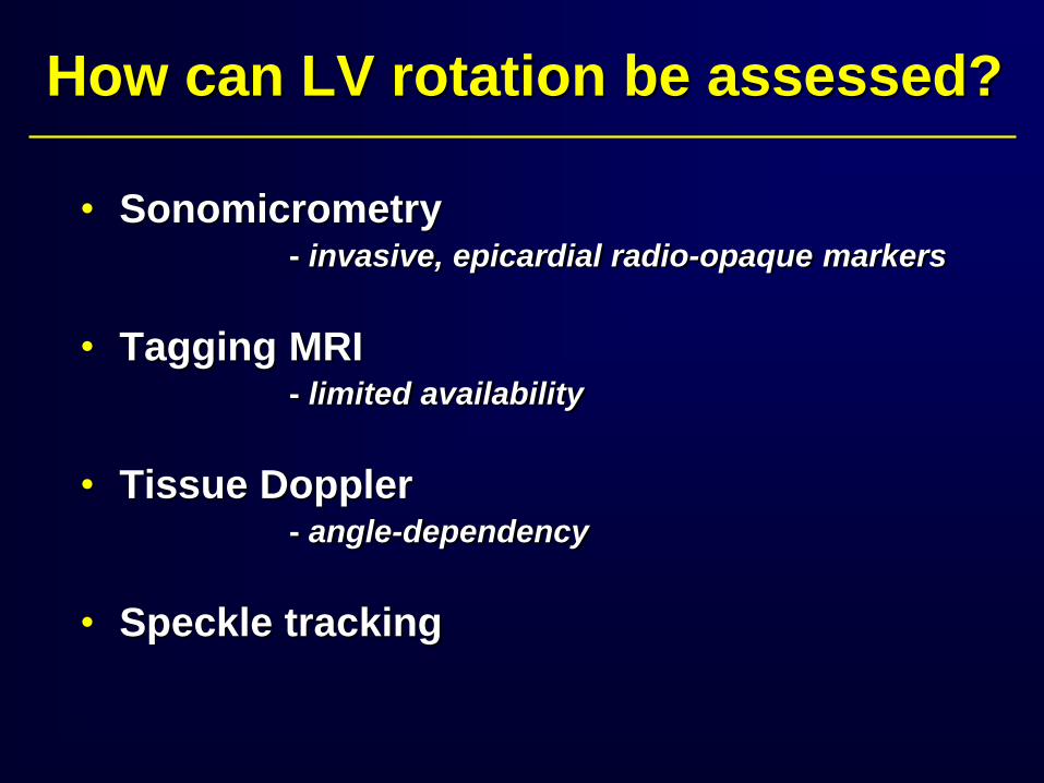
How can LV rotation be assessed?
• Sonomicrometry - invasive, epicardial radio-opaque markers
• Tagging MRI - limited availability
• Tissue Doppler - angle-dependency
• Speckle tracking

Left ventricular torsion
= rotation (rot) of the apex
relative to the base
• Apex: counterclockwise (+)
• Base: clockwise (-)
Twist (º) = apical rot – basal rot
Torsion (º/cm) = Twist
Apex-to-base length

Rotation vs time plots
BASE APEX

LV twist
Time-to-PUV Peak untwisting
velocity (PUV)
Base
Apex
Apex-Base = Twist
Temporal sequence of LV twist / untwist

Speckle Imaging Rotation
Validation vs Rotating Phantom
Courtesy of P. Lysyansky
Technical validation

LV rotation by STE : validation
Helle-Valle T et al. Circulation 2006.
Clinical validation
Experimental data
(13 dogs)
Clinical data
(29 normal subjects)

STE vs MR: Impact of missing the true apex
Goffinet C et al. Eur Heart J 2009
• 43 pts with various pathologies, 56±14 years (22–84)
• 2D-STE vs tagging MR
Apical rotation measured by 2D-STE significantly
underestimated that measured by tagging MR
Underestimation of apical rotation by 2D-STE is probably
not related to intrinsic inaccuracies of this technique,
but rather to its inability to image the true LV apex
(achieved in only 10% pts in this study!)

N = 100 Age (years)
LVESD (mm)
EF (%)
LAVi (ml/m2)
Vp (cm/s)
E/Vp
r = 0.4, p < 0.0001
r = -0.3, p = 0.004
r = 0.3, p = 0.002
r = 0.3, p = 0.003
r = 0.3, p = 0.003
r = -0.4, p = 0.003
LV torsion and its correlates in normals
Popescu BA et al. Van Dalen BM, et al. JASE 2008

LV torsion by STE: clinical studies
Wide variability in the reported values
for resting systolic torsion
Weyman AE. J Am Coll Cardiol 2007
Although conceptually simple,
torsion is more complex in practice

Physiological variables affecting LV twist/untwist
Sengupta PP et al. J Am Coll Cardiol Imaging 2008

Exercise
• Short-term exercise can almost double LV twisting
and untwisting (by augmented rotation that stores
additional potential energy released for improving
diastolic suction)
• Long-term exercise training may reduce the
values of resting LV twist (and increased torsional
reserves are being used in high-demand situations)
• With advancing age, the higher resting torsion is
associated with attenuation of torsional reserve at
peak exercise Neilan TG et al. J Am Soc Echocardiogr 2006
Notomi Y et al. Circulation 2006
Notomi Y et al. Am J Physiol Heart Circ Physiol 2008
Zocalo Y et al. Conf Proc IEEE Eng Med Biol Soc 2007
Burns AT et al. J Am Soc Echocardiogr 2007

Apical rotation and LV function
• Both LV twist and apical rotation are more closely
related to LV dP/dtmax than LV EF after ligation of
either LAD or LCx artery
Opdahl A et al. J Am Soc Echocardiogr 2008
Kim WJ et al. Circ Cardiovasc Imaging 2009
• Apical rotation measurement by STE is an effective
noninvasive index of global LV contractility
• Apical rotation by STE correlated well with LV twist
over a wide range of loading conditions and inotropic
states, and during myocardial ischemia

Anterior myocardial infarction
Takeuchi M et al. J Am Soc Echocardiogr 2007
30 pts with old anterior MI (>1 mo): 2 groups (LVEF ≥ 45%; < 45%)
• Reduced apical rotation in abnormal EF group (white
dots) results in severely depressed LV torsion and
reduced and delayed untwisting
• LV twist is preserved in the normal EF group
LV apex is the main determinant of LV torsion and
untwisting both in normal and diseased hearts

Exercise echo in HFNEF
In HFNEF - widespread abnormalities of both LV systolic and
diastolic function that become more apparent on exercise:
• At rest lower values of - Longitudinal and radial strain
- Apical rotation
- Reduced and delayed untwisting
- Mitral annular velocities
• At exercise, all parameters failed to normalize
HFNEF is not an isolated disorder of diastole
Correlated with peak VO2max
Tan YT. J Am Coll Cardiol 2009

Aortic stenosis Controls
(n=40)
AS
(n=61)
p value
Peak apical rotation (°) 15.7±5.9 21.0±7.6 <0.001
Peak basal rotation (°) -6.2±2.9 -6.7±3.2 0.4
Twist (°) 20.8±6.8 26.5±9.1 0.001
LV twist rate (°/s) 118±35 137±55 0.006
Peak systolic torsion (°/cm) 2.7±0.9 3.4±1.3 0.002
LV peak untwisting rate (°/s) -143±48 -158±59 0.18
Time to peak untwisting rate 1.23±0.09 1.21±0.08 0.2
LV peak apical back rotation rate (°/s) -93±47 -115±55 0.04
Time to peak apical back rotation rate 1.19±0.12 1.25±0.1 0.015
LV peak basal back rotation rate (°/s) 64±20 70±23 0.18
Time to peak basal back rotation rate 1.21±0.09 1.20±0.11 0.8
Popescu BA et al. Eur J Echocardiogr 2010
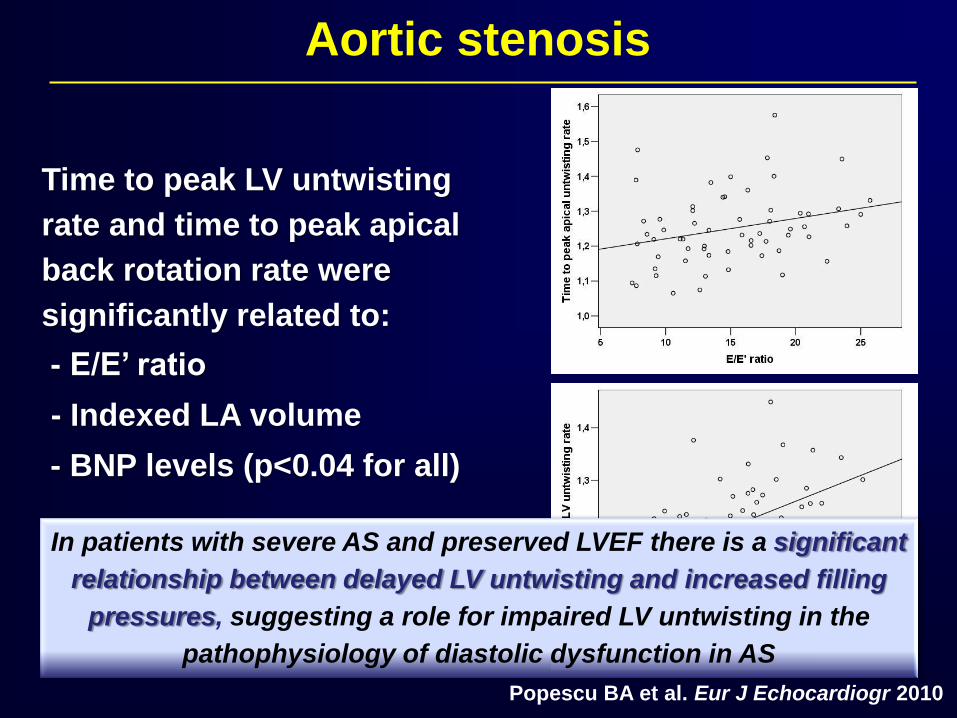
Aortic stenosis
In patients with severe AS and preserved LVEF there is a significant
relationship between delayed LV untwisting and increased filling
pressures, suggesting a role for impaired LV untwisting in the
pathophysiology of diastolic dysfunction in AS
Time to peak LV untwisting
rate and time to peak apical
back rotation rate were
significantly related to:
- E/E’ ratio
- Indexed LA volume
- BNP levels (p<0.04 for all)
Popescu BA et al. Eur J Echocardiogr 2010

LV torsion by STE in mitral regurgitation
• 38 pts with mod–severe MR (MVP) vs 30 controls
• LV remodeling and MR degree correlated with:
• reduced LV torsion
• reduced untwisting velocity
• delayed onset of untwisting
Borg A, et al. Heart 2008
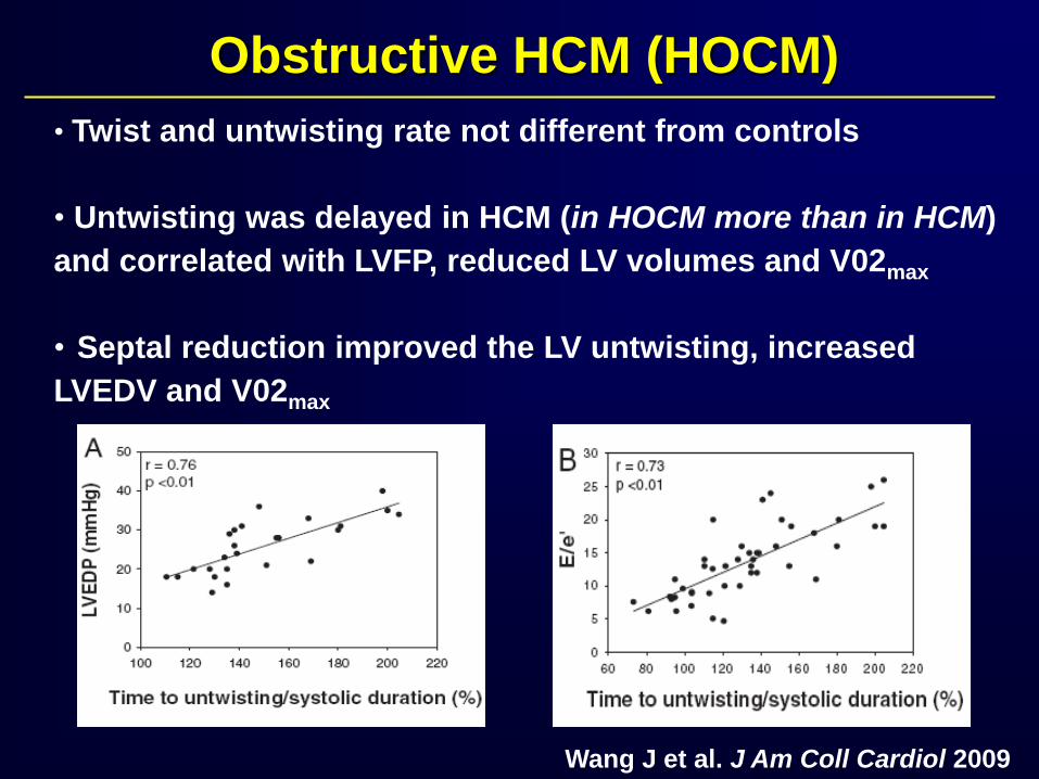
Obstructive HCM (HOCM)
• Twist and untwisting rate not different from controls
• Untwisting was delayed in HCM (in HOCM more than in HCM)
and correlated with LVFP, reduced LV volumes and V02max
• Septal reduction improved the LV untwisting, increased
LVEDV and V02max
Wang J et al. J Am Coll Cardiol 2009

Dilated cardiomyopathy
• LV systolic rotation at both basal and apical levels and LV
torsion are significantly reduced in pts, compared to controls (A)
Popescu BA et al. Eur J Heart Failure 2009
• 2 different patterns of apical
rotation:
- normally directed
(B - counterclockwise)
- reversed
(C - clockwise)
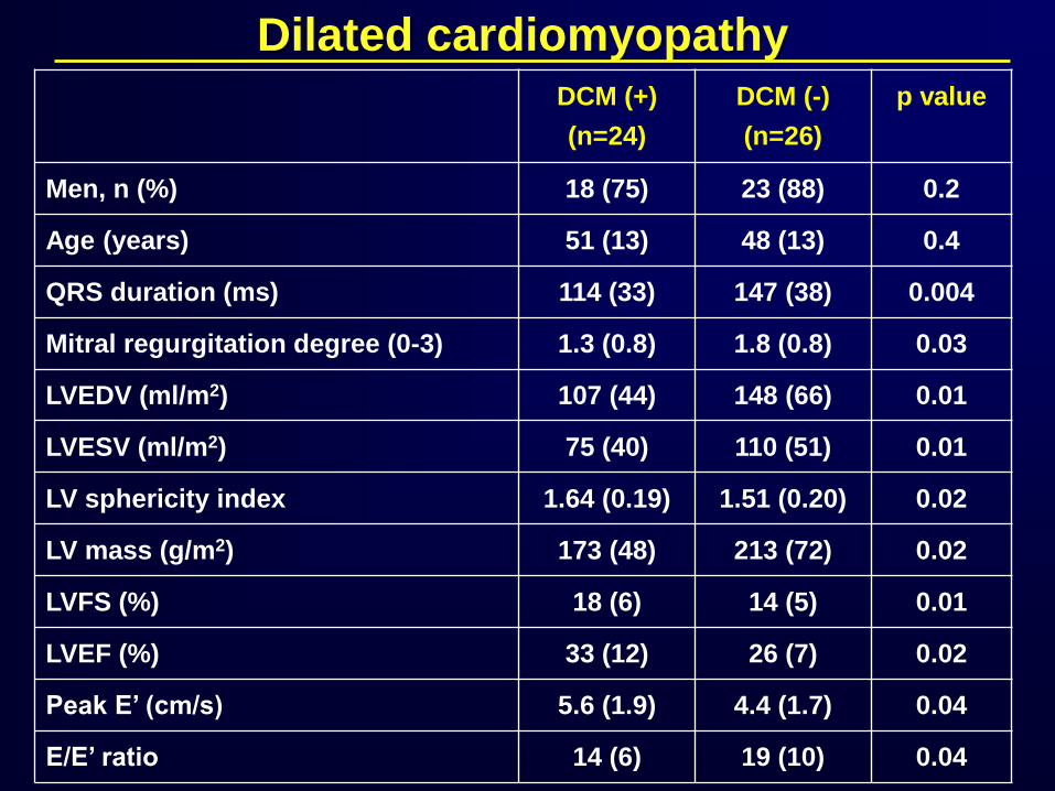
DCM (+)
(n=24)
DCM (-)
(n=26)
p value
Men, n (%) 18 (75) 23 (88) 0.2
Age (years) 51 (13) 48 (13) 0.4
QRS duration (ms) 114 (33) 147 (38) 0.004
Mitral regurgitation degree (0-3) 1.3 (0.8) 1.8 (0.8) 0.03
LVEDV (ml/m2) 107 (44) 148 (66) 0.01
LVESV (ml/m2) 75 (40) 110 (51) 0.01
LV sphericity index 1.64 (0.19) 1.51 (0.20) 0.02
LV mass (g/m2) 173 (48) 213 (72) 0.02
LVFS (%) 18 (6) 14 (5) 0.01
LVEF (%) 33 (12) 26 (7) 0.02
Peak E’ (cm/s) 5.6 (1.9) 4.4 (1.7) 0.04
E/E’ ratio 14 (6) 19 (10) 0.04
Dilated cardiomyopathy

Dilated cardiomyopathy
Reversed apical rotation and loss of LV
torsion in pts with DCM is associated with:
• significant LV remodeling
• increased electrical dyssynchrony
• reduced systolic function
• increased filling pressures
Indicating a more advanced disease stage

Am J Cardiol 2008;101:1163-9.
• 54 pts with HF; 33 underwent CRT
• 33 control subjects
• Radial & Long dyssynchrony by STE
• Apical & Basal rotation, twist & torsion by STE

• LV dyssynchrony is associated with discoordinate rotation
of the apical and basal regions, which in turn significantly
decreases peak LV twist and torsion.
• LV torsion and twist at AVC had the highest Sv (90%)
and Sp (77%) to predict CRT responders among all other
parameters, including radial and longitudinal dyssynchrony
Sade LE et al. Am J Cardiol 2008
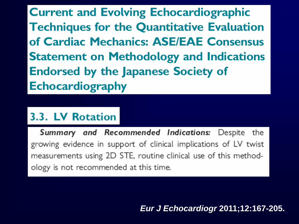
Eur J Echocardiogr 2011;12:167-205.

Conclusions
• LV twist/untwist play an important role in LV function,
in both ejection and filling
• Speckle tracking echocardiography allows the assessment
of LV rotation, twist/untwist
• Standardization of acquisition and processing is essential
for proper use of this technique
• Careful selection of the apical LV cut is mandatory, or else
underestimation of apical rotation/twist may result
• The incremental role of these parameters in clinical
decision-making needs further studies