The increasing importance of Plasmodium ovale and Plasmodium ...
Blood protozoa : Plasmodium & Babesia
Ass. Prof. Nader Alaridah MD, PhD

OverviewA parasite is an organism that lives on or in a host organism and gets its food
from or at the expense of its host.
Protozoa are unicellular eukaryotes that form an entire kingdom.The protozoa that are infectious to humans can be classified into four groups
based on their mode of movement:
• Sarcodina – the ameba
• Mastigophora – the flagellates, Giardia, Leishmania
• Ciliophora – the ciliates, e.g., Balantidium
• Sporozoa – organisms whose adult stage is not motile e.g., Plasmodium, Cryptosporidium undergo a complex life cycle with alternating sexual and asexual reproductive phases. The human parasites Cryptosporidium, Cyclospora, and Toxoplasma and the malarial parasites ( Plasmodium species) are all intracellular parasites.

• Over 2 billion (41% world population) lives in malaria-risk area.
• Infects 300-500 million people per year, 90% of whom are in sub-Saharan Africa.
• Kills over 1 million people each year and some estimate as many as 2.5 million.
• Leading Infectious killer of children. Worldwide a child dies of malaria every 30 seconds.
• Disease Burden increasing due to: weakening public health, agricultural practices, global warming, lack of vaccine, drug resistance in parasite and vector, population growth in endemic areas, increased travel.
Epidemiology

Plasmodium
• Plasmodium is a genus of parasitic alveolates, many of which cause malaria in their hosts.
• The parasite always has two hosts in its life cycle: Dipteran insect host and a vertebrate host.
• Species:
1.P. falciparum
2.P. malariae
3.P. vivax
4.P. ovale
5.Plasmodium knowlesi
- Plasmodium falciparum is the major species associated with deadly infections throughout the world.

• The vector for malaria is the female anopheline mosquito.
• When the vector takes a blood meal, sporozoites contained in the salivary glands of the mosquito are discharged into the puncture wound.
• Within an hour, these infective sporozoites are carried via the blood to the liver, where they penetrate hepatocytes and begin to grow, initiating the pre-erythrocytic or primary exoerythrocytic cycle.
• The sporozoites become round or oval and begin dividing repeatedly.
• Schizogony results in large numbers of exoerythrocytic merozoites.
• Once these merozoites leave the liver, they invade the red blood cells (RBCs), initiating the erythrocytic cycle.
Mechanism of Infection

• A dormant schizogony may occur in P. vivax and P. ovale organisms, which remain quiescent in the liver.
• These resting stages have been termed hypnozoites and lead to a true relapse, often within 1 year or up to more than 5 years later.

• Once the RBCs and reticulocytes have been invaded, the parasites grow and feed on hemoglobin.
• Within the RBC, the merozoite (or young trophozoite) is vacuolated, ring shaped, more or less ameboid, and uninucleate.
• The excess protein and hematin present from the metabolism of hemoglobin combine to form malarial pigment.
• Once the nucleus begins to divide, the trophozoite is called a developing schizont.
• The mature schizont contains merozoites (whose number depends on the species), which are released into the bloodstream.

Malaria transmission cycle

Malaria life cycle

Developmental stages of malarial parasites

PLASMODIUM VIVAX (BENIGN TERTIAN MALARIA)
• P. vivax infects only the reticulocytes.
• Splenomegaly occurs during the first few weeks of infection, and the spleen will progress from being soft and palpable to hard, with continued enlargement during a chronic infection.
• If the infection is treated during the early phases, the spleen will return to its normal size.
• A secondary or dormant schizogony occurs in P. vivax and P. ovale, which remain quiescent in the liver.
• These resting stages have been termed hypnozoites.

• After a few days of irregular periodicity, a regular 48-hour cycle is established.


Pathogenesis and Spectrum of Disease:
• In patients who have never been exposed to malaria:
Symptoms such as headache, photophobia, muscle aches, anorexia, nausea, and sometimes vomiting may occur before organisms can be detected in the bloodstream.
• In other patients with prior exposure to the malaria:
The parasites can be found in the bloodstream several days before symptoms appear.

PLASMODIUM OVALE
• Although P. ovale and P. vivax infections are clinically similar, P. ovale malaria is usually less severe, tends to relapse less frequently, and usually ends with spontaneous recovery.
• P. vivax, P. ovale infects only the reticulocytes.
• After a few days of irregular periodicity, a regular 48-hour cycle is established. Over time, the paroxysms become less severe and more irregular in frequency and then stop altogether.
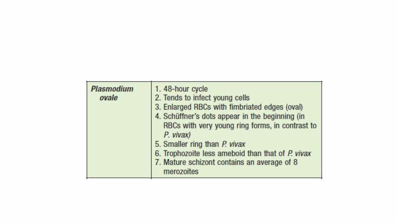


Pathogenesis and Spectrum of Disease:
• The incubation period is similar to that for P. vivax malaria, but the frequency and severity of the symptoms are much less, with a lower fever and a lack of typical rigors.

PLASMODIUM MALARIAE (QUARTAN MALARIA)

Pathogenesis and Spectrum of Disease:
• Proteinuria is common in P. malariae infections and may be associated with clinical signs of nephrotic syndrome.
• With a chronic infection, kidney problems result from deposition within the glomeruli of circulating antigen antibody complexes.
• A membrane proliferative type of glomerulonephritis is the most common lesion seen in quartan malaria.

PLASMODIUM FALCIPARUM (MALIGNANT TERTIAN MALARIA)• Plasmodium falciparum invades all ages of RBCs.
• Schizogony occurs in the spleen, liver, and bone marrow rather than in the circulating blood.
• Ischemia caused by the obstruction of vessels within these organs by parasitized RBCs will produce various symptoms, depending on the organ involved.

• A decrease in the ability of the RBCs to change shape when passing through capillaries or the splenic filter may lead to plugging of the vessels Also, only P. falciparum causes cytoadherence, a feature that is associated with severe malaria.
• In P. falciparum infections, as the parasite grows, the RBC membrane becomes sticky and the cells adhere to the endothelial lining of the capillaries of the internal organs.
• Thus, only the ring forms and the gametocytes (occasionally mature schizonts) normally appear in the peripheral blood.


Pathogenesis and Spectrum of Disease
• Symptoms such as aches, pains, headache, fatigue, anorexia, or nausea. This stage is followed by fever, a more severe headache, and nausea and vomiting.
• Severe or fatal complications can occur at any time and are related to the obstruction of vessels in the internal organs (liver, intestinal tract, adrenal glands, intravascular hemolysis/black water fever, and kidneys).
• Blackwater fever is a complication of malaria that is a result of red blood cell lysis, releasing hemoglobin into the bloodstream and urine, causing discoloration.
• Extreme fevers, 41.7° C (107° F) or higher, may occur in an uncomplicated malaria attack or in cases of cerebral malaria. Without vigorous therapy, the patient usually dies.
• Cerebral malaria is considered to be the most serious complication and the major cause of death with P. falciparum.


PLASMODIUM KNOWLESI (SIMIAN MALARIA,THE FIFTH HUMAN MALARIA)
• P. knowlesi invades all ages of RBCs.
• The early blood stages of P. knowlesi resemble those of P. falciparum.
• Whereas the mature blood stages and gametocytes resemble those of P. malariae.
• Unfortunately, these infections are often misdiagnosed as the relatively benign P. malariae; however, infections with P. knowlesi can be fatal.


CLINICAL FEATURES
• Malaria is a very common cause of fever in tropical countries. The first symptoms of malaria are nonspecific; the lack of a sense of wellbeing, headache, fatigue, abdominal discomfort, and muscle aches followed by fever are all similar to the symptoms of a minor viral illness.
• In some instances, a prominence of headache, chest pain, abdominal pain, cough, arthralgia, myalgia, or diarrhea may suggest another diagnosis. Although headache may be severe in malaria, the neck stiffness and photophobia seen in meningitis do not occur. While myalgia may be prominent, it is not usually as severe as in dengue fever, and the muscles are not tender as in leptospirosis or typhus. Nausea, vomiting, and orthostatic hypotension are common.
• The classic malarial paroxysms, in which fever spikes, chills, and rigors occur at regular intervals, are relatively unusual and suggest infection with P. vivax or P. ovale.
• The fever is usually irregular at first (that of falciparum malaria may never become regular); the temperature of nonimmune individuals and children often rises above 40C in conjunction with tachycardia and sometimes delirium. Although childhood febrile convulsions may occur with any of the malarias, generalized seizures are specifically associated with falciparum malaria and may herald the development of encephalopathy (cerebral malaria).

LABORATORY DIAGNOSIS (ALL SPECIES)1.Routine Methods:
- Thick and thin blood films.
- At least 200 to 300 oil immersion fields should be examined on both films before a negative report is issued.
- Stains:
1. Giemsa stain.
2. Wright’s stain.
3. Fluorescent nucleic acid stains, such as acridine orange.
- Blood collected using (EDTA) anticoagulant.

2.Serologic Methods:
- Several rapid malaria tests (RMTs):
1.Some of which use monoclonal antibodies against the histidine-rich protein 2 (HRP2).
2. Whereas others detect species-specific parasite lactate dehydrogenase (pLDH).
- These procedures are based on an antigen capture approach in dipstick or cartridge formats.
3. Molecular Diagnostics:
• Other methods include direct detection of the five species by using a specific DNA probe after PCR amplification of target DNA sequences.
4. Automated Instruments:
• Using automated flow cytometry hematology instruments, there are potential limitations related to the diagnosis of blood parasite infections.

THERAPY
• Antimalarial drugs are classified according to the stage of malaria against which they are targeted.
• QUINOLINES , ARTEMISININS • Tetracycline, doxycycline, and clindamycin are used increasingly in combination
with other antimalarials to improve their efficacy
• These drugs are referred to as :
1.Tissue schizonticides (which kill tissue schizonts).
2.Blood schizonticides (which kill blood schizonts).
3.Gametocytocides (which kill gametocytes).
4.Sporonticides (which prevent formation of sporozoites within the mosquito).

CONTROL

Babesiosis
• Babesiosis is an emerging tick-borne infectious disease caused by protozoan parasites of the genus Babesia that invade and eventually lyse red blood cells (RBCs).
• Most cases are due to Babesia microti. B. microti, a parasite of small rodents, is the most common etiologic agent of human babesiosis
• The primary causative agent of human babesiosis in Europe is B. divergens, but Babesia venatorum and B. microti also have been reported.
• The infection typically is mild in young and otherwise healthy individuals but can be severe and sometimes fatal in persons >50 years of age and in immunocompromised patients. Sporadic cases have been reported in Europe and the rest of the world.

Modes of Transmission
• B. microti is transmitted to humans primarily by the nymphal stage of the deer tick (Ixodes scapularis), the same tick that transmits the causative agents of Lyme disease .
• The vectors for transmission of B. duncani and B. divergens– like organisms are thought to be Ixodes pacificus and Ixodes dentatus, respectively.

babesia life cycle

CLINICAL MANIFESTATIONS• Asymptomatic B. microti Infection: At least 20% of adults and 40% of children do not
experience symptoms following B. microti infection. There is no evidence of long-term complications following asymptomatic infection; however, people who are asymptomatically infected may transmit the infection when they donate blood.
• Mild to Moderate B. microti Illness Symptoms typically develop following an incubation period of 1–4 weeks after tick bite and 1–9 weeks (but as long as 6 months) after transfusion of blood products. Patients experience a gradual onset of malaise, fatigue, and weakness. Fever can reach 40.9C and is accompanied by one or more of the following: chills, sweats, headache, myalgia, arthralgia, nausea, anorexia, and dry cough.
• Severe B. microti Illness Severe babesiosis requires hospital admission and typically occurs in patients with one or more of the following: age of >50 years, neonatal prematurity, male gender, asplenia, HIV/AIDS, malignancy, hemoglobinopathy, and immunosuppressive therapy.

PATHOGENESIS
• Anemia is a key feature of the pathogenesis of babesiosis. Hemolytic anemia caused by rupture of infected RBCs generates cell debris that may accumulate in the kidney and cause renal failure.
• Anemia also results from the clearance of intact RBCs as they pass through the splenic red pulp and encounter resident macrophages.
• Babesia antigens expressed at the RBC membrane promote opsonization and facilitate uptake by splenic macrophages. In addition, RBCs are poorly deformable as a result of oxidation generated by the parasite and the host immune response and are filtered out as they attempt to squeeze across the venous vasculature. Bone marrow suppression due to cytokine production may also contribute to anemia.

DIAGNOSIS
• Microscopic examination of Giemsa-stained thin blood smears
• Polymerase chain reaction (PCR)
• Serology can suggest or confirm the diagnosis of babesiosis. An indirect immunofluorescent antibody test for B. microti is most commonly used.


TREATMENT
• Atovaquone plus azithromycin is the recommended antibiotic treatment combination for mild to moderate babesiosis.
• Clindamycin plus quinones is the choice for severe infections.

Prevention
• Wear clothing that covers the lower part of the body, apply tick repellents (such as DEET) to clothing, and limit outdoor activities where ticks may abound from May through October.

The End