

Blood ppt for students 2015
42
Blood S Naghma Rizvi March, 2015. Acknowledgement :Muhammad Nisar
-
Upload
aku-karachi -
Category
Science
-
view
31 -
download
1
Transcript of Blood ppt for students 2015

Blood
S Naghma Rizvi
March, 2015.
Acknowledgement :Muhammad Nisar

Objectives
Define blood, its general properties and function.
Explain the composition.
Describe the formation of Blood cells
Discuss the structure and functions of blood cells
RBC (Erythrocytes)
WBC (Leukocytes)
Platelets (thrombocytes
Discuss the composition of plasma
Describe the three mechanism that contribute towards
hemostasis
Briefly discuss the ABO blood groups and Rh factor.
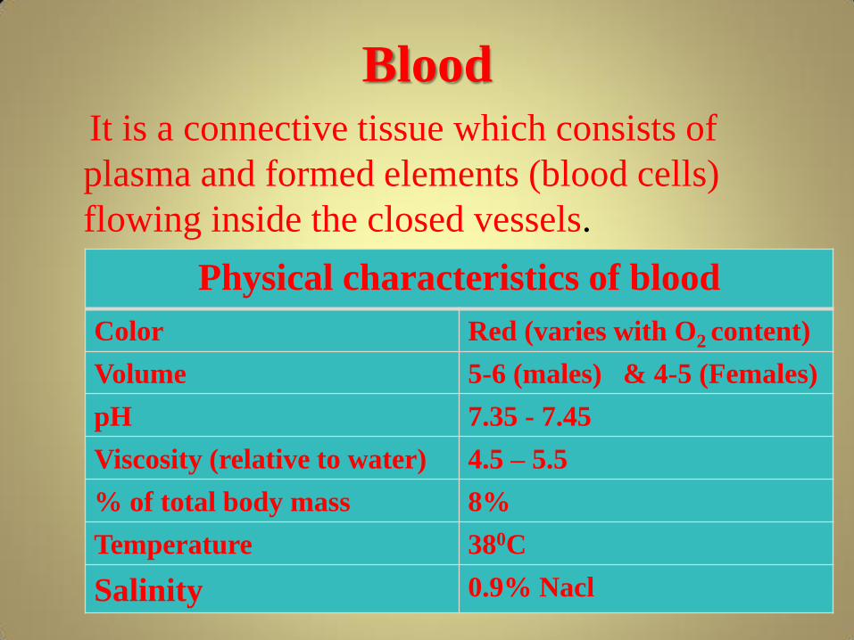
Blood
It is a connective tissue which consists of
plasma and formed elements (blood cells)
flowing inside the closed vessels.
Physical characteristics of blood
Color Red (varies with O2 content)
Volume 5-6 (males) & 4-5 (Females)
pH 7.35 - 7.45
Viscosity (relative to water) 4.5 – 5.5
% of total body mass 8%
Temperature 380C
Salinity 0.9% Nacl

Functions of Blood 1.Transportation
oxygen and carbon dioxide
food molecules (glucose, lipids, amino acids)
ions (e.g., Na+, Ca+2, HCO−3)
wastes (e.g., urea)
hormones
2.Regulation
Body temperature
pH
3.Protection
Clotting
Defense

Composition of blood
Centrifuged
blood RBCs & WBCs are whole cells platelets are
cell fragments
Un-
clotted/un-
centrifuged
blood

Plasma Yellowish clear liquid, composed of:
Water = 91%
Proteins = 7% (all synthesized by the liver)
• Albumin = 54%, regulates osmotic pressure of blood
• Globulins = 38%, alpha and beta globulins in transport,
• gamma globulins in defense (antibodies)
• Fibrinogen = 7%, coagulation
Other solutes =2%
• Electrolytes - Na+, K+, Ca++, Mg++
• Nutrients - glucose, amino acids, fatty acids, monoglycerides
• Gases - O2, N2, CO2
• Regulatory substance - hormones, enzymes
• Vitamins and wastes
• Wastes

Hemopoieses The process of development of blood cells

Erythrocytes (RBC)
Physical characteristics of Erythrocyte
Size 7 µm in diameter & 2.2 µm thick
Shape Flattened and biconcave disc
Mean count 4.5-5.5 per mm3 of blood
Morphology Red color, (variable) non-nucleated, each RBC
contains 280 millions Hb molecules, that can carry
over a billion O2 molecule
Site for production Bone marrow
Life span 120 days
Function Carries O2 and nutrients, removes CO2 & wastes

Structure of Erythrocyte
(Hemoglobin molecule)
•
HEME molecule
Globin Molecule
Hemoglobin molecule
contain 4 protein chains
called globins, each of
which is bound to 1 heme
(iron). Iron is able to
combine with oxygen in
the lungs and deliver it to
body tissues.
is able to combine with oxygen in the lung and release
oxygen in the tissue.
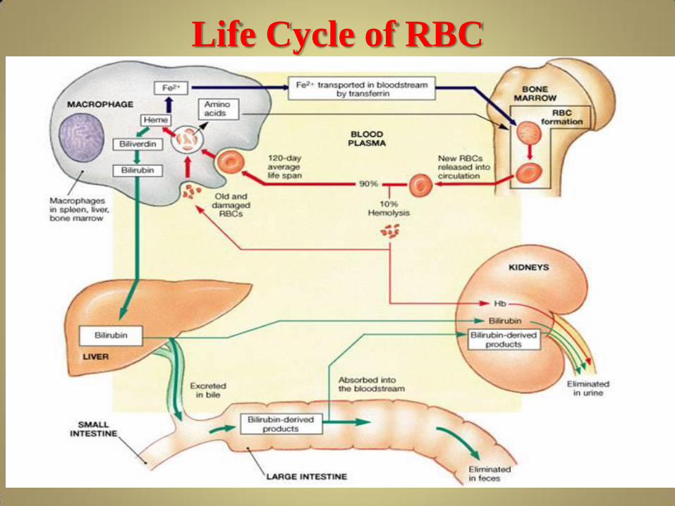
Life Cycle of RBC

• http://highered.mheducation.com/sites/007250
7470/student_view0/chapter19/animation__he
moglobin_breakdown.html
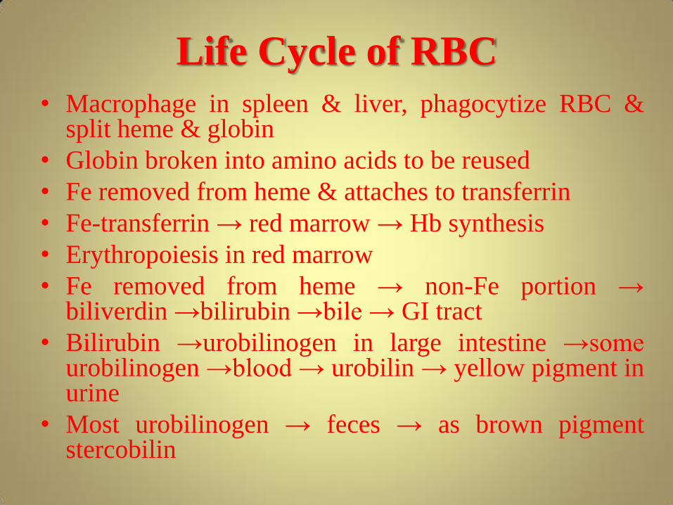
Life Cycle of RBC
• Macrophage in spleen & liver, phagocytize RBC & split heme & globin
• Globin broken into amino acids to be reused
• Fe removed from heme & attaches to transferrin
• Fe-transferrin → red marrow → Hb synthesis
• Erythropoiesis in red marrow
• Fe removed from heme → non-Fe portion → biliverdin →bilirubin →bile → GI tract
• Bilirubin →urobilinogen in large intestine →some urobilinogen →blood → urobilin → yellow pigment in urine
• Most urobilinogen → feces → as brown pigment stercobilin

Leukocytes (WBC) They are largest of all blood cells, have nuclei and
do not contain Hb.Forms 1% of blood volume
On the basis of presence of absence of chemical
filled cytoplasmic granules WBCs are classified
into two types:
Granular leukocyte
Agranular leukocyte
Granules
Granulocyte or PMNLS Agranulcytes

Granulocytes Multi-lobed nuclei
Named according to dyes they take up
Basophil Neutrophil Eosinophi
l
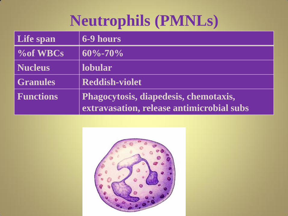
Neutrophils (PMNLs) Life span 6-9 hours
%of WBCs 60%-70%
Nucleus lobular
Granules Reddish-violet
Functions Phagocytosis, diapedesis, chemotaxis,
extravasation, release antimicrobial subs

Basophil (PMNLs) Life span
%of
WBCs:
0.5%-1%
Nucleus large and U to S shaped
Granules Blue
Functions Secretes histamine ,serotonin, prostaglandin, leukotrien & heparin

Eosinophil (PMNLs)
Life span
%of WBCs: 2-4%
Nucleus 2 large lobes
Granules Stain red
Functions Release enzyme that destroy parasites
Phagocytosis of antigen antibody complex, and allergens

Agranulocytes Their nucleus are round or slightly indented and
stains dark
Lymphocyte
T- lymphocyte(T cells)
B- lymphocyte(B cells)
Natural killer cells (NK cells)
Monocyte
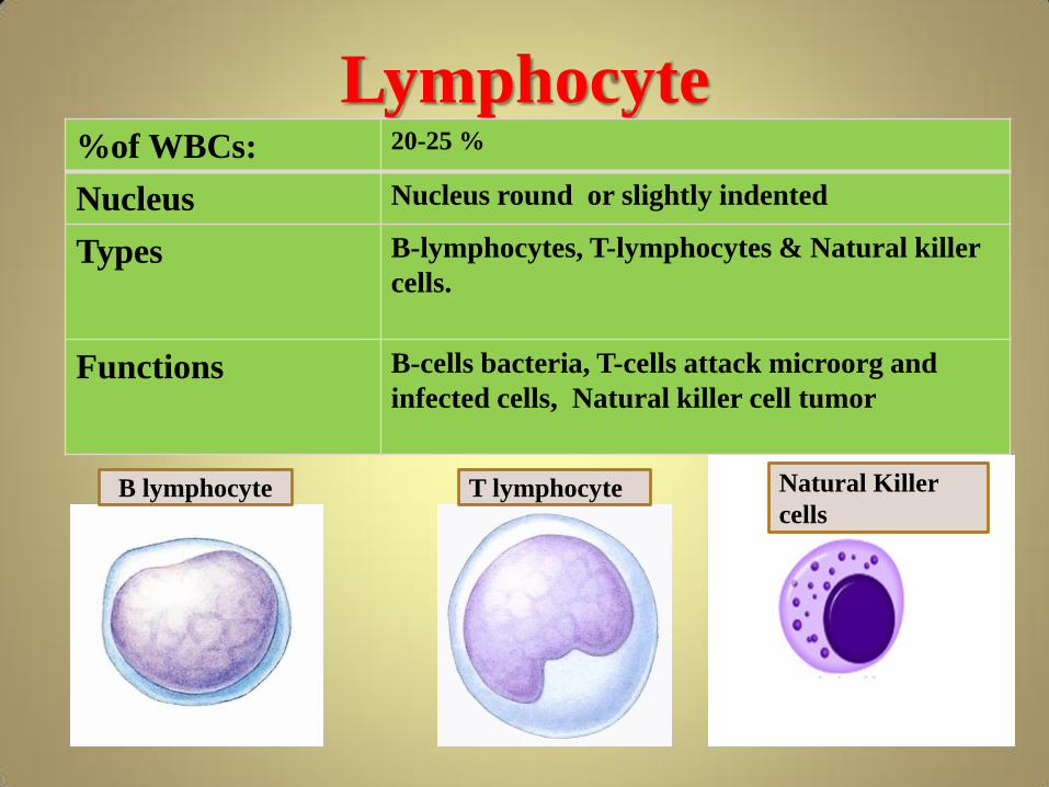
Lymphocyte %of WBCs: 20-25 %
Nucleus Nucleus round or slightly indented
Types B-lymphocytes, T-lymphocytes & Natural killer
cells.
Functions B-cells bacteria, T-cells attack microorg and
infected cells, Natural killer cell tumor
B lymphocyte T lymphocyte Natural Killer
cells

Monocyte
%of WBCs: 3-8
Nucleus Kidney or horse shoe shape nucleus
Functions
Differentiate into macrophages. Phagocytize pathogens, dead neutrophil and debris of dead cells

Platelets or
Thrombocytes
Size 4 µm in diameter
Shape Disc shape
Mean count 130000-400000 per mm3 of blood
Morphology Non-nucleated fragments of cells
Site for production Bone marrow
Life span 5-9 days
Function Blood clotting

Hemostasis/Haemostasis
Heamostasis or cessation of bleeding takes place through a series of responses, these include.
• Vasospasm
• Platelet plug formation:
• Coagulation
Follow the sequence to events initiated when platelets come into contact with an injured surface
Vasospasm
• Vessel wall constricts for a short period
• Platelets (sticky) adhere to the injured wall
• Platelets change their shape
• Platelets (activated) release serotonin that constrict
the vessel

Platelet Plug formation
• Passing platelets stick to the site and release
chemicals that attract more platelets.
• More platelets move leading to platelets
aggregation (positive feedback.
• Platelets form a plug or a temporary seal
within six minutes.

Coagulation
• Thromboplastin (TP) or tissue factor released by damaged tissue cells.
• TP activates (through series of chemical reactions) the inactive clotting factors already present in the blood.
• Prothrombin activato, first step in final common pathway.
This final common pathway can be initiated by two processes usually occurring together…

The insoluble fibrin threads
increase in number and forms a
meshwork that trap blood cells and
is much stronger than platelet plug

Vasospasm

Platelet Plug Formation

Plasma clotting Factors Factor Name Function
I Fibrinogen Converted to fibrin
II Prothrombin Enzyme
III Tissue Thromboplastin
Co factor
IV Ca++ Co factor
V proaccelerin, Labile factor
Co factor
VII proconvertin Enzyme
VIII AHF A Co factor

IX AHF B Enzyme
X Trombokinase Enzyme
XI AHF C Enzyme
XII Hageman Factor
Enzyme
XIII Fibrin stabilizing factor
Enzyme

ABO Blood System
This system consists of three types of antigens
Antigen A
Antigen B
Antigen Rh
Two types of antibodies
Anti-A antibody
Anti-B antibody
ABO system forms four major types of blood groups.
The table below shows the possible combinations of
antigens and antibodies with the corresponding ABO
type ("yes" indicates the presence of a component and
"no"indicates its absence in the blood of an individual).

ABO
blood type
Antigen
A
Antigen
B
Antibody
A
Antibody
B
A Yes No No Yes
B No Yes Yes No
AB Yes Yes No No
O No No Yes Yes

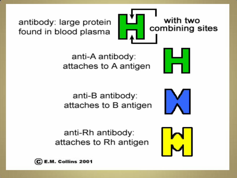

Anti-A antibody
Anti-B antibody Ant-Rh antyibody


Blood Transfusion

Hemolytic disease in Newborn


