The feasibility of real-time in vivo optical detection of blood–brain ...

Biophysical Properties of Red Blood Cells Using Optical Tweezers – Applications in Biomedicine
Carlos A. L. Silva1, Carinna N. Lima1,2, Yandilla S. S. Silva1, Diego C. N Silva3, Patricia Moura2, Beate S. Santos1, Goreti Pereira1 and Adriana Fontes1 1 Laboratório de Óptica Biomédica e Imagens, Universidade Federal de Pernambuco, Avenida da Arquitetura, S/N, Cidade
Universitária, Recife, Pernambuco 50740-530, Brazil. 2 Instituto de Ciências Biológicas, Universidade de Pernambuco, Recife, Pernambuco, Brazil. 3 Colegiado de Ciências Biológicas, Universidade Federal do Vale do São Francisco, Petrolina, Pernambuco, Brazil.
Advances in biophotonics have encouraged the development of novel tools applied to help understand a variety of biological events. Among these tools is the optical tweezer, a valuable microscopy technique, based on light momentum transfer, which allows not only capturing and manipulating cells and biomolecules, but also evaluating their mechanical properties. The optical tweezer is a highly sensitive tool with the following capabilities: (i) measuring cellular and molecular biophysical parameters without mechanical contact; (ii) detecting slight variations in biological properties and (iii) analyzing, one by one, biological systems, providing individual measurements, rather than only average values, which can help identify the signatures of several biological conditions. In particular, optical tweezers have allowed for the study of the aggregation and deformability of red blood cells (RBCs). Changes in these biophysical characteristics can lead to decreased, or even lost, RBC functionality and, consequently, reduced survival of erythrocytes in the blood circulation. In this context, this mini-review presents the fundamentals of the optical tweezer as a tool for biological manipulation, focusing mainly on the evaluation of the elastic constant, membrane viscosity, zeta potential and adhesion of RBCs. Applications of optical tweezers in the biomedical field will also be highlighted, including elucidative studies related to normal RBCs, as well as those affected by some intrinsic or extrinsic conditions such as diseases or storage for transfusion purposes.
Keywords: Optical trapping; hemotherapy; hematology; erythrocytes.
1. Introduction
In recent decades, with advances in the area of biophotonics, several techniques have been developed for the manipulation and characterization of individual cells, which have allowed for the study and comprehension of a variety of biological events. One of these tools is the optical tweezer, which is a versatile and valuable microscopy technique, well suitable for measuring forces and movement of microparticles. This technique is based on light momentum transfer, which allows for the capture and manipulation of cells, as well as biomolecules, and the evaluation of their mechanical properties [1-3]. This tool has been used for the study, at the cellular and molecular levels, of a variety of biochemical and biophysical processes without mechanical contact. Moreover, the optical tweezer is a highly sensitive tool capable not only of distinguishing small variations in biological properties, allowing for comparisons between different systems, but also of analyzing each biological system individually, providing single measurements rather than only average values, which can help identify the fingerprint of many biological conditions [3-6]. At the molecular level, optical tweezers have been used for studying the mechanics of DNA and RNA [7, 8], the forces associated with the folding/unfolding of proteins [9, 10], interactions between proteins and DNA [11, 12], among others. At the cellular level, we can highlight for instance the use of optical tweezers in studies of cell motility [13, 14], mitosis [15] and mechanical characteristics of red blood cells (RBCs) [4, 5]. The biophysical properties of RBCs are essential for the maintenance of homeostasis. Under normal conditions, mature RBCs have a biconcave shape and maintain unchanged their area/volume ratio, ionic composition, pH, as well as the structure and integrity of their membrane (lipid bilayer and cytoskeleton) [16, 17]. However, certain conditions can induce changes in the biophysical, biochemical and/or structural properties of erythrocytes. Patients with hematological diseases can present alterations in their erythrocyte rheological properties, which could result from modifications in the composition and organization of components of the lipid bilayer, cytoskeleton and/or hemoglobin [18]. Another condition that can change the biophysical and biochemical properties of erythrocytes is its storage in blood banks [19]. The modifications that occur as a result are known as the RBC storage lesion and include increased hemolysis, changes in shape [20], membrane blistering [21], tendency to cell aggregation [22], decrease in deformability [23, 24], and others. Among the important biophysical properties of RBCs we can highlight: (1) their deformability, or high flexibility, which allows RBCs to pass through microcapillaries much smaller than the size of the RBC (ca. 8 μm), thereby guaranteeing oxygen and carbon dioxide transport throughout the entire body [25] and (2) their ability to aggregate, which is closely related to the Zeta potential (ζ) of erythrocytes, a repulsive potential that arises as a consequence of the negatively charged surface of RBCs (conferred by the presence of sialylated glycoproteins). The ζ promotes repulsive
Microscopy and imaging science: practical approaches to applied research and education (A. Méndez-Vilas, Ed.)
45
___________________________________________________________________________________________

forces among RBCs, preventing their aggregation [26, 27]. Alterations in cellular biophysical properties may lead to the loss or decrease of the cells’ functionality and, consequently, lower survival of erythrocytes in circulation. Technical advances have been revolutionizing studies in erythrocyte biology, helping to elucidate the mechanisms related not only to the individual functions of RBCs, but also to those associated with cell-cell interactions. In this context, this mini-review presents the fundamentals of the optical tweezer as a microscopy tool for biological manipulation, focusing on studies related to RBCs in the biomedical area. Here, the potential of applying optical tweezers to investigating the biophysical properties of RBCs will be demonstrated through reports involving normal erythrocytes, as well as those affected by different conditions, such as storage or diseases.
2. Principles of Optical Tweezers
In 1609, Johannes Kepler observed that the tails of comets always point to the side opposite the sun, suggesting that the sun was producing a type of radiation pressure on comet tails. In 1873, James Clerk Maxwell formalized the idea that light could exert radiation pressure on matter and determined that the light reflected, refracted or absorbed by objects could generate optical forces. In 1901, Ernest Fox Nichols and Gordon Ferrie Hull, and independently Pyotr Lebedev, proved the existence of radiation pressure, confirming these predictions. Almost one hundred years after the Maxwell theory, in the early 1970s, Arthur Ashkin conducted experiments manipulating and trapping dielectric particles by using the radiation pressure of light [28]. The first works by Ashkin began with the use of counter-propagating laser beams, but by the mid-1980s, he proposed that only a highly focused laser beam would be sufficient to capture dielectric particles in three-dimension, thereby introducing the technique known as optical tweezers [29]. The optical tweezer technique has been consolidated as an optical tool capable not only of capturing and moving dielectric particles, but also of studying their mechanical properties [30]. The simplest way to understand how an optical tweezer works is by using the geometrical optical model, applied when r >> λ, where r is the particle radius and λ is the light wavelength. The trajectory of a light ray can be associated with a photon that carries a momentum . When an object, with a refractive index larger than the surrounding medium index (n2 > n1), changes the direction of the photon trajectory, it receives an impulse toward the bisectrix of the angle between the incident and the refracted photon, as a collision between particles, as shown in Figure 1A. It is like a pair of action and reaction forces. The object changes the light direction and the light pushes the object to the opposite side of the deviation. This is an intuitive way to understand the principles behind the operation of an optical tweezer.
Figure 1 – (A) Collision between a photon and a particle. (B) Forces and trajectories for two rays of a single, highly focused laser beam, for n2 > n1. Subsequently, if we consider two light rays of a highly focused laser beam, one on each side, as shown in Figure 1B, the rays a and b would meet at the focus (f), if there is no object. The refraction of these rays causes the forces Fa and Fb, and their sum leads to a resulting force F that tends to move the center of the particle towards the laser focus f. In this way, a highly focused laser beam creates a trap that keeps the center of dielectric particles in the laser focus. It is like a spring (F is a restoring force) connecting the center of the particle to the laser focus, when it moves, the particle follows this movement in three dimensions. Actually, the rays are not only refracted, but they are also reflected and then two resulting forces will act on the object, one named “the gradient force” that pulls the particle toward the laser focus, and the other called “the scattering force” that pushes the object away from the laser. When “the gradient force” is higher than “the scattering force”, we have an optical tweezer. Typically, since the optical tweezer works only in liquids, reflection is usually much smaller than refraction, but as the difference between the refractive indexes of the surrounding medium and the particle increases, the role of reflection becomes more significant, and may even prevent the capture of particles by the optical tweezer. It is not only reflection which can make the capture of particles by an optical tweezer difficult, but also absorption. From the point of view of optical force efficiency, thermal effects need to be avoided, even if they are small, since one of the main features of an optical tweezer is capturing and studying living biological systems. Most of the organic
Microscopy and imaging science: practical approaches to applied research and education (A. Méndez-Vilas, Ed.)
46
___________________________________________________________________________________________

substances that compose biological systems, such as the pigments melanin and hemoglobin, tend to present high absorption in the visible region. On the other hand, water (the most abundant component of living systems), absorbs mostly above 1300 – 1500 nm, leaving a window in the near-infrared region (NIR) with smaller absorptions, around 700 to 1300 nm. In the first demonstrations of the optical tweezer, when Arthur Ashkin used a visible laser to capture inorganic particles in water, he observed that bacteria were also captured and damaged by this system. Therefore, the visible-light laser was changed to one operating in the near-infrared region (1064 nm) and, then it was possible to trap and manipulate living cells, without considerable thermal injury [1]. The applications of optical tweezers in the biomedical sciences grew exponentially after the demonstration that it was possible to keep cells alive, for an extended time, if thermal effects were avoided with near-infrared lasers [30]. An upper estimate of the force magnitude of an optical tweezer can be made by assuming that a laser beam, with a power (P) of ca. 100 mW, can cause a maximum force in an object of ca. 400 pN, as described by Eq. 1 (where n1 is considered the water refractive index):
1.33 . ~400pN (Eq. 1)
Although very small, these forces are considerable on the microscopic level. Assuming that the captured object is a cubic particle of 1-μm sides with density equal to water, the acceleration caused by an optical force of 400 pN would be ca. 104 x g, where g is gravitational acceleration. Considering a particle 10-fold larger, the acceleration decreases by a factor 103, however, the acceleration caused by the optical force would still be in the range of 10 x g. However, for particles of 100-μm sides, the acceleration would be of ca. 10-2 x g, already negligible compared to other microscopic interactions. Thus, the optical tweezer can only capture and effectively manipulate particles smaller than approximately 50 μm. A typical optical tweezer system basically comprises one or two near-infrared laser beams, which pass by sets of lenses, and is/are tightly focused on a particle (presenting a refractive index larger than the surrounding medium) through of a microscope objective of high numerical aperture (N.A. > 1), as shown in Figure 2. The main set of lenses, indicated as telescope, has the function of capturing the particles in the microscope plane of view. Additional sets of lenses can also be used to bend the laser beams in order to approach and bring away captured particles in a dual-beam optical tweezer system. In a single-beam optical tweezer system, optical manipulation is usually performed by a motorized stage coupled to the microscope.
Figure 2 – Schematic representation of a single-beam optical tweezer system.
If the aim of the study is using the optical tweezer to perform force measurements of non-spherical biological systems, first of all it is necessary to calibrate the tool by using a silica or polystyrene bead, in order to find the trap stiffness. This calibration can be done, for example, by analyzing the viscous force when dragging beads at constant velocities using the optical tweezer system. In this case, Fviscous = k d, where k is the stiffness and d is the bead displacement from the equilibrium position (it is the distance between the bead center to the laser focus), as shown in Figure 3A. The bead will then be used as a calibrator; by coupling the microsphere to the biological system, it will be possible to evaluate the biological force of interest (in the horizontal plane) by using k and by measuring the displacement d caused by the target force, as shown in Figure 3B [30].
Microscopy and imaging science: practical approaches to applied research and education (A. Méndez-Vilas, Ed.)
47
___________________________________________________________________________________________

Figure 3 – (A) Bead displacement after being dragged at a constant velocity for optical tweezers calibration, in the horizontal plane. (B) Measuring erythrocyte adhesion by using dual-beam optical tweezers. The optical tweezer has proven to be a versatile tool in biomedical sciences, allowing not only for the capture and manipulation of cells and biomolecules, but also for the evaluation of biophysical properties related to biological systems [30]. In this context, this chapter will demonstrate the potential of applying optical tweezers to investigate the biophysical properties of red blood cells.
3. Biomedical Optical Tweezer Applications in Red Blood Cells
3.1. Agglutination/aggregation studies
Agglutination reactions are consequences of the mutual attraction of cell membranes that can occur among RBCs and between RBCs and other cells. In the blood stream, RBCs can aggregate and disaggregate spontaneously, and this phenomenon can affect transfusion procedures and the development of some diseases [31, 32]. These reactions depend on several factors, such as interactive forces and the environment. The interactive forces can be the intermolecular attractive (Van der Walls, hydrogen and electrostatic interactions) or repulsive (electrostatic interactions) forces. The electrostatic forces are a consequence of the Zeta potential (ζ), which can be used as a measure of the RBC surface charge. The presence of sialylated glycoproteins in the RBC membrane is responsible for the negative charges of the erythrocyte surface [32, 33]. Optical tweezers have been used in studies aiming to elucidate the mechanisms of RBC aggregation and coagulation. Khokhlova et al. [32] studied the aggregation speed and behavior of normal and pathological (systemic lupus erythematosus) RBCs. Systemic lupus erythematosus (SLE) is an autoimmune disease that enhances RBC agglutination forces. The authors observed that the aggregation force and speed of normal RBCs was lower than for SLE cells. The aggregation force ratio measured for SLE cells and normal RBCs, FSLE/Fnormal, was equal to 2.1, which indicates that this force is higher for SLE cells than for normal ones. Meanwhile, the aggregation speed value determined for normal RBCs was 0.30 ± 0.08 μm/sec and for SLE cells 0.53 ± 0.06 μm/sec, which was almost twice the speed of normal RBCs. These evaluations were performed using a dual-beam optical tweezer. The traps were turned on and the aggregation was studied by moving individual RBCs, in a pair rouleau, at a constant velocity of 0.3 μm/s with calibrated forces and measuring the maximum distance between the RBCs centers (Δx), as represented in Figure 4. The aggregation speed was evaluated by correlating the minimal RBC overlapping (dX, Figure 4) with the aggregate formation time. In this case, the trapping force value was ca. 29 pN.
Figure 4 - (A) Scheme demonstrating a pair rouleau being disaggregating by the dual-beam optical tweezers, the greater the force, the higher the Δx. (B) Scheme representing the dependence of the minimal RBC overlapping (dX) according to the rouleau formation time ta. In this case the trapping force was fixed at ca. 29 pN.
Microscopy and imaging science: practical approaches to applied research and education (A. Méndez-Vilas, Ed.)
48
___________________________________________________________________________________________

Fernandes et al. [33] compared the forces involved in RBC aggregation in normal serum, serum with anti-D antibodies and samples with potentiator solutions [Dextran, Bromelain, Papain and low ionic strength solution (LISS)]. Using a dual-beam optical tweezer, these authors measured a parameter called apparent membrane viscosity that is related to the type and the number of proteins involved in RBC aggregation. The measurement of apparent membrane viscosity was obtained using an RBC pair rouleau, by evaluating the optical force measured through the displacement of a silica bead attached to one RBC, while a second RBC was dragged as a function of constant velocities (0.2, 0.5, 0.7, 1.2, 1.8, 3.2 μm/s) [34]. They found values of 1x10⁻³ and 2x10⁻³ poise.cm, for normal serum and serum with anti-D antibodies, respectively. And, when using the agglutination potentiator solutions (Dextran, LISS and Bromelain), no agglutination was observed. Only Papain promotes high, non-specific agglutination, which was not possible to evaluate using optical tweezers. These results agree with immunohematological routine. Another research group [35] used optical tweezers to study the RBC coagulation process, and by observing the intensity of the interactions among the cells over time, they established three distinct phases in coagulation. At the beginning, RBC distribution is random and the erythrocytes begin to vibrate around their equilibrium positions. Then, with the formation of the coagulation region, RBCs are attracted and start to move slowly towards the coagulated group. In the third phase, RBC migration increases rapidly and proceeds until completing the process. These authors also demonstrated that the incorporation of additives in the RBC samples can alter the time of the coagulation process. The use of heparin extended the first phase of the coagulation, by attenuating intercellular interactions, and increased the overall coagulation time. On the other hand, the use of tranexamic acid reduced the coagulation time, making the RBCs enter directly into the third phase of the process.
3.2. Applications in hemoglobinopathies
Several studies have demonstrated that many biophysical changes can be evaluated in order to characterize hematologic diseases, such as sickle cell anemia, thalassemia, iron deficiency, and others. As many of these changes occur at the micrometric scale, or even at the nanoscale, optical tweezers have also been used for these analyses. Brandão et al. [36] used sickle cell disease as a clinical model to present and validate a new method for evaluating RBC deformability, using an optical tweezer operating at 1064 nm. These authors analyzed the deformability of erythrocytes obtained from patients with sickle cell disease (HbSS) or sickle cell trait (HbAS) and compared the results to normal controls. The optical tweezer system was capable of quantifying the global erythrocyte elastic constant, by capturing the RBCs and analyzing their elongations after being arrested using six different velocities (from 150 to 250 μm/s). The data showed that RBCs from HbSS patients are less deformable than the control group, but no significant difference was found between HbSS and HbAS patients. However, HbSS patients in treatment with hydroxyurea (HbSS/HU), for at least six months, presented RBC deformability similar to the control group (Figure 5).
Figure 5 - Elasticity evaluated for RBCs from subjects control, HbSS, HbAS, and HbSS/HU. Image adapted with permission from John Wiley and Sons: [EUROPEAN JOURNAL OF HAEMATOLOGY] [36], copyright (2003). In another work, Brandão et al. [37] studied stored RBCs from patients with sickle cell trait in order to analyze its elastic constant compared to a control group using the method previously described [33]. It is known that individuals with sickle cell trait, which are asymptomatic, are eligible for blood donation. Thus, an optical tweezer system was used to quantify the elastic constant of RBCs stored for 1, 14, 21, 28 and 35 days. These authors observed that HbAS RBCs stored up to 21 days showed elastic constant values significantly greater than control RBCs stored for the same period. The HbAS RBCs stored for 28 and 35 days exhibited higher rigidity, escaping from the optical trap, which made the measurements difficult. Therefore, the authors suggest that the transfusion of HbAS RBCs after a shorter storage period (below 21 days) may be desirable. In 2008, De Luca et al. [38] studied erythrocyte deformability in thalassemic and normal RBCs using a dual-beam optical tweezer to directly stretch the cells, and by evaluating elongation as a function of the trapping power, they
Microscopy and imaging science: practical approaches to applied research and education (A. Méndez-Vilas, Ed.)
49
___________________________________________________________________________________________

determined the erythrocyte stiffness. It was observed that the RBC membrane rigidity increased more than 40% for thalassemic patients, when compared to normal ones. The results showed that the RBC mechanical properties are greatly affected by the genetic defect in the hemoglobin structure associated with thalassemia disease. In 2009, Brandão and collaborators [39], using the same procedure described before, evaluated the deformability of RBCs from iron-deficient subjects and compared them to healthy donors. They observed that RBCs from iron-deficient patients were more rigid than the controls. The authors concluded that the results obtained for deformability were not related to cell geometry or volume, being more dependent on the hemoglobin content. Pellizzaro et al. [40] used an optical tweezer to evaluate the blood transfusion efficacy in sickle cell anemia patients. The properties of RBCs were studied analyzing the cell maximum and minimum diameters, when the RBC was trapped using different laser powers (8 to 80 mW). The time that the cells take to relax and recover their original size and shape after being released from the trap was also analyzed. The results showed that RBCs from the transfused sickle cell patient presented lower deformation capacity and relaxation rates, when compared to control donors. These authors concluded that the deformability and relaxation rates of the RBCs could be correlated to the transfusion efficiency in sickle cell patients.
3.3. Applications in hemotherapy
RBCs stored for transfusion purposes undergo progressive deterioration. As previously mentioned, glycoproteins, rich in sialic acids, are responsible for the negatively charged erythrocyte membrane surface, which creates a repulsive electrical Zeta potential preventing RBC aggregation in the blood stream. In this context, Silva et al. [19] evaluated changes in the sialic acid profile of membranes by measuring the RBC Zeta potential using an optical tweezer system. The experiments were done with RBCs, collected in CPD-SAGM as a function of storage time. In this same work, the authors also evaluated the elastic properties of erythrocytes during the same storage period. In Zeta potential experiments, the optical tweezer was used only for recapturing the cell after being submitted to a set of different constant voltages (30, 40, 50, 60, 70 and 80 V). Then, by using the Smoluchowski equation, the Zeta potential was obtained. In deformability experiments, optical tweezers were used for dragging RBCs using a set of different constant velocities (from 140 to 290 μm/s) and by correlating the cell elongation with each velocity, the erythrocyte elastic constant was determined, according to the method previously established by Brandão et al. [33]. Results showed that RBCs were ca. 134% less deformable at the end of storage (on the 36th day). Figure 6 illustrates differences of RBCs elongations at 200 and 260 μm/s on the day 8 and day 36. Moreover, the Zeta potential decreased by ca. 42% during that same period. The study pointed out that not only the elastic constant, but also the Zeta potential could provide new insights about RBC storage lesions.
Figure 6 – RBC elongation according to dragging velocities of 200 μm/s and 260 μm/s, on day 8 (A) and day 36 (B) of storage in CPD-SAGM bags. Image adapted with permission from PLoS ONE [19].
Transfusion-associated graft-versus-host disease (TA-GVHD) is a fatal complication of blood transfusion in immunosuppressed patients, in which blood donor leucocytes can cause an immune response against the blood cells of the receptor. The gamma irradiation of blood components is usually used as an effective way to prevent TA-GVHD. However, the question is this: can gamma irradiation change RBC biomechanical properties? It is known that irradiation can induce the production of reactive oxygen species and accelerate the leakage of potassium ions from cells, which can compromise the integrity of biological membranes. Moreover, blood banks, without immediate access to irradiators, can store pre-irradiated CPDA1 RBC units for 28 days, as a routine procedure. In this context, the aim of the study of Barjas-Castro and colleagues [41] was to evaluate the elastic constant of RBCs collected in CPDA1 units and irradiated with a dose of 25 Gy, using non-irradiated samples as controls. The RBC elastic properties were analyzed on days 1, 14, 21 and 28 by using an optical tweezer system applying the same experimental procedure described by Silva et al. [19]. Results showed that, for up to 14 days, the RBC elastic constant of irradiated units did not differ from the control. However, by the 21st day of storage, irradiated RBCs were 10-fold more rigid than the control, and on the 28th day, 30-fold more. Li et al. [42] used an optical tweezer to study the elastic properties of RBCs stored in Alsever’s Solution on days 2, 5, 7 and 14. For this, the optical tweezer was first calibrated by using the viscous force acting on a polystyrene bead of 5 µm. Then, a bead with a polylysine coating was trapped by the optical tweezer and bound to an RBC already attached to
Microscopy and imaging science: practical approaches to applied research and education (A. Méndez-Vilas, Ed.)
50
___________________________________________________________________________________________
the microscope chamber. In this configuration, stored cells were stretched at a maximum force of 315 pN and force–extension relation curves were acquired. Results showed that RBC deformability decreases with the storage time in this preservative solution, indicating that erythrocytes on day 14 were ca. 48% more rigid than on day 2.
3.4. Malaria studies
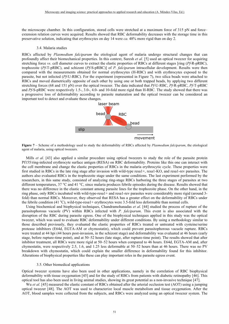
RBCs affected by Plasmodium falciparum the etiological agent of malaria undergo structural changes that can profoundly affect their biomechanical properties. In this context, Suresh et al. [5] used an optical tweezer for acquiring stretching force vs. cell diameter curves to extract the elastic properties of RBCs at different stages [ring (Pf-R-pRBC), trophozoite (Pf-T-pRBC), and schizont (Pf-S-pRBC)] of P. falciparum intracellular development. Results were then compared with the measurements obtained for normal erythrocytes (H-RBC) and with erythrocytes exposed to the parasite, but not infected (Pf-U-RBC). For the experiment (represented in Figure 7), two silica beads were attached to RBCs and moved diametrically opposite of each other by using one or both trapped beads, by applying two different stretching forces (68 and 151 pN) over the optical tweezer. The data indicated that Pf-U-RBC, Pf-R-pRBC, Pf-T-pRBC and Pf-S-pRBC were respectively 1.5-, 3.0-, 4.0- and 10-fold more rigid than H-RBC. The study showed that there was a progressive loss of deformability according to parasite maturation and the optical tweezer can be considered an important tool to detect and evaluate these changes.
Figure 7 – Scheme of a methodology used to study the deformability of RBCs affected by Plasmodium falciparum, the etiological agent of malaria, using optical tweezers.
Mills et al. [43] also applied a similar procedure using optical tweezers to study the role of the parasite protein Pf155/ring-infected erythrocyte surface antigen (RESA) on RBC deformability. Proteins like this one can interact with the cell membrane and change the elastic properties of RBCs in the malaria erythrocytic cycle. These properties were first studied in RBCs in the late ring stage after invasion with wild-type resa1+, resa1-KO, and resa1-rev parasites. The authors also evaluated RBCs in the trophozoite stage under the same conditions. The last experiment performed by the researchers, in this same study, consisted of analyzing ring-stage RBCs harboring the three stages of parasites at two different temperatures, 37 °C and 41 °C, since malaria produces febrile episodes during the disease. Results showed that there was no difference in the elastic constant among parasite lines for the trophozoite phase. On the other hand, in the ring phase, only RBCs incubated with wild-type-resa1+ and resa1-rev parasites were considerably more rigid (around 3-fold) than normal RBCs. Moreover, they observed that RESA has a greater effect on the deformability of RBCs under the febrile condition (41 oC), wild-type-resa1+ erythrocytes were 3.5-fold less deformable than normal cells. Using biochemical and biophysical techniques, Chandramohanadas et al. [44] studied the process of rupture of the parasitophorous vacuole (PV) within RBCs infected with P. falciparum. This event is also associated with the disruption of the RBC during parasite egress. One of the biophysical techniques applied in this study was the optical tweezer, which was used to evaluate RBC deformability under different conditions. By using a methodology similar to those described previously, they evaluated the elastic properties of RBCs treated or untreated with cysteine/serine protease inhibitors (E64d, EGTA-AM or chymostatin), which could prevent parasitophorous vacuole rupture. RBCs were treated at 44 hpi (44 hours post-invasion, in the schizont stage) and deformability was evaluated at 46 hours (early stage, before rupture-time point), and at 50–52 hours (late stage, after rupture-time point). The results showed that after inhibitor treatment, all RBCs were more rigid at 50–52 hours when compared to 46 hours. E64d, EGTA-AM and, after chymostatin, were respectively 2.5, 1.6, and 1.25 less deformable at 50–52 hours than at 46 hours. There was no PV breakdown with chymostatin, which could explain the smaller difference in deformability found for this inhibitor. Alterations of biophysical properties like these can play important roles in the parasite egress event.
3.5. Other biomedical applications
Optical tweezer systems have also been used in other applications, namely in the correlation of RBC biophysical deformability with tissue oxygenation [45] and for the study of RBCs from patients with diabetic retinopathy [46]. This optical tool has also been used in living animal studies, showing its great potential as a non-invasive technique [47]. Wu et al. [45] measured the elastic constant of RBCs obtained after the arterial occlusion test (AOT) using a jumping optical tweezer [48]. The AOT was used to characterize local muscle metabolism and tissue oxygenation. After the AOT, blood samples were collected from the subjects, and RBCs were analyzed using an optical tweezer system. The
Microscopy and imaging science: practical approaches to applied research and education (A. Méndez-Vilas, Ed.)
51
___________________________________________________________________________________________

results showed that there is a relationship between RBC elastic properties and their ability to supply oxygen to tissues. RBCs with greater deformability can deliver more oxygen to muscle tissue due to their greater ability to pass through clogged vessels. This phenomenon was observed by the slower increase/decrease of oxygenation tracings during AOT. The change of hemoglobin concentration dominates the correlation between the erythrocyte elastic constant and the dynamic tissue oxygenation induced by AOT. The acquired data highlighted the importance of correlating the contribution of the RBC elastic properties with the supply of oxygen to tissues. Agrawal et al. [46] used a dual-beam optical tweezer system comprises by the division and recombination of a single NIR laser to stretching RBCs from patients with diabetic retinopathy (DR), as well as type 2 diabetes mellitus (DM), and investigating erythrocyte deformability by comparison with normal, healthy controls. Diabetic retinopathy is an eye disease that results from a complication of diabetes and is caused by damage to blood vessels, which can also lead to blindness. They demonstrated that the deformability index [DI = (Imax – I0)/I0, where Imax is the final stretched length and I0 the initial length] of DR RBCs (0.0635 ± 0.028) was smaller than in DM (0.0645 ± 0.038) and in control (0.0698 ± 0.0224) RBCs, indicating that the DR erythrocytes are less deformable and more swollen when compared to DM RBCs. Furthermore, they associated a higher initial cell size (I0) with reduced deformability, observing that the I0 of RBCs from patients with diabetes was higher (DM 8.68 µm ± 0.49; DR 8.82 µm ± 0.32) than the control (8.45 µm ± 0.25). This study pointed out that the erythrocyte size in diabetic patients could be caused by metabolic disturbances, which could alter RBC deformability. Zhong et al. [47] presented the first in vivo cell manipulation using an optical tweezer system (Figure 8), showing that this tool could be used as non-invasive method. They used an NIR optical trap to manipulate RBCs present in blood capillaries of the surface of mouse ears. With this tool, they were able to induce the formation of artificial RBC clots in capillary vessels, and then recover blood flow by removing the blockage with the same tool. Moreover, they determined the optical trap stiffness in vivo of ~10 pN.µm-1, using the viscous drag force method. This work demonstrates the great potential of optical tweezers to be extended to in vivo studies, such as the ones related to hemodynamic parameters.
Figure 8 – In vivo optical tweezers: trapping a RBC within subdermal capillaries in a living mouse. Image adapted with permission from Macmillan Publishers Ltd: [NATURE COMMUNICATIONS] [47], copyright (2013).
4. Conclusions
About thirty years have now passed since Ashkin’s first experimental capture and manipulation of biological systems using NIR optical tweezers. Over the years, as described in this review, the optical tweezer technique has proved to be a valuable microscopy tool, capable not only of manipulating, but also of evaluating important biophysical properties associated with the aggregation and the deformability of RBCs, including in in vivo studies. Since aggregation and deformability are important parameters for RBCs in performing their biological functions in the blood stream, optical tweezers have opened up new possibilities in the biomedical field, allowing for quantitative understanding of the roles of intrinsic and extrinsic conditions, such as diseases and storage in blood banks, on RBC biomechanics. Certainly, the optical tweezer microscopy technique will set the pace of many biomedical studies for years to come.
Acknowledgments: The authors are grateful to the Coordenação de Aperfeiçoamento de Pessoal de Nível Superior (CAPES), the Conselho Nacional de Desenvolvimento Científico e Tecnológico (CNPq), and the Fundação de Amparo à Ciência e Tecnologia do Estado de Pernambuco (FACEPE). This work is also linked to the Instituto Nacional de Fotônica (INFo).
Microscopy and imaging science: practical approaches to applied research and education (A. Méndez-Vilas, Ed.)
52
___________________________________________________________________________________________

References [1] Ashkin A, Dziedzic J. Optical trapping and manipulation of viruses and bacteria. Science. 1987; 235:1517-20. [2] Molloy JE, Padgett MJ. Lights, action: Optical tweezers. Contemporary Physics. 2002; 43:241-58. [3] Stevenson DJ, Gunn-Moore F, Dholakia K. Light forces the pace: optical manipulation for biophotonics. J Biomed Opt. 2010;
15:041503-21. [4] Fontes A, Castro MLB, Brandão MM, Fernandes HP, Thomaz AA, Huruta RR, et al. Mechanical and electrical properties of
red blood cells using optical tweezers. J Opt. 2011; 13:044012. [5] Suresh S, Spatz J, Mills JP, Micoulet A, Dao M, Lim CT, et al. Connections between single-cell biomechanics and human
disease states: gastrointestinal cancer and malaria. Acta Biomater. 2005; 1:15-30. [6] Moffitt JR, Chemla YR, Smith SB, Bustamante C. Recent Advances in Optical Tweezers. Annu Rev Biochem. 2008; 77:205-
28. [7] Bryant Z, Stone MD, Gore J, Smith SB, Cozzarelli NR, Bustamante C. Structural transitions and elasticity from torque
measurements on DNA. Nature. 2003; 424:338-41. [8] Galla L, Meyer AJ, Spiering A, Sischka A, Mayer M, Hall AR, et al. Hydrodynamic Slip on DNA Observed by Optical
Tweezers-Controlled Translocation Experiments with Solid-State and Lipid-Coated Nanopores. Nano Lett. 2014; 14:4176-82. [9] Cecconi C, Shank EA, Bustamante C, Marqusee S. Direct Observation of the Three-State Folding of a Single Protein Molecule.
Science. 2005; 309:2057-60. [10] Rebane Aleksander A, Ma L, Zhang Y. Structure-Based Derivation of Protein Folding Intermediates and Energies from Optical
Tweezers. Biophys J. 2016; 110:441-54. [11] Huisstede JHG, Subramaniam V, Bennink ML. Combining optical tweezers and scanning probe microscopy to study DNA–
protein interactions. Microsc Res Tech. 2007; 70:26-33. [12] Heller I, Hoekstra TP, King GA, Peterman EJG, Wuite GJL. Optical Tweezers Analysis of DNA–Protein Complexes. Chem
Rev. 2014; 114:3087-119. [13] Schwingel M, Bastmeyer M. Force Mapping during the Formation and Maturation of Cell Adhesion Sites with Multiple Optical
Tweezers. Plos One. 2013; 8:e54850. [14] Stellamanns E, Uppaluri S, Hochstetter A, Heddergott N, Engstler M, Pfohl T. Optical trapping reveals propulsion forces,
power generation and motility efficiency of the unicellular parasites Trypanosoma brucei brucei. Sci Rep. 2014; 4:6515. [15] Ferraro-Gideon J, Sheykhani R, Zhu Q, Duquette ML, Berns MW, Forer A. Measurements of forces produced by the mitotic
spindle using optical tweezers. Mol Biol Cell. 2013; 24:1375-86. [16] Dao M, Lim CT, Suresh S. Mechanics of the human red blood cell deformed by optical tweezers. Journal of the Mechanics and
Physics of Solids. 2003; 51:2259-80. [17] Mohandas N, Chasis JA. Red blood cell deformability, membrane material properties and shape: regulation by transmembrane,
skeletal and cytosolic proteins and lipids. Semin Hematol. 1993; 30:171-92. [18] Baskurt OK, Meiselman HJ, editors. Blood rheology and hemodynamics. Seminars in thrombosis and hemostasis; 2003:
Thieme Medical Publishers. [19] Silva DCN, Jovino CN, Silva CAL, Fernandes HP, Filho MM, Lucena SC, et al. Optical Tweezers as a New Biomedical Tool
to Measure Zeta Potential of Stored Red Blood Cells. Plos One. 2012; 7:e31778. [20] Wolfe LC. The membrane and the lesions of storage in preserved red cells. Transfusion. 1985; 25:185-203. [21] Kriebardis Anastasios G, Antonelou Marianna H, Stamoulis Konstantinos E, Economou-Petersen E, Margaritis Lukas H,
Papassideri Issidora S. Storage-dependent remodeling of the red blood cell membrane is associated with increased immunoglobulin G binding, lipid raft rearrangement, and caspase activation. Transfusion. 2007; 47:1212-20.
[22] Hovav T, Yedgar S, Manny N, Barshtein G. Alteration of red cell aggregability and shape during blood storage. Transfusion. 1999; 39:277-81.
[23] Relevy H, Koshkaryev A, Manny N, Yedgar S, Barshtein G. Blood banking–induced alteration of red blood cell flow properties. Transfusion. 2008; 48:136-46.
[24] Henkelman S, Dijkstra-Tiekstra MJ, De Wildt-Eggen J, Graaff R, Rakhorst G, Van Oeveren W. Is red blood cell rheology preserved during routine blood bank storage? Transfusion. 2010; 50:941-8.
[25] Cluitmans JCA, Chokkalingam V, Janssen AM, Brock R, Huck WTS, Bosman GJCGM. Alterations in Red Blood Cell Deformability during Storage: A Microfluidic Approach. Biomed Res Int. 2014; 2014:9.
[26] Eylar EH, Madoff MA, Brody OV, Oncley JL. The Contribution of Sialic Acid to the Surface Charge of the Erythrocyte. J Biol Chem. 1962; 237:1992-2000.
[27] Fernandes HP, Cesar CL, Barjas-Castro MdL. Electrical properties of the red blood cell membrane and immunohematological investigation. Revista Brasileira de Hematologia e Hemoterapia. 2011; 33:297-301.
[28] Ashkin A. Acceleration and Trapping of Particles by Radiation Pressure. Phys Rev Lett. 1970; 24:156-9. [29] Ashkin A, Dziedzic JM, Bjorkholm JE, Chu S. Observation of a single-beam gradient force optical trap for dielectric particles.
Opt Lett. 1986; 11:288-90. [30] Jones P, Maragó O, Volpe G. Optical tweezers: Principles and applications. 1st Edition ed. Cambridge: Cambridge University
Press; 2015. [31] Lee K, Kinnunen M, Khokhlova MD, Lyubin EV, Priezzhev AV, Meglinski I, et al. Optical tweezers study of red blood cell
aggregation and disaggregation in plasma and protein solutions. J Biomed Opt. 2016; 21:035001. [32] Khokhlova MD, Lyubin EV, Zhdanov AG, Rykova SY, Sokolova IA, Fedyanin AA. Normal and system lupus erythematosus
red blood cell interactions studied by double trap optical tweezers: direct measurements of aggregation forces. J Biomed Opt. 2012; 17:0250011-6.
[33] Fernandes HP, Fontes A, Thomaz A, Castro V, Cesar CL, Barjas-Castro ML. Measuring red blood cell aggregation forces using double optical tweezers. Scand J Clin Lab Invest. 2013; 73:262-4.
Microscopy and imaging science: practical approaches to applied research and education (A. Méndez-Vilas, Ed.)
53
___________________________________________________________________________________________

[34] Fontes A, Fernandes HP, de Thomaz AA, Barbosa LC, Barjas-Castro ML, Cesar CL. Measuring electrical and mechanical properties of red blood cells with double optical tweezers. J Biomed Opt. 2008; 13:014001--6.
[35] Yang B-W, Li Z. Measuring micro-interactions between coagulating red blood cells using optical tweezers. Biomed Opt Express. 2010; 1:1217-24.
[36] Brandão MM, Fontes A, Barjas-Castro ML, Barbosa LC, Costa FF, Cesar CL, et al. Optical tweezers for measuring red blood cell elasticity: application to the study of drug response in sickle cell disease. Eur J Haematol. 2003; 70:207-11.
[37] Brandão MM, Saad STO, Cezar CL, Fontes A, Costa FF, Barjas-Castro ML. Elastic properties of stored red blood cells from sickle trait donor units. Vox Sanguinis. 2003; 85:213-5.
[38] De Luca AC, Rusciano G, Ciancia R, Martinelli V, Pesce G, Rotoli B, et al. Spectroscopical and mechanical characterization of normal and thalassemic red blood cells by Raman Tweezers. Opt Express. 2008; 16:7943-57.
[39] Brandao MM, Castro MdLR, Fontes A, Cesar CL, Costa FF, Saad ST. Impaired red cell deformability in iron deficient subjects. Clin Hemorheol Microcirc. 2009; 43:217-21.
[40] Pellizzaro A, Welker G, Scott D, Solomon R, Cooper J, Farone A, et al. Direct laser trapping for measuring the behavior of transfused erythrocytes in a sickle cell anemia patient. Biomed Opt Express. 2012; 3:2190-9.
[41] Barjas-Castro ML, Brandão MM, Fontes A, Costa FF, Cesar CL, Saad STO. Elastic properties of irradiated RBCs measured by optical tweezers. Transfusion. 2002; 42:1196-9.
[42] Li Y, Wen C, Xie H, Ye A, Yin Y. Mechanical property analysis of stored red blood cell using optical tweezers. Colloids Surf B Biointerfaces. 2009; 70:169-73.
[43] Mills JP, Diez-Silva M, Quinn DJ, Dao M, Lang MJ, Tan KSW, et al. Effect of plasmodial RESA protein on deformability of human red blood cells harboring Plasmodium falciparum. Proceedings of the National Academy of Sciences. 2007; 104:9213-7.
[44] Chandramohanadas R, Park Y, Lui L, Li A, Quinn D, Liew K, et al. Biophysics of Malarial Parasite Exit from Infected Erythrocytes. Plos One. 2011; 6:e20869.
[45] Wu Y-T, Chiou A, Sun C-W. Correlation between tissue oxygenation and erythrocytes elasticity. J Biophotonics. 2011; 4:224-8.
[46] Agrawal R, Smart T, Nobre-Cardoso J, Richards C, Bhatnagar R, Tufail A, et al. Assessment of red blood cell deformability in type 2 diabetes mellitus and diabetic retinopathy by dual optical tweezers stretching technique. Sci Rep. 2016; 6:15873.
[47] Zhong M-C, Wei X-B, Zhou J-H, Wang Z-Q, Li Y-M. Trapping red blood cells in living animals using optical tweezers. Nature Comm. 2013; 4:1768.
[48] Liao G-B, Bareil PB, Sheng Y, Chiou A. One-dimensional jumping optical tweezers for optical stretching of bi-concave human red blood cells. Opt Express. 2008; 16:1996-2004.
Microscopy and imaging science: practical approaches to applied research and education (A. Méndez-Vilas, Ed.)
54
___________________________________________________________________________________________


![Noninvasive Optical Measurement of Cerebral Blood Flow in ...€¦ · for the label-free measurement of blood velocity in small vessels in mice [7]; ... rapidly applied to humans](https://static.fdocuments.us/doc/165x107/5f34e2d7da53d34ad87c449d/noninvasive-optical-measurement-of-cerebral-blood-flow-in-for-the-label-free.jpg)