Bioinformatics-aided enzymatic process development
126
BIOINFORMATICS- AIDED ENZYMATIC PROCESS DEVELOPMENT PhD thesis Author: GERGELY BÁNÓCZI Supervisor: DR GÁBOR HORNYÁNSZKY Consultant: DR LÁSZLÓ POPPE Department of Organic Chemistry and Technology BME, 2017
Transcript of Bioinformatics-aided enzymatic process development
BIOINFORMATICS-AIDED ENZYMATIC
PROCESS DEVELOPMENT PhD thesis
Author: GERGELY BÁNÓCZI
Supervisor: DR GÁBOR HORNYÁNSZKY
Consultant: DR LÁSZLÓ POPPE
Department of Organic Chemistry and Technology BME, 2017

Cover art by Szabina Hunyadi.

ACKNOWLEDGEMENTS
First and foremost, I am extremely grateful – beyond words – for my
family, all the support and love that helped me get through all the hard times.
I would like to express my gratitude to my supervisor, Gábor
Hornyánszky, and to my consultant, László Poppe, for the chance to do my
research at the Bioorganic Research Group, and for all the support and
expertise that helped my work.
I thank for the hard work of all the undergraduate students who I have
mentored, namely Zsófia Bata, Zsuzsanna Fehérvári, Csilla Hargitai, Csongor
Szabó, Levente Mihalovits.
As I was part of a highly collaborative research team, it would have
been impossible to succeed without the help of group mates and technicians,
Diána Weiser, Evelin Bell, Eszter Kókai, Emese Abaházi, Flóra Nagy, Emese
Farkas, Zsófia Bata, Bianka Szokol, Balázs Komjáti, Zoltán Boros, Péter Falus,
Márk Oláh, László Nagy-Győr, Pál Csuka, Rita Molnárné Bernáth, Ádám
Czermann, and people from the Biocatalysis and Biotransformation Research
Centre at Kolozsvár, Csaba Paizs, László Csaba Bencze, Andrea Varga, Botond
Nagy, Alina Filip.
I would like to thank for all the help and advices for Ödön Farkas, Antal
Lopata, Ákos Gellért in computational chemistry, and for Károly Héberger in
statistics.
Financial support is gratefully acknowledged for Gedeon Richter
Pharmaceutical, József Varga Foundation, and Synbiocat LTD.
My apologies, if I missed somebody. The acknowledgements may seem
just a sheer list of people without any feelings, but believe me, it is not. And
thank you all for all the teachings that made me to surpass myself every time
in my life.

Convictions are more dangerous foes of truth than lies
Friedrich Nietzsche

CONTENTS Acknowledgements ............................................................................................................................ 2
List of abbreviations ........................................................................................................................... 6
Chapter 1 – Introduction .................................................................................................................... 7
1.1. Introduction to enzyme engineering ............................................................... 7
1.2. Aims of this research .................................................................................... 12
1.3. Philosophy of this research ........................................................................... 15
Chapter 2 – Theoretical background ................................................................................................. 17
2.1. Tasks in rational enzyme engineering ........................................................... 17
2.1.1. Enzyme and ligand structures ...................................................................... 17
2.1.2. Temperature tolerance of proteins ............................................................... 19
2.1.3. Components of the immediate enzyme environment .................................... 21
2.2. Enzyme engineering methods ....................................................................... 25
2.2.1. Mutagenesis of the amino acid sequence ...................................................... 25
2.2.2. Enzyme immobilization ............................................................................... 26
2.3. Studied enzymes .......................................................................................... 27
2.3.1. General overview of Aromatic amino acid Ammonia-lyases........................... 27
2.3.2. Phenylalanine ammonia-lyases (PALs) ......................................................... 29
2.3.3. Phenylalanine 2,3-aminomutases (PAMs) ..................................................... 33
2.3.4. Lipases ........................................................................................................ 36
Chapter 3 – Results and discussion ................................................................................................... 41
3.1. Modeling method development .................................................................... 41
3.1.1. Modeling ions around enzymes and non-specific ion binding ........................ 41
3.1.2. Modeling enzyme-substrate complexes ......................................................... 42
3.2. Examining PAL structural properties ............................................................. 44
3.2.1. Partial homology modeling .......................................................................... 44
3.2.2. Full homology modeling .............................................................................. 46
3.2.3. Examining PAL structural properties against temperature optima ................. 49
3.2.4. Examining the pH tolerance of Rubrobcater xylanophilus PAL ...................... 50
3.3. Investigating the reaction mechanism of MIO enzymes ................................. 52
3.4. Expanding the substrate scope of PcPAL towards styrylalanines .................... 56
3.4.1. Initial studies and mutant set construction ................................................... 56
3.4.2. Synthetic applications in kinetic resolution ................................................... 59
3.4.3. Rationalising the biotransformations of styrylalanines .................................. 61
3.5. Investigating phenylalanine aminomutase from Pantoea agglomerans ........... 67
3.5.1. Transformation of rac-α-arylalanines with PaPAM ........................................ 67

3.5.2. Transformation of rac-β-arylalanines with PaPAM ........................................ 69
3.5.3. Computational modeling of PaPAM-catalysed isomerizations ....................... 70
3.6. Kinetic resolution of Phenothiazine moiety containing secondary alcohols .... 74
3.6.1. Kinetic resolution of secondary alcohols ....................................................... 74
3.6.2. Kinetic resolution of secondary alcohol esters .............................................. 75
3.6.3. Assignement of Absolute configurations ....................................................... 76
3.7. Bioimprinting of lipases in Poly(Vinyl Alcohol) nanofiber ............................. 77
3.7.1. Testing potential bioimprinting agents ......................................................... 78
3.7.2. Effect of the additives on the polymer chain interactions .............................. 80
3.7.3. Molecular modeling of the potential bioimprinting effects ............................ 81
3.8. Computer-aided Selection of sol-gel precursor .............................................. 84
3.8.1. Virtual screening of organislane precursors for efficient entrapment ............. 84
Chapter 4 – Methods ....................................................................................................................... 87
4.1. Overview of the general modeling procedure ............................................... 87
4.1.1. Enzyme structure modeling ......................................................................... 89
4.1.2. Enzyme-ligand complex modeling ............................................................... 91
4.2. Homology modeling of MIO containing enzymes .......................................... 92
4.2.1. Homology modeling of Rubrobacter xylanophylus PAL................................. 92
4.2.2. Partial homology modeling of existing PDB structures .................................. 92
4.2.3. Large scale homology modeling of PAL enzymes .......................................... 93
4.3. QM/MM calculations on the covalent complexes of L-phenylalanine and L-
propargylglycine with PcPAL ...................................................................................................... 94
4.4. Modeling enzyme-substrate interactions of styrylalanine compounds ............ 96
4.5. Molecular modeling of the covalent enzymesubstrate N-MIO complexes in
PaPAM 99
4.6. Molecular modeling of bioimprinting agents with CaLB .............................. 100
4.7. Virtual screening for organosilane precursors of sol-gel matrices for the
immobilization of CaLB ............................................................................................................ 100
4.8. Assignment of the Absolute configuration of phenothyazine compounds in the
kinetic resolution with CaLB ..................................................................................................... 101
Chapter 5 – Conclusions ................................................................................................................ 103
Tézis pontok.................................................................................................................................. 107
Thesis points ................................................................................................................................. 109
NYILATKOZAT .............................................................................................................................. 111
References .................................................................................................................................... 112

LIST OF ABBREVIATIONS
3D three dimensional AvPAL phenylalanine ammonia-lyase from Anabaena variabilis CaLA lipase A from Candida antarctica CaLB lipase B from Candida antarctica DMDEOS dimethlydiethoxysilane ee enantiomeric excess FC Friedel-Crafts KR kinetic resolution MIO 3,5-dihydro-5-methylidene-4H-imidazol-4-one prosthetic
group MM molecular mechanics MM-GBSA molecular-mechanics-based generalized Born and surface
area continuum solvation method NID non-ionic detergent N-MIO C-N covalent bond forming MIO prosthetic group NMR nuclear magnetic resonance NpPAL phenylalanine ammonia-lyase from Nostoc punctiforme OTEOS octyltriethoxysilane PcPAL phenylalanine ammonia-lyase from Petroselinum crispum PAL phenylalanine ammonia-lyase PAM phenylalanine aminomutase PaPAM phenylalanine aminomutase from Pantoea agglomerans Phe phenylalanine PEG poliethylene glycol PG propargylglicine PpHAL histidine ammonia-lyase from Pseudomonas putida PTEOS phenyltriethoxysilane PVA poly(vinyl alcohol) QM/MM combined quantum mechanics/molecular mechanics method RgPAL phenylalanine ammonia-lyase from Rhodotorula glutinis RsTAL tyrosine ammonia-lyase from Rhodobacter sphaeroides RtPAL phenylalanine ammonia-lyase from Rhodosporidium
toruloides RxPAL phenylalanine ammonia-lyase from Rubrobacter
xylanophylus SgTAM tyrosine aminomutase from Streptomyces globisporus SmPAM phenylalanine aminomutase from Streptomyces maritimus TcanPAM phenylalanine aminomutase from Taxus canadensis TchinPAM phenylalanine aminomutase from Taxus chinensis TAL tyrosine ammonia-lyase TAM tyrosine aminomutase TEOS tetraethoxysilane wt wild-type enzyme

INTRODUCTION TO ENZYME ENGINEERING 7
CHAPTER 1 – INTRODUCTION
1.1. INTRODUCTION TO ENZYME ENGINEERING
Proteins are the major component of the living cells which are
composed of natural amino acids. They play crucial roles in the maintenance
of life, and their dysfunctions are known to cause different pathologies. One
of the best understood functions of proteins is catalysis (enzymatic activity),
which attracted much attention in the early days of protein science. By varying
the structure and incorporating co-factors, such as metal ions, and organic
molecules, such as flavins and porphyrins, a surprising diversity of activities is
displayed among the known several thousand enzymes. Also, enzymes show
selectivity and specificity towards a specified stereoisomer of the biologically
active small molecules classically applied in human healthcare as drugs that
led to the elaboration of the lock and key model by Fisher. [1]
However, there was an extremely rapid development in the
understanding of enzyme catalyzed processes at molecular level since then.
This enormous expansion was due to the developments in molecular genetics
(i.e. PCR, sequencing, novel expression systems, genome projects), structural
biology (i.e. protein crystallography, liquid-phase NMR protein structure
determinations) and bioinformatics (i.e. sequence alignments, protein
modeling, ligand-docking, quantum chemical calculations). Starting from the
20th century, over 130,000 3D structures have been deposited in the Protein
Data Bank (PDB) offering a wide range of data to explore the relationships
between structure and functionality. In the early days, the explanatory power
of these 3D structures reinforced the static view of protein structure until 1958
when Koshland suggested the induced-fit model based on the observations
that some enzymes could act on differently shaped substrates, and hence a
degree of flexibility is inevitable in function. [2] In fact, many proteins of
various and vital functions are, at least partially, intrinsically disordered. [3,
4]
Irrespective of the deep understanding of action, mankind has
harnessed the catalytic effect of enzymes for its own good since at least
antiquity (i.e. food production). But large-scale use started only in the the last
century, with one of the most eloquent example being the production of
penicillin. Nowadays, biocatalytic steps are already used industry-wide to
manufacture a wide range of products, i.e. drugs, food, organics, fine
chemicals or plastics (Table 1, [5, 6]) and the formed discipline is called
industrial biotechnology, also known as white biotechnology.

8 INTRODUCTION TO ENZYME ENGINEERING
Table 1 Enzymes used in various industrial segments and their application [5, 6]
Industry Enzyme class Application
Detergent protease protein stain removal amylase starch stain removal lipase lipid stain removal cellulase cleaning, color clarification,
antiredeposition (cotton) mannanase mannan stain removal (dressings,
sauces, ice cream, and personal care products)
pectate lyase fruit, marmalade, and tomato ketchup stains
Starch and fuel amylase starch liquefaction and saccharification
amyloglucosidase saccharification pullulanase saccharification glucose isomerase glucose to fructose conversion cyclodextrin-
glycosyltransferase cyclodextrin production
xylanase viscosity reduction (fuel and starch)
protease protease (yeast nutrition—fuel) Food protease milk clotting, infant formulas
(low allergenic), flavor lipase cheese flavor lactase lactose removal (milk) pectin methyl esterase firming fruit-based products pectinase fruit-based products transglutaminase modify visco-elastic properties Baking amylase bread softness and volume, flour
adjustment xylanase dough conditioning lipase dough stability and conditioning
(in situ emulsifier) phospholipase dough stability and conditioning
(in situ emulsifier) glucose oxidase dough strengthening lipoxygenase dough strengthening, bread
whitening protease biscuits, cookies transglutaminase laminated dough strengths Animal feed phytase phytate digestibility—
phosphorus release xylanase digestibility β-glucanase digestibility protease digestibility Beverage pectinase depectinization, mashing amylase juice treatment, low calorie beer β-glucanase mashing acetolactate decarboxylase maturation (beer) laccase clarification (juice), flavor (beer),
cork stopper treatment

INTRODUCTION TO ENZYME ENGINEERING 9
Textile cellulase denim finishing, cotton softening amylase desizing pectate lyase scouring catalase bleach termination laccase bleaching peroxidase excess dye removal Pulp and paper lipase pitch control, contaminant
control protease bio-film removal amylase starch coating, deinking,
drainage improvement xylanase bleach boosting cellulase deinking, drainage improvement,
fiber modification Fats and oils lipase transesterification phospholipase degumming, lyso-lecithin
production lipase production of bio-diesel (methyl
esters) Organic synthesis
lipase resolution of chiral alcohols and amides
acylase synthesis of semisynthetic penicillin
nitrilase synthesis of enantiopure carboxylic acids
Leather protease unhairing, bating lipase degreasing Personal care amyloglucosidase antimicrobial (combination with
glucose oxidase) glucose oxidase bleaching, antimicrobial peroxidase antimicrobial protease cleaning of contact lenses and
dentures catalase removing hydrogen peroxide
after contact lens disinfection
As nowadays industrial development needs more efficient, yet green
processes, new methods must meet more criteria than ever before. In
particular, one of the main challenges facing organic chemistry is the rational
synthesis of an ever growing number of complex, optically active natural
products and their analogues. [7] According to the policy of the FDA,
pharmacological investigations and production of chiral drugs is strongly
advised in enantiomerically pure form. [8, 9] The main idea of this regulation
is that significant portion of drugs and drug-like molecules is made up of chiral
compounds, and often, the corresponding therapeutic effect can be assigned
to only one stereoisomer, so it is extremely important to remove the ineffective
(or worse, toxic) other isomer(s). The most infamous example of this issue is

10 INTRODUCTION TO ENZYME ENGINEERING
the Contergan® scandal in West Germany, when the active pharmaceutical
ingredient thalidomid of the marketed drug Contergan® was formulated as a
racemate, and unfortunately, besides the sedative (R )-thalidomid, the (S )-
thalidomid antipode was teratogenic.
The greatest advantage of enzymes is the inherent capability to be
used in asymmetric synthesis, either in isolated form or within (mainly)
microorganisms. [10, 11, 12] These syntheses are regularly rewarded by
selectivity, sustainability, environment friendliness, economic viability, and
can avoid the formation of hazardous materials during the whole process.
These features are the outcomes of the facts that enzymes operate mainly
under mild reaction conditions and require/produce harmless materials
during their whole lifecycle.
It is common to refer to enzymes as „perfect catalysts” which may hold
true for i.e. triose phosphate isomerases, superoxide dismutases,
acetylcholinesterases, or carbonic anhydrases, in which cases the biocatalysed
reactions are essentially diffusion limited. [13, 14] Despite these outstanding
examples, an extensive analysis of nearly 2,000 enzymes showed that the
median turnover number (kcat) of these biocatalysts is about 10 s−1, much
smaller than what one would expect. [15, 16] This finding shockingly reminds
us that in nature, enzymes have rather evolved to operate under the specific
conditions provided by the host organism. Their degree of adeptness includes
diverse criteria such as the substrates they endogenously transform, their
effective reaction rate, the physico-chemical parameters of the environment in
which they function, how well they tolerate deviations from optimum
conditions etc. It is clear that the needs of a living organism and the industry
will seldom match exactly, which leaves plenty of room for improvement.
Technologies such as recombinant expression, protein engineering, and
directed evolution have revolutionized the development of industrial enzymes
and made it possible to tailor-make enzymes to display new activities and
adapt to new process conditions enabling a further expansion of their use.
Protein engineering emerged as an indispensable tool for the design
of new proteins and enzymes with enhanced properties. [17] There has been
a growing literature on protein engineering which may rely on different
principles, ranging from stochastic approaches, based on the selection of a
desired feature by directed evolution, to rational approaches, based on a
design procedure. [18] Choosing between stochastic and rational approaches
partly relies on the amount of the starting information, such that random
approaches are particluarly useful to cases when only little information is
available only. On the other hand, if the resolution of the starting information
is high, i.e. three-dimensional structure is available, rational approaches
become more viable. [19]

INTRODUCTION TO ENZYME ENGINEERING 11
In rational enzyme design, the availability of 3D protein structures is
a necessity. [17] Kinetics studies of numerous substrate analogues, direct
mechanistic investigations, environment dependence experiments
(temperature, pH, ionic strength tolerance etc.) provide data about the
elementary processes of biocatalysis, and statistics provides the means to
extract information from the gathered data. The newly acquired knowledge
serves as the foundation for rational enzyme engineering as simply illustrated
in Figure 1. Two main strategies are available for this purpose. The first one
is to modify the enzyme amino acid sequence pre-translationally
(„intrinsically”). The second one is to modify the enzyme post-translationally
(„externally”), to manipulate the structure with chemical or physical
modifications, most notably with immobilization.
A few challenges in enzyme engineering are mentioned in the
following. [17] Enzymes are often encountered not with their natural
𝑓(mechanism, enzyme-substrate interactions, statistics, etc.) =
rational enzyme engineering
Figure 1 Simple illustration of rational enzyme engineering
Figure 2 Recent expenditures of countries on biotechnology research and
development

12 AIMS OF THIS RESEARCH
substrate molecules but with congeneric compounds which demand the
modification of the active site, required for the the appropriate affinity and
activity. Furthermore, in case of chiral compounds, special attention should
be paid to the correct enantiomer selectivity. Another issue might arise when
enzymes are sensitive to changes in temperature and in pH, followed by
inactivation. The mechanism of inactivation and knowledge of weakpoints
gives us an opportunity to overcome or improve the issue. In addition, the
understanding of biosynthetic pathways and mechanisms of key enzymes
serves as basic research also, as many fundamental questions regarding
protein folding, stability, activity, and ligand binding can be answered.
Computers can provide powerful service for understanding the details
of biological phenomena ruled by enzymes and their partners. [17] In this
respect, computer-assisted approaches have become a requisite modeling tool
for protein engineering studies since they provide novel insights to complex
systems which may be challenging to be understood by experimental studies
only. [20, 21] Therefore, advances in computer technologies have a direct
impact on the success of rational design approaches for prediction of the
regulatory regions in proteins mediating stability, function, or selectivity.
In summary, biotechnology has a highly diversified industry that is
still growing in terms of both size and complexity, as indicated by the recent
expenditures of countries on biotechnology research and development (Figure
2). [22] In addition, industrial biotechnology is not dependent only on
governmental funds, as for example, biotech stock prices increased way above
average in 2013-14 at the NASDAQ stock exchange. [23] Europe holds a
competitive position in novel biocatalysts from environmental sources and in
advanced engineering of equipment for high-throughput experimentation, but
the core technology for directed evolution is a US patented technology, and
the largest company applying it for the optimization of enzyme catalysts for
industrial applications (Codexis Inc., USA) currently dominates the global
market.
1.2. AIMS OF THIS RESEARCH
During the course of my PhD studies, I was a member of the
Bioorganic Research Group at the Budapest University of Technology and
Economics. Our profile consists of the preparation of immobilized enzymes,
identification and production of novel enzymes, development of continuous
flow processes, modeling of enzymatic processes, and development of
analytical methods.

AIMS OF THIS RESEARCH 13
One my main goals was to reduce
the workhours needed for research and
development using rational enzyme design
and design and evaluation of experiments. It
was also important to evalute and interpret
both the experimental and computational
results, and to work with other members of
the group in close collaboration. Also, I tried
to learn and make use of other disciplines as
much as possible throughout my work
because, as an integrated whole, they can be more efficient. Because of these,
my work was inherently „multi dimensional”, and it can be partitioned mainly
along three dimensions, based on i) the aspect of scientific work (i. e. basic
research or process development), ii) the enzymatic process of interest (i. e.
kinetic resolution of secondary alcohols or aromatic amino acids), and iii) the
disciplines used (i.e. synthetic organic chemistry or modeling protocol
development).
During my spent time at the research group, I have worked mainly with
four enzymes, phenylalanine ammonia-lyase from Petroselinum crispum, and
from Rubrobacter xylanophilus, phenylalanine aminomutase from Pantoea
agglomerans, and lipase B from Candida antarctica.
Since phenylalanine is frequently found in biologically active peptides
and proteins, development of synthetic methods yielding sterically more
demanding and optically pure, aromatic α- and β-amino acids has gained
increased attention for possible applications in medicine. An attractive
enzymatic route for the synthesis of non-natural, enantiopure phenylalanine
analogues is provided by aromatic ammonia-lyases and aminomutases which
show high structural and sequence similarities and have a common
electrophilic prosthetic group, the autocatalytically formed 3,5-dihydro-5-
methylidene-4H-imidazol-4-one (MIO, Figure 3).
In nature, phenylalanine ammonia-lyases (PAL) catalyze the non-
oxidative ammonia elimination from (2S )-phenylalanine, yielding cinnamic
acid, an α,β-unsaturated carboxylic acid (panel A, Figure 4 ). Due to its broad
substrate tolerance, phenylalanine ammonia-lyase from Petroselinum crispum
(PcPAL) was one of the most frequently used aromatic amino acid ammonia-
lyase applied as a biocatalyst.
Another PAL from the thermophilic and radio-tolerant bacterium
Rubrobacter xylanophilus (RxPAL) holds the promise to be a potent
biocatalyst in synthetic biotransformations too. Further applications of stable
and biocompatible PAL formulations could be their use as therapeutic
enzymes in treatment of phenylketonuria or leukemia.
Figure 3 Structure of 3,5-
dihydro-5-methylidene-4H-
imidazol-4-one (MIO)

14 AIMS OF THIS RESEARCH
Phenylalanine aminomutase (PAM) from Pantoea agglomerans
(PaPAM) catalyzes the isomerisation of (S )-α-phenylalanine to (S )-β-
phenylalanine (panel B, Figure 4 ) and has important biological function in
the synthesis of the antibiotic andrimid. This enzyme was discovered recently,
and it was tested with a wide range of aromatic and heteroaromatic (S )-α-
arylalanines, both in isolated form and in whole cells, with promising results.
Lipases are serine hydrolases defined as triacylglycerol hydrolases, and
can be used for the kinetic resolution of i.e. racemic alcohols (panel C, Figure
4 ). Lipases represent one of the most frequently used enzyme classes due to
their ability to catalyze a wide range of reactions, and cover a wide range of
applications ranging from the production of biofuels, fragrances, food
ingredients, to enantioselective synthesis of active pharmaceutical
ingredients. Lipase B from Candida antarctica (CaLB) is one of the most widely
used lipases lending itself as an ideal target for enzyme engineering.
Figure 4 Industrially relevant reactions catalyzed by enzymes covered in my
doctoral thesis. A: kinetic resolution of aromatic α-amino acids by (S )-selective,
non-oxidative ammonia elimination catalyzed by phenylalanine ammonia-lyases
(EC 4.3.1.x, i.e. PcPAL) B: (S )-selective 2,3-isomerization of arylalanines by
phenylalanine aminomutases (EC 5.4.3.11, e.g. PaPAM). C: kinetic resolution of
racemic alcohols by stereoselective acylation catalyzed by lipases (EC 3.1.1.3,
e.g. CaLB).

PHILOSOPHY OF THIS RESEARCH 15
1.3. PHILOSOPHY OF THIS RESEARCH
In the beginning, I would like to introduce also my ideas, the
philosophy, about my work done, partially by staying true to the original
meaning of PhD (philosophiae doctor, doctor of philosophy).
Model building is a very useful way to explore relationships in nature.
Models are built partly relying on established theories and partly on newly
acquired principles, hypotheses which were formed through inductive
reasoning from experience. This model can be used then to deduce outcomes
from different inputs and make predictions. However, models may introduce
numerous assumptions and approximations, based on the limited available
information and limitation of resources. And further, hypotheses can never be
shown to be logically true by simply generalizing from confirming instances
(through inductive reasoning). [24] This means that models should be treated
with caution and should be extensively validated. This problem is illustrated
in Figure 5 through an instance of the game battleship. The real arrangement
of ships can be seen on the right, while the actual state of the game can be
seen on the left. Based only on the actual picture of the game, it is very hard
to guess the position of the entire fleet, and it is also very misleading, as the
actual arrangement resembles mostly an incomplete roman cross instead of
the real, spiral like arrangement. Also, human cognition is a very important
link in the chain of events belonging to scientific recognition. A simplified
model of the serial information processing is presented in Figure 6 [25]
revealing the importance of human perception and thought process which can
Figure 5 Illustration of the limited information through the game battleship. The
real arrangement of ships can be seen on the right, while the actual state of the
game can be seen on the left. Based only on the actual picture of the game, it is
very hard to guess the position of the entire fleet, and also it is very misleading, as
the actual arrangement resemebles mostly an incomplete roman cross instead of
the real spiral like arrangement.

16 PHILOSOPHY OF THIS RESEARCH
be infected, i.e. by the unwanted conformational bias. In this perspective, I
always tried to validate my models, and test assumptions as much as possible.
Also, I tried to be as comprehensive as possible in modeling, because, in my
view, treating certain phenomena even with basic tools is better than
completely neglecting them. The sections, covering my work in Results and
Discussion, are excerpts from different parts and phases of the rational enzyme
engineering. They are inherently not in a uniform state, but they are part of
an ongoing research to be completed in the manner of the prevoiusly reviewed
aims and philosophy.
Figure 6 Simplified model of the human serial information processing. [25]
Problem or task
Attention PerceptionThought process
DecisionResponse or action

TASKS IN RATIONAL ENZYME ENGINEERING 17
CHAPTER 2 – THEORETICAL BACKGROUND
2.1. TASKS IN RATIONAL ENZYME ENGINEERING
To establish a clear and explicit view on the main questions that have
to be answered, a simple but clever model may assist us. As our goal is to
design and develop enzymatic processes, enzymatic reactions, the description
of a typical enzyme kinetics assay reveals the most important factors (Figure
7 ).
Based on this model, the main questions of enzyme engineering are i)
the structure of the specific enzyme (enzyme structure), ii) the structural
properties, stability of the specific enzyme (solvent, (counter)ions, pH,
temperature), iii) and how interactions develop between the enzyme and the
current substrate molecule, including the reaction mechanism (enzyme-
substrate interactions). After obtaining sufficient experience and data, we can
start to manipulate the enzyme properties. In the following section, a brief
overview is presented about these factors, with more emphasis on topics that
are covered by my doctoral work.
2.1.1. ENZYME AND LIGAND STRUCTURES
The publicly available database for protein structures is the Protein
Data Bank (PDB), and the number of deposited strucures has grown over
130,000. Nowadays, three major experimental techniques have emerged for
protein structure determination, NMR, cryo-electron microscopy, and most
importantly, X-ray crystallography.
Figure 7 Explicit statement of the main factors to be addressed by enzyme
engineering through the instance of a description of an enzyme kinetics assay.
Enzyme activ ity assays were carried out at 31 °C, at various substrate concentrations,
in 1 mL quartz cuvette using 0.1 M Tris and 0.12 M NaCl (pH 8.8) as buffer.
(Counter)ions
Enzyme structure Temperature
pHSolvent
Enyzme-substrate interactions

18 TASKS IN RATIONAL ENZYME ENGINEERING
From a structural standpoint, the ideal method out of the three is
thought to be biomolecular NMR, because proteins can be investigated at their
natural conditions (i. e. ambient temperature, solution phase). Moreover, in
principle, simultaneous protein conformations and their molar ratios could be
determined from experiments. However, the size of the candidate proteins is
limited, and proteins need 13C and/or 15N isotope enrichment to gather a
meaningful dataset.
Cryo-electron microscopy is a form of transmission electron microscopy,
which was used to characterize large biological entities such as cells,
subcellular organelles, viruses etc. Flaws of this technique was the need to
cool biological samples way below room temperature, and that electron
density maps lacked resolution for atomic scale details. From continuous
development of instrumentation, improved specimen preparation, massive
advances in data processing and increased computational power electron
microscopy has become a technique capable of providing structural
information nowadays, recently providing a 3.3-Å-resolution structure of the
P22 bacteriophage virus. [26]
X-ray crystallography is the oldest and most widespread technique
today which utilizes the diffraction of X-rays on protein crystals. Experiments
provide an electron density map which needs further refinement and model
fitting. Refining models in an iterative fashion progressively improves the
agreement with experimental data. A structure is judged by its resolution and
the crystallographic Rfree-factor, for which a value of 0.20 or smaller is desired.
Further, the primary structure and/or a crude 3D model of the protein greatly
aides the structure determination. Advantages are the well established
infrastructure and protocols of this discipline, the independence on protein
size, and the high achiavable resolution. On the other hand, downsides consist
of the prerequisite of the single crystal of the protein, the infrequent ability to
determine hydrogen positions, and the unnatural conditions presented by the
crystal lattice and extreme cooling.
A possible problem, caused by the nature of the crystal lattice, is
demonstrated through the example of lipase B from Candida antarctica
(CaLB), which was also a subject of my doctoral work, incorporating a lid loop
near the active site that was believed to open or close the entrance to the
active site. The enzyme structure, with an open active site conformation (open
structure) was the only one available for years, and the closed active site
structure (closed structure) was reported only recently. [27] Analysis of the
previously available open structures (PDB IDs i.e. 1TCA, 1TCB, and 1TCC)
revealed that the amino acid segment Pro143-Asp145 would clash with
residue Leu199 of a symmetry-related CaLB monomer, if the enzyme would
adopt the more extended closed conformation. Unfortunately, an unusually

TASKS IN RATIONAL ENZYME ENGINEERING 19
loosely packed (yet open-structured) CaLB structure (PDB ID: 1LBT [28]) was
available earlier, but analysis showed that water content was over 60% for the
crystals of the open structure, compared to the 30% water content of the
crystals of the closed structure, indicating the possible role of the solvent in
the lid opening.
Further sources of errors can be refinement protocols using force fields
that employ united atom representations, neglect electrostatics, or have bad
quality parameters. [29] In particular, protein−ligand cocrystal structures are
prone to low-fidelity geometries. This is because, unlike in the case of the
protein when the amino acid sequence is known, the molecular structures
behind the unidentified electron densities is not trivial to recognize, leaving a
big role for intuition. Further, large errors in the ligand pose have only a small
effect on the overall goodness of fit, thus errors in ligand binding poses can
very well remain silent during the process.
2.1.2. TEMPERATURE TOLERANCE OF PROTEINS
An important feature of enzymes is their temperature tolerance. At
elevated temperatures the reaction rates are higher generally following the
rule of thumb that a 10 °C increase in temperature doubles the activity.
Further, the probability of the biocatalyst digestion by microbial infection is
lower at higher temperatures. Generally, thermostable enzymes can tolerate
heat treatment, as well as higher denaturing agent and substrate
concentrations. [30] On the other hand, the biotechnological value of cold-
adapted enzymes (stemming from psychrophiles) stems from their high
catalytic activity at low to moderate temperatures providing energy savings to
processes. However, the use of cold adapted enzymes can lower the risk of
undesirable chemical reactions and can enable the transformation of
thermally labile substrates. [31]
The structural features of thermotolerance of enzymes have been
intensively researched in the previous decades yielding a vast amount of data.
Szilágyi et al. [32] concluded that different protein families adapt to higher
temperatures by different sets of structural devices. Regarding the structural
parameters, the only generally observed rule was an increase in the number
of salt bridges with increasing growth temperature of host organisms. Other
parameters showed just a trend, whereas the number of hydrogen bonds and
the polarity of buried surfaces exhibited no clear-cut tendency to change with
growth temperature. They suggested that proteins from extreme thermophiles
are stabilized in different ways compared to the ones stemming from
moderately thermophilic organisms. The preferences of these two groups are

20 TASKS IN RATIONAL ENZYME ENGINEERING
different with regards to the number of ion pairs, the number of cavities, the
polarity of exposed surface and the secondary structural composition.
Similarly, according to Scandurra et al. [30], stability of a protein from
a thermophile, compared to its mesophilic homologue, is a property acquired
through many small structural differences such as the modulation of the
canonical forces, electrostatic (hydrogen bonds and ion-pairs) and
hydrophobic interactions. Thermostable proteins – produced by thermo- and
hyperthermophilic microorganisms growing between 45 and 110 °C – show
structural restriction on flexibility, which allows them to be functionally
competent at elevated temperatures, but renders them unusually rigid and
less active at mesophilic temperatures (10-45 °C) due to increased
compactness. From a comparative analysis of several families of proteins
including at least one thermophilic structure in each family, it appeared that
thermal stabilization was accompanied by an increase in hydrogen bonds and
salt bridges. Thermostability appeared to correlate also with a better packing
within buried regions.
Kurnikova et al. [33] have determined flexible regions for four
homologous pairs from thermophilic and mesophilic organisms by two
computational methods: FoldUnfold which is based on amino acid sequences,
and MD/First [34] which use three-dimensional structures and molecular
dynamics. Both methods allowed determining flexible regions in protein
structures. Supporting the previous statement by Scandurra et al., [30] MD
simulations showed that thermophilic proteins were more rigid in comparison
to their mesophilic homologues. It has been also found that the local networks
of salt bridges and hydrogen bonds in thermophiles render their structure
more stable with respect to fluctuations of individual contacts. This ionic
network connects α-helices and rigidifies the structure. Proteins from
mesophiles can be characterized by standalone salt bridges and hydrogen
bonds or small ionic clusters. Such difference in the network of salt bridges
results in different flexibility of homologous proteins. Despite these
generalizations, no universal rules were found as structural factors leading to
enhanced thermostability within a certain family of enzymes.
In contrast to their thermophilic counterparts, the structures of
thermolabile, cold-adapted enzymes generally permit high flexibility. [35, 36]
According to Saunders et al. [37] and Siddiqui et al. [35], cold adapted
enzymes tend to possess various combinations of the following features:
decreased core hydrophobicity, increased surface hydrophobicity, lower
arginine/lysine ratio, more and longer loops, decreased secondary structure
content, more glycine residues, less proline residues in loops and more proline
residues in α-helices, a reduced number of disulfide bridges, fewer

TASKS IN RATIONAL ENZYME ENGINEERING 21
electrostatic interactions, reduced oligomerization, and an increase in
conformational entropy of the unfolded state.
In summary, there is little consensus about the structural factors of
thermostability, but experts mainly agree on the importance of disulfide
bridges and electrostatic interactions such as salt bridges, hydrogen bonds.
2.1.3. COMPONENTS OF THE IMMEDIATE ENZYME ENVIRONMENT
The immediate environment of an enzyme generally consists of an
aqueous solution of different charged or neutral solutes (apart from the
substrates). Because the electromagnetic interaction is the second strongest
among the four fundamental interactions, charged solutes, including the
enzyme, must be properly incorporated into modeling.
The most important ions of aqueous solutions are the hydroxide and
hydronium ions. The negative of the base 10 logarithm of the concentration
of hydronium ions is defined as pH. On the enzymes’ side, the pKa values are
a measure of the protonation of ionizable groups in proteins and equal to the
pH when there is an equal concentration of the protonated and deprotonated
groups in solution. Ionizable groups play important roles in intra-protein,
protein-solvent, and protein-ligand interactions as well as solubility, protein
folding and catalytic activity. The pKa shift of a group from its intrinsic value
can be determined by the perturbation of the residue by the environment and
can be calculated from 3D structural data. [38]
Electrolytes are known to deviate from ideal solutions even at dilute
concentrations. This is the result of the explicit charge of the constituent ions,
and although the typical magnitude of enzyme net charge is just several e,
enzymes with vast net charges should be investigated with special attention.
One of the properties affected by the strong electric field is the pH itself that
caused the ionization of the enzyme. Deviation of the pH near the enzyme
surface compared to the bulk pH is accounted in Eq 1 [39] and Eq 2 [Figure
8, ψs=ψ(r=a)]. [40]] Thus an iterative pKa and surface pH (pHs) determination
in a self-consistent manner may be needed.
pHs = −log10(𝐻𝑠
+) ≈ −log10(𝐻𝑏+exp(
−𝑒𝜓𝑠
𝑘𝐵𝑇)) Eq 1
e : elementary charge ψ : electric potential
H+: hydromium ion concentration b : bulk
s : surface T : temperature k B : Boltzmann constant

22 TASKS IN RATIONAL ENZYME ENGINEERING
𝜓𝑆 =
𝑍𝑒
𝜀𝑅(1 −
𝜅𝑅
1 + 𝜅𝑎) Eq 2
ε : permittivity of the medium κ : Debye-Hückel parameter (see Eq 4) R : radius of the spherical protein model
a : sum of the protein and counterion radii Z : net charge of the protein
Further ions are also important components of the cell and affect the
corresponding biological macromolecules either via direct binding or as a
screening ion cloud. [41] Although some ion binding is highly specific and is
frequently associated with the function of the macromolecule (i.e. iron in
heme), other ions may bind to the protein surface nonspecifically (i.e. to
stabilize the protein structure by creating or maintaining secondary/tertiary
structural elements). [42]
Ion-binding sites in biological macromolecules are typically identified
through X-ray crystallography or NMR methods. [41] However, the proper
assignment of an ion’s position in X-ray structures of proteins is not always
trivial, i.e. sodium ions exhibit the same electron density as the oxygen atom
of water. [43]
There are numerous methods that treat the ions as non-interacting
mobile point charges present in the surrounding water phase. [41] These
approaches are based on either the Poisson-Boltzmann equation [44] or the
generalized Born model, [45] and result a charge density, rather than
explicitly modeled ions. A basic model, based upon the Poisson-Boltzmann
equation, comprises of a sphere with radius a and charge Z to represent the
enzyme (Figure 8 , [46]). This model can be used to calculate the total charge
Figure 8 Illustration of the diffuse ion cloud around the enzyme, the latter
modeled as a sphere with radius R and charge Z. Legend: κ-1: Debye length; ψ(r):
electric potential at distance r. [46]

TASKS IN RATIONAL ENZYME ENGINEERING 23
in the shell around the enzyme with the thickness of Rh-a (Eq 3 and Eq 4,
[40]). Ultimately, the amount of charge depends heavily on the ionic strength
(Eq 5), meaning that with more ions present in the bulk solution, the diffuse
counterion cloud of the enzyme becomes more compact, and vice versa. Also,
a more compact ion cloud can mean stronger non-specific binding of ions to
the enzyme. The Debye length (κ-1) is the multiplicative inverse of the Debye-
Hückel parameter (κ, Eq 4). The Debye length is equal to the distance where
the charge density function of the counterions has a maximum, thus it is a
very important quantity to characterise the size of the screening counterion
cloud.
𝑞 = −𝜖𝑍 [1 −
1 + 𝜅𝑅ℎ1 + 𝜅𝑎
e−𝜅(𝑅ℎ−𝑎)] Eq 3
ε : permittivity of the medium κ : Debye-Hückelparameter
a : sum of the protein and
counterion radius Z : net charge of the protein
Rh: outer radius of the
inspected shell q : total charge in the
shell
𝜅 = √
2𝑁𝐴𝑒2
𝜖𝑘𝐵𝑇√𝐼 Eq 4
I : ionic strength T : temperature
kB : Boltzmann constant e : elementary charge NA : Avogadro number
𝐼 =
1
2∑𝑐𝑖𝑧𝑖
2 Eq 5
ci: concentration of ion i zi: charge of ion i
Flaws of both the Poisson-Boltzmann and generalised Born methods
are that both can overestimate the screening by not accounting for mutual
repulsion between ions of the same polarity and the physical inability to build
an extremely high concentration due to volume exclusion effect (finite size of
ions). [41, 47] To minimize the errors originating from ions and solution
approximations, investigators have employed a set of corrections, including
alteration of dielectric function, [48] special treatment of the surface of the
macromolecule, [49] atomic radius adjustment, [45] and alternative
modifications. [50]
These kind of electrostatics models for ions in water provide
satisfactory long range electrostatic explanations for the forces of ions.
However the electro-chemical free energy of solvation of ions resides also in

24 TASKS IN RATIONAL ENZYME ENGINEERING
the first two water layers which requires explicit modeling to capture their
distinctive behaviors, as reviewed by Collins which is presented in the
remainder of this subsection. [51] The resulting short range forces produce
such surprising charge density-dependent behaviors as ion adsorption onto
nonpolar surfaces, charge aggregation of ions, or substantial ion pairing
preferences, which arise largely from the affinity of specific ions for individual
water molecules. Ions of low charge density adsorb onto weakly hydrated
surfaces, driven by the release of weakly held water which becomes strongly
interacting water in bulk solution. [52] This demonstrates that ion pairing
preferences are considerably affected by the water affinity of the ions. [53]
The Law of Matching Water Affinity states that ions of opposite charge
tend to form contact ion pairs in solution when they have matching water
affinities. [53] A chaotrope is an ion which in bulk solution binds water
weaker than water binds itself in bulk solution (has a positive entropy of
solvation), and a kosmotrope is an ion which in bulk solution binds water
stronger than water binds itself in bulk solution (has a negative entropy of
solvation). [52] Since water affinity is a strong function of ion size (small ions
of high charge density bind water strongly whereas large, monovalent ions of
low charge density bind water weakly), small ions tend to form contact ion
pairs with each other and large ions tend to form contact ion pairs with each
other, but large-small contact ion pairs tend not to form. The small ions of
opposite charge form contact ion pairs because of electrostatic attraction; the
large ions of opposite charge form contact ion pairs because this releases
weakly hydrated water which becomes strongly interacting water in bulk
solution.
Regarding proteins, the nitrogen-atom-containing, positively charged
amino acid side chains are weakly hydrated, whereas the negatively charged
carboxylate-containing amino acid side chains are strongly hydrated as
measured by Jones–Dole viscosity B-coefficients and dynamic hydration
numbers. [53, 54] Almost the total surface of a protein may be covered with
(strongly hydrated) negative charges, [55] but clusters of (weakly hydrated)
positive charges are rare. [56]
The high salt concentration in halophilic bacteria increases the surface
tension near the protein surface, encouraging the proteins to minimize their
solvent exposed surface area by aggregating. This aggregation can be resisted
by covering the surface of the protein with strongly hydrated carboxylate
groups, which is found to be the case for proteins from halophilic bacteria.
[57]

ENZYME ENGINEERING METHODS 25
2.2. ENZYME ENGINEERING METHODS
Enzyme engineering methods can be divided logically into two
groups, based on whether they are applied before translation (pre-
translationally) or after translation (post-translationally). The former means
the mutagenesis of the amino acid sequence coding DNA, the latter means the
chemical or physical modification of the produced enzyme, most notably with
enzyme immobilization.
2.2.1. MUTAGENESIS OF THE AMINO ACID SEQUENCE
Modifications to the amino acid sequence can be used to alter
specificity, stability, selectivity, and activity, and they can be introduced
stochastically or rationally.
In the directed evolution method, Darwinian principles of evolution
are applied and random mutagenesis is introduced in selected regions or in
the whole of the enzyme-coding gene. The ensemble of mutated genes is
transformed into a host microorganism, each cell containing a mutated gene.
Next, the mutated enzymes are expressed and subsequently screened to
distinguish the individuals based on the properties relevant for the desired
reaction. Finally, the best enzymes are isolated and may be used as a starting
point for the next round of mutagenesis, library generation, and screening.
[6] Advantages of this method are that no prior information about the enzyme
structure and reaction mechanism is needed, and that the method is generally
robust. Downsides of this method are that an efficient screening protocol is
needed to identify the improved enzymes, and that the mutational variations
increase exponentially with sequence length making the efficient sampling of
the available sequence space challenging.
In rational enzyme design, specific amino acids are selected for
modification resulting a much smaller number of enzyme variants. Since the
pioneering work in computationally designed protein sequences, [58, 59]
many remarkable achievements have been obtained. [6] Advantages of this
method are the much smaller number of enzyme variants and easier screening
compared to directed evolution. The advantages are well exemplified by the
redesign of limonene epoxide hydrolase to obtain highly stereoselective
mutants after the experimental screening of only 37 variants. [60] Downside
of the strategy is the requirement of structural/mechanistic knowledge about
the enzyme which limits its applicability to only certain properties of the
enzyme. This makes this method not robust and somewhat heuristic.
However, the literature contains a vastness of examples of rationally designed
26 ENZYME ENGINEERING METHODS
proteins [6] including the creation of new recognition, [61, 62] improved
protein stability, [63, 62] and protein-protein [64, 65] or protein-DNA
interactions. [66] We can find procedures to engineer a protein that binds a
specific cofactor [67] or a calcium-binding site, [68] redesign of an enzyme to
stabilize a transition state, [69] or create new activity from scratch, [70]
creation of new metal-binding sites to enhance stability. [71] It should be
mentioned that it is possible to design new proteins from „scratch”, commonly
known as de novo design. There are already a few examples of new proteins
acquired this way, [72] however the applicability of this method is still very
limited.
2.2.2. ENZYME IMMOBILIZATION
Immobilized enzymes are advantageous in various commercial
applications due to the convenience in handling, the ease of separation of
enzymes from the reaction mixture and reuse, the lowered production costs
and the possible increase in thermal and pH stability. [6]
Immobilization can take place inside or on the surface of whole
microbial cells. Cells may be further stabilized by entrapment in aqueous gel
beads or attached to the surface of spherical particles. Otherwise cells are
usually homogenized and cross-linked with glutaraldehyde to form an
insoluble yet penetrable matrix. Other immobilization methods are based on
chemical and physical binding to solid supports. [6]
A carrier or an entrapment matrix for enzyme immobilization should
be biocompatible and should provide an inert environment for the enzyme
molecules keeping the native conformation of the enzymes and not
compromising their biological activity. Although entrapment is a generally
applicable and robust enzyme immobilization method, many of the
traditionally obtained polymeric matrices provide too tight environment for
the enzyme molecules thereby reducing their catalytic activity. In case of
immobilized enzymes, biocatalysis is carried out in heterogeneous phases,
thus diffusion barrier is among the key factors governing the efficiency of the
process. [73, 74]
Recent developments in nanotechnology have provided a wealth of
diverse nano-scaffolds that could potentially serve as supports for enzyme
immobilization. [75] Among the several nano-sized materials, polymer
nanofibers are promising carriers or entrapment matrices for enzyme
immobilization. [76] Traditional methods for polymer fiber production
include melt spinning, dry spinning, wet spinning and gel-state spinning. The
electrospinning technology allows the production of long, three-dimensional,
ultrafine fibers with diameters in the range of a few nanometers to a few
STUDIED ENZYMES 27
microns and lengths up to kilometers by using an electrostatic field. [77] The
unique properties of nanofibers could be utilized, such as extraordinarily high
surface area per unit mass, very high porosity, tunable pore size, tunable
surface properties, layer thickness, high permeability, low basic weight, ability
to retain electrostatic charges and cost effectiveness, among others. the
preferred method.
The sol-gel encapsulation involving the formation of a silica matrix
with the biomolecule present during polymerization turned out to be a
versatile method. [78] The process proved to be an easy and effective way to
immobilize many different types of enzymes such as lipases. [79] The sol-gel
process is initiated by hydrolysis of tetraalkoxysilanes [Si(OR)4] followed by
polycondensation in the presence of the enzyme to be trapped producing a
dense silica gel polymeric network. Hydrolysis and condensation of the silane
precursors are catalyzed by weak acids or bases, promoting cross-linking and
simultaneous encapsulation of the protein by physical forces. Mass transfer
limitations of substrate influx and product efflux were found to be a
challenging disadvantage while entrapping enzymes within an inert matrix.
[80] The substrate-active site flux was found to depend on the pore size and
particles morphology. [80] Significant activity enhancement of the
immobilized lipases could be brought about when Si(OR)4 was applied in
admixture with hydrophobic silanes [R’Si(OR)3 or R’2Si(OR)2] bearing various
substituents (R’ being an alkyl or aryl group). [81] By mixing of Si(OR)4 with
more hydrophobic silanes of type R’Si(OR)3 or R’R’’Si(OR)2, hydrophobicity of
the matrix can be fine-tuned resulting in increase of lipase activity. High
variability of the precursors and entrapment conditions render the sol-gel
technology especially suitable to control the catalytic properties of the
immobilized enzymes.
2.3. STUDIED ENZYMES
2.3.1. GENERAL OVERVIEW OF AROMATIC AMINO ACID AMMONIA-
LYASES
Aromatic ammonia-lyases catalyze the non-oxidative ammonia
elimination from aromatic (2S )-amino acids, producing α,β-unsaturated
carboxylic acids. The members of this enzyme family are the histidine
ammonia-lyase (HAL, EC 4.3.1.3), tyrosine ammonia-lyase (TAL, EC
4.3.1.23), and phenylalanine ammonia-lyase (PAL, EC 4.3.1.24) catalyzing
the deamination of the corresponding (2S )-amino acids to (E )-cinnamic acid,
(E )-urocanic acid, and (E )-coumaric acid, respectively (Figure 9 ). Early
biochemical studies on both PAL and HAL identified an electrophilic prosthetic

28 STUDIED ENZYMES
group. First, X-ray crystallography of HAL revealed the 3,5-dihydro-5-
methylidene-4H-imidazol-4-one (MIO, Figure 10 , [82]) structure as the
electrophilic prosthetic group. [83] Since then, structures became available
from ten different organisms in which the MIO prosthetic group was identified
Pseudomonas putida HAL (PDB: 1GKM, [84]), Rhodosporidium toruloides
PAL (PDB: 1Y2M, [85]), Petroselinum crispum PAL (PDB: 1W27, [86]),
Anabaena variabilis PAL (PDB: 3CZO, [87]), Nostoc punctiforme PAL (PDB:
2NYF, [88]), Rhodobacter sphaeroides TAL (PDB: 2O6Y, [89]), Streptomyces
globisporus TAM (PDB: 2QVE, [90]), Taxus canadensis PAM (PDB: 3NZ4,
[91]), Taxus chinensis PAM (PDB: 2YII, [92]), and Pantoea agglomerans PAM
(PDB: 3UNV, [93]) (In several cases, more than one structure is available,
although only one representative is listed here for each MIO-enzyme). This
prosthetic group is formed autocatalytically by the elimination of two water
molecules from an XSG amino acid triad motif which is typically Ala-Ser-Gly
(Figure 10 ). Less frequently MIO could be formed from a Thr-Ser-Gly (in
PaPAM and in SmPAM), a Ser-Ser-Gly (in HAL from Fusobacterium
nucleatum, [94]) or a Cys-Ser-Gly (in HAL from Streptomyces griseus, [95])
motif as well.
Figure 9 Reactions catalyzed by aromatic ammonia-lyases.
Figure 10 Formation of the 3,5-dihydro-5-methylidene-4H-imidazol-4-one (MIO)
prosthetic group from an Ala-Ser-Gly peptide fragment.

STUDIED ENZYMES 29
2.3.2. PHENYLALANINE AMMONIA-LYASES (PALS)
PALs are essential in plants as a starting point of the phenylpropanoid
pathway, catalyzing the first step in the biosynthesis of numerous
phenylpropanoids, such as lignins, flavonoids and coumarins. PAL is coded by
a gene family and the presence of PAL isoforms is common in higher plants.
[86] It has been suggested that the phenylpropanoid metabolism is modulated
by PAL as a rate-limiting enzyme. [96] Because of its central role PAL is one
of the most extensively studied plant enzymes and a potential target for
herbicides. [97] Feedback inhibition of PAL activity by its own product, (E )-
cinnamic acid, has been demonstrated in vitro, [98], and it was proposed to
modify transcription of PAL genes in vivo. [99] The presence of PAL has been
reported in diverse plants and several fungi. [100] Isolation and properties
only of few PAL enzymes from bacteria have been reported. [101, 102] The
rarity of PAL in bacteria may be explained by the infrequency of
phenylpropanoids in these species. The bacterial PALs seem to be involved in
biosynthesis of the special bacterial products such as enterocin antibiotics by
Streptomyces maritimus and 3,5-dihydroxy-4-isopropylstilbene by
Photorhabdus luminescens from (E)-cinnamate as precursor. Structure
determination of PALs showed that the phenylalanine ammonia-lyases exist
as homotetramers possessing a conserved polypeptide chain fold. The
eukaryotic PALs are ca. 20 kDa per monomer larger than the prokaryotic
enzymes. The additional approximately 120-residue long C-terminal multi-
helix domain in the plant and fungal enzymes forms an arch over the active
site and has been proposed to function as a shielding domain restricting
substrate entry and product exit (Figure 11 ). Alternatively, it was
hypothesized that a role of this C-terminal extension is to decrease the lifetime
of eukaryotic PAL by destabilization of the conformation of a conserved Tyr
lid loop. [103] On that basis it might be hypothesized that prokaryotic PALs
are more thermostable than their eukaryotic counterparts. A comprehensive
study with over twenty enzymes including PAL from Rhodotorula glutinis
(RgPAL) indicated that enzyme thermal stability was correlated (if weakly)
with the growth temperature of the source organism. [104] The hypothesized
higher thermostability of the bacterial PALs was also supported by
investigations of the enzyme coded by EncP gene of the thermotolerant marine
bacterium Streptomyces maritimus. The EncP enzyme was shown to function
as a PAL at 30°C and its PAL activity increased exponentially from 30 to 64°C,
reaching a maximum activity at 74°C. [105]
In spite of their divergent properties, because of the identity of their
prosthetic group, it was believed that catalysis of MIO-enzymes involves
similar mechanisms. [106, 107] Active site mutagenesis studies on PAL, [108]

30 STUDIED ENZYMES
TAL, [109] and HAL [110] also demonstrated their similarity. Besides the Ala-
Ser-Gly triad as the precursor of the MIO prosthetic group, Tyr110 / Tyr351
in PcPAL, Tyr60 / Tyr300 in RsTAL, and Tyr54 / Tyr281 in PpHAL all proved
to play a crucial role too. One of the catalytically essential Tyr residues sits in
a mobile, lid-like loop (TyrA: Tyr110, Tyr60 and Tyr54 in PcPAL, RsTAL and
PpHAL, respectively, Figure 12 ), while the other one is buried inside the active
site close to the methylidene moiety of MIO (TyrB: Tyr351, Tyr300 and Tyr281
in PcPAL, RsTAL and PpHAL, respectively, Figure 12 ). Based on extensive
biochemical, structural and computational studies, three significantly
different mechanisms were suggested for the reactions catalyzed by HAL, PAL,
and TAL (Figure 12 ). First, it was suggested that an interaction between the
amino group of the substrate and the electrophilic prosthetic group of the
enzyme facilitated the ammonia-lyase reaction by generating a better leaving
group (N-MIO intermediate, Figure 12 ). Numerous evidences suggest that
interactions between the amino group of the substrate L-Phe and the
electrophilic MIO of PAL facilitate the ammonia-lyase reaction. [106] The
crystal structures of phenylalanine 2,3-aminomutases of PaPAM complexed
with L-phenylalanine [93] and TchiPAM complexed with L-,-difluoro--
phenylalanine, [111] the tyrosine 2,3-aminomutase SgTAM co-crystallized
A
B
Figure 11 A: Tetramer structure of TchinPAM (PDB: 2YII). Ribbons in red indicate
the catalytic core domain, common in MIO enzymes of both eukaryotic and
prokaryotic origin. Ribbons in green represent the C-terminal multihelix domain,
characteristic for enzymes of eukaryotic origin. B: Overlay of single chains of
several MIO-enzymes indicating high structural conservation of the protein fold.

STUDIED ENZYMES 31
with L-,-difluoro--tyrosine, [112] all with a covalent bond between the
amino group of the substrate/inhibitor and the exocyclic methylene carbon of
the MIO supported a reaction mechanism proceeding through the N-MIO
intermediate. Further experiments indicated that at least the TAL and the PAL
catalyzed reactions should have the same mechanism, because maize PAL
accepts both L-Phe and L-Tyr as substrate. [113] Moreover, the H89F mutation
[114] in RsTAL resulted in conversion to full PAL activity and a F144H
mutation in PAL1 from Arabidopsis thaliana led to full TAL activity. Therefore,
conclusions drawn from computational studies on TAL should be relevant for
the PAL-reaction as well. A detailed quantum mechanics/molecular mechanics
(QM/MM) study found that ammonia elimination may proceed through a
covalent N-MIO intermediate (“N-MIO intermediate”, Figure 12), followed
by a concerted elimination step in TAL. [107] This study suggested that in the
reactive state, both TyrA and TyrB are deprotonated and the L-Tyr substrate is
present as a zwitterion. A recent study, by independent QM/MM calculations,
also confirmed the possibility of the TAL reaction via the N-MIO intermediate.
[115] Proceeding from the N-MIO intermediate of PAL by the aid of the
catalytically essential, deprotonated Tyr110, the elimination would be either
stepwise i) through a benzylic carbanion (E1cB), ii) through a carbocation
Figure 12 Alternative pathways for the reaction catalyzed by the MIO-containing
aromatic amino acid ammonia-lyases (PAL, TAL and, HAL).

32 STUDIED ENZYMES
(E1), or iii) it would be a concerted process (E2). A thorough kinetic analysis
of the PAL reaction, [116] based on kinetic isotope effects of [15N]- and/or [3-2H2]-phenylalanine favored the mechanism via a carbanionic intermediate
(E1cB). In all three cases, Tyr110 must be deprotonated to function as a base.
By using L-Phe, stereospecifically deuterated at C3, it has been shown
that the PAL-catalyzed reaction proceeded with the loss of the pro-S -proton.
[117] Because abstraction of the non-acidic pro-S -proton was considered to
be difficult by an enzymatic base in the course of ammonia elimination, an
alternative mechanism has been proposed involving a Friedel-Crafts (FC,
Figure 12 ) type attack at the aromatic ring of the substrates by the
electrophilic prosthetic group which acidifies the pro-S -hydrogen by
generating a positive charge at the FC-complex (FC intermediate). [118]
Kinetic behaviour of racemic nitro-phenylalanines with PcPAL concluded the
preference of the Friedel–Crafts-type mechanism. [119]
Recently, a third, single-step mechanism was proposed for the TAL
reaction assuming a single transition state (TS) for the deamination without
the formation of a covalent bond between the substrate and the MIO group
(Single-step, TS, Figure 12 , [115]).
In a recent study, [120] a previously unrevealed, competing MIO-
independent reaction pathway was experimentally confirmed in connection
with AvPAL, RgPAL, and PcPAL, which proceeds in a non-stereoselective
manner and results in the generation of both L- and D-phenylalanine
derivatives. Isotope-labeling studies and mutagenesis of key active site
Figure 13 Enantiopure A) and racemic B) compounds investigated in the ammonia
elimination reaction with PcPAL.

STUDIED ENZYMES 33
residues were consistent with amino acid deamination occurring by a stepwise
E1cB elimination mechanism.
The synthetic application of PAL is most importantly based on the
stereoconstructive reverse reaction yielding (2S )-phenylalanine derivatives
from achiral precursors or the kinetic resolution of racemic amino acids
providing access to (2R )-phenylalanine derivatives as residual substrates
(panel A, Figure 4 ). Due to increasing consumption of the artificial sweetener
aspartame (an aspartic acid-phenylalanine dipeptide methyl ester), large scale
production of L-phenylalanine by addition of ammonia to (E )-cinnamic acid
was developed. [121, 122] PALs of plant and yeast origin were also useful as
biocatalysts in preparation of various unnatural L- and D--amino acids. [123,
124] At DSM Pharma Chemicals, large scale asymmetric synthesis of (S )-2-
indolinecarboxylic acid was developed using PAL biocatalysis as the key step.
[125]
Due to its broad substrate tolerance, phenylalanine ammonia-lyase
from Petroselinum crispum (PcPAL) was one of the most frequently used
aromatic amino acid ammonia-lyase applied as biocatalyst. Immobilization of
isolated PcPAL led to increased operational stability and enabled the
employment of PcPAL for the synthesis of non-natural substrates in
continuous-flow packed-bed reactors. [126] PcPAL was tested with a wide
range of enantiopure (panel A) and racemic (panel B) arylalanines (Figure 13
) in the ammonia elimination reaction. [127]
There are several potential applications of PALs also in medicine. In
addition to the known therapeutic enzymes, [128] PAL of sufficient stability
may be a potential therapeutic enzyme in cancer treatment as shown by in
vitro and in vivo (in mice) experiments [129, 130] The potential of chemically
modified PALs was also considered in enzyme replacement treatment of
phenylketonuria (PKU). [87] In 2011, BioMarin Pharmaceutical has
announced Pegvaliase (BMN 165) for the treatment of PKU which is currently
in Phase III of clinical trials.
2.3.3. PHENYLALANINE 2,3-AMINOMUTASES (PAMS)
In nature, phenylalanine 2,3-aminomutases catalyse the 2,3-
isomerization of (S )-phenylalanine to yield β-phenylalanine with the aid of
the MIO prosthetic group (Figure 14). Acting on α-phenylalanines, the (S )-
isomer-preferring PAM (EC 5.4.3.11) forms (S )-β-phenylalanine transposing
exclusively the amino group of the (S )-α-phenylalanine to give (S )-β-
phenylalanine (15. Figure). Among the known members of the PAM family
producing (S )-β-phenylalanine are AdmH from Pantoea agglomerans

34 STUDIED ENZYMES
(PaPAM) and EncP from Streptomyces maritimus (SmPAM). These enzymes
have important applications in the preparation of i.e. the antibiotic Andrimid.
[131]
The crystal structure of PaPAM complexed with phenylalanine to its
active site (PDB ID: 3UNV, [93]), supported a reaction mechanism proceeding
through two N-MIO containing intermediates (Figure 15 ). This is also in
agreement with QM/MM calculations on TAL [107] supporting ammonia
elimination via an N-MIO intermediate and suggesting the formation of
similar N-MIO complexes as a common feature of the mechanism for all MIO-
enzymes. According to these propositions, the steps starting from either α- or
β-phenylalanine are quite similar, i) formation of a covalent enzyme-substrate
complex via Michael addition of the amino group of the substrate onto MIO,
ii) ammonia elimination from the covalent N-MIO intermediate resulting in a
cinnamate binding intermediate state. The mechanism proceeds further with
iii) ammonia re-addition and iv) the release of the product. [93] Occasionally,
cinnamic acid can appear as a by-product (Figure 15 ), supposedly due to
intermittent opening of the Tyr78-containig loop resulting in a leak from the
main cycle at the cinnamate binding intermediate state. The isomerization
with opposing enantiopreference, resulting (R )-β-phenylalanine, is believed
to take place with a similar mechanism augmented with an intermittent flip
of the cinnamate intermediate.
Phenylalanine 2,3-aminomutases were discovered not so long ago, but
they saw a few application nevertheless.
PAM from Taxus canadensis (TcanPAM) forming (R )-β-phenylalanine
was used for the partial biotransformation of (S )-α-phenylalanine and its
derivatives into their (R )-β-isomer, [132] while the closely related Taxus
chinensis (TchiPAM) as a biocatalyst was employed in the enantioselective
ammonia addition to (E )-cinnamate producing a mixture of enantiopure (S )-
α- and (R )-β-phenylalanine. [133] In a later study, significant shift of the
regioisomeric preference towards the β-isomers was achieved by site directed
Figure 14 2,3-isomerization of (S )-phenylalanine yielding (S )-β-phenylalanine
with members of EC 5.4.3.11 family (i.e. PaPAM), and yielding (R )-β-
phenylalanine with members of EC 5.4.3.10 family (i.e. TchiPAM).

STUDIED ENZYMES 35
mutagenesis, [92] and several unnatural amino acids were used as amino
group donors alternatively to ammonia. [91] The amino group of these
substrates was transferred by TcanPAM intermolecularly to another aryl
acrylate skeleton to form mixtures of α- and β-arylalanines.
Substrate specificity of PaPAM was tested with a wide range of aromatic
and heteroaromatic (S )-α-arylalanines. Electronic and steric effects of
substituents at the aromatic ring significantly influenced both catalytic
efficiency and the formation of arylacrylates as by-products. It was observed
that 3-substituted (S )-α-phenylalanines were transformed faster than the 2-
or 4-substituted isomers. In order to explain these observations,
computational analysis of substratePaPAM structural interactions was
performed by substrate docking studies indicating that selectivity is
significantly influenced by steric barriers created by specific active-site residue
interactions with the substituted aryl portion of the substrate. [134] Recently
it was shown that recombinant whole cell E. coli expressing PaPAM could also
produce enantiopure (S )-β-arylalanines from (S )-α-arylalanines. [135]
SmPAM, described earlier as a lyase, has been shown lately to be closely
related to PaPAM (63% overall sequence identity). More recently, SmPAM
was investigated in the context of ammonia addition to several aryl-
substituted (E )-cinnamic acid analogues. [136] They found that SmPAM
Figure 15 The proposed mechanism of action of PaPAM starting from either
direction with the cooperation of two catalytically essential tyrosine residues
(TyrA and TyrB) and the MIO prosthetic group, N-MIO intermediates of α- or
β-phenylalanine are formed which interconvert by αβ migration of the
amino group involving a postulated (E )-cinnamate intermediate.

36 STUDIED ENZYMES
converted a range of arylacrylates to a mixture of (S )-α- and (S )-β-
arylalanines. The enzyme exhibited variable regioselectivity, much affected by
ring substituents, but introduction of certain active site mutations could shift
regioselectivity in either direction. However, in SmPAM-catalysed
isomerization of (S)-α- and (S)-β-arylalanines the enantioselectivity was
incomplete in many cases.
2.3.4. LIPASES
In nature, lipases catalyze the hydrolysis of the ester bond of tri-, di-,
and monoglycerides into fatty acids and glycerol (Figure 16 ), but lipases are
also capable of the hydrolysis of phospholipides, lysophospholipides,
cholesterol esters, cutin, or amides to a certain extent. [137, 138] Nowadays,
it is recognized that lipases are produced by various organisms, including
animals, plants, and microorganisms. [139] In eukaryotes, they are key
components of lipid and lipoprotein metabolism. [140] As so, they are
produced in the digestive system to hydrolyze absorbed triglycerides. In
insects, lipases are mainly found in muscles, plasma, digestive organs, and
salivary glands. [141] In plants, lipases are mainly located in seeds, as part of
the energy reserve tissues, and carry out the hydrolysis of reserve triglycerides
necessary for the seed germination and further growth of the plant. [142]
Lipases in plants also have an important role in the metabolism,
rearrangement, and degradation of chlorophyll and the ripening of fruits.
[143] Microorganisms use the production of extracellular lipases in order to
hydrolyze the triglycerides in the media and facilitate the ingestion of lipids.
Among the lipases of yeast origin, lipase B from Candida antarctica (CaLB)
has seen widespread industrial applications because of its broad substrate and
temperature tolerance and excellent enantioselectivity.
Figure 16 The natural reaction catalyzed by lipases

STUDIED ENZYMES 37
Lipases are structurally characterized by a common α/β hydrolase
fold. [138] The active site is located inside a pocket, while the surface of the
pocket’s border mainly consists of hydrophobic residues and, in the case of
CaLB, a funnel-like gorge is leading to the binding site. The catalytic site
consists of a catalytic triad, which is conserved among lipases, and comprises
of a serine as the nucleophile, an aspartate/glutamate as the acidic residue,
and a histidine as the transmitter between the two (Figure 17 ). [144, 145]
Further, a catalytic motif, the so-called oxyanion hole, is caractheristic for
lipases, as its backbone amide protons form hydrogen bonds with the
negatively charged oxygen of the substrate carbonyl group (Figure 17 ). [146]
The catalytic mechanism of lipases is well established, and is
described as a ping pong bi bi mechanism (Figure 17 ). [138] The mechanism
starts by acylation (Step I). This step consists of the transfer of a proton
between the serine and histidine residues of the catalytic triade causing the
activation of the hydroxyl group of the catalytic serine. As a consequence, the
hydroxyl residue of the serine, with subsequently increased nucleophilicity,
attacks the carbonyl group of the substrate. The first tetrahedral intermediate
is formed with a negative charge on the oxygen of the carbonyl group (Step
II). The oxyanion hole stabilizes the charge distribution and reduces the
energy of the tetrahedral intermediate by forming at least two hydrogen
bonds. Next, the alcohol leaves, and the acyl-enzyme is formed (Step III).
Then, the deacylation step takes place, where a nucleophile attacks the
enzyme releasing the carboxylyc acid (Step IV) and regenerating the enzyme
Figure 17 Ping pong bi bi mechanism of ester hydrolysis catalyzed by lipases. I:
free enzyme. II: addition of the ester onto the enzyme yielding the first tetrahedral
intermediate. III: elimination of the alcohol yielding the acyl-enzyme. IV: addition
of water yielding the second tetrahedral intermediate. After the elimination of the
carboxylic acid the free enzyme is restored.
38 STUDIED ENZYMES
(Step I). This nucleophile can be either water in the case of hydrolysis or an
alcohol in the case of alcoholysis. However, lipases can catalyze esterification
and transesterification (Figure 17 , direction I»IV»III»II»I) in
thermodynamically favorable conditions (i.e. low water activity). [147]
As it is well-known that lipids (the substrates) do not mix with water
(the solvent of the enzyme), the reactions inherently take place at an interface
of a biphasic system. This gives rise to a specific phenomenon, known as
interfacial activation, when lipase activity increases dramatically when the
substrate concentration is high enough to form micelles and emulsions, [147]
thus giving sigmoid curves when the reaction initial rate is plotted against the
substrate concentration (Figure 18 ). [148] It was postulated that mobile lid
loops uncover the active site in the presence of a lipid–water interface, thus
generating a conformational change and enabling the access of the substrate
Figure 18 Lipase specific activity plotted against p-nitropenyl butyrate (PNPB)
concentrations. Blue lines represent the saturation concentration of the substrate
and indicate the formation of the organic phase. Lipase from Humicola lanuginose
(HLL) and lipase A from Candida antarctica (CaLA) exhibit strong surface
activation, the former not even showing considerable activity in the monophasic
system. Lipase B from Candida antarctica (CaLB) is not affected by the organic
phase.
0
500
1000
1500
2000
0 1 2 3 4
Act
ivit
y (μ
mol
min
-1m
g-1
)
Concentration of PNPB (mM)
HLL
0
50
100
150
0 1 2 3 4
Act
ivit
y (μ
mol
min
-1m
g-1
)
Concentration of PNPB (mM)
CaLA
0
10
20
30
0 1 2 3 4
Act
ivit
y (μ
mol
min
-1m
g-1
)
Concentration of PNPB (mM)
CaLB
STUDIED ENZYMES 39
to the previously blocked active site (transitioning from closed to open
conformation) which would explain the non Michaelis–Menten behavior,
observed with most lipases. [149] Graphene-based immobilization of the
lipase QLM from Alcaligenes sp., backed up by molecular dynamics
simulations, also demonstrated that increasing hydrophobic surface increases
lipase activity due to opening of the helical lid present on lipase. [150]
The presence of such phenomenon was earlier one of the criteria to
distinguish lipases from esterase, as the latter does not demonstrate such
activation. However this criterion proved to be controversial throughout the
times, because some hydrolases exhibit no considerable activity before the
appearance of the organic phase (Humicola lanuginosa lipase, HLL, Figure 18
), some hydrolases exhibit activity to a certain extent before activation
(Candida antarctica lipase A, CalA, Figure 18 ), and some hydrolases exhibit
no deviation in their kinetic behaviour when an organic phase appears
(Candida antarctica lipase B, CalB, Figure 18 ). [148] Whether or not CaLB
lacked a lid structure was a matter of debate, until the crystal structures of the
open and closed conformations, at 0.91 Å resolution, were published. [27]
CaLB has two helices surrounding the active site, termed as α5 and α10, [28]
A B
Figure 19 Putative lipase open and closed conformations involved in surface
activation. A: closed conformation (red, PDB: 4X6U) of the lipase from Geobacillus
stearothermophylus and open conformation (magenta, PDB: 2W22) of the lipase
from Geobacillus thermocatenulatus. The two lipases share 94% sequence
identity. B: closed (red, PDB: 5A6V chain B) and open (magenta, PDB: 5A6V chain
A) conformations of lipase B from Candida antarctica. Structural elements directly
involved in the rearrangement are colored red and magenta, the rest of the
structures are colored green and white.

40 STUDIED ENZYMES
which were shown by several molecular dynamics simulation studies, [151,
152] to be highly dynamic regions and α5 was indicated as a „lipase” lid
depending on the working temperature [151] or the working organic solvents
[153]. Moreover, chimeras of CaLB, generated by lid swapping, displayed
large variations in kinetic constants and enantiomeric ratios for the hydrolysis
of p-nitrophenyl esters. [152]
However, such helix motions may not be large enough to produce
respectable variations in the hydrophobic surface and in the solvent-accessible
area or are simply not large enough to prevent access to the active site. This
is illustrated by the comparison of lipases from Geobacillus
stearothermophilus and Geobacillus thermocatenulatus (sharing 94%
sequence identity) to CaLB. Lipases from the Geobacillus show vast
displacements of structural elements compared to the moderate displacements
of CaLB (Figure 19 ). In summary, the abscence of surface activation and its
relationship to the actual active conformation of CaLB remains controversial
up today, leaving much confusion about the design principles of rational
enzyme and immobilization engineering.
Approximately 55% of all enzymes used in industrial biocatalysis are
hydrolases, lipases (~30%), and esterases (~8%) being the most
predominant enzymes besides proteases, nitrilases, and others. Chiral
intermediates and fine chemicals are in high demand from the pharmaceutical
and agrochemical industries for the preparation of bulk drug substances and
agricultural products. For example, the production of chiral alcohols brought
in 2002 more than 7 billion dollars in revenues worldwide. [154] A number
of multi-ton industrial processes use enzymatic resolution, often with lipases
that tolerate different substrates. BASF, for example, produces a range of
chiral alcohols and amines by acylating racemic compounds with proprietary
esters using the advantage of continuous-flow processes. [155] Only one
enantiomer is acylated to amide, which can be readily separated from the
unreacted amine, the unreacted amine can be racemized offline and fed back
into the process to increase the final yield. DSM uses enzymatic resolution for
racemic 2-pentanol and 2-heptanol with CaLB (S )-(+)-2-pentanol is a key
chiral intermediate required for synthesis of anti-Alzheimer’s drugs.

MODELING METHOD DEVELOPMENT 41
CHAPTER 3 – RESULTS AND DISCUSSION
The subsections, covering my work in the Results and Discussion, are
excerpts from different parts and phases of rational enzyme engineering.
Because my work was inherently „multi dimensional”, it can be further
partitioned mainly into molecular modeling method development, and
molecular modeling of certain enzymes.
Details about the modeling methods are presented in the Methods
section, however, in the beginning of Results and Discussion, the
development of two modeling methods is discussed. The rest of the section is
dedicated to the actual modeling of enzymes.
3.1. MODELING METHOD DEVELOPMENT
The overview of the most important factors regarding enzymatic
processes was presented in subsection 2.1. However, the modeling of enzyme-
substrate complexes and the modeling of ions around enzymes required more
elaborate work, and are presented in the actual subsection.
3.1.1. MODELING IONS AROUND ENZYMES AND NON-SPECIFIC
ION BINDING
MIO enzymes exhibited enormous negative net charges typically
between -48 and -52 resulting a considerable electric field. Therefore,
decreased pH near the enzyme surface and explicit modeling of counterions
and further ions were taken into account. The reduced pH was determined
according to Eq 1.
Surprisingly, very little was done in the field of explicit ion modeling
near biomacromolecules. Typically, modeling of ions in docking and other
modeling studies are neglected. In several cases, the electrolyte solution, with
full neutralization, is modeled in nanosecond scale molecular dynamics
simulation (The full neutralization is usually a requirement of the Particle
Mesh Ewald method). However, this method suffers from several pitfalls,
convergence is often overlooked in simulations, insufficient attention is paid
to non-specific ion binding, and even worse, according to Eq 3, the size of the
screening counterion cloud heavily depends on the ionic strength suggesting
that a fully neutralized, but relatively small unit cell may overemphasize the
bulk ionic strength, and the number of counterions.
The only available, straightforward solution to the non-specific ion
binding to surface and explicit ion modeling turned out to be the BION

42 MODELING METHOD DEVELOPMENT
webserver [156] which unfortunately produces counterion numbers beyond
neutralization. Therefore, the final protocol was developed as follows, the
charge, and ultimately the number of ions, is determined with Eq 3 in the first
shell of counterions. Dimensions of the first shell are obtained from i.e.
molecular dynamics simulations. Finally, only the strongest binding ions are
retained from BION server [156] calculations to match the previously
determined number. The pH determination, hydrogen bond and tautomeric
state prediction, and ion placement depend on each other and should be
carried out in an iterative manner, self-consistently. The obtained model is
than solvated and equilibrated with molecular dynamics. The remaining
number of counterions is determined on the size of the cell with Eq 3.
The utility of this elaborate ion modeling is demonstrated through the
example of PaPAM assays. Biotransformations of Phe by PaPAM were carried
out by Chesters et al. [105] in 0.1M phosphate buffer, and by us in 0.1M
TrisHCl. The pH of the assay buffers was 8.0 in both cases, however the ionic
strength was 0.3M and 0.05M for the phosphate and the TrisHCl buffer,
respectively. Shockingly, a point charge of -48 e, at room temperature,
produces a Debye length (see sub-subsection 2.1.3) of ~5 Å for the phosphate
buffer, and ~30 Å for the Tris buffer. This implies, that a fully neutralized unit
cell for molecular dynamics may be adequate simulating the assay with the
phosphate buffer, however such a simulation would way over exaggerate the
presence of ions in the immediate vicinity of the enzyme in the Tris buffered
assay.
3.1.2. MODELING ENZYME-SUBSTRATE COMPLEXES
To investigate enzyme-substrate interactions, ligand binding poses
were obtained with covalent docking. To model the tetrahedral intermediates
in CaLB, Schrodinger Covalent Docking [157] was used with soft potentials.
To model the N-MIO intermediates in MIO containing enzymes, a customized,
more efficient protocol was developed for these enzymes (Figure 20). MIO
enzymes share highly similar, compact active centers which almost entirely
desolvate ligands upon binding. For adequate docking, temporary removal of
the sidechains of the hydrophobic binding pocket and the MIO prosthetic
group was necessary. To account for the constraints exerted by the MIO group,
the simultaneous satisfaction of two distance constraints for the ligand amino
group was introduced. The intersection of the two spheres approximately
occupies the space available for the ligand amino group allowing a full
rotation around the C-C bond in MIO and even a small amount of tilting
motion (Figure 21 ). Next, the sidechains and MIO are restored, also leaving
the possibility to evaluate different enzyme mutations. A covalent N-MIO bond

MODELING METHOD DEVELOPMENT 43
is formed, and after subsequent minimization the redundant conformations
are removed. This method was validated to reproduce relevant poses of a test
set of compounds depicted in Figure 13 , obtained with systematic
conformational sampling. Both protocols incorporate sidechain prediction for
the induced-fit of the enzyme which phenomenon was confirmed
computationally and experimentally i.e. for CaLB (PDB: 3W9B) involving
Leu278. [158] Sidechain prediction consisted of the Monte Carlo
optimization of side chain rotamers involving also vectoring around the Cα-Cβ
bond mimicking small scale backbone movement. This method offers a viable
alternative to ensemble docking in different receptors, sampled from
molecular dynamics trajectories. In cases of non-covalent-binding ligands,
standard docking with soft potentials was carried out with Glide with optional
induced-fit modeling.
CaLB MIO
Figure 20 Flowchart of covalent docking protocols. Left: protocol for CaLB. The
steps in bracket are part of the Schrodinger Covalent Dock method. Right:
customized method for docking in MIO enzymes
Mutate the reactive residue to Ala
Dock compounds with distance contraints, keep poses with a
distance <5 Å to Cβ of the reactive residue
Restore the reactive residue with subsequent sidechain prediction,
keep poses with a distance <5 Å of the reacting atoms, create covalent
bond
Minimize in gas phase, cluster poses, optimize cluster
representatives
Predict sidechains of residues in the active center
Minimize with a final, extended radius
Mutate residues of the hydrophobic pocket and MIO to Ala
Dock compounds with two simultaneous distance contraints
Restore all sidechains and MIO, occasionally modify residues
according to the enzye mutant, create covalent bond
Minimize and eliminate redundant conformations
Predict sidechains of residues in the hydrophobic pocket
Minimize with a final, extended radius
44 EXAMINING PAL STRUCTURAL PROPERTIES
3.2. EXAMINING PAL STRUCTURAL PROPERTIES
As the heart of rational enzyme engineering is an intact structure of
the investigated enzymes, phenylalanine ammonia-lyases (PAL) were
subjected to partial homology modeling to correct eventual errors in available
PDB structures. The modified structures were prepared for subsequent
molecular modeling studies. A large scale homology modeling campaign was
used to create lower fidelity structures to investigate certain structural
proerties of PALs.
3.2.1. PARTIAL HOMOLOGY MODELING
First, we had to investigate, analyze and construct three-dimensional
structures of various phenylalanine ammonia-lyases and further MIO-
dependent enzymes. Experimentally determined protein structures from the
Protein Data Bank (PDB) were selected as representatives for each MIO-
enzyme type (encoded by genes of different organisms, Table 2 ). Selection
criteria were active site compactness, resolution, R-value, and R-free values.
After overlay of the single chains of the various MIO-enzymes, they were
systematically checked for missing side chains, residues, and loops, for
Figure 21 Spatial constraints for the covalent docking in MIO enzymes.

EXAMINING PAL STRUCTURAL PROPERTIES 45
inactive Tyr-loop conformations (the loop containing the catalytically
important Tyr), and for large backbone deviations. Errors were corrected as
much as possible by using partial homology modeling of the
missing/erroneous part(s). The templates for homology modelling were
chosen based on the maximum similarity of the corresponding amino acid
segments. The resulting checked/corrected experimental structures served as
templates for the large scale homology modeling for those MIO-enzymes
which did not have experimental structures.
In the PcPAL structure the Tyr-loop and an adjacent loop were present
in a catalytically inactive “loop-out” conformation, therefore these regions
were reconstructed. Templates for the residues 98-131 and 324-350 were
taken from TchiPAM. The Tyr-loop and a further loop which were both partly
missing and deformed from the structure of RtPAL crystal structure were
modeled as well. The coordinates for the missing residues 103-124 and 343-
358 of RtPAL were taken from TchiPAM as well. In the PpHAL and TcanPAM
structures the Tyr-loop is in a “loop-in” conformation, albeit in a loose, non-
Table 2 Experimental structures of aromatic ammonia-lyases and 2,3-
aminomutases (occasionally with partial corrections) chosen as homology
modeling templates.
Organism Func-tion
Abbreviation PDB code
Uniprot code
Errors Correction template
Anabaena variabilis
PAL AvPAL 3CZO Q3M5Z3 - -
Petroselinum crispum
PAL PcPAL 1W27 P24481 Inactive
loop 2YII
Rhodosporidium toruloides
PAL/
TAL RtPAL 1Y2M P11544
Missing Tyr-loop
2YII
Pseudomonas putida
HAL PpHAL 1GKM P21310 Loose Tyr-loop
2O6Y
Rhodobacter sphaeroides
TAL RsTAL 2O6Y Q3IWB0 - -
Streptomyces globisporus
TAM SgTAM 2QVE Q8GMG0 - -
Taxus canadensis
PAM TcanPAM 3NZ4 Q6GZO4 Loose Tyr-loop
2YII
Pantoea agglomerans
PAM PaPAM 3UNV Q84FL5 - -
Taxus chinensis PAM TchiPAM 2YII Q68G84 Missing residues
1W27
46 EXAMINING PAL STRUCTURAL PROPERTIES
active state. Residues 46-68 for PpHAL and 68-88 for TcanPAM were
remodeled using the templates 2O6Y and 2YII, respectively.
3.2.2. FULL HOMOLOGY MODELING
The checked/modified PDB structures of the MIO-enzymes served as
templates for a large scale homology modeling campaign for phenylalanine
ammonia-lyase (PAL) sequences with unknown structure to check later the
possible correlations between common factors of thermostability and
temperature optima.
Modeling in monomeric form followed by construction of the tetramer
resulted inappropriate, since it produced clashing adjacent loops and side
chains, and other steric clashes. Furthermore, energy minimization of such
tetrameric structures led to distortion. This effect could be visualized in the
case of partial homology modeling PcPAL (Figure 22 ). Significant differences
were found between the depicted loops of the two models. In the case of the
tetramer built from monomeric model (Model 1), energy minimization
resulted in a distortion of a helical part of chain B caused by the loop region
of chain A highlighting the importance of modeling the homotetrameric
structures as a complex (Model 2). Because the active site of the MIO-
containing enzymes is formed by three different chains of the tetramer, such
effects could not be neglected. Therefore, the structures of the MIO-containing
enzymes were always modeled in tetrameric form.
Table 3 Phenylalanine ammonia-lyases with known temperature optima but
without experimentally determined structures.
Organism Isoform Abbreviation Uniprot
code
Reference
Arabidobsis thaliana 1 AtPAL1 Q3M5Z3 [211]
Arabidobsis thaliana 2 AtPAL2 P24481 [211]
Arabidobsis thaliana 3 AtPAL3 Q7N4T3 [211]
Arabidobsis thaliana 4 AtPAL4 P11544 [211]
Bambusa oldhamii 1 BoPAL1 P21310 [212]
Bambusa oldhamii 2 BoPAL2 Q7N296 [212]
Bambusa oldhamii 4 BoPAL4 Q3IWB0 [212]
Cistanche deserticola - CdPAL Q8GMG0 [213]
Gossypium hirsutum - GhPAL Q6GZO4 [214]
Helianthus annuus - HaPAL Q84FL5 [215]
Jatropha curcas - JcPAL Q9KHJ9 [216]
Oryza sativa - OsPAL A2X7F7 [217]
Ustilago maydis - UmPAL Q96V77 [218]
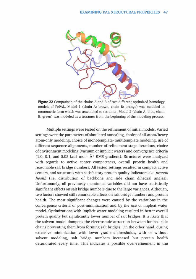
EXAMINING PAL STRUCTURAL PROPERTIES 47
Multiple settings were tested on the refinement of initial models. Varied
settings were the parameters of simulated annealing, choice of all-atom/heavy
atom-only modeling, choice of monotemplate/multitemplate modeling, use of
different sequence alignments, number of refinement stage iterations, choice
of environment modeling (vacuum or implicit water) and convergence criteria
(1.0, 0.1, and 0.05 kcal mol-1 Å-1 RMS gradient). Structures were analyzed
with regards to active center compactness, overall protein health and
reasonable salt bridge numbers. All tested settings resulted in compact active
centers, and structures with satisfactory protein quality indicators aka protein
health (i.e. distribution of backbone and side chain dihedral angles).
Unfortunately, all previously mentioned variables did not have statistically
significant effects on salt bridge numbers due to the large variances. Although,
two factors showed still remarkable effects on salt bridge numbers and protein
health. The most significant changes were caused by the variations in the
convergence criteria of post-minimization and by the use of implicit water
model. Optimizations with implicit water modeling resulted in better overall
protein quality but significantly lower number of salt bridges. It is likely that
the solvent model dampens the electrostatic attraction between ionized side
chains preventing them from forming salt bridges. On the other hand, during
extensive minimization with lower gradient thresholds, with or without
solvent modeling, salt bridge numbers increased but protein health
deteriorated every time. This indicates a possible over-refinement in the
Figure 22 Comparison of the chains A and B of two different optimized homology
models of PcPAL. Model 1 (chain A: brown, chain B: orange) was modeled in
monomeric form which was assembled to tetramer, Model 2 (chain A: blue, chain
B: green) was modeled as a tetramer from the beginning of the modeling process.

48 EXAMINING PAL STRUCTURAL PROPERTIES
protocol regarding extensive minimization. Therefore, the most reasonable
optimization protocol included a geometry pre-optimization without solvent
model to 1.0 kcal mol-1 Å-1 RMS gradient followed by further geometry
optimization with solvent modeling to only 1.0 kcal mol-1 Å-1 gradient. All final
models exhibit compact active centers, good protein health, and reasonable
salt bridge numbers (Table 4 ).
Table 4 Salt bridge and disulfide bridge statistics of theoretical (as the average of
numbers in the 16 models of each enzyme) and experimental PAL structures. For
the definitions of the C-terminal and core domains see Figure 11.
Enzyme Salt bridge
No.
(Mean)
Salt bridge
No. (RMSD)
Salt bridge
fraction in the core
domain [%]
Salt bridge
fraction in C-
terminal domain
[%]
Salt bridge
fraction between the two regions
[%]
Possible disulfide bridge
number
Temp. optimum
(°C)
Eukaryotic PALs
AtPAL1 230 8,9 70.7 18.9 10.5 0 47
AtPAL2 110 6.5 71.5 22.1 6.4 2 48
AtPAL3 116 6.3 69.9 14.7 15.4 0 31
AtPAL4 259 15.7 68.5 19.9 11.6 0 47
BoPAL1 253 11.3 70.1 18.9 11.0 4 60
BoPAL2 246 13.8 69.3 19.8 10.9 4 60
BoPAL4 238 9.7 72.9 17.9 9.2 8 50
CdPAL 253 12.7 68.1 21.5 10.4 4 55
GhPAL 244 8.0 68.6 21.0 10.3 2 30
HaPAL 214 8.7 67.1 21.8 11.1 0 55
JcPAL 243 13.2 71.6 17.3 11.1 2 60
OsPAL 248 9.5 68.4 20.1 11.5 4 50
PcPALa 208 - 67.0 22.7 11.3 0 58
UmPAL 177 14.5 80.5 14.6 4.9 0 30
Prokaryotic PAL
AvPALb 74 - 94.6 5.4c 0 0 40 aPartial homology model bExperimental structure (PDB: 3CZO) cFraction of saltbridges in the sequence segment corresponding to the C-terminal multihelix region in enzymes with eukaryotic origin

EXAMINING PAL STRUCTURAL PROPERTIES 49
3.2.3. EXAMINING PAL STRUCTURAL PROPERTIES AGAINST
TEMPERATURE OPTIMA
Although there is little consensus about the structural factors of
thermostability, experts mainly agree on the importance of disulfide bridges
and electrostatic interactions such as salt bridges, hydrogen bonds.
Mean salt bridge numbers obtained in this way were (Table 4 )
correlated with the temperature optima. When the correlation analysis was
performed including all data points, an insignificant correlation (R2=0.21,
N=13, α=0.05, threshold of significance: R2=0.31) was found. In contrary, a
much better, significant correlation (R2=0.42, N=12, α=0.05, threshold of
significance: R2=0.32) was found with the exclusion of the apparent outlier
GhPAL (blue point, Figure 23 ). Thus results for the main data group, based
on the comparison of salt bridge numbers from sophisticated homology
models and experimental temperature optima of the modeled MIO-enzymes,
implied moderate, but existing correlation between salt bridges and
temperature tolerance, despite the heavily multivariate nature of the
temperature optima of enzyme activities.
Regarding enzymes of eukaryotic and prokaryotic origin, the salt bridge
numbers were also decomposed, based the domain they belong to (Table 4 ).
The extremely low salt bridge number of AvPAL hinted that PALs of
prokaryotic origin may need significantly less salt bridges to achieve high
temperature optima than those of eukaryotic origin. Although building a
hypothesis upon only one data pair may be inadequate, this result would
Figure 23 Graphical representation of the eukaryotic PAL salt bridge numbers (split
into two groups) versus the temperature optima of enzyme activity.
0
10
20
30
40
50
60
70
75 125 175 225 275
Tem
per
atu
re o
pti
ma [
oC
]
Mean salt bridge numbers
Datapoints without the outlier Outlier

50 EXAMINING PAL STRUCTURAL PROPERTIES
further support the hypothesis on the Tyr-loop destabilizing / controlling role
of the extended C-terminal helix domain in the eukaryotic PALs, which in not
present in the prokaryotic ones. To test the validity of this assumption,
gathering more data in the future is required.
The number of possible disulfide bridges, however, showed no
correlation with temperature optima independently of the method of
modeling. One reason behind the absence of correlation between the disulfide
bridges and temperature optima may be the lack of certain disulfide bridges
despite the opportunity to be formed. This assumption led to a warning, it
could be possible that in several cases the actual recombinant host organisms
were not the most appropriate ones to form all the possible disulfide bonds.
3.2.4. EXAMINING THE PH TOLERANCE OF RUBROBCATER
XYLANOPHILUS PAL
The sequence of a phenylalanine ammonia-lyase from the
thermophilic and radio-tolerant bacterium Rubrobacter xylanophilus (RxPAL)
was identified previously in our research group by virtually screening the
genomes of bacteria for members of the phenylalanine ammonia-lyase family.
A synthetic gene encoding the RxPAL was cloned and overexpressed in
Escherichia coli TOP 10 was purified by Ni-NTA affinity chromatography.
Experimental work was carried out by Andrea Varga, Izabella Szabó, and
Klaudia Kovács.
The first strain of the genus Rubrobacter was isolated from gamma-
irradiated hot spring water. [159] This species, Rubrobacter radiotolerans was
A
B
Figure 24 The electrostatic potential representation of RxPAL (Panel A) compared
to that of less alkalophilic AvPAL (Panel B) indicating an excessive and
homogenous negative charge on the enzyme surface.

EXAMINING PAL STRUCTURAL PROPERTIES 51
slightly thermophilic with an optimum growth temperature of about 45°C.
Later, a true thermophile strain with an optimum growth temperature of
about 60°C was isolated from a hot runoff of a carpet factory and was
identified and named as a new species Rubrobacter xylanophilus. [160]
In order to explore the possible extreme properties of RxPAL,
homology modeling was implied. The model indicated several potential
disulfide bonds, and an unusually high number, 234 of salt bridges. These
findings could explain the high thermotolerance of the host organism and,
ultimately, the enzyme. Unfortunately, this cannot be confirmed for now,
because the activity temperature optimum of the isolated enzyme has not been
characterized.
Inspection of the surface electrostatic potential map, however, reveals
an excessive and, most importantly, homogenous negative charge on the
enzyme surface (Figure 24 ). This gave us the idea to monitor the enzymatic
activity upon an extended pH range of 6.5-12.0 (Figure 25 ). Values lower
than 6.5 were not tested because of precipitation of the protein. The rate of
the PAL catalyzed reaction increased slowly until pH 8.5. At higher pH values,
enzyme activity was further increasing and the maximal activity was reached
at pH 11.5, and activity was still retained up to pH 11.7. At higher pH values,
the activity abruptly decreased, potentially reflecting protein denaturation.
The PAL activity was fully stable for 1 h at room temperature at all pH ≤ 11.5.
The pH range of the phenylalanine ammonia lyases characterized so far is
clearly on the alkaline side, with an optimum pH of 8.5-9.5. In contrast to
other PALs, however, activity and stability of RxPAL was shown to be
significantly higher at very alkaline (around 11) pH, rendering the new
enzyme attractive as a biocatalyst under these conditions. The observed high
Figure 25 Plot of mean RxPAL activity (measured in triplicates) against pH values.
Data are presented as relative to the maximal activity observed at pH 11.5.

52 INVESTIGATING THE REACTION MECHANISM OF MIO ENZYMES
enzymatic activity at elevated pH values is especially beneficial with regard to
the reverse reaction, wherein ammonia addition to achiral arylacrylates
resulting enantiomerically pure L-configured (unnatural) α-amino acids in
presence of 5-6 M NH3/NH4+ in the pH 10-11 range.
3.3. INVESTIGATING THE REACTION MECHANISM OF MIO
ENZYMES
By using PAL, immobilized on magnetic nanoparticles and fixed in a
microfluidic reactor with an in-line UV detector, in our research group, we
first demonstrated that PAL can catalyze the ammonia elimination from the
acyclic propargylglycine (PG) to yield (E)-pent-2-ene-4-ynoate. This reaction
proved to be reversible and enantioselective also at high ammonia
concentrations yielding enantiopure L-PG (Figure 26 ). This opens new
perspectives to extend the MIO-enzyme toolbox towards acyclic substrates,
meanwhile grating valuable information about the catalytic mechanism of
MIO enzymes. Experimental work was done by Diána Weiser and László Csaba
Bencze.
The literature of MIO enzyme mechanisms has been exhaustively
reviewed in the introduction suggesting three possibilities, the FC, the TS, and
the most probable, N-MIO way (Figure 12).
The transformation of PG, a non-aromatic acyclic amino acid disfavors
the FC mechanism, although it was already demonstrated that PcPAL
transforms several non-aromatic cycloalkenylalanines such as rac-3-
(cycloocta-1,3,5,7-tetraen-1-yl)alanine and L-3-(cyclohexa-1,4-dien-1-
yl)alanine, [124] and these reactions could still be rationalized by a Friedel-
Figure 26 Preparation of enantiopure D-propargylglycine by PAL-catalyzed
ammonia elimination from rac-propargylglycine and enantiopure L-
propargylglycine by PAL-catalyzed ammonia addition to (E)-pent-2-ene-4-ynoate.
INVESTIGATING THE REACTION MECHANISM OF MIO ENZYMES 53
Crafts-like or a single-step mechanism. On the other hand, the constitutional
isomer L-3-(cyclohexa-2,5-dien-1-yl)alanine and the saturated version rac-β-
cyclohexylalanine were not accepted as substrates but proved to be moderate
inhibitors for PAL.
Two arguments were recently raised against the route through an N-
MIO intermediate in TAL, [115] i) it is unlikely to have two tyrosine residues
simultaneously deprotonated in the same active center without any strong
charge compensation, and ii) the calculated activation energy between the
lowest energy N-MIO intermediate and the elimination product (26.3
kcal mol-1 or 28.5 kcal mol-1) is rather high, meanwhile for the single
transition state (TS) mechanism a barrier of 16.6 kcal mol-1 was calculated.
[115] Still, according to these calculations, the single-step reaction is quite
endothermic and has a barrier of 64.7 kcal mol-1 for the reverse reaction [i.e.
ammonia addition to (E)-coumarate]. Therefore it was stated, that the “single-
step route” is in conformity with the apparent irreversibility of the reaction.
[115]
In fact, a reactive state with both TyrA and TyrB as deprotonated and
the L-Tyr substrate as zwitterionic for the PAL (or TAL) reaction can be easily
formed at pH ~9, if the amino group of anionic L-Phe (or L-Tyr) substrate
deprotonates TyrA–OH when entering into the active site containing only TyrB
in a deprotonated state. This would perfectly agree with the analysis of the
pH-profile of the PAL reaction showing that to be able to react the amino
group of the substrate must not be protonated but in turn, there should be a
group with a pKa of 9 on the enzyme which must be protonated. This
eliminates the first argument raised against the calculated N-MIO route. [116]
The calculated relative energies for the N-MIO intermediate varying
between -5.5 and -21.6 kcal mol-1 even in the study favoring the single-step
mechanism [115] indicated that the real N-MIO – product barrier may be
lower than the disputed values of 26.3 kcal mol-1 or 28.5 kcal mol-1.
Moreover, all structural data so far agree only with the N-MIO route. X-Ray
structural studies on various MIO-enzymes revealed that several substrates
and substrate analogues were found to be covalently bound via their N-atoms
to the MIO-group, which were covered in the literature review earlier.
To get molecular level insights into the biotransformation of L-PG,
QM/MM calculations were carried out. N-MIO and the constitutionally isomer
FC complexes were created for both L-PG and L-Phe in the PcPAL partial
homology model. Also, this was the first time QM/MM calculations were ever
done on the PAL mechanism.
Results indicate quite similar N-MIO-intermediate structures for L-
propargylglycine and L-phenylalanine (Figure 27 ), exhibiting consistency, a

54 INVESTIGATING THE REACTION MECHANISM OF MIO ENZYMES
feature what is expected from the real process. On the other hand, the much
higher Gibbs energies calculated for the FC covalent complexes relative to the
N-MIO states (166.7 kcal mol-1 from L-Phe and 9.5 kcal mol-1 from L-PG, Table
5 ) strongly disfavor the FC route as possible pathway for the PAL (or TAL)
reaction. Moreover, in both hypothetical FC complexes, the catalytically
relevant pro-S H-atoms attached to Cβ are quite distant from any of the two
catalytically relevant tyrosine residues (TyrA or TyrB), PG even forming a
potentially unreactive, “suicide” 3,4-dihydrofuranone type compound (Figure
28 panel A and B).
The single-step proposal
strongly contradicts to a number
of experimental results on the PAL
reaction with L-Phe because i)
negligible isotope effects were
found for the atoms which should
be involved in the single-step TS
(15N of 1.0021 and D of 1.15 for
Phe dideuterated at C3); [116] ii)
cinnamate as product was found
to be released prior to ammonia in
Figure 27 Model of the N-MIO intermediate of PAL with L-propargylglycine (solid
stick model) compared to the model of N-MIO intermediate of PAL with its natural
L-phenylalanine substrate (transparent stick model). The models were obtained by
ONIOM (ωB97XD/6-31g(d):AMBER) calculations.
Table 5 QM/MM Gibbs energies
(kcal mol-1) of the N-MIO and FC reaction
intermediates regarding L-Phe and L-PG.
Valueas are normalised to the minimum
energy intermediate in the case of each
compound.
Substrate Intermediate
N-MIO FC
L-Phe 0.0 166.7
L-PG 0.0 9.5

INVESTIGATING THE REACTION MECHANISM OF MIO ENZYMES 55
the catalytic sequence [161] and iii) the PAL reaction was found to be
reversible at elevated (>5 M) ammonia concentration. [162]
Moreover, we carried out equilibration studies with PAL starting from
both substrate and product at much lower ammonia concentrations indicated
that ammonia addition to (E )-cinnamate proceeded easily even at quite low
ammonia concentration (in 0.1 M, 0.5 M or 1.0 M ammonium carbonate
buffered to pH 8.8 with CO2). Equilibria were almost reached in 3 days from
both directions as the (E )-cinnamate / L-phenylalanine ratio values indicated
[in 0.1 M (NH4)2CO3 it was 7.6 in elimination and 10.8 in addition; in 0.5 M
(NH4)2CO3 it was 2.8 both in elimination and in addition; in 1.0 M (NH4)2CO3
it was 1.2 in elimination and 1.1 in addition]. This allowed us to calculate –
as the average for equilibrations in 0.5 M and 1 M (NH4)2CO3 – the apparent
equilibrium constant as 2.5 (at pH 8.8). Taking all these serious contradictions
into account, the single-step route cannot be considered as a real pathway for
the PAL (or TAL) reaction, and in summary, the N-MIO mechanism is left as
the only viable option.
Figure 28 Formation of the calculated relaxed structure A) of the hypothetical L-
Phe (S,R )-FC complex shown and B) hypothetical L-PG (E)-FC complex. In both
hypothetical FC complexes, the catalytically relevant pro-S H-atoms attached to
Cβ are quite distant from any of the two catalytically relevant tyrosine residues
(TyrA or TyrB).

56 EXPANDING THE SUBSTRATE SCOPE OF PCPAL TOWARDS STYRYLALANINES
3.4. EXPANDING THE SUBSTRATE SCOPE OF PCPAL
TOWARDS STYRYLALANINES
Styrylalanines are less studied and synthetically challenging
unnatural amino acids. It was already shown that phenylalanine 2,3-
aminomutase from Taxus canadensis (TcanPAM) could transform (S )-α-
styrylalanine into (E )-styrylacrilate as the major product. [163] Considering
the high structural and sequence similarities between TcanPAM and PcPAL, it
seemed commendable to explore the potential of PcPAL in the kinetic
resolution of α-styrylalanines (Figure 29 ). Experimental work was carried out
by Alina Filip and László Csaba Bencze.
3.4.1. INITIAL STUDIES AND MUTANT SET CONSTRUCTION
First, to assess the applicability of wt-PcPAL to the styrylalanine
compound family, the commercially available L-styrylalanine (L-1a) was tested
as substrate in the ammonia elimination reaction (Figure 29 , panel A). Kinetic
studies showed 14.7-fold lower kcat and 777-fold lower kcat/KM values for the
deamination of L-1a in comparison with the deamination of the natural
substrate, L-Phe (Table 6 ).
The low enzyme activity, observed with L-styrylalanine (L-1a), was
rationalized by molecular modeling. Evaluation of energetics indicated as the
most disadvantageous interaction the one between the phenyl ring of the N-
MIO intermediate from L-1a and the phenyl ring of residue F137 in the
aromatic binding pocket of wt-PcPAL (Figure 30 ). Moreover, enumeration of
the possible conformations indicated three major species. In panel A of Figure
30 , two apparently catalytically active conformations, resembling the active
arrangement of phenylalanine in PcPAL, are depicted. These two N-MIO
Figure 29 Ammonia elimination from racemic styrylalanines rac-1a-d, and
computer-only modeling and forecasting of the ammonia elimination from racemic
styrylalanines rac-1e-h, catalyzed by wt- and mutant PcPALs.

EXPANDING THE SUBSTRATE SCOPE OF PCPAL TOWARDS STYRYLALANINES 57
intermediate conformations can result in two different arrangements of the
diene product (2a), the s-cis [referred as pro-s-cis (psc)] or the s-trans
[referred as pro-s-trans (pst)] conformations. Panel B of Figure 30 depicts a
third conformation adopting a somewhat different arrangement with
significantly less stress exerted by F137. However, this specimen would lead
to (2Z ,4E )-5-phenylpenta-2,4-dienoic acid which was not detected by NMR
in the reaction mixture. Thus, conformation L-1au could be considered as an
unproductive state. These modeling results implied that the reaction rate
might be improved by reducing the clash between the aromatic moieties of L-
1a in its N-MIO state and F137 within the active site of wt-PcPAL, and
simultaneously by displacing the population of the unproductive
conformation. Notable, in case of 4-substituted cinnamates with PcPAL,
mutations of the F137 site proved efficacious. [164] Therefore, to provide
more space for the phenyl ring of L-1a, residue Phe137 in the hydrophobic
pocket was changed to smaller hydrophobic residues Val, Ala, and Gly in
mutants F137V-, F137A-, and F137G-PcPAL, respectively. The kinetic
parameters of the wild-type and the mutant PcPALs were determined for the
natural substrate L-Phe and for L-1a (Table 6 ). Not surprisingly, all mutants
A B
Figure 30 Active site models of the three major conformations of the N-MIO
complex of L-1a with wt-PcPAL [colors refer to the atomic contributions to the total
energy ranging from green (beneficial, ≤-5 kcal mol-1) to red (detrimental, ≥2 kcal
mol-1)]. A) The two allegedly catalytically active conformations L-1apsc and l-1apst
leading to the product 2a in s-cis (synperiplanar) or s-trans (antiperiplanar)
arrangements, respectively. B) the apparently catalytically inactive conformation
L-1au compared to the apparently catalytically active L-1apsc conformation.

58 EXPANDING THE SUBSTRATE SCOPE OF PCPAL TOWARDS STYRYLALANINES
showed decreased – declining with side chain volume – catalytic efficiencies
with the natural substrate, L-Phe. However, it can be clearly seen that F137V
has superior catalytic efficiency with L-1a, with the nearly 240-fold increased
kcat/KM value compared to wt-PcPAL. The F137A mutation also provided an
increased kcat/KM value with L-1a (10.5-fold enhancement), while the F137G
mutation showed no beneficial effect on the transformation of L-1a.
Interestingly, kcat values showed also substantial variances. The trends
and maxima were almost the same as in the case of the kcat/KM values.
Regarding L-Phe, kcat declined with decreasing side chain volume, except for a
surprising spike with F137A-PcPAL. Similarly, F137V-PcPAL with L-1a showed
a tremendous, almost 9-fold increase; F137A showed only a moderate 2.8-fold
increase; and F137G showed no improvement in kcat compared to wt-PcPAL.
Table 6 Kinetic parameters for wt-PcPAL and for F137X-PcPAL mutants in ammonia
elimination from L-phenylalanine and from L-styrylalanine (L-1a).
Substrate PcPAL KM (μM) kcat (s-1) kcat/KM (M-1 s-1)
L-Phe
wt 83 ± 5 0.694 ± 0.020 8361 ± 291 F137V 86 ± 10 0.173 ± 0.001 2011 ± 131 F137A 1732 ± 15 0.283 ± 0.001 163 ± 2 F137G 4969 ± 153 0.052 ± 0.003 10.4 ± 0.9
L-1a
wt 4384 ± 158 0.0471 ± 0.0003 10.7 ± 0.4 F137V 186 ±6 0.4220 ± 0.0280 2269 ± 168 F137A 1173 ± 70 0.1320 ± 0.0026 112.5 ± 7.2 F137G 4120 ± 270 0.0345 ± 0.0038 8.3 ± 0.2
A B
Figure 31 Molecular models of various N-MIO intermediates in PcPALs. A)
Arrangement of covalent intermediate L-1apsc in wt-PcPAL (relevant residues are in
red) and in F137V-PcPAL (relevant residues are in cyan). Note the different side
chain conformations of I460. B) Arrangement of the unproductive covalent
complex from D-1a (yellow) compared to the productive intermediate L-1apsc (red)
in wt-PcPAL.

EXPANDING THE SUBSTRATE SCOPE OF PCPAL TOWARDS STYRYLALANINES 59
A comparison of the covalent pro-s-cis intermediates from L-1a within
wt-PcPAL and F137V-PcPAL is shown in panel A of Figure 31 . The aromatic
moiety of the N-MIO intermediate from L-1a is forced towards I460 within the
active site of wt-PcPAL, unlike in the case of the more relaxed complex within
F137V-PcPAL, meanwhile the alanyl substructure of the substrate – which is
directly involved in bond rearrangement – is kept relatively unchanged in both
cases. It is worth noting that upon binding of L-1a to wt-PcPAL the active site
supposedly undergoes conformational change at the I460-side chain which
reshapes the active site and alters the energetics of distinct substrate
conformations.
3.4.2. SYNTHETIC APPLICATIONS IN KINETIC RESOLUTION
Next, we wanted to explore the potential of F137V-PcPAL in the
synthetically valuable ammonia elimination of various styrylalanines.
Therefore, racemic styrylalanines rac-1a-d were synthesized and tested with
F137V-PcPAL and wt-PcPAL as biocatalysts (Figure 29 ).
In the ammonia elimination reaction all styrylalanines (rac-1a-d) were
accepted as substrates by wt-PcPAL as well as by F137V-PcPAL. However, the
F137V mutant showed significantly improved catalytic activities (kcat and
kcat/KM) in comparison with the wild-type enzyme in all cases (Table 7 ).
The synthetic usefulness of F137V-PcPAL towards styrylalanines in the
ammonia elimination reactions was further tested by kinetic resolutions with
0.1 mmol rac-1a-d at 5 mM concentration using 0.5-1.0 mg of pure PcPALs.
The conversion of the PcPAL-catalyzed reactions from racemic styrylalanines
rac-1a-d (Figure 32 ), and the enantiomeric excess of the residual substrates
D-1a-d were both monitored (Table 8 ). In case of longer reaction times (>48
h), a fresh enzyme batch was added to the reaction mixture at the end of each
48 h period to overcome enzyme inactivation during long incubation times.
Table 7 Kinetic parameters for wt- and F137V-PcPALs in the ammonia elimination
of racemic styrylalanines rac-1a-d.
PcPAL Substrate KM (μM) kcat (s-1) kcat/KM (M-1 s-1)
wt-PcPAL
rac-1a 395 ± 6 0.00620 ± 0.00070 15.6 ± 0.9 rac-1b 154 ± 7 0.00024 ± 0.00001 1.5 ± 0.03 rac-1c 28 ± 1 0.00034 ± 0.00002 12.1 ± 0.6 rac-1d 287 ± 3 0.00320 ± 0.00030 11.1 ± 0.5
F137V-PcPAL
rac-1a 201 ± 12 0.2760 ± 0.0130 1373 ± 97 rac-1b 78.3 ± 2 0.0786 ± 0.0044 1004 ± 86 rac-1c 94.7 ± 2 0.1570 ± 0.0020 1657 ± 10 rac-1d 326 ± 10 0.0099 ± 0.0001 30.2 ± 1.9

60 EXPANDING THE SUBSTRATE SCOPE OF PCPAL TOWARDS STYRYLALANINES
Time course analysis of the conversion of the KRs of rac-1a-d (Figure
32 ) also indicated the beneficial catalytic properties of F137V-PcPAL
compared to that of wt-PcPAL. The theoretically possible conversion in a
kinetic resolution (KR) with exclusive selectivity (50%) could be almost
perfectly approached (c >49%) with the F137V mutant in reactions of rac-
1a,c after moderate reaction times (24 h and 134 h, respectively) and in the
cases rac-1b,d after longer incubation times (274 h and 300 h, respectively).
On the other hand, in reactions of racemic styrylalanines rac-1a-d with
wt-PcPAL, almost complete conversion of the L-enantiomer could be obtained
only with rac-1a (c >49% after 274 h), while the conversions of rac-1b-d could
Table 8 Kinetic resolution of styrylalanines rac-1a-d and enantiomeric excess of the
residual d-1a-d in the ammonia elimination reactions catalyzed by PcPAL variants.
Entry PcPAL Substrate Time (h) c (%) Eetheor (%)a Eeobs (%)
1 F137V rac-1a 24 50 100 >99 2 rac-1b 274 50 100 >99 3 rac-1c 134 50 100 >99 4 rac-1d 300 50 100 >99 5 wild-type rac-1a 274 50 100 >98 6 rac-1b 504 29 41 41 7 rac-1c 600 36 56 55 8 rac-1d 600 31 45 41
a Eetheor= c/(1-c) for a fully selective kinetic resolution
Figure 32 Comparison of the time profiles of the conversions in the ammonia
elimination reactions from rac-1a-d catalyzed by F137V-PcPAL (continuous lines) and
wt-PcPAL (dashed lines).
0
10
20
30
40
50
60
0 50 100 150 200 250 300
rac-1a F137V rac-1a wt rac-1b F137V rac-1b wt
rac-1c F137V rac-1c wt rac-1d F137V rac-1d wt
Reaction time (h)
Con
vers
ion
(%)

EXPANDING THE SUBSTRATE SCOPE OF PCPAL TOWARDS STYRYLALANINES 61
not approach the theoretically possible value within 500-600 h. KRs of rac-1a-
c with wt-PcPAL showed that enantiomeric with the theoretical values of a
fully selective KR indicating excellent enantiomer selectivity and preference of
the process to digest the L-enantiomers, L-1a-c (Table 8 ). Although the
enantiomeric excess of the residual D-1d fractions were somewhat smaller as
expected for a fully selective KR, it was not possible to determine whether this
happened due to experimental error of the HPLC process or due to a non-
perfect enantiomer selectivity of the KR from rac-1d.
KRs of rac-1a-d with F137V-PcPAL, however, showed that the residual
D-1a-d fractions had enantiomeric excess values over 99% indicating
unquestionably exclusive enantiomer selectivity in all cases. This was also
obvious from the time course curves of conversions approaching
asymptotically the 50% value when F137V-PcPAL was applied as biocatalyst
in the KRs (Figure 32 ). Time course profiles of the reactions and the
comparison of the kinetic constants with L-1a (Table 6 ) and with rac-1a
(Table 7 ) suggests that D-styrylalanine inhibits the ammonia elimination of L-
1a both with wt-PcPAL and F137V-PcPAL. If D-1a had no interaction with wt-
PcPAL than only a 2-fold higher apparent KM of the racemate rac-1a would be
observed as compared to the pure L-enantiomer L-1a without affecting the kcat
value. However, when rac-1a was applied as substrate instead of the pure L-
enantiomer L-1a, the Vmax (thus implicitly the apparent kcat value) decreased
7.6-fold, while the KM value decreased 11-fold. With F137V-PcPAL, the
differences became less significant, kcat decreased only 1.5-fold, and KM
showed only a minor variation, within the range of experimental errors.
Because the residual fractions from the KRs of rac-1a-d had quite high
enantiomeric excess at 50% conversion (ee >99%, see Table 3), the D-
enantiomers D-1a-d were apparently not converted. This implied D-1a being a
reversible inhibitor with considerable affinity forming an unproductive
enzyme–D-substrate complex. The covalent, unproductive N-MIO complex
from D-1a showed astonishing overlap with the productive intermediate L-1apst
within the active site of wt-PcPAL (panel B, Figure 31 ).
3.4.3. RATIONALISING THE BIOTRANSFORMATIONS OF
STYRYLALANINES
To get a more detailed view of the catalytic process, a quantitative
model was created and validated. N-MIO intermediates of L- and D-1a-d in the
corresponding PcPAL variants were constructed using the customized
induced-fit covalent docking protocol for MIO enzymes (see the overview of
the general modeling procedure). The non-covalent substrate-binding states
of L- and D-1a-d – directly preceding their N-MIO intermediate states – were

62 EXPANDING THE SUBSTRATE SCOPE OF PCPAL TOWARDS STYRYLALANINES
also modeled. Because the calculations did not reveal any unproductive states
for these non-covalent complexes, they will not be discussed hereafter.
The occurrence of the non-stereoselective, MIO-independent reaction
pathway [120] was ruled out experimentally. The attempted ammonia
elimination from rac-1a-d by wt-PcPAL with inactivated MIO group (obtained
by treatment with sodium borohydride) [165] showed no product formation.
The conversion of 4-nitrophenylalanine with the sodium borohydride treated
wt-PcPAL served as positive control for integrity of the residual parts of the
active site.
To investigate the catalytic activities quantitatively with all substrate
and enzyme mutant combinations, the apparent kcat values – as determined
from the maximum reaction rates according to the left-hand side of Eq 6 –
were decomposed into their constituents.
At substrate saturation, when the concentration of the free enzyme is
negligible, four species of enzyme–substrate intermediate complexes, can be
present in thermal equilibrium (allosteric inhibition was not considered).
From the L-enantiomer two catalytically active enzyme–substrate intermediate
complexes (the s-cis and s-trans L-1a-d–N-MIO states, abbreviated as E L-psc, E L-
pst) and an unproductive one (the unproductive L-1a-d–N-MIO states,
abbreviated as E L-u) could be formed, while another unproductive complex
could be formed from the D-enantiomers (the unproductive D-1a-d–N-MIO
states, abbreviated as ED). Thus, according to Eq 6, the catalytic activity is the
sum of the activities of E L-psc and E L-pst.
After dividing by [E*], the fractions of the concentrations of E L-psc and
E L-pst can be rewritten as molar fractions. Further, substituting the Eyring
equation (Eq 7) yields Eq 8,
𝑉max = 𝑘cat[𝐸∗] = 𝑘cat,L−𝑝s𝑐[𝐸𝑝s𝑐] + 𝑘cat,L−𝑝s𝑡[𝐸𝑝s𝑡] Eq 6
Vmax: maximal reaction rate [E*]: total enzyme concentration
𝑘 =
𝑘B𝑇
ℎ𝑒∆𝐺𝑅𝑇
#
Eq 7
𝑘cat =
𝑘B𝑇
ℎ𝑒∆𝐺L−𝑝s𝑐
# /𝑅𝑇 𝑥𝑝s𝑐 +𝑘B𝑇
ℎ𝑒∆𝐺L−𝑝s𝑡
# /𝑅𝑇 𝑥𝑝s𝑡 Eq 8
T: temperature kB: Boltzmann constant, h: Planck constant ΔG#: Gibbs energy of activation x: molar fraction

EXPANDING THE SUBSTRATE SCOPE OF PCPAL TOWARDS STYRYLALANINES 63
To approximate the Gibbs energy of activation, it was decomposed
into two parts, into an electronic term that is treated with quantum chemical
calculations, and a term specific for the steric stress that is treated with
molecular mechanics calculations (Eq 9).
Numerous evidences suggest that the elimination step happens with
an E1cB mechanism. Thus, Hammond’s postulate was applied, and a
descriptor – associated with the acidity of the pro-S β-proton (cBRΔE) – in a
linear relationship was used to calculate ΔE#Electronic (Eq 10). In the case of steric
strain, a simple rule was applied stating that N-MIO type enzyme–substrate
complexes with better interaction energies are able to “provide the energy
needed for activation proportionally easier”, thus ΔE#Steric is linearly dependent
on SterRΔE (Eq 11). This assumption is supported by the fact that the active
center is very compact in the N-MIO intermediate form also almost entirely
desolvating the substrate.
Because L-1a-d are congeneric – and thus it is expected to exhibit
fairly similar properties in many cases –, it was assumed that irrespectively of
the true dependence of the activation energy terms on the descriptors, linear
dependence should give a satisfactory description. However, using both
constants b and d from Eq 10 and Eq 11, respectively, in a non-linear
estimation would lead eventually to overfitting, thus, as a compromise, b was
considered equal to d.
Finally, substituting Eq 9, Eq 10 and Eq 11 into Eq 8, bringing the
molar fractions into the exponents, and applying that b=d, Eq 12 is obtained,
where a and b are constants.
Non-linear estimation of the constants in Eq 12 gave statistically
significant regression (R2=0.8, p=0.002) and parameters (a=90.63,
p=0.023; b=0.0791, p=0.014).
∆𝐺# ≈ ∆𝐸# = ∆𝐸𝐸𝑙𝑒𝑐𝑡𝑟𝑜𝑛𝑖𝑐# + ∆𝐸𝑆𝑡𝑒𝑟𝑖𝑐
# Eq 9
∆𝐸𝐸𝑙𝑒𝑐𝑡𝑟𝑜𝑛𝑖𝑐# = 𝑏 ∗ ∆𝐸𝑐𝑏
𝑅 + 𝑐 Eq 10
∆𝐸𝑆𝑡𝑒𝑟𝑖𝑐# = 𝑑 ∗ ∆𝐸𝑆𝑡𝑒𝑟
𝑅 + 𝑒 Eq 11
𝑘cat = 𝑎(𝑒𝑏∗∆𝐸L−𝑝s𝑐# /𝑅𝑇+ln 𝑥L−𝑝s𝑐 + 𝑒𝑏∗∆𝐸L−𝑝s𝑡
# /𝑅𝑇+ln𝑥L−𝑝s𝑡) Eq 12
a,b: constants of non-linear estimation

64 EXPANDING THE SUBSTRATE SCOPE OF PCPAL TOWARDS STYRYLALANINES
Evaluation of the F137V mutation based on computed data (Table 9
) clearly demonstrated that the affinities of L-1a-d in the pro-s-cis
conformation (bRΔE L-psc) increased substantially, as it was assumed. Moreover,
the affinities of L-1a-d in the pro-s-trans conformation increased even more,
as their population increased relative to the pro-s-cis conformation (x L-pst vs.
x L-psc), with the exception of L-1d with F137V-PcPAL. The unproductive
complexes of 1a-d (xD and x L-u), however, showed a more complex situation.
By the F137V-mutation, in some cases unproductive states were displaced
(e.g. 1c), and in some other cases they were introduced (e.g. 1d). These results
indicated that the simple increase of active site volume is not sufficient from
the standpoint of enzyme design, and a more sophisticated active site shape
altering scheme – taking the relative spatial requirements of the intermediate
states and unproductive states into account – should be employed. Inspection
of the descriptors for the acidity of the pro-S β-hydrogen (cBRΔE) – which was
derived from single point DFT calculations on a truncated part of the
intermediate model – revealed that in almost all cases acidity of the pro-s-
trans conformations compared to the pro-s-cis conformations (cBRΔEL-pst-cB
RΔEL-
Table 9 Calculated relative binding energies (bRΔE), rounded values of molar
fractions of the various covalent N-MIO complexes (x), energy-based descriptors
of the acidity of their pro-S β-hydrogen (cBRΔE) and steric strain of activation
(StrainRΔE) for intermediates of compound 1a-d in PcPAL variants. In the subscript L
and D correspond to the actual enantiomer; psc, pst, and u correspond to the s-cis,
s-trans product forming, and the unproductive 1a-d–N-MIO states. Energy related
term are in kcal mol-1. PcP
AL
Su
bstra
te
bRΔ
EL-p
sc b
xL-p
sc c (%)
xL-p
st c (%)
xDc( %
)
xL-u
c (%)
Strain
RΔE
L-psc b
Strain
RΔE
L-pst b
cBRΔ
EL-p
sc b
cBRΔ
EL-p
st b
cB RΔEL-p
st -cB RΔEL-
psc b
wt
1a 13.0 99.7 0.2 0.1 0.0 11.0 16.2 -2.2 3.8 6.1
1b 22.5 2.1 0.0 0.0 97.9 22.5 28.5 -1.5 -1.8 -0.3
1ca 13.6 41.5 0.0 0.7 57.8 12.9 20.4 11.9 8.2 -3.6
1d 33.7 93.4 0.0 0.0 6.6 33.9 42.4 -11.4 -11.5 0.1
F137V
1a -3.3 95.2 4.8 0.0 0.0 -0.7 1.1 -4.6 -10.7 -6.2
1b -3.2 99.9 0.1 0.0 0.0 1.4 5.7 -5.4 -7.7 -2.4
1ca 1.4 60.9 5.0 0.8 0.0 5.3 6.8 11.3 0.5 -10.8
1da 22.7 0.3 18.1 0.9 80.7 25.3 22.9 -31.6 -20.4 11.3
a In these cases multiple psc and pst conformations were obtained, but only the lowest energy
representatives were taken into account. b Quantities are related to the same property of L-
Phe with wt-PcPAL, in the form of RΔE=ΔE- ΔEl-Phe;wt-PcPAL. cMolar fractions were calculated
based on Boltzmann probabilities incorporating all substrate conformations.

EXPANDING THE SUBSTRATE SCOPE OF PCPAL TOWARDS STYRYLALANINES 65
psc) was comparable or higher, except for 1b/wt-PcPAL and 1d/F137V-PcPAL.
Thus, the higher proportion of the pro-s-trans conformations (x L-pst), enabling
more facilitated reaction due to the more acidic pro-S β-hydrogens, could
contribute to the increased activity of F137V-PcPAL. Moreover, the acidity
descriptors cBRΔEL-psc and cB
RΔEL-pst varied remarkably in wt- and F137V-PcPAL,
mostly due to the changes in dihedral angle between the phenyl ring and the
conjugated double bond of the styryl moiety. The more planar the skeleton
was; the better dispersion of the negative charge could happen due to the
higher degree of conjugation. This implied that in an efficient enzyme design
protocol the substrate affinity and electronic effects should be monitored
simultaneously.
This exploratory model validates the very concept of our
computations and it can be used as a tool to analyze various factors of
importance in catalysis and to provide valuable input for further studies.
Although this model was not expected to predict the apparent kcat with high
Table 10 Calculated relative binding energies (bRΔE), rounded values of molar fractions
of the various covalent N-MIO complexes (x), energy-based descriptors of the acidity of
their pro-S β-hydrogen (cBRΔE) and steric strain of activation (Strain
RΔE) for intermediates
of further 4-substituted styrylalanines (1e-h) in PcPAL variants, and predicted (kcat-pred)
values for the corresponding reactions. In the subscript L and D correspond to the actual
enantiomer; psc, pst, and u correspond to the s-cis, s-trans product forming, and the
unproductive 4-substituted styrylalanine–N-MIO states. Energy related term are in kcal
mol-1. PcP
AL
Su
bstra
te
bRΔ
EL-p
sc a
xl-p
sc b(%)
xL-p
st b(%)
xDb(%
)
xL-u
b(%)
Strain
RΔE
L-psc a
Strain
RΔE
L-pst a
cBRΔ
EL-p
sc a
cBRΔ
EL-p
st a
kcat-p
red
(s-1)
wt
1e 13.2 88.0 0.0 0.0 12.0 13.5 20.3 -0.3 -1.5 0.014
1f 38.7 0.2 0.0 0.0 99.8 40.1 48.0 -3.7 -3.8 <0.0001
1g 23.3 2.1 0.0 0.0 97.9 24.2 32.3 -1.7 -2.4 0.0001
1h 33.7 93.4 0.0 0.0 6.6 33.9 42.4 -11.4 -11.5 0.004
F137V
1e -4.3 99.3 0.7 0.0 0.0 0.6 3.6 -5.7 -6.9 0.180
1f 15.5 0.0 0.0 0.0 100.0 21.5 15.5 -6.8 -8.2 <0.0001
1g -2.6 95.9 0.0 0.0 4.1 2.9 8.2 -5.4 -1.6 0.120
1h 8.3 100.0 0.0 0.0 0.0 13.1 17.8 -13.7 -15.0 0.098
a Quantities are related to the same property of L-Phe with wt-PcPAL, in the form of RΔE=ΔE- ΔEL-
Phe;wt-PcPAL. bMolar fractions were calculated based on Boltzmann probabilities incorporating all
substrate conformations.

66 EXPANDING THE SUBSTRATE SCOPE OF PCPAL TOWARDS STYRYLALANINES
accuracy, it enabled us to predict the order of magnitude of the catalytic
activity for several further congeneric substrates.
With our model, we examined the behavior of further substituted
styrylalanines bearing fluoro, trifluoromethyl, bromo, and cyano substituents
at position 4 (1e-h, respectively, Table 10 ). Calculations with wt-PcPAL
predicted rac-1e and rac-1h to be poor substrates behaving similarly to rac-1a.
Modeling forecast rac-1g- and rac-1f to be extremely poor substrates or
reversible inhibitors (kcat-pred≤ 0.0001 s-1). The F137V mutation calculated no
significant improvement only for rac-1f (kcat-pred≤ 0.0001 s-1). Modeling
within this mutant indicated comparably high activity for rac-1e (kcat-
pred= 0.18 s-1) as for rac-1a (kcat= 0.28 s-1). Somewhat lower catalytic
activities were predicted for rac-1g and rac-1h (kcat-pred= 0.120 s-1
and 0.098 s-1, respectively) being in the range of the activity of rac-1c
(kcat= 0.16 s-1).

INVESTIGATING PHENYLALANINE AMINOMUTASE FROM PANTOEA AGGLOMERANS 67
3.5. INVESTIGATING PHENYLALANINE AMINOMUTASE
FROM PANTOEA AGGLOMERANS
Nowadays there is an ever increasing demand for optically pure β-
amino acids mainly by the pharmaceutical industry and peptide syntheses.
[166, 167] In particular, the biological characteristics of the β-amino acids,
along with their use as precursors of various heterocycles and as chiral
auxiliaries in enantioselective syntheses have aroused lively interest for their
chemistry. This induced rapid development of synthetic procedures for the
preparation of enantiopure β-amino acids and their congeners. [168] Until
recently most of the biocatalytic approaches for them relied on kinetic
resolution with hydrolytic enzymes, such as lipases, acylases and
hydantoinases. [169] To explore other synthetic routes, enantiomer selective
isomerization of various racemic α- and β-arylalanines catalysed by
phenylalanine 2,3-aminomutase from Pantoea agglomerans (PaPAM) were
investigated (Figure 33 ). Experimental work was carried out by Andrea Varga
and Botond Nagy.
3.5.1. TRANSFORMATION OF RAC-Α-ARYLALANINES WITH PAPAM
According to previous results, PaPAM failed to catalyze transformation
of 2-substituted (S )-α-phenylalanines bearing large electron withdrawing
substituents. [134] Our study starting from rac-α-arylalanines rac-3a-l (Figure
33 , panel A) confirmed the validity of this observation also for the racemic
mixtures (Table 11 ).
Figure 33 PaPAM-catalysed transformations of A) rac-α-arylalanines rac-3a-l and
B) rac-β-arylalanines rac-4a-l

68 INVESTIGATING PHENYLALANINE AMINOMUTASE FROM PANTOEA AGGLOMERANS
In the presence of PaPAM, no product could be detected with rac-α-(2-
chlorophenyl)alanine (rac-3g) or rac-α-(2-nitrophenyl)alanine (rac-3j), while
with rac-α-(2-fluorophenyl)alanine (rac-3d) being substituted with the
smallest halogen atom moderate mutase-activity was observed resulting in the
formation of enantiopure (S)-β-(2-fluorophenyl)alanine [(S)-4d].
Conversion of the rac-3-substituted-α-phenylalanines (rac-3e,h,k) was
moderately faster than that of the 2-substituted substrate rac-3d, but still
slower than with rac-α-phenylalanine (rac-3a).
Conversions of rac-4-substituted-α-phenylalanines (rac-3c,f,i,l)
indicated that with rac-4-fluoro- (rac-3f) and rac-4-bromo-α-phenylalanine
(rac-3c) about 1.2 times higher conversions were attained than with rac-4-
chloro-(rac-3i) or rac-4-nitro-α-phenylalanine (rac-3l).
The conversion of rac-α-arylalanine bearing thiophen-2-yl group as a
small heteroaromatic moiety (rac-3b) was similar to rac-α-phenylalanine (rac-
3a).
In an earlier study with (S )- phenylalanine, negligible ammonia-lyase
activity was predicted for PaPAM at low temperatures (under 30 °C). [105] In
another study and also in our present one, however, formation of significant
amount of (E )-arylacrylates was observed in many cases (5a,b,d,e,f,h,l, Table
11) indicating substantial lyase-activity of PaPAM. Note that with rac-4-
bromo-, rac-4-chloro, and rac-3-nitro-α-phenylalanine no such activity was
observed. Relatively high amounts of arylacrylates were formed in reactions
with substrates bearing the smallest aromatic moieties i.e. with rac-α-
phenylalanine, rac-α-(thiophen-2-yl)alanine, rac-2-fluoro-α-phenylalanine.
Table 11 Composition of mixtures obtained from rac-α-arylalanines rac-3a-l with
PaPAM (after 20 h incubation as determined by 1H-NMR)
Substrate x3 x(S)-4a x5
rac-3a 0.66 0.28 0.06 rac-3b 0.68 0.17 0.15 rac-3c 0.79 0.21 c
rac-3d 0.8 0.07 0.13 rac-3e 0.77 0.19 0.04 rac-3f 0.77 0.20 0.03 rac-3g 1.00b b b
rac-3h 0.84 0.13 0.03 rac-3i 0.90 0.10 c
rac-3j 1.00b b b
rac-3k 0.74 0.26 c
rac-3l 0.89 0.06 0.05 a ee >98% when not stated otherwise; b no reaction was observed; c not
observed

INVESTIGATING PHENYLALANINE AMINOMUTASE FROM PANTOEA AGGLOMERANS 69
Results with the rac-α-arylalanines demonstrated that mutase and/or
lyase activity of PaPAM was much affected by the nature of the aromatic
moiety of the substrates. Importantly, high enantiopurity of the products
[>98% ee for (S )-4a-f,h,i,k,l] indicated high enantioselectivity of the PaPAM-
catalysed isomerization in the αβ-direction. The observed high
stereoselectivity of the PaPAM-catalysed isomerization of the (S )-α- and (S )-
β-arylalanines was a major advantage compared to the incomplete
stereoselectivity of SmPAM-catalysedisomerizations.
3.5.2. TRANSFORMATION OF RAC-Β-ARYLALANINES WITH PAPAM
In order to explore the potential of kinetic resolutions of rac-α- and β-
arylalanines for the preparation of antipodal products, we extended our study
to the reactions of rac-β-arylalanines rac-4a-l (Figure Figure 33 , Table 12).
The similar time course profiles of the product formation in PaPAM
catalyzed reactions from (S )-β-phenylalanine and rac-β-phenylalanine
supported that the unreactive (R)-β-phenylalanine did not act as an inhibitor.
In contrast to the rac-2-substituted α-phenylalanines with large substituents
(rac-3g,j) which were apparently not accepted as substrates by PaPAM, all the
rac-2-substituted β-phenylalanines in the present study (rac-3d,g,j) were
smoothly transformed. On the other hand, sluggish or no conversion was
observed with rac-3- and rac-4-substituted β-phenylalanines bearing bulky
electron withdrawing substituents (rac-4c,h,i,k,l).
Table 12 Composition of the mixtures obtained from rac-β-arylalanines rac-4a-l
with PaPAM (after 20 h, as determined by 1H-NMR)
Substrate x4 x(S)-3a x5
rac-4a 0.71 0.28 0.01
rac-4b 0.69 0.27 0.04
rac-4c 0.91 0.08 0.01
rac-4d 0.61 0.37 0.02
rac-4e 0.68 0.25 0.07
rac-4f 0.76 0.23 0.01
rac-4g 0.54 0.40 0.06
rac-4h 1.00b b b
rac-4i 1.00b b b
rac-4j 0.87 0.13c d
rac-4k 1.00b b b
rac-4l 1.00b b b a ee >98% when not stated otherwise; b no reaction was observed; c ee= 92% d not observed

70 INVESTIGATING PHENYLALANINE AMINOMUTASE FROM PANTOEA AGGLOMERANS
The conversions of rac-β-(thiophen-2-yl)alanine and rac-3-fluoro-β-
phenylalanine were higher than that of rac -β-phenylalanine. Rac -4-fluoro-
phenylalanine was converted similarly as rac-β-phenylalanine, while rac-4-
bromo-β-phenylalanine was transformed to the α-isomer (S )-3c and a small
amount of 4-bromocinnamate (5c) at significantly lower conversion than rac-
β-phenylalanine. In all cases, except for rac-2-nitro-β-phenylalanine, reactions
proceeded with the formation of the enantiopure (S )-α-isomer [(S )-3a-g] and
some arylacrylate (5a-g). Transformation of rac-2-nitro-β-phenylalanine was
an exception owing to the absence of the by-product (5j) and incomplete
stereoselectivity (ee(S)-1j= 92%).
3.5.3. COMPUTATIONAL MODELING OF PAPAM-CATALYSED
ISOMERIZATIONS
To rationalize the effect of substituents of aromatic and heteroaromatic
rac-α- and β-arylalanines (rac-3a-l and rac-4a-l) on PaPAM-catalyzed
isomerization, computational and statistical analysis was performed based on
modeling and comparison of the N-MIO states (S )-3a-i,k,lN-MIO and (S )-4a-
A B
Figure 34 Comparison of the arrangements of regioisomeric N-MIO
intermediates in their PaPAM-catalysed transformations. A) N-MIO
intermediates of (S)-α-(3-chlorophenyl)alanine (solid model) and of (S)-β-
(3-chlorophenyl)alanine (transparent model) [(S)-3hN-MIO and (S)-4hN-MIO];
B) the N-MIO intermediates of (S)-α-(2-chlorophenyl)alanine (solid model)
and (S)-β-(2-chlorophenyl)alanine (transparent model) [(S )-3gN-MIO and
(S )-4gN-MIO]. Panel A depicts a case where the lowest energy conformations
of the two regioisomers adopt similar orientations of the aromatic ring. Panel
B illustrates a case where in the lowest energy conformations of the two
regioisomers the aromatic rings occupy different orientations (differing by a
flip).
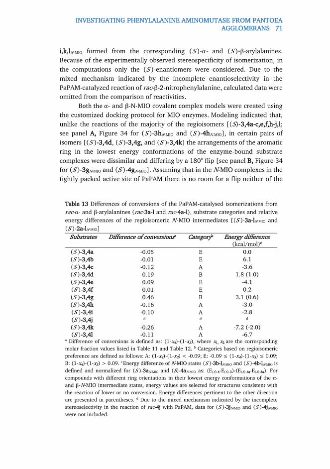
INVESTIGATING PHENYLALANINE AMINOMUTASE FROM PANTOEA AGGLOMERANS 71
i,k,lN-MIO formed from the corresponding (S )-α- and (S )-β-arylalanines.
Because of the experimentally observed stereospecificity of isomerization, in
the computations only the (S )-enantiomers were considered. Due to the
mixed mechanism indicated by the incomplete enantioselectivity in the
PaPAM-catalyzed reaction of rac-β-2-nitrophenylalanine, calculated data were
omitted from the comparison of reactivities.
Both the α- and β-N-MIO covalent complex models were created using
the customized docking protocol for MIO enzymes. Modeling indicated that,
unlike the reactions of the majority of the regioisomers [(S)-3,4a-c,e,f,h-j,l;
see panel A, Figure 34 for (S )-3hN-MIO and (S )-4hN-MIO], in certain pairs of
isomers [(S )-3,4d, (S )-3,4g, and (S )-3,4k] the arrangements of the aromatic
ring in the lowest energy conformations of the enzyme-bound substrate
complexes were dissimilar and differing by a 180° flip [see panel B, Figure 34
for (S )-3gN-MIO and (S )-4gN-MIO]. Assuming that in the N-MIO complexes in the
tightly packed active site of PaPAM there is no room for a flip neither of the
Table 13 Differences of conversions of the PaPAM-catalysed isomerizations from
rac-α- and β-arylalanines (rac-3a-l and rac-4a-l), substrate categories and relative
energy differences of the regioisomeric N-MIO intermediates [(S )-3a-lN-MIO and
(S )-2a-lN-MIO]
Substrates Difference of conversionsa Categoryb Energy difference (kcal/mol)c
(S )-3,4a -0.05 E 0.0 (S )-3,4b -0.01 E 6.1
(S )-3,4c -0.12 A -3.6
(S )-3,4d 0.19 B 1.8 (1.0)
(S )-3,4e 0.09 E -4.1
(S )-3,4f 0.01 E 0.2
(S )-3,4g 0.46 B 3.1 (0.6)
(S )-3,4h -0.16 A -3.0
(S )-3,4i -0.10 A -2.8
(S )-3,4j d d d
(S )-3,4k -0.26 A -7.2 (-2.0) (S )-3,4l -0.11 A -6.7
a Difference of conversions is defined as: (1-x4)-(1-x3), where x1, x2 are the corresponding
molar fraction values listed in Table 11 and Table 12. b Categories based on regioisomeric
preference are defined as follows: A: (1-x4)-(1-x3) < -0.09; E: -0.09 ≤ (1-x4)-(1-x3) ≤ 0.09;
B: (1-x4)-(1-x3) > 0.09. c Energy difference of N-MIO states (S )-3b-lN-MIO and (S )-4b-lN-MIO is
defined and normalized for (S )-3aN-MIO and (S)-4aN-MIO as: (E(S)-4-E(S)-3)-(E(S)-4a-E(S)-3a). For
compounds with different ring orientations in their lowest energy conformations of the α-
and β-N-MIO intermediate states, energy values are selected for structures consistent with
the reaction of lower or no conversion. Energy differences pertinent to the other direction
are presented in parentheses. d Due to the mixed mechanism indicated by the incomplete
stereoselectivity in the reaction of rac-4j with PaPAM, data for (S )-3jN-MIO and (S )-4jN-MIO
were not included.

72 INVESTIGATING PHENYLALANINE AMINOMUTASE FROM PANTOEA AGGLOMERANS
complete arylacrylate molecule nor of the aromatic ring only, it can be
predicted that in such cases one of the two N-MIO intermediate states is not
in its lowest energy state. Thus the calculated lowest energy differences in
such cases should be higher, corresponding to the allowed arrangements
without involving a sterically hindered ring flip (Table 13).
Statistical analysis of the experimental and computational data
revealed that compounds (S )-3,4a-i,k,l could be classified into three
categories (A, E, and B, Table 13) based on the difference of conversions
[defined as: (1-x4)-(1-x3), where x3, x4 are the corresponding molar fraction
values listed in Table 11 and Table 12]. The three categories: A: (1-x4)-(1-x3)
< -0.09; E: -0.09 ≤ (1-x4)-(1-x3) ≤ 0.09 and B: (1-x4)-(1-x3) > 0.09 could be
correlated with the regioisomeric preferences. One-way ANOVA test – treating
the categories (Table 13) as the independent and the conversion differences
(Table 11 and Table 12) as the dependent variables – indicated significant
difference between categories A and B at the α=0.050 level. In addition,
further nonparametric tests – having lower statistical power than parametric
methods but without the requirement of the normal distribution of the data –
were also performed. The Kruskal-Wallis ANOVA and Mann-Whitney U tests
resulted only somewhat higher p values (0.100 and 0.053, respectively) than
the threshold. Moreover, the median test showed also significant differences
between the categories. Overall, the statistical tests supported the finding that
categories A and B are truly separate, as it can be explicitly seen on the Figure
35.
Figure 35 Box-whisker plot depicting the energy differences of the
corresponding N-MIO intermediates [(S )-3a-i,k,l N-MIO and (S )-4a-i,k,l N-
MIO] as a function of the catalytic activity categories (A, B and E). Legend –
square: median; boxes: span between the lower and upper quartile,
whiskers: span of the entire set.

INVESTIGATING PHENYLALANINE AMINOMUTASE FROM PANTOEA AGGLOMERANS 73
The computational study extended with statistical analysis revealed
that the cases when one of the regioisomers was converted much faster (or is
the only one to be converted) the energetics of the regioisomeric N-MIO-type
enzyme-substrate complexes was one of the most important indicators of the
reaction outcome. In the case of the intermediate group (category E) involving
small aromatic moieties contribution of the steric effects are much less
pronounced.
The computational results suggest that the reactions of 2-substituted α-
phenylalanines [(S )-3d,g and (S )-4d,g: category B, Table 13 and Figure 35]
are strongly disfavored because the energy calculated for the α-N-MIO
intermediates [(S )-3d,gN-MIO] is much lower than that calculated for the
corresponding β-N-MIO intermediates [(S)-4d,gN-MIO]. This was in full
agreement with experimental observations indicating slow or no reaction from
the rac-2-substituted α-phenylalanines (rac-3d,g,j) with PaPAM, in contrast to
high conversions from 2-substituted rac-β-phenylalanines (rac-4d,g,j) under
the same conditions. The situation for the 3- and 4-substituted phenylalanines
with bulky substituents [(S )-3c,h,i,k,l and (S )-4c,h,i,k,l: category A, Table 13
and Figure 35] was the opposite. In good correlation with the experimental
regioisomeric preferences, much lower energies were calculated for the β-N-
MIO intermediates [(S )-4c,h,i,k,lN-MIO] than for the corresponding α-N-MIO
intermediates [(S )-3c,h,i,k,lN-MIO]. This could explain sluggish or no
conversion of β-arylalanines containing bulky substituents at positions 3 or 4
(rac-4c,h,i,k,l) with PaPAM and much higher conversions of the α compounds
(rac-3c,h,i,k,l) under the same conditions. In addition, energy differences
showed remarkable differences corresponding to the direction of reaction with
higher conversions and (assuming a disallowed flip of the aromatic ring, Table
13, values in parentheses) compared to the sterically non-restricted values,
and the hindered flip of the aromatic ring further amplified the difference in
reactivity mentioned before. Because of the hindered flip, in case of certain
regioisomeric amino acid pairs the aromatic ring in a given N-MIO
intermediate adopts different conformation depending on whether the actual
N-MIO intermediate is at the substrate side [step ii) in our postulated
mechanism] or at the product side [step iii) in our postulated mechanism].
This phenomenon would mean a hysteresis in the reaction mechanism which
is extremely rare.

74 KINETIC RESOLUTION OF PHENOTHIAZINE MOIETY CONTAINING SECONDARY ALCOHOLS
3.6. KINETIC RESOLUTION OF PHENOTHIAZINE MOIETY
CONTAINING SECONDARY ALCOHOLS
Phenothiazine is one of the valuable templates for a large number of
organic compounds which exhibit a wide variety of biological activities and
are used as antihistaminic, antipsychotic, anticholinergic, antiemetic, anti-
inflammatory, sedative or cytostatic drugs. [170] Especially in the case of
central nervous system drugs, [171] the number and positions of substituents
have a strong impact on the caused physiological effects. Therefore, the kinetic
resolution of a series of constitutional isomer 1-(10-ethyl-10H-phenothiazin-
yl)ethanols, rac-6a-c and of their acylated derivatives rac-7a-c were carried
out by certain lipase products (Figure 36 ). The absolute configurations of 7a,c
were predicted based upon QM/MM calculations in enzymatic environment.
The experimental work was carried out by Jürgen Brem.
3.6.1. KINETIC RESOLUTION OF SECONDARY ALCOHOLS
First, racemic 1-(10-ethyl-10H-phenothiazinyl)ethanols and their
corresponding acetates (rac-6a-c and rac-7a-c, respectively) were synthesized.
Next, the analytical scale, enantiomer selective lipase catalyzed acetylation of
rac-6a-c was screened in neat vinyl acetate using 14 free and immobilized
commercial lipases. Most of them exhibited low enantiomer selectivities and
activities, or were active in a non-selective manner. Out of them, the most
appropriate enzymes proved to be lipase A and B from Candida antarctica
(CaLA and CaLB). Next, the reaction media was tested with the most
aprropriate enzymes with vinyl acetate in the presence of several organic
solvents (Table 14 ). While CaLA catalyzed acylation with vinyl acetate as acyl
donor of rac-6a in polar solvents such as dichloromethane, chloroform,
dioxane proceeded with low activity (c<5% after 110 h), i.e. diethyl ether and
Figure 36 Biotransformation of the racemic 1-(10-ethyl-10H-
phenothiazinyl)ethanols rac-6a-c and their acetates rac-7a-c.
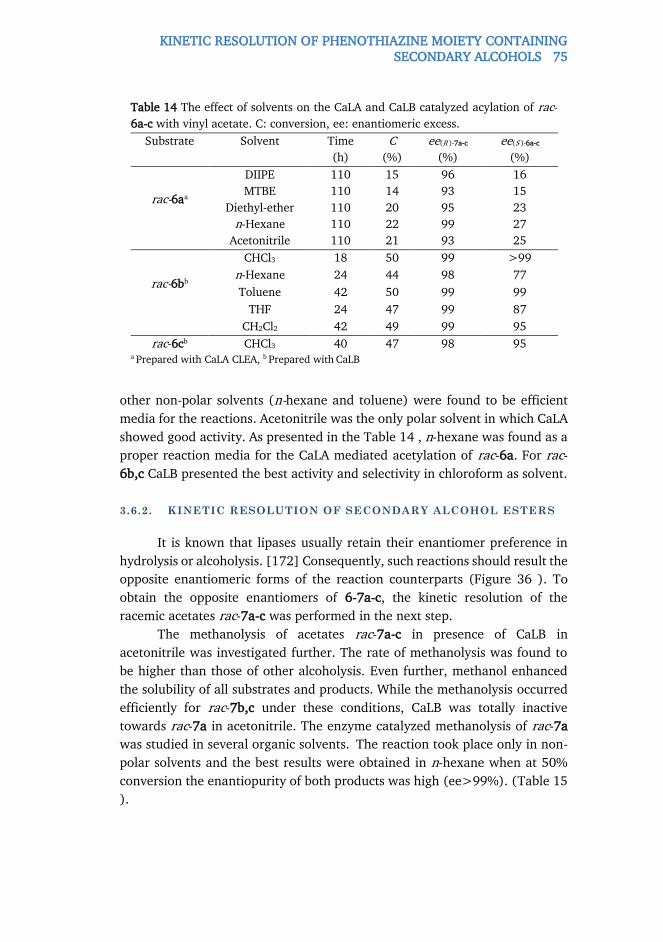
KINETIC RESOLUTION OF PHENOTHIAZINE MOIETY CONTAINING SECONDARY ALCOHOLS 75
other non-polar solvents (n-hexane and toluene) were found to be efficient
media for the reactions. Acetonitrile was the only polar solvent in which CaLA
showed good activity. As presented in the Table 14 , n-hexane was found as a
proper reaction media for the CaLA mediated acetylation of rac-6a. For rac-
6b,c CaLB presented the best activity and selectivity in chloroform as solvent.
3.6.2. KINETIC RESOLUTION OF SECONDARY ALCOHOL ESTERS
It is known that lipases usually retain their enantiomer preference in
hydrolysis or alcoholysis. [172] Consequently, such reactions should result the
opposite enantiomeric forms of the reaction counterparts (Figure 36 ). To
obtain the opposite enantiomers of 6-7a-c, the kinetic resolution of the
racemic acetates rac-7a-c was performed in the next step.
The methanolysis of acetates rac-7a-c in presence of CaLB in
acetonitrile was investigated further. The rate of methanolysis was found to
be higher than those of other alcoholysis. Even further, methanol enhanced
the solubility of all substrates and products. While the methanolysis occurred
efficiently for rac-7b,c under these conditions, CaLB was totally inactive
towards rac-7a in acetonitrile. The enzyme catalyzed methanolysis of rac-7a
was studied in several organic solvents. The reaction took place only in non-
polar solvents and the best results were obtained in n-hexane when at 50%
conversion the enantiopurity of both products was high (ee>99%). (Table 15
).
Table 14 The effect of solvents on the CaLA and CaLB catalyzed acylation of rac-
6a-c with vinyl acetate. C: conversion, ee: enantiomeric excess.
Substrate Solvent Time
(h)
C
(%)
ee(R )-7a-c
(%)
ee(S )-6a-c
(%)
rac-6aa
DIIPE 110 15 96 16
MTBE 110 14 93 15
Diethyl-ether 110 20 95 23
n-Hexane 110 22 99 27
Acetonitrile 110 21 93 25
rac-6bb
CHCl3 18 50 99 >99
n-Hexane 24 44 98 77
Toluene 42 50 99 99
THF 24 47 99 87
CH2Cl2 42 49 99 95
rac-6cb CHCl3 40 47 98 95 a Prepared with CaLA CLEA, b Prepared with CaLB

76 KINETIC RESOLUTION OF PHENOTHIAZINE MOIETY CONTAINING SECONDARY ALCOHOLS
3.6.3. ASSIGNEMENT OF ABSOLUTE CONFIGURATIONS
The absolute configurations of the (R )-1-(10H-phenothiazin-2-
yl)ethanol [non-ethylated analogue of (R )-6b] obtained by the reduction of
2-acetyl-phenothiazine in the presence of a chiral spiroborate ester catalyst
has already been described. [173] Since the sign of the specific rotation
proved to be independent of the nature of the N-substitution within a series
of (10-substituted-10H-phenothiazin-3-yl)ethanols, [174] the absolute
configuration of the N-ethylated 6b could be assigned to (R ). Also, the
absolute configuration of 6d was unambiguously assigned to (R ) by X-ray
crystallography. [174]
Absolute configuration assignment by QM/MM within the CaLB
enzymatic environment was applied for rac-6a and rac-6d. Rac-6d served as a
test compound to validate the protocols. The tetrahedral intermediates
(THI’s), formed by the reaction of the (R )- or (S )-alcohol with the acyl
enzyme, were used to model the transitions states. A THI was considered
catalytically active when the H atom on His224 kept equal and short (~2 Å)
distances from the oxygen atoms of Ser105 and the oxygen atom of the
substrate alcohol, as illustrated on the Figure 37 . First, the tetrahedral
intermediates were constructed within the CaL-B structure and systematic
conformation search was performed for each tetrahedral intermediate model
by molecular mechanics within a rigid enzyme environment. Next, the best
conformations were refined by two-
layer ONIOM calculations (HF/3 –
21g:uff; 955 atoms). Minimized
QM/MM potential energies indicated
the faster forming acetates of 6a and
6d with (R ) configuration (Table 16).
Based on these data and on the
consistent (+)-signs of optical
Table 15 Alcoholysis of rac-7a-c with methanol and CaLB in n-hexane or
acetonitrile. C: conversion, ee: enantiomeric excess.
Racemic
compound
Solvent Time
(h)
c
(%)
ee(R )-6
(%)
ee(S )-7
(%)
7a n-Hexane 420 50 >99 >99
7b n-Hexane 24 50 >99 >99
Acetonitrile 20 50 >99 >99
7c n-Hexane 24 50 >99 >99
Acetonitrile 20 50 >99 >99
Table 16 Comparison of the lowest
energy THIs of each antipode of 6a and
6d obtained with QM/MM calculations.
Energies are in kcal mol-1.
Configuration (R ) (S )
6a 0.0 7,9
6d 0.0 14,5

BIOIMPRINTING OF LIPASES IN POLY(VINYL ALCOHOL) NANOFIBER 77
rotation for (R )-6a,b,d, the (R )-(+)-absolute configuration was assigned to
the faster reacting enantiomer of 1-(10-ethyl-10H-phenothiazin-4-yl)ethanol,
6c as well.
3.7. BIOIMPRINTING OF LIPASES IN POLY(VINYL
ALCOHOL) NANOFIBER
One of the most successful strategies to enhance enzyme activity and
selectivity during immobilization involves shape tuning active sites by
molecular imprinting. [175] This bioimprinting effect on lipases can be
rationalized on the basis of the generally accepted hypothesis of the interfacial
activation mechanism. Thus, exploiting bioimprinting phenomena could
provide enhanced immobilized forms of CaLB providing molecular level
insights into the properties in the meantime. Although a number of examples
exist on enzyme immobilization on the surface of nanofibers, [74] only a few
experiments were reported on lipase entrapment within nanofibers. Thus,
immobilization of CaLB in electrospun poly(vinyl alcohol) (PVA) nanofibers
was attempted with different additives with aspired bioimprinting features.
Potential bioimprinting effects were investigated with molecular modeling
tools. Experimental work was carried out by Diána Weiser.
Figure 37 Putative catalytically active tetrahedral intermediate (THI) of (R )-6a
within the active site of CaLB determined by QM/MM (HF/3-21g:uff). Note the
equal distance of the H atom of His224 from the oxygen atom of Ser105 and the
substrate alcohol.

78 BIOIMPRINTING OF LIPASES IN POLY(VINYL ALCOHOL) NANOFIBER
3.7.1. TESTING POTENTIAL BIOIMPRINTING AGENTS
After selecting 5% lipase loading as optimal during PVA entrapment,
nanofibers were prepared containing CaLB and rationally selected additives
as bioimprinting agents (Table 17 , Figure 38 ).
Tween 80, Brij 30, PEG, olive oil, and lauric acid were studied as
potential imprinting agents for lipases in sol-gel matrices earlier (Figure 38 ).
[176] In addition to the fact that organosilane precursors were able to
influence the sol-gel matrix structure, it was assumed that they could act as
bioimprinting agents as well. The fact that lipases could accept alkoxysilanes
as substrates during oligomerization of dimethlydiethoxysilane (DMDEOS)
supported further this hypothesis. Therefore, four different organosilanes
Figure 39 Kinetic resolution of racemic secondary alcohols (rac-8a,b) catalyzed by
lipase biocatalysts immobilized by entrapment in PVA nanofibers with vinyl acetate
or vinyl butyrate as acylating agents.
Figure 38 Additives of PVA immobilization

BIOIMPRINTING OF LIPASES IN POLY(VINYL ALCOHOL) NANOFIBER 79
(TEOS, PTEOS, OTEOS, and DMDEOS, Figure 38 ) were investigated for the
first time as potential bioimprinting agents. Catalytic properties of the formed
biocatalysts were tested in kinetic resolution of racemic 1-phenylethanol (rac-
8a) using vinyl acetate as the acylating agent (Figure 39 ).
Immobilization by electrospun entrapment within PVA nanofibers
enhanced significantly the enzyme activity of entrapped CaLB (Table 17 ).
Even the simple PVA nanofiber-entrapped CaLB biocatalysts showed thirty
times higher activity than the non-immobilized CaLB powder. Introduction of
the bioimprinting agents further enhanced the catalytic properties of PVA-
entrapped CaLB in each cases. PEG 400 proved to be an especially efficient
additive with CaLB resulting a biocatalyst with more than fifty times higher
specific activity as the native powder with enhanced ee(R )-9a of the product
(99.9 %; compared to 98.7% with the native powder). Among organosilanes,
PTEOS and OTEOS exhibited significant bioimprinting effects on CaLB, with
more than forty-four times enhanced specific activity as the native enzyme
affording the product (R )-9 in high ee(R )-9a (99.5 %).
Next, the study was extended with 1-phenylpropan-2-ol (rac-8b) as
another alcohol and with vinyl butyrate as a second acylating agent. Catalysts
were non-immobilized CaLB or entrapped CaLB either in pure, or Brij 30, PEG
400, PTEOS, OTEOS doped PVA (Table 18 ). As expected, the overall
characteristics of the CaLB biocatalysts changed compared to the KR of rac-8a
with vinyl acetate, but the most powerful bioimprinting agents, Brij 30 and
Table 17 Effect of potential bioimprinting agents on performance of PVA-
entrapped CaLB in kinetic resolution of 1-phenylethanol rac-8a with vinyl acetate
(reaction time 2 h)
Additive c
(%)
ee(R)-9a
(%)
UBa
(U g-1)
YAb
(%) c 8.3 98.7 5.7 100 d 15.0 99.8 10.4 3605
Brij 30 13.0 99.8 9.0 3124
Tween 80 16.6 99.9 11.4 3983
PEG 400 21.3 99.9 14.7 5118
PEG 1000 14.8 99.5 10.2 3553
TEOS 8.8 98.6 6.1 2118
PTEOS 18.6 99.5 12.8 4460
OTEOS 18.8 99.5 12.9 4499
DMDEOS 12.3 99.7 8.5 2957 a UB is the effective specific activity of the biocatalyst [UB= (nrac×c)(t×mB)-1], mB is the mass
of the applied biocatalyst, UE is the effective specific activity of the crude, non-immobilized
enzyme [UE= (nrac×c)(t×mb)-1] mE is the mass of the applied crude biocatalyst ; b YA is the
activity yield [YA= 100×(UB×mB)/(UE×mE’)]; c Crude, non-immobilized CaLB; a CaLB
entrapped in PVA nanofiber, without any additive.
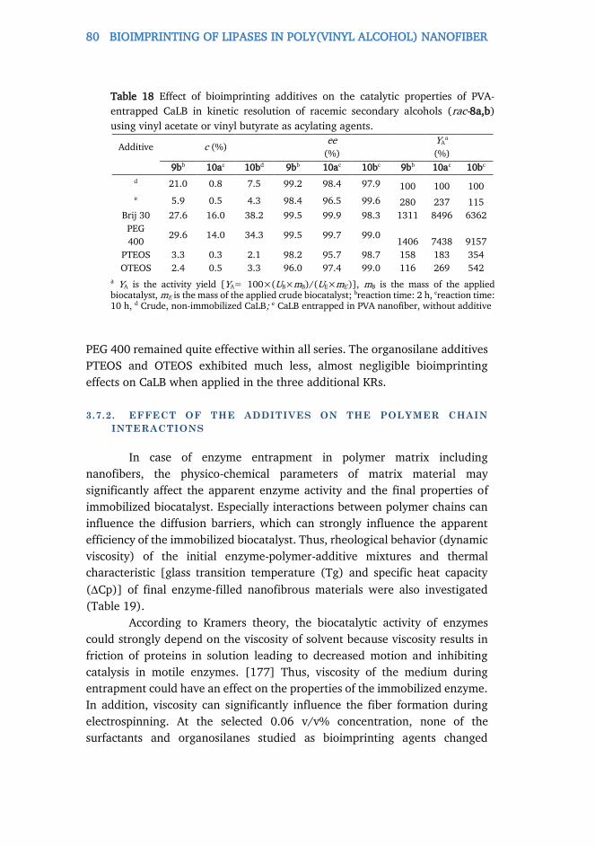
80 BIOIMPRINTING OF LIPASES IN POLY(VINYL ALCOHOL) NANOFIBER
PEG 400 remained quite effective within all series. The organosilane additives
PTEOS and OTEOS exhibited much less, almost negligible bioimprinting
effects on CaLB when applied in the three additional KRs.
3.7.2. EFFECT OF THE ADDITIVES ON THE POLYMER CHAIN
INTERACTIONS
In case of enzyme entrapment in polymer matrix including
nanofibers, the physico-chemical parameters of matrix material may
significantly affect the apparent enzyme activity and the final properties of
immobilized biocatalyst. Especially interactions between polymer chains can
influence the diffusion barriers, which can strongly influence the apparent
efficiency of the immobilized biocatalyst. Thus, rheological behavior (dynamic
viscosity) of the initial enzyme-polymer-additive mixtures and thermal
characteristic [glass transition temperature (Tg) and specific heat capacity
(Cp)] of final enzyme-filled nanofibrous materials were also investigated
(Table 19).
According to Kramers theory, the biocatalytic activity of enzymes
could strongly depend on the viscosity of solvent because viscosity results in
friction of proteins in solution leading to decreased motion and inhibiting
catalysis in motile enzymes. [177] Thus, viscosity of the medium during
entrapment could have an effect on the properties of the immobilized enzyme.
In addition, viscosity can significantly influence the fiber formation during
electrospinning. At the selected 0.06 v/v% concentration, none of the
surfactants and organosilanes studied as bioimprinting agents changed
Table 18 Effect of bioimprinting additives on the catalytic properties of PVA-
entrapped CaLB in kinetic resolution of racemic secondary alcohols (rac-8a,b)
using vinyl acetate or vinyl butyrate as acylating agents.
Additive c (%) ee
(%)
YAa
(%)
9bb 10ac 10bd 9bb 10ac 10bc 9bb 10ac 10bc
d 21.0 0.8 7.5 99.2 98.4 97.9 100 100 100 e 5.9 0.5 4.3 98.4 96.5 99.6 280 237 115
Brij 30 27.6 16.0 38.2 99.5 99.9 98.3 1311 8496 6362
PEG
400 29.6 14.0 34.3 99.5 99.7 99.0
1406 7438 9157
PTEOS 3.3 0.3 2.1 98.2 95.7 98.7 158 183 354
OTEOS 2.4 0.5 3.3 96.0 97.4 99.0 116 269 542
a YA is the activity yield [YA= 100×(UB×mB)/(UE×mE’)], mB is the mass of the applied biocatalyst, mE is the mass of the applied crude biocatalyst; breaction time: 2 h, creaction time: 10 h, d Crude, non-immobilized CaLB; e CaLB entrapped in PVA nanofiber, without additive

BIOIMPRINTING OF LIPASES IN POLY(VINYL ALCOHOL) NANOFIBER 81
significantly the viscosity of the solution compared to the pure PVA solution,
except DMDEOS causing a slight increase of the solution’s viscosity. Therefore,
effects of the additives on the entrapped lipase could not be attributed to a
change of the viscosity of the medium. Among the additives, only PEG 400
had a significant effect on the Tg of nanofibers (a decrease of Tg 25.5 oC,
compared to the PVA fiber without additive). In the case of PEG 400 as
additive, the specific heat capacity also decreased significantly, indicating
weaker interactions between polymer chains. The higher degree of branching
within the entrapment matrix could result in lower diffusion barrier for
substrate / product mobility, thereby contributing to the increase of apparent
activity of the immobilized lipases. In summary, except for PEG400, there is
no strong evidence that the activity enhancement of the immobilized enzyme
products would stem mainly from the modification of the polymer chain
interactions.
3.7.3. MOLECULAR MODELING OF THE POTENTIAL
BIOIMPRINTING EFFECTS
To understand the molecular level reasons of the outstanding activity
enhancement of Brij 30 and PEG 400 and the moderate activity enhancement
of substituted organosilanes with relatively large apolar substituents, such as
OTEOS and PTEOS, molecular docking studies were performed on the active
sites of the open and a closed lid structures of CaLB. Our goal was to draw
conclusions based on the binding poses of Brij 30, PEG 400, the partially
hydrolyzed derivatives of OTEOS, PTEOS, DMDEOS, and the bulkiest product,
(R )-10b. Poses found for Brij 30 and PEG 400 in the open structure of CaLB
Table 19 The effect of different additives on the viscosity, glass transition
temperature (Tg) and specific heat capacity (Cp) of electrospun PVA nanofibers.
Additive Viscosity
(mPas)
Tg
(oC)
Cp
(J g-1oC-1)
- 500.9 71.4 0.30
Brij 30 551.9 66.8 0.24
Tween 80 532.3 67.9 0.29
PEG 400 516.7 45.9 0.08
PEG 1000 525.8 67.7 0.18
TEOS 493.5 71.0 0.28
PTEOS 496.6 70.7 0.29
OTEOS 500.4 70.6 0.30
DMDEOS 575.2 70.8 0.30

82 BIOIMPRINTING OF LIPASES IN POLY(VINYL ALCOHOL) NANOFIBER
Figure 40 Docking of various additives and the product (R )-10b into CaLB with open
lid conformation (refined model of 5A6V: chain A, in cyan) and CaLB with closed lid
conformation (refined model of 5A6V: chain B, in light brown). Panels: A) Brij 30 in
A B
D C
E F
G H

BIOIMPRINTING OF LIPASES IN POLY(VINYL ALCOHOL) NANOFIBER 83
closed CaLB; B) Brij 30 in open CaLB; C) PEG 400 in closed CaLB; D) PEG 400 in open
CaLB; E) overlay of poses of partially hydrolyzed OTEOS in both CaLB forms; F)
overlay of poses of partially hydrolyzed PTEOS in both CaLB forms; G) overlay of poses
of partially hydrolyzed DMDEOS in both CaLB forms; H) overlay of poses of (R )-10b
in both CaLB forms.
(Figure 40 , A and C, respectively) were in good agreement with the X-ray
structure of CaLB complexed with a long chain methylpenta(oxyethyl)
heptadecanoate substrate analogue (PDB code: 1LBT) thus supporting our
conclusions concerning the bioimprinting effect of these two additives.
The docking results revealed that PEG 400 and Brij 30 have distinct
arrangements in the case of open CaLB active site (Figure 40 , A and C,
respectively) and in the closed lid conformations (Figure 40 , B and D,
respectively. It could be seen that both additives embrace the mobile lid loop
when binding to the closed form, and it is probable that during the
electrospinning most CaLB molecules adopt such form. This means at the same
time protection of the closed reactive form during entrapment and also
reserving an enlarged space available for the active site lid after washing out
the additive from the active site. The removal of the additives is obvious,
otherwise they would act as inhibitors. This double effect – conserving the
reactive active site conformation but providing free space to active site lid
movements – can significantly improve the catalytic activity during the
acylation reactions. Thus the docking study suggests two synergistic effects
(reactive conformation stabilization and mobility enhancement of the active
lid) resulting in the beneficial bioimprinting effects on CaLB and probably on
the other lipases as well.
Based on the experimental results, significant differences were
expected between the most effective medium length PEG units-containing
additives (Figure 40 , A-D) and the organosilane compounds (Figure 40 E-G).
The computed spatial arrangements of the organosilanes were compared to
the computed arrangement of the product (R )-10b (Figure 40 , H). In all the
cases it is clearly visible that the Si atom of the partially hydrolyzed derivatives
can occupy a location next to the catalytic serine side chain. Although
DMDEOS could be docked also with its Si atom close to the catalytic triad, it
seems not large enough to significantly stabilize the active conformation of
the active site, while OTEOS and PTEOS share similar sizes and overall
arrangements compared to (R )-10b in both active sites, therefore suggesting
overall active site stabilization during immobilization by substrate mimicry.
This effect, however, without providing the space for free movements of the
active site lid, could cause only much smaller and variable activation. The
facilitation of the lid-loop movement ultimately suggests that in the case of

84 COMPUTER-AIDED SELECTION OF SOL-GEL PRECURSOR
CaLB, the open conformation may not be the catalytically active one. Because
the stabilization of the closed structure would ruin enzyme activity, if it was
the inactive one (as it is however for the majority of lipases). But, with a
catalytically active closed conformation, and with the facility for the enzyme
to alternate between the open closed forms, ligand association/dissociation
would be eligible. Moreover, the existence of such a catalytically active closed
enzyme conformation would mean the protection of the reactive
intermediates, thus it would be consistent with the concept of “negative
catalysis”. [178]
This was the first time to ever hypothesize that minor amounts of well
selected additives can cause significant activity enhancement of the entrapped
CaLB by two enzyme structural factors: i) by stabilizing the integrity of the
active center; ii) and by providing extra room for the mobile active site-
covering lids after their removal, thus enhancing the turnover frequency of the
reaction.
3.8. COMPUTER-AIDED SELECTION OF SOL-GEL
PRECURSOR
To find organosilanes, serving not only as entrapment matrix precursors
for another kind of entrapment, sol-gel immobilization, but also as
bioimprinting agents, binding poses of several partially hydrolyzed
organosilanes were predicted within the open and closed lid structures of
CaLB. An advanced method was introduced to model the effects of such
bioimprinting agents. This protocol was based on our previous, exploratory
method applied to PVA nanofiber entrapment. Induced-fit docking and
subsequent scoring with a modified version of MM-GBSA scoring (referred
simply as ΔEb) was introduced. Experimental work was done by Flóra Nagy.
3.8.1. VIRTUAL SCREENING OF ORGANISLANE PRECURSORS FOR
EFFICIENT ENTRAPMENT
Within the open structure of CaLB each of the partially hydrolyzed
silanes behaved similarly, in full agreement with our previous study. Within
the open structure of CaLB, docking revealed only substrate mimicking poses.
Every silane precursor occupied the active site and formed hydrogen bonds
with the catalytic triade and its close proximity (ΔE b-o, Table 20 ).
Within the closed structure of CaLB, docking revealed the already
identified substrate mimicking poses. In addition, several partially hydrolyzed

COMPUTER-AIDED SELECTION OF SOL-GEL PRECURSOR 85
silanes could occupy multiple poses interacting directly with lid-loop too.
These results reproduced our previous finding again, albeit with modest
differences (Figure 41).
Consequently, our screening was focused on selecting those particular
precursors which can adopt both binding poses in the closed form of CaLB
with high affinities, but slightly preferring the lid-loop-interacting pose (ΔΔEb-
cc-cl). Balanced affinity was important to manifest both effects with comparable
frequency. The major enhancement in the biocatalyst’s activity, however, was
expected from the enlarged space for the lid-loop movements after
entrapment of enzymes complexed with bioimprinting agents in lid-loop-
interacting poses. In the Table 20, binding energies of the partially hydrolyzed
forms of potential silane precursors are presented within the open and closed
conformations of CaLB. On the basis of the above considerations, the four best
organosilanes resulting in partially hydrolyzed forms with ΔΔEb-cc-cl in the
range of -2 – 6 kcal mol-1 (TEOS, PTEOS, DMDEOS, and OTEOS) were
selected for further investigations.
Next, statistical tools were applied to determine optimum compositions
of organosilane mixtures for entrapment. Biocatalytic properties and
durability of the new preparations were evaluated in kinetic resolutions of two
racemic alcohols and racemic amines, using vinyl acetate and isopropyl 2-
ethoxyacetate as acylating agents, both in batch and continuous-flow systems.
Optimized enzyme preparates showed outstanding performance, for example
Table 20 Binding energy values of various partially hydrolysed silane precursors in open-lid and closed-lid conformations of CaLB (refined models of 5A6V chain A and chain B, respectively).
Ra ΔEb-ob/
kcal mol-1 ΔEb-cc
c/
kcal mol-1 ΔEb-cl
d/
kcal mol-1 ΔΔEb-cc-cl
e/ kcal mol-1
octadecyl -27.1 -56.6 -16.6 -40.0 propyl -30.1 -30.7 -26.5 -4.2 - -27.3 -31.2 -29.9 -1.3 phenyl -23.2 -29.0 -31.0 2.0 dimethyl -17.9 -24.7 -27.0 2.3 octyl -21.8 -28.1 -33.3 5.2 decyl -15.5 -27.8 -34.1 6.3 vinyl -25.3 -25.7 -33.2 7.5 methyl -32.0 -29.9 -42.7 12.8 dodecyl -23.4 f -36.6 -
hexyl -25.6 -23.1 f -
a Substituent(s) in partially hydrolysed alkoxysilanes [Si(OEt)2(OH)2, R-Si(OEt)(OH)2 or R2-Si(OEt)(OH)]. b Binding energy values within open-lid CaLB structure. c Binding energy values within active centre of closed-lid CaLB structure in substrate mimicking pose. d Binding energy values within the active centre of closed-lid CaLB structure in lid-bound pose. d Differences of binding energy values of lid-bound and substrate mimicking poses in the closed-lid CaLB structure. f No such pose was identified by the docking calculations

86 COMPUTER-AIDED SELECTION OF SOL-GEL PRECURSOR
retaining more than 91% of its initial activity after 10 cycles of use.
Astonishingly, certain immobilized enzymes retained their high activity up to
100 °C meanwhile preserving also excellent enantioselectivity.
A
B
Figure 41 Major docking poses of two partially hydrolyzed silane
precursors, A) PhSi(OEt)(OH)2 (hydrolyzed form of PTEOS) and
B) CH3(CH2)7Si(OEt)(OH)2 (hydrolyzed form of OTEOS) in CaLB with closed
conformation (refined model of 5A6V: chain B). In both panels, the full structure
with the bound molecule mimicking the pose of a substrate is in cyan, the loop in
the model with the bound molecule in lid-loop-interacting pose is in light brown,
Ser130 is colored in red and His249 in blue.
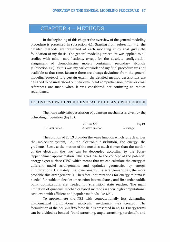
OVERVIEW OF THE GENERAL MODELING PROCEDURE 87
CHAPTER 4 – METHODS
In the beginning of this chapter the overview of the general modeling
procedure is presented in subsection 4.1. Starting from subsection 4.2, the
detailed methods are presented of each modeling study that gives the
foundation of my thesis. The general modeling procedure was applied to all
studies with minor modifications, except for the absolute configuration
assignment of phenothiazine moiety containing secondary alcohols
(subsection 4.8), as this was my earliest work and my final procedure was not
available at that time. Because there are always deviations from the general
modeling protocol to a certain extent, the detailed method descriptions are
designed to be understood on their own to aid comprehension, however cross
references are made when it was considered not confusing to reduce
redundancy.
4.1. OVERVIEW OF THE GENERAL MODELING PROCEDURE
The non-realtivistic description of quantum mechanics is given by the
Schrödinger equation (Eq 13).
𝐻Ψ = 𝐸Ψ Eq 13 H: Hamiltonian ψ : wave function E: energy
The solution of Eq 13 provides the wave function which fully describes
the molecular system, i.e. the electronic distribution, the energy, the
gradients. Because the motion of the nuclei is much slower than the motion
of the electrons, the two can be decoupled according to the Born–
Oppenheimer approximation. This gives rise to the concept of the potential
energy hyper surface (PES) which means that we can calculate the energy at
different nuclei arrangements and optimize geometries by energy
minimizations. Ultimately, the lower energy the arrangement has, the more
probable this arrangement is. Therefore, optimizations for energy minima is
needed for stable molecules or reaction intermediates, and first-order saddle
point optimizations are needed for stransition state seaches. The main
limitation of quantum mechanics based methods is their high computational
cost, even with efficient and popular methods like DFT.
To approximate the PES with computationally less demanding
mathematical formulations, molecular mechanics was created. The
formulation of the AMBER ff96 force field is presented in Eq 14. Energy terms
can be divided as bonded (bond stretching, angle stretching, torsional), and
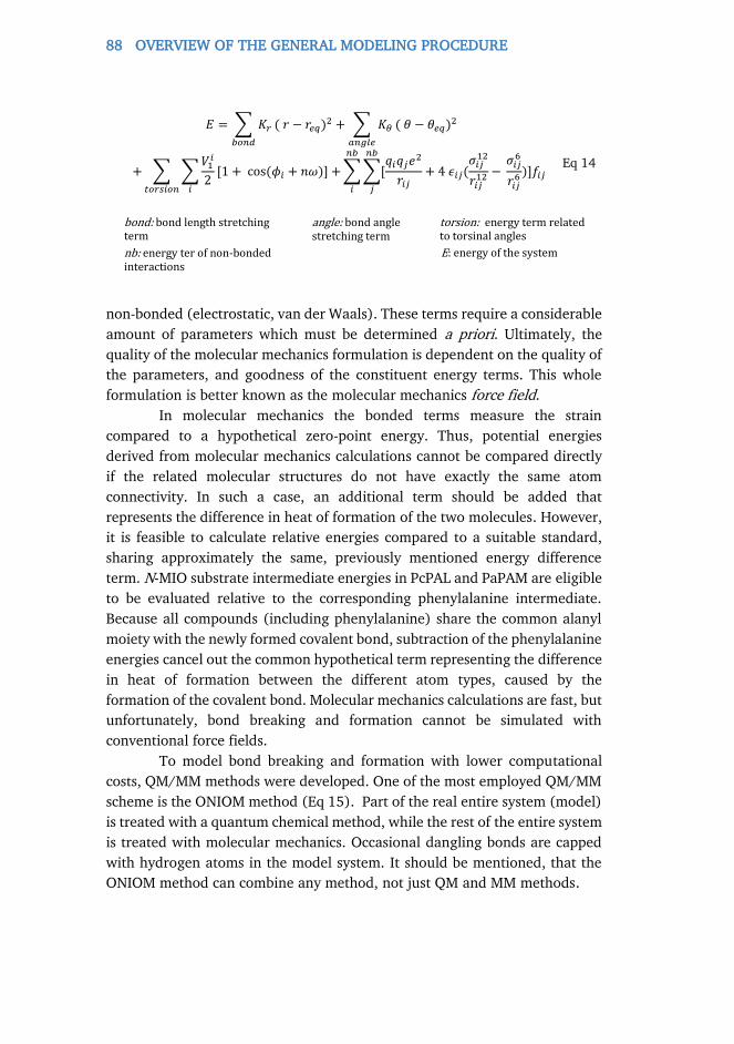
88 OVERVIEW OF THE GENERAL MODELING PROCEDURE
non-bonded (electrostatic, van der Waals). These terms require a considerable
amount of parameters which must be determined a priori. Ultimately, the
quality of the molecular mechanics formulation is dependent on the quality of
the parameters, and goodness of the constituent energy terms. This whole
formulation is better known as the molecular mechanics force field.
In molecular mechanics the bonded terms measure the strain
compared to a hypothetical zero-point energy. Thus, potential energies
derived from molecular mechanics calculations cannot be compared directly
if the related molecular structures do not have exactly the same atom
connectivity. In such a case, an additional term should be added that
represents the difference in heat of formation of the two molecules. However,
it is feasible to calculate relative energies compared to a suitable standard,
sharing approximately the same, previously mentioned energy difference
term. N-MIO substrate intermediate energies in PcPAL and PaPAM are eligible
to be evaluated relative to the corresponding phenylalanine intermediate.
Because all compounds (including phenylalanine) share the common alanyl
moiety with the newly formed covalent bond, subtraction of the phenylalanine
energies cancel out the common hypothetical term representing the difference
in heat of formation between the different atom types, caused by the
formation of the covalent bond. Molecular mechanics calculations are fast, but
unfortunately, bond breaking and formation cannot be simulated with
conventional force fields.
To model bond breaking and formation with lower computational
costs, QM/MM methods were developed. One of the most employed QM/MM
scheme is the ONIOM method (Eq 15). Part of the real entire system (model)
is treated with a quantum chemical method, while the rest of the entire system
is treated with molecular mechanics. Occasional dangling bonds are capped
with hydrogen atoms in the model system. It should be mentioned, that the
ONIOM method can combine any method, not just QM and MM methods.
𝐸 = ∑ 𝐾𝑟𝑏𝑜𝑛𝑑
(𝑟 − 𝑟𝑒𝑞)2 + ∑ 𝐾𝜃
𝑎𝑛𝑔𝑙𝑒
(𝜃 − 𝜃𝑒𝑞)2
+ ∑ ∑𝑉1𝑖
2[1 +cos(𝜙𝑖 + 𝑛𝜔)] +
𝑖𝑡𝑜𝑟𝑠𝑖𝑜𝑛
∑∑[𝑞𝑖𝑞𝑗𝑒
2
𝑟𝑖𝑗+ 4𝜖𝑖𝑗(
𝜎𝑖𝑗12
𝑟𝑖𝑗12 −
𝑛𝑏
𝑗
𝑛𝑏
𝑖
𝜎𝑖𝑗6
𝑟𝑖𝑗6 )]𝑓𝑖𝑗
Eq 14
bond: bond length stretching term
nb: energy ter of non-bonded interactions
angle: bond angle stretching term
torsion: energy term related to torsinal angles
E: energy of the system

OVERVIEW OF THE GENERAL MODELING PROCEDURE 89
𝐸𝑂𝑁𝐼𝑂𝑀 = 𝐸𝑄𝑀,𝑚𝑜𝑑𝑒𝑙 + 𝐸𝑀𝑀,𝑟𝑒𝑎𝑙 − 𝐸𝑀𝑀,𝑚𝑜𝑑𝑒𝑙 Eq 15
QM: quantum chemical method MM: molecular mechanics method
model: model system real: the entire system
After we have chosen the appropriate method for the description of
the PES, the search for relevant conformations, or for the global minimum
conformation may be pursued. Two established methods for the sampling of
the PES are molecular dynamics and Monte Carlo simulations. Molecular
dynamics simulates the motion of a model accordingly to the classical
Newton’s equation to result a trajectory, Monte Carlo methods simulate the
dynamics of the system by stochastic motion of the system to assemble a non-
time-dependent trajectory. Samples from trajectories are subjected to energy
minimizations to evaluate the obtained conformation.
4.1.1. ENZYME STRUCTURE MODELING
The heart of this modeling process is the intact model of the enzyme.
Good quality X-ray structures are available for PaPAM (PDB: 3UNV, [93]) and
CaLB (5A6V, [27] 1LBT, [28]). On the other hand, the partial homology
model of PcPAL and the full homology model of RxPAL was required.
Homology modeling methods use the phenomenon that evolutionary-
related proteins share similar structures. The method predicts the 3D structure
of a protein only available with its amino acid sequence. The target sequence
is aligned with a related, homologous protein sequence with experimentally
determined structure which is referred to as a template. Homology modeling
can generally be performed in six steps:
1. Template selection
2. Sequence alignment
3. Backbone generation
4. Loop modeling
5. Side chain modeling
6. Validation
For template selection, a sequence identity of at least 30% is desired.
There are many strategies for side chain modeling. Two of the most frequently
applied methods are simulated annealing and the optimization of side chain
rotamers. Simulated annealing consists of molecular dynamics simulations
with applied constraints at rapidly increasing temperatures, and after an
optional plato at rapidly decreasing temperatures reaching room temperature
finally. On the other hand, side chain rotamer optimization consists of a Monte

90 OVERVIEW OF THE GENERAL MODELING PROCEDURE
Carlo optimization of a restricted number of the most probable side chain
conformations, also known as rotamers. Simulated annealing is a robust and
well established method, however the final conformation is obtained in a
random manner, and may not resemble the dominant conformations of the
„real” structure. On the other hand, rotamer optimizations may converge
towards relevant enzyme conformations, even small-scale backbone
movements can be incorporated when the variation of the Cα and Cβ bond
vector is incorporated during rotamer generation. However, rotamer
optimizations may face convergence issues and may require considerable
more time, especially with rapidly increasing enzyme size. In summary,
simulated annealing can be the better choice, when there is only low
resolution information available about ternary structure of the enzyme,
and/or the lower-fidelity structures are sufficient. Rotamer optimization can
be a better choice, when higher-fidelity structures are needed, and we have
the necessary amount of high resolution information, i.e. in the case of partial
homology modeling.
Validation consists of the comparison of certain quantities of the
homology models to experimentally derived distributions, i.e. the
experimentally derived distribution of ψ and φ back bone dihedral angles
(Ramachandran plot).
If any of steps 3-5 are omitted, or the whole procedure is applied only
to a segment of the target sequence, the method may be referred as partial
homology modeling.
The obtained structures may be completed and adjusted in a
straightforward manner, i) hydrogen atoms are added and bond orders are
assigned, ii) artefacts of the protein crystallization procedure are removed
(not a case of homology models) iii) hydrogen bond network, tautomeric
states, side chain conformations of amino acids forming contacts with crystal
mates, and ionization states are determined and optimized corresponding to
the experimental assay conditions.
The modeling of ions around MIO enzymes is described as follows. The
charge, and ultimately the number of ions, is determined with Eq 3 in the first
shell of counterions. Dimensions of the first shell are obtained from the
literature. Finally, only the strongest binding ions are retained from BION
server [156] calculations to match the previously determined number. The pH
determination, hydrogen bond and tautomeric state prediction, and ion
placement depend on each other and should be carried out in an iterative
manner, self-consistently. The remaining number of counterions is determined
on the size of the cell with Eq 3. The obtained model is than solvated and
equilibrated with molecular dynamics. The standard equilibration protocol
looks as follows: 112 ps simulation at 10 K, NVT ensemble; 12 ps simulation

OVERVIEW OF THE GENERAL MODELING PROCEDURE 91
at 10 K, NPT ensemble, 36 ps simulation at room temperature, NPT ensemble.
Harmoinc potentials were applied to atoms of the enzymes during simulation,
except for polar surface hídrogens.
The more appropriate solvation of the active centres and their
immediate vicinity were achieved through grand canonical Monte Carlo
simulations. Spherical models of the active centers are cut off from the enzyme
models with a radius of approximately 27 Å which is believed to be big enough
to neglect the error of truncation, even with considerable remaining charge.
This was a compromise, because MIO enzymes have enormous size and net
charge. To obtain an electroneutral enzyme model, either the model should
be that big, that softwares couldn’t handle it, or the elimination of the net
charge of the system would introduce unknown errors. CaLB was used in
organic solvents experimentally, therefore ionization, explicit solvation and
ion placement was omitted, only implicit solvation models, if any, were used.
4.1.2. ENZYME-LIGAND COMPLEX MODELING
Enzyme-ligand complexes were obtained with molecular docking.
Molecular docking predicts the three-dimensional models of intermolecular
biological complexes in the bound state. Regarding small molecules in
general, the doking is carried out with a flexibile ligand, and a frozen receptor.
However, enzyme flexibility can be modeled with downscaled van der Waals
radii, or the sidechain prediction and minimization of the receptor during
docking.
The final, lowest energy binding states can be selected for each ligand
with scoring with the MM-GBSA method. The standard MM-GBSA method
involves the minimization, incorporating also implicit solvation, of the ligand-
receptor complex, followed by the subsequent minimization of the receptor
and the ligand individually after separation. Finally, the MM-GBSA score
(simply denoted as ΔEb) is calculated according to Eq 16,
∆𝐸𝑏 = 𝐸𝑐𝑜𝑚𝑝𝑙𝑒𝑥 − 𝐸𝑟𝑒𝑐𝑒𝑝𝑡𝑜𝑟 − 𝐸𝑙𝑖𝑔𝑎𝑛𝑑 Eq 16
where the three terms are the final energies of the previously mentioned
minimizations, respectively. However, the during minimization, the ligand
relaxes in the adopted conformation which was forced by the receptor.
Unfortunately, this conformation often does not match with dominant
conformation adopted in the solution.
To approximate the true binding energies with higher fidelity, our
modifications involved i) the substitution of the ligand energy term with a

92 HOMOLOGY MODELING OF MIO CONTAINING ENZYMES
value obtained after mixed Monte Carlo/low-mode conformational search and
ii) the substitution of the receptor energy term uniformly for all ligands with
the corresponding energy of an apoenzyme structure.
Most of the molecular modeling work was done with different versions
of the Schrödinger Suite modeling suite. Frequently used modules were Glide,
[179, 180, 181, 182] MacroModel, [183] Prime, [184] Protein Preparation
Wizard, [185, 186] ProPKa, [187] Desmond. [188, 189, 190] Images were
created with Maestro [191] and PyMOL. [192] All statistical analyses were
carried out with the help of Statistica. [193]
4.2. HOMOLOGY MODELING OF MIO CONTAINING
ENZYMES
4.2.1. HOMOLOGY MODELING OF RUBROBACTER XYLANOPHYLUS
PAL
Homology model was generated with MODELLER [194, 195, 196,
197]using PAL from Anabaena variabilis as the template (UniProt code
Q3M5Z3; PDB code 3CZO; 35 % identity with BLOSUM62 matrix). The raw
model was refined with MacroModel (implicit water solvent model,
OPLS2005 force field, threshold 0.1 kcal mol-1 Å-1). Poisson-Boltzmann
electrostatic potential surfaces were created with Maestro with default
settings.
4.2.2. PARTIAL HOMOLOGY MODELING OF EXISTING PDB
STRUCTURES
Experimentally determined protein structures from the Protein Data
Bank (PDB) were selected as representatives for the MIO-enzymes from each
organism. Selection criteria were active site compactness, resolution, R-value,
and R-free values. After the overlay of the single chains of the various MIO-
enzymes, they were systematically checked for missing side chains, residues,
and loops, for inactive Tyr-loop conformations and for large backbone
deviations. The templates for the partial homology modeling were chosen
based on the maximum similarity of the corresponding amino acid segments.
In the PcPAL structure the Tyr-loop and an adjacent loop were present
in a catalytically inactive “loop-out” conformation, [86] therefore these
regions were reconstructed. Templates for the residues 98-131 and 324-350
were taken from TchiPAM.
The Tyr-loop and a further loop which were both partly missing and
deformed from the structure of RtPAL [85] crystal structure were modeled as
HOMOLOGY MODELING OF MIO CONTAINING ENZYMES 93
well. The coordinates for the missing residues 103-124 and 343-358 of RtPAL
were taken from TchiPAM as well.
In the PpHAL [84] and TcanPAM [91] structures the Tyr-loop is in a
“loop-in” conformation, albeit in a partially open, non-active state. Residues
46-68 for PpHAL and 68-88 for TcanPAM were remodeled using the templates
2O6Y and 2YII, respectively.
4.2.3. LARGE SCALE HOMOLOGY MODELING OF PAL ENZYMES
The resulting checked/corrected experimental structures served as
templates for the large scale homology modeling for those MIO-enzymes
which did not have experimental structures. In the BRENDA enzyme database
[198] 13 different MIO-containing phenylalanine ammonia-lyases were found
with known temperature optima but without experimentally determined
structure. For each of the various PALs sixteen homology models of the
catalytically active homotetrameric form were constructed each time, using
MODELLER [194, 195, 196, 197]. From the beginning of the optimization
process, the ASG amino acid triads in the conserved position of the MIO
prosthetic group were replaced by the corresponding MIO-structures.
Symmetry constraints were applied to the monomers forming the
homotetramer.
Refinement of the raw models constructed by MODELLER was
performed with Maestro and Protein Preparation Wizard (bond order
assignment, addition of hydrogens). The refined models were minimized with
TINKER [199](force field: OPLS, minimization method: TNCG).
For the refinement of the initial models of Rubrobacter xylanophylus
PAL, multiple settings were tested (max. 8 models for each) for MODELLER
and for post-minimization with within TINKER. Varied settings were i) in
MODELLER: the level of simulated annealing, all-atom/heavy atom-only
modeling, monotemplate/multitemplate modeling, different sequence
alignments, number of refinement stage iterations; ii) in TINKER post-
minimization: solvent model (vacuum or implicit water) and convergence
criterion (1.0, 0.1, and 0.05 kcal mol-1 Å-1 RMS gradient).
The number of salt bridges were determined in the final models. For
salt bridge statistics ANOVA, Mann-Whitney U-tests, and Wilcoxon matched-
pair tests were applied. The structures were validated with PROCHECK [200]
using SAVES (Structure Analysis and Verification Server). Salt bridge numbers
were determined with VMD with default parameters. [201] Sulfur atoms of
cysteine residues were considered as disulfide bridge candidates with an
interatomic distance less than or equal to 4 Å, or less than or equal to 7 Å
when at least one of the residues was located on a loop.

94 QM/MM CALCULATIONS ON THE COVALENT COMPLEXES OF L-PHENYLALANINE AND L-PROPARGYLGLYCINE WITH PCPAL
The final procedure consisted of multitemplate modeling,
preoptimization followed by a thorough simulated annealing (warming: 150,
250, 400, 700, 1000K, 0.8 ps simulation at each stage; cooling: 1000, 800,
600, 500, 400, 300K, 2.4 ps simulation at each stage, force field: CHARMM)
and minimization in vacuum to a RMS gradient threshold of 1.0 kcal mol-1 Å-
1 followed by further geometry optimization with implicit water solvation to a
RMS gradient of 1.0 kcal mol-1 Å-1.
4.3. QM/MM CALCULATIONS ON THE COVALENT
COMPLEXES OF L-PHENYLALANINE AND L-
PROPARGYLGLYCINE WITH PCPAL
The all atom homotetramer model of PcPAL, obtained from the
previously discussed partial homology model campaign, was used for this
study. The more detailed method description of the partial homology
modeling is also presented here.
As templates, the homotetrameric PcPAL structure (PDB: 1W27, [86])
without residues 98-131 and 324-350 (in each chains) and the residues 66-
103 and 293-321 (in each chains) from the TcinPAM homotetramer (PDB:
2YII, [92]) were applied. All structural waters from the original PcPAL
structure (1W27) were retained during modeling, except for the ones being
closer than 5 Å to the remodeled loops. The raw model was adjusted with
Protein Preparation Wizard (bond order assignment, hydrogen bond
optimization) and pre-optimized with Prime (implicit water model: VSGB2.0,
optimization method: LFBGS, convergence criterion: 0.1 kcal mol–1 Å–1 RMS
gradient). The refined model was then solvated explicitly with water, without
any additional ions, using Desmond. A spherical active site model (25 Å
radius, centered to the Cα-atom of N-MIO-ligand) was cut-off from the refined
model and capped with acetyl and N-methylamino groups. Initial N-MIO and
FC complex models for the PAL reaction with L-Phe or L-propargylglicine were
obtained with systematic conformational search and subsequent induced fit
calculations.
For the covalently bound L-Phe complexes, three initial models were
created, taking into account the diastereotopicity of the C2-atom in the
aromatic ring by the FC attack [one N-MIO model and two FC models, (S,R )-
FC and (S,S )-FC], and subjected to systematic conformational search, using
MacroModel (method: SPMC, maximum resolution for torsional alteration:
60°, convergence criterion for minimization: 0.1 kJ mol –1 Å –1 RMS gradient,
energy window: 150 kJ mol –1). Torsional angles were sampled around the
following bonds for the FC models: bonds between the C-atoms of the former

QM/MM CALCULATIONS ON THE COVALENT COMPLEXES OF L-PHENYLALANINE AND L-PROPARGYLGLYCINE WITH PCPAL 95
exocyclic C=C double bond in MIO, C2 of L-Phe and the exocyclic C-atom of
MIO, Cα and Cβ of L-Phe, Cβ and C1 of L-Phe; and for the N-MIO model: bonds
between the C-atoms of the former exocyclic C=C double bond in MIO, Cα
and Cβ of L-Phe, Cβ and C1 of L-Phe, the N-atom and Cα of L-Phe. The three
initial L-Phe complex model sets represented two constitutionally different
cases (L-Phe N-MIO model and L-Phe Friedel-Crafts models).
For the covalently bound propargylglycine complexes, six initial models
were created taking into account both enentiomers of propargylglycine.
Because only the attack at Cδ-atom of propargylglycine results in a vinyl cation
at the Cγ position of propargylglycine acidifying the H-atoms at Cγ position,
only the structural variants from these type of FC attack on the triple bond
were taken into acount. In this way, six initial models [two N-MIO models,
(R ) -N-MIO, (S )-N-MIO; and four FC models, (R )/(E )-FC, (R )/(Z )-FC,
(S )/(E )-FC and (S )/(Z )-FC] were generated and investigated. Torsional
angles were sampled around the following bonds for the FC models: bonds
between the C-atoms of the former exocyclic C=C double bond in MIO, Cδ of
propargylglycine and the exocyclic C-atoms of MIO, Cα and Cβ of
propargylglycine, Cβ and Cγ of propargylglycine; and for the N-MIO model:
bonds between the C-atoms of the former exocyclic C=C double bond in MIO,
Cα and Cβ of propargylglycine, the N-atom and Cα of propargylglycine. The six
initial propargylglycine complex model sets represented also two
constitutionally different cases (the two propargylglycine N-MIO model as one
group and the four propargylglycine FC models as another).
Water molecules were removed from the active site, and the bound L-
Phe and residues in the 3 Å proximity of L-Phe (except Tyr110) in the N-MIO
model were subjected to minimization for each conformational search step for
all models during the calculations. Residues selected variable in the
minimization of the L-Phe N-MIO model and the ligand (L-Phe, L-
propargylglycine or D-propargylglycine) were kept freely moving in
minimizations of all other complexes. The substrates and Tyr110 were set
deprotonated and Tyr351 was set protonated for all models. The residues
Ile460, Leu134, Leu138, Leu206, Leu256, Val259, Asn260, Phe116 around
the calculated initial conformations were subjected to sidechain predictions
using Prime. (number of iterations: 3, with Cα-Cβ vectorization of 35o
magnitude). In the final models, the active site water molecules were placed
back and residues in the 7 Å proximity of L-Phe (including Tyr110) in the N-
MIO model were allowed to move freely in a final minimization using
MacroModel (no implicit water model, optimization method: PRCG,
convergence criterion: 0.01 kJ mol–1 Å–1 RMS gradient). For each of the four
constitutionally different complex classes, the model sets within the lowest 20
96 MODELING ENZYME-SUBSTRATE INTERACTIONS OF STYRYLALANINE COMPOUNDS
kJ mol-1 energy window were selected for further investigations. The
OPLS2005 force field was applied in all molecular mechanics calculations.
QM/MM calculations on the intermediate models of PcPAL were
performed by two-layer ONIOM method with mechanical embedding using
Gaussian 09 Rev B. [202] Long-range corrected density functional ωB97XD
[203] theory with the 6-31G(d) basis set was used as the QM method in the
high layer, the low layer was treated with molecular mechanics using the
AMBER force-field. [204] Partial charges for the ligand and MIO in each of
the four constitutionally different complex classes were derived using the
webserver RED. [205] The high layer consisted of the entire MIO and the
actual ligand, the sidechains of Tyr110, Asn260, Gln348, Arg354, Tyr351,
Asn384, Phe400, Asn487, Gln488, parts of Phe116, Gly117, Gly261, and a
water molecule. Residues in the 7 Å proximity of L-Phe in the L-Phe N-MIO
model were allowed to move freely in final minimizations for all four
constitutionally different complex classes. Optimizations have been performed
using the quadratic coupled macrosteps algorithm with mechanical
embedding, with reduced step size when needed. [206] The resulting
stationary structures have been characterized as minima using analytic
second derivative calculations in the Freq facility of Gaussian resulting
no imaginary frequencies, the relating themochemical calculations were
carried out considering the temperature as 298.15K, and the pressure as 1
atmosphere.
4.4. MODELING ENZYME-SUBSTRATE INTERACTIONS OF
STYRYLALANINE COMPOUNDS
This time, a slightly modified modeling protocol was used for the
tetrameric partial homology model. The previously discussed PcPAL model
was used as a starting structure. The crude model was completed and adjusted
using the Protein Preparation Wizard in two steps: i) hydrogen atoms were
added and bond orders were assigned, and ii) the hydrogen bond network,
tautomeric states, side chain conformations of selected amino acids, and
ionization states were determined and optimized corresponding to the
experimental assay conditions. In all four active centers, Tyr110 was set
deprotonated and Tyr351 protonated. Protein pKa, enzyme charge, and
surface pH were determined according to Eq 1, Eq 2, and Eq 3 in an iterative
manner using PROPKA (bulk pH=8.8, sodium ions, enzyme+ion radius=50Å,
ionic strength=10 mol m-3, thickness of first ion shell: 3 Å). Next, residues
103-126 of the catalytically essential Tyr110 containing loop and further
residues within 3 Å distance were subjected to loop modeling. This step

MODELING ENZYME-SUBSTRATE INTERACTIONS OF STYRYLALANINE COMPOUNDS 97
consisted of two repetitions of side chain prediction of the previously
mentioned residues with Cα-Cβ vectoring incorporated, thus simulating also
the small scale movement of the backbone (implicit water solvation model:
VSGB, number of steps:2) using Prime. The final model was minimized with
Prime [RMSG: 0.1 kcal mol-1 Å-1, algorithm: TNCG, implicit water solvation
model: VSGB).
The refined and completed structure served as a starting point to create
an overall protein model corresponding to the experimental assay conditions
(0.1M TrisHCL, bulk pH:8.8). Sodium ions from the BION webserver [156]
simulation were substituted to quaternary Tris. The buffer solution solvated
model was created by the Desmond program suite. The buffer solvated model
was then equilibrated with a slightly modified default equilibration protocol,
applying harmonic constraints to the Cartesian coordinates of buried
hydrogen atoms and all protein heavy atoms. A spherical model of the active
site with a radius of 27 Å, centered on the exocyclic methylene carbon of the
MIO prosthetic group of chain C, was cut off and capped with acetyl and N-
methylamino groups.
N-MIO type covalent complexes of the substrates were constructed by
our induced-fit covalent docking protocol. This involved the creation of initial
conformations of compounds by docking with Glideinto a modified and
artificially enlarged active site in which the residues Leu134, Phe137, Leu138,
Leu256, Val259, and Ile460 were exchanged to Ala residues, further the MIO
prosthetic group was reduced to Ala + Gly and three water molecules in the
active site were removed.
After having docked into the enlarged active site, all side chains and the
MIO group were restored. Residue 137 was in all cases changed from Ala to
the residue corresponding to the actual enzyme mutant. For the amino acid
compounds, a covalent bond between the nitrogen atom of the amino group
and the exocyclic carbon of MIO was created. The ligands and the residues in
close proximity were minimized, and finally, redundant conformations were
eliminated with MacroModel. After this step, side chain conformations of
residues Leu134, Phe137, Leu138, Leu256, Val259, Ile460, and Asn260 were
predicted with Prime (with Cα-Cβ vectoring, implicit water solvation model:
VSGB, number of steps:2). Next, water molecules in the active center and its
immediate vicinity were predicted and placed with grand canonical Monte
Carlo simulation using Desmond. Finally, a minimization of all ligands and
the same set of atoms in the 6 Å proximity resulted in the final models and
energies using Prime. The OPLS3 force field was applied in all molecular
mechanics calculations and simulations.
Binding energy values (bΔE) were calculated with a modified MM-GBSA
methodology according to Eq 16.

98 MODELING ENZYME-SUBSTRATE INTERACTIONS OF STYRYLALANINE COMPOUNDS
Solvation of the N-MIO intermediate complexes were done with
explicit solvation rather than implicit solvation with the aid of grand canonical
Monte Carlo simulation using Desmond. The mixed use of implicit and explicit
solvation is undesired in general, however the application of implicit solvation
to the minimization of the complexes introduces unacceptably large errors,
and in addition, the favorable cancellation of errors made the mixed scheme
convenient to provide relevant molecular structures and energies.
Values of the descriptor SterΔE were calculated according to Eq 16 with
the small change that the term Eligand was obtained with minimization without
the application of any solvation model. Values of descriptor cBRΔE were
calculated according to Eq 17,
where L-Phe;wt-PcPAL in the subscript refers to the same quantity calculated
with L-Phe in the wt-PcPAL active center, EcB is the single point energy of the
model of the conjugated base, and EN-MIO is the single point energy of the N-
MIO model, both calculated at the B3LYP/6-311+G(dp) level with the GD3
dispersion correction model using the program suite MRCC (www.mrcc.hu).
[207] For the N-MIO model, the previously described enzyme-substrate
models were used such as the atoms of the enzyme were deleted, and the
dangling substrate-MIO bond was capped with a methyl group. For the
conjugated base model, the pro-S β-proton was removed from the N-MIO
model, and the remaining one was adjusted to help the β-carbon of the alanyl
substructure to form sp2 hybrid state arrangement.
Quantities SterΔE and bΔE (denoted here as Y in general) were also
evaluated relative to the corresponding values of L-Phe with wt-PcPAL
throughout the text, according to Eq 18
F137XYΔE has the purpose to incorporate the change when the mutation F137V
is introduced, and it is calculated according to Eq 19.
∆𝐸𝑐𝐵𝑅 = (𝐸cB − 𝐸𝑁−MIO) − (𝐸cB − 𝐸𝑁−MIO)L−Phe;wt−PcPAL Eq 17
∆𝐸Y𝑅 = 𝛥𝐸Y
𝑋137 − 𝛥𝐸YF137
L−Phe + ∆𝐸YF137X Eq 18
∆𝐸Y
F137X = (𝐸𝑐𝑜𝑚𝑝𝑙𝑒𝑥 − 𝐸𝑟𝑒𝑐𝑒𝑝𝑡𝑜𝑟)X137
− (𝐸𝑐𝑜𝑚𝑝𝑙𝑒𝑥 − 𝐸𝑟𝑒𝑐𝑒𝑝𝑡𝑜𝑟)F137 Eq 19

MOLECULAR MODELING OF THE COVALENT ENZYMESUBSTRATE N-MIO COMPLEXES IN PAPAM 99
4.5. MOLECULAR MODELING OF THE COVALENT
ENZYMESUBSTRATE N-MIO COMPLEXES IN PAPAM
The homotetrameric X-ray structure of PaPAM [PDB ID: 3UNV] was
completed and adjusted using the Protein Preparation Wizard in four steps: i)
hydrogen atoms were added and bond orders were assigned, ii) artefacts of
the protein crystallization procedure were removed, except for two phosphate
ions, iii) hydrogen bond network, tautomeric states, side chain conformations
of selected amino acids and ionization states were determined and optimized
corresponding to the experimental assay conditions and iv) a constrained
minimization was performed. Protein pKa, enzyme charge, and surface pH
were determined according to Eq 1, Eq 2, and Eq 3 in an iterative manner
using PROPKA and the BION webserver [156] (bulk pH=8.0, sodium ions,
enzyme+ion radius=50Å, ionic strength=60 mol m-3, thickness of first ion
shell: 3 Å). In the further modelling process for the N-MIO intermediates, in
accordance with to the proposed mechanism, Tyr78 was set deprotonated and
Tyr320 protonated.
The refined and completed X-ray structure served as a starting point to
create an overall protein model (bulk pH: 8.0, buffer: 0.01M Na2HPO4) The
buffer solution solvated model was created by the Desmond program suite.
The PaPAM model was solvated explicitly with water and additional ions were
added with respect to the experimental assay conditions. The buffer solvated
model was then equilibrated with a slightly modified default equilibration
protocol, applying harmonic constraints to the Cartesian coordinates of
protein heavy atoms. A spherical model of the active site with a radius of 27
Å and centred on the exocyclic methylene carbon of the MIO prosthetic group
of chain C was cut off and capped with acetyl and N-methylamino groups.
N-MIO type covalent complexes of our substrates were created by our
induced-fit covalent docking protocol. This involved the creation of initial
conformations of all the compounds by docking with Glide program suite into
a modified and artificially enlarged active site in which the residues Leu216,
Ile219, Leu104, Val108, Met84, Leu421, Leu171, and Phe428 were exchanged
to Ala residues, further the MIO prosthetic group was reduced to Thr + Gly
and three water molecules in the active site were removed.
After having docked into the enlarged active site, all side chains and the
MIO group were restored, a covalent bond between the nitrogen atom of the
amino group and the exocyclic carbon of MIO was created, the covalently
bound ligands and the residues in close proximity to them were minimized
and finally, redundant conformations were eliminated with MacroModel.
100 MOLECULAR MODELING OF BIOIMPRINTING AGENTS WITH CALB
During restoring the mutated side-chains, conformations of several active site
residues were predicted with Prime.
After replacing the three active site water molecules removed earlier, a
final minimization in the 6Å proximity of the covalently bound ligand using
Prime resulted in the final models and energies. OPLS2005 force field was
applied in all molecular mechanics calculations and simulations.
Statistical tests (one-way ANOVA, Kruskal-Wallis ANOVA, median test
and Mann-Whitney U tests) were carried out. Non-significant Levene and
Shapiro-Wilk tests justified the use of one-way ANOVA. The probability value
of type I error (α) was chosen to be 0.05 in all the cases.
4.6. MOLECULAR MODELING OF BIOIMPRINTING AGENTS
WITH CALB
X-ray structure of CaLB [PDB ID: 5A6V] chain A (with open lid
conformation) and chain B (with closed lid conformation) [27] were
completed and adjusted using the Protein Preparation Wizard in four steps: i)
hydrogen atoms were added and bond orders were assigned, ii) artifacts of
the protein crystallization procedure and waters were removed, iii) hydrogen
bond network, tautomeric states, and ionization states were determined and
optimized, and iv) a constrained minimization was performed.
A possible set of states of PEG 400, Brij 30, (R )-1-phenylpropan-2-ol
butyrate, and the partially hydrolyzed derivatives of octyltriethoxysilane
(OTEOS), phenyltriethoxysilane (PTEOS), and dimethlydiethoxysilane
(DMDEOS) bound to CaLB with open lid conformation (refined model of
5A6V: chain A) and CaLB with closed lid conformation (refined model of
5A6V: chain B), were obtained by molecular docking using Glide [standard
precision with expanded sampling]. The final, lowest energy binding states
were selected for each molecule after standard MM-GBSA rescoring (force
field: OPLS3, solvation model: chloroform with an external relative dielectric
constant of 2.0) using Prime.
4.7. VIRTUAL SCREENING FOR ORGANOSILANE
PRECURSORS OF SOL-GEL MATRICES FOR THE
IMMOBILIZATION OF CALB
This protocol is heavily based on the exploratory one, used in the
previous section, for the modeling of bioimprinting agents of nanofiber
entrapment. Most notable difference is the introduction of the modeling of
enzyme induced-fit.

ASSIGNMENT OF THE ABSOLUTE CONFIGURATION OF PHENOTHYAZINE COMPOUNDS IN THE KINETIC RESOLUTION WITH CALB 101
The two X-ray structures of CaLB [PDB ID: 5A6V chain A (with open-
lid conformation) and chain B (with closed-lid conformation)] [27] were
completed and adjusted using the Protein Preparation Wizard, as described
earlier. The process consisted four steps: i) hydrogen atoms were added and
bond orders were assigned, ii) artifacts of the protein crystallization procedure
and waters were removed, iii) hydrogen bond network, tautomeric states, and
ionization states were determined and optimized, and iv) a constrained
minimization was performed.
For computations, partially hydrolyzed forms [Si(OEt)2(OH)2,
RSi(OEt)(OH)2, and R2Si(OEt)(OH)] of tetraethoxysilane, monosubstituted
triethoxysilanes [RSi(OEt)3, R= methyl, vinyl, n-propyl, n-hexyl, n-octyl, n-
decyl, n-dodecyl, n-octadecyl, phenyl], and a disubstituted diethoxysilane,
dimethyl diethoxysilane were prepared. An initial set of binding poses of the
partially hydrolyzed silanes bound to CaLB with open lid conformation
(refined model of 5A6V: chain A) and CaLB with closed lid conformation
(refined model of 5A6V: chain B) were obtained by induced fit docking. Glide
was used to get the initial docking poses [standard precision with expanded
sampling, max. minimization iterations: 200, max. number of poses per
ligand: 10, receptor and ligand Van der Waals radii were scaled to 90% of the
original values]. Residues Thr40, Thr76, Asn79, Gln106, Thr138, Leu140,
Leu144, Ser153, Val154, Gln157, Ile189, Leu278, Ile285 were subjected to
protein sidechain conformation prediction [with Cα-Cβ vectoring that
simulates small-scale backbone movement, force field: OPLS3, solvation
model: chloroform with an external relative dielectric constant of 2.0, number
of steps: 2] and a final minimization [force field: OPLS3, method: BFGS,
solvation model: chloroform with an external relative dielectric constant of
2.0, RMSG: 0.01 kcal mol-1 Å-1] using Prime. The final, lowest energy binding
states were selected for each ligand after rescoring with a modified version of
MM-GBSA. The standard MM-GBSA method involves the minimization,
incorporating also implicit solvation, of the ligand-receptor complex, followed
by the subsequent minimization of the receptor and the ligand individually
after separation. Finally, the MM-GBSA score (ΔE b) is calculated according to
Eq 16.
4.8. ASSIGNMENT OF THE ABSOLUTE CONFIGURATION OF
PHENOTHYAZINE COMPOUNDS IN THE KINETIC
RESOLUTION WITH CALB
Calculations were performed within the active site of the crystal
structure CaLB containing Tween80 (T80) as substrate mimic (PDB code:

102 ASSIGNMENT OF THE ABSOLUTE CONFIGURATION OF PHENOTHYAZINE COMPOUNDS IN THE KINETIC RESOLUTION WITH CALB
1LBT). From this CaLB structure, a part containing the residues within 10 Å
spheres around the Ser105 O, T80 C10 and T80 C20 atoms was cut off as the
active site model. T80 was removed and by the Hyperchem [208] standard
procedure, hydrogens were added to the amino acid residues and water
oxygens. The C- and N-termini at cutting were completed to neutral aldehyde
and amino moieties. During our calculations, His224 was protonated and
Asp187 was deprotonated.
The tetrahedral intermediates (THI) were built in the active site model
by creating a covalent bond between the O atom of Ser105 and the carbonyl
carbon of the (R )- and (S )-enantiomers of 7a and 7d acetates. In this way,
starting structures were constructed for the eight THI states [(R)-N-(R)-6-(R)-
THI, (S)-N-(R)-6-(R)-THI, (R)-N-(R)-6-(S)-THI, (S)-N-(R)-6-(S)-THI, (R)-N-
(S)-6-(R)-THI, (S)-N-(S)-6-(R)-THI, (R)-N-(S)-6-(S)-THI, (S)-N-(S)-6-(S)-
THI] for acetylation of each racemic alcohols rac-6a and rac-6d. The initial
THI's including Cα and Cβ (with their hydrogens) of Ser105 (47 atoms for 7a
and 7d acetates) were optimized by the MM+ method of Hyperchem [208]
within the rigid enzymatic environment (including no waters).
The conformational search (CS) within this active site model was
performed by the CS module implemented in Hyperchem [208] at molecular
mechanics (MM+) level using default settings (MM+ force field; gradient: 0.1
kcal/mol; Polak-Ribiere method until 0.05 kcal/mol RMS gradient or
maximum 600 cycles; limits: 300 iterations, 150 optimizations, 15
conformations; test options: "skip if atoms are closer than 0.3 Å"). During CS,
five torsion angles and no rings of the alcohols 6a and 6d were chosen to vary
in a rigid enzymic environment (including no waters). The three lowest energy
conformations of each 6a and 6d alcohol - acyl enzyme THI systems were
investigated further by QM/MM methods.
All QM/MM optimizations were carried out using the ONIOM protocol
as implemented in Gaussian03 [209] with the aid of GaussView [210] for
selecting and preparing the calculation levels.
The best conformations for each THI's were optimized using a two-layer
ONIOM method (HF/3-21g:uff) over 955 atoms. The high layer (HL) included
the atoms of the alcohols 6a and 6d, the acetate part of THI, the side chains
of the catalytic triad (Ser105, His224, Asp187 + one water), the relevant parts
of the residues forming the so-called oxyanion hole (Gln106, Thr40). The HL
was relaxed with Hartree-Fock (HF/3-21g) method. Residues from the low
layer (LL), except Trp104 (+ one water), Asp134, Gln157, Ile189, Ala281,
Ala282 and Ile285, were kept fixed during the molecular mechanic (UFF)
optimization.

CONCLUSIONS 103
CHAPTER 5 – CONCLUSIONS
Excerpts from the field of rational enzyme engineering was presented
thorughout this PhD thesis for potential industrial applications. Enyzmes
covered were phenylalanine ammonia-lyases from Petroselinum crispum
(PcPAL) and Rubrobacter xylanophylus (RxPAL), phenylalanine aminomutase
from Pantoea agglomerans (PaPAM), and lipase B from Candida antarctica
(CaLB). Segments of my work were inherently not in a uniform state, they
were heavy on the (basic) research side, but they are part of an ongoing work,
but I believe, we are ready to move on towards enzymatic process
development, towards the enzyme engineering in the classical meaning.
As the heart of rational enzyme engineering is an intact structure of
the investigated enzymes, partial homology modeling was applied to correct
eventual errors in available PDB structures used in further detailed studies.
Large scale homology model was used to create lower fidelity structures for
lower resolution studies. A novel method was developed and applied to
account for the more adequate modeling of ions around enzymes. Induced-fit
molecular docking was used to obtain ligand binding poses, and in the cases
of covalent docking, two different proprietary protocols were developed, one
for CaLB, and another one for MIO containing enzymes. The developed
methodology was applied for all the cases except for the absolute
configuration assignment of 1-(10-ethyl-10H-phenothiazinyl)ethanol isomers,
as it was my earliest work.
PDB structures of various phenylalanine ammonia-lyases and other
MIO dependent enzymes were investigated, analyzed, and corrected. The
checked – and necessarily modified – PDB structures served as templates for
the large scale homology modeling for PAL sequences with unknown
structures. Multiple settings were tested in the modeling workflow, based on
physicochemical considerations, to result structures with intact active center,
good overall protein quality, and reasonable number of salt bridges.
The homology model of RxPAL indicated possible disulfide bridges,
high degree of salt-bridge formation and excess of negative electrostatic
potential on the surface of the enzyme. The unusually high negative charge of
the enzyme surface gave us the idea to extend the investigated interval of pH
to extreme high values. Indeed, the activity assay of RxPAL with L-
phenylalanine exhibited a local maximum at pH 8.5 and the global maximum
extremely high, at 11.5.
Experimental activity temperature optima of PALs were correlated with
two common factors of thermal stability: salt bridge and disulfide bridge
numbers, obtained from homology models. Our results indicate i) a moderate

104 CONCLUSIONS
correlation between salt bridge numbers and temperature optima, ii) and
negligible effects of disulfide bridges. The negligible effect of disulfide bridges
raises the question, if the host organisms of recombinant enzyme production
are adequate for disulfide bridge formation. Homology models may be
prepared for subsequent molecular modeling projects. The gathered
information about temperature and pH tolerance may be incorporated into
algorithms searching for new, efficient and stable enzymes in genomic
databases.
By using PAL, we first demonstrated that PAL can catalyze the ammonia
elimination from the acyclic propargylglycine to yield (E )-pent-2-ene-4-
ynoate. The biotransformation of propargylglicine encourages us to explore
the application of similar non-aromatic, or other “unconventional” molecules
as PAL substrates.
Computations on the QM/MM models of the N-MIO intermediates
from L-propargylglycine and L-Phe in PAL, showing similar arrangements
within the active site, support a mechanism via the N-MIO intermediate. Based
on the acquired knowledge, we focused on the expansion of the substrate
scope of PcPAL towards the L-enantiomers of racemic styrylalanines. Ammonia
elimination from L-styrylalanine, catalyzed by wild-type PcPAL (wt-PcPAL),
happened with 777-fold lower kcat/KM value as the deamination of the natural
substrate, L-Phe. Computer modeling of the reactions catalyzed by wt-PcPAL
indicated an unproductive and two major catalytically active conformations
and detrimental interactions between the aromatic moiety of L-styrylalanine,
and the phenyl ring of F137 residue. Replacement of F137 by smaller
hydrophobic residues resulted a small mutant library, from which F137V-
PcPAL showed superior catalytic efficiency in the ammonia elimination
reaction of several racemic styrylalanine derivatives providing access to the D-
enantiomers by kinetic resolution, even though the D-enantiomers proved to
be reversible inhibitors. The enhanced catalytic efficiency could be
rationalized by molecular modeling indicating more relaxed enzyme-substrate
complexes and the promotion of conformations with higher catalytic activities
as the main reasons. Our statistical model predicted the F137V mutation
beneficial for the kinetic resolutions of several congeneric, but experimentally
not tested compounds.
We conducted enantiomer selective isomerization of various racemic α-
and β-arylalanines catalysed by phenylalanine 2,3-aminomutase from Pantoea
agglomerans (PaPAM). Substituent effects were rationalized by
computational modeling revealing that one of the main factors correlating
with biocatalytic activity was the energy difference between the covalent
regioisomeric enzyme-substrate complexes. In several cases “hysteresis” was
postulated, the conformation (thus energy) of a given regioisomeric N-MIO
CONCLUSIONS 105
intermediate formed from the given regioisomeric substrate differed from that
conformation which resulted in the given regioisomer as product. This
putative hysteresis mechanism could be confirmed by X-ray crystallography.
Phenylalanine aminomutases and phenylalanine ammonia-lyases are
interrelated, many of the methods are interchangeable between the two. Using
the customized covalent docking, a larger scale docking campaign should be
pursued that would yield sophisticated QSPR statistical models. QM/MM
calculations on the reaction mechanisms are already on the way. Combining
the two, an optimization protocol for enzyme mutations could be developed
to tailor enzymes for specific compounds. Also, virtual screening for potential
substrates could take place.
Immobilization of CaLB was carried out using multiple techniques.
First, CaLB was investigated by entrapment in electrospun poly(vinyl alcohol,
PVA) nanofibers. Potential bioimprinting effect of eight substrate mimicking
additives (polyethylene glycols, PEGs), non-ionic detergents (NIDs), and
various organosilanes were tested with the fiber-entrapped lipases. In addition
to the known lipase-bioimprinting agents (PEGs, NIDs), phenyl- and
octyltriethoxysilane also enhanced substantially the biocatalytic properties of
lipases in their electrospun PVA fiber-entrapped forms. The reasons of the
bioimprinting effect of several additives were rationalized by docking studies
in the open and closed form of CaLB, suggesting significant activity
enhancement of the entrapped lipases by two factors regarding enzyme
structure, i) by stabilization of the active conformation of the lipases which is
hypothesized to be the closed form; and ii) by providing extra room for the
mobile active-site-covering lids after their removal, thus enhancing the
turnover frequency of the reaction.
CaLB was immobilized also with the sol-gel enzyme entrapment. In
silico modeling within the open-lid and closed-lid conformations of CaLB
enabled efficient selection of organosilanes, based on the previously
postulated two criteria, serving as components of ternary compositions to
prepare sol-gel matrices for enzyme entrapment. The sol-gel-entrapped forms
of CaLB were excellent biocatalysts in kinetic resolutions of secondary alcohols
and secondary amines with aromatic or aliphatic substituents both in batch
and continuous-flow biotransformations. The encouraging results further
support the hypotheses that the closed form is the catalytically active
conformation. Immobilization studies of CaLB continue with further
immobilization methods. The incorporation of materials science simulations,
most notably molecular dynamics, will be inevitable in the future. Also, the
confirmation of the real active conformation of CaLB should be attempted.
Docking, QM/MM, and mutational studies should be applied to CaLB after
that.

106 CONCLUSIONS
Three racemic 1-(10-ethyl-10H-phenothiazinyl)ethanol isomers and
their corresponding acetate esters were subjected to kinetic resolution yielding
both enantiomers of the alcohols and esters with the aid of lipase A and B
from Candida antarctica. The absolute configuration of the forming
enantiopure compounds were assigned as (R ) by using QM/MM(Hartree-
Fock/3-21g:UFF) calculations within the of CaLB enzymatic environment
(PDB: 1LBT).
The “Easter egg” of my work was the modeling of electrolytes around
enzymes. Most of the time, enzymes have a relatively low net charge,
however, substantial net charges imply phenomena that are overlooked
experimentally and computationally nowadays. The compactness of the
screening (counter)ion cloud of the enzyme, and non-specific ion binding
could seriously affect local electrostatic properties, and i.e. alter the energies
of highly polar transition states, thus reaction rates ultimately. Strictly
speaking, high net charge is not a must for non-specific ion binding. Binding
sites, where ionized sidechains and hydrophobic pockets are next to each
other, could demonstrate unfavorable non-specific ion binding. Analogy for
this phenomenon is the unfavorable binding of water in protein binding sites.
The displacement of such waters by drug-like molecules is beneficial for ligand
binding and are extensively exploited nowadays in drug design. Could it be,
that such “unstable” bound ions exist, and if yes, could they be modeled and
displaced by drugs? Worthy candidates for such research are ion channels,
and fragment based drug design.

TÉZIS PONTOK 107
TÉZIS PONTOK
I. Elkészítettük több fenilalanin ammonia-liáz teljes vagy
részleges homológia modelljét, majd kapcsolatot mutattunk ki a
szerkezetekben található sóhidak és az enzimaktivitás hőmérséklet optimuma
között. [1]
II. Homológia modell segítségével megjósoltuk a Rubrobacter
xylanophylus fenilalanin ammónia-liáz enzim (RxPAL) különleges pH tűrését,
majd kísérletek segítségével bizonyítottuk, hogy az enzimaktivitás pH 11,5-ös
pufferoldatban lényegesen nagyobb, mint a fenilalanin ammónia-liázokra
jellemző pH 9-es optimum tartományban. [2]
III. Alátámasztottam QM/MM számításos módszerekkel,
fenilalanin és propargilglicin petrezselyem fenilalanin ammonia-liáz enzim
(PcPAL) által katalizált reakcióján keresztül, az aromás aminosav ammónia-
liázok N-MIO mechanizmusának létjogosultságát. [3]
IV. Molekulamodellezéssel megterveztük a petrezselyem
fenilalanin ammonia-liáz enzim (PcPAL) három új mutációját. Egy kiválasztott
mutánssal és a vad típusú enzimmel előállítottunk enantiomertiszta formában
D-sztirilalanint és nagy térkitöltésű analogonjait kinetikus rezolválással.
Azonosítottam a biokatalízist befolyásoló főbb tényezőket
molekulamodellezés és statisztikai eszközök felhasználásával. [4]
V. Megvalósítottuk több, nem természetes, aromás α- és β-
alaninszármazék enantioszelektív 2,3-izomerizációját Pantoea agglomerans
fenilalanin aminomutáz enzimmel. Szignifikáns korrelációt mutattam ki a
kovalens intermedierek energiája és a reakció regiopreferenciája között
molekulamodellezés és statisztikai eszközök felhasználásával. [5]
VI. Előállítottunk kinetikus rezolválással enantiomertiszta
formában fentiazin részt tartalmazó szekunder alkoholokat és azok acetát-
észtereit Candida antarctica A és B lipáz enzim (CaLA és CaLB) segítségével,
majd meghatároztuk a vegyületek abszolút konfigurációját
molekulamodellezési eljárásokkal. [6]
VII. Megvalósítottuk a Candida antarctica B lipáz enzim (CaLB)
rögzítését poli(vinil-alkohol) nanoszálakban, különböző adalékanyagok
jelenlétében. Kimutattuk, hogy egyes adalékok különböző mértékben növelik
az enzim aktivitását és szelektivitását. Ezt a jelenséget enzimoldalról két okra
vezettük vissza: az enzim aktív centrum integritásának megőrzésére rögzítés
közben, és az aktív helyet fedő hurok mobilitásának elősegítésére. Sikeresen

108 TÉZIS PONTOK
választottam ezen két szempont alapján, számítógépes tervezéssel,
organoszilán komponenseket a CaLB szol-gél rögzítéséhez, mely jobb
hatékonyságot, hőstabilitást és újrafelhasználhatóságot eredményezett. [7]
A tézisek alapjául szolgáló közlemények:
[1] Gergely Bánóczi, Csongor Szabó, Zsófia Bata, Gábor
Hornyánszky, László Poppe, STUDIA UBB CHEMIA, 2015, 60(4), 213-228.
IF: 0,136 (BG részesedése: 70%)
[2] Klaudia Kovács, Gergely Bánóczi, Andrea Varga, Izabella Szabó,
András Holczinger, Gábor Hornyánszky, Imre Zagyva, Csaba Paizs, Beáta G.
Vértessy, László Poppe, PLOS-ONE, 2014, 9(1), e85943. IF:3,730 (BG
részesedése: 20%)
[3] Diána Weiser, László Csaba Bence, Gergely Bánóczi, Ferenc Ender,
Róbert Kiss, Eszter Kókai, András Szilágyi, Beáta G. Vértessy, Ödön Farkas,
Csaba Paizs, László Poppe, ChemBioChem, 2015, 16(16), 2283-2288. IF:
3,088 (BG részesedése: 15%)
[4] László Csaba Bencze, Alina Filip, Gergely Bánóczi, Monica-Ioana
Toşa, Florin-Dan Irimie, Ákos Gellért, László Poppe, Csaba Paizs, Org Biomol
Chem, 2017, 15. 3717˗3727. IF: 3,559 (BG részesedése: 100%)
[5] Andrea Varga, Gergely Bánóczi, Botond Nagy, László Csaba
Bencze, Monica Ioana-Toşa, Ákos Gellért, Florin Dan Irimie, János Rétey,
László Poppe, Csaba Paizs, RSC Adv., 2016, 6, 56412-56420. IF: 3,289 (BG
részesedése: 100%)
[6] Jürgen Brem, Sarolta Pilbák, Csaba Paizs, Gergely Bánóczi, Florin-
Dan Irimie, Monica-Ioana Toşa, László Poppe, 2011, Tetrahedron Asymmetry,
22 (8), 916-923. IF: 2,165 (BG részesedése: 100%)
[7] Diána Weiser, Péter L. Sóti, Gergely Bánóczi, Viktória Bódai,
Bálint Kiss, Ákos Gellért, Zsombor K. Nagy, Béla Koczka, András Szilágyi,
György Marosi, László Poppe, Tetrahedron, 2016, 72(46). 7335-7342, IF:
2,645 (BG részesedése: 60%)

THESIS POINTS 109
THESIS POINTS
I. We built the partial or full homology models of numerous
phenylalanine ammonia-lyases and found correlation between the salt bridge
numbers of the models and the temperature optima of the enzyme activity.
[1]
II. We predicted extraordinary pH tolerance for the
phenylalanine ammonia-lyase from Rubrobacter xylanophylus, based on
homology modeling, and we later confirmed experimentally that enzyme
activity was considerably higher at pH 11.5 compared to the pH 9 range,
characteristic for phenylalanine ammonia-lyases. [2]
III. I provided arguments with QM/MM calculations on the
ammonia elimination of phenylalanine and propargylglicine, catalyzed by
phenylalanine ammonia-lyase from parsley, to support the N-MIO mechanism
for aromatic amino acid ammonia-lyases. [3]
IV. We designed three mutants for the phenylalanine ammonia-
lyase from parsley using molecular modeling. Later, we synthesized
enantiopure D-styrylalanine and its sterically more demanding analogues with
the wild-type and with one of mutant enzymes through kinetic resolution. I
identified the main underlying factors of catalysis with the means of molecular
modeling and statistics. [4]
V. We carried out the enantioselective 2,3-isomerization of
several non-natural, aromatic α- and β-alanine analogues catalyzed by the
phenylalanine aminomutase from Pantoea agglomerans. I found significant
correlation between the energy of the covalent intermediates and the
regiopreference of the reactions with the means of molecular modeling and
statistics. [5]
VI. We synthesized enantiopure secondary alcohols and their
corresponding acetate esters containing a phenothiazine moiety through
kinetic resolution using lipase A and B from Candida antarctica (CaLA and
CaLB). Subsequently, we assigned the absolute configurations of the
synthesized compounds with means of molecular modeling. [6]
VII. We carried out the immobilization of lipase B from Candida
antarctica by entrapment in poly(vinyl alcohol) nanofibers in the presence of
different additives. We demonstrated that several additives improved
stereoselectivity and activity. From the standpoint of the enzyme, two causes
of the enhanced properties were proposed, preservation of the active site

110 THESIS POINTS
integrity during entrapment; and the facilitation of the mobility of the active-
site-covering lid loop. Based on these theses, I identified computationally
several organosilane components for the sol-gel entrapment of CaLB resulting
enhanced efficiency, thermotolerance, and reusability. [7]
Scientific articles giving the foundation of the thesis points:
[1] Gergely Bánóczi, Csongor Szabó, Zsófia Bata, Gábor
Hornyánszky, László Poppe, STUDIA UBB CHEMIA, 2015, 60(4), 213-228.
IF: 0.136 (Share of GB: 70%)
[2] Klaudia Kovács, Gergely Bánóczi, Andrea Varga, Izabella Szabó,
András Holczinger, Gábor Hornyánszky, Imre Zagyva, Csaba Paizs, Beáta G.
Vértessy, László Poppe, PLOS-ONE, 2014, 9(1), e85943. IF:3.730 (Share of
GB: 20%)
[3] Diána Weiser, László Csaba Bence, Gergely Bánóczi, Ferenc Ender,
Róbert Kiss, Eszter Kókai, András Szilágyi, Beáta G. Vértessy, Ödön Farkas,
Csaba Paizs, László Poppe, ChemBioChem, 2015, 16(16), 2283-2288. IF:
3.088 (Share of GB: 15%)
[4] László Csaba Bencze, Alina Filip, Gergely Bánóczi, Monica-Ioana
Toşa, Florin-Dan Irimie, Ákos Gellért, László Poppe, Csaba Paizs, Org Biomol
Chem, 2017, 15. 3717˗3727. IF: 3.559 (Share of GB: 100%)
[5] Andrea Varga, Gergely Bánóczi, Botond Nagy, László Csaba
Bencze, Monica Ioana-Toşa, Ákos Gellért, Florin Dan Irimie, János Rétey,
László Poppe, Csaba Paizs, RSC Adv, 2016, 6, 56412-56420. IF: 3.289 (Share
of GB: 100%)
[6] Jürgen Brem, Sarolta Pilbák, Csaba Paizs, Gergely Bánóczi, Florin-
Dan Irimie, Monica-Ioana Toşa, László Poppe, 2011, Tetrahedron Asymmetry,
22 (8), 916-923. IF: 2.165 (Share of GB: 100%)
[7] Diána Weiser, Péter L. Sóti, Gergely Bánóczi, Viktória Bódai,
Bálint Kiss, Ákos Gellért, Zsombor K. Nagy, Béla Koczka, András Szilágyi,
György Marosi, László Poppe, Tetrahedron, 2016, 72(46). 7335-7342, IF:
2.645 (Share of GB: 60%)

NYILATKOZAT 111
NYILATKOZAT
Alulírott Bánóczi Gergely kijelentem, hogy ezt a doktori értekezést
magam készítettem és abban csak a megadott forrásokat használtam fel.
Minden olyan részt, amelyet szó szerint, vagy azonos tartalomban, de
átfogalmazva más forrásból átvettem, egyértelműen, a forrás megadásával
megjelöltem.
Budapest, 2017.05.19.
Bánóczi Gergely

112 REFERENCES
REFERENCES
[1] E. Fischer, "Einfluss der Configuration auf die Wirkung der Enzyme," Berichte
der deutschen chemischen Gesellschaft, vol. 27, no. 3, pp. 2985-2993, 10 1894.
[2] D. E. Koshland, "Application of a theory of enzyme specificity to protein
synthesis," PNAS, vol. 44, no. 2, pp. 98-104, 1958.
[3] H. J. Dyson and P. E. Wright, "Intrinsically unstructured proteins and their
functions," Nature Reviews Molecular Cell Biology, vol. 6, no. 3, pp. 197-208,
3 2005.
[4] J. Habchi, P. Tompa, S. Longhi and V. N. Uversky, "Introducing Protein Intrinsic
Disorder," Chemical Reviews, vol. 114, no. 13, pp. 6561-6588, 9 7 2014.
[5] O. Kirk, T. V. Borchert and C. C. Fuglsang, "Industrial enzyme applications,"
Current Opinion in Biotechnology, vol. 13, no. 4, pp. 345-351, 2002.
[6] S. Lobedanz, T. Damhus, T. V. Borchert, T. T. Hansen, H. Lund, W. Lai, M. Lin,
M. Leclerc, O. Kirk, S. Lobedanz, T. Damhus, T. V. Borchert, T. T. Hansen, H.
Lund, W. Lai, M. Lin, M. Leclerc and O. Kirk, "Enzymes in Industrial
Biotechnology," in Kirk-Othmer Encyclopedia of Chemical Technology,
Hoboken, NJ, USA, John Wiley & Sons, Inc., 2016, pp. 1-73.
[7] A. Liese, K. Seelbach and C. Wandrey, Industrial biotransformations, Wiley-
VCH, 2006, p. 556.
[8] H. Y. Aboul-Enein and I. W. Wainer, The impact of stereochemistry on drug
development and use, Wiley, 1997, p. 695.
[9] "FDA'S policy statement for the development of new stereoisomeric drugs,"
Chirality, vol. 4, no. 5, pp. 338-340, 1992.
[10] L. Poppe and L. Novák, Selective Biocatalysis. A Synthetic Approach., vol. 105,
WILEY‐VCH Verlag GmbH, 1993, pp. 1156-1156.
[11] H.-J. Rehm, G. Reed, A. Puhler, P. Stadler, D. R. Kelly and Wiley InterScience
(Online service), Biotechnology. Volume 8a, Biotransformations I, Wiley-VCH,
1998, p. 607.
[12] K. Faber, Biotransformations in Organic Chemistry, Berlin, Heidelberg:
Springer Berlin Heidelberg, 2011.
[13] S. H. Koenig, R. D. Brown and III, "H2CO3 as substrate for carbonic anhydrase
in the dehydration of HCO3 .," PNAS, vol. 69, no. 9, pp. 2422-5, 9 1972.
[14] M. Alcalde, Directed enzyme evolution: advances and applications.
[15] A. Bar-Even, E. Noor, Y. Savir, W. Liebermeister, D. Davidi, D. S. Tawfik and R.
Milo, "The Moderately Efficient Enzyme: Evolutionary and Physicochemical
Trends Shaping Enzyme Parameters," Biochemistry, vol. 50, no. 21, pp. 4402-
4410, 31 5 2011.
[16] R. Milo and R. L. Last, "Achieving Diversity in the Face of Constraints: Lessons
from Metabolism," Science, vol. 336, no. 6089, pp. 1663-1667, 29 6 2012.
[17] A. Glieder, C. P. (. P. Kubicek, D. Mattanovich, B. Wiltschi and M. Sauer,
Synthetic biology, p. 368.
[18] P. R. Carey, Protein engineering and design, Academic Press, 1996, p. 361.

REFERENCES 113
[19] G. Raphael and d. l. P. M. López, Protein Design, vol. 340, New Jersey: Humana
Press, 2006.
[20] S. J. Park and J. R. Cochran, Protein engineering and design, CRC Press, 2010,
p. 402.
[21] A. A. Lazakidou, Biocomputation and biomedical informatics : case studies and
applications, Medical Information Science Reference, 2010, p. 338.
[22] K. B. I. OECD, "www.oecd.org/sti/msti.htm," October 2016. [Online].
[23] P. Mourdoukoutas,
"www.forbes.com/sites/panosmourdoukoutas/2014/03/03/how-to-invest-in-
biotech/," March 2014. [Online].
[24] K. Popper, "The Logic of Scientific Discovery".
[25] M. W. Eysenck and M. T. Keane, Cognitive psychology : a student's handbook,
p. 838.
[26] C. F. Hryc, D.-H. Chen, P. V. Afonine, J. Jakana, Z. Wang, C. Haase-Pettingell,
W. Jiang, P. D. Adams, J. A. King, M. F. Schmid and W. Chiu, "Accurate model
annotation of a near-atomic resolution cryo-EM map.," PNAS, vol. 114, no. 12,
pp. 3103-3108, 21 3 2017.
[27] B. Stauch, S. J. Fisher and M. Cianci, "Open and closed states of Candida
antarctica lipase B: protonation and the mechanism of interfacial activation.,"
Journal of lipid research, vol. 56, no. 12, pp. 2348-58, 12 2015.
[28] J. Uppenberg, N. Oehrner, M. Norin, K. Hult, G. J. Kleywegt, S. Patkar, V.
Waagen, T. Anthonsen and T. A. Jones, "Crystallographic and molecular-
modeling studies of lipase B from Candida antarctica reveal a stereospecificity
pocket for secondary alcohols," Biochemistry, vol. 34, no. 51, pp. 16838-16851,
12 1995.
[29] C. H. Reynolds, "Protein–Ligand Cocrystal Structures: We Can Do Better," ACS
Medicinal Chemistry Letters, vol. 5, no. 7, pp. 727-729, 10 7 2014.
[30] R. Scandurra, V. Consalvi, R. Chiaraluce, L. Politi and P. C. Engel, "Protein
thermostability in extremophiles," Biochimie, vol. 80, no. 11, pp. 933-941, 11
1998.
[31] R. Cavicchioli, T. Charlton, H. Ertan, S. Mohd Omar, K. Siddiqui and T.
Williams, "Biotechnological uses of enzymes from psychrophiles," Microb
Biotech, vol. 4, no. 4, p. 449, 2011.
[32] A. Szilágyi and P. Závodszky, "Structural differences between mesophilic,
moderately thermophilic and extremely thermophilic protein subunits: results
of a comprehensive survey.," Structure, vol. 8, p. 493, 2000.
[33] T. B. Mamonova, A. V. Glyakina, O. V. Galzitskaya and M. G. Kurnikova,
"Stability and rigidity/flexibility—Two sides of the same coin?," Biochimica et
Biophysica Acta, vol. 1834, p. 854, 2013.
[34] T. Mamonova, B. Hespenheide, R. Straub, M. F. Thorpe and M. Kurnikova,
"Protein flexibility using constraints from molecular dynamics simulations,"
Physical Biology, vol. 2, p. 137, 2005.
[35] K. S. Siddiqui and R. Cavicchioli, "Cold-Adapted Enzymes," Annual Review of
Biochemistry, vol. 75, p. 403, 2006.
[36] G. Feller, "Protein Adaptation in Extremophiles," New York,, Nova Science
Publishers, 2008, p. 35.
114 REFERENCES
[37] N. F. Saunders, T. Thomas, P. M. Curmi, J. S. Mattick, E. Kuczek and R. Slade,
"Mechanisms of thermal adaptation revealed from the genomes of the Antarctic
Archaea Methanogenium frigidum and Methanococcoides burtonii.," Genome
Research, vol. 13, p. 1580, 2003.
[38] M. N. Davies, C. P. Toseland, D. S. Moss and D. Flower, "Benchmarking pK(a)
prediction," BMC Biochem, vol. 7, p. 18, 2006.
[39] P. A. Parsegian, "Possible modulation of reactions on the cell surface by changes
in electrostatic potential that accompany cell contact," Ann. N.Y. Acad. Sci., vol.
238, p. 362, 1974.
[40] C. Tanford, Physical Chemistry of Macromolecules, New York: Johy Wiley and
Sons, 1961.
[41] M. Petukh, M. Zhenirovskyy, C. Li, L. Li, L. Wang and E. Alexov, "Predicting
Nonspecific Ion Binding Using DelPhi," Biophysical Journal, vol. 102, p. 2885,
2012.
[42] P. Müller, S. Köpke and G. M. Sheldrick, "Is the bond-valence method able to
identify metal atoms in protein structures?," Acta Crystallogr. D Biol.
Crystallogr, vol. 59, p. 32, 2003.
[43] M. M. Harding, "Metal-ligand geometry relevant to proteins and in proteins:
sodium and potassium," Acta Crystallogr. D Biol. Crystallogr, vol. 58, p. 872,
2002.
[44] F. Fogolari, A. Brigo and M. H, "The Poisson-Boltzmann equation for
biomolecular electrostatics: a tool for structural biology.," J. Mol. Recognit.,
vol. 15, p. 377, 2002.
[45] W. C. T. A. Still, R. C. Hawley and T. Hendrickson, "Semianalytical treatment
of solvation for molecular mechanics and dynamics.," J. Am. Chem. Soc., vol.
112, p. 6127, 1990.
[46] Y. Zhang, E. Farrell, D. Mankiewicz and Z. Weiner, "Study of Protein
Hydrodynamics with Light Scattering: Size and Charge of Lysozyme,"
Brrokhaven Instruments Corporation.
[47] R. H. Fowler, Statistical Mechanics: The Theory of the Properties of Matter in
Equilibrium, Cambridge : Cambridge University Press, 1980.
[48] S. Miertus, E. Scrocco and J. Tomasi, "Electrostatic interaction of a solute with
a continuum—a direct utilization of ab initio molecular potentials for the
prevision of solvent effects," Chem. Phys, vol. 55, p. 117, 1981.
[49] J. A. Grant, B. T. Pickup and A. Nicholls, "A smooth permittivity function for
Poisson-Boltzmann solvation methods," J. Comput. Chem, vol. 22, p. 608,
2001.
[50] A. V. Onufriev and G. Sigalov, "A strategy for reducing gross errors in the
generalized Born models of implicit solvation," J. Chem. Phys, vol. 134, p.
164104, 2011.
[51] K. D. Collins, "Why continuum electrostatics theories cannot explain biological
structure, polyelectrolytes or ionic strength effects in ion–protein interactions,"
Biophysical Chemistry, vol. 167, p. 43, 2012.
[52] M. W. Washabaugh and K. D. Collins, "The systematic characterization by
aqueous column chromatography of solutes which affect protein stability," The
Journal of Biological Chemistry, vol. 261 , p. 2477, 1986.

REFERENCES 115
[53] K. D. Collins, "Charge density-dependent strength of hydration and biological
structure," Biophysical Journal , vol. 72 , p. 65, 1997.
[54] M. Y. Kiriukhin and K. D. Collins, "Dynamic hydration numbers for biologically
important ions," Biophysical Chemistry, vol. 99 , p. 155, 2002.
[55] N. K. Vyas, M. N. Vyas and F. A. Quiocho, "Crystal structure of M-tuberculosis
ABC phosphate transport receptor: specificity and charge compensation
dominated by ion–dipole interactions," Structure , vol. 11 , p. 765, 2003.
[56] Z. Y. Zhu and S. Karlin, "Clusters of charged residues in protein three-
dimensional structures," PNAS, vol. 93 , p. 8350, 1996.
[57] O. Dym, M. Mevarech and J. L. Sussman, "Structural features that stabilize
halophilic malate-dehydrogenase from an archaebacterium," Science , vol. 267
, p. 1344, 1995.
[58] H. W. Hellinga, J. P. Caradonna and F. M. Richards, "Construction of new
ligand binding sites in proteins of known structure: II. Grafting of a buried
transition metal binding site into Escherichia coli thioredoxin," J Mol Biol, vol.
222, no. 3, p. 787, 1991.
[59] J. S. Richardson and D. C. Richardson, "The de novo design of protein
structures," Trends Biochem Sci, vol. 14, no. 7, p. 304, 1989.
[60] H. J. Wijma, R. J. Floor, S. Bjelic, S. J. Marrink, D. Baker and D. B. Janssen,
"Enantioselective Enzymes by Computational Design and In Silico," Angew
Chem Int Ed, vol. 54, p. 1, 2015.
[61] N. S. Scrutton, A. Berry and R. N. Perham, "Redesign of the coenzyme
specificity of a dehydrogenase by protein engineering," 1990, vol. 343, no.
6253, p. 38, Nature.
[62] H. Yu and H. Huang, "Engineering proteins for thermostability through
rigidifying flexible sites.," Biotechnol Adv, vol. 32, no. 2, p. 308, 2014.
[63] R. Guerois, J. E. Nielsen and L. Serrano, " Predicting changes in the stability of
proteins and protein complexes: a study of more than 1000 mutations," J Mol
Biol, vol. 320, no. 2, p. 369, 2002.
[64] T. Kortemme and D. Baker, "Computational design of protein–protein
interactions," Curr Opin Chem Biol, vol. 8, no. 1, p. 91, 2004.
[65] T. Kortemme, L. A. Joachimiak, A. N. Bullock, A. D. Schuler, B. L. Stoddard and
D. Baker, "Computational redesign of protein-protein interaction specificity,"
Nat Struct Mol Biol, vol. 11, no. 4, p. 371, 2004.
[66] J. Ashworth, J. J. Havranek, C. M. Duarte, D. Sussman, R. J. Monnat, B. L.
Stoddard and D. Baker, "Computational redesign of endonuclease DNA binding
and cleavage specificity," Nature, vol. 441, no. 7093, p. 656, 2006.
[67] F. V. Cochran, S. P. Wu, W. Wang, V. Nanda, J. G. Saven, M. J. Therien and W.
F. DeGrado, "Computational de novo design and characterization of a four-
helix bundle protein that selectively binds a nonbiological cofactor," J Am Chem
Soc, vol. 127, no. 5, p. 1346, 2005.
[68] W. Yang, A. L. Wilkins, Y. Ye, L. Z-r, L. S-y, J. L. Urbauer, H. W. Hellinga, A.
Kearney, P. A. van der Merwe and J. J. Yang, "Design of a calcium-binding
protein with desired structure in a cell adhesion molecule," J Am Chem Soc,
vol. 127, no. 7, p. 2085, 2005.

116 REFERENCES
[69] J. K. Lassila, J. R. Keeffe, P. Oelschlaeger and S. L. Mayo, "Computationally
designed variants of Escherichia coli chorismate mutase show altered catalytic
activity," Protein Eng Des Sel, vol. 18, no. 4, p. 161, 2005.
[70] U. T. Bornscheuer and M. Pohl, "Improved biocatalysts by directed evolution
and rational protein design," Curr Opin Chem Biol, vol. 5, no. 2, p. 137, 2001.
[71] J. R. Desjarlais and T. M. Handel, "Side-chain and backbone flexibility in
protein core design," J Mol Biol, vol. 290, no. 1, p. 305, 1999.
[72] C. E. Tinberg, S. D. Khare, J. Dou, L. Doyle, J. W. Nelson, A. Schena, W.
Jankowski, C. G. Kalodimos, K. Johnsson, B. L. Stoddard and D. Baker,
"Computational design of ligand binding proteins with high affinity and
selectivity," Nature, vol. 501, no. 7466, p. 212, 2013.
[73] S. Cantone, V. Ferrario, L. Corici, C. Ebert, D. Fattor, P. Spizzo and L. Gardossi,
"Efficient immobilisation of industrial biocatalysts: criteria and constraints for
the selection of organic polymeric carriers and immobilisation methods.,"
Chem. Soc. Rev., vol. 42, p. 6262, 2013.
[74] U. Hanefeld, L. Gardossi and L. Magner, "Understanding enzyme
immobilisation.," E. Chem. Soc. Rev., vol. 38, p. 453, 2009.
[75] S. A. Ansari and Q. Husain, "Potential applications of enzymes immobilized
on/in nano materials: A review.," Biotechnol. Adv., vol. 30, p. 512, 2012.
[76] L. Wu, X. Yuan and J. Sheng, "Immobilization of cellulase in nanofibrous PVA
membranes by electrospinning," J. Membr. Sci., vol. 250, p. 167, 2005.
[77] A. Arinstein and E. Zussman, "Electrospun polymer nanofibers: Mechanical and
thermodynamic perspectives," J. Pol. Sci. B: Polym. Phys., vol. 49, p. 691, 2011.
[78] L. L. Hench and J. K. West, "The sol-gel process," Chem. Rev., vol. 90, p. 33,
1990.
[79] P. Tielmann, H. Kierkels, A. Zonta, A. Ilie and M. T. Reetz, "Increasing the
activity and enantioselectivity of lipases by sol–gel immobilization: further
advancements of practical interest," Nanoscale, vol. 6, p. 6220, 2014.
[80] C. Paul, P. Borza, A. Marcu, G. Rusu, M. Birdeanu, S. M. Zarcula and F. Péter,
"Influence of the Physico-chemical," Nanomater. Nanotech, vol. 6, p. 3, 2016.
[81] D. Weiser, Z. Boros, G. Hornyánszky, A. Tóth and L. Poppe, "Disubstituted
dialkoxysilane precursors in binary and ternary sol–gel systems for lipase
immobilization," Proc. Biochem., vol. 47, p. 428, 2012.
[82] T. F. Schwede, J. Rétey and G. E. Schulz, "Characterization of the active site of
histidine ammonia-lyase from Pseudomonas putida.," Biochemistry , vol. 38, p.
5355, 1999.
[83] L. Poppe, "Methylidene-Imidazolone: A Novel Electrophile for Substrate
Activation," Curr. Opin. Chem. Biol, vol. 5, p. 512, 2001.
[84] M. Baedeker and G. E. Schulz, "Structures of two histidine ammonia-lyase
modifications and implications for the catalytic mechanism," European Journal
of Biochemistry, vol. 269, no. 6, p. 1790, 2002.
[85] L. Wang, A. Gamez, C. N. Sarkissian, M. Straub, M. G. Patch, G. W. Han, S.
Striepeke, P. Fitzpatrick, C. R. Scriver and R. C. Stevens, "Structure-based
chemical modification strategy for enzyme replacement treatment of
phenylketonuria," Molecular Genetics and Metabolism, vol. 86, p. 134, 2005.

REFERENCES 117
[86] H. Ritter and G. E. Schulz, "Structural Basis for the Entrance into the
Phenylpropanoid Metabolism Catalyzed by Phenylalanine Ammonia-Lyase,"
Plant Cell, vol. 16, p. 3426, 2004.
[87] L. Wang, A. Gamez, H. Archer, E. E. Abola, C. N. Sarkissian, P. Fitzpatrick, D.
Wendt, Y. Zhang, M. Vellard, J. Bliesath, S. M. Bell, J. F. Lemontt, C. R. Scriver
and R. C. Stevens, "Structural and biochemical characterization of the
therapeutic Anabaena variabilis phenylalanine ammonia lyase," Journal of
Molecular Biology, vol. 380, p. 623, 2008.
[88] M. C. Moffitt, G. V. Louie, M. E. Bowman, J. Pence, J. P. Noel and B. S. Moore,
"Discovery of two cyanobacterial phenylalanine ammonia lyases: kinetic and
structural characterization.," Biochemistry, vol. 46, no. 4, p. 1004, 2007.
[89] V. Louie, M. E. Bowman, M. C. Moffitt, T. J. Baiga, B. S. Moore and J. P. Noel,
"Structural determinants and modulation of substrate specificity in
phenylalanine-tyrosine ammonia-lyases.," Chemistry and Biology, vol. 13, p.
1327, 2006.
[90] D. Christenson, W. Liu, M. D. Toney and B. Shen, "A Novel 4-
Methylideneimidazole-5-one-Containing Tyrosine Aminomutase in Enediyne
Antitumor Antibiotic C-1027 Biosynthesis," Journal of the American Chemical
Society, vol. 125, no. 20, p. 6062, 2003.
[91] L. Feng, U. Wanninayake, S. Strom, J. Geiger and W. D. Walker, "Mechanistic,
mutational, and structural evaluation of a Taxus phenylalanine aminomutase.,"
Biochemistry, vol. 50, no. 14, p. 2919, 2011.
[92] B. Wu, W. Szymański, G. G. Wybenga, M. M. Heberling, S. Bartsch, S. de
Wildeman, G. J. Poelarends, B. L. Feringa, B. W. Dijkstra and D. B. Janssen,
"Mechanism-Inspired Engineering of Phenylalanine Aminomutase for
Enhanced β-Regioselective Asymmetric Amination of Cinnamates,"
Angewandte Chemie International Edition, vol. 51, no. 2, p. 482, 2012.
[93] S. Strom, U. Wanninayake, N. D. Ratnayake, K. D. Walker and J. H. Geiger,
"Insights into the mechanistic pathway of the Pantoea agglomerans
phenylalanine aminomutase," Angewandte Chemie International Edition, vol.
51, no. 12, p. 2898, 2012.
[94] V. Kapatral, I. Anderson, N. Ivanova, G. Reznik, T. Los, A. Lykidis, A.
Bhattacharyya, A. Bartman, W. Gartner, G. Grechkin, L. Zhu, O. Vasieva, L.
Chu, Y. Kogan, O. Chaga, E. Goltsman, A. Bernal, N. Larsen and R. Overbeek,
"Genome Sequence and Analysis of the Oral Bacterium Fusobacterium
nucleatum Strain ATCC 25586," J. Bacteriol, vol. 184, p. 2005, 2002.
[95] P.-C. Wu, T. A. Kroening, P. J. White and K. E. Kendrick, "Purification of
Histidase from Streptomyces griseus and Nucleotide Sequence of the hutH
Structural Gene," Journal of Bacteriology, vol. 174, no. 5, p. 1647, 1992.
[96] J. L. Ferrer, M. B. Austin, J. C. Stewart and J. P. Noel, "Structure and function
of enzymes involved in the biosynthesis of phenylpropanoids," Plant Physiol
Biochem, vol. 46, p. 356, 2008.
[97] M. M. Nemat Alla and M. E. Younis, "Herbicide effects on phenolic metabolism
in maize (Zea mays L.) and soybean (Glycine max L.) seedlings," J Experiment
Bot, vol. 46, p. 1731, 1995.
[98] C. Appert, E. Logemann, K. Hahlbrock, J. Schmid and N. Amrhein, "Structural
and catalytic properties of the four phenylalanine ammonia-lyase isoenzymes
118 REFERENCES
from parsley (Petroselinum crispum Nym.)," Eur J Biochem, vol. 225, p. 491,
1994.
[99] M. Mavandad, R. Edwards, X. Liang, C. J. Lamb and R. A. Dixon, "Effects of
trans-Cinnamic Acid on Expression of the Bean Phenylalanine Ammonia-Lyase
Gene Family," Plant Physiol, vol. 94, p. 671, 1990.
[100] W. M. Hyun, Y. H. Yun, J. Y. Kim and S. H. Kim, "Fungal and Plant
Phenylalanine Ammonia-lyase.," Mycobiology, vol. 39, p. 257, 2011.
[101] V. Emes and L. C. Vining, "Partial purification and properties of L-phenylalanine
ammonia-lyase from Streptomyces verticillatus," Can J Biochem, vol. 48, p.
613, 1970.
[102] L. Xiang and B. S. Moore, "Biochemical Characterization of a Prokaryotic
Phenylalanine Ammonia Lyase," J Bacteriol, vol. 187, p. 4286, 2005.
[103] S. Pilbák, A. Tomin, J. Rétey and L. Poppe, "The essential tyrosine-containing
loop conformation and the role of the C-terminal multi-helix region in
eukaryotic phenylalanine ammonia-lyases," FEBS J , vol. 273, p. 1004, 2006.
[104] R. M. Daniel, M. J. Danson, R. Eisenthal, C. K. Lee and M. E. Peterson, "The
effect of temperature on enzyme activity: new insights and their implications,"
Extremophiles , vol. 12, p. 51, 2008.
[105] C. Chesters, M. Wilding, M. Goodall and J. Micklefield, "Thermal
Bifunctionality of Bacterial Phenylalanine Aminomutase and," Angew. Chem.
Int. Ed, vol. 51, p. 4344, 2012.
[106] H. A. Cooke, C. V. Christianson and S. D. Bruner, "Structure and chemistry of
4-methylideneimidazole-5-one containing enzymes.," Curr. Opin. Chem. Biol,
vol. 13, p. 460, 2009.
[107] S. Pilbák, Ö. Farkas and L. Poppe, "Mechanism of the Tyrosine Ammonia Lyase
Reaction—Tandem Nucleophilic and Electrophilic Enhancement by a Proton
Transfer," Eur. J. Chem., vol. 18, p. 7793, 2012.
[108] D. Röther, L. Poppe, G. Morlock, S. Viergutz and J. Rétey, "An active site
homology model of phenylalanine ammonia-lyase from P. crispum," Eur. J.
Biochem, vol. 269, p. 3065, 2002.
[109] A. C. Schroeder, S. Kumaran, L. M. Hicks, R. M. Cahoon, C. Halls, O. Yu and J.
M. Jez, "Contributions of Conserved Serine and Tyrosine Residues to Catalysis,
Ligand Binding, and Cofactor Processing in the Active Site of Tyrosine
Ammonia Lyase," Phytochem, vol. 69, p. 1496, 2008.
[110] D. Röther, L. Poppe, S. Viergutz, B. Langer and J. Rétey, "Characterization of
the active site of histidine ammonia-lyase from Pseudomonas putida," Eur. J.
Biochem, vol. 268, p. 6011, 2001.
[111] G. G. Wybenga, W. Szymanski, B. Wu, B. L. Feringa, D. B. Janssen and B. W.
Dijkstra, "Structural Investigations into the Stereochemistry and Activity of a
Phenylalanine-2,3-aminomutase from Taxus chinensis," Biochemistry , vol. 53,
no. 19, p. 3187, 2014.
[112] C. V. Christianson, T. J. Montavon, G. M. Festin, H. A. Cooke, B. Shen and S.
D. Bruner, "The mechanism of MIO-based aminomutases in beta-amino acid
biosynthesis," J. Am. Chem. Soc, vol. 129, p. 15744, 2007.

REFERENCES 119
[113] J. Rösler, F. Krekel, N. Amrhein and J. Schmid, "Maize Phenylalanine
Ammonia-Lyase Has Tyrosine Ammonia-Lyase Activity," Plant Physiol., vol.
113, no. 1, p. 175, 1997.
[114] K. T. Watts, B. N. Mijts, P. C. Lee, A. J. Manning and C. Schmidt-Dannert,
"Discovery of a substrate selectivity switch in tyrosine ammonia-lyase, a
member of the aromatic amino acid lyase family," Chem. Biol, vol. 13, p. 1317,
2006.
[115] G. P. Pinto, A. J. M. Ribeiro, M. J. Ramos, P. A. Fernandes, M. Toscano and N.
Russo, "New insights in the catalytic mechanism of tyrosine ammonia-lyase
given by QM/MM and QM cluster models," Archives of Biochemistry and
Biophysics, vol. 582, p. 107, 2015.
[116] J. D. Hermes, P. M. Weiss and W. W. Cleland, "Use of nitrogen-15 and
deuterium isotope effects to determine the chemical mechanism of
phenylalanine ammonia-lyase," Biochemistry, vol. 24, no. 12, p. 2959, 1985.
[117] R. H. Wightman, A. R. Battersby and K. R. Hanson, "Studies of enzyme-
mediated reactions. Part I. Syntheses of deuterium- or tritium-labelled (3S)-
and (3R)-phenylalanines: stereochemical course of the elimination catalysed
by L-phenylalanine ammonia-lyase," J. Chem. Soc., Perkin Trans. 1, p. 2355,
1972.
[118] L. Poppe and J. Rétey, "Friedel–Crafts-Type Mechanism for the Enzymatic
Elimination of Ammonia from Histidine and Phenylalanine," Angew. Chem. Int.
Ed., vol. 44, p. 3668 , 2005.
[119] M. Ioana-Toşa, J. Brem, A. Mantu, F. D. Irimie, C. Paizs and J. Rétey, "The
Interaction of Nitrophenylalanines with Wild Type and Mutant 4-
Methylideneimidazole-5-one-less Phenylalanine Ammonia Lyase,"
ChemCatChem, vol. 5, no. 3, p. 779, 2013.
[120] S. L. Lovelock, R. C. Lloyd and N. J. Turner, "Phenylalanine ammonia lyase
catalyzed synthesis of amino acids by an MIO-cofactor independent pathway.,"
Angew. Chem. Int. Ed, vol. 53, p. 4652, 2014.
[121] B. K. Hamilton, H. Y. Hsiao, W. E. Swann, D. M. Anderson and J. J. Delent,
"Manufacture of L-amino acids with bioreactors," Trends Biotechnol, vol. 3, p.
64, 1985.
[122] A. Klausner, "Building for success in phenylalanine," Nature Biotechnol, vol. 3,
p. 301, 1985.
[123] G. Renard, J. C. Guilleaux, C. Bore, V. Malta-Valette and D. A. Lerner,
"Synthesis of L-Phenylalanine Analogs by Rhodotorula glutinis. Bioconversion
of Cinnamic acid derivatives," Biotechnol Lett, vol. 14, p. 673, 1992.
[124] A. Gloge, B. Langer, L. Poppe and J. Rétey, " The behavior of substrate
analogues and secondary deuterium isotope effects in the phenylalanine
ammonia-lyase reaction.," Arch Biochem Biophys, vol. 359, p. 1, 1998.
[125] B. de Lange, D. J. Hyett, P. J. D. Maas, D. Mink and F. B. J. van Assema,
"Asymmetric Synthesis of (S)-2-Indolinecarboxylic Acid by Combining
Biocatalysis and Homogeneous Catalysis," ChemCatChem, vol. 3, p. 289, 2011.
[126] J. H. Bartha-Vári, M. I. Toşa, F. D. Irimie, D. Weiser, Z. Boros, B. G. Vértessy,
C. Paizs and L. Poppe, "Immobilization of Phenylalanine Ammonia-Lyase on
Single-Walled Carbon Nanotubes for Stereoselective Biotransformations in

120 REFERENCES
Batch and Continuous-Flow Modes," ChemCatChem, vol. 7, no. 7, p. 1122,
2015.
[127] L. Poppe, C. Paizs, K. Kovács, F.-D. Irimie and B. G. Vértessy, "Preparation of
Unnatural Amino Acids with Ammonia-Lyases and 2,3-Aminomutases," in
Unnatural amino acids, Humana Press, 2012, p. 3.
[128] B. M. McGrath and G. Walsh, Directory of Therapeutic Enzymes, Boca Raton:
CRC Press, 2006.
[129] C. W. Abell, W. J. Stith and D. S. Hodgins, "The Effects of Phenylalanine
Ammonia-Lyase on Leukemic Lymphocytes in Vitro," Cancer Res, vol. 32, p.
285, 1972.
[130] R. Shen, R. R. Fritz and C. W. Abell, "Clearance of Phenylalanine Ammonia-
lyase from Normal and Tumor-bearing Mice," Cancer Res, vol. 37, p. 1051,
1977.
[131] N. D. Ratnayake, U. Wanninayake, J. H. Geiger and K. D. Walker,
"Stereochemistry and Mechanism of a Microbial Phenylalanine Aminomutase,"
J. Am. Chem. Soc, vol. 133, p. 8531, 2011.
[132] K. Klettke, S. Sanyal, W. Mutatu and K. D. Walker, "β-Styryl- and β-Aryl-β-
alanine Products of Phenylalanine Aminomutase Catalysis," J. Am. Chem. Soc,
vol. 129, p. 6988, 2007.
[133] B. Wu, W. Szymanski, P. Wietzes, S. Wildeman, G. J. Poelarends, B. L. Feringa
and D. B. Janssen, "Enzymatic Synthesis of Enantiopure α- and β-Amino Acids
by Phenylalanine Aminomutase-Catalysed Amination of Cinnamic Acid
Derivatives," ChemBioChem, vol. 10, p. 338, 2009.
[134] N. D. Ratnayake, N. Liu, L. A. Kuhn and K. D. Walker, "Ring-Substituted α-
Arylalanines for Probing Substituent Effects on the Isomerization Reaction
Catalyzed by an Aminomutase," ACS Catalysis, vol. 4 , no. 9, p. 3077, 2014.
[135] N. D. Ratnayake, C. Theisen, T. Walker and K. D. Walker, "Whole-cell
biocatalytic production of variously substituted β-aryl- and β-heteroaryl-β-
amino acids," J. Biotechnol, vol. 217, p. 12, 2016.
[136] N. J. Weise, F. Parmeggiani, S. T. Ahmed and N. J. Turner, "The Bacterial
Ammonia Lyase EncP: A Tunable Biocatalyst for the Synthesis of Unnatural
Amino Acids," J. Am. Chem. Soc, vol. 137, p. 12977, 2015.
[137] A. Svendsen, "Lipase protein engineering," Biochim Biophys Acta Protein Mol
Enzymol , vol. 1543, p. 223, 2000.
[138] L. Casas-Godoy, S. Duquesne, F. Bordes, G. Sandoval and A. Marty, "Lipases:
An Overview," in Lipases and Phospholipases: Methods and Protocols, Methods
in Molecular Biology, New York, Springer Science+Business Media, 2012, p. 3.
[139] J. Vakhlu and A. Kour, "Yeast lipases: enzyme purifi cation, biochemical
properties and gene cloning," Electron J Biotechnol, vol. 9, p. 69, 2006.
[140] R. Sharma, Y. Chisti and U. C. Banerjee, "Production, purifi cation,
characterization, and applications of lipases," Biotechnol Adv, vol. 19, p. 627,
2001.
[141] W. M. Pahoja and M. A. Sethar, "A review of enzymatic properties of lipase in
plants, animals and microorganisms," J Appl Sci, vol. 2, p. 474, 2002.
REFERENCES 121
[142] P. Adlercreutz, T. Gitlesen and I. Ncube, "Vernonia lipase: a plant lipase with
strong fatty acid selectivity," in Methods in Enzymology, vol 284., Academic
Press, 1997, p. 220.
[143] T. Tsuchiya, H. Ohta and K. Okawa, "Cloning of chlorophyllase, the key enzyme
in chlorophyll degradation: fi nding of a lipase motif and the induction by
methyl jasmonate," PNAS, vol. 96, p. 15362, 1999.
[144] L. Brady, A. M. Brzozowski and Z. S. Derewenda, "A serine protease triad forms
the catalytic centre of a triacylglycerol lipase," Nature , vol. 343, p. 767, 1990.
[145] F. K. Winkler, A. Darcy and W. Hunziker, "Structure of human pancreatic
lipase," Nature , vol. 343, p. 771, 1990.
[146] J. Pleiss, M. Fischer and M. Peiker, "Lipase engineering database—
understanding and exploiting sequence-structure-function relationships," J Mol
Catal B: Enzym, vol. 10, p. 491, 2000.
[147] P. Reis, K. Holmberg and H. Watzke, "Lipases at interfaces: a review," Adv
Colloid Interface Sci, Vols. 147-48, p. 237, 2009.
[148] M. Martinelle, M. Holmquist and K. Hult, "On the interfacial activation of
Candida antarctica lipase A and B as," Biochimia et Biophysica Acta, vol. 1258,
p. 272, 1995.
[149] A. M. Brzozowski, H. Savage and C. S. Verma, "Structural origins of the
interfacial activation in Thermomyces (Humicola) lanuginosa lipase,"
Biochemistry , vol. 39, p. 15071, 2000.
[150] M. Mathesh, B. Luan, T. O. Akanbi, J. K. Weber, J. Liu, C. J. Barrow, R. Zhou
and W. Yang, "Opening Lids: Modulation of Lipase Immobilization by Graphene
Oxides," ACS Catalysis, vol. 6, no. 7, pp. 4760-4768, 7 2016.
[151] M. R. Ganjalikhany, B. Ranjbar, Taghavi and T. T. Moghadam, "Functional
motions of Candida antarctica lipase B: a survey through open-close
conformations," PLoS One, vol. 7 , p. e40327, 2012 .
[152] M. Skjøt, L. De Maria, R. Chatterjee, A. Svendsen and P. R. Ø. ,. a. J. B. .. a.
atkar, "Understanding the plasticity of the alpha/beta hydrolase fold: lid
swapping on the Candida antarctica lipase B results in chimeras with intere,"
ChemBioChem, vol. 10, p. 520, 2009 .
[153] P. Trodler and J. Pleiss, "Modeling structure and fl exibility of Candida
antarctica lipase B in organic solvents," BMC Struct. Biol, vol. 8 , p. 9 , 2008 .
[154] A. M. Rouhi, "Fine Chemicals," Chem. Eng. News, vol. 82, p. 49, 2004.
[155] G. Hieberm and K. Ditrich, "Introducing ChiPros biocatalytic production of
chiral intermediates on a commercial scale," Chim. Oggi, vol. 19, no. 6, p. 16,
2001.
[156] M. Petukh, T. Kimmet and E. Alexov, "BION web server: predicting non-
specifically bound surface ions," Bioinformatics, vol. 29, no. 6, p. 805, 2013.
[157] K. Zhu, K. W. Borrelli, J. R. Greenwood, T. Day, R. Abel and R. S. Farid,
"Docking Covalent Inhibitors: A Parameter Free Approach To Pose Prediction
and Scoring," Journal of Chemical Information and Modeling, vol. 54, p. 1932,
2014.
[158] S. K. Kim, H. H. Lee, Y. C. Park, S. T. Jeon, S. H. Son, W. K. Min and J. H. Seo,
"Simple amino acid tags improve both expression and secretion of Candida

122 REFERENCES
antarctica lipase B in recombinant Escherichia coli," Biotechnology and
Bioengineering, vol. 112, no. 2, p. 346, 2014.
[159] T. Yoshinaka, K. Yano and H. Yanaguchi, "Isolation of a highly radioresistant
bacterium, Arthrobacter radiotolerans nov. sp.," Agric Biol Chem, vol. 37, p.
2269, 1973.
[160] L. Carreto, E. Moore, M. F. Nobre, R. Wait, P. W. Riley, R. J. Sharp and M. S.
da Costa, "Rubrobacter xylanophilus sp. nov., a new thermophilic species
isolated from a thermally polluted effluent," Int J Syst Bacteriol, vol. 46, p. 460,
1996.
[161] P. G. Strange, J. Staunton, H. R. Wiltshire, A. R. Battersby, K. R. Hanson and
E. A. Havir, "Studies of enzyme-mediated reactions. Part II. Stereochemistry of
the elimination of ammonia from L-tyrosine catalysed by the enzyme from
maize," J. Chem. Soc, vol. 18, p. 2364, 1972.
[162] K. Yamada, N. Nabe, K. Izuo, I. Nakamichi and Chibata, "Production of L-
Phenylalanine from trans-Cinnamic Acid with Rhodotorula glutinis Containing
L-Phenylalanine Ammonia-Lyase Activity," Appl. Environ. Microbiol, vol. 42, p.
773, 1981.
[163] U. Wanninayake, Y. DePorre, M. Ondari and K. D. Walker, "Mechanistic,
Mutational, and Structural Evaluation of a Taxus Phenylalanine Aminomutase,"
Biochemistry, vol. 50, p. 10082, 2011.
[164] S. Bartsch and U. T. Bornscheuer, "Mutational analysis of phenylalanine
ammonia lyase to improve reactions rates for various substrates," Prot. Eng.
Des. Sel., vol. 23, p. 929, 2010.
[165] B. Schuster and J. Rétey, "The mechanism of action of phenylalanine ammonia-
lyase: the role of prosthetic dehydroalanine.," PNAS, vol. 92, p. 8433, 1995.
[166] F. Fülöp, T. A. Martinek and G. K. Tóth, "Stereoselective synthesis of
perillaldehyde-based chiral β-amino acid derivatives through conjugate
addition of lithium amides," Chem. Soc. Rev., vol. 35, p. 323, 2006.
[167] D. Seebach and J. Gardine, "β-Peptidic Peptidomimetics," Acc. Chem. Res, vol.
41, p. 1366, 2008.
[168] L. Kiss and F. Fülöp, "Synthesis of Carbocyclic and Heterocyclic β-
Aminocarboxylic Acids," Chem. Rev, vol. 114, p. 1116, 2014.
[169] J. Altenbuchner, M. Siemann-Herzberg and C. Syldatk, "Hydantoinases and
related enzymes as biocatalysts for the synthesis of unnatural chiral amino
acids," Curr. Opin. Biotechnol, vol. 12, p. 559, 2001.
[170] A. D. Mosnaim, V. V. Ranade, M. E. Wolf, J. Puente and V. M. Antonieta,
"Phenothiazine molecule provides the basic chemical structure for various
classes of pharmacotherapeutic agents," Am. J. Ther. , vol. 13, p. 261, 2006.
[171] A. P. Feinberg and S. S. H, "Chlorpromazie and Dopamine: Conformational
Similarities that Correlate with the Antischizophrenic Activity of Phenothiazine
Drugs," PNAS , vol. 72, p. 1899, 1975.
[172] A. R. Katritzky, H. Y. He, Q. Long, X. Cui, J. Level and A. V. Wilcox, "The
preparation of some heteroaromatic and aromatic aldehydes," ARKIVOC, vol.
3, p. 240, 2000.
[173] V. Stepanenko, M. De Jesus, W. Correa, I. Guzman, C. Vazquez, L. Ortiz and M.
Ortiz-Marciales, "Spiroborate esters in the borane-mediated asymmetric

REFERENCES 123
synthesis of pyridyl and related heterocyclic alcohols," Tetrahedron:
Asymmetry, vol. 18, no. 23, p. 2738, 2007.
[174] J. Brem, M. I. Tosa, C. Paizs, A. Munceanu, D. Matkovic-Calogovic and F. D.
Irimie, "Lipase-catalyzed kinetic resolution of racemic 1-(10-alkyl-10H-
phenothiazin-3-yl)ethanols and their butanoates," Tetrahedron: Asymmetry,
vol. 21, p. 1993, 2010.
[175] J. O. Rich, V. Mozhaev, J. S. Dordick, D. S. Clark and Y. L. Khmelnitsky,
"Molecular Imprinting of Enzymes with Water-Insoluble Ligands for
Nonaqueous Biocatalysis," J. Am. Chem. Soc, vol. 124, p. 5254, 2002.
[176] M. T. Reetz, P. Tielmann, W. Wiesenhöfer, W. Könen and A. Zonta, "Continuous
Flow Enzymatic Kinetic Resolution and Enantiomer Separation using Ionic
Liquid/Supercritical Carbon Dioxide Media," Adv. Synth. Catal, vol. 345, p.
717, 2003.
[177] S. Uribe and J. G. Sampedro, "Measuring solution viscosity and its effect on
enzyme activity," Biol Proced Online, vol. 258, p. 14276, 1983.
[178] J. Rétey, "Enzymic Reaction Selectivity by Negative Catalysis or How Do
Enzymes Deal with Highly Reactive Intermediates?," Angew Chem Int Ed, vol.
29, no. 4, p. 355, 1990.
[179] Glide, Schrödinger, LLC, New York, NY, USA.
[180] R. A. Friesner, J. L. Banks, R. B. Murphy, T. A. Halgren, J. L. Klicic, S. T. Mainz,
M. T. Repasky, E. H. Knoll, D. E. Shaw, M. Shelley, J. K. Perry, P. Francis and
P. S. Shenkin, "Glide: a new approach for rapid, accurate docking and scoring.
1. Method and assessment of docking accuracy," J. Med. Chem., vol. 47, p.
1739, 2004.
[181] R. A. Friesner, R. B. Murphy, M. P. Repasky, L. L. Frye, J. R. Greenwood, T. A.
Halgren, P. C. Sanschagrin and D. Mainz, "Extra Precision Glide: Docking and
Scoring Incorporating a Model of Hydrophobic Enclosure for Protein−Ligand
Complexes," J. Med. Chem., vol. 49, p. 6177, 2006.
[182] T. A. Halgren, R. B. Murphy, R. A. Friesner, H. S. Beard, L. L. Frye, W. T. Pollard
and J. L. Banks, "Glide: a new approach for rapid, accurate docking and scoring.
2. Enrichment factors in database screening," J. Med. Chem., vol. 47, p. 1750,
2004.
[183] MacroModel, Schrödinger, LLC, New York, NY.
[184] Prime, Schrödinger, LLC, New York, NY, USA..
[185] Protein Preparation Wizard, Epik, Impact, Schrödinger LLC, New York, NY,
USA..
[186] G. M. Satry, M. Adzhigirey, T. Day, R. Annabhimoju and W. Sherman, "Protein
and ligand preparation: Parameters, protocols, and influence on virtual
screening enrichments," J. Comput. Aid. Mol. Des, vol. 27, p. 221, 2013.
[187] M. H. M. Olsson, C. R. Søndergard, M. Rostkowski and J. H. Jensen, "PROPKA3:
Consistent Treatment of Internal and Surface Residues in Empirical pKa
Predictions," J. Chem. Theor. Comput., vol. 7, p. 525, 2011.
[188] Desmond Molecular Dynamics System, D. E. Shaw Research, New York, NY,
USA.
[189] S. N. Y. N. U. Maestro-Desmond Interoperability Tools.
124 REFERENCES
[190] K. J. Bowers, E. Chow, H. Xu, R. O. Dror, M. P. Eastwood, B. A. Gregersen, J.
L. Klepeis, I. Kolossvary, M. A. Moraes, F. D. Sacerdoti, J. K. Salmon, Y. Shan
and D. E. Shaw, in Proceedings of the ACM/IEEE Conference on Super-
computing (SC06), Tampa, FL, 2006.
[191] Maestro, New York, NY: Schrödinger, LLC..
[192] The PyMOL Molecular Graphics System, Schrödinger, LLC..
[193] software.dell.com., Dell Inc. (2016). Dell Statistica (data analysis software
system)..
[194] N. Eswar, M. A. Marti-Renom, B. Webb, M. S. Madhusudhan and D. Eramian,
"Comparative Protein Structure Modeling With MODELLER," 2006, p. 5.6.1.
[195] M. A. Marti-Renom, A. Stuart, A. Fiser, R. Sánchez, F. Melo and A. Sali,
"Comparative protein structure modeling of genes and genomes," in Annu Rev
Biophys Biomol Struct , 29, 2000, p. 291.
[196] A. Sali and T. L. Blundell, "Comparative protein modelling by satisfaction of
spatial restraints," J Mol Biol, vol. 234, p. 779, 1993.
[197] A. Fiser, R. K. Do and A. Sali, "Modeling of loops in protein structures," Protein
Sci, vol. 9, p. 1753, 2000.
[198] M. Scheer, A. Grote, A. Chang, I. Schomburg, C. Munaretto, M. Rother, C.
Söhngen, M. Stelzer, J. Thiele and D. Schomburg, "BRENDA, the enzyme
information system in 2011.," Nucleic Acids Research, vol. 39, p. 670, 2011.
[199] TINKER Suite 7.1; http://dasher.wustl.edu/tinker/.
[200] R. A. Laskowski, M. W. MacArthur, D. S. Moss and J. M. Thornton,
"PROCHECK: a program to check the stereochemical quality of protein
structures," Journal of Applied Crystallography, vol. 26, p. 283, 1993.
[201] W. Humphrey, A. Dalke and K. Schulten, "VMD: Visual molecular dynamics,"
Journal of Molecular Graphics, vol. 14, p. 33, 1996.
[202] M. J. Frisch, G. W. Trucks, H. B. Schlegel and G. E. e. a. G. I. W. C. 2. Scuseria.
[203] J.-D. Chai and M. Head-Gordon, "Long-range corrected hybrid density
functionals with damped atom–atom dispersion corrections," Phys. Chem.
Chem. Phys., vol. 10, p. 6615, 2008.
[204] W. D. Cornell, P. Cieplak, C. I. Bayly, I. R. Gould, K. M. Merz Jr., D. M.
Ferguson, D. C. Spellmeyer, T. Fox, J. W. Caldwell and P. A. Kollman, "A Second
Generation Force Field for the Simulation of Proteins, Nucleic Acids, and
Organic Molecules," J. Am. Chem. Soc., vol. 117, p. 5179, 1995.
[205] E. Vanquelef, S. Simon, G. Marquant, E. Garcia, G. Klimerak, J. C. Delepine, P.
Cieplak and F.-Y. Dupradeau, "R.E.D. Server: a web service for deriving RESP
and ESP charges and building force field libraries for new molecules and
molecular fragments," Nucl. Acids Res., vol. 39, p. 511, 2011.
[206] T. Vreven, K. S. Byun, I. Komáromi, S. Dapprich, J. A. Montgomery Jr., K.
Morokuma and M. J. Frisch, "Combining Quantum Mechanics Methods with
Molecular Mechanics Methods in ONIOM," J. Chem. Theory and Comput, vol.
2, p. 815, 2006.
[207] M. Kállay, Z. Rolik, J. Csontos, I. Ladjánszki, L. Szegedy, B. Ladóczki, G. Samu,
K. Petrov, M. Farkas, P. Nagy, D. Mester and Hégely, "MRCC, a quantum
chemical program suite," J. Chem Phys, vol. 139, p. 94105, 2013.
[208] Hyperchem 7.5, Hypercube, Inc., 2003..

REFERENCES 125
[209] M. J. Frisch, G. W. Trucks, H. B. Schlegel and G. E. Scuseria, Gaussian 03,
Revision B.05.
[210] R. Dennington II, T. Keith, J. Millam, K. Eppinnett, W. L. Hovell and R.
Gilliland, GaussView, Version 3.09, Semichem, Inc., Shawnee Mission, KS,
2003..
[211] C. F. Cochrane, B. L. Davin and G. N. Lewis, "The Arabidopsis phenylalanine
ammonia lyase gene family: kinetic characterization of the four PAL isoforms.,"
Phyochemistry, vol. 65, p. v, 2004.
[212] L. S. Hsieh, G. J. Ma, C. C. Yang and P. D. Lee, "Cloning, expression, site-
directed mutagenesis and immunolocalization of phenylalanine ammonia-lyase
in Bambusa oldhamii," Phytochemistry, vol. 71, no. 17-18, p. 1999, 2010.
[213] S. G. Hu, M. J. Jia, J. Y. Hur, S. Y. Chung, H. J. Lee, J. D. Yuun, S. W. Chung,
H. G. Yi, H. T. Kin and H. D. Kim, "Molecular Biology Reports," in Molecular
characterization of phenylalanine ammonia-lyase gene from Cistance
deserticola, Atlanta, ScholarlyEditions, 2011, p. 3741.
[214] A. J. Dubery and F. Smit, "Phenylalanine ammonia-lyase from cotton (
Gossypium hirsutum) hypocotyls: properties of the enzyme induced by a
Verticillium dahliae phytotoxin," Biochimica et Biophysica Acta, vol. 1207, p.
24, 1994.
[215] J. Jorrín, R. López-Valbuena and M. Tena, "Purification and properties of
phenylalanine ammonia-lyase from sunflower (Helianthus annuus L.)
hypocotyls," Biochimica et Biophysica Acta, vol. 964, p. 73, 1988.
[216] J. Gao, S. Zhang, F. Cai, X. Zheng, N. Lin, X. Qin, Y. Ou, X. Gu, X. Zhu, Y. Xu
and F. Chen, "Characterization, and expression profile of a phenylalanine
ammonia lyase gene from Jatropha curcas L.," in Molecular Biology Reports,
Atlanta, ScholarlyEditions, 2012, p. 3443.
[217] D. A. Sarma and R. Sharma, "Purification and characterization of uv-b induced
phenylalanine ammonia-lyase from rice seedlings," Phytochemistry, vol. 50, p.
729, 1999.
[218] H. S. Kim, W. J. Kronstad and E. B. Ellis, "Purification and characterization of
phenylalanine ammonia-lyase from Ustilago maydis," Phytochemistry, vol. 43,
no. 2, p. 351, 1996.