
Biofilm Formation by Pathogens Causing Ventilator ...
10
Research Article Biofilm Formation by Pathogens Causing Ventilator-Associated Pneumonia at Intensive Care Units in a Tertiary Care Hospital: An Armor for Refuge Sujata Baidya , 1 Sangita Sharma, 1 Shyam Kumar Mishra , 1,2 Hari Prasad Kattel, 1 Keshab Parajuli, 1 and Jeevan Bahadur Sherchand 1 1 Department of Clinical Microbiology, Institute of Medicine, Tribhuvan University Teaching Hospital, Kathmandu, Nepal 2 School of Optometry and Vision Science, University of New South Wales, Australia Correspondence should be addressed to Sujata Baidya; [email protected] Received 11 September 2020; Revised 26 January 2021; Accepted 21 May 2021; Published 29 May 2021 Academic Editor: Stanley Brul Copyright © 2021 Sujata Baidya et al. This is an open access article distributed under the Creative Commons Attribution License, which permits unrestricted use, distribution, and reproduction in any medium, provided the original work is properly cited. Background. Emerging threat of drug resistance among pathogens causing ventilator-associated pneumonia (VAP) has resulted in higher hospital costs, longer hospital stays, and increased hospital mortality. Biofilms in the endotracheal tube of ventilated patients act as protective shield from host immunity. They induce chronic and recurrent infections that defy common antibiotics. This study intended to determine the biofilm produced by pathogens causing VAP and their relation with drug resistance. Methods. Bronchoalveolar lavage and deep tracheal aspirates (n = 70) were obtained from the patients mechanically ventilated for more than 48 hours in the intensive care units of Tribhuvan University Teaching Hospital, Kathmandu, and processed according to the protocol of the American Society for Microbiology (ASM). Antibiotic susceptibility testing was done following Clinical and Laboratory Standards Institute (CLSI) 2017 guidelines. Biofilm formation was determined using the microtiter plate method described by Christensen and modified by Stepanovoic et al. Results. Significant microbial growth was seen in 78.6% of the total samples with 52.7% monomicrobial, 45.5% polymicrobial, and 1.8% fungal infection. Among the 71 isolates obtained, bulk was gram-negative (n = 64, 90.1%). Pseudomonas aeruginosa (31.0%) was the predominant isolate followed by Acinetobacter calcoaceticus baumannii complex (16.9%), Klebsiella pneumoniae (16.9%), Citrobacter freundii (15.5%), Staphylococcus aureus (7.0%), Escherichia coli (5.6%), Citrobacter koseri (2.8%), Enterococcus faecalis (1.4%), Burkholderia cepacia complex (1.4%), and Candida albicans (1.4%). Of the total isolates, 56.3% were biofilm producers. Multidrug-resistant (MDR) organisms, extended- spectrum β-lactamase (ESBL), and metallo-β-lactamase (MBL) producers were preeminent among the biofilm producers. The highest producer of biofilm was P. aeruginosa (19.7%). Among gram-negative biofilm producers, 42.2% were MDR, 21.9% were ESBL producers, and 7.8% were MBL producers. Conclusion. Gram-negative nonfermenter bacteria account for the bulk of nosocomial pneumonia. MDR, ESBL, and MBL production was preponderant among the biofilm producers. The rampant spread of drug resistance among biofilm producers is summoning novel interventions to combat multidrug resistance. 1. Introduction Nosocomial infections are those infections occurring after more than 48 hours of hospital admission [1]. These infec- tions pose a massive challenge which has fueled up the causes of hospital morbidity and mortality. Ventilator-associated pneumonia (VAP), a form of hospital-associated pneumonia (HAP), specially refers to pneumonia occurring in patients mechanically ventilated for more than 48 hours after tracheal intubation [2]. VAP is characterized by the presence of lung infiltration (new or progressive), fever, altered white blood cell count, changes in sputum characteristics, and occurrence of a causative agent [3]. These infections account for the most complications (9–27%) associated with patients in intensive care units (ICUs) receiving mechanical ventilation [4]. The major risks of VAP include length of hospital stay, exposure to ventilator, invasive procedures, host immunity, virulence of the invading microorganisms, and cross- Hindawi BioMed Research International Volume 2021, Article ID 8817700, 10 pages https://doi.org/10.1155/2021/8817700
Transcript of Biofilm Formation by Pathogens Causing Ventilator ...
Research ArticleBiofilm Formation by Pathogens Causing Ventilator-AssociatedPneumonia at Intensive Care Units in a Tertiary Care Hospital: AnArmor for Refuge
Sujata Baidya ,1 Sangita Sharma,1 Shyam Kumar Mishra ,1,2 Hari Prasad Kattel,1
Keshab Parajuli,1 and Jeevan Bahadur Sherchand1
1Department of Clinical Microbiology, Institute of Medicine, Tribhuvan University Teaching Hospital, Kathmandu, Nepal2School of Optometry and Vision Science, University of New South Wales, Australia
Correspondence should be addressed to Sujata Baidya; [email protected]
Received 11 September 2020; Revised 26 January 2021; Accepted 21 May 2021; Published 29 May 2021
Academic Editor: Stanley Brul
Copyright © 2021 Sujata Baidya et al. This is an open access article distributed under the Creative Commons Attribution License,which permits unrestricted use, distribution, and reproduction in any medium, provided the original work is properly cited.
Background. Emerging threat of drug resistance among pathogens causing ventilator-associated pneumonia (VAP) has resulted inhigher hospital costs, longer hospital stays, and increased hospital mortality. Biofilms in the endotracheal tube of ventilated patientsact as protective shield from host immunity. They induce chronic and recurrent infections that defy common antibiotics. This studyintended to determine the biofilm produced by pathogens causing VAP and their relation with drug resistance. Methods.Bronchoalveolar lavage and deep tracheal aspirates (n = 70) were obtained from the patients mechanically ventilated for morethan 48 hours in the intensive care units of Tribhuvan University Teaching Hospital, Kathmandu, and processed according tothe protocol of the American Society for Microbiology (ASM). Antibiotic susceptibility testing was done following Clinical andLaboratory Standards Institute (CLSI) 2017 guidelines. Biofilm formation was determined using the microtiter plate methoddescribed by Christensen and modified by Stepanovoic et al. Results. Significant microbial growth was seen in 78.6% of the totalsamples with 52.7% monomicrobial, 45.5% polymicrobial, and 1.8% fungal infection. Among the 71 isolates obtained, bulk wasgram-negative (n = 64, 90.1%). Pseudomonas aeruginosa (31.0%) was the predominant isolate followed by Acinetobactercalcoaceticus baumannii complex (16.9%), Klebsiella pneumoniae (16.9%), Citrobacter freundii (15.5%), Staphylococcus aureus(7.0%), Escherichia coli (5.6%), Citrobacter koseri (2.8%), Enterococcus faecalis (1.4%), Burkholderia cepacia complex (1.4%), andCandida albicans (1.4%). Of the total isolates, 56.3% were biofilm producers. Multidrug-resistant (MDR) organisms, extended-spectrum β-lactamase (ESBL), and metallo-β-lactamase (MBL) producers were preeminent among the biofilm producers. Thehighest producer of biofilm was P. aeruginosa (19.7%). Among gram-negative biofilm producers, 42.2% were MDR, 21.9% wereESBL producers, and 7.8% were MBL producers. Conclusion. Gram-negative nonfermenter bacteria account for the bulk ofnosocomial pneumonia. MDR, ESBL, and MBL production was preponderant among the biofilm producers. The rampantspread of drug resistance among biofilm producers is summoning novel interventions to combat multidrug resistance.
1. Introduction
Nosocomial infections are those infections occurring aftermore than 48 hours of hospital admission [1]. These infec-tions pose a massive challenge which has fueled up the causesof hospital morbidity and mortality. Ventilator-associatedpneumonia (VAP), a form of hospital-associated pneumonia(HAP), specially refers to pneumonia occurring in patientsmechanically ventilated for more than 48 hours after tracheal
intubation [2]. VAP is characterized by the presence of lunginfiltration (new or progressive), fever, altered white bloodcell count, changes in sputum characteristics, and occurrenceof a causative agent [3]. These infections account for the mostcomplications (9–27%) associated with patients in intensivecare units (ICUs) receiving mechanical ventilation [4].
The major risks of VAP include length of hospital stay,exposure to ventilator, invasive procedures, host immunity,virulence of the invading microorganisms, and cross-
HindawiBioMed Research InternationalVolume 2021, Article ID 8817700, 10 pageshttps://doi.org/10.1155/2021/8817700

contamination [5]. VAP develops by direct entry of the bac-teria into the lower respiratory tract which may be innateoropharyngeal flora or those in the environment drew invia aspiration, pooling, and trickling of secretions aroundcuff, impaired ciliary action, or biofilm inside the tube [6].
In course of expanding hospital setting, prescribing anti-biotics under a crippled antibiotic stewardship is habitude.VAP itself deals with resistant pathogens, and use of broad-spectrum antibiotics for its control further aggravates thecondition. In a systematic review of VAP in south-east Asiancountries, the incidence of VAP ranges from 2.13 to 116 perthousand ventilator days [7]. According to the systemic meta-analysis of the incidence of adult VAP in Asian countries(2018), Nepal has VAP incidence density of 21.4 per 1000ventilator days and period prevalence of 21.7% [8]. The inci-dence of VAP is higher in Nepal when compared with dataof developed countries. High prevalence inNepal was reportedin various studies by Shrestha et al. (34%) [9], Mishra et al.(41.6%) [10], and Lamichhane and Mishra (24.2%) [11].
Biofilm, an assemblage of microbial cells, is irreversiblyassociated with a surface and enclosed in a matrix of polysac-charide material [12]. The increased resistance demonstratedby biofilm-forming organisms may be contributed by theexopolysaccharide matrix, production of exotoxin thwartinghost immunity, or plasmid exchange of resistant genes [13].Biofilm formation in the endotracheal tube of ventilatedpatients plays a major role in the occurrence of VAP [14].Biofilm-producing organisms are said to be associated withnearly 50% of the nosocomial infection. They cause chronicand recurrent infections that are highly tolerant to commonantibiotics [15, 16]. Bacterial cells in biofilm are about 10 to1000 times more resistant to the antimicrobial agents incontrast to their planktonic analogues [17].
Bacteriological etiology of VAP depends on the durationof mechanical ventilation. Early onset occurs within the first4 days of hospitalization with better prognosis while late-onset VAP occurs after 5 days or more which is mostly dueto MDR organism [3]. Gram-negative bacteria (GNB) likeAcinetobacter baumannii, Klebsiella pneumoniae, Pseudo-monas aeruginosa, Escherichia coli, and Enterobacter spp.are often associated with VAP [6]. Bacteria causing early-onset VAP constitute Streptococcus pneumoniae, Haemophi-lus influenzae, methicillin-sensitive Staphylococcus aureus(MSSA), antibiotic-sensitive enteric GNB like Escherichiacoli, Klebsiella pneumoniae, Enterobacter spp., Proteus spp.,and Serratia marcescens while bacteria causing late-onsetVAP include multidrug-resistant (MDR) bacteria such asmethicillin-resistant Staphylococcus aureus (MRSA), Acine-tobacter baumannii, Pseudomonas aeruginosa, and ESBLproducers [18].
The incidence of VAP is commendable in previousstudies in Nepal. Moreover, MDR organisms have out-smarted the treatment protocols. It is evident in variousstudies that biofilms play an important role in the gravityof medical-device-associated infections; however, there areno adequate studies done in Nepal in the purview of VAPto the best of our knowledge. The rationale of the studywas to unravel the relation of biofilm production with anti-microbial resistance.
2. Materials and Methods
This was a laboratory-based cross-sectional study carried outfrom July 2019 to January 2020 (seven months). A total of 70lower respiratory tract samples (bronchoalveolar lavage(BAL) and deep tracheal aspirate) were taken from patientsunder mechanical ventilation for more than 48 hours in theintensive care units (ICUs) of Tribhuvan University Teach-ing Hospital (TUTH), a 700-bed tertiary care center of Nepal.
2.1. Inclusion Criteria. Patients admitted and put on mechan-ical ventilation in ICU for >48 hours were included.
2.2. Exclusion Criteria. Patients with pneumonia prior tomechanical ventilation or before 48 hours of mechanicalventilation were excluded.
2.3. Sample Collection and Processing. Samples were col-lected, transported, and processed for culture and sensitivityfollowing standard laboratory protocol of the American Soci-ety for Microbiology (ASM) [19]. Biofilm formation wasdetected by the microtiter plate technique as described byChristensen et al. and modified by Stepanovic et al. [20].
2.4. Quantitative Culture. First, one tube of 5ml of 1xphosphate-buffered saline (PBS) was labelled as “1 : 100.” Tothis tube, 50μl of undiluted sample was added and vortexedfor 30 to 60 seconds. 100μl of “1 : 100” dilution fluid wastransferred to a plate marked “×103” (each colony on thisplate = 103CFU/ml). Then, another tube of 5ml of 1x PBSwas labelled “1 : 10,000.” To this tube, 50μl of “1 : 100” dilutionwas transferred and vortexed. Then, 100μl of the “1 : 10,000”dilution was transferred to a plate marked “×105” (each colonyon this plate = 105CFU/ml). The inoculum was spread overeach plate evenly by using a disposable plastic rod [19].
2.5. Identification of the Organism. The organism isolatedwas identified using the protocol provided by the ASM. First,gram staining of the isolates was performed. Then, catalaseand coagulase tests were done to differentiate S. aureus fromother gram-positive isolates. Growth inbile esculin agar (BEA)and 6.5% NaCl were assessed. A sensitivity test to meropenemdisks was performed to differentiate Enterococcus faecium(resistant) from Enterococcus faecalis (sensitive). A panel ofbiochemical tests was performed to identify different gram-negative isolates (Table 1). Oxidase tests were done to differ-entiate gram-negative bacteria. Resistance to the colistingroup of antibiotics and cotrimoxazole was used to differen-tiate BCC (Burkholderia cepacia complex) (colistin-resistantand cotrimoxazole-sensitive) from other pseudomonads.
2.6. Antibiotic Susceptibility Test. The antibiotic susceptibil-ity tests of the isolated organisms were done using MuellerHinton Agar (MHA) by the standard disk diffusion tech-nique of the Kirby-Bauer method as recommended byCLSI [21]. The following antibiotics with specified concen-trations were used: ampicillin (10μg), ampicillin-sulbactam(10/10μg), amoxycillin-clavulanic acid (20/10μg), cefixime(5μg), ceftazidime (30μg), ceftriaxone (30μg), cefepime(30μg), cefoxitin (30μg), piperacillin (100μg), piperacillin-
2 BioMed Research International
tazobactam (100/10μg), cotrimoxazole (1.25/23.75μg),gentamicin (10μg), amikacin (30μg), imipenem (10μg),meropenem (10μg), ciprofloxacin (5μg), levofloxacin(5μg), colistin sulfate (10μg), erythromycin (15μg), clinda-mycin (2μg), vancomycin (30μg), teicoplanin (30μg), doxy-cycline (30μg), and chloramphenicol (30μg) from HiMediaLaboratories, India.
2.7. Microtiter Plate Method. A 0.5 McFarland adjusted stan-dard of the bacterial suspension was diluted 100 times inBrain Heart Infusion (BHI) broth with 1% glucose. Then,200μl of the diluted bacterial suspension was sampled intowells of microtiter plates. The test was run in triplicates.The negative control wells were sampled with sterile BHIbroth only. All other procedures were performed in the samemanner as the test. The microtiter plates were incubated instatic condition for 24 hours at 37°C in a normal incubator.After completion of incubation, the microtiter plate wasvigorously washed by physiological saline three times toremove planktonic bacteria and nonbiofilm adhesion ofbacteria. The adherent bacteria were fixed with 200μl 99%methanol then emptied after 15 minutes and left to dry.Plates were stained with a 2% Hucker crystal violet stain for5 minutes and rinsed with tap water. After complete drying,160μl of 33% glacial acetic acid was added to dissolve crystalviolet and OD was measured at 570 nm using an automatedELISA reader. Quantification of biofilm was done fromnegative controls, and average OD was calculated from trip-licates of each isolate [20] (Figure 1).
2.8. Detection of ESBL. Potential ESBL producers were deter-mined by testing with ceftazidime (CAZ, 30μg) and cefotax-ime (CTX, 30μg) disk on MHA. Those isolates with zone ofinhibition ðZOIÞ < 18mm for CAZ or <23mm for CTX wereconfirmed by the combination disk method. Two disks: onecontaining CAZ (30μg) alone and the other CAZ in combi-nation with clavulanic acid (30/10μg), were placed apart.The plate was incubated at 35 ± 2°C for 16-18 hours; anincrease in zone diameter of more than 5mm around disk
containing clavulanic acid than the other disk was confirmedas positive ESBL producer [21].
2.9. Detection of MBL. The isolates were subjected to MBLdetection when meropenem was resistant (ZOI < 25mm).The phenotypic test for MBL production was performed bythe combination disk method described by Tsakris et al.[22]. In this test, two meropenem disks (10μg) were placedon MHA. To one disk, 10μl of 0.1M (292μg) anhydrousethylene diamine tetra-acetic acid (EDTA) was dispensed.The plate was incubated at 37°C. After overnight incubation,an increase in zone diameter of more than 5mm around theEDTA disk compared to the meropenem disk alone was con-firmed as positive MBL production.
2.10. MIC. Colistin strip (E-strip from HiMedia LaboratoriesPvt. Ltd., Bombay, India) was used for MIC determination. A0.5 McFarland suspension of each isolate was prepared andlawn cultured onMHA. The colistin sulfate E-strip with anti-biotic concentration from 0.016 to 256μg/ml in gradient wasplaced onto the inoculated plate. The plate was incubated at37°C for 24 hours according to the manufacturer’s instruc-tion. Then, the MIC value was determined.
2.11. Data Analysis. All data were entered in computer andanalyzed using SPSS version 20.0. Tables and charts wereprepared using Microsoft Excel. The chi-square test wasapplied to test the significance of the relation between cate-gorical values, and the level of significance was set at p < 0:1.
2.12. Ethical Consideration. The study was conducted aftertaking written approval from the Institutional Review Com-mittee of Institute of Medicine (Ref: 424(6-11)E2/075/76).In addition, written consent was taken from patient’s localguardian for participation in the study before enrolment.
2.13. Limitation of the Study. This was a laboratory-basedcross-sectional study conducted in a short time period ofseven months, and the outcome of the patients with VAPcould not be assessed.
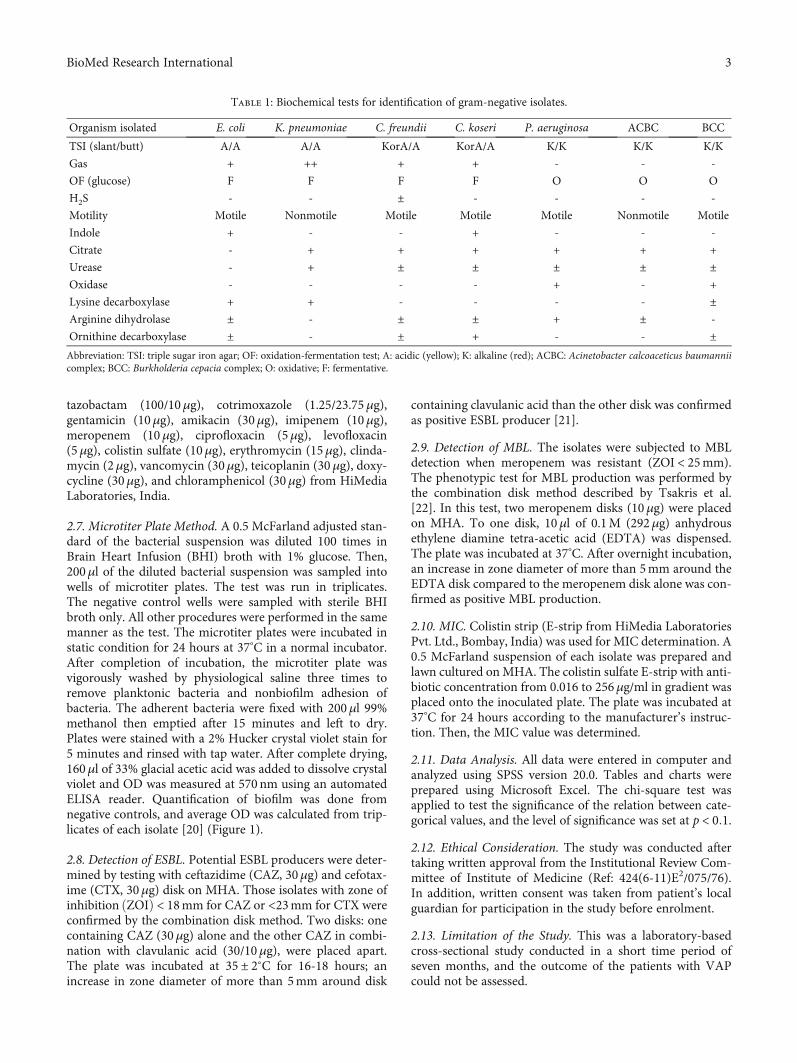
Table 1: Biochemical tests for identification of gram-negative isolates.
Organism isolated E. coli K. pneumoniae C. freundii C. koseri P. aeruginosa ACBC BCC
TSI (slant/butt) A/A A/A KorA/A KorA/A K/K K/K K/K
Gas + ++ + + - - -
OF (glucose) F F F F O O O
H2S - - ± - - - -
Motility Motile Nonmotile Motile Motile Motile Nonmotile Motile
Indole + - - + - - -
Citrate - + + + + + +
Urease - + ± ± ± ± ±Oxidase - - - - + - +
Lysine decarboxylase + + - - - - ±Arginine dihydrolase ± - ± ± + ± -
Ornithine decarboxylase ± - ± + - - ±Abbreviation: TSI: triple sugar iron agar; OF: oxidation-fermentation test; A: acidic (yellow); K: alkaline (red); ACBC: Acinetobacter calcoaceticus baumanniicomplex; BCC: Burkholderia cepacia complex; O: oxidative; F: fermentative.
3BioMed Research International

3. Results
3.1. Demographics and Study Setup. A total of 70 samples (7bronchoalveolar lavage or BAL and 63 deep tracheal aspi-rates) from patients under mechanical ventilation wereassessed. Significant growth of organisms was observed inall BAL specimens (n = 7), while 76.2% (n = 48) of the totaldeep tracheal aspirates yielded significant microbial growth.The median age of the study population was 50 years witha range of infants below 1 year to 85 years. Male participantswere preponderant accounting for 69% (n = 48) while femaleparticipants summed to 31% (n = 22) only. The highest num-ber of significant growth was seen in the 50 to 60 years agegroup and infants below 1 year of age each sharing 20% ofthe significant growth (Table 2).
3.2. Distribution of Isolates according to Growth. Among thesamples received, 78.6% (n = 55) yielded significant growth,of which 52.7% (n = 29) were monomicrobial, 45.5%(n = 25) were polymicrobial, and 1.8% (n = 1) were fungalinfection. A total of 71 organisms were isolated of which90.1% (n = 64) were gram-negative, 8.5% (n = 6) weregram-positive, and 1.4% (n = 1) was Candida spp.
Among the total isolates, Pseudomonas aeruginosa(31.0%) was the predominantly isolated organism followedby Acinetobacter calcoaceticus baumannii complex (ACBC)summing to 16.9% (Table 3). P. aeruginosa was predominantin both early- and late-onset VAP accounting for 34.8% and37.5% of the isolates, respectively. There was a variable levelof susceptibility of isolates to different groups of antibioticsand 100% susceptibility to vancomycin, teicoplanin (gram-positive), and colistin (gram-negative). In our study, less than50% gram-negative isolates were sensitive to carbapenem.
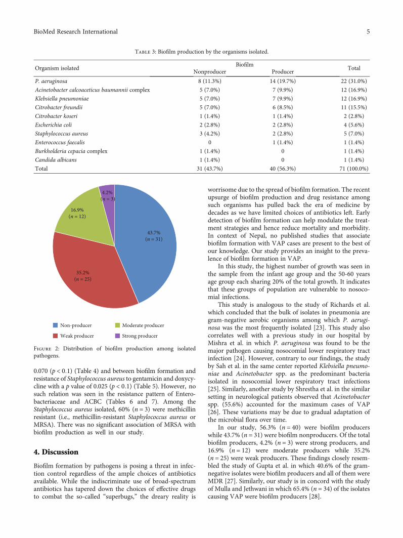
3.3. Distribution of Biofilm Production. Out of the totalorganisms, 56.3% (n = 40) were biofilm producers while43.7% (n = 31) were biofilm nonproducers. Among the bio-film producers, the majority were weak producers accounting
for 35.2% (n = 25) followed by moderate producers summingto 16.9% (n = 12) and only 4.2% (n = 3) were strongproducers of biofilm (Figure 2). Among the total isolates, P.aeruginosa (19.7%) was the major one producing biofilmfollowed by ACBC (9.9%) and K. pneumoniae (9.9%)(Table 3). Only two organisms, namely, P. aeruginosa andACBC, were strong biofilm producers with the share of2.8% and 1.4%, respectively (4.2% altogether).
3.4. Biofilm Production and Drug Resistance Patterns (MDR,ESBL, and MBL). Of the total isolates, 77.5% (n = 55) wereMDR. Among the total gram-negative isolates, 42.2%(n = 27) were MDR biofilm producer, 21.9% (n = 14) wereESBL biofilm producer, and 7.8% (n = 5) were MBL biofilmproducer (Figure 3). MDR organisms were preeminent inboth early- and late-onset VAP. There was a high prevalenceof multidrug resistance in biofilm-producing organisms.However, there was no statistically significant relationbetween the biofilm production and multidrug resistance,ESBL as well as MBL production. There was, nonetheless, sig-nificant association between biofilm production and resistanceof P. aeruginosa to piperacillin/tazobactam with a p value of
Figure 1: Detection of biofilm formation by a microtiter plate method.
Table 2: Age-wise distribution of growth samples.
Age group No significant growth Significant growth
<1 year 6 (8.5%) 14 (20.0%)
1 to 10 years 0 1 (1.4%)
10 to 20 years 0 2 (2.9%)
20 to 30 years 2 (2.9%) 5 (7.1%)
30 to 40 years 0 4 (5.7%)
40 to 50 years 3 (4.3%) 3 (4.3%)
50 to 60 years 2 (2.9%) 14 (20.0%)
60 to 70 years 0 4 (5.7%)
70 to 80 years 2 (2.9%) 6 (8.5%)
80 to 90 years 0 2 (2.9%)
4 BioMed Research International
0.070 (p < 0:1) (Table 4) and between biofilm formation andresistance of Staphylococcus aureus to gentamicin and doxycy-cline with a p value of 0.025 (p < 0:1) (Table 5). However, nosuch relation was seen in the resistance pattern of Entero-bacteriaceae and ACBC (Tables 6 and 7). Among theStaphylococcus aureus isolated, 60% (n = 3) were methicillinresistant (i.e., methicillin-resistant Staphylococcus aureus orMRSA). There was no significant association of MRSA withbiofilm production as well in our study.
4. Discussion
Biofilm formation by pathogens is posing a threat in infec-tion control regardless of the ample choices of antibioticsavailable. While the indiscriminate use of broad-spectrumantibiotics has tapered down the choices of effective drugsto combat the so-called “superbugs,” the dreary reality is
worrisome due to the spread of biofilm formation. The recentupsurge of biofilm production and drug resistance amongsuch organisms has pulled back the era of medicine bydecades as we have limited choices of antibiotics left. Earlydetection of biofilm formation can help modulate the treat-ment strategies and hence reduce mortality and morbidity.In context of Nepal, no published studies that associatebiofilm formation with VAP cases are present to the best ofour knowledge. Our study provides an insight to the preva-lence of biofilm formation in VAP.
In this study, the highest number of growth was seen inthe sample from the infant age group and the 50-60 yearsage group each sharing 20% of the total growth. It indicatesthat these groups of population are vulnerable to nosoco-mial infections.
This study is analogous to the study of Richards et al.which concluded that the bulk of isolates in pneumonia aregram-negative aerobic organisms among which P. aerugi-nosa was the most frequently isolated [23]. This study alsocorrelates well with a previous study in our hospital byMishra et al. in which P. aeruginosa was found to be themajor pathogen causing nosocomial lower respiratory tractinfection [24]. However, contrary to our findings, the studyby Sah et al. in the same center reported Klebsiella pneumo-niae and Acinetobacter spp. as the predominant bacteriaisolated in nosocomial lower respiratory tract infections[25]. Similarly, another study by Shrestha et al. in the similarsetting in neurological patients observed that Acinetobacterspp. (55.6%) accounted for the maximum cases of VAP[26]. These variations may be due to gradual adaptation ofthe microbial flora over time.
In our study, 56.3% (n = 40) were biofilm producerswhile 43.7% (n = 31) were biofilm nonproducers. Of the totalbiofilm producers, 4.2% (n = 3) were strong producers, and16.9% (n = 12) were moderate producers while 35.2%(n = 25) were weak producers. These findings closely resem-bled the study of Gupta et al. in which 40.6% of the gram-negative isolates were biofilm producers and all of them wereMDR [27]. Similarly, our study is in concord with the studyof Mulla and Jethwani in which 65.4% (n = 34) of the isolatescausing VAP were biofilm producers [28].
Table 3: Biofilm production by the organisms isolated.
Organism isolatedBiofilm
TotalNonproducer Producer
P. aeruginosa 8 (11.3%) 14 (19.7%) 22 (31.0%)
Acinetobacter calcoaceticus baumannii complex 5 (7.0%) 7 (9.9%) 12 (16.9%)
Klebsiella pneumoniae 5 (7.0%) 7 (9.9%) 12 (16.9%)
Citrobacter freundii 5 (7.0%) 6 (8.5%) 11 (15.5%)
Citrobacter koseri 1 (1.4%) 1 (1.4%) 2 (2.8%)
Escherichia coli 2 (2.8%) 2 (2.8%) 4 (5.6%)
Staphylococcus aureus 3 (4.2%) 2 (2.8%) 5 (7.0%)
Enterococcus faecalis 0 1 (1.4%) 1 (1.4%)
Burkholderia cepacia complex 1 (1.4%) 0 1 (1.4%)
Candida albicans 1 (1.4%) 0 1 (1.4%)
Total 31 (43.7%) 40 (56.3%) 71 (100.0%)
43.7%(n = 31)
35.2%(n = 25)
16.9%(n = 12)
4.2% (n = 3)
Non-producer
Weak producer
Moderate producer
Strong producer
Figure 2: Distribution of biofilm production among isolatedpathogens.
5BioMed Research International

35.3%
14.0%
3.1%
42.2%
21.9%
7.8%
0.0
5.0
10.0
15.0
20.0
25.0
(%)
30.0
35.0
40.0
45.0
MDR ESBL MBL
Biofilm Non-producerBiofilm Producer
Figure 3: Relation between biofilm production and drug resistance of gram-negative isolates.
Table 4: Relation between drug resistance and biofilm production in Pseudomonas aeruginosa.
AntibioticsBiofilm
p valueProducer NonproducerSensitive Resistant Sensitive Resistant
Gentamicin 5 (35.7%) 9 (64.3%) 4 (50.0%) 4 (50.0%) 0.512
Amikacin 5 (35.7%) 9 (64.3%) 4 (50.0%) 4 (50.0%) 0.512
Ciprofloxacin 4 (28.6%) 10 (71.4%) 1 (12.5%) 7 (87.5%) 0.387
Levofloxacin 4 (28.6%) 10 (71.4%) 2 (25%) 6 (75.0%) 0.856
Ceftazidime 4 (28.6%) 10 (71.4%) 3 (37.5%) 5 (62.5%) 0.665
Cefepime 10 (71.4%) 4 (28.6%) 4 (50.0%) 4 (50.0%) 0.315
Piperacillin 7 (53.8%) 6 (46.2%) 3 (37.5%) 5 (62.5%) 0.466
Piperacillin/tazobactam 12 (85.7%) 2 (14.3%) 4(50.0%) 4(50.0%) 0.070
Cefoperazone/sulbactam 11 (78.6%) 3 (21.4%) 4 (50.0%) 4 (50.0%) 0.166
Meropenem 8 (57.1%) 6 (42.9%) 2 (25.0%) 6 (75.0%) 0.145
Imipenem 8 (57.1%) 6 (42.9%) 2 (25.0%) 6 (75.0%) 0.145
Colistin 14 (100.0%) 0.0% 8(100.0%) 0.0%
Table 5: Relation between drug resistance and biofilm production in Staphylococcus aureus.
AntibioticsBiofilm
p valueProducer NonproducerSensitive Resistant Sensitive Resistant
Ampicillin 0 2 (100.0%) 0 3 (100.0%)
Cephalexin 1 (50.0%) 1 (50.0%) 1 (33.3%) 2 (66.7%) 0.709
Cotrimoxazole 0 2 (100.0%) 1 (33.3%) 2 (66.7%) 0.361
Gentamicin 0 2 (100.0%) 3 (100.0%) 0 0.025
Ciprofloxacin 0 2 (100.0%) 0 3 (100.0%)
Levofloxacin 0 2 (100.0%) 0 3 (100.0%)
Cefoxitin 1 (50.0%) 1 (50.0%) 1 (33.3%) 2 (66.7%) 0.709
Vancomycin 2 (100.0%) 0 3 (100.0%) 0
Teicoplanin 2 (100.0%) 0 3 (100.0%) 0
Erythromycin 0 2 (100.0%) 2 (66.7%) 1 (33.3%) 0.136
Clindamycin 0 2 (100.0%) 2 (66.7%) 1 (33.3%) 0.136
Doxycycline 0 2 (100.0%) 3 (100.0%) 0 0.025
Chloramphenicol 1 (50.0%) 1 (50.0%) 3 (100.0%) 0 0.171
6 BioMed Research International
Our study complies well with the study of Mishra et al.in the same setting (2013) in which 46.3% of the total iso-lates obtained from various clinical specimens were foundto be biofilm producers [29]. Meanwhile, the pattern oforganisms producing biofilm reported by Mishra et al. wasdifferent from our study who elucidated Klebsiella pneumo-niae (30%) as the major biofilm-producing gram-negativeisolate [29].
It is of note that, in our study, the MDR pathogens werepredominant (77.5%) among biofilm producers as well asnonproducers. Among the MDR isolates, 35.3% were biofilmnonproducers while 42.2% were biofilm producers. Thisaccords to the conclusion stated by Cepas et al. that MDRisolates may not be greater biofilm producers than non-MDR isolates. They also reported that P. aeruginosa fromrespiratory samples were more biofilm forming than thosefrom the other types of samples [30].
In a similar study by Dumaru et al. in another tertiarycare center in Nepal, 62.7% of the isolates were biofilm pro-ducers; 26.8% and 16.7% were ESBL and MBL producers,
respectively. It shows escalating drug resistance as animpending threat in our country. Likewise, the same studyshowed a significant association between MBL productionand biofilm production while no association between ESBLproduction and biofilm production [31]. Our study, however,could not address such statistical relation between multidrugresistance and biofilm formation.
In our study, 77.5% (n = 55) of the isolated pathogenswere MDR. This is in concordance with the study of Khanalet al. in a hospital in Kathmandu which showed bulk of theisolates causing respiratory infection in ICU were gram-negative isolates (92.2%) and 68.8% of them were MDR.Their study concluded that P. aeruginosa was the highestESBL-producing isolate, summing up to 42.8% [5]. Our studydissents with the latter fact as in our study; ACBC (10.9%)was the highest ESBL producer while P. aeruginosa (20.3%)was the most copious MDR isolate.
In our study, gram-negative nonfermenters including P.aeruginosa and ACBC were predominant isolates in bothearly- and late-onset VAP. Contrary to our study, some
Table 6: Relation between drug resistance and biofilm production in Enterobacteriaceae.
AntibioticsBiofilm
p valueProducer NonproducerSensitive Resistant Sensitive Resistant
Ampicillin 0 16 (100.0%) 0 13(100.0%)
Cefixime 3 (18.7%) 13 (81.3%) 1 (7.7%) 12(92.3%) 0.390
Cotrimoxazole 4 (25.0%) 12 (75.0%) 2 (15.4%) 11(84.6%) 0.525
Gentamicin 7 (43.7%) 9 (56.3%) 7 (53.8%) 6(46.2%) 0.588
Levofloxacin 5 (31.2%) 11 (68.8%) 4 (30.7%) 9 (69.3%) 0.976
Amoxycillin/clavulanic acid 3 (18.7%) 13 (81.3%) 1 (7.7%) 12(92.3%) 0.390
Cefepime 6 (37.5%) 10 (62.5%) 4 (30.7%) 9 (69.3%) 0.705
Piperacillin/tazobactam 9 (56.2%) 7 (43.8%) 6 (46.2%) 7(53.8%) 0.588
Cefoperazone/sulbactam 10 (62.5%) 6 (37.5%) 6 (46.2%) 7(53.8%) 0.379
Meropenem 9 (56.2%) 7 (43.8%) 7 (53.8%) 6(46.2%) 0.897
Imipenem 10 (62.5%) 6 (37.5%) 7 (53.8%) 6(46.2%) 0.638
Colistin 16(100.0%) 0.0% 13(100.0%) 0.0%
Table 7: Relation between drug resistance and biofilm production in Acinetobacter calcoaceticus baumannii complex.
AntibioticsBiofilm
p valueProducer NonproducerSensitive Resistant Sensitive Resistant
Amikacin 1 (14.3%) 6 (85.7%) 1 (20.0%) 4 (80.0%) 0.793
Levofloxacin 1 (14.3%) 6 (85.7%) 2 (40.0%) 3 (60.0%) 0.310
Ceftazidime 0 7 (100.0%) 0 5(100.0%)
Cefepime 0 7 (100.0%) 0 5(100.0%)
Piperacillin/tazobactam 1 (14.3%) 6 (85.7%) 2 (40.0%) 3 (60.0%) 0.310
Cefoperazone/sulbactam 2 (28.6%) 5 (71.4%) 2 (40.0%) 3 (60.0%) 0.679
Ampicillin/sulbactam 2 (28.6%) 5 (71.4%) 2 (40.0%) 3 (60.0%) 0.679
Meropenem 1 (14.3%) 6 (85.7%) 1 (20.0%) 4 (80.0%) 0.793
Imipenem 1 (14.3%) 6 (85.7%) 1 (20.0%) 4 (80.0%) 0.793
Colistin 7 (100.0%) 0 5(100.0%) 0
Doxycycline 0 7 (100.0%) 0 5(100.0%)
7BioMed Research International

studies suggest that early-onset VAP is linked to high rates ofinfection of oropharyngeal flora, while late-onset VAP isfound mostly associated with P. aeruginosa, ACBC, MRSA,and MDR gram-negative bacteria [3, 32]. The occurrence ofMDR nonfermenters as an eminent isolate in the early-onset VAP in our study may be due to the colonization bynonfermenters during hospitalization before shifting to ICUand acquisition of mechanical ventilation. It can also beattributed to the fact that patients under critical care areexposed to indiscriminate use of broad-spectrum antibioticsduring their care or prior to the critical care which may haveprovoked the resistance.
In a similar study done in TUTH by Shrestha et al., thebulk of the organism associated with VAP was found to beACBC (43.47%) which grosses the second position in ourstudy. The antibiogram of P. aeruginosa in their studyshowed high resistance to ciprofloxacin similar to our study[9]. On the other hand, the susceptibility rate of P. aerugi-nosa to cefoperazone/sulbactam and carbapenem in theirstudy was 82% and 91%, respectively, while our studyshows the sensitivity rate has fallen to 68.2% and 45.5%,respectively. This is quite infuriating because carbapenemsare the drugs of choice for ESBL producing MDR P. aeru-ginosa. This situation may be attributed to wide use ofcarbapenems in our ICUs due to past record of high prev-alence of resistant bacteria. Similarly, 35.9% of all gram-negative isolates were ESBL producers in our study whichis slightly higher than the findings of Shrestha et al. whichwas 32.8% [9].
In the antibiogram study of gram-positive isolates, thesensitivity rate of doxycycline and chloramphenicol wascommendable accounting to 60% and 80%, respectively.These isolates had low sensitivity towards ciprofloxacin,cotrimoxazole, and erythromycin probably due to rampantempirical use of these drugs.
Our study showed statistically significant association ofpiperacillin/tazobactam resistance and biofilm productionwith p value of 0.070 (i.e., p value < 0.1) in P. aeruginosaand similar association with gentamicin and doxycyclineresistance (p value of 0.025) in gram-positive isolates. But,the association of biofilm production and multidrug resis-tance was statistically nonsignificant. This is in accordancewith the study of Cepas et al. that showed despite relation-ships being found between individual drug resistance, thereis no relation between multidrug resistance and biofilmformation [30]. It is also supported by the studies of Shresthaet al. in uropathogens [33] and Eyoh et al. [34].
However, the antibiogram study of P. aeruginosa andrelation with biofilm formation does not concur with thestudy of Neopane et al. done in another tertiary care centerin Nepal which found biofilm production of P. aeruginosato be strongly associated with multidrug resistance andESBL production [35]. The same study observed the resis-tance of ceftazidime in biofilm-producing isolates was57.1% which in our study was 68.2%. It can roughly pointat impending threat of resistance among the biofilm pro-ducers. There are studies that show significant correlationof multiple drug resistance and biofilm formation includingBardbari et al. [36]. There are other studies conducted in
tertiary care centers in Nepal as well. Nepal et al. [37] andShrestha et al. [38] unveiled high antimicrobial resistanceseen in organisms producing biofilm on inanimate surfaces.These variations could be because of isolation of differentstrains with different propensity of biofilm formation indifferent studies.
The fact that biofilm in ETT causes bad prognosis of VAPis well supported by the study of Gil-Perotin et al. whichdetected bacteria causing VAP in ETT biofilm and ETAdespite an appropriate antibiotic therapy [14]. Organismsin biofilm can bypass the antimicrobial mechanism and thehost response via exopolysaccharide layer, quorum sensing,retarded growth, sequestration of resistance genes, and manyother mechanisms [15, 16]. High concentration of drugs thatcan control biofilm organisms cannot be obtained in thehuman body. This can put light on the emergence of resis-tance among the biofilm-producing isolates due to theconsistent use of antibiotics. A strong positive correlationwas reported by the study of Uppe et al. between biofilmformation and occurrence of VAP as well [39].
The pathogen causing VAP and other nosocomial infec-tions is dynamic which may greatly vary among the hospitals,at the smallest unit of wards and even within the ICUs. Thereis a scarcity of local data on surveillance of infection in hos-pitals in context of Nepal [40]. It is unwise to administerempirical therapy without proper surveillance. Shifting tothe deescalation regime is a golden tool that can diminishthe emanating problem of antimicrobial resistance in criticalcare units. Revision of the antimicrobial resistance patterncan also help attest appropriate empirical drug choice for aparticular ward and hospital.
The only drug of last regime that is left in the chest ofpharmaceuticals to intervene is colistin. This has broughtup a global challenge to capsize the realm of so-called“superbugs.” Being a responsible medical professional, onemust intend to preserve or restore the efficacy of currentlyavailable drugs. Furthermore, coating of endotracheal tubeswith antimicrobial peptides or their peptidomimetics couldbe an option to reduce the occurrence of VAP [41].
5. Conclusion
This study acquainted us about the biofilm formation andits relation with drug resistance. The data support thatinfants and population above 50 years are more vulnerableto nosocomial infections. Gram-negative nonfermenterbacteria constituted the bulk of isolates. Over 50% of theisolates were found to be biofilm producers. The hike inmultidrug resistance, ESBL, and MBL production was alsopreeminent among biofilm producers. There is an increasein drug resistance reported in our study compared to simi-lar settings. This indicates emerging drug resistance amongthe biofilm producers that may have grave outcomes. Thisstudy advocates further research in convenient methods ofdetection of biofilm in vivo as well as in vitro in the routinelaboratory and to look up for alternative therapies forbiofilm-associated pathogens to unburden the threat ofresistant pathogens.
8 BioMed Research International

Data Availability
The data supporting the findings of this study is availablefrom the corresponding author upon request.
Conflicts of Interest
The author reports no conflicts of interest in this work.
Acknowledgments
We are thankful to the laboratory staffs of Department ofMicrobiology and the staffs of ICUs of TUTH for their coop-eration throughout the study. The study was supported byNepal Health Research Council through UndergraduateHealth Research Grant (Reference no. 1583).
References
[1] G. Ducel, J. Fabry, and L. Nicolle, Prevention of HospitalAcquired Infections: A Practical Guide, World Health Organi-zation, Geneva, Switzerland, 2nd edition, 2002.
[2] M. H. Kollef, “What is ventilator-associated pneumonia andwhy is it important?,” Respiratory Care, vol. 50, no. 6,pp. 714–724, 2005.
[3] American Thoracic Society, “Guidelines for the managementof adults with hospital-acquired, ventilator-associated, andhealthcare-associated pneumonia,” American Journal of Respi-ratory and Critical Care Medicine, vol. 171, no. 4, pp. 388–416,2005.
[4] A. A. Kalanuria, W. Zai, and M. Mirski, “Ventilator-associatedpneumonia in the ICU,” Critical Care, vol. 18, no. 2, p. 208,2014.
[5] S. Khanal, D. R. Joshi, D. R. Bhatta, U. Devkota, and B. M.Pokhrel, “β-Lactamase-producing multidrug-resistant bacte-rial pathogens from tracheal aspirates of intensive care unitpatients at National Institute of Neurological and Allied Sci-ences, Nepal,” ISRN Microbiology, vol. 2013, Article ID847569, 5 pages, 2013.
[6] E. Shahrokhi, A. Hasani, K. Ansarin et al., “Bacterial biofilm inventilator-associated pneumonia: a clinical concern,” Journalof Research in Medical and Dental Science, vol. 6, no. 4,pp. 46–51, 2018.
[7] S. Kharel, A. Bist, and S. K. Mishra, “Ventilator-associatedpneumonia among ICU patients in WHO southeast Asianregion: a systematic review,” PLoS One, vol. 16, no. 3, articlee0247832, 2021.
[8] A. Bonell, R. Azarrafiy, V. T. L. Huong et al., “A systematicreview and meta-analysis of ventilator-associated pneumoniain adults in Asia: an analysis of national income level on inci-dence and etiology,” Clinical Infectious Diseases, vol. 68, no. 3,pp. 511–518, 2019.
[9] R. K. Shrestha, R. K. Dahal, S. K. Mishra et al., “Ventilatorassociated pneumonia in tertiary care hospital, Maharajgunj,Kathmandu, Nepal,” Journal of Institute of Medicine Nepal,vol. 35, no. 3, 2013.
[10] D. R. Mishra, N. Shah, and D. S. Shah, “Incidence and outcomeof ventilator associated pneumonia in ICU of a tertiary carehospital in Nepal,” Journal of Nepal Medical Association,vol. 56, no. 207, pp. 304–308, 2017.
[11] A. Lamichhane and A. Mishra, “Prevalence of ventilator asso-ciated pneumonia in neonates in a tertiary care hospital inwestern Nepal,” Journal of Nepal Medical Association,vol. 57, no. 216, 2019.
[12] R. M. Donlan, “Biofilms: microbial life on surfaces,” EmergingInfectious Diseases, vol. 8, no. 9, pp. 881–890, 2002.
[13] R. M. Donlan and J. W. Costerton, “Biofilms: survival mecha-nisms of clinically relevant microorganisms,” Clinical Microbi-ology Reviews, vol. 15, no. 2, pp. 167–193, 2002.
[14] S. Gil-Perotin, P. Ramirez, V. Marti et al., “Implications ofendotracheal tube biofilm in ventilator-associated pneumoniaresponse: a state of concept,” Critical Care, vol. 16, no. 3,p. R93, 2012.
[15] T.-F. C. Mah and G. A. O'Toole, “Mechanisms of biofilm resis-tance to antimicrobial agents,” Trends in Microbiology, vol. 9,no. 1, pp. 34–39, 2001.
[16] J. W. Costerton, P. S. Stewart, and E. P. Greenberg, “Bacterialbiofilms: a common cause of persistent infections,” Science,vol. 284, no. 5418, pp. 1318–1322, 1999.
[17] P. Gilbert, T. Maira-Litran, A. J. McBain, A. H. Rickard, andF. W. Whyte, “The physiology and collective recalcitrance ofmicrobial biofilm communities,” Advances in Microbial Physi-ology, vol. 46, pp. 202–256, 2002.
[18] J. D. Hunter, “Ventilator associated pneumonia,” BMJ,vol. 344, article e3325, 2012.
[19] H. D. Isenberg, Clinical Microbiology Procedures Handbook,vol. 1, American Society for Microbiology; ASM Press, Wash-ington DC, 2nd edition, 2004.
[20] S. Stepanović, D. Vuković, I. Dakić, B. Savić, and M. Švabić-Vlahović, “A modified microtiter-plate test for quantificationof staphylococcal biofilm formation,” Journal of Microbiologi-cal Methods, vol. 40, no. 2, pp. 175–179, 2000.
[21] CLSI, Performance Standards for Antimicrobial SusceptibilityTesting, Clinical and Laboratory Standards Institute, Wayne,PA, 2017.
[22] A. Tsakris, A. Poulou, S. Pournaras et al., “A simple pheno-typic method for the differentiation of metallo-β-lactamasesand class A KPC carbapenemases in Enterobacteriaceae clini-cal isolates,” The Journal of Antimicrobial Chemotherapy,vol. 65, no. 8, pp. 1664–1671, 2010.
[23] M. J. Richards, J. R. Edwards, D. H. Culver, and R. P. Gaynes,“Nosocomial infections in medical intensive care units in theUnited States,” Critical Care Medicine, vol. 27, no. 5,pp. 887–892, 1999.
[24] S. Mishra, H. Kattel, J. Acharya et al., “Recent trend of bacterialaetiology of lower respiratory tract infection in a tertiary carecentre of Nepal,” International Journal of Infection and Micro-biology, vol. 1, no. 1, pp. 3–8, 2012.
[25] M. K. Sah, S. K. Mishra, H. Ohora et al., “Nosocomial bacterialinfection and antimicrobial resistant pattern in a tertiary carehospital in Nepal,” Journal of Institute of Medicine, vol. 36,no. 3, 2014.
[26] D. K. Shrestha, B. Rajbhandari, A. Pradhanang, G. Sedain, S. K.Shilpakar, and S. Pradhan, “Ventilator-associated pneumonia inneurosurgical patients: a tertiary care center study,” Journal ofInstitute of Medicine Nepal, vol. 41, no. 2, pp. 40–44, 2019.
[27] R. Gupta, A. Malik, M. Rizvi, andM. Ahmed, “Biofilm produc-ing multidrug and extensive drug resistant bacterial pathogensfrom tracheal aspirates of intensive care unit patients-a threatto combat,” International Journal of Current Microbiology andApplied Sciences, vol. 1, pp. 1–9, 2015.
9BioMed Research International

[28] S. A. Mulla and U. N. Jethwani, “Assessment of biofilm forma-tion by the causative organism of ventilator associated pneu-monia at intensive care unit of a tertiary care hospital,” TheNational Journal of Emergency Research, vol. 2, pp. 15–19,2012.
[29] S. K. Mishra, P. Basukala, O. Basukala, K. Parajuli, B. M.Pokhrel, and B. P. Rijal, “Detection of biofilm productionand antibiotic resistance pattern in clinical isolates fromindwelling medical devices,” Current Microbiology, vol. 70,no. 1, pp. 128–134, 2015.
[30] V. Cepas, Y. López, E. Muñoz et al., “Relationship between bio-film formation and antimicrobial resistance in gram-negativebacteria,” Microbial Drug Resistance, vol. 25, no. 1, pp. 72–79, 2019.
[31] R. Dumaru, R. Baral, and L. B. Shrestha, “Study of biofilm for-mation and antibiotic resistance pattern of gram-negativeBacilli among the clinical isolates at BPKIHS, Dharan,” BMCResearch Notes, vol. 12, no. 1, p. 38, 2019.
[32] J. Chastre and J.-Y. Fagon, “Ventilator-associated pneumo-nia,” American Journal of Respiratory and Critical Care Medi-cine, vol. 165, no. 7, pp. 867–903, 2002.
[33] B. Shrestha, B. Shrestha, A. Poudel, B. Lekhak, and M. K.Upreti, “In-vitro biofilm detection among uropathogens andtheir antibiogram profile,” Tribhuvan University Journal ofMicrobiology, vol. 5, pp. 57–62, 2018.
[34] A. B. Eyoh, M. Toukam, J. Atashili et al., “Relationshipbetween multiple drug resistance and biofilm formation inStaphylococcus aureus isolated from medical and non-medical personnel in Yaounde, Cameroon,” Pan African Med-ical Journal, vol. 17, p. 186, 2014.
[35] P. Neopane, H. P. Nepal, R. Gautam et al., “Is there correlationof biofilm formation with multidrug resistance and ESBL pro-duction in Pseudomonas aeruginosa?,” European Journal ofBiomedical, vol. 4, no. 1, pp. 366–372, 2017.
[36] A. M. Bardbari, M. R. Arabestani, M. Karami, F. Keramat,M. Y. Alikhani, and K. P. Bagheri, “Correlation between abilityof biofilm formation with their responsible genes and MDRpatterns in clinical and environmental Acinetobacter bauman-nii isolates,” Microbial Pathogenesis, vol. 108, pp. 122–128,2017.
[37] H. P. Nepal, P. Neopane, R. Shrestha et al., “Biofilm formationand antimicrobial resistance in Klebsiella pneumoniae isolatedfrom patients visiting a tertiary care center of Nepal,” AsianPacific Journal of Tropical Disease, vol. 7, no. 6, pp. 347–351,2017.
[38] R. Shrestha, N. Nayak, D. R. Bhatta, D. Hamal, S. H.Subramanya, and S. Gokhale, “Drug resistance and biofilmproduction among Pseudomonas aeruginosa clinical iso-lates in a tertiary care hospital of Nepal,” Nepal MedicalCollege Journal, vol. 21, no. 2, pp. 110–116, 2019.
[39] A. Uppe, D. Gupta, S. Sawant, and G. Nair, “Incidence of bio-film formation in ET tube and correlation with occurrence ofVAP in a tertiary care ICU,” American Journal of InfectiousDiseases, vol. 7, no. 2, pp. 65–72, 2019.
[40] N. P. Parajuli, S. P. Acharya, S. K. Mishra, K. Parajuli, B. P.Rijal, and B. M. Pokhrel, “High burden of antimicrobial resis-tance among gram negative bacteria causing healthcare associ-ated infections in a critical care unit of Nepal,” AntimicrobialResistance and Infection Control, vol. 6, no. 1, p. 67, 2017.
[41] B. Ozcelik, P. Pasic, P. Sangwan et al., “Evaluation of the novelantimicrobial BCP3 in a coating for endotracheal tubes,” ACSOmega, vol. 5, no. 18, pp. 10288–10296, 2020.
10 BioMed Research International


![Pneumonia (Ventilator-associated [VAP] and non-ventilator ...](https://static.fdocuments.us/doc/165x107/61c3dfa934191a172140c0d5/pneumonia-ventilator-associated-vap-and-non-ventilator-.jpg)


