
BIO208 SEEDLESS PLANT - nou.edu.ng!
117
BIO 208 SEEDLESS PLANTS 1 NATIONAL OPEN UNIVERSITY OF NIGERIA SCHOOL OF SCIENCE AND TECHNOLOGY COURSE CODE: BIO208 COURSE TITLE: SEEDLESS PLANTS
Transcript of BIO208 SEEDLESS PLANT - nou.edu.ng!

BIO 208 SEEDLESS PLANTS
1
NATIONAL OPEN UNIVERSITY OF NIGERIA
SCHOOL OF SCIENCE AND TECHNOLOGY
COURSE CODE: BIO208
COURSE TITLE: SEEDLESS PLANTS
BIO 208 SEEDLESS PLANTS
2
MODULE 1.
UNIT 1: MORPHOLOGY OF ALGAE
CONTENTS
1.0 Introduction
2.0 Objectives
3.0 Main Content
3.1 Morphology of algae
3.1.1 Anacystis
3.1.2 Chlamydomonas
3.2 Colonial Forms
3.2.1 Microcystis
3.2.2 Volvox
3.3 Filamentous Forms
3.3.1 Nostoc
3.3.2 Ulothrix
3.4 Heterotrichous Forms
3.4.1 Draparnaldiopsis
3.4.2 Ectocarpus
3.5 Thalloid Forms
3.5.1 Ulva
3.5.2 Fucus
3.6 Polysiphonoid Forms
3.6.1 Polysiphonia
4.0 Conclusion
5.0 Summary
6.0 Tutor Marked Assignment
7.o References/Further Reading

BIO 208 SEEDLESS PLANTS
3
1.0 INTRODUCTION
Algae are placed in Kingdom Protista along with protozoa. Earlier they were
classified with plants as they are photosynthetic autotrophs-possess
chlorophyll and chloroplasts and superficially appear like plants. Since their
gametes do not have protective cells around them they are no longer classified
with plants.
In this unit on algae, you will study the morphology of algae. Although simple
in structure, lacking differentiation, algae exhibit great diversity in size and
appearance. Their size ranges from simple microsocple to giant thallus
extending several metres in length as in kelps. Algal morphology varies from
simple unicellular form to complex thallus as found in seaweed.
Algae are widely distributed in nature whenever there is plenty of water and
sunshine. They also occur abundantly on wet rocks, wet ground and a pool of
water. They also inhabit harsh habitats.
2.0 OBJECTIVES
After studying this unit you will be able to:
• know where algae can be found
• describe the basic types of thallus in algae
• compare the morphology of unicellular, colonial, filamentous,
heterotrichous, thalloid and polysiplionoid forms of algae.
• draw the morphology of. Anacystis, Chlamydomonas, Microystis, Volvox,
Ulva, and Fucus and describe their special features.
3.2 ALGAL MORPHOLOGY
The science or study of algae is called ‘Phycology’. One who speciallises in the
study of algae is called ‘Phycologist’ or ‘Algologist’.
BIO 208 SEEDLESS PLANTS
4
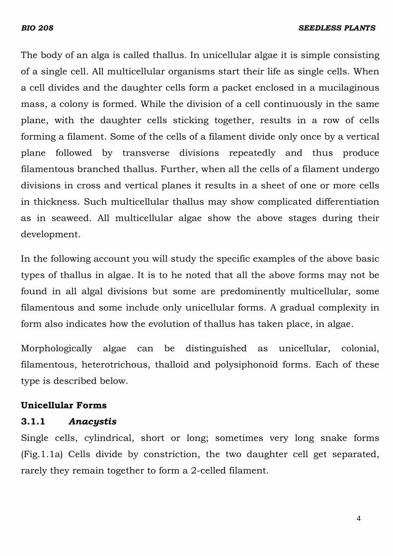
The body of an alga is called thallus. In unicellular algae it is simple consisting
of a single cell. All multicellular organisms start their life as single cells. When
a cell divides and the daughter cells form a packet enclosed in a mucilaginous
mass, a colony is formed. While the division of a cell continuously in the same
plane, with the daughter cells sticking together, results in a row of cells
forming a filament. Some of the cells of a filament divide only once by a vertical
plane followed by transverse divisions repeatedly and thus produce
filamentous branched thallus. Further, when all the cells of a filament undergo
divisions in cross and vertical planes it results in a sheet of one or more cells
in thickness. Such multicellular thallus may show complicated differentiation
as in seaweed. All multicellular algae show the above stages during their
development.
In the following account you will study the specific examples of the above basic
types of thallus in algae. It is to he noted that all the above forms may not be
found in all algal divisions but some are predominently multicellular, some
filamentous and some include only unicellular forms. A gradual complexity in
form also indicates how the evolution of thallus has taken place, in algae.
Morphologically algae can be distinguished as unicellular, colonial,
filamentous, heterotrichous, thalloid and polysiphonoid forms. Each of these
type is described below.
Unicellular Forms
3.1.1 Anacystis
Single cells, cylindrical, short or long; sometimes very long snake forms
(Fig.1.1a) Cells divide by constriction, the two daughter cell get separated,
rarely they remain together to form a 2-celled filament.

BIO 208 SEEDLESS PLANTS
5
Individual single cells may have their own mucilagenous cover around them.
Several such cells may be enclosed in common colourless mucilage giving the
impression of a colony.
3.1.2 Chlamydomonas
This single celled alga contains a nucleus, a cup-shaped chloroplast in which
one pyrenoid is commonly present (Fig. 1.1b) The chloroplast on the anterior
side shows 2 to 3 rows of fatty redcoloured granules. This is known as eyespot
or stigma which is helpful for the alga to respond to light. The cell wall is firm
and distinct. A small contractile vacuole is found at the base of each flagellum.
Chlamydomonas cells under partially dry conditions divide and the daughter
cells without flagella remain enclosed by a common mass of mucilage. Such a
colony is known as palmella stage of Chlamydomonas (Fig.1.1c). This is only a
temporary stage and on flooding with water individual cells develop flagella
and escape swimming away from the colony. Thus the beginning of the colony
construction found in Volvox can be seen in Chlamydomonas.
a
c
Fig. 1.1: Unicellular algae: a) Anacystis nidulans, b) Chlamydomonas

BIO 208 SEEDLESS PLANTS
6
3.2 Colonial Algae
When a cell divides and the daughter cells formed remain together within a
common mucilage mass, it is known as a colony. A colony may contain large
number of cells. Sometimes it may be so big that one can see it with unaided
eyes.
3.2.1 Microcystis
This is a colonial alga, most common in polluted ponds and lakes (Fig.1.2e)
Sometimes the colonies are big and can be seen by unaided eyes. They
accumulate on the surface of water forming quite a thick layer in some
seasons (water blooms).
Single cells are spherical and colony is formed because of loose aggregates of
several thousand cells held by mucilage (Fig. 1.2e). The colonies float on the
surface of water because of the presence of elongated cylindrical gas vesicles
inside the individual cells.
3.2.2 Volvox
The colonies of Volvox are spherical, ball-like and big enough to be seen with
unaided eye (Fig.1.2g) Each colony contains l000-5000 cells arranged on the
outside of a mucilagenous ball called coenobiuim. Coenobium is a colony in
which the number of cells is fixed at the time of formation. No further addition
of cells occurs. Generally the cells are also in a special arrangement. Two types
of cells can be seen generally, vegetative or somatic and gonidia. In younger
colonies cytoplasmic connections - plasmodesmata between individual cells
can be seen under the microscope.
Vegetative cells are more or less like ChIamydomonas with two flagella, cell
wall, single cup-shaped chloroplast, eyespot, pyrenoid, contractile vacuole and

BIO 208 SEEDLESS PLANTS
7
a nucleus (Fig. 1.2h). The cells on the posterior side of the colony may be
larger than in the front.
In Volvox all the cells of a colony are derived from a single parental cell. They
are arranged on the surface of mucilaginous ball, connected with other cells by
cytoplasmic connections. Some cells behave as sex cells meant for
reproduction whereas others remain vegetative and ultimately grow old and
die. This differentiation into vegetative and reproductive cells is a very
important feature in the development of multicellular organisms.
e f
g h
Fig. 1.2 Colonial algae; e) Microcystis aeruginosa, f) Portion of e magnified g) Volvox aureus h)
Cells of in the interior polar view.

BIO 208 SEEDLESS PLANTS
8
Filamentous Forms
When a cell divides always cross-wise and the daughter cells do not separate
from each other, it results in a linear row of cells as in Nostoc, Ulothrix and
Oedogonium. However, the three algae show different levels of differentiation.
3.3.1 Nostoc
This is a simple, filamentous form, a single row of cells, uniseriate (Fig.1.3a).
Several filaments of Nostoc are generally enclosed within a common mucilage
envelop to form a colony (Fig. 1.3b). Some cells in between the vegetative cells
are modified into heterocysts. Heterocyst are a highly differentiated cell in
some filamentaous blue-green algae that is a site of nitrogen fixation. All the
vegetative cells are capable of developing into spores called akinetes. Akinete
are a thick-walled, nonmotile reproductive cell found in algae.
a b
C d
Fig. 1.3: Filamentous algae; (a) filaments of Nostoc showing akinetes and (b)
heterocysts and aggregate of Nostoc filaments forming a ball, (c) germilings of
Ulothrix and (d) cell structure of UIothrix showing gridle shaped chloroplasts.

BIO 208 SEEDLESS PLANTS
9
3.4 Heterotrichous Forms
When some cells of a filament divide vertically it results in a branch. Many
filamentous forms show extensive branching of the main filament giving it a
bushy appearance.
In some algae the branches at the base remain horizontal, attached to the
substratum known as prostrate system from which erect system of vertical
branched filaments arise. This type of body is known as heterotrichous habit.
Heterotrichous habit is the most highly developed filamentous construction in
algae.
3.4.1 Draparnaldiopsis
It is a heterotrichous alga which shows greater differentiation in plant body.
The prostate system is very much reduce The main axis contains long
internodal cells alternating with short nodal cells (Fig. 1.4). The short nodal
cells bear a bunch of short branches. Some of the side branches may develop
into long colourless hairs or setae. The main axis produces at the base long
multicellular colourless rhizoids in large number to form a kind of cortex.
Their main function is to attach the alga to the substratum.
Fig. 1.4: Draparnaldiopsis indica (photograph by late Prof. Y.B.K Chowdarv).

BIO 208 SEEDLESS PLANTS
10
a b
Fig. 1.5: Heterotrichous algae; a, Ectocarpus showing habit and b) thalli with unilocular
sporangia or gametangia.
3.4.2 Ectocarpus
It is another heterotrichous alga (Fig. 1.5).The prostrate system which attaches
the alga to the substratum is made of branched filaments. The erect system is
in the form of uniseriate (single row of cells) branched filaments forming loose
tufts of 1mm to 10 mm or more. The branches arise just below the cross walls
of the cells of the main filament. Most of these branches terminate in elongated
hairs.
3.5 Thalloid Forms
When the cells of a filament divide in more than one plane, that is not only
cross-wise but also lengthwise it results in a sheet of cells. The thallus may be
one cell or many cells in thickness.
3.5.1 Ulva
Ulva is a very common alga found on rocky coasts of sea (Fig. 1.6a). The
thallus is attached to the substrate such as rocks by rhizoids at the base.

BIO 208 SEEDLESS PLANTS
11
Fig. 1.6: Ulva lactuca; a) habit of growth b) Fucus vesiculosus- morphology of the thallus.
Fucus
Fucus is a brown algal seaweed common on the rocky coasts of sea in
temperate countries (Fig. 1.6b). The body of Fucus is large about half a metre
or so in length. It has a basal discoid holdfast, a short stipe and long flat and
dichotomously branched fronds or blades. Dichotomous branching pattern is
one in which the two arms of the branch are more or less equal in length. At
the tip of the blade are found air bladders which make the plant float in water.
3.6 Polysiphonoid Forms
This form of algae is more complex than the earlier described forms. It is found
in the red alga Polysiphonia (Fig. 1.7) which is marine in habitat.
3.6.1 Polysiphonia
The algae shows in general heterotrichous habit. The prostrate system is in the
form of an elongated rhizoid which attaches the algae to the substratum. The
erect system is highly branched. The branches are of two kinds, some are long
and some short and hair-like. The main filament grows by the division of a
single apical cell. The mature plant body is made up of central row of cells -
central siphon, surrounded by vertical rows of cells, 4 to 24 - pericentral
siphons.

BIO 208 SEEDLESS PLANTS
12
All the pericentral cells are connected with the cells of central siphon and are
also connected with each other.
When the cytoplasm of one cell is connected to the cytoplasm of the
neighbouring cell through a pit in their wall, it is known as pit connection. In
Polysiphonia although all the cells are separate, their cytoplasm is connected
by means of pit connections.
Fig.1.7:Polysiphonia; habit showing multicellular construction of several
interconnected rows of siphons
New branches may develop from the cells of central siphon or from the
pericentral cells. The trichoblasts which are simple or branched hair-like
lateral branches arise from the pericentral cells.
4.0 CONCLUSION
Algae are diverse in the group and forms. They can be distinguished as
unicellular, colonial, filamentous, heterotrichous, thalloid and pohysiphonoid
forms. The unicellular algae are simplest in form while the thalloid are sheet-
like.
BIO 208 SEEDLESS PLANTS
13
5.0 SUMMARY
• Algae are diverse group of organisms ranging from microscopic unicellular
to giant thalloid forms anchored to rocks in the sea. Morphologically they
can be distinguished as unicellular, colonial, filamentous, hetertrichous,
thalloid and polysiphonoid forms.
• The unicellular algae are simplest in morphology. Some advancement is
observed in colonial forms. The cells of a colony may communicate through
plasmodesmata. There is division of labour between cells, some remain
vegetative while others take part in reproduction.
• Some algae have a prostrate system attached to the substratum and an
erect system of vertical branches. This is called heterotrichous habit.
• Thalloid forms are sheet like polysiphonoid forms are more complex. They
possess rhizoids and branched erect system. Mature thallus consists of
central row of cell-central siphon surrounded by pericentral siphon.
6.0 TUTOR MARKED ASSIGNMENT
1. Describe the structure of two simplest forms of algae.
7.0 REFERENCES/FURTHER READINGS
Dutta, A.C. (1981). Botany for degree students. Oxford University Press. 909p.
IGNOU (1991 )Indira Gaahdi National Open University. Plant Diversity-Algae.

BIO 208 SEEDLESS PLANTS
14
UNIT 2: CLASSIFICATION OF ALGAE
CONTENTS
1.0 Introduction
2.0 Objectives
3.0 Main Content
3.1 Criteria for Classification of Algae
3.1.1 Prokaryotic Algae
3.1.2 Division Cyanophyta (BIue-green algae)
3.2 Eukaryotic Algae
3.2.1 Division Chlorophyta (Green algae)
3.2.2 Division Phacophyta (Brown algae)
3.2.3 Division Rhodophyta (Red algae)
3.2.4 Division Xanthophyta (Yellow-green algae)
3.2.5 Division Chrysophyta (Golden-brown algae)
3.2.6 Division Euglenophyta (Euglenoids)
3.2.7 Division Dinophyta (Dinoflagellales)
3.2.8 Division Cryptopliyta (Cryptomonad)
3.2.9 Division Bacillariophyta (Diatoms)
4.0 Conclusion
5.0 Summary
6.0 Tutor Marked Assignment
7.0 References/Further Reading
1.0 INTRODUCTION
From the previous two units it is evident that algae show a great diversity in
structure and reproduction. In this unit you will learn classification of this
diverse group. Classification means grouping of organisms according to the
similarity in their characters. It is not far fetched but true that organisms
showing similar morphology, life cycle, physiology and biochemistry are

BIO 208 SEEDLESS PLANTS
15
genetically related from the evolutionary point of view (phylogenetically related)
and one is justified in grouping them together.
Algae could be classified according to their common characters into 8 divisions
of’ Kingdom Protista.
In this unit you are introduced to the characteristics of different divisions of
algae.
2.0 Objectives
After studying this unit you should be able to:
• list the various criteria used for the classification of algae,
• explain why algae are classified as protists instead of plants,
• list the various divisions of algae and describe the characteristics of each,
• classify the genera of algae into division, order and family and
• give common examples of algae from each division.
3.0 MAIN CONTENT
3.1 CRITERIA FOR CLASSIFICATION OF ALGAE
The criteria used by phycologists are quite varied. Generally a number of
characters are used together ranging from external morphology,
ultrastructure, chromosome number and their morphology, pigment
composition, nature of cellular storage products, enzymes, isoenzymes, DNA
homology, and DNA banding etc. As new techniques are developed they are
used to decide more precisely the relatedness (or absence of it) of organisms
which seem otherwise’ related to each other.
Given below are the salient characters of each of the divisions of the algae. It is
to be noted that each division is again divided into orders, families, genera and
species. Because of the restriction of time representatives of other divisions are

BIO 208 SEEDLESS PLANTS
16
not included in your course, not because they are any less important in the
biological world.
3.1.1 PROKARYOTIC ALGAE
3.l.2 Division CYANOPHYTA (Cyanobacteria or Blue-green algae)
Prokaryotic algae are placed in Division Cyanophyta. Algae of this division may
be unicellular, colonial, and filamentous, with or without branches, branching
may be ‘true’ or ‘false’ type. Most forms are embedded in mucilaginous or
gelatinous sheaths.
The composition of cell wall is similar to bacterial cell wall. It is, made up of
distinctive mucopeptides and muramic acid.
The ultrastructure of the cell shows no organised nucleus, mitochondria or
chloroplasts. Photosynthetic lamellae and ribosomes of 70s type are present in
the cytoplasm of the cells. Some filamentous forms possess specialised cells
termed as ‘heterocysts’ which are involved in nitrogen fixation.
The main photosynthetic pigments are chlorophyll a and phycobilins -
(phycocyan and phycoerythrin). A number of carotenoids including β carotene
are also present some of which are specific to the division. Carbon is reserved
in the cells as glycogen granules and nitrogen as cyanophycear granules.

BIO 208 SEEDLESS PLANTS
17
Other granules like polyphosphate granules, some enzyme aggregates Iike
carboxysomes may also be present.
Reproduction occurs by simple cell division. No motile cells are found in
cyanobacteria and they do not have sexual method of reproduction. Thick
walled cells called ‘akinetes’ or spores are present in some forms for
perennation and asexual reproduction.
Cynobacteria are distributed all over the earth in diverse habitats, fresh water
lake ponds, rivers, arctic, antarctic areas, hot water springs, brine salt pans,
desert soils, subaerial surfaces like tree trunks, building terraces and rock
surfaces.
Examples: Anacystis, Microcystis, Nostoc, Anabaena, Oscillatoria, Spirulina,
Calothrix, Gleotrichia, and Scytonema (Fig. 2.1).
Anacystis Microcystis
AnabaenaNostoc
Spirulina
Calothrix
Fig. 2.1: Some examples of blue-green algae

BIO 208 SEEDLESS PLANTS
18
3.2 EUKARYOTIC ALGAE
As you have learnt earlier, that Kingdom Protista includes eight divisions of
some phycologists make nine divisions treating Bacillariophyta separate from
Chrysophyta. You may note that we have also taken it as a separate division
following account they are described in detail below.
3.2.1 Division CHLOROPHYTA (Green algae)
This includes unicellular to multicellular forms of green algae. The multicell
forms may be in the form of filamentous, branched or unbranched, thalloid, or
sheet like arrangement of cells. Some of the green algae are colonial in form,
cell structure is eukaryotic type as in higher plants with membrane bound
organised nucleus, plastids, mitochondria, and cytoplasmic ribosomes of 80s
type.
The cell wall is generally made up of cellulose. Sometimes the cells are also
with chitin.
The principal photosynthetic pigments are chlorophyll a and b, carotenes an
xanthophyIls located in the thylakoids.
Chlorella
Chlamydomonas
Trentephohlia
Coleochaete Cladophora
Spirogyra
Fig. 2.2: Some members of Division Chlorophyta.

BIO 208 SEEDLESS PLANTS
19
The storage products of the cell are mostly starch, but in some algae lipids.
Reproduction occurs by asexual and sexual methods. Asexual reproduction is
biflagellate or quadri-flagellate zoospores whereas gametes (sexual
reproductive biflagellate). The flagella are anterior and of whiplash type. Sexual
reproduction includes isogamy, anisogamy, and oogamy.
Green algae are distributed in fresh water and marine habitats; some may be
subaerial on wet soil or bark of trees.
ExampIes: Chlorella, Chlamydomonas, Pediastrum, Spirogyra, Cladophora,
Acelabularia, Trentephohlia, Micrasterias and Caulerpas (Fig. 2.2).
3.2.2 Division PHAEOPHYTA (Brown algae)
Structurally, they are most complex in morphology. They range from simple
branched filaments to massive bodies.Cell wall composition is complex.
Besides cellulose, it may contain algin, fucoidin
Principal photosynthetic pigments are chlorophyll a and c and carotenoids.
Fucoxanthin (brown in colour) is present in large amount that gives alga
brown colour by masking the green colour of chlorophyll. Photosynthetic
storage product is mannitol, some times laminarin. Rarely, lipid droplets may
be found in the cells.
Sexual reproduction ranges from isogamy to oogamy. The motile swarmers
have two unequal laterally inserted flagella, one of the flagella is larger and
anterior and the other is smaller and posterior.
Most of the brown algae are seaweed. very large in size, commonly known as
kelps. They are the main source of iodine, agar and related products.
Examples: Ectocarpus, Fucus, Laminaria, Sargassum, Dictyota, Alaria,
Macrocystis, Nereocystis and Padina (Fig. 2.3)

BIO 208 SEEDLESS PLANTS
20
Fig. 2.3: Some common brown-algae.
3.2.3 Division RHODOPHYTA (Red algae)
Most forms are multicellular and highly branched, a few are ihalloid and one
algae Porphyridium is unicellular. The body may be covered with calcium
carbonate incrustations.
Besides cellulose their cell wall contains pectin, polysulphate, esters and large
amount of polysaccharides on the outside of their surface. These
polysaccharides are the source of agar and carageenans. Certain red algae for
example coralline algae secrete calcium carbonate around their cells and form
stiff thalli.
Caralline algae are a group of red algae that secrete calcium carbonate around
their cells and form still thalli. Caralline algae are important builders of coral
reefs in tropical water, contrary to the believe that coral animals alone make
up oral reefs.

BIO 208 SEEDLESS PLANTS
21
Fig. 2.4: Some common Red algae.
The main photosynthetic pigments are chlorophyll a, d and phycoerythrin.
Some red algae contain phycocyanin also. The algae appear red or pink in
colour because of large amounts of phycoerythrin. The food reserve in the cells
is floridian starch.
No motile cells are found at any stage of reproduction. Sexual reproduction is
advanced oogamous type. Male gametes spermatia are passively transported
by water movements to the tip of trichogyne of the female carpogonium. After
fertilisation, special developmental changes occur, that are not found in any
other division of the algae.
Most of the red algae are marine in habitat. A few are found in fresh water
lakes, rivers, streams and ponds. Some are epiphytic or parasitic in nature.
Example: Porphyridium (unicellular), Porphyra, Polysiplonia, Gracilaria,
Gelidium and Corallina (Fig. 2.4).`
3.2.4 Division XANTHOPHYTA (Yellow-green algae)
Some forms are unicellular and motile while others are filamentous, with
multinucleate cells. Photosynthetic pigments are chlorophyll a, c, β carotene

BIO 208 SEEDLESS PLANTS
22
which is present in large amount, and xanthophylls giving the cells greenish-
yellow colour. Food reserves include lipid and chrysolaminarin (β-1,3 - linked
polymer of glucose, also known as leucosin). Cell wall frequently consists of
two overlapping halves, containing pectin, silica and small amount of cellulose.
Sexual reproduction is rare. The motile cells have two.unequal flagella present
on the anterior end; one is tinsel and the other whiplash type (Fig. 2.5)
Yellow-green algae are widely distributed in aquatic, fresh water habitats.
Some are sub-aerial and a few are marine in distribution.
Examples: Vaucheria, Botrydium (Fig.2.5)
Fig. 2.5: Two members of yellow-green algae
3.2.5 Division CHRYSOPHYTA (Golden brown algae)
Mostly unicellular or colonial, filamentous forms are rare.
Motile cells have two equal or unequal flagella present on the anterior end. The
longer one has stiff hairs and the shorter is smooth. The cell wall is made of
pentin and silica or scales of carbonate. The chloroplasts are deeply lobed.

BIO 208 SEEDLESS PLANTS
23
Fig. 2.6 Some members of Chrysophyta. Principal pigments are chlorophyll a, c, and carotenoids like β-carotene,
fucoxanthin, diatoxanthin and neofucoxanthin.
Storage products are mostly oil droplets, and true starch is absent but glucan
granules or leucosin are present.
Sexual reproduction is rare. Most common features are the formation of
resting cysts, resting spore (statospores), with silica walls. The cysts are
formed as a result of asexual or sexual reproduction.
Golden-brown algae are distributed in marine and fresh water habitats, and in
fast flowing mountain streams. Marine coccolithophorides are responsible for
the formation of chalk beds on the bottom of the sea.
Examples: Synura, Chromulina, Ochromonus, Mallomonas, and Dinobryon (Fig.
2.6).

BIO 208 SEEDLESS PLANTS
24
3.2.6 Division EUGLENOPHYTA (Euglenoids)
Most of the euglenoids are simple unicellular motile flagellates. They have no
firm cell wall, and possess characteristics like protozoans. They have a
contractile vacuole. Cell surface is pellicle (thin membrane) and has helical;
knob like projections. Cell shape changes constantly (euglenoid-movements).
Chloroplasts show variety of shapes such as discoid, ribbon like or stellate.
Cells are biflagellate but only one flagellum emerges anteriorly (Fig. 2.7)
Members of some algal divisions such as the englenoids, cryplophytes
dinolligellates, chrysophytes are predominantly unicellular. Some biologists
consider these organisms to be more related to the animal kingdom and
classify them under protozoa.
The photosynthetic pigments located in the plastids include chlorophyll a, b
and carotenoids including β-carotene. Some euglenoids are also colourless. A
form of starch-paramylon is present as distinct granules. Oil droplets and
polyphosphate granules are also common in the cells.
Cells divide by binary fission. Many species produce cysts under adverse
conditions. Sexual reproduction is absent.
Fig. 2.7: Euglena

BIO 208 SEEDLESS PLANTS
25
Euglenoids occur in fresh water and brackish water and very commonly
polluted ponds and temporary rain water pools.
Examples: Euglena, Trachelomonas, Phacus.
3.2.7 Division DINOPHYTA (Dinollagellates)
Cell wall consists of cellulose plates which are inside the plasma membrane
and a number of plates or body scales may be present on the cell wall. Cell
structure is complex. Majority of forms are unicellular and motile. Many
dinoflagellates e.g Noctiluca, are luminescent. They glow in the dark when they
are disturbed
(Fig. 2.8).
Most of these algae contain chlorophyll, a and c and distinctive carotenoid
specific to dinoflagellates. Reserve foods are mostly in the form of starch and
oil.
Asexual method of reproduction is by cell division. Parent cell divides into a
number of aplanospores or zoospores or non-motile cells. Sexual reproduction
has been recently reported, gametes are smaller than the vegetative cells and
the fusion is isogamous. Formation of cysts with or without gametic fusion it
found.
Dinoflagellates are mostly found as marine phytoplankton, sometimes as
‘redtide’ blooms. Many occur as symbionts in marine animals like corals
(zooxanthellae).
Examples: Noctiluca, Gonyaulax, Peridinium, Ceratium.
Fig. 2.8: Members of Division Dinophta

BIO 208 SEEDLESS PLANTS
26
3.2.8 Division CRYPTOPHYTA (Crytomonads)
Unicellular motile organisms, when alive they are brown in colour. Several
genera are animal like in morphology and mode of nutrition, some are
colourless and saprophytic in nature. Cells are without cell wall ovoid and
dorsiventrally flattened. The two flagella are apical and unequal in length. The
chloroplasts may be single or many in a cell. In some cryptomonads there are
two, large parietal choroplasts, or many disc like ones.
Pigments include chlorophyll a, c, phycocyanin, phycoerythrin,and diverse
carotenoids. Reserve photosynthate is starch.
Reproduction is by longitudinal division of the cell. Palmelloid forms may
produce zoospores. Sexual reproduction has not been reported so far.
Examples : Crytomonas, Chroomonas.
3.2.9 Division BACILLAR1OPHYTA (Diatom)
Mostly unicellular forms, some are colonial and filamentous in structure. Cell
wall is silicified, consisting of two perforated overlapping plates. It is highly
ornamented on the surface. Chromatophores are brownish in colour due to
large amount of carotenoids. Diatoms (cut in half) each cell is made up of two
parts. The larger part fitting tighty over the slightly smaller part like a
petridish (Fig. 2.9)
Photosynthetic pigments are chlorophyll a and c, fucoxanthin, diatoxanthin
and diadinoxanthin. Common storage product is oil and chrysolaminarin.
Reproduction occurs by vegetative and sexual methods. Diatom cells unlike
other algae are diploid in nature. Sexual fusion is homothallic, within the
individuals of the same clone. Two amoehoid gametes fuse to form a zygote
which develops into an auxospore. Fusion may be isogamous, anisogamous or
oogamous type.

BIO 208 SEEDLESS PLANTS
27
Diatoms are widely distributed in fresh water and sea as planktons, on mud
surfaces, moist rocks, and sand. They may even be epiphytic, epizoid or
endozoid. Large deposits of fossil diatom shells known as diatomaceous earth
are mined and used in various industries.
Examples: Navicula, Cymbella, Coscinodiscus, Diatoma and Fragilaria.
At the end it has to be pointed out that classification of algae is tentative and
can be improved by using new and advanced techniques like DNA
fingerprinting which can clarify the genetic relatedness of organisms.
Fig. 2.9: Members of Divison Bacillariophyta. Some diatoms as seen under scanning
electron microscope (Courtesy of P. D. yanandan)
4.0 CONCLUSION
Algae could be classified according to their common characters into 8 divisions
of Kingdom Protista. The relationship among different groups and differences
was also discussed.

BIO 208 SEEDLESS PLANTS
28
5.0 SUMMARY
In this unit you have learnt:
• Algae have been grouped into two major types: prokaryotes and
eukaryotes because of the basic differences in the ultrastructure of the
cells.
• Cyanobacteria or blue-green algae although related to bacteria, are
grouped with oilier algae because of the similarity in pigment composition
and presence of oxygenic photosynthesis.
• Eukaryotic algae can be classified into 9 divisions each sharing a large
number of common characters. All photosynthetic algae have chlorophyll
a and β-carotene, but other pigments may vary.
• Three divisions Cyanophyta, Rhodophyta and Cryptophyta have similar
phycobilin pigments blue phycocyanin, and red phycoerythrin, otherwise
they are unrelated in any of the other characters.
• Green algae (Division Chlorophyta) are unicellular, colonial and
filamentous in forms, motile and free floating. The photosynthetic
pigments are chlorophyll a, b, β-carotene and xanthophylls. Food is
stored as starch. Though euglenoids also contain chlorophyll a and b, but
they are different from green algae.
• Brown algae (Division Phaeophyta) are mostly marine, large, complex
usually multicellular and non-motile. The chlorophylls are masked by
brown pigment fucoxanthin. Food is stored as oil and complex
carbohydrate-laminarin. The zoospores and gametes are motile.
• Red algae (Division Rhodophyta) are marine, multicellular and
filamentous. The chlorophylls are masked by phycobilins. Food is stored
as floredian starch. There are no motile cells in the life cycle of the algae.

BIO 208 SEEDLESS PLANTS
29
• Members of Xanthophyta, Chrysophyta, Dinophyta and Cryptophyta are
mostly unicellular. They contain chlorophyll a and c and are collectively
called chromophytes.
• In Xanthophyta, Chrysophyta, Dinophyta the cell wall is made either of
cellulose or is absent. In Euglenophyta and Cryptophyta cell wall is
absent.
6.0 TUTOR MARKED ASSIGNMENT
1) In the following statements fill in the blank spaces with appropriate
words.
i) In cyanobacteria carbon in reserved as …………………………………
ii) The colour of red algae is due to …………………………………………
iii) The storage material in the algae of Division Phaeophyta is……….
iv) Sexual reproduction in Xanthophyta is ……………………………….
7.0 Reference/Further Reading
Dutta, A.C. (1981). Botany for degree students Academy Press U.K.
IGNOU. (2002). Indira Gandhi National Open University. Plant Diversity-Fungi.

BIO 208 SEEDLESS PLANTS
30
Unit 3: REPRODUCTION AND LIFE CYCLE IN ALGAE
1.0 Introduction
2.0 Objectives
3.0 Main Content
3.1 Types of Reproduction
3.1.1 Vegetative Reproduction
3.1.2 Asexual Reproduction
3.1.3 Sexual Reproduction
3.2 Reproduction and Life Cycle
3.2.1 Chlamydomonas
3.2.2 Ulothrix
3.2.3 Fucus
4.0 Conclusion
5.0 Summary
6.0 Tutor Marked Assignment
7.0 References / Further Reading
1. INTRODUCTION
In unit you have learn that algae vary in size from small microscopic
unicellular forms like chlamydomonas to large macroscopic
multicellular forms like Polysiphonia. The multicellular forms show
great diversity in their organization and include filamentous,
heterotrichous, thalloid and polysiphnoid forms. In this unit we will
discuss the types of reproduction and life cycle in algae taking suitable
representative examples from various groups. Algae show all the three
types of reproduction vegetative, asexual and sexual. Vegetative
method solely depends on the capacity of bits of algae accidentally

BIO 208 SEEDLESS PLANTS
31
broken to produce a new one by simple cell division. Asexual methods
on the other hand involve production of new type of cells, zoospores.
In sexual reproduction gametes are formed. They fuse in pairs to form
zygote.
Zygote may divide and produce a new thallus or it may secrete a thick
wall to form a zygospore.
You will see that sexual reproduction in algae has many interesting
features.
2.0 OBJECTIVES
After studying this unit you should be able to:
• describe with suitable examples the three types of reproduction
vegetative, asexual and sexual in algae.
• distinguish the three types of union of gametes – isogamy, anisogamy
and oogamy in algae.
• illustrate diagrammatically reproduction and life cycle in
Chlamydomonas, Ulothrix, and Fucus describe their special features,
• describe the four basic types of life cycle found in algae
3.0 MAIN CONTENT
3.1 TYPES OF REPRODUCTION
Reproductive processes found in various groups of algae can be
broadly divided into three types: vegetative, asexual and sexual
methods.
3.1.1 Vegetative Reproduction
The most common type of reproduction in algae is by binary fission. In
unicellular prokaryotic algae like Anacystis it is the only method of
reproduction found in nature. In filamentous and multicellular forms,

BIO 208 SEEDLESS PLANTS
32
the algae may get broken accidentally into small pieces, -each
developing into a new one. The above methods of propagation are know
as vegetative reproduction.
3.1.2 Asexual Reproduction
When vegetative reproduction takes place through specialized cells
(other than sex cells), it is descried as asexual reproduction.
Anabaena and Nostoc
The cells accumulate food materials; develop thick walls to become
spores or akinetes (Fig. 3.1). Akinetes can withstand dryness (lack of
water) and high temperature for a long time, but when conditions are
suitable they germinate to form new filaments.
Akinete
Fig. 3.1 : Anabaena showing akinetes.
Ulothrix
Filamentous algae (like Ulothrix) may reproduce by producing motile
cells called zoospores (Fig. 3.2). The protoplast of a single cell divides
many times by mitosis to produce several zoospores.
Fig. 3.2: Formation of zoospores in Ulothrix
Each zoospore has 2-4 flagella with which it swims for sometime and
then settles by its anterior end. It subsequently divides into a lower

BIO 208 SEEDLESS PLANTS
33
cell which becomes the holdfast and the upper cell which by further
divisions becomes the vegetative filament. Zoospores are produced in
other algae also.
Asexual reproduction in other algae is described below.
Chlamydomonas
Although this is a unicellular motile algae but it produces zoospores.
The parent cell divides inside the cell –envelop and each daughter cell
develops two flagella each. These zoospores look exactly like the parent
cell except they are smaller in size. When the zoospores are fully
developed the parent cell wall dissolves, releasing them free into the
surrounding water (Fig.3.3)
Fig. 3.3 :Formation of zoospores and palmella stage in Chlamydomonas
Sometimes when there is less water outside, zoospores may lose
flagella and round up. These non-motile spores are called
aplanospores which develop into thick walled hypnospores.
On moist soil when zoospores can not be released due to lack of free
water, they get embedded within a gelatinous material formed from
parent cell wall. Such cells do not have flagella but whenever they
become flooded with water they develop flagella and swim away in the

BIO 208 SEEDLESS PLANTS
34
water. These gelatinous masses containing thousands of non-motile
cells are known as palmella stage of Chlamydomonas.
Oedogonium Zoospores are produced singly in a cell. Each has one
nucleus and a crown of flagella at the apex.
Many zoospores are produced from a single cell, as in Ulothrix. They
have single nucleus and 2-4 flagella.
Ectocarpus
Zoospores are produced in sporangia which are of following two types:
I Plurilocular Sporangia: The sporangium is made up of many
cells and several biflagellate zoospores are produced (Fig. 3.4).
II Unilocular Sporangia: The sporangium is made up of one cell
which produces single biflagellate zoospore (Fig. 3.4)
Fig. 3.4: Unilocular and Plurilocular sporangia of Ectocarpus
3.1.3 Sexual Reproduction
Sexual reproduction in algae like in other organisms involves the
fusion of two cells from opposite sex called gametes, resulting in the
formation of a zygote. Some basic features of this method of
reproduction are as follows:

BIO 208 SEEDLESS PLANTS
35
Gametes are always haploid and may or may not be different in
morphology. If both the sex cells look alike, they could be male called
plus (+) or female called minus (-) mating types of strains. Gametes
can fuse only when one is plus and the other is minus.
Both of them + and – may be produced by a single parent. This is
called monoecious or homothallic condition. When they come from
different plus or minus thallus types it is called dioecious or
heterothallic condition.
There are three types of gametic fusion (Fig. 3.5).
a. Isogamy: When both the gametes are of the same size and
morphology.
b. Anisogamy: The two gametes are distinctly different in size or
shape, the larger of the two is minus (female) type.
c. Oogamy: The female gamete, egg or ovum is big in size and has no
flagella hence it is non-motile. Male gametes are flagellated and
highly motile. They are also known as antherozoids,
spermatozoids or sperms.
The male gametes are attracted by the female cells because of special
hormones called gamones (a violatile hydrocarbon) produced by them.
Fusion of the gametes leads to the formation of a zygote. If the
conditions are unsuitable for growth, the zygote may develop a thick
wall and become a resting zygospore. Gametes being haploid, are
produced by mitosis in a haploid thallus. If the thallus is diploid as in
Fucus the reproductive cells undergo meiosis or reduction division to
form haploid gametes.

BIO 208 SEEDLESS PLANTS
36
Fig. 3.5: Three types of gametic fusion-isogamy, anisogamy and oogamy.
In haploid thallus, after the fusion of gametes, the diploids zygote
undergoes meiosis during germination. However, in diploid algae a
zygote may divide mitotically and give rise to a diploid thallus (Fucus).
Both haploid and diploid thallus are found in Ulva. They look very
similar in size and shape.
3.2 REPRODUCTION AND LIFE CYCLE
We have given above the basic modes of reproduction in algae. Now we
take up some specific algal types to illustrate their life cycle in nature.
It is to be noted that the life cycle of an alga is very much controlled by
environmental factors like temperature, light, seasons, and availability
of nutrient, and also salinity, wave action and periodicity of tides in the
case of marine forms. Observations made by people during different
times from various geographical locations and sometimes
experimentally studied under controlled conditions, give us fairly
comprehensive if not a complete picture of the life cycle of an alga.
3.2.1 Chlamydomonas
Sexual reproduction in this alga shows all the three different types
depending on the species (Fig. 3.6). Isogamy is found in C. reinhardii,
C. gynogama and C. media.

BIO 208 SEEDLESS PLANTS
37
Fig. 3.6 :Sexual reproduction in Chlamydomonas: Isogamy, anisogamy and oogamy.
Isogamy is of two types:
In clonal population (cells obtained by the repeated divisions of a
single parent cell) fusion may take place between gametes which are
homothallic or in self compatible strains. For example, fusion occurs
between any two cells of C. gynogama and C. media.
In C. moewusii and C. reinhardii fusion of gametes can take place only
when they come from two different unrelated (heterothallic, self
incompatible) strains.
In many isogamous species the parent cell may divide to produce 16 to
64 biflagellate gametes while in some the adult cells themselves may
directly behave as gametes and fuse.
Anisogamous form of gametic fusion is found in C. braunii. A female
cell divides and produces four large cells. Each of these cells have two
flagella but are less active. The male cells are about 8 in number but
smaller in size.

BIO 208 SEEDLESS PLANTS
38
Oogamy is the advanced type of sexual reproduction found in C.
coccifera. A parent cell discards its flagella and directly becomes a non-
motile egg or ovum. While male parent cell by repeated divisions
produces sixteen male gametes. These are biflagellate and highly
motile.
The process of gametic attraction, fusion and related phenomena have
been studied in some detail in the laboratory. Under proper light
condition and carbon dioxide concentration, production of gametes can
be initiated by nitrogen starvation. The formation of male or female
gametes (even in the case of isogamy) is attributed to the varying
concentration of gamones produced by them. The attraction between
gametes was found due to the presence of glycosidic mannose at the
tips of the flagella of one strain which in a complementary way binds
with the substance present in the flagella of the gamete of the opposite
stain. Once this sticking of the flagella of plus and minus gametes
takes place, flagella twist about each other bringing the anterior ends
of the gametes close. This is followed by cellular and nuclear fusion.
The zygote secrets a thick wall and accumulates large amount of food
materials like starch. Lipids and orange – red pigments. It is now
known as zygospore which remains dormant till the environmental
conditions are favourable for its germination
It has been shown that during germination of zygospore meiosis takes
place followed by mitosis resulting in haploid Chlamydomonas cells.
Life Cycle
Chlamydomonas is unicellular, haploid and reproduces asexually
many times by forming zoospores. Under unfavourable environmental
conditions it produces gametes which fuse to form diploid zygospore.

BIO 208 SEEDLESS PLANTS
39
During germination reduction division takes place and haploid cells
are formed (Fig. 3.7).
Chlamydomonas is of great interest to biologists. Its study has brought
to light several interesting features of biological importance, some of
which are listed below.
i Presence of DNA in the chloroplasts of the alga
ii Presence of cytoplasmic genes
iii Production of genetic mutations – affecting nutrition,
photosynthesis and production of mutants without flagella or cell
wall.
iv Dicovery of gamones and their role in sexual reproduction.
v Presence of isogamy, anisogamy and oogamy in a single genus.
vi Control of reproduction by environmental conditions.
Fig. 3.7: Life cycle of Chlamydomonas
Alternation of Generations
The type of life cycle of an organism in which reproduction
alternates with each generation between sexual reproduction and

BIO 208 SEEDLESS PLANTS
40
asexual reproduction is called alternation of generations. The two
generations are termed as gametophytic and sporophytic
generations. The gametophytic generation is haploid(n) and the
sporophytic generation is diploid(2n)
The fusion of two gametes(n) results in zygote(2n) which on
germination forms the plant / thallus called sporophyte. The
sporophyte in turn produces haploid spores by meiosis. When a
spore germinates it develops into gametophyte which bears male
or female gametes or both on the same plant / thallus.
In some bryophytes the gametophytic generation is more
conspicuous. While in ferns the sporophytic generation is more
prominent. In angiosperms main plant body is sporophyte and
the gametophytic generation is reduced to a few cells. You will see
that all type of situations prevail in algae. In some algae
gametophyte is prominent while in others sporophyte is
prominent.
3.2.2 Ulothrix
Sexual reproduction takes place by means of isogamous,
biflagellate.
Fusion takes place only between plus and minus mating types.
The gametes are from different filaments (heterothallic). The
zygote develops a thick wall and remains dormant till the
conditions are favourable for germination. When conditions
become favourable meiosis takes place and 4 – 16 haploid
zoospores are produced which settle down and give rise to
vegetative filaments (Fig. 3.8)

BIO 208 SEEDLESS PLANTS
41
It has been found that Ulothrix produce gametes when grown
under long day conditions while short day conditions initiate the
formation of zoospores.
Life Cycle
Look at Fig. 3.8 : showing the life cycle of Ulothrix.
Which is the diploid stage of the algae?
The thallus of Ulothrix is haploid and the diploid stage is
represented by the zygote only.
We would like to draw your attention to the fact that in some
species (U. speciosa, u. flcca and and in U. implexa) the zygote
develops into an independent, unicellular, thallus which is
diploid in nature. It produces zoospores asexually by meiosis. The
zoospores develop into haploid filaments.
Thus in Ulothrix two types of life cycles can be distinguished:
Haplobiontic:
The thallus is haploid and only the zygote is diploid e.g. U.
zonata?
Diplobiontic:
In diplobiontic cycle, the alga consists of a haploid thallus that
produces gametes and a diploid unicellular stalked thallus which
produces zoospores after meiotic division. The two generations –
haploid and diploid, alternate with each other. (alternation of
generations). Because the two thalli are very different in size and
morphology it is known as heteromorphic, diplobiontic life
cycle.

BIO 208 SEEDLESS PLANTS
42
Fig. 3.8: Life cycle of Ulothrix
3.2.3 Fucus
uocus has advanced type of reproductive structure, termed as
receptacles, which are swollen at the tips of branches (Fig. 3.9 A)
Distributed over the surface of each receptacle are small pores,
known as ostioles which lead into the cavities, called
conceptacles (Fig. 3.9B). Each conceptacle may produce only
eggs, only sperms or as in some cases both. A thallus may be
unisexual – either having male receptacle or only female ones.
Fig. 3.9 :Fucus : A) Structure of thallus, B) Enlarged receptacle
BIO 208 SEEDLESS PLANTS
43
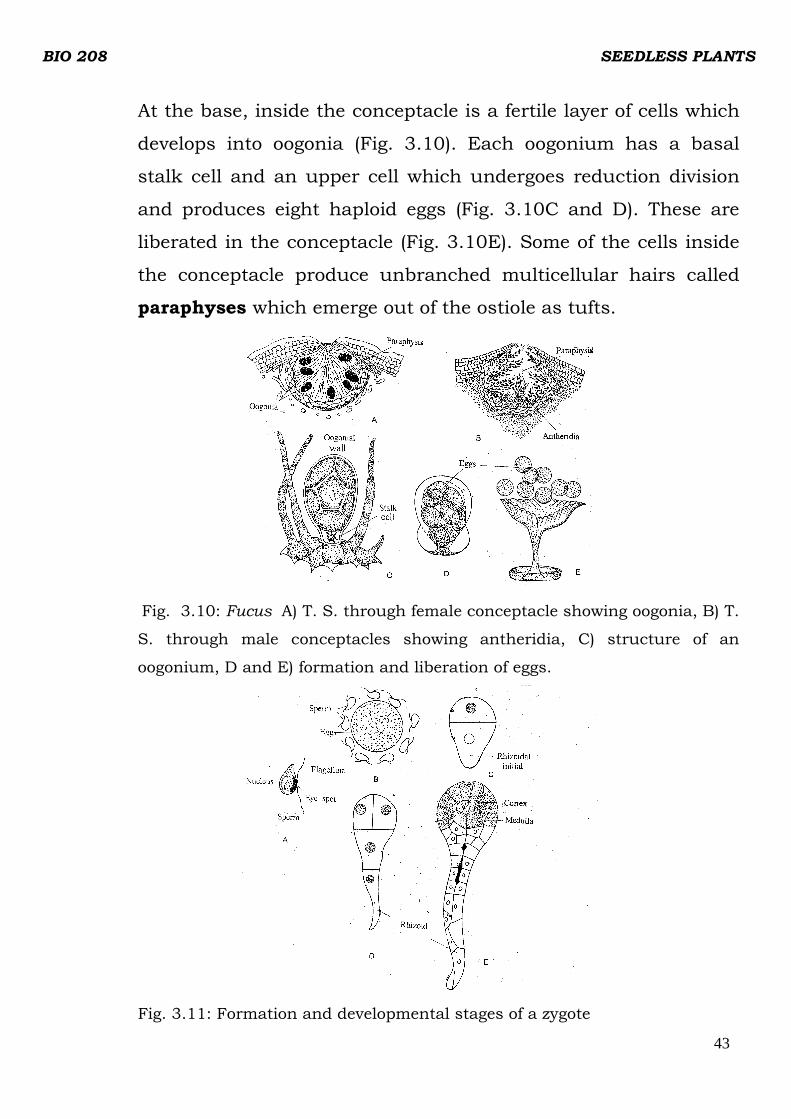
At the base, inside the conceptacle is a fertile layer of cells which
develops into oogonia (Fig. 3.10). Each oogonium has a basal
stalk cell and an upper cell which undergoes reduction division
and produces eight haploid eggs (Fig. 3.10C and D). These are
liberated in the conceptacle (Fig. 3.10E). Some of the cells inside
the conceptacle produce unbranched multicellular hairs called
paraphyses which emerge out of the ostiole as tufts.
Fig. 3.10: Fucus A) T. S. through female conceptacle showing oogonia, B) T.
S. through male conceptacles showing antheridia, C) structure of an
oogonium, D and E) formation and liberation of eggs.
Fig. 3.11: Formation and developmental stages of a zygote

BIO 208 SEEDLESS PLANTS
44
Antheridia are produced on branched paraphyses inside the
concptacle ( Fig. 3.10B). Each antheridium is like a unicellular
sporangium which divides meiotically and then by further
divisions produced 64 haploid sperms. The biflagellate sperm has
a longer flagellum pointing backwards and a shorter one
projecting towards the front. It has a single chloroplast and a
prominent orange eye spot
The release of the gametes is connected with the sea tides. At low
tide, Fucus fronds shrink due to loss of water, and when such
fronds are exposed to an on coming tide, the eggs and sperms are
released into the surrounding sea water.
The egg of Fucus are known to attract sperms (Fig. 3.11 A and B)
by secreting a gamone. Immediately after fertilization a all is
secreted around the zygote. It has been shown that unfertilized
eggs can develop into germlings parthenogenetically if treated
with dilute acetic acid.
The diploid zygote germinates by producing a rhizoidal outgrowth
on one side. It is later cut by wall formation to form a lower
rhizoidal cell and apical cell (Fig. 3.11 C) which by further
divisions (Fig. 3.11 D and E) gives rise to the Fucus fronds.
4.0 CONCLUSION
Reproduction in algae could be by vegetative method (binary
fission), asexual through specialized cells or sexual by fusion of
two cells from opposite sex called gametes.
The life cycles of Chlamydomonas, Ulothrix and Ficus were
discussed. There are other genera in this group. It should be
BIO 208 SEEDLESS PLANTS
45
noted that algae also exhibit alternation of generations in their life
cycles.
5.0 SUMMARY
• Reproduction in algae is by asexual and sexual methods.
• Asexual method involves fission of cells are regeneration of
new ones
• Sexual method involves fusion of male gamete and female
gamete resulting in the formation of a zygote.
• The life cycle in algae demonstrates clearly a marked
alternation of generations especially in the higher forms like
Ulva, Laminaria and Ficus.
6.0 TUTOR MARKED ASSIGNMENT
1) Which of the following algae reproduce only by binary
fission?
a. Volvox
b. Chlamydomonas
c. Anacystis
d. Microcystis
2) In the following statements fill in the blank spaces with
appropriate words:
i ………………… is an enlarged cell in blue-green algae
which accumulates food reserve, develops a thick wall and
functions as a resting spore.
ii Under unfavourable conditions the zoospores lose their
flagella and round up, they are called ………………
iii When a filamentous alga is accidently broken it
develops into a ………………………...

BIO 208 SEEDLESS PLANTS
46
iv The stage when thousands of zoospores of
Chlamydomonas cluster together in a gelatinious mass is
called ……………………..
v When both plus (+) and minus (-) strains are produced
by the same parent the condition is called ………………….
vi When two gametes (plus and minus) arise from
different parent algae the condition is called …………………..
vii Fusion of gametes of same size and morphology is
called …………………..
viii In anisogamy the two gametes are of …………………..
7.0 REFERENCES
Dutta A.C. (1981). Botany for degree students.
Oxford University Press. 909p.
IGNOU. (1991). Indra Gandhi Nationl Open University. Plant
Diversity - Algae.

BIO 208 SEEDLESS PLANTS
47
MODULE 2
UNIT 1: FUNGI MORPHOLOGY
CONTENTS
1.0 Introduction
2.0 Objectives
3.0 Main Content
3.1 Fungal Morphology
3.2 Unicellular Forms
3.3 Filamentous Forms
3.4 Pseudoparenchymatous Forms
4.0 Conclusion
5.0 Summary
6.0 Tutor Marked Assignment
7.o References/Further Readings
1.0 INTRODUCTION
You are probably familiar with yeast, bread mould, rust, smut and
mushrooms. They all are members of the fungal Kingdom. Fungi exhibit a
range of structures: unicellular, plasmodium like filamentous and
pseudoparenchy -matous. However, the different forms show common cellular,
physiological and biochemical characteristics. In this unit, you will study these
forms in some detail.
2.O OBJECTIVES
After studying this unit you should be able to:
i. distinguish fungi from other groups of organisms on the basis of
morphological features.
ii. describe the range of morphological forms in fungi.
3.0 MAIN CONTENT
BIO 208 SEEDLESS PLANTS
48
3.1 FUNGAL MORPHOLOGY
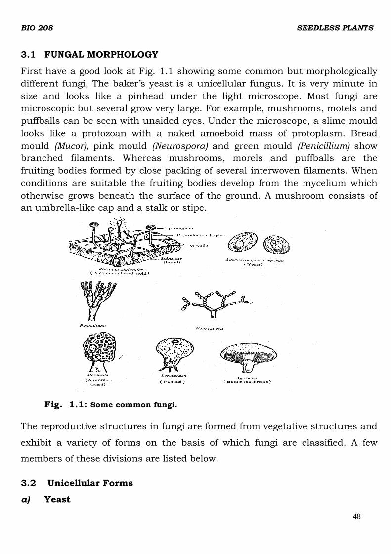
First have a good look at Fig. 1.1 showing some common but morphologically
different fungi, The baker’s yeast is a unicellular fungus. It is very minute in
size and looks like a pinhead under the light microscope. Most fungi are
microscopic but several grow very large. For example, mushrooms, motels and
puffballs can be seen with unaided eyes. Under the microscope, a slime mould
looks like a protozoan with a naked amoeboid mass of protoplasm. Bread
mould (Mucor), pink mould (Neurospora) and green mould (Penicillium) show
branched filaments. Whereas mushrooms, morels and puffballs are the
fruiting bodies formed by close packing of several interwoven filaments. When
conditions are suitable the fruiting bodies develop from the mycelium which
otherwise grows beneath the surface of the ground. A mushroom consists of
an umbrella-like cap and a stalk or stipe.
Fig. 1.1: Some common fungi.
The reproductive structures in fungi are formed from vegetative structures and
exhibit a variety of forms on the basis of which fungi are classified. A few
members of these divisions are listed below.
3.2 Unicellular Forms
a) Yeast

BIO 208 SEEDLESS PLANTS
49
The fungi are unicellular, often multicellular or acellular epkaryotic organisms.
The most common unicellular fungi are yeasts. Which are of wide occurrence.
Yeast is found on the sticky sugary surface of ripe fruit and grows in any sugar
solution. The individual cells adhere to one another forming a chain. Single
Cells are hyaline but the colonies appear greenish or brownish in colour. The
fine structure of a yeast cell as shown in Fig. 1.2a, is of the eukaryotic type. It
has a well-defined nucleus, mitochondria, endoplasmic reticulum and other
organelIes. Close to the nucleus, a large area of cytoplasm is occupied by
vacuole. The cell wall of yeast has 2-3 layers made of chitin and
polysaccharides - glucan and mannans, Depending upon the stage of
development variable amounts of proteins, lipids and other substances are
found accumulated in the cell.
Yeasts are distributed well over the surface of earth. They are abundant on
substrates that contain sugars, like the nectar of flowers and surface of fruits.
They are also found in soil, animal excreta, milk and on the vegetative parts of
plants and also in some other habitats.
B
Saccharomyces Olpidium in algal cell
A
Fig.1.2 A) The fine structure of yeast Saccharomyces B) Olpidium
Yeasts are noted particularly for their ability to utilise carbohydrates, hence
the name Saccharomycetes is applied to this group.
BIO 208 SEEDLESS PLANTS
50
Another unicellular fungus is Olpidium (Fig. 1.2b), the simplest chytrid, which
is a simple globular cell without branches.
b) Slime Moulds
Unicellular forms are also seen in slime during a certain stage of their life
cycle (Fig.1.3). You must remember that slime moulds are not considered true
fungi.
Their characteristics resemble both protozoa and fungi. That is why it has
been difficult to classify them. These curious organism show unicellular
(multinucleate) protozoan-like or multicellular fungus-like stages during the
course of their life cycle. Slime moulds are further classified as cellular slime
moulds and plasmodial slime moulds.
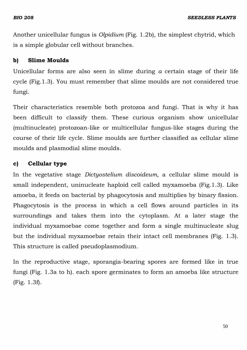
c) Cellular type
In the vegetative stage Dictyostelium discoideum, a cellular slime mould is
small independent, uninucleate haploid cell called myxamoeba (Fig.1.3). Like
amoeba, it feeds on bacterial by phagocytosis and multiplies by binary fission.
Phagocytosis is the process in which a cell flows around particles in its
surroundings and takes them into the cytoplasm. At a later stage the
individual myxamoebae come together and form a single multinucleate slug
but the individual myxamoebae retain their intact cell membranes (Fig. 1.3).
This structure is called pseudoplasmodium.
In the reproductive stage, sporangia-bearing spores are formed like in true
fungi (Fig. 1.3a to h). each spore germinates to form an amoeba like structure
(Fig. 1.3f).
BIO 208 SEEDLESS PLANTS
51
Fig. 1.3: Life Cycle of a cellular slime mould, Dictyostelium discoideum
d) Plasmodial Type
In plasmodial slime moulds, for example Echinosteliurn minutum, in the
vegetative stage, a large mass of multinucleate amoeboid cytoplasm with
characteristic diploid nuclei is formed (Fig. 1.4). But unlike cellular slime
moulds, the individual cells are not delimited by cell membrane. The cell wall
is absent. It feeds on encysted myxamoebae and bacteria and may spread over
a large area. The plasmodium does not have a definite size or shape. It may be
globose, flat and sheet-like spreading over a large area in the form of a very
thin network (Fig. 1.4b). When the plasmodium creeps over the surface of the
substratum, it changes its shape accordingly and engulfs particles of food on
its way. Finally, it matures and changes into the fructification typical of the
species (Fig. 1.4c and d). The entire plasmodium takes part in the formation of
fructifications, which bear spores resulting from meiosis. The spores germinate
to produce flagellated cells which develop into plasmodium (myxamoeba Fig.
1.4e to i).

BIO 208 SEEDLESS PLANTS
52
Slime mould plasmodia are often brilliantly coloured ranging from colourless
to shiny grey, black, violet, blue, green, yellow, orange and red. The yellow and
the white plasmodia are probably the most commonly encountered. Colour
changes have been observed to occur within a plasmodium under laboratory
conditions. Most slime moulds live in cool, shady, moist places in the woods,
on decaying logs. Dead leaves or other organic matter which holds abundant
moisture.
Zygote (Karyogamy) Fig.1.4: Life cycle of a plasmodial slime mould, Echimosteliumminutum.
3.3 Filamentous Forms
Most fungi are filamentous. You may have noticed on a piece of stale bread a
web of very fine and delicate threads. These are formed when a fugal spore
lands on the bread and germinates into a small tube-like outgrowth, which
further grows as transparent, tubular filaments in all directions. Each of these
filaments is called hypha, the basic unit of fungal body. The mass of
interwoven hyphae constituting the body of a fungus is called mycelium
(Fig.1.5) It may consist of highly dispersed hyphae, or it may be a cottony
mass of hyphae. The aerial hyphae that bear reproductive structures are called

BIO 208 SEEDLESS PLANTS
53
reproductive hyphae. The fugal mycelium has an enormous surface to volume
ratio and is close to the food source. This large surface-to- volume ratio is a
marvellous adaptation for absorptive mode of nutrition.
The mycelium of fungi is covered with a cell wall made of chitin, a
polysaccharide that is also found in the exoskeleton of insects and
crustaceans However, in some fungi the cell wall contains cellulose and lignin-
like substances. The protoplasm of mycelium may be continuous throughout
the mycelium so there will he several nuclei scattered throughout the
cytoplasm. This condition is termed as coenocytic (Fig. 1.5b), Such non-
septate hyphae are observed in the members of the Division Zygomycetes e. g.
Mucor and Rhizopus.. The septa or cross walls in the non-septate mycelia are
formed only to cut off reproductive structuies or to seal off a damaged portion.
Such septa are solid plates without any pores.
The members of other classes of fungi like ‘Ascomycetes and Basidiomycetes
e.g. Aspergillus and Penicilliun develop internal cross walls i.e., septa, which
divide the hyphae into segments. The septa appear at regular intervals. The
segments may be uninucleate or multinucleate.
The septa, in these cases have perforations through which cytoplasmic strands
including nuclei can migrate from one cell to the other (Fig. 1.5a). The
presence of septa gives mechanical support to the hyphae. The reproductive
structures are also separated from vegetative structures h septa but these are
not perforated.

BIO 208 SEEDLESS PLANTS
54
Fig. 1.5: Typical septate and non-septate hyphae of fungi.
In some groups of fungi the mycelium formed on germination of spores
consists of uninucleate segments (monokaryotic) initially. This is called
primary mycelium. Later when fusion occurs either between hyphal segments
of the same mycelium or different mycelium, the segments contain two nuclei
(dikaryotic). This conversion is called dikaryotisation and the mycelium is
called secondary mycelium. This stage may last for a long period. When this
mycelium gets organised into a specialized structure, it is termed tertiary
mycelium.
3.4 Pseudoparenchymatous Forms
The fungus mycelium normally, as mentioned above, is a mass of loosely
interwoven hyphae which form a network. In some fungi the entire mycelium
or its parts undergo various modifications. The walls of the hyphae in the
mass get fused and they lose their individuality. As a result the hyphal mass,
in cross section appears to he a continuous structure. It resembles the
parenchymatous tissue of higher plants, but it is not a true parenchyna as
found in higher plants. In fungi such a tissue is called plectenchyma.
Plectenchyma can further be differentiated into two types. The plectenchyma
with rounded fungal cells is called pseudoparenchyma and with less
compacted elongated cells is called prosenchyma.
BIO 208 SEEDLESS PLANTS
55
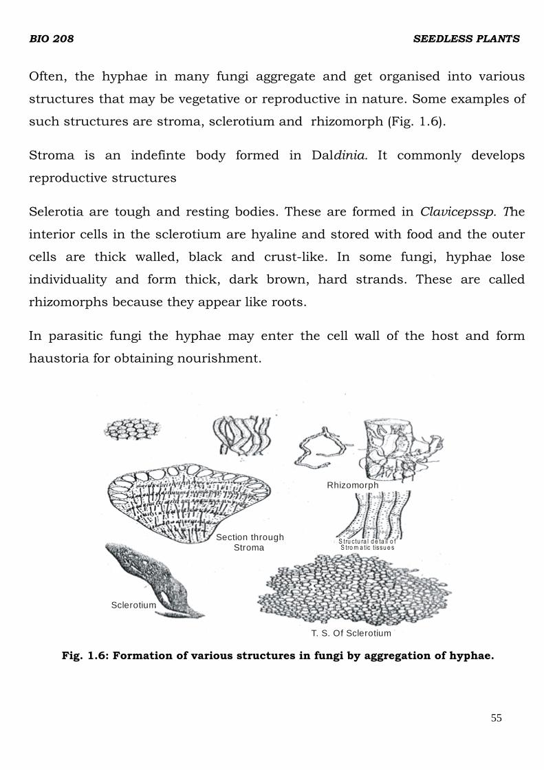
Often, the hyphae in many fungi aggregate and get organised into various
structures that may be vegetative or reproductive in nature. Some examples of
such structures are stroma, sclerotium and rhizomorph (Fig. 1.6).
Stroma is an indefinte body formed in Daldinia. It commonly develops
reproductive structures
Selerotia are tough and resting bodies. These are formed in Clavicepssp. The
interior cells in the sclerotium are hyaline and stored with food and the outer
cells are thick walled, black and crust-like. In some fungi, hyphae lose
individuality and form thick, dark brown, hard strands. These are called
rhizomorphs because they appear like roots.
In parasitic fungi the hyphae may enter the cell wall of the host and form
haustoria for obtaining nourishment.
Section through Stroma
Rhizomorph
S tru ctu ra l d e ta il o f S trom a tic t issu e s
Sclerotium
T. S. Of Sclerotium
Fig. 1.6: Formation of various structures in fungi by aggregation of hyphae.

BIO 208 SEEDLESS PLANTS
56
4.0 CONCLUSION
Fungi exhibit a range of structures from unicellular, plasmodium-like
filamentous to pseudoparenchymatous. Most fungi are multicellular and
branched filaments. The mycelium is the main part of fungal body and it may
aggregate to produce stoma, sclerotia and rhizomorphs.
5.0 SUMMARY
In this unit you have learnt that:
i. Fungi grow on variety of substrates that contain traces of organic
compounds. Some of the members can grow under extreme conditions of
temperature and osmotic concentration of the solute.
ii. Fungi show a range of morphological forms. Unicellular fungi like yeast
are rare. Slime moulds are either unicellular or plasmodium like at a
certain stage of the life cycle.
iii. Most fungi are multicellular, branched filaments. The mycelium is the
main part of the fungal body. The reproductive structures are born on the
reproductive hyphae.
iv Various kinds of structures arise when the entire mycelium or its part
aggregate and give rise to special structures such as stroma, sclerotia,
rhizomorphs arid others.
6.0 Tutor Marked Assignment
Indicate which of the following statements are true or false. Write T for true or
F for false;
i) Fungi are achlorophyllous organisms.
ii) Fungi prefer acidic medium for growth.
iii) The cell wall of fungi belonging to the division Oomycota is made of
chitin.
iv) Fungi can utilise organic substances.

BIO 208 SEEDLESS PLANTS
57
v) Yeast cell is prokaryotic type.
vi) Most genera in fungi are multicellular and some are unicellular.
vii) In slime moulds the cell wall is absent
7.0 Reference/Further Reading
1. Dutta: A. C. (1981). Botany for degree students. Oxford University Press
909p.
2. IGNOU. (1991). Indra Gandhi National Open University. Plant Diversity.
BIO 208 SEEDLESS PLANTS
58
UNIT 2: LIFE CYCLES IN FUNGI
CONTENTS
1.0 Introduction
2.0 Objectives
3.0 Main Content
3.1 Types of Life cycles and alteration of generation s
3.1.2 Phytopthora
3.1.3 Rhizopus
4.0 Conclusion
5.0 Summary
6.0 Tutor Marked Assignment
7.o References/Further Reading
I.0 INTRODUCTION
In the last units you learnt about the morphology of fungi. In this unit we
shall discuss a detailed study of reproduction and alternation of generations in
Phytophthora and Rhizopus.
2.0 OBJECTIVES
After studying the units you should be able to;
i. describe reproduction processes in Phytopthora and Rhizopus
ii. illustrate the life cycles of Phytopthora and Rhizopus.
3.0 MAIN CONTENT
3.1 TYPES OF LIFE CYCLES AND ALTERNATION OF GENERATIONS
3.1.2 Phytophthora
This fungus belongs to the Division Oomycota. There are about 75 species in
this genus, most of which live as parasites on flowering plants. The species

BIO 208 SEEDLESS PLANTS
59
Phytophthora infestans is of great economic importance. It causes a serious
potato disease called potato blight or late blight of potato.
Morphology
The mycelium of Phytophthora is profusely branched and consists of aseptate,
hyaline and coenocytic hyphae. The hyphae ramify in the intercellular spaces
of the host tissues. The mycelium produces haustoria which penetrate the
host cell wall and enter the cells to draw nourishment (Fig. 2.1a). The
haustoria may be simple or branched. Phytophthora reproduces both asexually
and sexually.
Asexua1 Reproduction
In warm and humid weather it normally reproduces asexually. During this
stage a tuft of slender, branched hyphae usually arise from the internal
mycelium. They come out through the stomata or pierce through the epidermal
cell on the lower surface of the leaf (Fig. 2.1b). In tubers they come out
through the injured portions of the skin. These aerial hyphae are hyaline and
branched. They bear a sporangium at their tip. You have learnt earlier that the
hyphae-bearing sporagia or conidia are called sporangiophores or
conidiophores respectively. The sporangia are thin-walled, hyaline and lemon-
shaped and have a beak-like projection or papilla at their tips.
The mature sporangia can easily be separated from the sporangiophore. The
sporangiophore is branched. It bears nodular swellings which denote the point
of detachment of sporangia. Wind, rain drops or contact with neighbouring
leaves detach and scatter the ripe sporangia on neighbouring potato plants.
They may fall on the ground and get spread into the soil. The sporangia lose
their viability if they fail to germinate within a few hours.

BIO 208 SEEDLESS PLANTS
60
When the sporangia fall on the leaf of a host plant, they germinate. Moisture
and temperature are the determinants for germination. In the presence of
water and low temperatures (upto 12°C) the sporangiun behaves as a
zoosporangium. The protoplast divides into 5-8 uninucleated daughter
protoplasts which transform into zoospores.
Fig. 2.1: Phytophthora infestans; a) Intercellular mycelium forming hapstoria, b) Soranglophores
coming out of stoma bearing sporangia c) flagellared zoospores.
The zoospores are uniform and biflagellate (Fig. 2.1c) of the two flagella one is
of the whiplash type and the other is of the tinsel type. The zoospores are set
free through the apical papilla into a vesicle in some species. The vesicle soon
bursts open to liberate the zoospores. The liberated zoospores swim for some
time, and later settle on a substratum losing the flagella and germinate.
During germination, the zoospore puts out a short hypha called appressorium.
The appressoria help to fix the fungus on the surface of the host leaf. From the
appresorium, a narrow, peg- like infection hypha develops which forces its way
into the host leaf.

BIO 208 SEEDLESS PLANTS
61
At temperature up to 24°C, and low relative humidity the sporangium
germinates directly behaving like a conidium. It germinates producing a germ
tube or a short hypha, which enters into the host leaf.
The sporangia, which are washed into the soil, germinate and infect the
tubers. As a result the tubers rot by harvest time or during storage. Under
favourable conditions a number of asexual generations may be produced in
one growing season. This results in rapid propagation of the fungus to spread
the disease.
Sexual Reproduction
Sexual reproduction is of the oogamous type. The male sex organs are
antheridia and the female oogonia. They arise at the tips of short lateral
branches as antheridial and cogonial initials respectively (Fig. 2.2a).
Phytophthora infestans is heterothallic.
The antheridium is a club-shaped structure with one or two nuclei to begin
with. Later the nuclei divide and produce about 12 nuclei (Fig.2.2b). At the
time of the fertilization only one functional nucleus persists and the others
degenerate. The oogonium develops on a neighbouring hypha of the antheridial
branch. It grows across the antheridium and swells to form a pear-shaped or
spherical structure (Fig.2.2 c). It contains dense cytoplasm and many nuclei
(about 40).The protoplast of the oogonium becomes differentiated into an outer
multinucleate periplasm and a central uninucleate ooplasm. The central
nucleus divides into two and one of them disappears. The surviving nucleus
functions as the egg nucleus. The nuclei of the periplasm later degenerate.
The oogonial wall bulges out at a certain point to make a receptive spot . The
oogonial wall disintegrates at this spot. Through this opening the antheridium
pushes a short fertiIization tube (Fig 2.2e). The fertilization tube penetrates the
periplasm and reaches the ooplasm. Here it opens and delivers the male
nucleus along with the surrounding cytoplasm. The male and female nuclei
fuse, thus bringing out fertilization (Fig. 2.2f)

BIO 208 SEEDLESS PLANTS
62
Antheridialinitial Antheridia
Oogonia
a
Oogonial initial
b c
d e
Periplasm
Ooplasm
Oospore
f
Fig. 2.2: Stages of sexual reproduction in Phytophthora infestans
The fertilized egg secretes a thick wall around itself and becomes the oospore.
When the conditions are favourable the oospore germinates. It is believed that
meiosis takes place during germination. The germination of oospore takes
place after the decay of the host tissue. A germ tube develops from the oospore
and may directly develop into a mycelium or oospore may bear a terminal
sporangium. Inside the sporangium zoospores are produced which after
liberation develop into new mycelia.
ln the life cycle of Phytophthora there is an asexual cycle which may repeat
during favourable conditions. The sexual cycle takes place prior to the onset of
unfavourable conditions forming a resting spore. These cycles normally
alternate with each other.
3.1.3 Rhizopus
Rhizopus isa member of Division Zygomycota. It is commoly called bread
mould since it is frequently found growing on stale bread. It is a saprophytic
fungus. It also grows on decaying fruits, vegetables and other food materials.
Rhizopus stolonifer sometimes grows as a facultative parasite on strawberries
causing a transit disease called ‘leak’ and also causes ‘soft rot’ disease of sweet
potatoes, yam and cassava tubers.

BIO 208 SEEDLESS PLANTS
63
The mycelium is a white cotton-like fluffy mass with numerous, slender,
branched hyphae. The mycelium has three types of hyphae: i) rhizoidal ii)
stolons and iii) sporangiophores (Fig, 2.3).
The rhizoidal hyphae are a cluster of brown, slender and branched rooting
hyphae which arise from the lower surface of the stolon at certain points
which are the apparent nodal points. These hyphae help in anchorage and in
the absorption of water and nourishment from the substratum.
The aerial hyphae which grow horizontally over the surface of the substratum
are called stolons. These hyphae are comparatively large, and slightly arched.
The stolons grow rapidly in all directions, completely filling the surface of the
substratum.
The third kind of hyphae called sporangiophores develop during the
reproductive phase. The sporangiophores arise from the apparent nodal
regions, opposite to the rhizoidal hyphae in a cluster. They grow vertically
bearing sporangia at their tips.
Asexual Reproduction
Rhizopus reproduces asexually by multinucleate, non-motile spores which are
produced in small, round, black sporangia. The sporangia are borne
terminally, and singly on unbranched sporangiophores (Fig 2.3b). A mature
sporangium is differentiated into two regions, a central less dense, vacuolated
region with fewer’ nuclei called columella and a peripheral dense region with
many nuclei called sporoferous region. The protoplast in the columella is
continuous with that of the sporangiophore.
The sporeferous region undergoes cleavage to form a number of muItinucleate
segment. These segments round off and secrete walls around them to become
sporangiospores. These are unicellular, multinucleate, non-motile
aplanospores, globose or oval in shape (Fig. 2.3c). As the spores mature the

BIO 208 SEEDLESS PLANTS
64
sporangium bursts open liberating the spore mass (Fig. 2.3d and e). A part of
the wall remains as a collar-like fringe at the base of the sporangium.
The spores are dispersed away by the wind. Falling on a suitable substratum,
under suitable conditions a spore germinates producing a short germ tube
which grows further and branches profusely to produce three types of hypae.
Sporangiosphores
Sporangia
aRhizoidalhyphae
Spores
Spores
Columella
Spores
Feedingmycelium
Columella
Columella
d
e
c
b
Rhizoids
Sporangiophores
Fig.2.3: Rhizopus stolonifer a) three kinds of hyhae of the mycelium, b) sporangiophores
developing at the point of rhizoidal hyhae, c) structure of the sporangium in detail, d)
invaginated columella, e) dehiscence of the spores.
Under unfavourable conditions Rhizopus produces chlamydospores. As you
learnt they are thick-walled spores with accumulated reserve food. They are
produced intercalarily. They help to tide over unfavourable conditions during
which time the mycelium perishes. With the return of favourable conditions
they germinate and produce normal mycelium.
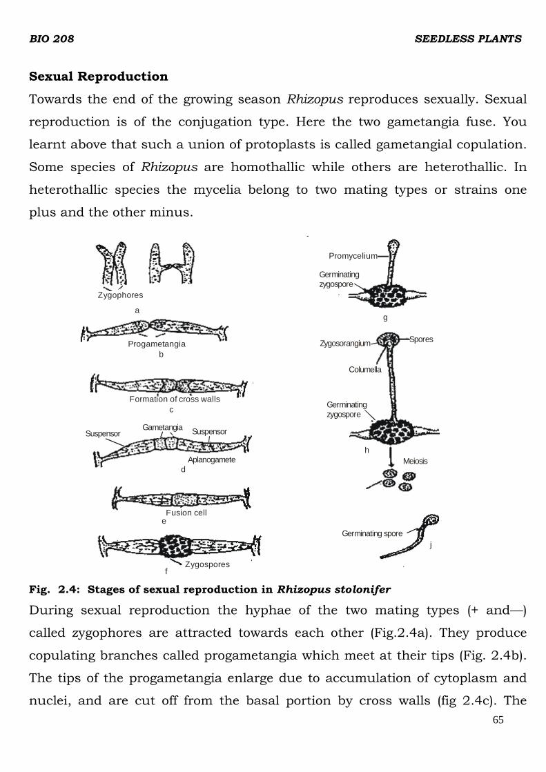
BIO 208 SEEDLESS PLANTS
65
Sexual Reproduction
Towards the end of the growing season Rhizopus reproduces sexually. Sexual
reproduction is of the conjugation type. Here the two gametangia fuse. You
learnt above that such a union of protoplasts is called gametangial copulation.
Some species of Rhizopus are homothallic while others are heterothallic. In
heterothallic species the mycelia belong to two mating types or strains one
plus and the other minus.
Zygophores
a
Progametangiab
Promycelium
Germinating zygospore
Zygosorangium
g
Columella
Spores
h
Germinating zygospore
Meiosis
Germinating sporej
Gametangia
c
Fusion celle
Zygosporesf
Formation of cross walls
Suspensor Suspensor
Aplanogameted
Fig. 2.4: Stages of sexual reproduction in Rhizopus stolonifer
During sexual reproduction the hyphae of the two mating types (+ and—)
called zygophores are attracted towards each other (Fig.2.4a). They produce
copulating branches called progametangia which meet at their tips (Fig. 2.4b).
The tips of the progametangia enlarge due to accumulation of cytoplasm and
nuclei, and are cut off from the basal portion by cross walls (fig 2.4c). The

BIO 208 SEEDLESS PLANTS
66
terminal portion is call suspensor. The entire gametangium transforms into an
aplanogamete (Fig. 2.4d). The two gametangia may be of the same size or one
of them slightly smaller than the other.
When the gametangia mature the intervening, waIls dissolve and the two
gametes and their nuclei fuse producing a zygospore (fig. 2.4e and f). The
zygospore increases in size and secretes a thick two layered wall around it. The
outer layer is dark and warty. It is called extine or exospore. The inner layer is
thick and is called intine or endospore. As the zygospore increases in size, the
wall of the fusion cell containing the zygospore ruptures and it is set free.
Prior to germination of the zygospore, the diploid nuclei divide meiotically
producing numerous haploid nuclei. During meiosis segregation of strains
takes place. The zygospore during germination absorbs water and swells. As a
result the outer wall extine breaks open. The inner wall intine with the inner
contents grows out as a germ tube or promycelium (Fig 2.4g). The
promyceliurn is of limited growth and produces a terminal sporangium. It is
called zygosporangium or germ sporangium (Fig 2.4h). Inside the sporangium
numerous, non-motile germ spores called meiospores are produced. They are
liberated at maturity which develop into new mycelia (Fig 2.4i and j).
The life cycle of Rhizopus consists of two phases, asexual and sexual. The
asexual phase consists of myelium, sporangiophores, sporangia and the
sporangiospores. This phase in the life cycle serves to propagate the haploid
phase of the fungus during favourable conditions. The sexual phase, consists
of mycelia of the plus and minus strains, the progametangia, gametangia,
aplanogametes, zygospore, promycelium, germ sporangium and the germ
spores. Among these, the zygospore is the only diploid structure. All others are
hapoid. Such a sexual cycle is called haplontic characterized by zygotic
meiosis and haploid mycelium as the only adult fungi

BIO 208 SEEDLESS PLANTS
67
Rhizopus exhibits heterothallism wherein the mycelia of a single species are
morphologically similar but physiologically different. There is no apparent
distinction between male and female mycelia except in their sexual behaviour.
Such a distinction is designated by the terms plus and minus. This was first
discovered by Blakeslee in 1904. This is the first indication of the origin of
dioecious condition of sexual phase in an organism.
4.0 CONCLUSION
Reproduction in fungi occurs by vegetative asexual and sexual methods.
Vegetative reproduction occurs by budding, fission, framentation, formation of
oidia and rhizomorphs. Asexual reproduction occurs more frequently. Sexual
reproduction involves plasmogamy, karyogamy and meiosis. Phytophthora
infestans and Rhizopus are some of the examples of fungi treated in this unit.
5.0 SUMMARY
In this unit you have learnt that;
i. Phytophthora infestans causes serious potato disease. The coenocytic
hyphae ramify in the host tissue and draw nutrition through haustoria.
Asexual reproduction takes place by the formation of sporangia.
Depending upon the condition of temperature and humidity the
sporangia may produce zoospores or germinate directly. Sexual
reproduction is of oogamous type.
ii. In Rhizopus the mycelium has rhizoidal hyphae, stolons and the hyphae-
bearing sporangiophores. The sporangiophores bear non-motile
aplanospores, the asexual reproductive bodies. Sexual reproduction
occurs by the fusion of gametangia of opposite strains forming
zygosporangia which bear numerous non-motile zygospores.

BIO 208 SEEDLESS PLANTS
68
6.0 Tutor Marked Assignment
a. How many types of sexual reproduction are found in fungi? List them.
b. What are Sclerotia?
7.0 Reference/Further Reading
Dutta, A.C. (1981). Botany for degree students Academy Press U.K.
IGNOU. (2002). Indra Gandhi National Open University. Plant Diversity-Fungi.

BIO 208 SEEDLESS PLANTS
69
UNIT 3 :REPRODUCTION IN FUNGI
CONTENTS
1.0 Introduction
2.0 Objectives
3.0 Main Content
3.1 Types of Reproduction
3.1.1 Vegetable reproduction
3.1.2 Asexual reproduction
3.1.3 Sexual reproduction
4.0 Conclusion
5.0 Summary
6.0 Tutor Marked Assignment
7.o References/Further Reading
I.0 INTRODUCTION
In the previous unit you learnt about fungal morphology. In this unit we will
discuss the process of reproduction in fungi. Like algae, reproduction in fungi
occurs by vegetative, asexual and sexual methods. In the following account we
will describe the various types .of production in fungi with suitable examples.
2.0 OBJECTIVES
After studying this unit you should be able to:
i. describe the types of reproduction in fungi.
ii. distinguish between vegetative and asexual methods of reproduction,
iii. describe plasmogamy in details.
iv. describe the phases of sexual reproduction in fungi.
3.0 MAIN CONTENT
3.1 TYPES OF REPRODUCTION
In the previous unit you have learnt that a fungus hypha elongates by apical
growth, but most parts of a fungus are potentially capable of growth. When the

BIO 208 SEEDLESS PLANTS
70
mycelium of a fungus reaches a certain stage of maturity and accumulates
reserve food, it starts reproducing. As in algae, reproduction in fungi is of three
kinds:
i) vegetative,
ii) asexual and
iii) sexual
Budding b
Chlamydospore
Fission
a
Ooidia c d
Fig.3.1:Vegetative reproduction in fungi
Vegetative (Fig. 3.1)and asexual methods of reproduction which do not involve
the fusion of nuclei or sex cells or sex organs are, however, clubbed by many
mycologists into asexual methods of reproduction. Thus, they recognise only
two methods, asexual and sexual.
3.1.2 Asexual Reproduction
In fungi, asexual reproduction is a more common method than sexual
reproduction. It is usually repeated several times in a season. It takes place by
the formation of special reproductive cell called spores. The formation of
spores in fungi is called sporulation. Each spore develops into a new
mycelium. These spores are produced as a result of mitosis in the parent cell
and hence they are also called mitospores. The spores vary in colour, shape
and size, number, arrangement on hyphae and in the way in which they are

BIO 208 SEEDLESS PLANTS
71
borne. They may be hyaline, green, yellow, orange, red, brown to black in
colour and are minute to large in size. In shape they vary from globose to oval,
oblong, needle-shaped to helical. Thus an infinite variety of spores can be
observed in fungi (Fig.3.2) and you will find them very fascinating under the
microscope.
Fig. 3.2 Variety of spores in fungi
Usually the spores are unicellular. They may be uninucleate or multinucleate.
In some fungi like Alternaria and Curvularia they are multicellular. The
mitospores produced in fungi are of two types, sporangiospores and conidia.
The sporangiospores are produced inside a sac-like structure called
sporangium. The hypha bearing a sporangium is called sporangiophore
(Fig.3.3a). They are characteristically branched. The sporangiospores may be
motile or non-motile. The non-motile sporangiospores are called aplanospores-
from Greek a, not + planets wanderer + spores seed, spore (Fig. 3.3a). These
are characteristic of terrestrial species like Mucor and Rhizopus. In aquatic
fungi like Pythium of’ the Division Oomycota motile biflagellate
sporangiospores are produced. These are called zoospores and the
sporangium bearing them is called zoosporangium (Fig.3.3b and c). A
zoospore is a motile spore lacking a cell wall. After a swarming period it
secretes a wall and germinates to form a germ tube. In contrast to zoospores,
the aplanospores have a definite spore wall and are dispersed by wind and
insects.

BIO 208 SEEDLESS PLANTS
72
The conidia are non-motile, deciduous mitospores formed externally as single
separate cells. They develop either directly on the mycelium or on
morphologically differentiated hyphae called conidiophores (Fig.3.3d). The
conidiophores may be simple or branched, septate or aseptate. The conidia are
produced singly e.g., Phytophthora or in chains at the tips of the conidiophores
e.g. Aspergillus (Fig.3.3d) or at the tips of their branches e.g., Penicillium
(Fig.3.3e).
Often the conidiophores arise singly and are scattered in the mycelium.
Sometimes they arise in specialised structures called fruiting bodies.
According to their appearance they are termed as synnema, sporodochia,
acervuli (sucer-shaped), pycnidia (flask-shaped, globular) or pustules. These
are shown in Fig.3.4
Aplanospores
Sporangium
Sprangiosphore
a
ZoosporesZoosporangium
Sporangiophore
Conidia
d
e
b
Sterigma
Conidia
Secondarysterigma
Primarysterigma
VesicleConidiophoreSterigmata
Zoosporangium
Zoospores
c
Fig.3.3: Formation of sporangiospores and conidia in fungi: sporangia containing (a)
aplanospores, (b) and (c) zoospores (d) conidiophores showing conidia on branches
and (e) conidophores bearing conidial chains on branches.

BIO 208 SEEDLESS PLANTS
73
Fig. 3.4: Various types of fruiting bodies in fungi.
3.1.3 Sexual Reproduction
The sexual stage in fungi is called the perfect state in contrast to the
imperfect state which is the asexual stage. Sexual reproduction involves the
fusion of two compatible sex cells or gametes of opposite strains. Fungal sex
organs are called gametangia. They may be equal in size. In many higher
ascomycetes morphologically different gametangia are formed. The male
garnetangia are called antheridia and the female ones ascogonia.
The fungus may be homothallic, that is, the fusing gametes come from the
same mycelium or may be heterothallic, that is, the fusing gametes come from
different strains of mycelia.
In fungi, sexuaI reproduction involves the following three phases: i)
plasmogamy, ii) karyogamy and iii) meiosis (Fig.3.5). These three processes
occur in a regular sequence and at a specific time, during the sexual stage of
each species.
i. Plasmogamy: It is the union of protoplasts of reproductive hyphae or
cells, one from the male and the other from the female to bring about the

BIO 208 SEEDLESS PLANTS
74
nuclei of the two parents close together as a pair. However, the two nuclei
do not fuse with each other. Such a cell is called a dikaryon. The
dikaryotic condition is unique to fungi and may continue for several
generations as the two nuclei (dikaryon) divide simultaneously during cell
division. These are passed on to the daughter hypha.
ii. Karyogamy: The fusion of the two nuclei which takes place in the next
phase is called karyogamy. It may immediately follow plasmogamy as in
lower fungi, or it may be delayed for a long time as in higher fungi.
iii. Meiosis: Karyogamy which eventually occurs in all sexually reproducing
fungi is sooner or later followed by meiosis producing four genetically
different spores.
We will now discuss plasmogamy in detail. There are different methods of
plasmogamy in fungi.
Haploid spores
Haploids
Fig.3.5: Three phases of sexual cycle in fungi
i) Planogametic copulation: It involves fusion of two gametes. Like in
algae sexual union in fungi may be isogamous, anisogamous or
oogamous (Fig.3.6a ). Anisogamy and oogamy are together called
heterogamous sexual reproduction. Isogamy is the simplest type of
sexual reproduction, where the fusing gametes are morphologically
similar e.g. Olpidium and Catenaria. Anisogamy, where the fusing
gametes are dissimilar is found in one genus, Allomyces, a chitrid. In
oogamy as you may recall the motile antherozoid enters oogonium and
unites with egg or oosphere forming a zygote. Oogamy is seen in fungi
like Pythium and Albugo.

BIO 208 SEEDLESS PLANTS
75
Fig. 3.6: Different ways of plasmogamy in fungi
ii) Gametangial copulation : The two gametangia make contact and the
entire contents of the two fuse together and become one e. g., Mucor and
Rhizopus. In some fungi the entire protoplast of one gametangia flows
into the other through a pore (Fig. 3.6b). Among the two, the recipient is
the female and the donor is the male.

BIO 208 SEEDLESS PLANTS
76
iii) Gametangial contact The male gamete is not a separate entity but the
nucleus in the antheridium represents the gamete. As you can see in the
Fig. the oogonium and antherididm form a contact through a tube and
one or more nuclei inside the antheridium migrate into the oogonium.
You may note that in this case the two gametangia do not fuse. It is
observed in Penicillium (Fig.3.6c).
iv) Spermatization This mode is quite remarkable as the minute conidia
like gametes called spermatia are produced externally on special hyphae
called spermatiophore (Fig. 3.6d). Spermatia may develop inside the
cavities called spermatogonia. The female cell may be a gametangium, a
specialised receptive hypha or even a vegetative hypha.
v) Somatogamy: In higher fungi like Ascomycetes and Basidiomycetes there
is a progressive degeneration of sexuality. The entire process is very
much simplified by the fusion of two mycelia which belong to opposite
strains (Fig. 3.6e). The post-fertlization changes result in the production
of a fruiting body which is called ascocarp in Ascomycetes and
basidiocarp in Basidiomycetes.
The gametangial fusion followed by the fusion of male and female nuclei
results in diploid nuclei. Subsequently, reduction division occurs and haploid
spores are formed. In fungi, the spores may be formed in specialised
structures characteristic of a division. In Ascornycetes the spores called
ascospores are formed within the ascus (plur. Asci, Fig.3.7). The asci reside
enclosed within the fruiting body- the ascocarp. According to the
characteristics, the ascocarps are distinguished as cleistothecium
(indehiscent) apothecium, (cup or saucer shaped), perithecium (flask-shaped)
and pseudoperithecium.

BIO 208 SEEDLESS PLANTS
77
In basidiomycetes, sexual spores are termed basidiospores which develop on
club- shaped structure, called basidium.
Fig. 3.7: a to f) Stages of sexual reproduction in Penicillium; a to c) development of sex organs,
antheridia and ascogonia, c and f) stages in plasmogamy, g and h) ascocarp-cleistothecium formed
when surrounding hyphae enclose a number of asci, i) apothecium of Ascobolus sp j) perithecium, k)
an immature pseudothecium of Leptosphaera.
4.0 CONCLUSION
Fungi reproduce when the mycelium reaches a certain stage of maturity.
Phytophthora. Intestans the crusal organism for potato blight or late blight of
potato reproduces sexually and asexually. Both the asexual and sexual phases
alternate with each other depending on how favourable the weather condition
is. Rhizopus, also a parasite, have a similar life cycle like Phytophthora

BIO 208 SEEDLESS PLANTS
78
5.0 SUMMARY
In this unit you have learnt that;
i. Fungi reproduce by vegetative, asexual and sexual methods.
ii. Vegetative reproduction takes place by fission, budding, fragmentation.
iii. Asexual reproduction occurs more frequently then sexual method. The
sporangiophores or conidiophores formed bear spores and conidia
respectively.
iv. In sexual reproduction, depending upon the species the entire thallus or
a portion of it may take part in the formation of reproductive bodies.
6.0 Tutor Marked Assignment
a. List the main types of reproduction in fungi.
b. List the types of plasmogamy in fungi.
7.0 Reference/Further Reading
Dutta, A.C. (1981). Botany for degree students. Oxford University Press. 909 p.
IGNOU. (1991). Indra Gandhi National Open University . Plant Diversity 1.

BIO 208 SEEDLESS PLANTS
79
MODULE 3
UNIT 1: MORPHOLOGY, LIFE CYCLE AND CLASSIFICATION
OF BRYOPHYTES
CONTENTS
1.0 Introduction
2.0 Objectives
3.0 Main Content
3.1 General Characteristics
3.2 Life Cycle / Adaptation to land
3.3 Morphology Classification of Bryophytes
3.2.1 Hepaticopsida
Riccia
Marchantia
3.3.2 Anthocerotopsida
Anthoceros
3.3.3 Bryopsida
Sphagnum
Funaria
4.0 Conclusion
5.0 Summary
6.0 Tutor Marked Assignment
7.o References/Further Reading
1.0 INTRODUCTION
In the previous units, you have studied about algae in general.
In this unit, you will learn about the Bryophytes.
Bryophytes are considered to be the first land plants among embryophytes.
Exactly how this happened is not clear because the fossil records are not

BIO 208 SEEDLESS PLANTS
80
complete. When there was a shift from aquatic mode of life to land habit the
species had to face many challenges. How could water and minerals be taken
from the soil and transported to parts that are not in contact with
soil? How could the soft bodies keep from drying out? To meet these challenges
there was a need to develop certain structural modifications. The land plants
belonging to various groups have continued to exist approximately from the
Devonian period. This demonstrates that they are well adapted to their
particular niche on land. It is the nature of these adaptations that is of interest
to us in this unit.
In this unit we will deal with the characteristic features of each group and
describe a few genera belonging to these groups. You will study how these
genera differ from each other and also from the majority of flowering plants
which are so commonly growing around you.
2.0 OBJECTIVES
After studying this unit, you should be able to:
• describe the general characteristics of bryophytes,
• give reasons why algae are considered to be ancestors of the first land
plants,
• classify the bryophytes
• describe the reproduction and life cycle of bryophytes,
• describe the morphology of some members of the group.
3.0 MAIN CONTENT
3.1 GENERAL CHARACTERISTICS AND LIFE CYCLE
The Division Bryophyta includes the simplest and the most primitive members
of land plants that lack roots, and do not have a vascular system. There are
some mosses that have a primitive system of tubes that conduct-water and

BIO 208 SEEDLESS PLANTS
81
food. The water-conducting tubes are called hydroids. They have elongated,
thick, dead cell. But they are not lignified like tracheids and vessels. The food-
conducting tubes are called leptoids, and they are connected through
plasmodesmata.
A single plant is very small, hardly a few cm in size. It seldom grows large
because of lack of supporting tissues. Thousands of tiny moss plants often
grow together and given thick, green carpet-like appearance. The morphology
of some common bryophytes is given in (Fig.1.1). Have a good look at them.
Can you recall seeing any in their natural habitats? Bryophytes show two
distinct and well defined phases of life cycle, sexual and asexual, which follow
each other. The gametophyte is haploid and produces gametes. The
sporophyte is diploid and produces spores. The haploid generation alternates
with diploid .generation (recall alternation of generations in algae). Both the
gametophyte and sporophyte may be several centimetres in length but the
gametophyte is the long-lived phase of life cycle. You may note that in other
land plants the sporophyte is the dominant generation.
The gametophyte may be thalloid (Fig. 1.1 A, B and D) or has an axis
differentiated into stem-like and leaf-like structures (Fig. 1.1C, E and F) which
lack xylem and phloem. You may note that these leaf-like structures are part
of gametophyte, whereas in vascular plants the leaves strictly develop on
sporophyte. The gametophyte is green, photosynthetic and nutritionally
independent, and anchors to the soil by unicellular or multicellular filaments
called rhizoids. Rhizoids appear like roots but unlike roots they lack vascular
tissues and are much simpler in structure.
Now try to list a few points that distinguish bryophytes from algae.
Bryophytes are most abundant in moist tropical areas. But they also grow in
deserts, mountains and are observed in parts of Antarctica. In dry areas their

BIO 208 SEEDLESS PLANTS
82
growth and activity is restricted to wet seasons only. Some mosses grow in
fresh water streams but they are not found in sea flora.
3.2 Life Cycle
We are illustrating here the life cycle of bryophytes taking Funaria as an
example. The gametophyte of Funaria (Fig. 1.2 A) bears two types of specialised
multicellular reproductive organs (Fig.1.2B and C) called the gametangia
(gamete holders) which protect egg and sperm during the development.
The male gametangia, called antheridia (sing, antheridium, Fig.1.2B), produce
sperms. The female gametangia, called archegonia (sing, archegonium,
Fig.1.2C), produce eggs. The gametangiá have outer sterile layer of cells forming
protective jacket.
Fig.:1.1: Morphology of bryophytes : A and B) thalloid liverworts - Riccia and Marchanlia, C) a leafy Liverwort - Porella,, D) a hornwort - Anthoceros, E and F) mosses - Sphagnum and Funaria.
Bryophytes are oogamous i.e. the egg is larger, nonflagellated and non-motile,
and the sperm is smaller and motile.
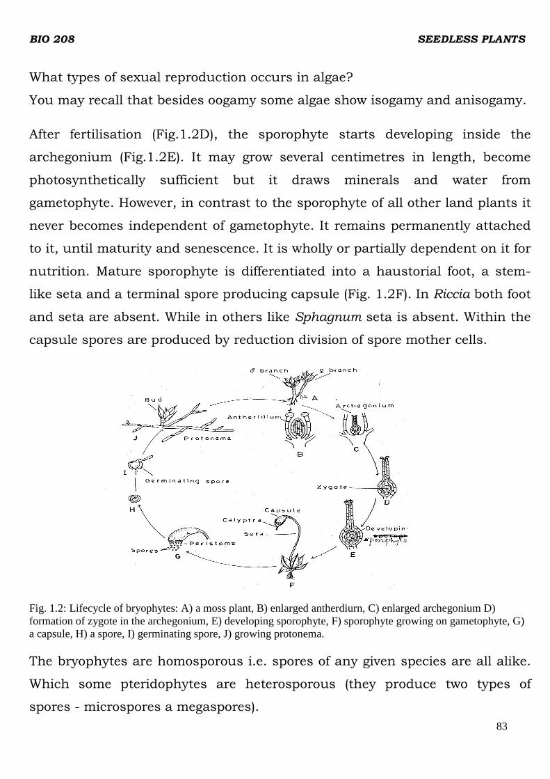
BIO 208 SEEDLESS PLANTS
83
What types of sexual reproduction occurs in algae?
You may recall that besides oogamy some algae show isogamy and anisogamy.
After fertilisation (Fig.1.2D), the sporophyte starts developing inside the
archegonium (Fig.1.2E). It may grow several centimetres in length, become
photosynthetically sufficient but it draws minerals and water from
gametophyte. However, in contrast to the sporophyte of all other land plants it
never becomes independent of gametophyte. It remains permanently attached
to it, until maturity and senescence. It is wholly or partially dependent on it for
nutrition. Mature sporophyte is differentiated into a haustorial foot, a stem-
like seta and a terminal spore producing capsule (Fig. 1.2F). In Riccia both foot
and seta are absent. While in others like Sphagnum seta is absent. Within the
capsule spores are produced by reduction division of spore mother cells.
Fig. 1.2: Lifecycle of bryophytes: A) a moss plant, B) enlarged antherdiurn, C) enlarged archegonium D) formation of zygote in the archegonium, E) developing sporophyte, F) sporophyte growing on gametophyte, G) a capsule, H) a spore, I) germinating spore, J) growing protonema.
The bryophytes are homosporous i.e. spores of any given species are all alike.
Which some pteridophytes are heterosporous (they produce two types of
spores - microspores a megaspores).

BIO 208 SEEDLESS PLANTS
84
A spore represents the first stage of gametophytic generation (Fig. 1.2H). It is
unicellular haploid and germinates (Fig.1.2H,I) to produce a short-lived green
protonema (Fig.1.2j)
The adult gametophore develops on this protonema. Protonema may be
thalloid, globular or filamentous. The protonema and the adult gametophore
are strikingly different from each other.
An adult gametophyte bears gametangia which produce haploid male and
female gametes. The gametes represent the last stage of gametophytic
generation and the zygote represents the first stage of sporophytic generation,
whereas the spore mother cells (diploid) represent the last. The spore mother
cells undergo reduction division to form haploid spores. So, any stage in the
life cycle which is haploid, belongs to gametophytic generation, whereas the
diploid stage belong to sporophytic generation.
Now let us sum up the distinguishing features of bryophytes.
1. They lack vascular system. In some of the mosses a primitive conducting
system is present that transports food and water.
2. The gametophyte is dominant generation and sporophyte remains
attached to it. In other land plants the sporophyte is dominant and
independent
There are strong reasons to believe that green algae served as ancestors of
bryophytes.
The move from water to land offers an organism some distinct competitive
advantages as well as challenges. What could be the advantages of the
terrestrial habitat over the aquatic? Some of the advantages are as follows:
i) greater availability of sunlight for photosynthesis,
ii) increased level of carbon dioxide, and

BIO 208 SEEDLESS PLANTS
85
iii) decreased vulnerability to predation.
If some more points cross your mind, add to this list.
Can you now think what are the challenges of land environment? Try to list
them below.
Compare your points with the following:
1. Plants on land are exposed to direct sunlight and air. Hence there is danger
of drying out or desiccation because of evaporation. Gametes and zygotes
are also susceptible to desiccation.
2. The aquatic plants are supported by the buoyancy of water, but on land,
plants need some anchor to fix to the ground and also require support to
stand erect.
3. Absorption of minerals and water, and their transportation to the parts
which are not in contact with soil. In other words, land plants need supply
lines for the distribution of water and nutrients.
4. Effective dispersal of spores at right time and at right place for the survival
of progeny, with the help of hygroscopic structures like elaters and
peristome teeth.
3.3 MORPHOLOGY OF BRYOPHYTES
So far you have studied the general characteristics of bryophytes. You may
recall that the Division Bryophyla is divided into three classes (a)
Hepaticopsida (liverworts) (b) Anthocerotopsida (hornworts) and (c) Bryopsida
(mosses). Let us now study the representative genera from each class.
3.3.1 Hepaticopsida
The gametophyte of liverworts usually lies close to the ground. There are two
forms of liverworts. In some, the gametophyte is dorsi-ventral, thalloid in form
with obvious upper and lower surfaces. These are thalloid liverworts. While in
others it is differentiated into leaf-like and stem-like structures like those of

BIO 208 SEEDLESS PLANTS
86
mosses. The latter are known as leafy liverworts. The leaves of leafy liverworts
are without midrib, whereas midrib is present in the leaves of mosses.
Internally, the gametophytes of liverworts may be homogenous or composed of
different types of tissues. Liverworts grow on moist ground or rocks that are
always wet. They can be found in muddy areas near streams. In greenhouses
you may find them growing in flower pots.
In this course you will study two representatives of the order Marchantiales
(Riccia and Marchantia) and one of the order Jungermanniales (Pellia).
Take a look at this classification:
Division - Bryophyta
Class - Hepaticopsida
Order - Marchantiales
Family - Ricciaccae
The gametophytes of Marchantiales are exclusively thalloid. The order
Marchantiales consists of about 35 genera and approximately 420 species.
We will first study in detail the genus Riccia and then Marchantia.
Riccia
Riccia belongs to the family Ricciaceae which is the most primitive and the
smallest family of the order Marchantiales. Riccia has more than 130 species
and is very widely distributed. Most of the species are terrestrial and grow
mainly on moist soil and rocks. Riccia fluitans is an aquatic species.
In structure, Riccia represents the simplest of the bryophytes. Its gametophyte
is small green fleshy, thalloid. It grows prostrate on the ground and branches
freely by dichotomy. Several Riccia plants grow together and take the form of
circular patches, which are typically resette-like (Fig. 1.3A). The thallus
bearing female and male sex organs are shown in (Fig. 1.3B and C.)

BIO 208 SEEDLESS PLANTS
87
Fig. 1.3: Morphology and internal structure of Riccia A) a rosette of Riccia trichocarpa, B) a female thallus of R. discolor, C) a male thahlus of R. discolor, D)ventral surface of the thallus, E) a smooth walled rhizoid, F) a tuberculate rhizold, ) transverse vertical section of female thallus.
The branches of the thallus are called thallus-lobes. According to the species,
thallus lobes are linear to wedge-shaped. The dorsal surface of the thallus has
a prominent midrib, represented by a shallow groove called the dorsal groove.
At its apex there is a depression termed as apical-notch. The sporophytes are
sunk deeply, in the dorsal groove, each in a separate cavity. Both male and
female sex organs may develop on the same tha]lus (monoecious) or on
different thalli (dioecious) (Fig.1.3B and C). On its ventral surface (Fig.1.3D)
There are a number of slender, colourless, unicellular. unbrandhed processes
called rhizoids that help to attach the thallus to the substratum. The rhizoids
are of two types: (a) smooth walled - these have smooth walls (Fig1.3E) and (b)
tuberculate - these have peg-like ingrowths of wall projecting into the lumen
(Fig.1.3F). On the ventral surface towards the apex and along the margins of
thallus small plate like structures are also present (Fig.1.3D). These are scales
which are arranged in a single row and are single cell in thickness. These
scales project forward and overlap the growing point to protect it from
desiccation. The growing point is located in the notch and consists of a
transverse row of 3 to 5 cells. The growth of the thallus occurs in length as
well as in width by the divisions of these cells. Each thallus branches

BIO 208 SEEDLESS PLANTS
88
dichotomously and several dichotomies lie close to one another forming a
typical rosette.
Again take a look at this simple classification of the bryophyte
Division -Bryophyta
CIass -Hepaticopsida
Order -Marchantiales
Family -Marchantiaceae
Marchantia
The family Marchantiaceae, to which Marchantia belongs, includes about 23
genera and approximately 200 species. The special feature of this family is that
in all the genera the gametophyte bears archegonia on vertical stalked
receptacles called archegoniophore (carpocephala). In Marchantia anthoridia
are also produced in stalked receptacles known as antheridiophores. The type-
genus Marchantia is placed among the most advanced members with about 65
species, of which Marchantia polymorpha is the most widely distributed.
Marchantia usually grows in cool moist places along with mosses and in areas
of burnt ground. It is deep green in colour. Like Riccia its gametophyte is flat,
prostrate, dorsi-ventral and dichotomously branched thallus (Fig.1.4A ). There
is a prominent midrib low ridge covered with rhizoids (Fig.1.4B). Along the
midrib there are a number of cuplike structures with frilled margins. These are
called gemma cups (Fig. 1.4C) which contain numerous vegetative
reproductive bodies called gemmae (sing. gemma). In mature thalli
antheridiophores and archegoniophores, which bear antheridia and
archegonia (Fig.1.4D and E) respectively, are also present at the growing
apices of certain branches. Marchantia is dioecious. Like Riccia the apex of
each branch is notched and a growing point is situated in it. You will note that
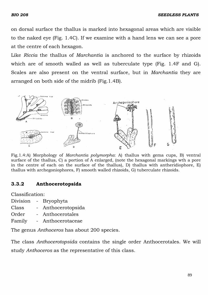
BIO 208 SEEDLESS PLANTS
89
on dorsal surface the thallus is marked into hexagonal areas which are visible
to the naked eye (Fig. 1.4C). If we examine with a hand lens we can see a pore
at the centre of each hexagon.
Like Riccia the thallus of Marchantia is anchored to the surface by rhizoids
which are of smooth walled as well as tuberculate type (Fig. 1.4F and G).
Scales are also present on the ventral surface, but in Marchantia they are
arranged on both side of the midrib (Fig.1.4B).
Fig.1.4:A) Morphology of Marchantia polymorpha: A) thallus with gema cups, B) ventral surface of the thallus, C) a portion of A enlarged, (note the hexagonal markings wth a pore in the centre of each on the surface of the thallus), D) thallus with antheridiophore, E) thallus with archegoniophores, F) smooth walled rhizoids, G) tuberculate rhizoids.
3.3.2 Anthocerotopsida
Classification:
Division - Bryophyta
Class - Anthocerotopsida
Order - Anthocerotales
Family - Anthocerotaceae
The genus Anthoceros has about 200 species.
The class Anthocerotopsida contains the single order Anthocerotales. We will
study Anthoceros as the representative of this class.

BIO 208 SEEDLESS PLANTS
90
Anthoceros
It grows principally in moist shady places on the sides of ditches, or in moist
cracks of rocks. The gametophytes of Anthoceros are dorsi-ventral, thallose,
somewhat lobed or dissected, and sometimes have a tendency toward
dichotomous branching (Fig.1.5A).
The thallus of Anthoceros is dark green, velvety on the upper surface and
variously lobed. The midrib is either indistinct or absent. It also lacks
tuberculate rhizoids and scales. Only smooth walled rhizoids are present.
Thallus
Sporophytes
A
Fig.1.5:Anthoceros; A) mature thallose gametophyte with attached sporophytes.
Note the sheath at the base of each sporophyte.
3.3.3 Bryopsida
Classification:
Division - Bryophyta
Class - Bryopisida
Order - Sphagnales
Family - Sphagnaceae
This is the largest class of bryophytes and includes about 600 genera and
14,500 species. Bryopsida is divided into three subclasses: Sphagnidae (peat

BIO 208 SEEDLESS PLANTS
91
mosses), Andreaeidae (rock mosses) and Bryidae (true mosses). Bryidae
include about 14,000 species. You will study the genus Funaria as a
representative of this order. Order Sphgnales is represented by a single genus
Sphagnum which includes about 300 species. Let us first study Sphagnum.
Sphagnum
Sphagnum forms peat bogs in northern parts of the world. In some countries
peat is burnt as fuel. Sphagnum is also used in plant nurseries as packing
material. Mats of this moss hold moisture and help the seeds of other plants to
germinate and grow.
Sphagnum is confined to acidic, water-logged habitat. It is the principal
component of peat bogs where it forms a more or less continuous spongy layer.
The adult gametophyte develops as an upright leafy-shoot, called
gametophores from a simple thallose, one cell thick protonema. The
gametophore is differentiated into stem and leaves. The terminal growth of the
stem is due to an apical cell. The axis is attached to the soil by means of
multicellular, branched rhizoids with oblique cross walls. Rhizoids are present
only in young gametophores and disappear when it matures. Afterwards, the
gametophore absorbs water directly.
Look at Fig.1.6A, the mature gametophores consists of an upright stem
bearing leaves. Every fourth leaf of the stem bears a group of three to eight
lateral branches in its axis. These branches are of two types: (i) divergent and
(ii) lying next to the stem (Fig.1.6B). Sometimes, one of the branches in a tuft
continues upward growth to the same height as the main axis and resembles it
in structure. These strongly developed branches are called innovations and
they ultimately get detached and become independent plants. The branches
near the apex of a stem are short and densely crowded in a compact head
called coma.

BIO 208 SEEDLESS PLANTS
92
The leaves lack midrib (Fig.1.6C and D). They are small and arranged in three
vertical rows on the stem. In the surface view of a leaf one can observe two
types of cells: (i) narrow, living, chlorophyll containing cells and (ii) large dead,
empty, rhomboidal, hyaline (glass-like, transparent) cells with pores and s
spiral as well as annular wall thickenings (Fig.1.6E). In transverse section, leaf
shows beaded appearance, with large, dead hyaline cells regularly alternating
with the small, green, chlorophyllous cells (Fig.1.6F). The spiral thickenings
provide mechanical support and keep the hyaline cells from collapsing when
they are empty.
S tem
D ive rg en t b ra nches
Penden t b ra nch es
B
DC
AH y a line
c e l l
C h l o ro p h y l lc o n ta i n i n g
c e l l
EF
Fig. 1.6:Structure and morphology of Sphagnum: A) a mature gametophyte with attached
sporophyte at the apex, B) portion of a shoot showing divergent and drooping (pendent)
branches, C) leaf of a divergent branch enlarged. Note the apex. The midrib is absent, D)
leaf of the main stem without midrib, E) leaf cells in surface. Note the network of
chlorophyllous cells, surrounding porous hyaline cells; also the fibrillar thickenings of walls
of hyaline cells. F) T. S. of a leaf.
The pores help in rapid intake of water and also in exchange of cations for
H+ions which are the metabolic products of Sphagnum. Hence, they create
acidic environment in their immediate surrounding. The hyaline cells take up

BIO 208 SEEDLESS PLANTS
93
and hold large quantities of water, sometimes as much as twenty times the
weight of the plant. The narrow chloroplast containing cells carry on
photosynthesis. In a mature leaf these two types of cells are arranged in a
reticulate manner. This peculiar leaf structure accounts for the ability of the
Sphagnum plant to absorb and retain large quantities of water and
consequently for its outstanding bog-building properties. Because of their
water absorbing quality they are used in gardening.
Funaria
Classification:
Division - Bryophyta
Class - Bryopsida
Sub-class - Bryidae
Order - Funariales
Family - Funariacea
Funaria is a very common moss. It is very widely distributed throughout the
world. One species, Funaria hygrometrica is cosmopolitan and it the best
known of all the mosses.
Like other bryophytes that you have studied, the most conspicuous form of
the moss plant is the adult gametophyte. This consists of a main erect axis
bearing leaves which are arranged spirally (Fig. 1.7A). This adult gametophyte
is called gametophores. It is small, about 1-3 cm high. The leaves do not have
a stalk but show a distinct midrib. The gametophore is attached to the
substratum by means of rhizoids which are multicellular, brached and have
oblique septae. The gametophyte bears sporophyte which has foot, seta and
capsule (Fig.1.7A).
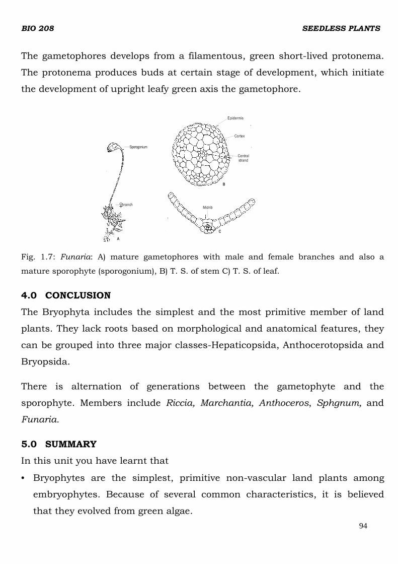
BIO 208 SEEDLESS PLANTS
94
The gametophores develops from a filamentous, green short-lived protonema.
The protonema produces buds at certain stage of development, which initiate
the development of upright leafy green axis the gametophore.
Fig. 1.7: Funaria: A) mature gametophores with male and female branches and also a
mature sporophyte (sporogonium), B) T. S. of stem C) T. S. of leaf.
4.0 CONCLUSION
The Bryophyta includes the simplest and the most primitive member of land
plants. They lack roots based on morphological and anatomical features, they
can be grouped into three major classes-Hepaticopsida, Anthocerotopsida and
Bryopsida.
There is alternation of generations between the gametophyte and the
sporophyte. Members include Riccia, Marchantia, Anthoceros, Sphgnum, and
Funaria.
5.0 SUMMARY
In this unit you have learnt that
• Bryophytes are the simplest, primitive non-vascular land plants among
embryophytes. Because of several common characteristics, it is believed
that they evolved from green algae.
Epidermis
Cortex
Central strand
B
Midrib
C
A
branchO+
Sporogonium

BIO 208 SEEDLESS PLANTS
95
• There is alternation of generations between green independent gametophyte
and sporophyte which is wholly or partially dependent on it. Sporophyte is
generally a small capsule with or without foot and seta. The gametophyte
develops from protonema and bears sex organs - archegonia and antheridia.
Bryophytes are homosporous.
• The challenges of land environment for a plant are fixation to the ground,
desiccation, conduction of water and dispersal of sperms and spores. These
are taken care of by developing land adaptations such as epidermis, cuticle,
stomata, airpores, rhizoids, multicellular jacket of cells for the protection of
developing gametes, and retention of zygote in the archegonium. In some
bryophytes the primitive conducting tissues - hydroids and leptoids have
also developed.
• The gametophyte of liverworts - Riccia and Marchantia is dorsi-ventral,
thalloid structure and is internally differentiated. The pores on the dorsal
surface allow exchange of gases and are much advanced in Marchantia.
The leafy liverworts have leaf-like and stem-like appendages. The
gametophyte of Anthoceros, is also dorsiventral, but not differentiated
internally. Blue green algae Nostoc live in mucilage cavities of the thallus.
• Bryophytes are classified into liverworts (Hepaticopsida) hornworts
(Anthocerotopsida) and Mosses (Bryopsida).
• Mosses – Sphagnum and Funaria have erect axis and bear leaf-like
structures.
6.0 TUTOR MARKED ASSIGNMENTS
a) In the following statement choose the alternative correct word given in
the parentheses. In bryophytes.
i. the dominant phase of life cycle is (gametophyte/sporophyte)
ii. (roots/rhizoids) anchor the plant to the soil.
iii. the protonema is (haploid/diploid)

BIO 208 SEEDLESS PLANTS
96
iv. the sporophyte is (dependent/not dependent) on gametophyte.
b) Which of the following statements are true and which are false about
bryophytes? Write T for true and F for false against each statement.
i. Some mosses have hydroids and leptoids for the conduction of
water and food, respectively.
ii. The gametophyte is an independent plant.
iii. They produce two types of spores
iv. Protonema is the transitional stage between spore and adult
gametophyte.
7.0 REFERENCES/FURTHER READING
Dutta A.C. (1981). Botany for degree students. Oxford University Press. 909p.
IGNOU. (1991). Indra Gandhi National Open University. Plant Diversity-Algae.

BIO 208 SEEDLESS PLANTS
97
UNIT 2: CHARACTERISTICS, LIFE CYCLE CLASSIFICATION
AND MORPHOLOGY OF SOME PTERIDOPHYTES.
1.0 Introduction
2.0 Objectives
3.0 Main Content
3.1 Pteridophytic Life Cycle
3.2 General Characteristics and Relationship with Other Groups
3.3 Formation of Fossils and Their Types
3.4 Morphology
Lycopodium
Selaginella
Pteris
4.0 Conclusion
5.0 Summary
6.0 Tutor Marked Assignment
7.0 Reference / Further Reading
1.0 INTRODUCTION
Now we come to the last group of non-flowering plants, the
pteridophytes, included in this course. The most familiar plants of this
group are ferns which we commonly see as houseplants, in parks and
also in house landscapes along with other ornamental plants. Ferns
are rather small plants with graceful, often delicate compound leaves.
Because of their beauty and difficulty in propagation, they are
considered very precious plants.

BIO 208 SEEDLESS PLANTS
98
In this unit you will study the general characteristics and life cycle of
pteridophytes and the structure and morphology of some represent -
tative genera.
Scientists got the idea about the early vascular land plants from fossils
– the extinct members. Rhynia and Cooksonia were the simple and
most primitive pteridophytes. One of the simplest living members of
this group is Psilotum.
You know, pteridophytes are vascular plants and they possess root,
stem and leaves. All vascular plants posses water- and food-
conducting pipelines made up of xylem and phloem tissues,
respectively. In different groups of plants, a great variation is found in
the relative position and arrangement of xylem and phloem, other
associated tissues and in the presence or absence of pit. In
pteridophytes a natural gradation in vascular tissues from simple
(primitive) to complex forms is observed.
2.0 OBJECTIVES
After studying this unit you will be able to:
• list characteristics of pteridophytes
• outline the life cycle of a typical pteridophytes
• compare the general features and life cycle of pteridophytes with
bryophytes
• differentiate between different types of fossils
• give examples of fossil pteridophytes
• describe morphology of the genera included in this unit
• distinguish among groups of pteridophytes on the basis of
morphological character.

BIO 208 SEEDLESS PLANTS
99
3.0 MAIN CONTENT
3.1 PTERIDOPHYTIC LIFE CYCLE
Have a good look at the pictures of a pteridophyte.
What you see are sporophytes of these plants. Their gametophytes are
very small only a few millimeters in size, and are short-lived. Let us
first learn about the life cycle of pteridophytes because then it would
be easier for us to list their characteristics. Like bryophytes,
pteridophytes also have two distinct phases in the life cycle:
gametophyte and sporophte (Fig. 2.1) that follow each other in regular
succession. Since the two generations look different, they are termed
heteromorphic. Under normal circumstances, gametophyte produces
motile male gametes (sperms) and non-motile female gametes (eggs).
Fusion between an egg cell and male gamete results in the formation of
a zygote which is diploid. The zygote divides by mitotic divisions and
forms the sporophyte. On sporophyte a number of haploid, non-motile
spores are produced by meiosis. The life cycle is then completed when
a spore germinates and produces a haploid gametophytes by mitotic
divisions, (Fig.2.1).
You have studied that in bryophytes, the dominant phase in the life
cycle is the gametophyte, and the sporophyte is either partially or
completely dependent on it for nutrition. But in pteridophytes the
sporophyte very soon becomes independent of the gametophyte and is
the dominant generation.
The sporophyte shows greater degree of complexity in structural
organization. It is organized into stem, root and leaves, except in the
most ancient fossil pteridophytes and in the most primitive living
member. The vascular tissues (xylem and phloem) are developed only
in the sporophytes.

BIO 208 SEEDLESS PLANTS
100
Fig. 2.1: Potential typical life cycle of pteridophytes.
Furthermore, the aerial parts are covered with a layer of cuticle. On
the epidermis there are stomata for the exchange of gases. These
anatomical complexities of the sporophyte helped in inhabiting a much
wider range of environmental conditions than the gametophyte could.
The life cycle of a typical fern e.g. Dryopteris felixma is as shown
in(Fig. 2.2). It is annotated and self explanatory.

BIO 208 SEEDLESS PLANTS
101
Sporangia release spores, which give
rise to gametophytes
7
Most ferns are homosporous, meaningthat they produce a
single type of spore
1
6
The fern spore develops into a smal l , heat-shaped gametophytethat sustains i tsel f by photo-
synthesis.
2 Each gametophyte has both male & female sex organs, but the archegonia antheridia usually mature at different times, ensuring cross-fetiliation between gametophytes.
3
MEIOSIS
FERTILIZATION
Meiosis
Gametophyte The spots on the underside of
reproductive leaves(sporophylls) are
called Sori. Each sorus is a cluster
of sporangia.
A fertilized egg develops into a new sporophyte, and the young plant grows out from an archegonium of itsparent, the gametophyte.
5
Fern sperm, l ike those of al l seedlessvascular plants, use flagel la to swim
through moisture from antheridia toeggs in the archegonia and then feti l izethe eggs.
4
Sporangium
Mature sporophyte
New sporophyte
Zygote
Egg
Archegonium
Younggametophyte
Spore
Sperm
Haploid
Diploid
Fig. 2.2: The life cycle of a fern. After Campbell and Reece (1991).
3.2 GENERAL CHARACTERISTICS AND RELATIONSHIP WITH OTHER
GROUPS
In the previous section, you have learnt that in pteridophyte is the
dominant phase. It possesses a vascular system and is differentiated
into true root, stem and leaves. Pteridophytes exhibit a great variation
in form, size and structure.
Most of the pteridophtes are herbaceous except a few woody tree ferns.
They may be dorsi-ventral or radial in symmetry and have
dichotomously or laterally branched stems that bear microphyllous
or megaphyllous leaves

BIO 208 SEEDLESS PLANTS
102
The organization of vascular cylinder (also called stele, see box item) in
the sporophyte varies from simple primitive type to more complex
forms. Besides, vessels are also present in some members.
The roots are generally adventitious, the primary embryonic root being
short-lived.
The spores are produced in special structures called the sporangia
that are invariably subtended by leaf-like appendages known as
sporophylls (Fig.2.3C).
Fig. 2.3 C: Lycopidium showing a sporophyll
The sporangia may be scattered throughout the vegetative axis or may
be restricted to a particular area. They are in many cases compacted to
form distinct spore producing regions called the cones or the strobill
(sing, strobulus). The sporangia in some cases, may be produced
within specialized structures called the sporocarp. Distinct segregation
of vegetative and reproductive shoots and leaves has also been
observed in some other species. Have you ever noticed brown-black
dots on the underside of a fern leaf? Each dot is a reproductive
structure called sorus (plura, sori, Fig.2.5C). It is a cluster of
sponrangia that contain spores.

BIO 208 SEEDLESS PLANTS
103
S o r i
C
Fig. 2.5: Plant of Cyathea. C) A portion of leaf showing sori
Pteridophytes, in general, are homosporous i.e. they produce only one
type of spores. However, a few species are heterosporous i.e. they
produce two types of spores, microspores and megaspores
(Fig.2.4D,B). A spore on germination produces gametophyte.
Heterosporous species produce microgametophyte as well as
megagametophyte.
Me g a sp o ro p h y l l
M e g a sp o ra n g i um
M e g a sp o re
M i c r o s p o r o p h y l l
M i c r o s p o r a n g i u m
CBA
M i c r o s p o r a n g i um
M i c r o s p o r e
D
M e g a s p o r a n g i u m
Fig. 2.4: Selaginella: A) A megasporophyll. B) V.S. of A. C) Microsporophyll.
D) V. S of microsporophyll.

BIO 208 SEEDLESS PLANTS
104
In general, pteridophytes form green, dorsiventrally differentiated,
thallose gametophytes with sex organs restricted to the ventral
surface. The sex organs may be embedded or projecting. They resemble
those of bryophytes in general plan. The female reproductive structure
is archegonium and the male reproductive structure is an
antheridium.
The archegonium has invariably four longitudinal rows of neck cells
whose height varies in different genera. The antheridium consists of a
single layer of sterile jacket of cells enclosing a mass of androcytes or
antherozoid mother cells. Each androcyte gives rise to a single ciliated,
motile antherozoid. The opening of the mature sex organs and the
subsequent fertilization is still conditioned by the presence of water.
Hence like bryophytes, they could also be called amphibians of plant
kingdom.
Now that you have studied the life cycle and the general characteristics
of pteridophytes, can you compare them with bryophytes?
Bryophytes resemble pteridophytes in the following features:
1. Thallose liverworts and pteridophyte show similarity in vegetative
structure of gametophytes.
2. Their female and male reproductive structures are archegonium and
antheridium, respectively.
3. The opening of the mature sexual reproductive organs and the
subsequent fertilization are conditioned by the presence of water in
liquid state, i.e., both require water for fertilization.
4. They usually show a distinct and clearly defined heteromorphic
alternation of generations and the two generations follow each other in
regular succession.
BIO 208 SEEDLESS PLANTS
105
5. The spores arise in the same manner in both the groups. The spore
mother cells are produced by the last division of the sporogenous
tissues. Each of the spore mother cells undergoes meiotic division
resulting in a tetrads of spores.
6. Development of embryo occurs in the archegonium
7. The young sporophyte or embryo is partially parasitic upon the
gametophyte.
Now try to list the characteristics which distinguish pteridophyte from
bryophytes
1. ……………………………………….............
2. ………………………………………………..
3. …………………………………………………
Compare your points with the following:
1. Unlike bryophytes, in which sporophyte is dependent upon
gametophyte physically and physiologically, the sporophyte is
independent at maturity in pteridophytes, and is the dominant phase
of life cycle instead of gametophyte.
2. In pteridophytes the sporophyte has true roots, stem and leaves and
well developed conducting tissues – xylem and phloem, which are
absent in bryophytes.
3. Some of the pteridophytes are heterosporous but all the bryophytes are
homosporous.
As mentioned earlier,pteridophytes form an important link between
bryophytes and seed plants. This suggests that they also resemble in some
respects with spermatophytes.

BIO 208 SEEDLESS PLANTS
106
Pteridophytes resemble seed plants in the following respects:
1. The sporophytes is dominant, typically photosynthentic phase of life
cycle.
2. It is organized into stem, root and leaves.
3. The roots and the leafy shoots are provided with a conducting system
made of specialized cells.
4. Some pteridophytes do approach seed-habit and some fossil
pteridophytes had seed-like structure.
Due to their affinities with bryophytes as well as with higher vascular,
plants, pteridophytes are also known as “Vascular Cryptogams” (non-seed
producing plants).
In the above account you have learnt about the characteristics of
pteridophytes and their relation to other plant groups. Now we will describe
the formation of various types of fossils and how they reveal life forms that
occurred millions of year ago.
3.3 FORMATION OF FOSSILS AND THEIR TYPES
You may raise a question as to how can one know “Where, when and
from what ancestral group did the first vascular land plant and seed-
like structure evolve?” To find the answer to these questions we have
to depend on fossils. Let us first try to define a fossil and the ways in
which fossils came to be formed. We will also try to know the extent to
which they may be expected to provide information useful to the
morphologist.
What are Fossils? Fossils are the remains and / or impressions of organisms that lived in
the past. In its correct sense fossils include the remains of organisms
or their parts and also anything connected with an organism proving

BIO 208 SEEDLESS PLANTS
107
its existence, i.e., anything which gives evidence that an organism once
lived.
How are fossils formed?
The actual nature of fossilization depends on the environmental
conditions in which it takes place. Dead plant remains are liable to get
disintegrated and it is only rarely that they get fossilized. Chances of
fossilization are better for organisms having stiff tissues / skeletons.
The details of fossilization process are discussed below.
Fossilization Process
The process of formation of fossils is going on ever since the
sedimentary rocks began to deposit and it is going on in nature even
now.
In some cases plants may be deposited on the site where they grow (in
situ), such as swamps and small inland lakes. Due to low oxygen
content and presence of toxic substance in the water, microbial growth
is inhibited, so the plants do not decay. This results in the
preservation of the plant remains until they were covered by layers of
sediments. European and Enugun coal forests are the example of this
type of fossilization.
In other cases plant parts are carried down by flowing water and
finally sink to the bottom of a lake or estuarine water where they are
less susceptible to decay by microbes.
During fossilizations the protoplasmic contents and softer
parenchymatous cells disappear first, while the harder wood and other
sclerenchymatous or cutinized tissues resist to the last. The growing
pressure of the heavy sedimentary rocks above, fist reduces the vacant

BIO 208 SEEDLESS PLANTS
108
spaces inside the cells and forces the liquids substances out. Some
organic substances may also escape as marsh gas. Naturally, all
fossils get highly compressed and the final result depends on how far
the conditions were favourable for good fossilization. In spite of all
hazards sometimes fossils are formed, which retain their cellular
structure beautifully and sometimes even some of the cell contents.
Types of fossils
According to the nature of fossilization, fossils may be of the following
types:
1. Petrifaction
It is the best type of fossilization. In this type buried plant material
gets decayed with the passage of time and gets replaced, molecule for
molecule by mineral solutions. The impregnation of silica, calcium
carbonate, magnesium carbonate, iron sulphide takes place within the
tissues. Most of the plant material may get decayed but at least some
original cell wall components remain. After fossilization the whole
structure becomes stone-like and it can be cut into fine section. The
structure of the tissues may be observed by examining the section
under the microscope. Anatomical structures of ancient plants are
beautifully obtained from such petrifactions. Silicified and calcified
pieces of wood are quite common.
II. Cast or incrustation
This type of fossilization is also quite common. The plant parts get
covered up by sand or mud. After sometime the plant material inside
degenerates leaving a cavity known as mold. This cavity, again gets
filled up by some rock-forming material which in course of time
solidifies into an exact cast of the plant material, showing all its

BIO 208 SEEDLESS PLANTS
109
surface features. A cast fossil does not actually contain any part of the
original plant but it is of great use as the cast correctly shows the
original features of plant part.
III. Impression
These are found when a leaf or any other part of the plant falls on and
leave an impression on the surface of semisolid clay. In course of time
this impression becomes permanent when the clay turns into stone.
Such impressions often very clearly show details of external features
and structures like stomata are clearly seen in good preparation. IV.
IV. Compression
In a compression the organic remains of the plant part actually remain
in the fossils but in a highly compressed state. During fossilization the
great pressure of sediments above causes flattening of plant parts. In
the fossil usually a carbonaceous film remains which represents the
surface features. However, in good compressions it has been possible
to swell out the organ by some chemical treatments so that plant some
details become visible. A good type of compressed fossil is the “clay
nodule”. In this the plant material gets encased in a ball of clay, gets
compressed and the clay ball turns into stone.
Nomenclature of fossils
Mostly, fossils consist of fragments of plants. Sometimes it may take
many years to find the fossil of a stem to which a particular kind of
leaf belonged. Therefore, in the meantime each fragment of fossil plant
is described under a separate generic name and such genera are
known as “Form genera”. In naming such form genera we usually add
suffixes, signifying which part of the plant it came from.
Following are a few examples:

BIO 208 SEEDLESS PLANTS
110
Plant Part Suffix Used
a) Leaf -phyllum
b) Fern-like or frond -pteris
c) Tree trunk -dendron
d) Woody part -xylon
e) Seed-like structure -spermum, -carpon, -carpus, -storms
f) Microsporangium -theca
g) Cone -strobilus, - strobes
It is the work of palaeobotanists to collect bits of such fossils, i.e, form
general, and to reconstruct the form, structure and mode of life of the plant
from which they came. Success has been achieved in reconstructing a few
fossil plants.
3.4 MORPHOLOGY
As you have noticed in the earlier units on Algae, Fungi and
Bryophytes, each of these major plant groups are classified into
smaller groups on the basis of distinguishable characteristics. You
may recall the following major divisions of extant and extinct
pteridophytes.
Extinct Pteridophytes (known only from fossil records):
Rhyniophta
Zosterophyllophyta
Tremerophyta
Living Pteridophytes
Psilotophyta
Lycopodiophyta
Equisetophyta
BIO 208 SEEDLESS PLANTS
111
Pterophyta (= polypodiophyta, Filicopsida)
In the following text we will learn in detail about representative types
of some of these classes. As you know that during evolution, advanced,
complex forms evolved from primitive simpler forms. So we will first
study simple, primitive forms and subsequently the advanced, complex
forms.
Lycopodium
Classification:
Division - Lycopodiophyta
Class - Lycopsida
Order - Lycopodiales
Lycopodium, popularly known as club moss, is a large genus with
about 180 species of which approximately 33 species are found in
India. They are distributed world –wide in tropical, sub-tropical forests
and in temperate regions. Some species are abundant in hills at
comparatively high altitude. They grow in cool climate on moist
humus-rich soil.
The adult sporophyte is herbaceous and with a wide range of habits.
Generally in tropics they are pendulous epiphytes, whereas in
temperate regions they are prostrate or erect (Fig.2.6A, B, C). They
usually grow about 30 to 60 cm in length. The stem may be
unbranched or dichotomously branched which later becomes
monopodial. It is covered with microophylls which in most species are
spirally arranged. However, in some species leaves are arranged in
whorled or decussate manner (Fig. 2.6D-G) decussate x-shaped, with
pair of opposite leaves each at right angles to the pair below:

BIO 208 SEEDLESS PLANTS
112
Strobila
Fig.2.6: Lycopodium: A-C) Portion of plant of Lycopodium phlegmaria, L.
volubile and L. clavatum respectively. D-G) Leaf form and arranged in
different species.
Selaginella
Classification;
Division - Lycopodiophyta
Class - Isoetatae
Order - Selaginellales.
Most of the species of Selaginella are restricted to damp areas of the tropical
and subtropical regions of the world. A few species are markedly xerophytic
and inhabit desert regions. These are sometimes called “resurrection plants”
because of their extra-ordinary power of recovery after prolonged drought.
The plant may be prostrate, erect or sub-erect. Only a few are epiphytic.
Some form delicate green mossy cushions, others are vine-like, with stems
growing to a height of several metres, while many have creeping axes, from
which arise leafy branch systems that bear a striking superficial
resemblance to a frond of fern.

BIO 208 SEEDLESS PLANTS
113
Branching in Selaginella is characteristic, terminal and unequal, forming
weaker and stronger branches. At each dichotomy there are one or two
meristems on either side. These angle-meristems develop into cylindrical
outgrowths known as “rhizophores” (Fig. 2.7A). In most species only the
ventral angle-meristem develops into rhizophore, while the other remains as
dormant papilla. The rhizophores grow downwards into group and give rise
to a small tuft of adventitious roots at their tips.
The morphological nature of rhizophore has been controversial. It has been
held to be a (a) root, (b) a branch of stem (c) a structure sui generis (falling
in neither of the categories). Earlier investigators reported a unique
combination of characters of rhizophore:
i exogenous origin from the stem at the time of branching,
ii lack of root cap,
iii production of roots endogenously behind the tip, and
iv ability, in some instance, to be converted into leafy shoots. Since these
features are not typical of root these outgrowths are called
rhizophores.
The features suggestive of their root nature are;
i positive geotropism,
ii in some species when these structure re less than 1 mm the root cap
develops, in S. martensil cap differentiates when it nears the soil.
Using labeled auxin (C14 IAA) ‘it has shown that auxin transport in
rhizophores of Selaginella is acropetalous as in case of angiosperms root,
whereas it is basipetalous in stems. Therefore, now the term “rhizophore”
as well as the arguments regarding its nature are of historical significance.
In Selaginella the leaves are sessile with a single unbranched vein
(Fig.2.7A). Leaves of Selaginella are ligulate. The ligule is present in or near

BIO 208 SEEDLESS PLANTS
114
the axil of each leaf as a laminate outgrowth (Fig. 2.7B). It differentiates
and matures very early in the ontogeny of leaf. A mature ligule is tongue-to
fan-shaped. Its basal region is made up of tubular, hyaline cells forming
the sheath is a hemispherical region of thin and greatly vacuolated cells
referred to as glossopodium. The remaining cells are isodiametric. The
apical region is one cell thick and is made up of elongated cells with scanty
contents.
Fig. 2.7 Salaginella A) Portion of a plant B) T. S. of a part of leaf
Pteris
Classification;
Division - Pterophyta (= Filicophyta)
Class - Polypodiatae
Order - Filicales
Pteris is a widely distributed genus with about 250 species. It grows
abundantly in cool, damp and shady places in tropical and subtropical
regions of the world. In all there are 19 species recorded from Ind. Pteris
vitata is a low level fern which brings out new leaves throughout the year.
Its is very common along mountain walls and grows up to 1200 metres
above sea level. Pteris quadriauriata grows abundantly along roadsides and
in the valley throughout North-Western Himalayas. Another species, Pteris
cretica grows well from 1200 to 2400 metres above sea level. Some species
Body of ligule
Glosspodium
Sheath
Leaves
Rhyzophores

BIO 208 SEEDLESS PLANTS
115
are also found in the rainforest but of South-West South-East and South-
South parts of Nigeria.
All the species of Pteris are terrestrial, perennial herbs with either creeping
or semi-erect rhizome covered by scales. Roots arise either from the lower
surface or all over the surface of rhizome. You may have noticed that the
most conspicuous part of a fern plant is its leaves which are called fronds.
The leaves are compound in most species but a few have simple leaves, for
example Pteris cretica. Look at (Fig. 2.8A), the stalk of leaf continues as
rachis and bears leaflets called pinnae.
In Pteris vittata the pinnae present near the base and tip are smaller than
those in the middle. The leaf apex is occupied by an odd pinna. Every pinna
is transverse by a central midrib which gives off lateral veins that bifurcate.
The pinnae are sessile and broader at the base gradually decreasing in
width towards the apex (Fig. 2.8B). The leaves are bipinnate in P. biauriata.
The pinnules are rough in texture. The young leaves show typical incurving
known as circinate vernation. The leaves bear spore producing structures
on the underside of the leaflets. They appear as rows of brown dots (sori,
sing, sorus). Each sorus is a cluster of sporangia.
`
Fig. 2.8:Pteris A) A plant of Pteris vittata. B) A leaf showing mid-rib and dichotomous vein
Pinnae
Rachis
Bifurcation

BIO 208 SEEDLESS PLANTS
116
4.0 CONCLUSION
The Pteridophytes are vascular plants (with xylem and phloem) that
produce neither flowers nor seeds. They are therefore called vascular
cryptogams.
Pteridophytes include horsetails, ferns and club mosses. Specific
generic members include Cooksonia, Psilotum, Lycopodium, Selaginella,
Equisetum, Pteris, Dryopteris felixma, (Nigerian fern) and Marsilea. Some
members are fossils. They are used for medicinal purposes and as
ornamentals.
5.0 SUMMARY
In these units you have learnt that
• Pteridophytes are primitive, vascular, non-flowering land plants,
• Like bryophytes, they show distinct alternation of generations, but
instead of gametophyte, sporophyte is the dominant phase of life
cycle.
• Fossils provide evidence for extinct member plants. They are of four
types: Petrifaction, Cast, Impression and Compresion,
• Lycopodium stem is densely covered with microphylls. It is also
protostellic. Roots are from pericycle and are diarchy.
• In Selaginella the main stem may be prostrate, semi-erect or erect,
branched or unbranched. It posses microphylls which are spirally
arranged on the stem and are ligulate.
• Pteris has a creeping rhizome which bears scales or branched hairs.
The plant is characterized by prominent pinnately compound or
digitate leaves. The sporangia are generally grouped together in sori.
• Pteridophytes are found abundantly in Nigeria and are common in wet
and hilly wet areas.

BIO 208 SEEDLESS PLANTS
117
6.0 TUTORMARKED ASSIGNMENTS
a. Match the fossil plant with the suffix used for its naming
Plant Part -Suffix
i Woody -pteris
ii Microsporangium -canpon
iii Cone -theca
iv Fern-like -strobilus
v Seed-like structure -xylon.
b. Describe the typical life cycle of Pteridophytes
7.0 REFERENCES / FURTHER READING
Campbell, Neil A and Reece Jane B. (1991). Biology. 6th Edition.
Benjamin Cummings, New York, Jan Francisc.
IGNOU. (1997). Indra Gandhi National Open University. Plant Diversity
– Pteridophytes.