
BALKAN JOURNAL OF STOMATOLOGY - zahnaerzte-do24.de · preventive dental care of Serbian population,...
66
BALKAN JOURNAL OF STOMATOLOGY Official publication of the BALKAN STOMATOLOGICAL SOCIETY ISSN 1107 - 1141 Volume 12 No 2 June 2008
Transcript of BALKAN JOURNAL OF STOMATOLOGY - zahnaerzte-do24.de · preventive dental care of Serbian population,...

BALKAN JOURNAL OF STOMATOLOGYOfficial publication of the BALKAN STOMATOLOGICAL SOCIETY
ISSN 1107 - 1141
Volume 12 No 2 June 2008

BALKAN JOURNAL OF STOMATOLOGY ISSN 1107 - 1141 TUPNBUPMPHJD
BM!
!TP
DJF
UZ
ALBANIARuzhdie QAFMOLLA - Editor Address:Emil KUVARATI Dental University Clinic Besnik GAVAZI Tirana, Albania BOSNIA AND HERZEGOVINA Address:Maida GANIBEGOVIĆ Faculty of DentistryNaida HADŽIABDIĆ Bolnička 4aMihael STANOJEVIĆ 71000 Sarajevo BIHBULGARIANikolai POPOV - Editor Address:Nikola ATANASSOV Faculty of StomatologyNikolai SHARKOV G. Sofiiski str. 1 1431 Sofia, BulgariaFYROMJulijana GJORGOVA - Editor Address:Ana STAVREVSKA Faculty of StomatologyLjuben GUGUČEVSKI Vodnjanska 17, Skopje Republika MakedonijaGREECEAnastasios MARKOPOULOS - Editor Address:Haralambos PETRIDIS Aristotle University Grigoris VENETIS Dental School Thessaloniki, Greece
ROMANIAAndrei ILIESCU - Editor Address:Victor NAMIGEAN Faculty of StomatologyCinel MALITA Calea Plevnei 19, sect. 1 70754 Bucuresti RomaniaSERBIAMarko VULOVIĆ - Editor Address:Zoran STAJČIĆ Faculty of Stomatology Miloš TEODOSIJEVIĆ Dr Subotića 8 11000 Beograd SerbiaTURKEYEnder KAZAZOGLU - Editor Address:Pinar KURSOGLU Yeditepe University Arzu CIVELEK Faculty of Dentistry Bagdat Cad. No 238 Göztepe 81006, Istanbul TurkeyCYPRUSGeorge PANTELAS - Editor Address:Huseyn BIÇAK Gen. Hospital NicosiaAikaterine KOSTEA No 10 Pallados St. Nicosia, Cyprus
Editorial board
Editor-in-Chief Ljubomir TODOROVIĆ, DDS, MSc, PhD Faculty of Stomatology, University of Belgrade Clinic of Oral Surgery PO Box 506 Dr Subotića 4, 11000 Belgrade Serbia
CouncilPresident: Prof. A. IliescuPast President: Prof. N. AtanassovPresident Elect: Prof. M. VulovićVice President: Prof. P. KoidisSecretary General: Prof. L. ZouloumisTreasurer: Dr. G. TsiogasEditor-in-Chief: Prof. Lj.Todorović
Members: R. Qafmolla P. Kongo H. Sulejmanagić S. Kostadinović N. Sharkov J. Mihailov M. Carčev J. Gjorgova T. Lambrianidis
E. Hasapis D. Bratu A. Creanga D. Stamenković M. Barjaktarević E. Kazazoglu H. Bostançi G. Pantelas F. Kuntay
BALKAN STOMATOLOGICAL SOCIETYTUPNBUPMPHJD
BM!
!TP
DJF
UZ
The whole issue is available on-line at he web address of the BaSS (www.e-bass.org)

BALKAN JOURNAL OF STOMATOLOGYOfficial publication of the BALKAN STOMATOLOGICAL SOCIETY
ISSN 1107 - 1141
Volume 12 No 2 June 2008
BALKAN JOURNAL OF STOMATOLOGY ISSN 1107 - 1141 TUPNBUPMPHJD
BM!
!TP
DJF
UZ
Obituary 68
RP K.A. Louloudiadis Oral Health Promotion: It is Time for Action 70
OP H. Kilicoglu Prediction of Impacted Maxillary Canine Eruption using 76 I.S. Aksu Warford Method
OP N. Gkantidis Differential Diagnosis and Combined Treatment of 81 N. Topouzelis Maxillary Midline Diastema Caused by Labial Fraenum and/or L. Zouloumis Intermaxillary Suture
OP E.A. Koulaouzidou Cytotoxicity of 2 Bleaching Agents: An In Vitro Study 89 K.T. Papazisis N. Economides A. Karanika-Kouma A.H. Kortsaris
OP U. Tunga Electron Microscopic Features of Effects of 93 B. Sonat Different Intracoronal Bleaching Methods and Materials on the Structure of Dentin
OP O. Şakar Denture Related Stomatitis and Candida Counts of a 98 H. Bilhan Rest Home Population: An Epidemiologic Pilot Study in T. Sülün Patients Wearing Upper Full Removable Dentures F. Çalışır E. Ispirgil Z. Erturan G. Erköse
OP K. Dejanoski Modification of Impressions to Prevent 103 A. Angelovska Supporting Tissues Overloading S. Pancevska L. Popovska N. Janeva
TR Ş.B. Türker Using a Modified Neutral Zone Technique to Obtain Maxillary and 107 E. Kazazoğlu Mandibular Impressions in 1 Stage for Construction of a Denture for a Mandibular Defect Patient: A Technical Report
Contents
VOLUME 12 NUMBER 2 March 2008 PAGES 65-128

Balk J Stom, Vol 12, 2008 67
CR T. Ilgenli Platelet-Rich Plasma and Bone Graft Combined with 112 O. Bengisu Partial Thickness Mucosal Flap Technique in the N. Dundar Treatment of Deep Intrabony Defects B.I. Kal
CR B. Obradovic Osteosarcoma of the Mandible. A Case Report 118 D. Dizdarevic F. Foco
CR S. Iordanidis Mandibular Infected Buccal Cyst 122 G. Venetis (Buccal Bifurcation Cyst). A Case Report A. Epivatianos
CR B. Gavazi Hemisection and Root Amputation: Report of a Case 126 D. Brovina

Two months have already past since Prof. Marko Vulović, a full professor at the Faculty of Dentistry in Belgrade, Serbia, and President of the BaSS, died.
Born in Ivanjica (Serbia) in 1943, where he finished his elementary and secondary school, he enrolled the Faculty of Dentistry in Belgrade, where he graduated in 1968. Soon after graduating, he joined the academic staff of the Clinic of Paediatric and Preventive Dentistry, Faculty of Dentistry in Belgrade, in 1970. Shortly after that he started his postgraduate studies, specializing in paediatric and preventive dentistry in 1976. It was during this period that he developed what was to become a lifelong interest in the area of cariology - the research of factors provoking tooth caries the theme which was the subject of research in his PhD Thesis (“The role of microelements in developing tooth caries”), which he completed in 1980. His major interest then extended to several other aspects in preventive and paediatric dentistry, especially cariology and programmes of oral diseases prevention. The findings of his research were presented at national and international meetings, many at several BaSS congresses. His is the author of the Programme of preventive dental care of Serbian population, and many other textbooks for undergraduate and postgraduate studies of paediatric and preventive dentistry, as well as
brochures and posters in the field of dental health care and training.
He was an active member of the Serbian Medical Society (the member of the Council and Executive Board), and the president of the Association of paediatric and preventive dentists of Serbia and (former) Yugoslavia. Also, he was a member of several international associations, like British Assotiation for the Study of Comunity Dentistry, FDI and European Assotiation for the Public Dental Health. He received several national and international awards for his notable scientific contribution.
He was especially close to the idea of BaSS. In fact, he was one of the founders of our society, and regularly attended its reunions and congresses. Not only was Prof. Vulović a very active member of the BaSS, its Vice President, President Elect and actual President, he was also extremely popular among colleagues, always relaxed in manner, with a great sense of humour, loved and highly respected by all who knew him.
Marko managed to be what I always admired most - an outstanding academic and clinician. He proved to be a gifted dental surgeon who cared deeply for his young and small patients, whose skill was combined with a scientific mind. He wore his knowledge lightly and was a delightful colleague, no less a scholar than a friend. His remarkable personality and charm
Obituary
Prof. Marko Vulović
(1943 - 2008)
BALKAN JOURNAL OF STOMATOLOGY ISSN 1107 - 1141 TUPNBUPMPHJD
BM!
!TP
DJF
UZ

attracted warm loyalty and commitment from all those around him. He was fiercely supportive of his younger colleagues to whom he pointed out the significance of international communication for professional development. In fact, he was the one who introduced many of his colleagues to the BaSS.
The dental profession has lost a great clinician, scientist and teacher, a modest person particularly devoted to his Faculty and to the BaSS. He truly was
a man of talent, determination and charm, and will be sorely missed by all members of the BaSS who knew him. His many friends will miss his honesty, integrity and warm personality. This unique and charismatic man, who gave a personal charm to everything, will always be re membered by all who knew him.
Dragan Beloica
Balk J Stom, Vol 12, 2008 69

SUMMARYMore than 50 years ago WHO has recognized that health is a state of
complete physical, mental and social well-being. The purpose of oral health promotion is to achieve a continuation of improvements in oral health and reduction of inequalities by actions directed at the underlying determinants of oral health. An indispensable factor of this process is a multiplex action that utilizes a number of complementary strategies. Oral diseases are major pub-lic health problem, especially on the disadvantaged and low socio-economic population groups. The current pattern of oral diseases reflects distinct risk factors related to living conditions, lifestyles and environmental and merely the implementation of preventive oral health strategies. Thus, the implementation of effective oral disease prevention measures and heath promotion strategies is urgently needed, and common risk factors and whole population ap proaches should be used to integrate oral health with national general health pro-grammes.Keywords: Health; Oral Health, promotion; Prevention
Kostas A. Louloudiadis
Aristotle University of ThessalonikiDental SchoolThessaloniki, Greece
REVIEW PAPER (RP)Balk J Stom, 2008; 12:70-75
BALKAN JOURNAL OF STOMATOLOGY ISSN 1107 - 1141
Oral Health Promotion: It is Time for Action
TUPNBUPMPHJDB
M!!T
PD
JFU
Z
Introduction
Oral health is an inseparable element of general health and well-being. Acceptable oral health enables individuals to communicate and eat effectively, enjoy a variety of foods, and it’s important in quality of life, self-esteem and social confidence1. A range of diseases and conditions can be classified as oral diseases, including dental caries and periodontal diseases, and their consequences (endodontics, surgical and prosthetic interventions), oral cancers, dental erosion and fluorosis. They are very prevalent and their impact on society and the individual are significant. Pain, discomfort, limitations in eating function leading to poor nutrition, and time off school or work as a result of dental problems are all common effects of oral diseases. Despite the improvement of oral health in most of the developed countries in the last 30 years, inequalities in oral health are a major problem. However, oral diseases affect a significant proportion of the population. Disadvantaged population groups suffer higher rates of oral diseases than population groups of high socio-economic level2. Therefore, oral diseases consider as an important public health problem. We know now the aetiology of oral diseases and the methods to control and prevent their development. Despite
the improvements in clinical operative techniques that made treatment more effective, treatment approaches alone will never eradicate oral diseases3.
Nowadays health care must be evidence-based. For that, there are valid reasons about the effectiveness of used health education methods3,5 for improving oral health and if these methods can affect oral health inequalities6. Since Miller’s era, the dental profession has had a long-standing interest in the prevention of oral diseases. We know more today about oral disease processes and we have began to identify risk factors and methods for reducing these conditions at biological and clinical levels by changing behaviours and actions of individuals, professionals and public, and the dominant preventive approach has been based on a behavioural model7. This approach places emphasis on providing oral health information to patients and to the public with the assumption that improvement in knowledge will lead to changes in oral health behaviours and ultimately better oral health status. The health education model has been very popular within the dental professional as it is applicable to clinical approach for care and treatment of individual patients. Recent reviews of the health education and promotion literature have, however, identified that this old approach fails to realize the complexities of human

behaviour and the importance of socio-economic and environmental factors which determining behaviour change that will last4,8,9. It is accepted that the relationship of oral health and disease is changing worldwide in response to new social, cultural and economic patterns, and also, despite remarkable gains in oral health, particularly in dental caries and periodontal diseases, people still suffer from these diseases. To meet these new challenges, oral health professionals and other health related scientists tray to find ways to minimize the continual existence of oral diseases for which preventive strategies are known. To achieve this, a new approach, largely influenced by the WHO10, lead to health promotion movement, which places emphasis on reducing health inequalities through actions on changing the determinants of health11,12.
“Health” is a complex issue involving both the pre-vention of disease and promotion of health. For more than 50 years ago it has been recognized that “health is a state of complete physical, mental and social well-being and ability to function and not simple the absence of il lness or infirmity”13. The ability to promote health in addition to preventing a disease has became increasingly possible and nowadays health is promoted by providing a decent standard of living, good work conditions, education, physi-cal culture, rest and recreation. WHO14 describes health promotion as a “process that enables individuals and com-munities to increase control over the factors of health and thereby improve their health through personal choice and social responsibility”. “Health promotion” constitutes a range of commentary actions combin ing the diverse social and behavioural sciences and other health related disci-plines15. “Health education” is a critical part of health promotion and is defined as “any combination of lear ning experiences designed to facilitate voluntary adaptations of behaviours conducive to health”16. Education alone is insuf-ficient to guarantee health, but appropriate information can provide the foundation for making informed decisions about one’s health. Studies demonstrated that health edu-cation of decision makers (community leaders and health care providers) is a potential powerful instrument for social change. By the accumulation of scientific knowledge, we know that it is possible to prevent the 2 most prevalent oral diseases (caries and periodontal disease); however, events, actions and behaviours at cultural, social, com-munity, family and individual levels continue to impede full realization of complete oral health.
“Disease prevention” is another key in health promo-tion. It includes biomedical and public health approaches ranges from use of appropriate fluorides and dental sealants for dental caries prevention to protective masks and gloves which limit the health care professionals’ opportunities for infection. Disease prevention is characterized as:
Primary = reducing the risk of disease;Secondary = screening and early intervention to arrest
the progress of disease;
Tertiary = minimizing a disease’s effects on functional and activity.
Research on oral health promotion has expanded from water fluoridation prevention strategies and oral health delivery systems to studies designed with focus on socio-cultural, political and economic contexts within which prevention and promotion activities occur17. Utilizing the results of research, oral health professionals must counsel individuals on appropriate oral hygiene procedures, help children avoid risky behaviours, develop programmes to eliminate risky behaviours, encourage behavioural changes to improve disease management and treatment benefits, advocate social or public initiatives to promote a healthful environment. It is essential to realize that oral diseases are highly complex, resulting from biological and genetic conditions, aggravated physiological vulnerabilities, adverse environmental effects, loss of social and economic supports and related individual, social, environmental and cultural factors. Oral diseases also are accelerated by the absence of positive factors (e.g. lack of access to known efficacious preventive strategies). Because of this broad multifactorial aetiology of oral diseases, approaches to oral health promotion must be diversified and comprehensive. Given that the efficacy and effectiveness of prevention strategies for most oral diseases are well established, the goal of oral health promotion is to achieve oral health by using these specific strategies, as supported by positive life-styles, appropriate services and an environment that reinforces healthy personal behaviours. In these efforts, many barriers to oral health promotion will have to be overcome, as the integration of curative and preventive approaches to health care, the variety of existing views about specific objectives for oral health promotion, the lack of realization by some health professionals that the oral cavity is part of the human body and that treating oral diseases involves an understanding of systemic health and illness, as well as an individual’s place and function in the social world. Oral health promotion can be the route for ensuring that each individual and all members of society share the same responsibility, that is, to maintain oral function and health throughout life18. As U.S. surgeon, Everett Koop said “you are not healthy without good oral health”19.
Oral health promotion and disease prevention are accomplished by:
individual oral health practices;1.
practitioners’ health-enhancing activities, including edu-2. cation, diagnosis, as well as therapeutic prophylactic and preventive services, such as providing sealants and fluoride applications; and
environmental support changes, such as national 3. nutrition policies or regulations requiring optimal level of community water fluoridation.
Balk J Stom, Vol 12, 2008 Oral Health Promotion 71

72 K.A. Louloudiadis Balk J Stom, Vol 12, 2008
Strategies and Approaches for Oral Health Promotion
A debate continues over the most appropriate metho-dology for assessing different intervention approaches. However, the question remains which oral health promo-tion approaches oral professionals should adopt. A basic element of health promotion is the development and imple-mentation of a range of complementary strategies to pro-mote health7. This can be accomplished as follows20:
Promoting health through public policy1. - by focus our attention on the impact on health of public policies from all sectors, and not just from the health sector;Creating a supportive environment2. - by assessing the impact on health of the environment and clarifying opportunities to make changes conducive to health;Developing personal skills3. - by moving beyond the transmission of information, to promote understanding and to support the development of personal, social and political skills that enable individuals to take action to promote health;Strengthening community action4. - by supporting concrete and effective community action in defining priorities, making decisions, planning strategies and implementing them to achieve better health;Reorienting health services5. - by focusing attention away from the responsibility to provide curative and clinical services towards the goal of health gain.
A strategy for oral health promotion approach must be effective, minimize oral health inequalities, have the minimal possible cost, be consisted with existing programmes of general health promotion, analyze and understand the broad beliefs of the community as well as those of the professionals who act as advocates, develop a range of clearly stated and challenging goals, and ensure that actions are evidence based.
Until today, 2 main strategies are proposed for oral health promotion21:
The Common Risk/Health Factor Approach (CRHFA); 1. andPopulation Strategies (PS).2.
The Common Risk/Health Factor Approach (CRHFA)
People of all ages during their life time are exposed to unlimited number of risks to their health. Risk is the probability that an event will occur within a given period of time. The World Workshop on Periodontics (1996) adopted the following definition of risk factor, as “an environmental, behavioural or biological factor confirmed by temporal sequence, usually in longitudinal studies, which if present directly increases the probability of a disease occurring, and if absent or removed reduces the probability.
Risk factors are part of the causal chain or exposure of the host to the causal chain. Once disease occurs, removal of a risk factor may not result in a cure”23.
The factors that lead to the development of disease at a given period of time are likely to have their roots in a complex chain of environmental events that may begin years previously24. The common oral and dental diseases are chronic diseases and the solutions to prevent them must share with other health professionals, educators and the com munity. Our task as oral health professionals is to convince policy makers and society to undertake the specific social measures which are necessary to solve oral health problems and to participate in the implementation of these policies. By utilizing this approach, health promotion is directed at the underlying factors. The main factors of the major dental and oral diseases are diet, plaque, smo king, alcohol, stress, and trauma to teeth and jaws (Fig. 1).
As these factors are common to a number of other chronic diseases, it is rationale to use the common risk factors approach25. Decision makers and individuals will be more readily influenced by measures directed to preventing major general diseases, as well as dental caries, than if dental disease-specific recommendations are made alone. The CRHFA distinguishes between action at reducing risk factors and actions promoting health factors. One of the principles of general and oral health promotion is to focus on the whole population rather than on disease-specific at risk groups. A major benefit of CRHFA is the focus on improving health conditions in general for the whole population and for groups at high risk. This benefit reduces social inequalities. Preventing strategies based upon CRHFA will exert a favourable effect not only on a single disease but simultaneously on several conditions. A number
Figure 1. The common health risk factor approach

Balk J Stom, Vol 12, 2008 Oral Health Promotion 73
of risk factors in individuals and groups, particularly those at the lower social groups, suggests that preventive approaches should be directed at clusters of risk factors common to a number of disease and the social structures which influence individuals health risk26. The CRHFA addresses risk factors that are common to many chronic conditions and the potential benefits of such an approach are far greater than isolated interventions24.
Population StrategiesPopulation health strategies address the entire range
of factors that affect health, rather than focusing on specific risks and clinical signs related to particular oral disease27.
1. High Risk Approach (HRA)Concern for reducing disease in people with severe
caries or periodontal diseases rests on the assumption that those predisposed to develop many cavities and pockets are distinguished from those at low risk. That implies some means of identifying those in special need. The high risk strategy aims to identify people who may develop disease in the future by the use either of a predictive marker or of an early feature of the disease which precedes its clinical manifestations so that efforts can be focused on them. Screening is used to detect those individuals at high risk for close monitoring and special preventive treatment. The high risk approach can be regarded as the traditional and medically oriented approach to disease prevention, but this approach has a number of limitations, the most important are the poor power of prediction of risks, of labelling of individuals and low cost-effectiveness of intervention24.
Advantages of the high risk strategy are:Any preventive intervention must be appropriate to ●the individual needs for future disease;Those not at risk do not have to undergo preventive treat- ●ment;Services and resources must be directed where the need ●and potential benefits are likely to be greatest.Disadvantages of high risk strategy are: ●The test to identify the high risk individuals must ●have high sensitivity and specificity (until now none is sufficient)28,29;Those that are not high risk don’t mean that they are not ●at risk;Is costly; ●Manpower is needed. ●
2. Whole Population Approach (WPA)The Whole Population Approach (WPA) assumes
that all people are at risk of developing an oral disease and therefore preventive interventions should be directed to all members of the society30. Nowadays, comparing with 20 years ago, in many industrialized countries dental health in children and young adults is markedly better. This improvement has come as a result of changed norms of behaviour in the population as a whole, together with alteration in manufacturing practices and the addition
of fluoride to toothpaste. The aim of the WPA is to alter social norms and to control the determinants removing the underlying causes, and can flexibly direct at designated part of the whole population (school, district, and town). The WPA differs from the high risk approach in that it doesn’t use screening of individuals for risk factors29, and relies on inter-sectoral planning (politicians, health educators, physicians, teachers, etc.).
Stages of Oral Health Promotion
Over the past several decades, increasing interest has been shown in preventing diseases and disabilities by modifying behaviours, lifestyles and social and environmental conditions. Changes in a nation’s political and economic structures and its delivery and financing of health care services can affect situations predisposing to health or disease. Setting priorities for services, instituting incentives for delivering key services and ensuring access to these services, all can affect health outcomes.
In the past dental health education was undertaken within schools targeting school children. Nowadays, a more holistic approach has been adopted, which involves activities in a range of different settings with a variety of partners who have an important part to play in the promotion of oral health.1st stage: Assess the needs of the population
It is important, before any intervention, to know the needs of the targeting population.2nd stage: Set goals for change
The main oral health goal is to maintain “natural, functional, acceptable dentition, which enables an individual to eat, speak and socialize without discomfort, pain or embar rassment for a lifetime, and which contributes to general well being”31.3rd stage: Develop an action and evaluation plan
Depending of the goals that we set, an action and evaluation plan is required to outline the scope and detail of the strategy. The evaluation of oral health promotion (OHP), until today, is a neglected area of clinical practice. Health promotion evaluation can highlight changes in a range of outcomes relevant to the actions implemented32. A quality evaluation requires adequate resources and personnel with the necessary skills and experience33. In oral health evaluation, a variety of outcome measures can be used to assess changes achieved at different points in the process of implementation34.4th stage: Implement plan
Failure to complete the first stages invariably results in a disappointing outcome.5th stage: Evaluate and review progress
This stage identifies successes and failures, both of which are important.

74 K.A. Louloudiadis Balk J Stom, Vol 12, 2008
The Role of Dentists in Oral Health Promotion
In the near future the dentist involvement will be as oral health advocates. Their actions will be to influence the decisions and actions of individuals, communities and government authorities that influence health. This can be achieved by educating the decision-makers in general, about specific oral health issues, and setting the agenda to obtain political decisions to improve oral health of the population. To increase effective ness, dentists must build partnerships with the community, other professionals and other sectors. Dentists must place their skills at the disposal of the community, but until today the role of the private practitioners is limited26.
Public health dentists must develop the following approaches:
Maximize use of available other health related profes- ●sionals;Agree on local initiatives, for example, to provide ●susceptibility to behaviour change;Agree on means for assessing, recording and ●monitoring diet in the whole practice population;Develop means for the delivery of effective counselling ●to promote healthy nutrition;Agree on targets, which will allow these practice-based ●initiatives to be evaluated.
Conclusion
The main reasons for the dramatic decline in dental caries in industrialized countries are related more to health promotion than to dental services8.
The future of oral health promotion lies in:Targeting characteristics of individuals and populations ●at risk;Undertaking multiple approaches simultaneously; ●Expanding the scope and settings of oral health ●promotion;Emphasizing families and communities; ●Creating integrated and comprehensive programmes; ●Influencing programmes shown to be effective; and ●Encouraging coordinated efforts across disciplines. ●
All preventive measures require economic, social, and political support to ensure their acceptance, implementation and effectiveness. By adopting a health promotion CRHFA and integrating oral health with general health policies, policies to promote oral health should become more effective and efficient. Oral health should cease to be marginalized in overall health and dentists must become team members in advocacy and education with other organizations, government sectors and community organizations.
WHO adopted the following priority actions on oral health promotion for the whole population:Effective use of fluorides - through fluoridated drinking water, salt, milk or affordable toothpaste35;Health diet - through the reduction of consumption of sugars and increased intake of fruits and vegetables36;Control of tobacco-related oral diseases37;Health promoting schools38;Primary health care of elderly people39;Oral health-general health-quality interrelationships40;Development of oral health systems - oriented towards prevention and health promotion40;Prevention of HIV/AIDS-related oral diseases41;Development of oral health information systems of goals, targets42 and progress43;Research for oral health - bringing the gaps between developed and developing countries44.
References
Locker D1. . Measuring oral health: a conceptual framework. Comm Dent Health, 1988; 5:3-18.Petersen PE2. . The World Oral Health Report 2003. Continuous improvement of oral health in the 21st century - the approach of the WHO Global Oral Health Programme. Comm Dent Oral Epidemiol, 2003; 31(suppl 1):3-24.Yee R, Sheiham A3. . The burden of restorative treatment for children in third world countries. Int Dent J, 2002; 52:1-9.Brown L4. . Research in dental health education and health promotion: a review of the literature. Health Educ Quart, 1994; 21:83-102.Kay L, Locker D5. . Is dental health education effective? A systematic review of current evidence. Comm Dent Oral Epidemiol, 1996; 24:231-235.Schou I, Wight C6. . Does dental health education affect inequalities in dental health? Comm Dent Health, 1994; 11:97-100.Towner E7. . The history of dental health education: a case study in Britain. In: Shou L, Blinkhorn A (eds). Oral health promotion. Oxford: Oxford University Press, 1993.Nadanovsky P, Sheiham A8. . The relative contribution of dental services to the changes in caries levels of 12 year-old children in 18 industrialized countries in the 1970s and early 1980s. Dent Oral Epidemiol, 1994; 23:231-239.Watt RG, Sheiham A9. . Inequalities in oral health: a review of the evidence and recommendations for action. Br Dent J, 1999; 187:6-12.World Health Organization, Health and Welfare. Canada 10. and Canadian Public Health Association. Ottawa, Charter for health promotion. Ottawa, Ontario, Canada, 1986.Marmot M, Wilkinson R11. . Social Determinants of Health. Oxford: Oxford Univ Press, 1999.Smedley B, Syme L12. . Promoting Health. Intervention strategies from social and behavioral research. Washington DC: Institute of Medicine; 2000.

Balk J Stom, Vol 12, 2008 Oral Health Promotion 75
World Health Organization. Preamble to the Constitution of 13. the World Health Organization as adopted by the International Health Conference. New York: World Health Organization; 1946.World Health Organization. Targets for health for all. 14. Copenhagen: World health organization, 1985.World Health Organization. Health promotion. A discussion 15. document on the concept and principles. World Health Organization, Regional Office for Europe, Copenhagen, 1984.Green LW, Kreuter MW16. . Health promotion planning and education: an environmental approach. 2nd ed. Mountain View, CA: Mayfield Publ Co, 1991.Ingersol BD17. . Behavioral aspects in dentistry. New York: Appleton-Century-Croft, 1982.Kikbush I18. . Self-care in health promotion. Soc Sci Med, 1989; 29:125-130.Koop CE19. . Oral health 2000. Chicago: American Fund for Dental Health, 1993.Watt RG20. . Strategies and approaches in oral disease prevention and health promotion. Bulletin of WHO, 2005; 83:711-718.Rose G21. . The strategy of preventive medicine. Oxford Univ Press, 1992.World Health Organization. The World Health Report 2002. 22. Reducing risks, promoting life. Geneva: WHO, 2002.Beck JD23. . Risk revisited. Comm Dent Oral Epidemiol, 1998; 26:220-225.Petersen PE24. . Sociobehavioral risk factors in dental caries-international perspectives. Comm Dent Oral Epidemiol, 2005; 33:274-279.Sheiham A, Watt R25. . The common risk factor approach - a rationale basis for promoting oral health. Comm Dent Oral Epidemiol, 2000; 28:399-406.Sheiham A, Watt R26. . Oral health promotion and policy. In: Murray JJ, Nunn JH, Steele JG (eds).The prevention of oral disease. 4th ed. Oxford: Oxford Univ Press, 2003; pp 243-257.Young TK27. . Population health; concepts and methods. New York: Oxford University Press, 1998.Johnson N. Risk for oral diseases. Dental caries: Markers 28. of high and low risk groups and individuals. Cambridge: Cambridge Univ Press, 1991.Sheiham A, Joffe M29. . Public dental health strategies for identifying and controlling dental caries in high and low risk populations. In: Johnson NW (ed). Risk Markers for oral diseases. Dental Caries: Markers of high and low risk groups and individuals. Vol 1. Cambridge: Cambridge Univ Press, 1991; pp 445-448.Kalio PJ30. . Health promotion and behavioral approaches in the prevention of periodontal disease in children and adolescents. Periodontology 2000, 2001; 26:135-145.
World Health Organization. A review of current 31. recommendations for the organization and administration of community oral health services in Northern and Western Europe. Report of a WHO Workshop. World Health Organization, Regional Office of Europe, Copenhagen, 1982.Nutbeam D32. . Evaluating health promotion-progress, problems and solutions. Health Promotion Int, 1998; 13:27-44.World Health Organization. Health Promotion Evaluation: 33. Recommendations to policy makers. Copenhagen: WHO, Geneva, 1998.Watt RG, Fuller SS, et al34. . Oral health promotion evaluation - time for development. Comm Dent Oral Epidemiol, 2001; 29:161-166.Petersen PE, Lennon MA35. . Effective use of fluorides for the prevention of dental caries in the 21st century: The WHO approach. Comm Dent Oral Epidemiol, 2004; 32:319-321.Moynihan P, Petersen PE36. . Diet, nutrition and the prevention of dental diseases. Public Health Nutrition, 2004; 7:2012-2026.Petersen PE37. . Tobacco and oral health - the role of the WHO. Oral Health and Preventive Dentistry, 2003; 1:309-315.World Health Organization. Oral health promotion through 38. schools. Geneva: WHO. WHO Information Series on School Health, Document 11, 2003.Petersen PE, Yamamoto T39. . Improving the oral health of older people: the approach of the WHO Global Oral Health Programme. Comm Dent Oral Epidemiol, 2005; 33:81-92.Petersen PE, Estupinan-Day S, Ndiaye C40. . WHO’s action for continuous improvement in oral health. Bulletin of WHO, 2005; 83(9):642-643.Petersen PE41. . Strengthening the prevention of HIV/AIDS-related oral disease: a global approach. Comm Dent Oral Epidemiol, 2004; 32:399-401.Hobdell M, Petersen PE, Clarkson J, Johnson N42. . Global goals for oral health 2020. Int Dent J, 2003; 53:285-288.Petersen PE, Kwan S43. . Evaluation of community based oral health promotion and oral disease prevention - WHO recommendations for improved evidence in public health practice. Comm Dent Health, 2005; 22:71-74.Petersen PE44. . Priorities for research for oral health in the 21st century - the approach of the WHO Global Oral Health Programme. Comm Dent Health, 2005; 22:71-74.
Correspondence and request for offprints to:
Kostas A. LouloudiadisAristotle University of Thessaloniki, Dental SchoolThessaloniki, GreeceE-mail: [email protected]

SUMMARYImpaction most commonly involves lower third molars, followed by
permanent upper canines. Warford et al11 used a bi-condylar line as a hori-zontal reference line to predict the maxillary canine impaction in mixed den-tition. The measurement was taken of the mesial angle formed by using the constructed horizontal and the long axis of the maxillary canine.
In this study, a modified Warford method was used for young adults and results after orthodontic treatments were discussed. 4 patients with impac-ted canines were examined using the Warford method. When evaluating the impacted canine cases, the modified Warford method must be applied care-fully and local conditions, like root anatomy, should also be evaluated. Keywords: Impacted Canine; Orthodontics; Warford Method
Hulya Kilicoglu, Irem Sakarya Aksu
Istanbul University, Faculty of DentistryDepartment of OrthodonticsIstanbul, Turkey
ORIGINAL PAPER (OP)Balk J Stom, 2008; 12:76-80
BALKAN JOURNAL OF STOMATOLOGY ISSN 1107 - 1141
Prediction of Impacted Maxillary Canine Eruption using Warford Method
TUPNBUPMPHJDB
M!!T
PD
JFU
Z
Introduction
Impaction most commonly involves lower third molars, followed by permanent upper canines. Dachi and Howel2 reported that the incidence of maxillary canine impaction is 0.92%, whereas Thilander and Myrberg10 estimated the prevalence of canine impaction in 7- to 13-year-old children to be 2.2%. Ericson and Kurol4 estimated the incidence at 1.7%. Impactions are twice as common in females (1.17%) than in males (0.51%). 85% of impacted canines are located palatally. Fournier et al7 reported a palatal-to-buccal impaction ratio of 3:1, and Jacoby8 reported a ratio of 12:1. Of all patients with maxillary impacted canines, it is estimated that 8% have bilateral impactions. The incidence of mandibular canine impaction is 0.35%2.
Besides generalized causes, including endocrine deficiencies, febrile diseases, and irradiation, there are loca lized factors that cause for canine impactions, like prolonged retention or early loss of the deciduous canine, abnormal position of the tooth bud, tooth size-arch length discrepancies, the presence of an alveolar cleft, ankylosis, cystic or neoplastic formation and dilaceration of the root1.
The aim of this study was to use Warford’s method11 in young adults, and evaluate the treatment results of impacted canines according to their angulations.
Method
To predict the maxillary canine impaction, Warford et al11 drawn a bi-condylar line and used as a horizontal reference. The measurement was taken of the mesial angle formed by using the constructed horizontal and the long axis of the unerupted tooth (Fig. 1). Mean angulation was found to be 75.10 for non-impacted teeth, and 63.20 for impacted canines11.
According to Warford’s method, horizontally posi-tioned canine tooth supposed to be impacted, and canines that positioned at an angle of 750 or higher showed no dif-ficulties on eruption .
Case 1E.S. was a 14-year-old female patient. She had
symmetric face and straight profile. Intraoral examination showed a spaced upper arch with unerupted maxillary canines and a mildly crowded lower arch (Fig. 2). In occlusion, she had a 1 mm overjet, and a 4 mm overbite. Radiographic examination showed all teeth, including the third molars. Both maxillary canines having well developed roots, were impacted. According to Warford’s method, right canine’s angulation was 610 and left canine’s angulation was 440. During the fixed orthodontic treatment, the right canine tooth erupted spontaneously while the left canine tooth was forced to erupt (Fig. 3).

Case 2
M.C. was a 18-year-old female patient. She had symmetric face and straight profile. Intraoral examination showed a spaced upper arch with unerupted right maxil-lary canine and a mildly crowded lower arch (Fig. 4). In
occlusion, she had a 3 mm overjet and a 4 mm overbite. Radiographic examination showed all teeth, including the third molars. According to Warford’s method right canine’s angulation was 620. With orthodontic treatment the impacted right canine tooth was forced to erupt and brought into occlusion (Fig. 5).
Figure 1. Angular measurement of unerupted canines accoding to Warford method. 7
Figure 2. Case 1. Before treatment.
Figure 3. Case 1. After canine brought into occlusion.
Balk J Stom, Vol 12, 2008 Prediction of Impacted Maxillary Canine Eruption 77

78 H. Kilicoglu, I.S. Aksu Balk J Stom, Vol 12, 2008
Figure 4. Case 2. Before treatment.
Figure 5. Case 2. After orthodontic treatment.
Figure 6. Case 3. Before treatment.
Balk J Stom, Vol 12, 2008 Prediction of Impacted Maxillary Canine Eruption 79
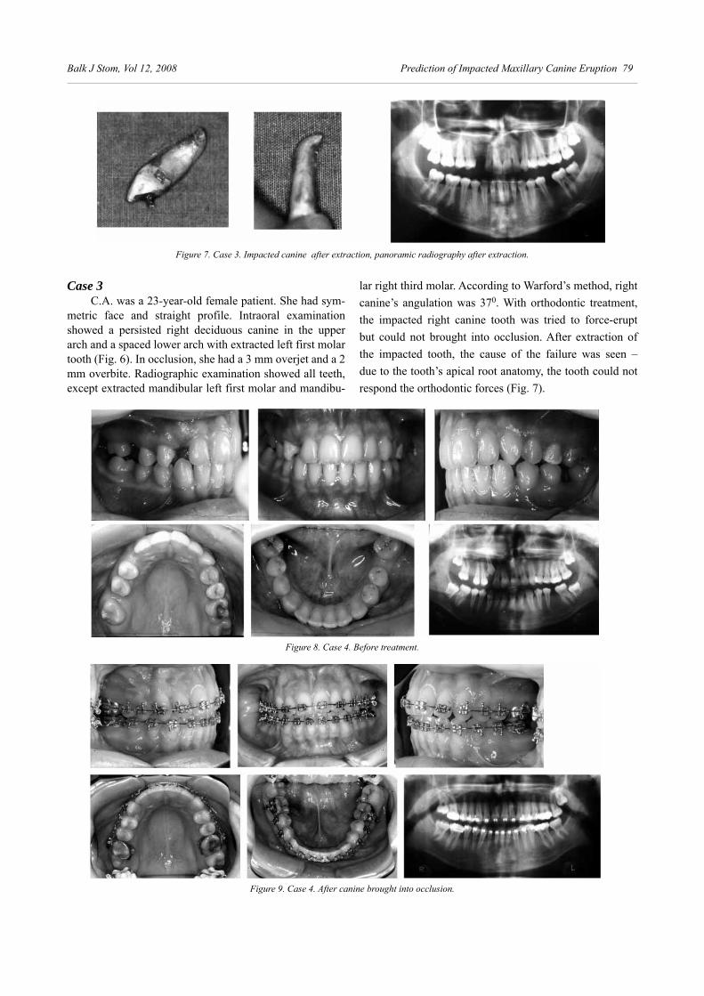
Case 3C.A. was a 23-year-old female patient. She had sym-
metric face and straight profile. Intraoral examination showed a persisted right deciduous canine in the upper arch and a spaced lower arch with extracted left first molar tooth (Fig. 6). In occlusion, she had a 3 mm overjet and a 2 mm overbite. Radiographic examination showed all teeth, except extracted mandibular left first molar and mandibu-
lar right third molar. According to Warford’s method, right canine’s angulation was 370. With orthodontic treatment, the impacted right canine tooth was tried to force-erupt but could not brought into occlusion. After extraction of the impacted tooth, the cause of the failure was seen – due to the tooth’s apical root anatomy, the tooth could not respond the orthodontic forces (Fig. 7).
Figure 7. Case 3. Impacted canine after extraction, panoramic radiography after extraction.
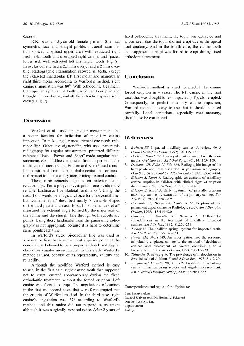
Figure 8. Case 4. Before treatment.
Figure 9. Case 4. After canine brought into occlusion.
80 H. Kilicoglu, I.S. Aksu Balk J Stom, Vol 12, 2008
Case 4R.K. was a 15-year-old female patient. She had
symmetric face and straight profile. Intraoral examina-tion showed a spaced upper arch with extracted right first molar tooth and unerupted right canine, and spaced lower arch with extracted left first molar tooth (Fig. 8). In occlusion, she had a 2.5 mm overjet and a 2 mm over-bite. Radiographic examination showed all teeth, except the extracted mandibular left first molar and mandibular right third molar. According to Warford’s method, right canine’s angulation was 800. With orthodontic treatment, the impacted right canine tooth was forced to erupted and brought into occlusion, and all the extraction spaces were closed (Fig. 9).
Discussion
Warford et al11 used an angular measurement and a sector location for indication of maxillary canine im paction. To make angular measurement one needs refe-rence line. Other investigators5,6,9, who used panoramic radiography for angular measurement, preferred dif ferent reference lines. Power and Short9 made angular mea-surements via a midline constructed from the perpendicular to the central incisors, and Ericson and Kurol5 used a mid-line constructed from the mandibular central incisor proxi-mal contact to the maxillary incisor interproximal contact.
These measurements depends on anterior dental relationships. For a proper investigation, one needs more re liable landmarks like skeletal landmarks11. Using the nasal floor would be a logical choice for a horizontal line, but Damante et al3 described nearly 7 variable shapes of the hard palate and nasal fossa floor. Fernandez et al6 measured the external angle formed by the major axis of the canine and the straight line through both suborbitary points. Using these landmarks from the panoramic radio-graphy is not appropriate because it is hard to determine same points each time.
In Warford’s study, bi-condylar line was used as a reference line, because the most superior point of the condyle was believed to be a proper landmark and logical choice for angular measurement. In this study Warford’s method is used, because of its repeatability, validity and reliability.
Although the modified Warford method is easy to use, in the first case, right canine tooth that supposed not to erupt, erupted spontaneously during the fixed orthodontic treatment, without the forced eruption. Left canine was forced to erupt. The angulations of canines in the first and second cases that were force-erupted met the criteria of Warford method. In the third case, right canine’s angulation was 370 according to Warford’s method, and this canine did not respond to treatment although it was surgically exposed twice. After 2 years of
fixed orthodontic treatment, the tooth was extracted and it was seen that the tooth did not erupt due to the apical root anatomy. And in the fourth case, the canine tooth that supposed to erupt was forced to erupt during fixed orthodontic treatment.
Conclusion
Warford’s method is used to predict the canine forced eruption in 4 cases. The left canine in the first case, that was thought to rest impacted (440), also erupted. Consequently, to predict maxillary canine impaction, Warford method is easy to use, but it should be used carefully. Local conditions, especially root anatomy, should also be considered.
References
1. Bishara SE. Impacted maxillary canines: A review. Am J Orthod Dentofac Orthop, 1992; 101:159-171.
2. Dachi SF, Howell FV. A survey of 3874 routine full mouth radio-graphs. Oral Surg Oral Med Oral Path, 1961; 14:1165-1169.
3. Damante JH, Filho LI, Sila MA. Radiographic image of the hard palate and nasal fossa floor in panoramic radiography. Oral Surg Oral Pathol Oral Radiol Endod, 1998; 85:479-484.
4. Ericson S, Kurol J. Radiographic assessment of maxillary canine eruption in children with clinical signs of eruption disturbances. Eur J Orthod, 1986; 8:133-140.
5. Ericson S, Kurol J. Early treatment of palatally erupting maxillary canines by extraction of the primary canines. Eur J Orthod, 1988; 10:283-295.
6. Fernandez E, Bravo LA, Canteras M. Eruption of the permanent upper canine: A Radiologic study. Am J Dentofac Orthop, 1998; 113:414-420.
7. Fournier A, Turcotte JY, Bernard C. Orthodontic considerations in the treatment of maxillary impacted canines. Am J Orthod, 1982; 81:236-239.
8. Jacoby H. The “ballista spring” system for impacted teeth. Am J Orthod, 1979; 75:143-151.
9. Power SM, Short MB. An investigation into the response of palatally displaced canines to the removal of deciduous canines and assessment of factors contributing to a favourable eruption. Br J Orthod, 1993; 20:215-223.
10. Thilander B, Myrberg N. The prevalence of malocclusion in Swedish school children. Scand J Dent Res, 1973; 81:12-20.
11. Warford JH, Grandhi RK, Tira DE. Prediction of maxillary canine impaction using sectors and angular measurement. Am J Orthod Dentofac Orthop, 2003; 124:651-655.
Correspondence and request for offprints to:
Irem Sakarya AksuIstanbul Universitesi, Dis Hekimligi FakultesiOrtodonti ABD 5. kat. Capa/IstanbulTurkey

SUMMARYMidline diastema between maxillary central incisors is a common
occurrence, especially in primary and mixed dentition. Its presence has been attributed to genetic and/or environmental factors. However, it is often a normal characteristic of growth. Many times the development of upper mid-line diastema is related to the presence of hypertrophic or inferiorly attached upper labial fraenum and/or imperfect fusion at the midline of premaxilla. These 2 conditions are frequently confusing in clinical practice, thus the diagnosis and treatment of the problem would be false. The probable thera-peutic approaches for the maxillary midline diastema provoked by abnormal labial fraenum and/or intermaxillary suture include orthodontics, restorative dentistry, surgery or various combinations of them. The necessity of treat-ment is mainly conducted by aesthetic and psychological rather than func-tional reasons. Irrespectively of the optional treatment, permanent retention of the result should be adapted in most cases. The purpose of this study was to analyze the relation between hypertrophic or inferiorly attached upper labial fraenum and imperfect fusion at the midline of premaxilla, with the maxillary midline diastema. Additionally, appropriate clinical and labora-tory examinations are described, plus therapeutic alternates, which are pro-posed in each case. Keywords: Maxillary Midline Diastema; Upper Labial Fraenum; Intermaxillary Suture
Nikolaos Gkantidis1, Nikolaos Topouzelis2, Lampros Zouloumis3 1Private Dentist, Thessaloniki, Greece2Aristotle University, School of DentistryDepartment of OrthodonticsThessaloniki, Greece3Aristotle University, School of DentistryDepartment of Oral and Maxillofacial SurgeryThessaloniki, Greece
ORIGINAL PAPER (OP)Balk J Stoma, 2008; 12:81-88
BALKAN JOURNAL OF STOMATOLOGY ISSN 1107 - 1141
Differential Diagnosis and Combined Treatment of Maxillary Midline Diastema Caused byLabial Fraenum and/or Intermaxillary Suture
TUPNBUPMPHJDB
M!!T
PD
JFU
Z
Introduction
In 9-year-old children the prevalence of maxillary midline diastema is high, with a 48.8% rate of children presenting diastema larger than 0.5 mm49, but this rate is decreased with age27,49,56,67. In adults, the prevalence of upper midline diastema is considered to range between 1.6% and 25.4%14,38,40,44,56,63. In the majority of studies the predominant opinion is that the upper midline diastema occurs under the effect of multiple environmental factors6,9,12,31,44,46. However, there is a number of well documented studies that support the possibility of genetic predispose in the development of this condition29,44,58,60.
Moyers42 examined 82 patients with upper midline diastema and reported the following causative factors: (a) imperfect fusion at midline of premaxilla (32.9%); (b) enlarged or malposed upper labial fraenum (24.4%); (c) midline diastema as part of normal growth (23.2%);
(d) congenitally missing lateral incisors (11%); (e) supernumerary teeth at the midline (3.7%); (f) unusually small teeth (2;4%); and (g) combination of imperfect fusion and congenitally missing lateral incisors (2.4%). Furthermore, additional causes for the development of upper midline diastema have been reported in the literature, such as para-functional oral habits12,46,64, increased overbite of anterior teeth46,49 or pathologic teeth migration due to periodontal disease18,54.
Imperfect Fusion at Midline of Premaxilla
The relationship between the imperfect fusion at the midline of premaxilla and the upper midline diastema has

82 N. Gkantidis et al. Balk J Stom, Vol 12, 2008
been recognized over the past years, but it did not gain too much emphasis27,64. The discontinuity of the bony plates may be superficial or extend deeper in the alveolar process24. The gap within the maxilla is occupied by epithelial and connective tissue. Often fraenum or gingival fibres (especially interdental fibres) are attached at that site. Normally, interdental fibres functionally contribute in the retention of the teeth in position23,34,39,64. Because of the disturbance of the continuity and arrangement of the interdental gingival fibres, their ability to resist in expressed forces to teeth is compromised. As a result, there is a tendency for distal movement of upper central incisors, leading in some cases to the development of midline diastema. For the same reason, this diastema is usually accompanied by rotation of upper lateral incisors and ectopic eruption of canines64. Moyers42 stated that imperfect fusion at the midline of premaxilla is the most common cause of maxillary midline diastema, with a rate of 32.9%. Also, Popovich et al48,50 suggested that the combination of imperfect fusion with several other predisposal factors is the most significant cause of maxillary midline diastema.
The diagnosis of imperfect fusion at midline is performed radiographically. It is fundamental for the central ray to be precisely perpendicular to the alveolar process42. The normal radiographic image of the suture is a V-shaped structure (Fig. 1). The suture is characterised pathologic: (a) when it is displayed wider than normal (approximately 2mm) (Fig. 2); (b) when a circumscribed irregular ovoid area (spade-shaped) is displayed in this region (Fig. 3); or (c) when the alveolar process is W-shaped in the region between the maxillary central incisors, in cases with extended separation of the bone plates (Fig. 4a). The latest 2 instances are often accompanied by abnormal labial fraenum48,50.
Figure 4. (a) Pathological W-shaped intermaxillary suture; (b) Post-treatment radiograph, after orthodontic closure of the diastema, followed
by surgical intervention in the intermaxillary suture
Figure 1. Normal V-shaped intermaxillary suture. The present diastema is due to congenitally missing upper lateral incisors
Figure 2. Pathological intermaxillary suture, wider than normal
Figure 3. Pathological spade-shaped intermaxillary suture
Management consists of orthodontic closure of the diastema, followed by a surgical intervention in the suture35. The orthodontic closure of the diastema is performed first, so that tissue healing and fibre remodelling take place in the new position, where we desire to retain the final treatment outcome51,64,69. In cases where the imperfect fusion makes the diastema closure impossible, the surgery must be performed before the closure of the midline diastema. During the surgical procedure, a trapezoidal flap with 2 perpendicular incisions (bilaterally to the midline interdental papilla), which are joined with a horizontal incision at the interdental gingiva, is created. After the

Balk J Stom, Vol 12, 2008 Diagnosis and Treatment of Maxillary Midline Diastema 83
elevation of mucoperiosteal flap and the apocalypse of the alveolar bone, a surgical fissure bur is inserted in the midline suture and detracts the soft tissues, while abrading the bone35,64. The orthodontic appliance which was utilized for the dental movement must be replaced by a retention appliance during the phase of healing (Fig. 4b). In certain cases, there is a chance for relapse, demanding revision of the same procedure35. At this point it is critical to mention that Sullivan et al65 and Shashua and Artun60 failed to confirm the relation between relapse and imperfect fusion at midline of premaxilla.
Hypertrophic or Inferiorly Attached Upper Labial Fraenum
The hypertrophic or inferiorly attached upper labial fraenum is considered for many years the commonest cause of the maxillary midline diastema4,27,39,61. Several authors differentiate from this aspect, as Moyers42 does. He supports that the upper labial fraenum is the second commonest cause of this condition, with an incidence of 24.4%. Referring to the fraenum composition, it consists of epithelium, collagen fibres, blood vessels and nerves, and sometimes few elements of minor salivary glands and isolated stratified muscle fibers28,57. Henry et al30 in the remarkable histological study concluded that, except from the elements mentioned above, there are also elastic fibres, which extend sometimes to the whole length of the fraenum, even perforating the periosteum. Those authors considered that the harmful effect of the fraenum is due to the presence of the elastic and collagen fibres, while no evidence of substantial differences in composition of normal and abnormal fraena were identified.
There is controversy among researchers concerning the presence of an immediate causative relation between hypertrophic or inferiorly attached upper labial fraenum and the maxillary midline diastema. Several authors do not support the existence of this relation. Tait66 considered that the fraenum has no effect to the maxillary central incisors, while Ceremelo17 concluded that the fraenum is not related to the presence or the width of the diastema. Bergstrom et al11 stated that the long term potential for spontaneous diastema closure in patients with abnormal fraenum, remains the same independently to the implementation of a previous surgical intervention. Popovich et al48,50 suggested that the presence of the diastema leads to the abnormal fraenum and not the adverse.
In contrast, several authors stated that the fraenum is involved in the pathogenicity of the midline diastema1,4,6,16,19,27,39,61. Adams1 suggested that a specific type of fraenum, which is not necessarily large but interrupts the continuity of interdental fibres, forms the factor that inducts the reactions for the development of the
diastema. Otherwise, this author supports that diastema develops only when there is additional presence of other predisposing factors. Campbell et al16 supported the same statement as well. The disruption of the interdental (trans-septal) gingival fibres continuity, due to the fraenum, is considered by several studies as presupposing for the development of a pathological diastema16,24,59,64. Edwards23 supported the presence of a strong but not absolute correlation between the fraenum and the upper midline diastema. Shashua and Artun60 found that there is a relation between the width of the maxillary midline diastema and the abnormal labial fraenum.
Regarding the physiology of the upper labial fraenum and its relation with age, the fraenum found to be smaller in length, thicker and more inferiorly attached in infants17,39. Normally, the fraenum does not follow the growth of the alveolar process that occurs during tooth eruption6,19, since the erupting central incisors exercise pressure on it. This fact makes the fraenum appear to be at a more nasal position with age, while in fact, it remains more or less in the same position20. In certain instances, the fraenum attachment does not obviously “migrate nasally” by the elapse of time, but continues to develop between the 2 upper central incisors and remains there forming a residual fibre zone23. More detailed, these fibres can be attached to the periosteum and to the connective tissue of the abnormal intermaxillary suture6,19,27, or only disrupt the continuity of the interdental gingival fibre system16,24,59,64. Under these circumstances, we must not expect spontaneous correction of the diastema with the eruption of the maxillary lateral incisors and canines3,27,42,46,49. It is obvious that the nearer to the incisive papilla and the deeper within the tissues the fraenum is attached, the more possible for it to cause a diastema20. The sum of these clinical data should be always taken under consideration, concerning the age and the other parameters which affect the problem20,23,39. Popovich et al48,50 supported the presence of an adverse relationship between the inferiorly attached and/or hypertrophic fraenum and maxillary midline diastema. They stated that due to the presence of the diastema the fraenum still develops coronally, along with the alveolar process, as teeth erupt. This happens because the dentition exerts minor or zero pressure on the fraenum.
Occasionally, when the fraenum fibres are inserted quite deep, the hypertrophic or inferiorly attached upper labial fraenum could be diagnosed by simple clinical observation (Fig. 5) or by observing ischaemia provoked at the interdental papilla when stretching the upper lip (Fig. 6)1,39,42. According to Miller41, the fraenum should be judged as pathological when it is uncommonly wide, when there is insufficient attached gingival zone in the midline (Fig. 7), and when the interdental papilla moves by stretch of the fraenum. However, the evaluation of the fraenum is sometimes difficult, especially in borderline cases11,23,46.

84 N. Gkantidis et al. Balk J Stom, Vol 12, 2008
This specific pathological situation is treated with orthodontic closure of the maxillary midline diastema, followed by surgical intervention22,27,41. The orthodontic appliances must be kept in place during the whole phase of healing (Fig. 8,a-c). With this approach, the new scar tissue that is going to be formed will contribute in the desirable retention of the result of treatment69. The above should be advocated only if the diastema remains open after the eruption of permanent canines19,26,39. Nevertheless, in specific cases, when the fraenum is significantly hypertrophic and so inhibits the orthodontic closure of the diastema, surgical intervention is required at an earlier stage than usual69.
Figure 5. Diagnosis of abnormal labial fraenum only by observation
Figure 6. Diagnosis of abnormal labial fraenum with extension of the upper lip and observation of ischaemia in the interdental papilla
Figure 7. Diagnosis by observation of the abnormal labial fraenum, which is unusually broad, and there is no apparent attached gingiva at
the midline
Figure 8. (a) Abnormal labial fraenum after orthodontic diastema correction; (b) Surgical intervention in the fraenum; (c) Result of surgery and retention of orthodontic appliances in place during healing
Various surgical techniques have been described for the management of the abnormal upper labial fraenum that causes a midline diastema. Those include: (a) the classic technique of frenectomy, in which the fraenum, the interdental soft tissue, and the palatal interdental papilla are completely removed, leaving uncovered bone or periosteum; (b) osteotomy of alveolar process under the apices of the teeth10; (c) corticotomies33; (d) septotomies62;
(e) ‘’Z-plasty’’ technique, which does not reposition the fraenum but aims to reduce the traction that is exerted from the fraenum to the interdental soft tissues36; (f) reverse-bevel gingivectomy; (g) circumferential supracrestal fibrotomy technique16; and (h) frenectomy in combination with free gingival graft from palate. This technique seems to create aesthetic problems because of a difference in the colour between physiological gingiva

Balk J Stom, Vol 12, 2008 Diagnosis and Treatment of Maxillary Midline Diastema 85
and transplantation site13. Possibly, the most effective and less invasive surgical technique for the treatment of the hypertrophic or inferiorly attached upper labial fraenum still is the one proposed by Edwards23. This technique includes 3 different steps: (1) apical reposition of the fraenum with apocalypse of alveolar bone; (2) distraction of interdental (transseptal) fibres between approximated central incisors; and (3) gingivoplasty or re-contouring of gingiva at the labial or palatal interdental papilla when it is necessary22. Moreover, with purpose to further decrease the potential for relapse, it is seemingly important to remove the elastic fibres that impregnate the periosteum underline the fraenum, because nowhere in the normal human periodontium is there such an elastic tissue involvement with attached gingiva22,30. The major purpose of the current procedure is to reposition the fraenum from the site of diastema by a triangle incision which is performed from the top to the base of the fraenum. The remaining area is healed within 7-10 days, with full epithelization completed in 2-3 weeks22. In cases with more aesthetic expectations, the technique proposed by Bagga et al5 seems to be really effective. This technique includes the coverage of the bony surface by 2 triangle laterally repositioning partially thickness flaps, but it presupposes the presence of adequate width of the attached gingiva in order to be performed. It must be noted that occasionally the pressure provoked to the fraenum fibres during the orthodontic approximation of maxillary central incisors leads to avascular necrosis along with fraenum and gingival fibres remodelling, so making the surgical intervention useless23,42,60.
Management of the Maxillary Midline Diastema
The clinician before selecting the appropriate treatment plan must definitely define a sound diagnosis. The diagnosis requires the evaluation of several factors, as age, stage of growth, relations of teeth with adjacent, their antagonists and their skeletal base, possible presence of malocclusion, tooth-size relationship, other pathological conditions, and the presence of diastemas in other segments of the arch. Consequently, the selection of the appropriate management for the diastema between the maxillary central incisors is often difficult, as it presupposes a sound diagnosis and mainly the recognition and treatment of the aetiology of the problem. In a study of Almog et al2, 87.5% of the subjects mentioned that they prefer the method of computer-imaging simulation for the acceptance of the proposed treatment plan, concerning the closure of diastema in the anterior segment of the maxilla, because they understood better the treatment plan that way.
The treatment of the diastema is usually postponed until the eruption of permanent canines, but it may start earlier, depending on the cause of the problem and in cases with a very extensive diastema15,31. As a general principle, only for diastemas between maxillary central incisors larger than 2 mm, there is a danger for not spontaneous closure with the eruption of lateral incisors and canines, while for a diastema smaller than 2 mm it is almost impossible to remain open23,52. The early closure of a diastema (during the mixed dentition) is performed when: (a) the diastema creates severe aesthetic problem to the patient and (b) the position of central incisors inhibits the eruption of lateral incisors or canines, as the lateral incisors may have been displaced in the space which is normally occupied by canines52.
In the period of permanent dentition, when the diastema is smaller than 2 mm it can be treated successfully by the orthodontist, probably by simple tipping of the maxillary central incisors. This can be performed with a removable appliance with clasps, finger-springs and, possibly an anterior bow52. When this approach is selected, there is a strong need for retention as it often leads to relapse22,41. The use of elastic bands around teeth for the closure of the diastema is strictly prohibited because the bands may move apically and destruct the periodontal ligament, provoking even loss of teeth55. When there is a diastema larger than 2mm, in the majority of cases there is a need for bodily movement of teeth, and a more complicated treatment with fixed orthodontic appliances is required52.
In recent years, various authors suggested that the closure of the diastema between the upper central incisors or other diastemas in the anterior segment of the maxillary arch (whether created by the orthodontic therapy or pre-existed) may be achieved with the placement of crown veneers45 or with teeth restorations with resin composite68. Nevertheless, the long-term prognosis in these cases should be further investigated47. In specific cases, this approach can be adopted when the patient does not desire to be treated with orthodontics, when there are other coexisting aesthetic problems (i.e. amelogenesis imperfecta), and when the treatment demands a combined orthodontic and restorative approach, in cases with large diastema8.
Retention of the Treatment Result
The retention after treatment of the maxillary midline diastema is considered to be necessary, especially if lateral incisors and canines have not yet erupted. The prevalence of relapse according to Sullivan et al65 is 34%, while according to Shashua and Artun60 it approaches approximately 50%. The reason for relapse is the placement of teeth in a position where the teeth are not in equilibrium

86 N. Gkantidis et al. Balk J Stom, Vol 12, 2008
5. The need for permanent retention of the result of treatment, in almost all cases, is inevitable.
References
Adams CP1. . The relation of spacing of the upper central incisors to abnormal labial frenum and other features of the dento-facial complex. Dent Pract Dent Rec, 1954; 74:72-86. Almog D, Sanchez Marin C, Proskin HM, Cohen MJ, 2. Kyrkanides S, Malmstrom H. The effect of esthetic consultation methods on acceptance of diastema-closure treatment plan: a pilot study. J Am Dent Assoc, 2004; 135:875-881.Andrews LF3. . The six keys to normal occlusion. Am J Orthod, 1972; 62:296-309. Angle EH4. . Treatment of malocclusion of the teeth, 7th ed. Phila-delphia: S.S. White dental manufacturing Co, 1907; pp 167.Bagga S, Bhat KM, Bhat GS, Thomas BS5. . Esthetic management of the upper labial frenum: a novel frenectomy technique. Quintessence Int, 2006; 37:819-823.Baum AT6. . The midline diastema. J Oral Med, 1966; 21:30-39.Bearn DR7. . Bonded orthodontic retainers: a review. Am J Orthod Dentofacial Orthop, 1995; 108:207-213.Beasley WK, Maskeroni AJ, Moon MG, Keating GV, 8. Maxwell AW. The orthodontic and restorative treatment of a large diastema: a case report. Gen Dent, 2004; 52:37-41.Becker A9. . The median diastema. Dent Clin North Am, 1978; 22:685-710.
with their functional environment. Thus, a balance between the external forces exerted to teeth and internal forces that retain teeth in position is not preserved53. The main reason for this is the inability for exception of the factor that disturbs this equilibrium9,12,43. For instance, this disturbance may occur with the disruption of the continuity of the interdental gingival fibres, whose functional role is the preservation of teeth position, like in cases with an abnormal upper labial fraenum. Moreover, the equilibrium may be disturbed when the interdental fibres are compressed by the orthodontic closure of the diastema, so exerting distal forces to maxillary central incisors64.
The main risk factors for relapse include: (a) increased pre-treatment width of the maxillary midline diastema;
(b) the presence of another member of the family with a diastema; and (c) more than one diastema in the maxillary anterior region60. However, according to other authors, prognostic factors for relapse can not be defined65. Taking under consideration recent studies, the cases with a risk for long term relapse can not be safely predicted21,25,37,70. Consequently, it would be wise in almost every case to select permanent retention for the preservation of the therapeutic outcome and the avoidance of undesirable clinical and legal matters. The retainer may be used for a specific period of time or for the patient’s whole life69,70. According to Lang et al37, the time for retention must in every case exceed 2 years.
Figure 9. (a) Maxillary midline diastema; (b) Lingual retainer fabricated by multi-stranded stainless steel wire and bonded to maxillary anterior teeth for retention after orthodontic treatment
The most appropriate solution for permanent retention is the bonding of a multi-stranded stainless steel wire, constructed by 6 strands of wire, with a 0.0215 inch diameter each7,32. The basic advantage of this retainer is that its flexibility permits teeth to retain their physiologic movement, plus it is placed and bonded easily69. The multi-stranded wire is bonded with resin composite, usually from canine to canine in the middle of the palatal surface of anterior teeth (Fig. 9). In cases where the retainer interferes in functional movements of the mandible, it can be bonded cervically or within a shallow rim constructed to the enamel of the teeth69.
Conclusions
1. The imperfect fusion at midline of premaxilla and the abnormal labial fraenum are the commonest causes of the maxillary midline diastema.
2. The differential diagnosis and the appropriate management of these 2 conditions require careful evaluation of a variety of parameters.
3. The most effective treatment for the maxillary midline diastema is the treatment that faces the cause of the diastema.
4. The optional treatment often requires a multidisciplinary approach.

Balk J Stom, Vol 12, 2008 Diagnosis and Treatment of Maxillary Midline Diastema 87
Bell WH10. . Surgical-orthodontic treatment of interincisal diastemas. Am J Orthod, 1970; 57:158-163.Bergstrom K, Jensen R, Martensson B11. . The effect of superior labial frenectomy in cases with midline diastema. Am J Orthod, 1973; 63:633-638. Bishara SE12. . Management of diastemas in orthodontics. Am J Orthod, 1972; 61:55-63.Breault LG, Fowler EB, Moore EA, Murray DJ13. . The free gingival graft combined with the frenectomy: A clinical review. Gen Dent, 1999; 47:514-518.Brunelle JA, Bhat M, Lipton JA14. . Prevalence and distribution of selected occlusal characteristics in the US population, 1988-1991. J Dent Res, 1996; 75:706-713.Campbell A, Kindelan J15. . Maxillary midline diastema: a case report involving a combined orthodontic/maxillofacial approach. J Orthod, 2006; 33:22-27. Campbell PM, Moore JW, Matthews JL16. . Orthodontically corrected midline diastemas. A histologic study and surgical procedure. Am J Orthod, 1975; 67:139-158. Ceremelo PJ17. . The superior labial frenum and the midline diastema and their relation to growth and development of the oral structures. Am J Orthod, 1953; 39:120-139.Cirelli JA, Cirelli CC, Holzhausen M, Martins LP, Brandao 18. CH. Combined periodontal, orthodontic, and restorative treatment of pathologic migration of anterior teeth: a case report. Int J Periodontics Restorative Dent, 2006; 26:501-506.Dewel BF19. . The labial frenum, midline diastema, and palatine papilla: a clinical analysis. Dent Clin North Am, 1966; pp 175-184. Diaz-Pizan ME, Lagravere MO, Villena R20. . Midline diastema and frenum morphology in the primary dentition. J Dent Child, 2006; 73:11-14.Durbin DD21. . Relapse and the need for permanent fixed retention. J Clin Orthod, 2001; 35:723-727. Edwards JG22. . Soft-tissue surgery to alleviate orthodontic relapse. Dent Clin North Am, 1993; 37:205-225.Edwards JG23. . The diastema, the frenum, the frenectomy: a clinical study. Am J Orthod, 1977; 71:489-508. Ferguson MW, Rix C24. . Pathogenesis of abnormal midline spacing of human central incisors. A histological study of the involvement of the labial frenum. Br Dent J, 1983; 154:212-218.Fidler BC, Artun J, Joondeph DR, Little RM25. . Long-term stability of Angle Class II, division 1 malocclusions with successful occlusal results at end of active treatment. Am J Orthod Dentofacial Orthop, 1995; 107:276-285.Finn SB26. . Clinical Pedodontics. Philadelphia: WB Saunders, 1971; pp 416-418.Gardiner JΗ27. . Midline spaces. Dent Pract Dent Rec, 1967; 17:287-298.Gartner LP, Schein D28. . The superior labial frenum: a histologic observation. Quintessence Int, 1991; 22:443-445.Gass JR, Valiathan M, Tiwari HK, Hans MG, Elston RC29. . Familial correlations and heritability of maxillary midline diastema. Am J Orthod Dentofacial Orthop, 2003; 123:35-39.Henry SW, Levin MP, Tsaknis PJ30. . Histologic features of the superior labial frenum. J Periodontol, 1976; 47:25-28.Huang WJ, Creath CJ31. . The midline diastema: a review of its etiology and treatment. Pediatr Dent, 1995; 17:171-179.
32. Karaman AI, Polat O, Buyukyilmaz T. A practical method of fabricating a lingual retainer. Am J Orthod Dentofacial Orthop, 2003; 124:327-330.Kole H33. . Surgical operations on the alveolar ridge to correct occlusal abnormalities. Oral Surg Oral Med Oral Pathol, 1959; 12:515-529. Κonstantinidis Α34. . Periodontology. Vol. 1. Thessaloniki: Κonstantinidis Α, 2003; p 77.Kraut R, Payne J35. . Osteotomy of intermaxillary suture for closure of median diastema. J Am Dent Assoc, 1983; 107:760-761.Kruger GO36. . Oral Surgery. Ed. 2. St. Louis: CV Mosby, 1964; pp 146-147.Lang G, Alfter G, Goz G, Lang GH37. . Retention and stability - taking various treatment parameters into account. J Orofac Orthop, 2002; 63:26-41.Lavelle CL38. . The distribution of diastemas in different human population samples. Scand J Dent Res, 1970; 78:530-534.Lindsey D39. . The upper mid-line space and its relation to the labial fraenum in children and in adults. A statistical evaluation. Br Dent J, 1977; 143:327-332.McVay TJ, Latta GH Jr40. . Incidence of the maxillary midline diastemas in adults. J Prosthet Dent, 1984; 52:809-811. Miller PD Jr41. . The frenectomy combined with a laterally positioned pedicle graft. Functional and esthetic considerations. J Periodontol, 1985; 56:102-106.Moyers R42. . Handbook of Orthodontics. 4th Ed. Chicago: Year Book Medical Publishers, 1988; pp 357-360.Mulligan TF43. . Diastema closure and long-term stability. J Clin Orthod, 2003; 37:560-574.Nainar SM, Gnanasundaram N44. . Incidence and etiology of midline diastema in a population in south India (Madras). Angle Orthod, 1989; 59:277-282.Nazarian A45. . Closing the gap with minimal preparation veneers. Dent Today, 2006; 25:70-71.Oesterle LJ, Shellhart WC46. . Maxillary midline diastemas: a look at the causes. J Am Dent Assoc, 1999; 130:85-94.Peumans M, Van Meerbeek B, Lambrechts P, Vanherle 47. G. The 5-year clinical performance of direct composite additions to correct tooth form and position. I. Esthetic qualities. Clin Oral Investig, 1997; 1:12-18.Popovich F, Thompson GW, Main PA48. . The maxillary interincisal diastema and its relationship to the superior labial frenum and intermaxillary suture. Angle Orthod, 1977; 47:265-271.Popovich F, Thompson GW49. . Maxillary diastema: indications for treatment. Am J Orthod, 1979; 75:399-404.Popovich F, Thompson GW, Main PA50. . Persisting maxillary diastema: differential diagnosis and treatment. Dent J, 1977; 43:330-333.Proffit W, Fields H51. . Contemporary Orthodontics. 2nd ed. St. Louis: Mosby-Year Book 1993; pp 128-129, 486.Proffit W, Fields H52. . Contemporary Orthodontics. 3rd ed. St. Louis: Mosby, 2000; pp 77, 87, 170, 429-430, 463-465.Proffit WR53. . Equilibrium theory revisited: Factors influencing position of the teeth. Angle Orthod, 1978; 48:175-186. Rahilly G, Crocker C54. . Pathological migration: an unusual cause of midline diastema. Dent Update, 2003; 30:547-549.

88 N. Gkantidis et al. Balk J Stom, Vol 12, 2008
Sullivan TC, Turpin DL, Artun J65. . A postretention study of patients presenting with maxillary median diastema. Angle Orthod, 1996; 66:131-138.Tait CW66. . The median frenum of the upper lip and its influence on the spacing of the upper central incisor teeth. Dent Cosmos, 1924; 76:991-992.Weyman J67. . The incidence of median diastema during the eruption of the permanent teeth. Dent Pract Dent Rec, 1967; 17:276-278.Willhite C68. . Diastema closure with freehand composite: controlling emergence contour. Quintessence Int, 2005; 36:138-140. Zachrisson BU69. . Orthodontics and periodontics In: Lindhe J. Clinical periodontology and implant dentistry. 3rd ed. Copenhagen: Munksgaard, 1997; pp 741-793.Zachrisson BU70. . Use of self-ligating brackets, superelastic wires, expansion/proclination, and permanent retention - a word of caution. World J Orthod, 2006; 7:198-206.
Correspondence and request for offprints to:
Prof. Nikolaos Topouzelis Aristotle University, School of DentistryDepartment of OrthodonticsGR-54124, ThessalonikiGreece E-mail: [email protected]
Redlich M, Galun EA, Zilberman Y55. . Orthodontic-prosthetic treatment to replace maxillary incisors exfoliated because of improper use of orthodontic elastics: a case report. Quintessence Int, 1997; 28:241-244.Richardson ER, Malhotra SΚ, Henry M, Little RG, Coleman 56. HT. Biracial study of the maxillary midline diastema. Angle Orthod, 1973; 43:438-443.Ross RO, Brown FH, Houston GD57. . Histologic survey of the frena of the oral cavity. Quintessence Int, 1990; 21:233-237. Schmitt E, Gillenwater JY, Kelly TE58. . An autosomal dominant syndrome of radial hypoplasia, triphalangeal thumbs, hypospadias, and maxillary diastema. Am J Med Genet, 1982; 13:63-69.Scott JH, Symons NB59. . Introduction to dental anatomy. 7th ed. Edinburgh: Churchill Livingstone, 1974; p 264.Shashua D, Artun J60. . Relapse after orthodontic correction of maxillary median diastema: a follow-up evaluation of consecutive cases. Angle Orthod, 1999; 69:257-263.Sicher H61. . Oral Anatomy. 2nd ed., St. Louis: CV Mosby Company, 1952; pp 73-75.Skogsborg C62. . The use of septotomy (surgical treatment) in connection with orthodontic treatment, and the value of this method for proof of Wolkoff’s theory of tension of the bone tissue after regulation of teeth. Int J Orthod, 1932; 18:1044. Steigman S, Weissberg Y63. . Spaced dentition. An epidemiologic study. Angle Orthod, 1985; 55:167-176.Stubley R64. . The influence of transseptal fibers on incisor position and diastema formation. Am J Orthod, 1976; 70:645-662.
SUMMARYTooth bleaching procedures have become very popular since their first
introduction in dental clinical practice. There are concerns about the effects of exposure of oral tissues to bleaching agents. The purpose of the present study was to determine the cytotoxic effect of 2 bleaching agents (sodium perborate and carbamide peroxide) against 2 fibroblastic cell lines. BHK21/C13 baby hamster kidney fibroblasts and RPC-C2A rat pulp cells were used for the experiment. The cells were grown as monolayer cultures at 37°C in an atmosphere containing 5% CO2 in air, and 100% relative humidity. Cells were plated in multiwell plates and serial dilutions of the bleaching agents were prepared and placed in contact with cell cultures. The anti-proliferative effect was determined after 24 and 48 hours of exposure by means of the colorimetric sulphorodamine B assay, in reference to controls.
BHK21/C13 and RPC-C2A reacted to the bleaching agents with dif-ferent intensity. Cell proliferation was reduced compared to the controls after 24 and 48 hours of exposure in both cell lines. The cytotoxic effect was concentration- and time-dependent. Sodium perborate was the most potent material tested.Keywords: Cytotoxicity; Sodium Perborate; Carbamide Peroxide
Elisabeth A. Koulaouzidou1, Konstantinos T. Papazisis2, Nikolaos Economides3, Artemis Karanika-Kouma1, Alexander H. Kortsaris4
1Aristotle University, Faculty of DentistryDepartment of Operative DentistryThessaloniki, Greece2Theagenion Cancer InstituteThessaloniki, Greece3Aristotle University, Faculty of Dentistry, Department of EndodontologyThessaloniki, Greece4Democretian University of ThraceFaculty of Medicine, Department of Biochemistry Greece
ORIGINAL PAPER (OP)Balk J Stom, 2008; 12:89-92
BALKAN JOURNAL OF STOMATOLOGY ISSN 1107 - 1141
Cytotoxicity of 2 Bleaching Agents: An In Vitro Study
TUPNBUPMPHJDB
M!!T
PD
JFU
Z
Introduction
Tooth discoloration is an important concern in aes-thetic dentistry. There are several reasons why teeth may change colour, including food, drinks, tobacco, poor oral hygiene, diseases, medications, dental materials, age, genetics, environment, dental trauma, endodontic treat-ment, systemic diseases etc1-4.
Tooth bleaching techniques were developed a century ago and all of them involved the process of oxidation. Generally, bleaching includes 2 types of techniques: vital and non-vital. There are 2 main methods of vital tooth bleaching. One involves the application of an oxidizing agent for a short period of time and may include activation of the process by heat or light. The other method involves using a specially prepared mouth guard to hold the oxidizing agent next to the teeth for few hours a day, usually during night, for 1 to 3 weeks.
Non-vital tooth bleaching techniques include mainly the thermocatalytic method5 and the walking bleach method6. In the thermocatalytic method, heat is used to activate the oxidizing agent placed in the pulp chamber. In
the walking bleach technique, the bleaching agent is sealed in the pulp chamber for 3-7 days and thereafter it is replaced regularly until a satisfactory result is obtained (usually 2-3 times)7.
The most commonly used bleaching agents are hydro-gen peroxide and sodium perborate, either used alone or in combination. More recently carbamide peroxide has also been recommended. Carbamide peroxide is mainly pro-posed for vital tooth bleaching at concentrations ranging from 10% to 35%. Sodium perborate alone or in combi-nation with hydrogen peroxide is the main medicament in non-vital tooth bleaching techniques8-10.
Although generally positive results have been reported concerning the whitening ability of the bleaching agents, rather few studies have addressed their possible undesirable effects on living tissues11-14. The risk of bleaching agents to be a threat to the adjacent tissues is correlated to several factors, such as the used amount and concentration, the diffusion through dental tissues, the contact to the gingival tissues.
The biological risk from bleaching agents can be estimated by tests in vitro. In vitro methods are routinely

90 E.A. Koulaouzidou et al. Balk J Stom, Vol 12, 2008
used in toxicity testing, safety assessment, and risk evaluation. Cell cultures can be used for screening the cytotoxicity of materials. The purpose of the present study was to evaluate the cytotoxic effect of sodium perborate and carbamide peroxide using 2 fibroblastic cell lines.
Material and Methods
Sodium perborate and carbamide peroxide were purchased from Sigma Aldrich Co (USA). 2 established cell lines were used: BHK21/C13 (baby hamster kidney fibroblasts) and RPC-C2A (rat pulp cells). BHK21/C13 were obtained from ICRF (London, UK) and RPC-C2A cells were a generous offer from Prof. S. Kasugai (Tokyo, Japan). Cells were grown as monolayer cultures in T-75 flasks (Corning Costar), sub-cultured twice a week at 37°C in an atmosphere containing 5% CO2 in air and 100% relative humidity, and maintained at a low passage number (5-20). The culture medium was Dublecco’s modified Eagle’s medium (DMEM, Gibco, Glasgow, UK), supplemented with 10% foetal bovine serum (FBS, Gibco, Glasgow, UK), 100 IU/ml penicillin and 100 μg/ml streptomycin.
Adherent cells at a logarithmic growth phase were detached by the addition of 2-3 ml of a 0.05% trypsin (Gibco Brl, 1:250) - 0.02% EDTA mixture and incubation for 2-5 min at 37°C. Cells were plated (5,000 cells in 100 μl of culture medium per well) in 96-well flat-bottomed micro-titre plates (Corning Costar). Micro-plates were left for 24 h at 37°C to allow cells attach to the bottom of the wells and resume exponential growth. 100 μl of serial dilutions of the bleaching agents (diluted in the culture medium) were added on the cells (final volume in each well 200 μl) after 24 h. 6 replicate wells for each concentration were used. Negative control wells containing the same volume of complete medium, were included in each experiment. Cell growth was evaluated 24 and 48 h later by means of the SRB assay. All experiments were repeated at least twice.
The SRB assay was carried out as previously described15 and modified by Papazisis et al16. In brief, 70 μl 0.4% (w/v) sulforhodamine B (SRB, Sigma) in 1% acetic acid solution were added to each well and left at room temperature for 20 min. SRB was removed and the plates washed 5 times with 1% acetic acid before air drying. Bound SRB was solubilized with 200 μl 10 mM un-buffered Tris-base solution (Sigma) and plates were left on a plate shaker for at least 10 min. Absorbance was read in a 96-well plate reader (Anthos-2001, Anthos labteck instruments, A-5022, Salzburg) at 492 nm subtracting the background measurement at 620 nm. The test optical density (OD) value was defined as the absorbance of each individual well, minus the blank value (“blank” is the mean optical density of the background control wells,
n = 8). Mean values and CV from 6 replicate wells were calculated automatically. Dose-response curves were plotted (values expressed as percentage of control optical density). The data were analyzed by ANOVA and the Student-Newman-Keul test (p<0.05).
Results
0%
25%
50%
75%
100%
0.000001 0.00001 0.0001 0.001 0.01 0.1 1concentrations
(a)ce
ll n
um
ber
Carbamide peroxide
Sodium perborate
0%
25%
50%
75%
100%
0.000001 0.00001 0.0001 0.001 0.01 0.1 1concentrations
(b)
cell
nu
mb
er
Carbamide peroxide
Sodium perborate
0%
25%
50%
75%
100%
0.000001 0.00001 0.0001 0.001 0.01 0.1 1
concentrations(a)
cell
nu
mb
er
Carbamide peroxide
Sodium perborate
0%
25%
50%
75%
100%
0.000001 0.00001 0.0001 0.001 0.01 0.1 1
concentrations(b)
cell
nu
mb
er
Carbamide peroxide
Sodium perborate
Figure 1. Effect of carbamide peroxide and sodium perborate on BHK21/C13 cells after (a) 24 hours and (b) 48 hours of exposure. Each
point and error bar represents mean ± SD of 6 replicate wells
Figure 2. Effect of carbamide peroxide and sodium perborate on RPC-C2A cells after (a) 24 hours and (b) 48 hours of exposure. Each point
and error bar represents mean ± SD of 6 replicate wells

Balk J Stom, Vol 12, 2008 Cytotoxicity of Bleaching Agents 91
The results are shown in figure 1 for BHK21/C13 and in figure 2 for RPC-C2A cells. BHK21/C13 and RPC-C2A cells reacted to the bleaching agents with different intensity and BHK21/C13 cells were more sensitive than RPC-C2A. Cell number was significantly reduced compared to controls after 24 and 48 hours of exposure in both cell lines (p<0.05). The cytotoxic effect was concentration- and time-dependent. Sodium perborate was the most potent material tested.
Discussion
In this study both bleaching agents tested sup-pressed the proliferation of BHK21/C13 and RPC-C2A cells as analyzed by the SRB assay, which is a well established method for material testing17,18. The method is based on the measurement of cellular protein content. Sulphorhodamine-B (SRB) stains vital cells and the amount of dye taken up is measured in a spectrophotometer15.
The mechanism of action of sodium perborate and carbamide peroxide is based on the production of hydrogen peroxide7. Hydrogen peroxide is a reactive oxidizing agent, along with superoxide (O2−), hydroxyl (HO), peroxyl (ROO) and alkoxyl (RO) radicals. Oxygen radicals are a potential source of cell damage through causing DNA strand breaks, genotoxicity, and cytotoxicity19.
Woolverton et al11 compared 2 carbamide peroxide oxygenating agents with 7 widely used dental products in L929 cells and found that both agents were no more toxic than the other materials. In another study, carbamide peroxide was less cytotoxic than hydrogen peroxide although both materials exerted cytotoxic effect to fibroblasts12. Today, carbamide peroxide concentrations of either 35% or 22% may be used for external tooth bleaching. However, to avoid hazardous side effects (tooth sensitivity, mucosal irritation, alteration of enamel surface) concentrations higher than 10% should be avoided7.
Although recently an intracoronal bleaching method20,21 with carbamide peroxide has been proposed, sodium perborate and hydrogen peroxide are still the most common materials used in clinical practice.
External cervical resorption of endodontically treated teeth has been reported after intracoronal bleaching22. One possible mechanism for this effect is that the bleaching agent reaches the periodontal tissues through dentinal tubules and causes an inflammatory reaction23.
Asfora et al14 evaluated the biocompatibility of sodium perborate and 30% hydrogen peroxide using the analysis of adherence capacity and morphology of macrophages. They showed that hydroxide perborate caused irreversible cellular damage while sodium perborate caused neither morphological nor functional alteration in macrophages. Kinomoto et al13 examined the cytotoxicity
of internal bleaching agents on human periodontal ligament cells in vitro. The least cytotoxic material after 24h was sodium perborate and the most toxic the mixture of sodium perborate and hydrogen peroxide. However, after 48 hours the cytotoxicity of sodium perborate increased and it was more cytotoxic than hydrogen peroxide. The results of our study showed that both bleaching agents tested were cytotoxic compared to controls, and sodium perborate was the most potent material.
It is clear that it is not possible to extrapolate the in vitro results to in vivo conditions since various mechanisms exist in human tissues protecting them from damage24. On the other hand, the bleaching agents remain in direct contact with living tissues for prolonged time thus increasing the risk of undesirable biological effects.
Acknowledgements. The authors are grateful to Prof. S. Kasugai, Department of Pharmacology, Faculty of Dentistry, Tokyo, Japan, for the offer of the RPC-C2A cells.
References
Van der Burght TP, Mullaney TP, Plasschaert AJ1. . Tooth discolouration induced by endodontic sealers. Oral Surg Oral Med Oral Pathol, 1986; 61:84-89.Watts A, Addy M2. . Tooth discoloration and staining: a review of the literature. Br Dent J, 2001; 190:309-316. Lorenz K, Bruhn G, Heumann C, Netuschil L, Brecx M, 3. Hoffman T. Effect of two new chlorhexidine mouthrinses on the development of dental plaque, gingivitis and discoloration. A randomized, investigator-blind, placebo controlled, 3 week experimental gingivitis study. J Clin Periodontol, 2006; 33:561-567. Omata Y, Uno S, Nakaoki Y, Tanaka T, Sano H, Yoshida S, 4. Sidhu SK. Staining of hybrid composites with coffee, oolong tea or red wine. Dent Mater J, 2006; 25:125-131. Brown G5. . Factors influencing successful bleaching of the discolored root-filled tooth. Oral Surg Oral Med Oral Pathol, 1965; 20:238-244.Nutting EB, Poe GS6. . Chemical bleaching of discolored endodontically treated teeth. Dent Clin North Am, 1967; 11:655-662.Dahl JE, Pallesen U7. . Tooth bleaching - A critical review of the biological aspects. Crit Rev Oral Biol Med, 2003; 14:292-304. Rotstein I, Zalkind M, Mor C, Tarabeah A, Friedman S8. . In vitro efficacy of sodium perborate preparations used for intracoronal bleaching of discolored non-vital teeth. Endod Dent Traumatol, 1991; 7:177-180. Perrine GA, Reichl RB, Baisden MK, Hondrum SO9. . Comparison of 10% carbamide peroxide and sodium perborate for intracoronal bleaching. Gen Dent, 2000; 48:264-270.Lim MY, Lum SOY, Poh RSC, Lee GP, Lim KC10. . An in vitro comparison of the bleaching efficacy of 35% carbamide peroxide with established intracoronal bleaching agents. Int Endodont J, 2004; 37:483-488.

92 E.A. Koulaouzidou et al. Balk J Stom, Vol 12, 2008
Woolverton CJ, Haywood VB, Heymann O11. . Toxicity of two carbamide peroxide products used in nightguard vital bleaching. Am J Dent, 1993; 6:310-314. Koulaouzidou E, Lamprianidis T, Konstantinidis A, 12. Kortsaris AH. In vitro evaluation of the cytotoxicity of a bleaching agent. Endod Dent Traumatol, 1998; 14:21-25.Kinomoto Y, Carnes DL, Ebisu S13. . Cytotoxicity of intracanal bleaching agents on periodontal ligament cells in vitro. J Endod, 2001; 27:574-577.Asfora K, Santos M, Montes M, Castro C14. . Evaluation of biocompatibility of sodium perborate and 30% hydrogen peroxide using the analysis of the adherence capacity and morphology of macrophages. J Dent, 2005; 33:155-162.Skehan P15. . Assays of cell growth and cytotoxicity. In: Studzinski GP (ed). Cell growth and apoptosis. A practical approach. Oxford, New York, Tokyo: IRL Press, 1995; pp 169-191.Papazisis KT, Geromichalos GD, Dimitriadis KA, Kortsaris 16. AH. Optimization of the sulforhodamine B colorimetric assay. J Immunol Meth, 1997; 208:151-158.Vajrabhaya L, Sithisarn P, Wilairat P, Leelaphiwat17. . Comparison between sulphorhodamine-B dye staining and 51Cr-release method in cytotoxicity assay of endodontic sealers. J Endod, 1997; 23:355-357.Koulaouzidou EA, Papazisis KT, Economides NA, Beltes P, 18. Kortsaris AH. Antiproliferative effect of mineral trioxide aggregate, zinc oxide-eugenol cement, and glass-ionomer cement against three fibroblastic cell lines. J Endod, 2005; 31:44-46.
Naik S, Tredwin CJ, Scully C19. . Hydrogen peroxide tooth whitening (bleaching): review of safety in relation to possible carcinogenesis. Oral Oncol, 2006; 42:668-674.Settembrini L, Gultz J, Kaim J, Scherer W20. . A technique for bleaching nonvital teeth: Inside/Outside bleaching. J Am Dent Assoc, 1997; 128:1283-1284.Sulieman M21. . An overview of bleaching techniques: 2. Night guard vital bleaching and non-vital bleaching. Dent Update, 2005; 32:39-46.Friedman S, Rotstein I, Libferd H, Stabholz A, Heling I22. . Incidence of external root resorption and esthetic results in 58 bleached pulpless teeth. Endod Dent Traumatol, 1988; 4:23-26.Cvek M, Lindvall AM23. . External root resorption following bleaching of pulpless teeth with oxygen peroxide. Endod Dent Traumatol, 1985; 1:56-60. Tse CS, Lynch E, Blake DR, Williams DM24. . Is home tooth bleaching gel cytotoxic? J Esthet Dent, 1991; 3:162-168.
Correspondence and request for offprints to:
Nikolaos Economides1 Vamvaka, streetGR-54631 ThessalonikiGreeceE-mail: [email protected]

SUMMARYThe study used electron microscopy to evaluate the effects of various
concentrations of hydrogen peroxide and sodium perborate, which are the chief agents used in bleaching, on the surface of dentin. 18 freshly extracted, unerupted mandibular third molars were split along the sagittal plane (lin-gually and buccally) with Carborundum disks so that 36 nearly equal intact teeth parts were obtained. In these 36 specimens, dentin surfaces were eva-luated. Each specimen was randomly assigned to 1 of 6 experimental treat-ments: distilled water, 10% hydrogen peroxide, 30% hydrogen peroxide, dis-tilled water plus sodium perborate, 10% hydrogen peroxide plus sodium per-borate, and 30% hydrogen peroxide plus sodium perborate. Changes in the surface morphology of the dentin were recorded according to their severity.
The most changes occurred in the intact dentin surfaces treated with 30% hydrogen peroxide, and with 30% hydrogen peroxide and sodium per-borate. The use of bleaching agents was accompanied by morphological changes in the dentin; the most severe changes were found with the higher concentration of the hydrogen peroxide.Keywords: Dentin; Electron Microscopy; Intracoronal Bleaching
Umut Tunga1, Bade Sonat2
1Ondokuz Mayıs UniversityFaculty of Dentistry, Department of Endodontics Samsun, Turkey2Ankara University, Faculty of Dentistry Department of EndodonticsAnkara, Turkey
ORIGINAL PAPER (OP)Balk J Stom, 2008; 12:93-97
BALKAN JOURNAL OF STOMATOLOGY ISSN 1107 - 1141
Electron Microscopic Features of Effects of Different Intracoronal Bleaching Methods and Materials on the Structure of Dentin
TUPNBUPMPHJDB
M!!T
PD
JFU
Z
Introduction
Increased acidity is a common side effect of the bleaching technique. The mechanism by which the bleaching agents penetrate dental tissues and initiate the resorptive process remains unclear and studies should be intensified to throw more light on this process. Additionally, information regarding the effects of various types of sodium perborate on the bleaching technique is limited. Agents currently being used for bleaching may cause damage to the structure of the teeth as a result of reactions between the hard tissues of the tooth and the chemical agent1. Deleterious effects of bleaching agents include apical and horizontal leakage, decreased stability of restorations, increased fragility of the tooth, and external resorption2. Although these effects are known side effects of the bleaching process, the ultra-structural pattern of this process remains to be elucidated1. The adverse effects of peroxide-containing bleaching agents have long been reported, but there is disagreement regarding the effects of
different concentrations on the surface structure of the hard tissues of teeth.
The purpose of this investigation was to evaluate the effects of various concentrations of hydrogen peroxide alone and in combination with sodium perborate in the walking bleach method. The study presented here concerned changes to the surface structure of dentin, and compared the results by means of electron microscopy.
Materials and Methods
18 extracted, unerupted mandibular third molars were used in the study. The soft tissues covering the root surfaces were gently removed with thin brushes and the teeth were placed in distilled water. Using rotary Carborundum discs under water-cooled conditions, the teeth were split along the sagittal plane (lingually and buccally) so that 36 nearly equal tooth sides were obtained (Fig. 1).

94 U. Tunga1, B. Sonat Balk J Stom, Vol 12, 2008
Pulp tissue remnants were removed from the pulp chambers by excavators, and intact dentin surfaces were obtained. In these 36 specimens, intact dentin surfaces were obtained by leaving the pulp chambers in their original form and the samples were evaluated only in intact regions. Each specimen was then treated in an ultrasonic bath with distilled water for 25 minutes and randomly assigned to 1 of 6 experimental groups:Group 1 - distilled water (control group);Group 2 - 10% hydrogen peroxide; Group 3 - 30% hydrogen peroxide; Group 4 - distilled water plus sodium perborate (Sodium perborate tri-hydrate, Merck, Art 6560);
Group 5 - 10% hydrogen peroxide and sodium perborate;Group 6 - 30% hydrogen peroxide and sodium perborate tri-hydrate (2x [NaBO2(OH)2]- 4H2O).
Specimens from each group were stored in their respective bleaching materials at 37ºC for 3 days. After the initial 3 days, fresh solutions were prepared and the specimens were stored at 37ºC for an additional 3 days. At the end of the 6-day period, the specimens were taken out, rinsed in tap water and left to dry at room temperature for 1 hour. The specimens were then covered with 300º Angstrom gold film and prepared for scanning electron microscopy (Jeol, JSM Serie 6400).
Electron Microscopic EvaluationSpecimens were evaluated at magnifications of 500x,
750x, 1000x, and 1500x. Changes in the surface structure of the dentin were classified as slight, moderate, or severe. Because smear layers may have occurred, especially over the dentin, during the cutting procedure with drills, only intact dentin surfaces were evaluated.
Results
Results from the electron microscopic evaluation are summarized in table 1.
Figure 1. Teeth were split along the saggital plane (lingually and buccally).
Table 1: Influence of dentin surfaces as a result of various types and concentrated bleaching agents
Slight irregularity in the surface structure (out of 6)
Moderate irregularity in the surface structure (out of 6)
Severe irregularity in the surface structure (out of 6),
(dehydratation in the structure)
Group 1 =6= - -
Group 2 =4= =2= -
Group 3 - =2= =4=
Group 4 - =5= =1=
Group 5 - =4= =2=
Group 6 - =1= =5=
Intact dentin surfaces to which distilled water was applied as a control agent had remained unchanged (Group 1, Fig. 2). Specimens treated with 10% hydrogen peroxide (Group 2, Fig. 3) had fewer observable changes than any other bleached group; deformations in the surface structure did occur, but less frequently than in other groups. Severe changes occurred in most of the intact dentin surfaces treated with 30% hydrogen peroxide (Group 3, Fig. 4). In group 4, in most of the samples moderate surface structural changes were seen (Fig. 5). In group 5, moderate surface
structural changes were seen in most of the samples; besides, severe surface structural changes were also found in 2 samples (Fig. 6). With 30% hydrogen peroxide and sodium perborate tri-hydrate (Group 6, Fig. 7), the surface became almost completely irregular.
Interesting finding was the presence of different amounts and locations of sodium perborate residues and particles on all the specimens subjected to sodium perborate applications (Fig. 8).

Balk J Stom, Vol 12, 2008 The Effect of Intracoronal Bleaching on Dentin 95
Figure 2. Group 1 (X 1500 magnification): Distilled water applied dentin surfaces. Entrance of dentin tubules are clear and regular and the
prismatic structure kept original structure.
Figure 5. Group 4 (X 1500) Distilled water and sodium perborate applied dentin surfaces. It is seen that the prismatic properties of dentin
tubules are lost and the tubule crevices are widened. Deformation in dentin structure is visible.
Figure 3. Group 2 (X 1500) 10% hydrogen peroxide applied dentin surfaces. It took attention, but dentin tubules kept their original
structures.
Figure 6. Group 5 (X 1500) In this group in which 10% hydrogen peroxide was used together with sodium perborate, deformation in the tubular entrances took attention and also disregulation in the tubular
pattern was seen.
Figure 4. Group 3 (X1500) 30% hydrogen peroxide applied in dentin surfaces. Randomly located tissue crevices due to dehydratation are visible. Although the regular form prisms of dentin tubules kept its
original structure few deformations especially in the tubule entrances.
Figure 7. Group 6 (X 1500) In this group whereas the %30 hydrogen peroxide was used together with sodium perborate; dentin tubules over
the dentin surface was enlarged and their tubular structure disappeared in most although in some regions still the tubular may be seen.

96 U. Tunga1, B. Sonat Balk J Stom, Vol 12, 2008
Arı and Üngör10 studied different types of sodium perborate for intracoronal bleaching efficacy and stated that duration of the application of the bleaching agent is more crucial than the concentration. They claimed that to mix the sodium perborate with water rather than the hydrogen peroxide is more advisable to prevent further damage on hard tissues. In the present study, we elected to assess the effects of the trihydrate form of sodium perborate instead of the monohydrate form because of its frequent use in practice; no significant difference was found between them in this in vitro study.
One potential explanation for the discrepancies associated with the use of sodium perborate is the type of sodium perborate used. Unfortunately, Zalkind et al9 did not record the type of sodium perborate they used. Interestingly, in our study, in the group in which sodium perborate was used with water, moderate effects were seen. This accords with results recorded by Lewinstein et al11. Additionally, in our study, sodium perborate was found to be more effective when used with hydrogen peroxide.
Rotstein et al12 reported that among perborates, the highest content (16%) of active oxygen occurs in sodium perborate monohydrate. When combined with water or hydrogen peroxide, it changes to a solid form in less than 1 hour and becomes inactive. In comparison, the active oxygen content of sodium perborate tri-hydrate is 11.8% and it becomes solid in 24 hours or more. The active oxygen content of sodium perborate tetra-hydrate is 10.4%. Peroxide compounds are widely used in different concentrations for bleaching purposes. Oxidation is the main cause of varying degrees of surface porosity and structural change depending on the bleaching agent. In addition to differences caused by modes of use, the
Discussion
Previous studies have used teeth extracted for orthodontic purposes. In this study, in order to avoid incidental effects of the oral environment, fully impacted teeth were used.
Most researchers have attributed cervical resorption to the caustic effect of H2O2. The lack of electron-microscopic evaluation of this procedure prompted us to undertake this study. The present study demonstrated a reduction in regularity of the surface structure associated with the use of hydrogen peroxide plus sodium perborate, as is commonly reported with the walking bleach technique. In the thermo-catalytic method, however, hydrogen peroxide is used with heat alone. The more common occurrence of external resorption with thermo-catalysis suggests that damage can be accelerated by the acidity of hydrogen peroxide and/or the heat used in this method.
Several investigations have evaluated differences in external resorption after bleaching with hydrogen peroxide plus heat3-7. External resorption after the use of hydrogen peroxide plus sodium perborate in the walking bleach technique has been reported by Latcham8. Also, Zalkind et al9, using a scanning electron microscope, observed damage in enamel, dentin, and cement caused by bleaching agents (30% hydrogen peroxide, sodium perborate); 30% hydrogen peroxide solution was associated with severe changes to dentin surfaces, whereas no changes were observed with sodium perborate. In our study, specimens treated with 30% hydrogen peroxide (Group 3) showed definitive changes to both the intact and cut dentin structures. Prepared surfaces showed differential penetration of bleaching material (Fig. 9). Therefore, in order to maximize the standardization of the experimental groups, intact dentin is preferred when the effects of different bleaching agents on dentin surfaces are evaluated. The greatest effect was found with 30% hydrogen peroxide (Group 3). However, in contrast to
findings by Zalkind et al9, in the present study changes were also observed in specimens treated with sodium perborate plus water.
Figure 8. (X 500) Presence of different amounts of sodium perborate residues and particles in specimens.
Figure 9. (X 750 magnification): Both prepared and intact dentin surfaces are seen together. In prepared regions tubular entrances are
closed and the prismatic properties were lost. In intact surfaces regular tubular structure can be seen.

Balk J Stom, Vol 12, 2008 The Effect of Intracoronal Bleaching on Dentin 97
bleaching agents may not spread uniformly throughout the surfaces, and the deformation caused by this process needs to be confirmed.
It is believed that in the majority of cases, the use of bleaching agents and the bleaching process itself are not solely responsible for cervical resorption. Thus, it is important that clinicians are aware of the potential for penetration, and take precautions while trying to increase the dentin permeability12. The few studies in the dental literature that have evaluated the effects of bleaching agents on the surfaces of dental tissues have most often been concerned with carbamide peroxide.
Conclusion
Evaluation with electron microscopy showed that the intact dentin surfaces treated with 30% hydrogen peroxide (Group 3) and 30% hydrogen peroxide plus sodium perborate (Group 6) had severe surface changes more often than specimens from any other group. We have observed that over the dentin hard tissues, effects of sodium perborate were similar to those of H2O2 (10% and 30%). Therefore, the belief that sodium perborate causes less surface destruction than hydrogen peroxide is not supported by the evidence.
References
1. Kawamoto K, Tsujimoto Y. Effects of the hydroxyl radical and hydrogen peroxide on tooth bleaching. J Endod, 2004; 30:45-50.
2. Attin T, Paqué F, Ajam F, Lennon AM. Review of the current status of tooth whitening with the walking bleach technique. Int Endod J, 2003; 36:313-329.
3. Harrington GW, Natkin E. External resorption associated with bleaching of pulpless teeth. J Endod, 1979; 5:344-348.
4. Lado EA, Stanley HR, Weismann MI. Cervical resorption in bleached teeth. Oral Surg Oral Med Oral Pathol, 1983; 55:78-80.
5. Cvek M, Lindvall AM. External root resorption following bleaching of pulpless teeth with oxygen peroxide. Dental Traumatol, 1985; 1:56-60.
6. Friedman S, Rotstein I, Libfeld H, Stabholz A, Heling I. Incidence of external root resorption and esthetic results in 58 bleached pulpless teeth. Dental Traumatol, 1988; 4:23-26.
7. Gimlin DR, Schindler WG. The management of postbleaching cervical resorption. J Endod, 1990; 16:292-297.
8. Latcham NL. Postbleaching cervical resorption. J Endod, 1986; 12:262-264.
9. Zalkind M, Arwaz JR, Goldman A, Rotstein I. Surface morphology changes in human enamel, dentin and cementum following bleaching. Dental Traumatol, 1996; 12:82-88.
10. Ari H, Üngör M. In vitro comparison of different types of sodium perborate used for intracoronal bleaching of discolored teeth. Int Endod J, 2002; 35:433-436.
11. Lewinstein I, Hirschfeld Z, Stabholz A, Rotstein I. Effect of hydrogen peroxide and sodium perborate on the microhardness of human enamel and dentin. J Endod, 1994; 2:61-63.
12. Rotstein I, Zalkind M, Mor C, Tarabeah A, Friedman S. In vitro efficacy of sodium perborate preparations used for intracoronal bleaching of discolored non-vital teeth. Dental Traumatol, 1991; 7:177-180.
Correspondence and request for offprints to:
Prof. Umut TungaOndokuz Mayıs University Faculty of DentistryDepartment of Endodontics55130 Kurupelit-Samsun TurkeyE-mails: [email protected] & [email protected]
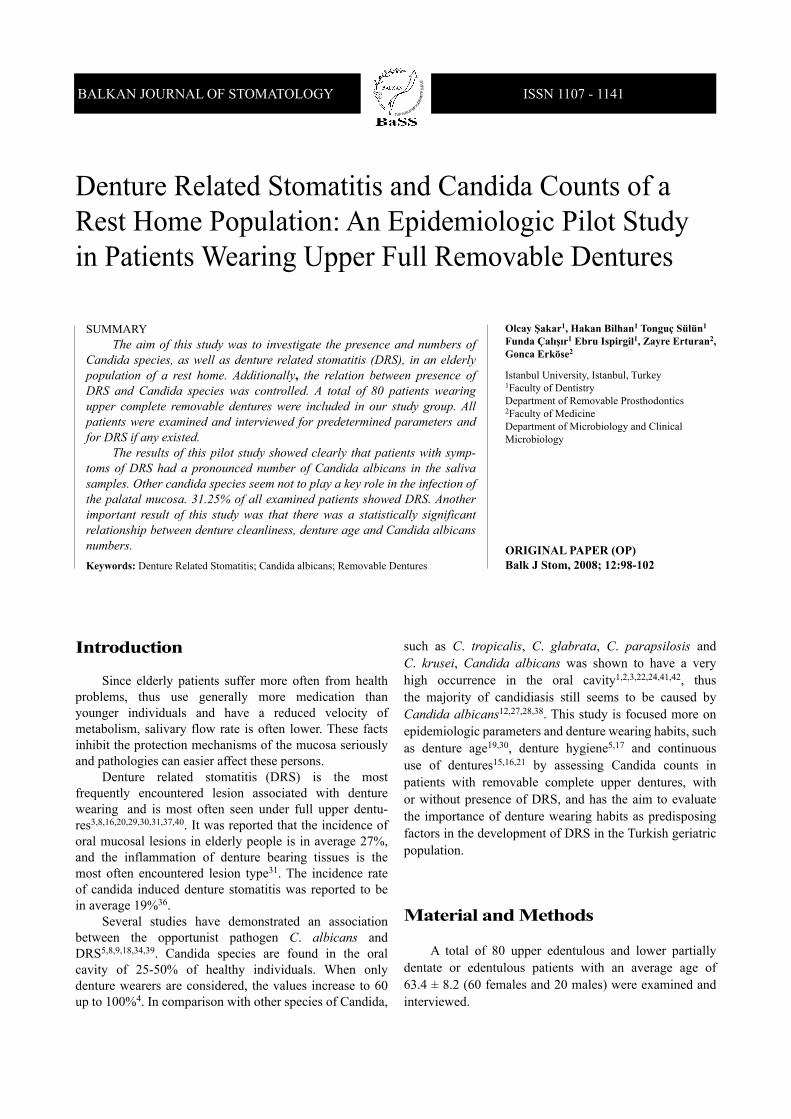
SUMMARYThe aim of this study was to investigate the presence and numbers of
Candida species, as well as denture related stomatitis (DRS), in an elderly population of a rest home. Additionally, the relation between presence of DRS and Candida species was controlled. A total of 80 patients wearing upper complete removable dentures were included in our study group. All patients were examined and interviewed for predetermined parameters and for DRS if any existed.
The results of this pilot study showed clearly that patients with symp-toms of DRS had a pronounced number of Candida albicans in the saliva samples. Other candida species seem not to play a key role in the infection of the palatal mucosa. 31.25% of all examined patients showed DRS. Another important result of this study was that there was a statistically significant relationship between denture cleanliness, denture age and Candida albicans numbers.Keywords: Denture Related Stomatitis; Candida albicans; Removable Dentures
Olcay Şakar1, Hakan Bilhan1 Tonguç Sülün1 Funda Çalışır1 Ebru Ispirgil1, Zayre Erturan2, Gonca Erköse2
Istanbul University, Istanbul, Turkey1Faculty of DentistryDepartment of Removable Prosthodontics2Faculty of MedicineDepartment of Microbiology and Clinical Microbiology
ORIGINAL PAPER (OP)Balk J Stom, 2008; 12:98-102
BALKAN JOURNAL OF STOMATOLOGY ISSN 1107 - 1141
Denture Related Stomatitis and Candida Counts of aRest Home Population: An Epidemiologic Pilot Study in Patients Wearing Upper Full Removable Dentures
TUPNBUPMPHJDB
M!!T
PD
JFU
Z
Introduction
Since elderly patients suffer more often from health problems, thus use generally more medication than younger individuals and have a reduced velocity of metabolism, salivary flow rate is often lower. These facts inhibit the protection mechanisms of the mucosa seriously and pathologies can easier affect these persons.
Denture related stomatitis (DRS) is the most frequently encountered lesion associated with denture wearing and is most often seen under full upper dentu-res3,8,16,20,29,30,31,37,40. It was reported that the incidence of oral mucosal lesions in elderly people is in average 27%, and the inflammation of denture bearing tissues is the most often encountered lesion type31. The incidence rate of candida induced denture stomatitis was reported to be in average 19%36.
Several studies have demonstrated an association between the opportunist pathogen C. albicans and DRS5,8,9,18,34,39. Candida species are found in the oral cavity of 25-50% of healthy individuals. When only denture wearers are considered, the values increase to 60 up to 100%4. In comparison with other species of Candida,
such as C. tropicalis, C. glabrata, C. parapsilosis and C. krusei, Candida albicans was shown to have a very high occurrence in the oral cavity1,2,3,22,24,41,42, thus the majority of candidiasis still seems to be caused by Candida albicans12,27,28,38. This study is focused more on epidemiologic parameters and denture wearing habits, such as denture age19,30, denture hygiene5,17 and continuous use of dentures15,16,21 by assessing Candida counts in patients with removable complete upper dentures, with or without presence of DRS, and has the aim to evaluate the importance of denture wearing habits as predisposing factors in the development of DRS in the Turkish geriatric population.
Material and Methods
A total of 80 upper edentulous and lower partially dentate or edentulous patients with an average age of 63.4 ± 8.2 (60 females and 20 males) were examined and interviewed.

The patients’ (1) age, (2) gender, (3) existence of a den-ture actually being used, (4) denture age, (5) frequency and (6) method of denture cleaning and (7) dental history were recorded. Clinical examination performed the same investi-gator for standardization reasons. The type of dentures, presence and localization of denture induced lesions, such as stomatitis, inflammatory papillary hyperplasia, and the cleanliness of the dentures was noted. Only individuals who were not undergoing antibiotic or antifungal therapy for at least 6 months before sampling were selected for this study.
In the case of DRS, the erythema was scored by using Newton’s classification index32:(1) Slight inflammation (localized slight hyperaemia);(2) Moderate inflammation (generalized erythema); (3) Severe inflammation (diffuse and papillary hyperplasia).
A subjective denture hygiene index6 was used to score the plaque at the intaglio surface in 3 groups. (1) Excellent - no or very little plaque;(2) Fair - less than half of the denture base covered by
plaque;(3) Poor - more than half of the denture base covered by
plaque.In all cases, a mycological test for Candida species
was made. In order to provide standardization for collected samples, the overall investigation was carried out at midmorning and at least 2 hours after eating, drinking or any hygiene procedure. Smear samples from a triangular area of the palate and saliva samples were taken from the patients. 1ml of saliva as well as the smear samples were cultured in Chromagar medium, incubated at 37°C for 48 hours, and subsequently the count of Candida colony-forming units of all different morphologies was recorded. For identification of the yeasts, the morphologies were examined in cornmeal agar with Tween 80 and the carbohydrate assimilation was investigated with API ID 32 C (Biomerieux® - France). For differentiation of Candida albicans and C. dubliniensis, the colony morphology and chlamydospore development in Staib agar, chlamydospore development in Casein agar and the capability to grow in 45°C was investigated.
The growth of yeasts from saliva samples were determined as colony forming units (cfu). The growth of smear samples was determined as follows:- In case of growth in the first region - low;- In case of growth in the first and second region - dense;- In case of growth in all three regions - very dense.
The relationship between DRS, denture age and patient age was analyzed by using Whitney-Mann U-test. The relationship between other denture related factors and DRS was investigated by the use of Chi-square test.
Results
60 isolates (75%) of Candida species were obtained from the oral cavity of the elderly subjects. Distribution
of the different Candida species were shown in table 1. 32.25% of all examined patients had symptoms of DRS.
Table 1. Distribution of Candida Species
Candida Types n - %C. albicans 50 (62.5)C. glabrata 20 (25)C. tropicalis 9 (11.25)C. parapsilosis 9 (11.25)C. crusei 2 (2.5)C. crevisiae 1 (1.25)C. guilliermondii 1 (1.25)C. dubliniensis 1 (1.25)C. lusitaniae 1 (1.25)C. kefyr 1 (1.25)
Smear samples: The relationship between DRS symptoms, such as palatal erythema and Candida albicans growth, was not statistically significant. Additionally, other Candida species also had no effect on the DRS.
Candidal growth showed clear differences within the 3 stomatitis types, although there was no statistical significance, thus patients with any type of inflammation regardless of the Newton classification, were considered as DRS cases.
Saliva samples: The numbers of Candida albicans in saliva samples had a statistically significant effect on the clinical symptoms (Tabs. 2 and 3). Other candida species had no effect on the DRS.
Table 2. The relationship between C. albicans count and Denture Related Stomatitis
Candida Albicans Countn mean SD
Denture Related Stomatitis
+ 25 820,2 1290,4- 55 434,6 996,4
Table 3. The relationship between C.albicans and Denture Related Stomatitis
Denture Related Stomatitis
+ -
Candida albicans
+n 21 30% 84 53,7
-n 4 25% 16 46,3
Totaln 25 55% 100 100
There was also a statistically significant association between Candida albicans numbers and denture cleanliness (Tab. 4). The relation between denture age and DRS was found to be statistically very significant (p = 0.008). There was no statistical relation between patient gender, denture
Balk J Stom, Vol 12, 2008 Candida Counts in Full Removable Denture Wearers 99

100 O. Şakar et al. Balk J Stom, Vol 12, 2008
cleaning methods, denture cleaning frequency, continous denture wearing and frequency of denture cleaning and DRS.
Table 4. The relationship between denture cleanliness and C.albicans count
Candida Albicans Countn mean SD
Denture CleanlinessExcellent 15 34,2 123,5Fair 32 234,2 801,2Poor 33 1074,4 1345,0
Discussion
The patients in this study were drawn from a population of people living in a rest home. Therefore, this sample group may not be representative of the population wearing removable dentures on a whole, but is a cross-section.
Budtz-Jorgensen and Bertram5 suggested that the denture plaque on tissue surfaces of dentures must have an irritating effect on mucosa. Catalan et al11 have reported that denture plaque in patients with DRS mostly show a considerable thickness. The toxic effects of plaque masses in contact with oral mucosa, for extended period of time, are predictable and similar as in the periodontal patient. Candida albicans and other related species are the most common type of bacteria found in oral candidal infections43. A significant number of DRS cases were encountered especially among patients wearing maxillary complete dentures: the palate is the most frequently affected region and is more susceptible to yeast colonization. This finding was usually reported in similar investigative studies40,43, which is also confirmed by our results. This fact can be explained by the greater area which is covered by the denture base and thus prevented from contacting saliva and being subject to anaerobic conditions37.
Denture plaque is mainly composed of Candida albicans, which is the main cause of DRS9-11,13,19,35. The importance of Candida species, especially Candida albicans, in provoking DRS was reported by several other investigators9,10,13,19,24,35,41. In comparison with other species of Candida, such as C. tropicalis, C. glabrata, C. parapsilosis and C. krusei, Candida albicans was shown to have a very high occurrence in the oral cavity1-3,22,24,41,42, thus the majority of candidiasis still seems to be caused by Candida albicans12,27,28,38. These findings were confirmed by our results too, but no statistically significant differences in yeast colonization between the various stages of DRS could be shown; however, 75% of all patients had at least one kind of Candida species in their samples. The comparison of our findings with other studies about the distribution of Candida species showed generally similar
numbers and percentages. The only 2 differences were that the percentage of C. tropicalis in our study group was lower than other reports, and in contrast, the C. glabrata percentage was much higher. This could indicate to a difference for the Turkish geriatric patient as well as being a coincidence due to the low number of cases14,33.
The denture cleanliness, according to numerous stu-dies5,10,17,21,23,24,26, is an important factor in the development of DRS. Similarly, in Kulak and Arikan’s study23 the results showed a significant association between DRS, denture hygiene and candidal colonization. Our results indicated a significant association between DRS and denture cleanliness, and a tendencial relation to Candida albicans growth, too.
Some studies have shown that denture hygiene habits (frequency and method) are important factors in the development of DRS21; however, in accordance to our findings, others found no relationship23,24,31. The reason for these controversial findings could be the fact that many patients are not properly informed about the brushing and cleaning methods and the frequency, and believe that their habits are adequate.
According to the results of many studies showing a high incidence of yeasts in the saliva or on palatal tissue of DRS patients in comparison to the control groups, it has been speculated that the presence of yeasts is an important factor in the development of the disease. However, many subjects with intraoral yeast presence can from time to time be free of symptoms of DRS. On the other hand, some cases with significant symptoms of DRS can have a relatively low count of yeasts. There must be other factors, besides the presence of yeasts, that are important in the development of the disease. The filamentous growth in the hyphae form of Candida albicans has been reported to enhance the adherence to the tissues and consequently contributing to the virulence of this pathogen25,38. It can be speculated due to our low yeast counts from the palatal smears, that the hyphae form is resistant to smear sampling due to the strong adherence, thus in spite of heavy inflammation, no yeast colonization can be detected. Understanding of the potential role of hyphae in the pathogenicity of Candida albicans would be of significant benefit in view of the increasing incidence of candidiasis7.
In the light of these findings, for the future of this project it is planned to increase the number of patients and to take epithelial samples of a permitting group to ascertain the role of hyphae in the development of DRS.
Conclusions
The results of this pilot study showed clearly that:1) The relationship between DRS symptoms, such as
palatal erythema, and Candida albicans growth was not statistically significant in palatal smear samples;

Balk J Stom, Vol 12, 2008 Candida Counts in Full Removable Denture Wearers 101
2) Patients with symptoms of DRS had a pronounced number of Candida albicans in the saliva;
3) Other Candida species seem not to play a key role in the infection of the palatal mucosa;
4) Poor denture hygiene seems to increase Candida albicans numbers;
5) Denture age influences DRS development, too.
References
Arendorf TM, Walker DM1. . Denture stomatitis: a review. J Oral Rehabil, 1987; 14:217-227.Arendorf TM, Walker DM2. . The prevalence and intra-oral distribution of Candida albicans in man. Arch Oral Biol, 1980; 25:1-10.Barbeau J, Seguin J, Goulet JP, de Koninck L, Avon SL, 3. Lalonde B, Rompre P, Deslauriers N. Reassessing the presence of Candida albicans in denture-related stomatitis. Oral Surg Oral Med Oral Pathol Oral Radiol Endod, 2003; 95:51-59.Berdicevsky I, Aryeh H, Szargel R, Gutman D4. . Oral Candida of asymptomatic denture wearers. Int J Oral Surg, 1980; 9:113-115.Budtz-Jorgensen E, Bertram U5. . Denture stomatitis. I. The etiology in relation to trauma and infection. Acta Odontol Scand, 1970; 28:71-92. Budtz-Jorgensen E, Bertram U6. . Denture stomatitis II. The effect of antifungal and prosthetic treatment. Acta Odontol Scand, 1970; 28:283-304. Budtz-Jorgensen 7. E, Stenderup A, Grabowski M. An epidemiologic study of yeasts in elderly denture wearers. Community Dent Oral Epidemiol, 1975; 3(3):115-119.Budtz-Jorgensen E8. . Oral mucosal lesions associated with the wearing of removable dentures. J Oral Pathol, 1981; 10:65-80.Budtz-Jorgensen E9. . The significance of Candida albicans in denture stomatitis. Scand J Dent Res, 1974; 82:151-190.Cardash HS, Helft M, Shani A, Marshak B10. . Prevalence of Candida albicans in denture wearers in an Israeli geriatric hospital. Gerodontology, 1989; 8:101-107.Catalan A, Herrera R, Martinez A11. . Denture plaque and palatal mucosa in denture stomatitis: scanning electron microscopic and microbiologic study. J Prosthet Dent, 1987; 57:581-586.Cross LJ, Williams DW, Sweeney CP, Jackson MS, Lewis 12. MAO, Bagg J. Evaluation of the recurrence of denture stomatitis and Candida colonization in a small group of patients who received itraconazole. Oral Surg Oral Med Oral Pathol Oral Radiol Endod, 2004; 97:351-358.Davenport JC13. . The oral distribution of candida in denture stomatitis. Br Dent J, 1970; 129:151-156. De Resende MA, de Sousa LV, de Oliveira RC, Koga-Ito 14. CY, Lyon JP. Prevalence and antifungal susceptibility of yeasts obtained from the oral cavity of elderly individuals. Mycopathologia, 2006; 162(1):39-44.Fenlon MR, Sherriff M, Walter JD15. . Factors associated with the presence of denture related stomatitis in complete denture wearers: a preliminary investigation. Eur J Prosthodont Restor Dent, 1998; 6:145-147.
Hand JS, Whitehill JM16. . The prevalence of oral mucosal lesions in an elderly population. J Am Dent Assoc, 1986; 112:73-76.Hoad-Reddick G, Grant AA, Griffiths CS17. . Investigation into the cleanliness of dentures in an elderly population. J Prosthet Dent, 1990; 64:48-52.Hoad-Reddick G18. . Oral pathology and prostheses - are they related? Investigations in an elderly population. J Oral Rehabil, 1989; 16:75-87.Holbrook WP, Rodgers GD19. . Candidal infections: experience in a British dental hospital. Oral Surg Oral Med Oral Pathol, 1980; 49:122-125.Jainkittivong A, Aneksuk V, Langlais RP20. . Oral mucosal conditions in elderly dental patients. Oral Dis, 2002; 8:218-223.Jeganathan S, Payne JA, Thean HP21. . Denture stomatitis in an elderly edentulous Asian population. J Oral Rehabil, 1997; 24:468-472.Koneman EW, Allen SD, Janda WM, Schreckenberger PC, 22. Winn WC Jr. Textbook and atlas of diagnostic microbiology. 5th ed. Philadelphia: JB Lippincott; 1997; pp 1040-5106.Kulak Y, Arikan A23. . Aetiology of denture stomatitis. J Marmara Univ Dent Fac, 1993; 1:307-314.Kulak-Ozkan Y, Kazazoglu E, Arikan A24. . Oral hygiene habits, denture cleanliness, presence of yeasts and stomatitis in elderly people. J Oral Rehabil, 2002; 29:300-304.Leberer E, Ziegelbauer K, Schmidt A, Harcus D, Dignard 25. D, Ash J, Johnson L, Thomas D. Virulence and hyphal formation of Candida albicans require the Ste20p-like protein kinase CaCla4p. Curr Biol, 1997; 7:539-546.MacEntee MI, Glick N, Stolar E26. . Age, gen der, dentures and oral mucosal disorders. Oral Dis, 1998; 4:32-36.Meiller TF, Jabra-Rizk MA, Baqui A, Kelley JI, Meeks 27. VI, Merz WG, Falkler WA. Oral Candida dubliniensis as a clinically important species in HIV-seropositive patients in the United States. Oral Surg Oral Med Oral Pathol Oral Radiol Endod, 1999; 88:573-580.Merz WG28. . Candida albicans strain delineation. Clin Microbiol Rev, 1990; 3:321-334.Mikkonen M, Nyyssonen V, Paunio I, Rajala M29. . Prevalence of oral mucosal lesions associated with wearing removable dentures in Finnish adults. Community Dent Oral Epidemiol, 1984; 12:191-194.Moskona D, Kaplan I30. . Oral lesions in elderly denture wearers. Clin Prev Dent, 1992; 14:11-14.Nevalainen MJ, Narhi TO, Ainamo A31. . Oral mucosal lesions and oral hygiene habits in the home-living elderly. J Oral Rehabil, 1997; 24:332-337.Newton AV32. . Denture sore mouth. Br Dent J, 1962; 112:357-360.Pires FR, Santos EB, Bonan PR, De Almeida OP, Lopes 33. MA. Denture stomatitis and salivary Candida in Brazilian edentulous patients. J Oral Rehabil, 2002; 29:1115-1119.Radford DR, Challacombe SJ, Walter JD34. . Denture plaque and adherence of Candida albicans to denture-base materials in vivo and in vitro. Crit Rev Oral Biol Med, 1999; 10:99-116.Renner RP, Lee M, Andors L, McNamara TF, Brook S35. . The role of C. albicans in denture stomatitis. Oral Surg Oral Med Oral Pathol, 1979; 47:323-328.Samaranayake LP, Wilkieson CA, Lamey PJ, MacFarlane 36. TW. Oral disease in the elderly in long-term hospital care. Oral Dis, 1995; 1(3):147-155.

102 O. Şakar et al. Balk J Stom, Vol 12, 2008
Sherman RG, Prusinski L, Ravenel MC, Joralmon RA37. . Oral candidosis. Quintessence Int, 2002; 33:521-532.Sudbery P, Gow N, Berman J38. . The distinct morphogenic states of Candida albicans. Trends Microbiol, 2004; 12:317-324.Theilade E, Budtz-Jorgensen E39. . Predominant cultivable micro-flora of plaque on removable dentures in patients with denture-induced stomatitis. Oral Microbiol Immunol, 1988; 3:8-13.Vigild M40. . Oral mucosal lesions among institutionalized elderly in Denmark. Community Dent Oral Epidemiol, 1987; 15:309-313.Webb BC, Thomas CJ, Willcox MD, Harty DW, Knox KW41. . Candida-associated denture stomatitis. Aetiology and management: a review. Part 2. Oral diseases caused by Candida species. Aust Dent J, 1998; 43:160-166.
Wingard JR42. . Importance of Candida species other than C albicans as pathogens in oncology patients. Clin Infect Dis, 1995; 20:115-125.Zegarelli DJ43. . Fungal infections of the oral cavity. Otolaryngol Clin North Am, 1993; 26:1069-1089.
Correspondence and request for offprints to:
Olcay Şakarİstanbul Üniversitesi, Dişhekimliği Fakültesi Protetik Diş Tedavisi A.B.D.Total Parsiyel Protezler B.D. Kat 2 PK: 34093Çapa İstanbul TürkiyeE-mail: [email protected]

SUMMARYWhile taking impressions from the displaceable mucosa, the tissue dis-
location should be minimal. Our aim was to control the tissue dislocation from flabby ridges during taking impressions with a modified method of “free impression”. 14 edentulous patients with displaceable alveolar ridges were treated using this modified method of functional impression and mild pressure on the tissue. The method is called “free impression”. Custom tray perfo-rated in the soft tissue region was constructed. To keep the impression mate-rial not to leak through the perforation, gauze was used. After completing of the border moulding procedures and taking selective - compressive impres-sion, as usual, we completed the procedure with soft tissue impression. The impression material was applied between the gauze and the oral mucous with a syringe. Control of tissue dislocation was measured with the distance of 3 points marked on the soft tissue, which were transferred from the impression to the plaster model. The distances were measured with accuracy of 0.02.
There were not significant differences between the values of the distan-ces measured in the mouth and on the models (p<0.0002).This results encourage us to prefer the method of “free impression”, as a method with minimal tissue dislocation.Keywords: Denture, complete; Functional Impressions; Displaceable Mucosa, Flabby Ridges
K. Dejanoski, A. Angelovska, S. Pancevska, L. Popovska, N. Janeva
Dental Clinical Centre “St Pantelejmon”Faculty of DentistrySkopje, FYROM
ORIGINAL PAPER (OP)Balk J Stom, 2008; 12:103-106
BALKAN JOURNAL OF STOMATOLOGY ISSN 1107 - 1141
Modification of Impressions to PreventSupporting Tissues Overloading
TUPNBUPMPHJDB
M!!T
PD
JFU
Z
Introduction
Interaction between the complete denture and the other structures from the stomathognatic system should be well balanced from functional aspect. This should be well composed, harmonious, functional unit, which on a standard level should effectively satisfy the individual needs of the patient.
The functional impression is a part of the designing of the complete denture5,7,9. With this impression, we define the interaction of the denture with the oral mucosa, such as its dimension, pressure distribution and border moulding, as important factors for retention and stability of the complete denture. When there is severe mobility of the denture supporting tissues, the impression methods with controlled selective pressure should be preferred. With these methods the pressure is the most similar to the physiological one, the pressure distribution is adequate to the oral base quality, the subjective influence of the therapist is avoided and the displacement of the tissue is minimal6.
Our aim was to control tissue dislocation, experimentally and clinically, during taking impressions from the displaceable oral mucosa using a modified method called as “free impression method”.
Material and Method
The control of the tissue dislocation during the usage of the modified method of “free impression” was performed by the comparison of marked distances in the mouth and on the models. We were able to control tissue dislocation in horizontal plain only. We marked by using an indelible pencil 3 points before taking impression, 1 in the area of the rigid mucosa and 2 on the movable area. These distances were constant, so that we could compare them. A model from Plexiglas was constructed. These points were transferred, over to the impression, on the model. The distances between the points on the pattern

104 K. Dejanoski et al. Balk J Stom, Vol 12, 2008
were determined with precision of 0.02 mm. The casts were made of plaster with expansion of 0.1%.
A modified method known as “free impression method” requires primary impression and production of special tray fenestrated in the region of the movable tissue. While taking an impression from the upper jaw, we put gauze on the fenestrated area, which was supposed to hold the impression material in close contact with the oral mucosa and to avoid deformation of the impression material.
After completing of the border moulding procedure and taking selective compressive impression of the unchanged oral tissues, as usual, we completed the procedure with soft tissue impression (Fig. 1). We took the impression out of the mouth and cut the excess of the impression material from the edges of the perforation (Fig. 2). A peace of gauze was stuck to the tray above the perforated area. Then the impression was returned in the mouth (Fig. 3). In addition, the impression material was put with a syringe between the gauze and the oral mucosa (Fig. 4). The quantity of the material should be adequate to the surface that would be copied, so as not to make pressure between the gauze and the mucosa, and to have sufficient quantity that will enable the proscribed thickness of the manufacturer, usually 2-4 mm. Afterwards, by coping the movable area, we completed the impression13,16 (Fig. 5). We used the light body silicon materials (Xantopren LV -Heraus Kulzer).
In this impression technique the pressure comes only from the weight of the impression material itself. On the maxilla it is minor because of the gravity.
“Free impression” clinical method was used with 14 patients, complete denture users with displaceable mucosa and signs of mechanical irritation. The control was made on plaster models from 28 impressions, from 14 patients, from the maxilla, the second impression was taken after the rest of the tissue for at least 2 hours. Student’s T-test was used for comparison.
Figure 1. Border moulding procedure and functional impression from the unchanged mucosa
Figure 5. Completed functional impression
Figure 3. Preparation for taking impression from the flabby ridges
Figure 4. Application of the impression material for taking impression of the flabby parts
Figure 2. Functional impression from the unchanged mucosa
Balk J Stom, Vol 12, 2008 Prevention of Supporting Tissues Overloading 105
Results
The results of the measured distances did not show significant differences between the values in the mouth and on the model (p<0.05), and their differences were close to the expected volume changes of the plaster (Tab. 1).
Table 1. Comparison of values of the measured distances in mouth and on the working model
Distance AB in mouth AB on modelN 14 14M 15.01 15.00SD 0.01 0.02p 0.07
Clinical examinations showed effective results, objective and subjective, in all cases according to the oral mucosa condition.
Discussion
Changes of the oral mucosa may appear as undesired effects of some drugs, antagonists and blockers of the calcium which initiate collagen production, or drugs which interact and activate the fibroblasts. Mc Cord et al11,12 found gingival hyperplasia in approximately 65% of the complete denture users who were using Phenytoin. However, a special aspect should be given to changes of the denture supporting areas that appear as a result of irregular dentures which have been worn for a longer period, or which were not used or maintained properly. If these changes are of proliferative type, they are known as denture related hyperplasia. According to literature, changes like these can be found in 15-18% of denture wearers, mostly over 50 years of age; they appear more frequently in women, than in men11,12.
Permanent irritation or pressure over the oral mucosa initiate inflammatory changes. The oral mucosa is not functionally predetermined to receive and distribute the masticatory pressure over the bone base even though in some areas it can endure larger pressure. If such uneven forces are larger and exceed the physiological oral tissue tolerance, pathological changes in the sub-epithelial tissue occur. At the beginning, these changes are exudative, but they often turn into proliferative changes. This forces result in continuing resorption of the bone - oral mucosa loses its firm tissue and becomes displaceable. The epithelium may be healthy or with persistent lesions or ulcers in more severe mechanical irritations. Deterioration of the circulation, increased temperature under the dentures; incorporated infective, chemical or allergic factors may initiate even more serious manifestation.
From the prosthetic aspect, it is important that the changed mucosa and the denture may create reciprocal actions - the unstable denture due to its constant irritation leads to these changes, and they decrease the stabilization of the denture. The unburdening of certain areas by using lead foil, adding phosphate cement, free scrubbing from the denture should not be allowed, because those procedures are subjective, freely dosed and may have a negative effect of chamber.
The impression technique is one of the preventive ways to avoid the appearance of oral hyperplasia in the removable denture wearers. The other phases of the complete denture fabrication must be paid attention to. Prevention is very important and doctors from different specialties must be involved and consulted. The impressions from the areas with changed mucosa should be taken without compression, deformation and without displaceable tissue distortion. From that point of view, impressions with controlled and minor compression are preferred because they allow minimal deformation and dislocation of the changed tissue.
There are also other indications and techniques in the literature for the modified method of functional impressions1-4,8,10,14,15.
Conclusion
The method of “free impression”, when the procedure is followed, gives favourable results, which insure good relationship between the oral tissues and the denture.
References
A1-Hadi LA1. . A simplified technique for prosthetic treatment of microstomia in a patient with scleroderma: A case report. Quintessence Int, 1994; 25(8):531-533.Basker RM, Ogden AR, Ralph JP2. . Complete denture prescription - an audit of performance. Br Dent J, 1993; 174:278-284.Conroy B, Reitzik M3. . Prosthetic restoration in microstomia. J Prosthet Dent, 1971; 26:324-327.Devlin H, Cash AJ, Watts DC4. . Mechanical behaviour and structure of light-cured special tray materials. J Dent, 1995; 23:255-259. Fel ton DA, et al5. . Pre dic tab le im pres si on pro ce du res for comp le te den tu res. Dent Clin North Am, 1996; 40(1):39-51.Frohlich V6. . Ist die Incorporationeiner Prothese pathologisch-anatomisch moglich? Dtsch Zaharztl Z, 24(6):578-592.Grant AA, Heath RJ, McCord FJ7. . Complete Prosthodontics Problems, Diagnosis and Management. Wolfe, 1994.Lay LS, et al8. . Ma king the tre at ment try-in, al te red cast im pressi on and oc clu sal registration in one ap po in tment. J Pros thet Dent, 1996; 75(40):446-448. Lejoyeux J9. . Protese complete. 3rd ed. Paris: Malone, 1986.

106 K. Dejanoski et al. Balk J Stom, Vol 12, 2008
Lurbke JR10. . Sectional impression tray for patients with constricted opening. J Prosthet Dent, 1984; 52(1):135-137.Mc Cord JF, Tyson KW, Blair IS11. . A sectional complete denture for a patient with microstomia. J Prosthet Dent, 1989; 61:645-647.Mc Cord J, Sloan P, Hussey D12. . Phenytoin hyperplasia occurring under complete dentures. J Prosth Dent, 1992; 68:569-571.Phillps RW13. . Science of dental materials. Philadelphia-London-Toronto: WB Sounders Co. Suzuki Y, Abe M, Hosoi T, Kurtz K14. . Sectional collapsed denture for a partially edentulous patient with microstomia: A clinical report. J Prosthet Dent, 2000; 84:256-259.
Wahle JJ, Gardner LK, Fiebiger M15. . The mandibular swing-lock complete denture for patients with microstomia. J Prosthet Dent, 1992; 68:523-527.Williams JR, Craig RG16. . Physical properties of addition silicones as a function of composition. J Oral Rehabil, 1988; 15:639-650.
Correspondence and request for offprints to:
Dr. Aneta Angelovska Draga Stojanova St. 21 1000 Skopje, FYR MacedoniaE-mail: [email protected]
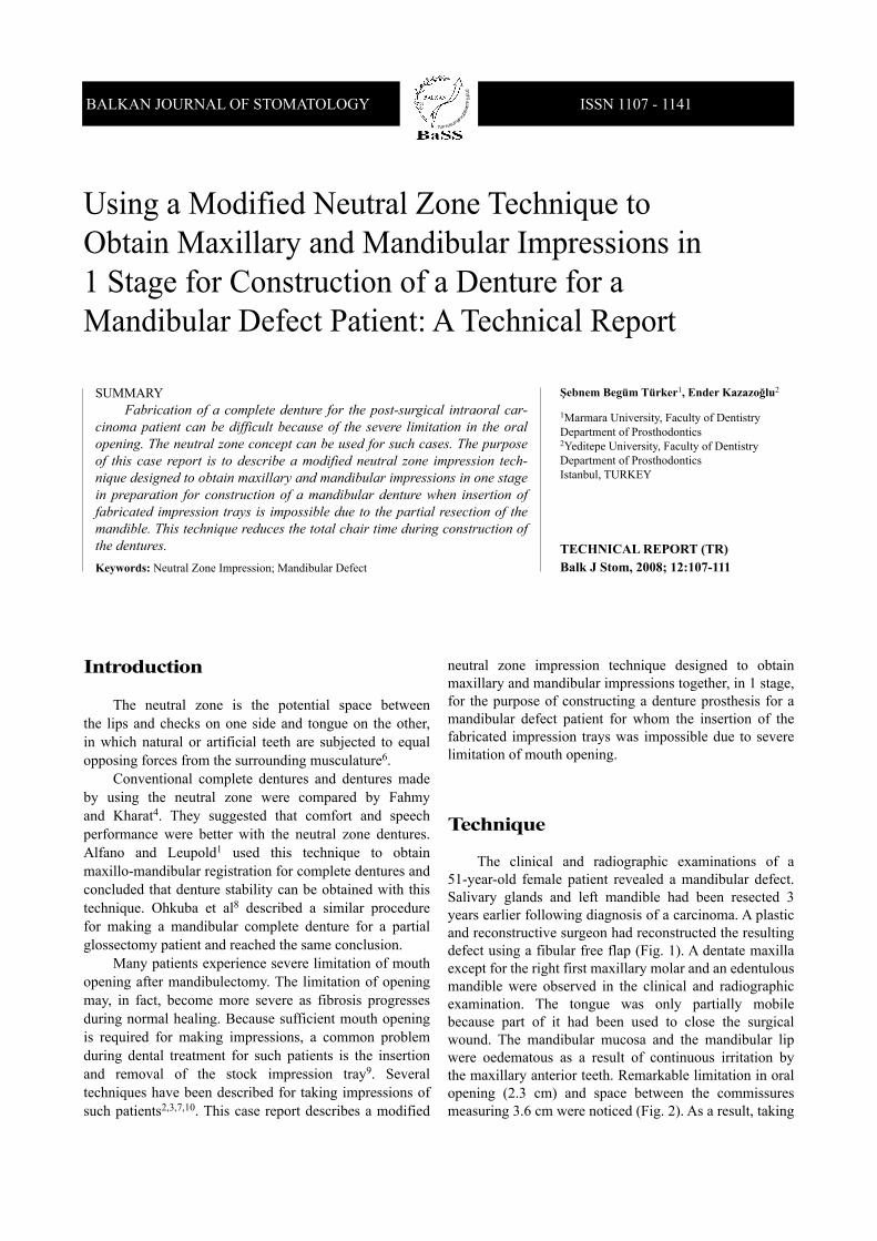
SUMMARYFabrication of a complete denture for the post-surgical intraoral car-
cinoma patient can be difficult because of the severe limitation in the oral opening. The neutral zone concept can be used for such cases. The purpose of this case report is to describe a modified neutral zone impression tech-nique designed to obtain maxillary and mandibular impressions in one stage in preparation for construction of a mandibular denture when insertion of fabricated impression trays is impossible due to the partial resection of the mandible. This technique reduces the total chair time during construction of the dentures. Keywords: Neutral Zone Impression; Mandibular Defect
Şebnem Begüm Türker1, Ender Kazazoğlu2
1Marmara University, Faculty of DentistryDepartment of Prosthodontics2Yeditepe University, Faculty of Dentistry Department of ProsthodonticsIstanbul, TURKEY
TECHNICAL REPORT (TR)Balk J Stom, 2008; 12:107-111
BALKAN JOURNAL OF STOMATOLOGY ISSN 1107 - 1141
Using a Modified Neutral Zone Technique to Obtain Maxillary and Mandibular Impressions in 1 Stage for Construction of a Denture for a Mandibular Defect Patient: A Technical Report
TUPNBUPMPHJDB
M!!T
PD
JFU
Z
Introduction
The neutral zone is the potential space between the lips and checks on one side and tongue on the other, in which natural or artificial teeth are subjected to equal opposing forces from the surrounding musculature6.
Conventional complete dentures and dentures made by using the neutral zone were compared by Fahmy and Kharat4. They suggested that comfort and speech performance were better with the neutral zone dentures. Alfano and Leupold1 used this technique to obtain maxillo-mandibular registration for complete dentures and concluded that denture stability can be obtained with this technique. Ohkuba et al8 described a similar procedure for making a mandibular complete denture for a partial glossectomy patient and reached the same conclusion.
Many patients experience severe limitation of mouth opening after mandibulectomy. The limitation of opening may, in fact, become more severe as fibrosis progresses during normal healing. Because sufficient mouth opening is required for making impressions, a common problem during dental treatment for such patients is the insertion and removal of the stock impression tray9. Several techniques have been described for taking impressions of such patients2,3,7,10. This case report describes a modified
neutral zone impression technique designed to obtain maxillary and mandibular impressions together, in 1 stage, for the purpose of constructing a denture prosthesis for a mandibular defect patient for whom the insertion of the fabricated impression trays was impossible due to severe limitation of mouth opening.
Technique
The clinical and radiographic examinations of a 51-year-old female patient revealed a mandibular defect. Salivary glands and left mandible had been resected 3 years earlier following diagnosis of a carcinoma. A plastic and reconstructive surgeon had reconstructed the resulting defect using a fibular free flap (Fig. 1). A dentate maxilla except for the right first maxillary molar and an edentulous mandible were observed in the clinical and radiographic examination. The tongue was only partially mobile because part of it had been used to close the surgical wound. The mandibular mucosa and the mandibular lip were oedematous as a result of continuous irritation by the maxillary anterior teeth. Remarkable limitation in oral opening (2.3 cm) and space between the commissures measuring 3.6 cm were noticed (Fig. 2). As a result, taking

108 Ş.B. Türker, E. Kazazoğlu Balk J Stom, Vol 12, 2008
impressions of the maxillary and mandibular arches using stock impression trays was impossible. Therefore, an altered neutral zone impression technique was planned in order to fabricate a mandibular denture.
mandibular alveolar ridge to support silicone impression material during construction of a neutral zone impression. A silicone impression material with medium viscosity (Speedex, Coltene, Whaledent Inc, New Jersey) was injected on this wire using a 5 cc dental injector while the patient was at the rest position. Then, the patient was instructed to make functional movements for several minutes as the silicon impression material was setting. Functional movements included: pronouncing words with “S” sounds, taking frequent sips of water and swallowing to activate the floor of the mouth, pursing the lips to activate the tongue, and whistling to contract the muscles around the lips. The forces exerted during functioning of the lips, cheeks, and tongue shaped the neutral zone. When the setting time for the first injection was completed, a second injection of the same material was placed onto the first set impression material and the patient was again instructed to make functional movements.
Figure 1. Extraoral view of the mandibular defect
Figure 2. Remarkable limited oral opening
Figure 3b. The wire supported silicone impression
Figure 3a. Intraoral view of the mandibular wire supported silicone impression model with the occlusal scheme of the maxillary arch
Impression ProcedureAn orthodontic 0.9 mm stainless steel wire was
prepared and adapted along the centre of the patient’s
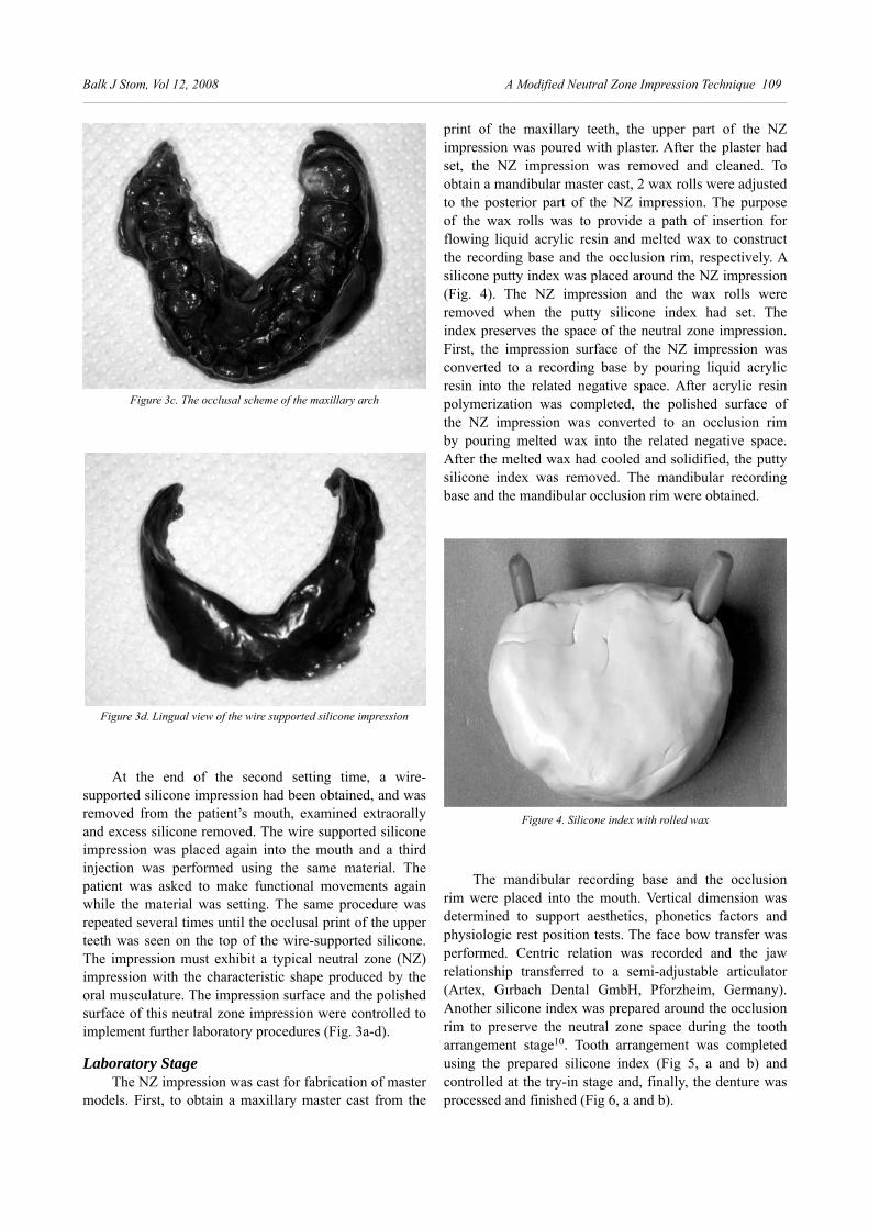
Balk J Stom, Vol 12, 2008 A Modified Neutral Zone Impression Technique 109
At the end of the second setting time, a wire-supported silicone impression had been obtained, and was removed from the patient’s mouth, examined extraorally and excess silicone removed. The wire supported silicone impression was placed again into the mouth and a third injection was performed using the same material. The patient was asked to make functional movements again while the material was setting. The same procedure was repeated several times until the occlusal print of the upper teeth was seen on the top of the wire-supported silicone. The impression must exhibit a typical neutral zone (NZ) impression with the characteristic shape produced by the oral musculature. The impression surface and the polished surface of this neutral zone impression were controlled to implement further laboratory procedures (Fig. 3a-d).
Laboratory StageThe NZ impression was cast for fabrication of master
models. First, to obtain a maxillary master cast from the
print of the maxillary teeth, the upper part of the NZ impression was poured with plaster. After the plaster had set, the NZ impression was removed and cleaned. To obtain a mandibular master cast, 2 wax rolls were adjusted to the posterior part of the NZ impression. The purpose of the wax rolls was to provide a path of insertion for flowing liquid acrylic resin and melted wax to construct the recording base and the occlusion rim, respectively. A silicone putty index was placed around the NZ impression (Fig. 4). The NZ impression and the wax rolls were removed when the putty silicone index had set. The index preserves the space of the neutral zone impression. First, the impression surface of the NZ impression was converted to a recording base by pouring liquid acrylic resin into the related negative space. After acrylic resin polymerization was completed, the polished surface of the NZ impression was converted to an occlusion rim by pouring melted wax into the related negative space. After the melted wax had cooled and solidified, the putty silicone index was removed. The mandibular recording base and the mandibular occlusion rim were obtained.
Figure 3c. The occlusal scheme of the maxillary arch
Figure 3d. Lingual view of the wire supported silicone impression
Figure 4. Silicone index with rolled wax
The mandibular recording base and the occlusion rim were placed into the mouth. Vertical dimension was determined to support aesthetics, phonetics factors and physiologic rest position tests. The face bow transfer was performed. Centric relation was recorded and the jaw relationship transferred to a semi-adjustable articulator (Artex, Gırbach Dental GmbH, Pforzheim, Germany). Another silicone index was prepared around the occlusion rim to preserve the neutral zone space during the tooth arrangement stage10. Tooth arrangement was completed using the prepared silicone index (Fig 5, a and b) and controlled at the try-in stage and, finally, the denture was processed and finished (Fig 6, a and b).

110 Ş.B. Türker, E. Kazazoğlu Balk J Stom, Vol 12, 2008
Discussion
The neutral zone denture functionally contours all the polishing surfaces of the denture and this may be an important contributing factor to better speech and comfort1. The most critical stage for the rehabilitation of the maxillofacial prosthodontic’ patient is the impression procedure. Because this patient’s oral topographies had been changed as a result of surgical procedures, taking impressions presents, sometimes, severe difficulties. The technique described here offers an alternative impression technique for the situation in which the conventional impression technique cannot be used.
In the case presented here, the patient’s tongue location had been changed to obtain primary closure of the surgical area. The patient’s mandibular opening was limited due to the surgical resection. In addition, cicatrized tissue was found near the site of the surgery. All of these factors had negative effects on the possibility of taking accurate impressions using conventional methods.
Using the neutral zone impression technique as described in this case report allowed the clinician to obtain maxillary and mandibular models, mandibular recording
base and occlusion rim at only one visit. At the second appointment, vertical dimension and centric relation were determined. The vertical dimension of occlusion was reduced to ensure sufficient inter-occlusal space for mastication and improved speech with the finished denture. The try-in stage was performed at the third appointment. Thus, the time required to construct the mandibular denture was decreased.
As the lower incisor teeth were arranged with silicone index, the continuous trauma caused by the maxillary incisors was prevented.
The technique presented allows the dentist to construct a more stable record base. Although the technique is relatively simple for the dentist who uses the neutral zone technique in general practice, it is more time consuming. Another disadvantage of the technique is the necessity for increased laboratory time. The laboratory technician must be trained to support all the laboratory procedures.
Conventionally, the tongue provides support for lower denture stability, but in the case presented here, because the patient’s tongue had been used to close the surgical area, lower denture stability was provided by the patient’s surrounding tissues including the lips, cheek and tongue.
Figure 5a. Maxillary master cast Figure 5b. Tooth arrangement
Figure 6a. Denture processed with Molloplast-B relining material Figure 6b. Intraoral view of the denture

Balk J Stom, Vol 12, 2008 A Modified Neutral Zone Impression Technique 111
Conclusion
The neutral zone approach can be easily used for limited oral opening cases. It offers an alternative technique for patients with partially resected mandible for whom the insertion of conventional impression trays is impossible.
References
Alfano SG, Leupold RJ.1. Using the neutral zone to obtain maxillomandibular relationship records for complete denture patients. J Prosthet Dent, 2001; 85:621-623.Cheng AC, Wee AG, Shiu-Yin C, Tat-Keung L.2. Prosthodontic management of limited oral access after ablative tumor surgery: a clinical report. J Prosthet Dent, 2000; 84:269-273.Dhanasomboon S, Kiatsiriroj K3. . Impression procedure for a progressive sclerosis patient: A clinical report. J Prosthet Dent, 2000; 83:279-286.Fahmy FM, Kharat DU.4. A study of the importance of the neutral zone in complete dentures. J Prosthet Dent, 1990; 64:459-462.
Gahan MJ, Walmsley AD. 5. The neutral zone impression revisited. Br Dent J, 2005; 198:269-272.Glossory of prosthodontic terms. 6. J Prosthet Dent, 1999; 81:86.McCord JF, Tyson K, Blair IS7. . A sectional complete denture for a patient with microstomia. J Prosthet Dent, 1989; 61:645-647.Ohkubo C, Hanatani S, Hosoi T, Mizuno Y. 8. Neutral zone approach for denture fabrication for a partial glossectomy patient: A clinical report. J Prosthet Dent, 2000; 84:390-393.Taylor TD. 9. Clinical Maxillofacial Prosthetics. Chicago, Berlin, London: Quint Pub Co, Inc, 2000; pp 155-170. Whitsitt JA, Battle LW. 10. Technique for making flexible impression trays for the microstomic patient. J Prosthet Dent, 1984; 52:608-609.
Correspondence and request for offprints to:
Dr. Ş. Begüm TürkerMarmara UniversitesiDiş Hekimligi Fakultesi34365 Nisantasi, IstanbulTurkey E-mail: [email protected]

SUMMARYRegenerative results of intrabony defect treatments are seriously com-
promised when the flap cannot be kept completely closed. To optimize the clinical outcomes of regenerative procedures, primary closure over treated area seems to be very important. The aim of the present study was to describe a surgical technique modification, which is appropriate to the anatomical features of the operation site and which allows for the primary closure of huge intrabony defects. For these purposes, we report 2 patients in whom previous biomaterial applications had failed and resulted with progressive alveolar bone loss and development of a huge cavity. Clinical parameters and radiographic bone fill were assessed at baseline and 12th months. The results of a specific flap design in relation to anatomical characteristics of gingival thickness, adequate graft and biologic agent combination for large intrabony defects are reported.Keywords: Platelet Rich Plasma; Partial Thickness Mucosal Flap; Infrabony Defects
Tunc Ilgenli1, Orhun Bengisu1, Nesrin Dundar2, Betul Ilhan Kal2
Ege University, Faculty of DentistryBornova, Izmir1Department of Periodontology2Department of Oral Diagnosis and Oral Radiology
CASE REPORT (CR)Balk J Stom, 2008; 12:112-117
BALKAN JOURNAL OF STOMATOLOGY ISSN 1107 - 1141
Platelet-Rich Plasma and Bone Graft Combined withPartial Thickness Mucosal Flap Technique in theTreatment of Deep Intrabony Defects
TUPNBUPMPHJDB
M!!T
PD
JFU
Z
Introduction
During the past decades, treatment modalities like grafting of biomaterials and application of biological agents have been used with varying success to accomplish the reconstruction of lost attachment apparatus in deep intra-osseous defects1. However, retention of the graft matrix can present a clinical challenge and graft containment would seem necessary for an optimal regenerative response to occur2,3. Autologous platelet rich plasma (PRP) is a novel method for obtaining autologous platelet growth factors (PGFs), especially for platelet derived growth factor (PDGF) and transforming growth factor ß (TGF ß)4. PRP use is a way to accelerate and enhance body’s natural wound-healing mechanisms4. The added benefit of PRP is its ability to form a biologic gel that may provide containment, clot stability and function as an adhesive5. An important decrease in healing time of bone grafts was shown when they are used in combination with PRP5. Numerous reports have shown that the use of PRP facilitates clinical handling of graft material6-8.
In periodontal diseases, the graft material and/or regenerative potential of biological agents are not
the only factors that have an influence on the variety in the management of intrabony defect fill. Clinically, several factors, including the patient selection, defect morphology, biological and physiochemical characteristic of grafted biomaterials/biomimetic substances, as well as surgical variables and postoperative maintenance, may influence the extent of clinical attachment gain and bone re-growth following a grafting procedure1-3. Periodontal reconstructive surgery for intra-osseous defects is a technically sensitive procedure9. Selection of a specific flap design in relation to anatomical characteristics of interdental space and location/morphology of bony lesion and proper suturing technique may significantly contribute in determining the amount of soft and hard tissue changes following surgery9. Preservation of the flap tissue is important for regenerative techniques to ensure coverage and containment of the graft post surgically3. The goal of flap management is to obtain tension-free primary closure over the entire graft or defect complex. While most clinicians agree that primary soft tissue closure, which is maintained throughout the course of regeneration, is ideal; opinions vary greatly concerning how best to accomplish this goal10,11.

The purposes of this article were to describe a surgical technique modification, which is appropriate to the anatomical features of the operation site and which allows for the primary closure of huge intrabony defects, and also to evaluate the obtained regenerative outcomes using this procedure. For these purposes, we report 2 patients in whom previous biomaterial applications had failed and resulted in a progressive alveolar bone loss leading to a huge cavity around the involved region.
Case 1
A 45-year-old female was referred to the Department of Periodontology at University of Ege, Faculty of Dentistry, for periodontal diagnosis and treatment. The patient complained about a swelling around the missing mandibular left canine area on which there was a 3 unit fixed adhesive restoration between mandibular left second incisor (32) and left first premolar (34). Clinical examination revealed an abscess formation under the pontic, which was deeply seated on the alveolar mucosa (Fig. 1). She stated that biomaterial application was performed in the region 4 years ago. Radiographic examination revealed a biomaterial application failure, which resulted with abscess formation causing to an increase in alveolar bone loss. A huge intrabony defect was observed involving the mesial aspect of mandibular left first premolar and the alveolar area under the pontic (Fig. 2). The clinical measurements for mandibular left first premolar including probing pocket depth (PPD), probing attachment level (PAL) and the position of gingival margin recession (REC) were obtained by a graded periodontal probe (CP-15UNC HU-FRIEDY). During initial therapy (root planning and scaling) individual acrylic occlusal stents were prepared for standardized intraoral radiographs (Ekta-speed, Eastman Kodak Co, Rochester, NY, USA). An individualized film holder consisting of a Rinn bite block (KKD, Ellwangen/Jagst, Germany), which was rigidly connected to the acrylic dental stent and X-ray tube was used (10 mA, 70 kVp, Trophy Radiologie, Vincennes, France). Before surgery, the pontic of prosthetic restoration was shortened to expose the incision line.
Surgical TechniqueThe main objective of the following flap design is
to allow passive advancement of both lingual and buccal flaps on the defect side. The surgical technique is outlined in drawing 1.
Figure 2. Intrabony defect involving the mesial aspect of premolar and the alveolar ridge under the pontic
Figure 1. Abscess formation around missing mandibular left canine
Drawing 1. (1) Intrasulcular incisions were performed lingually and extended interproximally; (2) The incision followed the mucogingival
line extending to 2 adjacent teeth on either side of the defect; (3) Horizontal releasing incisions were later continued by 2 vertical
releasing incisions which were connected horizontally through the vestibular sulcus
1. Starting lingually 1 or 2 teeth from each side of the defect, intrasulcular incisions were performed lingually and extended interproximally, which were later followed on the buccal aspect by oblique/vertical incisions terminating at mucogingival line;
2. From this point, the incision followed the mucogingival line extending to 2 adjacent teeth on either side of the defect;
3. These horizontal releasing incisions were later continued by 2 vertical releasing incisions that were connected horizontally through the vestibular sulcus. Starting from the most apical position, a partial-thickness mucosal flap was carefully elevated. This preparation ran apico-coronary over the defect and ended up at lingual alveolar crest. Here the blade was positioned slightly deeper, touching the bone, so that a full-thickness flap could be elevated on the lingual aspect of the mandible.
Balk J Stom, Vol 12, 2008 Treatment of Deep Intrabony Defects 113
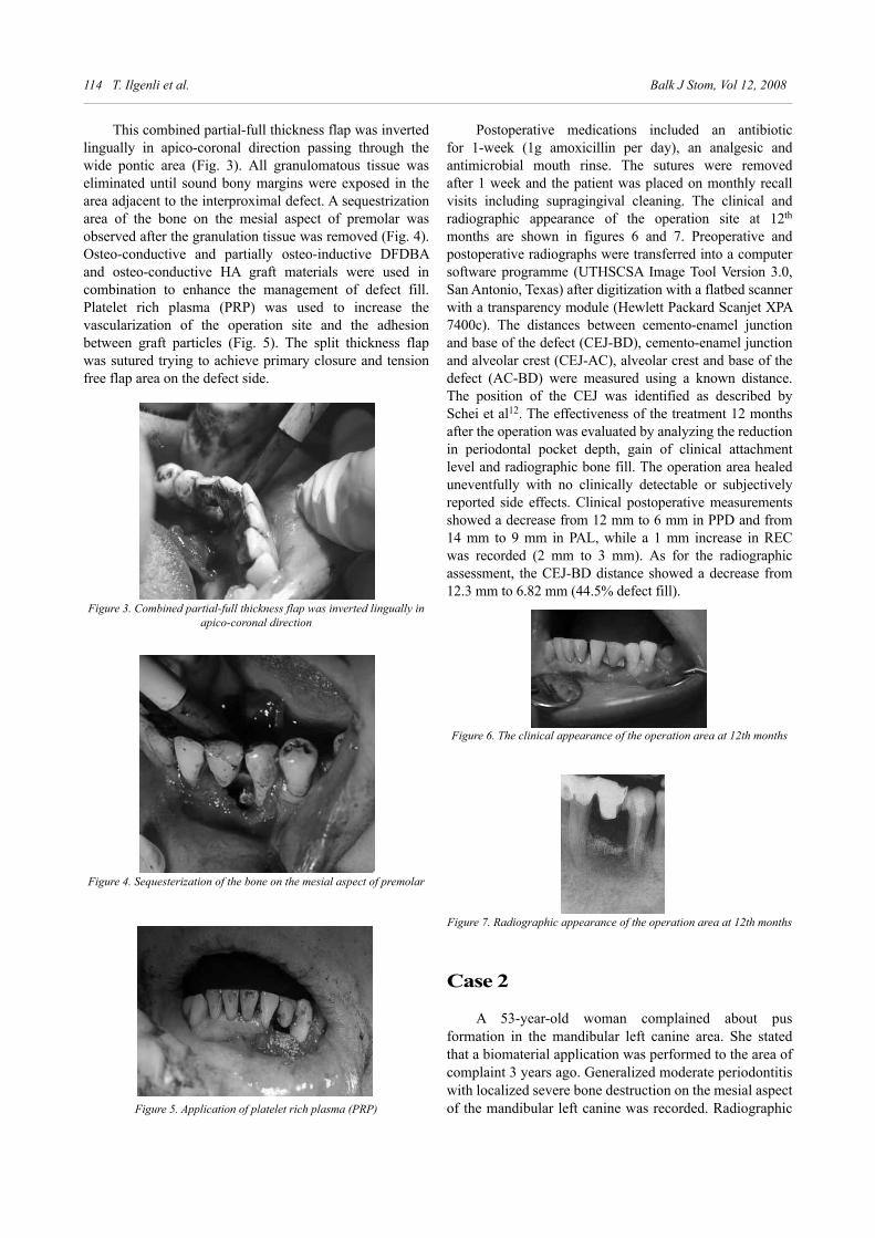
114 T. Ilgenli et al. Balk J Stom, Vol 12, 2008
This combined partial-full thickness flap was inverted lingually in apico-coronal direction passing through the wide pontic area (Fig. 3). All granulomatous tissue was eliminated until sound bony margins were exposed in the area adjacent to the interproximal defect. A sequestrization area of the bone on the mesial aspect of premolar was observed after the granulation tissue was removed (Fig. 4). Osteo-conductive and partially osteo-inductive DFDBA and osteo-conductive HA graft materials were used in combination to enhance the management of defect fill. Platelet rich plasma (PRP) was used to increase the vascularization of the operation site and the adhesion between graft particles (Fig. 5). The split thickness flap was sutured trying to achieve primary closure and tension free flap area on the defect side.
Postoperative medications included an antibiotic for 1-week (1g amoxicillin per day), an analgesic and antimicrobial mouth rinse. The sutures were removed after 1 week and the patient was placed on monthly recall visits including supragingival cleaning. The clinical and radiographic appearance of the operation site at 12th months are shown in figures 6 and 7. Preoperative and postoperative radiographs were transferred into a computer software programme (UTHSCSA Image Tool Version 3.0, San Antonio, Texas) after digitization with a flatbed scanner with a transparency module (Hewlett Packard Scanjet XPA 7400c). The distances between cemento-enamel junction and base of the defect (CEJ-BD), cemento-enamel junction and alveolar crest (CEJ-AC), alveolar crest and base of the defect (AC-BD) were measured using a known distance. The position of the CEJ was identified as described by Schei et al12. The effectiveness of the treatment 12 months after the operation was evaluated by analyzing the reduction in periodontal pocket depth, gain of clinical attachment level and radiographic bone fill. The operation area healed uneventfully with no clinically detectable or subjectively reported side effects. Clinical postoperative measurements showed a decrease from 12 mm to 6 mm in PPD and from 14 mm to 9 mm in PAL, while a 1 mm increase in REC was recorded (2 mm to 3 mm). As for the radiographic assessment, the CEJ-BD distance showed a decrease from 12.3 mm to 6.82 mm (44.5% defect fill).
Figure 3. Combined partial-full thickness flap was inverted lingually in apico-coronal direction
Figure 6. The clinical appearance of the operation area at 12th months
Figure 7. Radiographic appearance of the operation area at 12th months
Figure 4. Sequesterization of the bone on the mesial aspect of premolar
Figure 5. Application of platelet rich plasma (PRP)
Case 2
A 53-year-old woman complained about pus formation in the mandibular left canine area. She stated that a biomaterial application was performed to the area of complaint 3 years ago. Generalized moderate periodontitis with localized severe bone destruction on the mesial aspect of the mandibular left canine was recorded. Radiographic
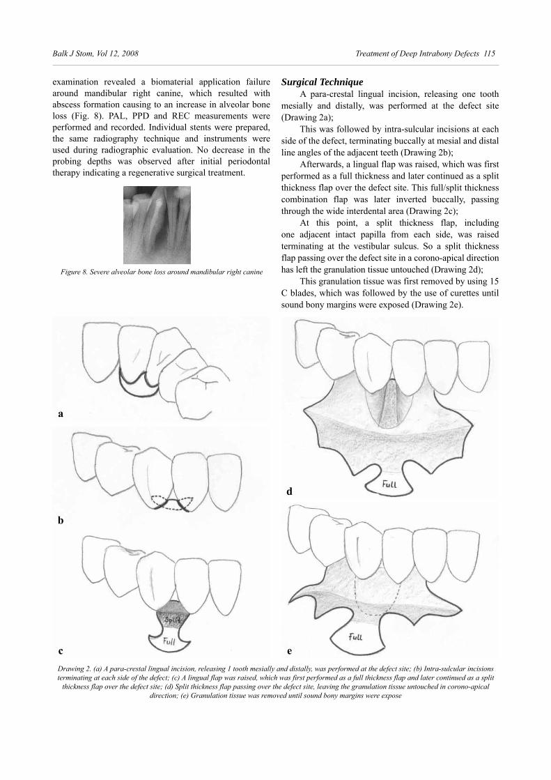
Balk J Stom, Vol 12, 2008 Treatment of Deep Intrabony Defects 115
examination revealed a biomaterial application failure around mandibular right canine, which resulted with abscess formation causing to an increase in alveolar bone loss (Fig. 8). PAL, PPD and REC measurements were performed and recorded. Individual stents were prepared, the same radiography technique and instruments were used during radiographic evaluation. No decrease in the probing depths was observed after initial periodontal therapy indicating a regenerative surgical treatment.
Surgical TechniqueA para-crestal lingual incision, releasing one tooth
mesially and distally, was performed at the defect site (Drawing 2a);
This was followed by intra-sulcular incisions at each side of the defect, terminating buccally at mesial and distal line angles of the adjacent teeth (Drawing 2b);
Afterwards, a lingual flap was raised, which was first performed as a full thickness and later continued as a split thickness flap over the defect site. This full/split thickness combination flap was later inverted buccally, passing through the wide interdental area (Drawing 2c);
At this point, a split thickness flap, including one adjacent intact papilla from each side, was raised terminating at the vestibular sulcus. So a split thickness flap passing over the defect site in a corono-apical direction has left the granulation tissue untouched (Drawing 2d);
This granulation tissue was first removed by using 15 C blades, which was followed by the use of curettes until sound bony margins were exposed (Drawing 2e).
Figure 8. Severe alveolar bone loss around mandibular right canine
Drawing 2. (a) A para-crestal lingual incision, releasing 1 tooth mesially and distally, was performed at the defect site; (b) Intra-sulcular incisions terminating at each side of the defect; (c) A lingual flap was raised, which was first performed as a full thickness flap and later continued as a split
thickness flap over the defect site; (d) Split thickness flap passing over the defect site, leaving the granulation tissue untouched in corono-apical direction; (e) Granulation tissue was removed until sound bony margins were expose
a
d
c e
b

116 T. Ilgenli et al. Balk J Stom, Vol 12, 2008
In this case, regarding the morphological potential of the intrabony defect, osteo-conductive and partially osteo-inductive DFDBA and PRP was used in combination to enhance the management of defect fill (Fig. 9). Tensile-free primary flap closure was performed.
Discussion
These data appear to emphasize the clinical importance of adapting surgical approach to the anatomy of the treated area, as well as to the physical/chemical characteristics of the regenerative material. Incision design critically influences the postoperative wound healing process in terms of blood supply and flap survival13. A surgical procedure especially designed to preserve the interdental tissues and to obtain primary closure of the flaps over the membranes in GTR was introduced by Cortellini et al14. In the present surgical study, the primary closure of the flaps in the interdental space was not obtained as described by Cortellini et al, i.e. by not releasing the flap coronary with a periosteal incision.
It is well known that healing and regeneration results are seriously compromised when the complete closure of the flap is not maintained15. This problem can be solved with primary flap coverage and adequate passive flap adaptation and vertical tensile force distribution during healing. Wang et al10 and Obarrio et al11 preferred split thickness flaps for primarily closure in order to provide the flap to be tension-free. Apart from these investigators who combined reflected full and partial thickness flaps during the operation, we reflected partial thickness flaps without involving the adjacent periosteum and only the wall components of the defect area were exposed after granulation tissue was removed. Blank et al16 stated the importance of an adequate zone of gingiva in the combined treatment of large defects, and emphasized that gingival recession and a lack of keratinized gingiva were present after healing in their cases. Positioning the flap coronary to achieve primary closure over the membrane and graft may have created these mucogingival problems. They performed a second surgical procedure, a sub-epithelial connective tissue graft, to treat the gingival recession and lack of attached gingiva. In our cases, the technique we applied avoided the coronal repositioning of the marginal gingiva and did not disturb the normal relationship of the existing surrounding tissues nor reduced the vestibular depth. Trombelli et al9 reported that selection of a specific flap design in relation to anatomical characteristics of interdental space and location/morphology of intrabony defect, and proper suturing technique, may contribute in limiting the apical shift of gingival margin.
Wound stabilization appears to be a critical factor for success in regeneration procedures17. Movement of only 10 to 20 μm during early stages of fracture healing is enough to divert the differentiation of mesenchymal cells into fibroblasts instead of osteoblasts17. This enables organization of coagulum and graft material during the early phases of wound healing and thereby maximum bone regeneration may be achieved. Confirming previously reports, our results also showed that PRP can serve both in haemostasis and adhesion of graft material, as well as contributing physiologically to provide rapid healing of
Figure 9. Application of combined osteoconductive and partially osteoinductive HA/DFDBA and platelet rich plasma (PRP)
Postoperative medications included a 1-week regi-men of antibiotic (1g amoxicillin BID) and antimicrobial mouth rinse. The sutures were removed 10 days after the sur gery and the patient was placed on monthly recall visits including supragingival cleaning. The second standardized radiographs were taken 12 months after the ope ration (Fig. 10). Previously described radiographic measurements were performed on preoperative and post-operative radiographs. The effectiveness of the treatment was evaluated by ana lyzing reduction of periodontal pocket depths, gain of clinical attachment level and radio-graphic bone fill. The operation area healed uneventfully. No clinically detectable or subjectively reported side effects were noted (Fig. 11). Clinically, PPD decreased from 10 mm to 3 mm, and PAL from 13 mm to 8 mm, while REC increased from 3 mm to 5 mm. Radiographic assessment showed an improvement of 13.83 mm to 6.07 mm (63.08% defect fill) in the CEJ-BD distance.
Figure 10. Radiographic appearance of the operation area at 12th months
Figure 11. The clinical appearance of the operation area at 12th months

Balk J Stom, Vol 12, 2008 Treatment of Deep Intrabony Defects 117
the surgical site6-8. PRP, while improving angiogenesis (new blood vessel formation), is a potent mitogen (stimulator of cell proliferation) and chemotactic (causes directed cell migration) protein for PDL fibroblasts and alveolar bone cells, while bone allograft offers a biological matrix conducive to cell growth and may contribute osteo-inductive bone matrix proteins. The present aim was to combine the mentioned properties of these 2 materials. As declared by Lindhe et al18, the clinician should be aware that graft material, intra-osseous defect morphology, technical factors and selection of patient may account for variability in clinical performance. Regenerative potential of the graft material is not the only factor to account for the variability of clinical results about the defect fill and the elimination of pocket depth.
In conclusion, selection of a specific flap design in relation to anatomical characteristics of the treated area is as important as the selection of a biomaterial and/or combination adequate for the morphology of intrabony defects. More research is needed to identify patient, site, choice of material and technique factors associated with successful outcomes of the treatment of intra-osseous defects.
References
Trombelli L, Heitz-Mayfield L, Needleman I, Moles 1. D, Scabbia A. A systematic review of graft materials and biological agents for intraosseous defects. J Clin Periodontol, 2002; 29(Suppl 3):117-135.Kornman KS, Robertson PB2. . Fundamental principles affecting the outcomes of therapy for osseous lesions. Periodontol 2000, 2000; 22:22-43.Rosen PS, Reynolds MA, Bowers GM3. . The treatment of intrabony defects with bone grafts. Periodontol 2000, 2000; 22:88-103.Carlson NE, Roach RB4. . Platelet rich plasma: Clinical appli-cations in dentistry. J Am Dent Assoc, 2002; 133:1383-1386.Kassolis JD, Rosen PS, Reynolds MA5. . Alveolar ridge and sinus augmentation utilizing platelet rich plasma in combination with freeze-dried bone allograft: Case series. J Periodontol, 2000; 71:1654-1661.Whitman DH, Berry RL6. . A technique for improving the handling of particulate cancellous bone and marrow grafts using platelet gel. J Oral Maxillofac Surg, 1998; 56:1217-1218.
Marx RE, Carlson ER, Eichstaedt RM, Schimmele SR, 7. Strauss JE, Georgeff KR. Platelet-rich plasma: Growth factor enhancement for bone grafts. Oral Surg Oral Med Oral Pathol Oral Radiol Endod, 1998; 85:638-646.Whitman DH, Berry RL, Green DM8. . Platelet Gel: An autologous alternative to fibrin glue with applications in oral and maxillofacial surgery. J Oral Maxillofacial Surg, 1997; 55:1294-1299.Trombelli L, Bottega S, Zuchelli G9. . Supracrestal soft tissue preservation with enamel matrix proteins in treatment of deep intrabony defects. A report of 35 consecutively treated cases. J Clin Periodontol, 2002; 29:433-439.Wang HL, Kimble K, Eber R10. . Use of bone grafts for the enhancement of a GTR-Based root coverage procedure: A pilot case study. Int J Periodontics Restorative Dent, 2002; 22:118-127.Obarrio JJ. Dutari JA, Chamberlain TM, Croston A11. . The use of autologous growth factors in periodontal surgical therapy: Platelet gel bio-technology - Case reports. Int J Periodontics Restorative Dent, 2000; 20:487-497.Schei O, Waerhaug J, Lovdal A, Arno A12. . Alveolar bone loss as related to oral hygiene and age. J Periodontol, 1959; 30:7-16.Mörmann W, Ciancio SG13. . Blood supply of human gingiva following periodontal surgery. A fluorescein angiographic study. J Periodontol, 1977; 48:681-692.Cortellini P, Pini Prato G, Tonetti M14. . The modified papilla preservation technique. A new surgical approach for interproximal regenerative procedures. J Periodontol, 1995; 66:261-266.Reynolds MA, Bowers GM15. . Fate of demineralized freeze-dried bone allografts in human intrabony defects. J Periodontol, 1996; 67:150-157.Blank B, Levy AR16. . Combined treatment of a large periodontal defect using GTR and DFDBA. Int J Periodontics Restorative Dent, 1999; 19:481-487.Wikesjo U, Nilveus R17. . Periodontal repair in dogs: effect of wound stabilization on healing. J Periodontol, 1990; 61:719-724.Lindhe J, Palmer R18. . Group C Summary. J Clin Periodontol, 2002; 29 (Suppl. 3):160-162.
Correspondence and request for offprints to:
Prof. Tunç IlgenliEge University, Faculty of DentistryDepartment of Periodontology35100 IzmirTurkeyE-mail: [email protected]

SUMMARYOsteosarcoma is a malignant tumour of bone mesenchymal cells. It
accounts for 20% of all primary bone malignancies, but only 5-6% of jaw malignancies. The peak incidence is during the third decade of life. There is a 2:1 male-to-female preponderance. This is a report of a 30-year-old man with painful swelling on the right side of the mandible and parotid on the same side. He was previously treated under a diagnosis of dental abscess (that diagnosis was made by a general dentist). Over a very short period of time tumour progressed and the patient lost weight. After the clinical and radiographic examination (CT scan, MRI) and biopsy, we diagnosed osteo-sarcoma of the mandible, grade 3.
In consultation with an oncologist, the first step was preoperative chemotherapy and after that a radical surgical procedure. The wound after surgery was repaired with myocutaneous pectoral major flap. PH examina-tion was osteosarcoma with clear margins. In perspective this patient is a candidate for reconstruction of bone defect with one of familiar methods - a second stage procedure.Keywords: Osteosarcoma; Mandible; Reconstruction; Myocutaneous Pectoral Major Flap
B. Obradovic1, D. Dizdarevic2, F. Foco2
1University of Banja Luka, Medical School Department of Maxillofacial SurgeryBanja Luka2Clinic of Maxillofacial SurgerySarajevo, Bosnia and Herzegovina
CASE REPORT (CR)Balk J Stom, 2008, 12:118-121
BALKAN JOURNAL OF STOMATOLOGY ISSN 1107 - 1141
Osteosarcoma of the Mandible. A Case Report
TUPNBUPMPHJDB
M!!T
PD
JFU
Z
Introduction
Osteosarcoma is a malignant tumour of bone mesenchymal cells. It accounts for 20% of all primary bone malignancies4, but it represents only 5-6% of jaw mali-gnancies8. The peak incidence is during the third decade of life; it is rare in children. There is a 2:1 male to female preponderance7. Trauma has been implicated as a causative factor. Paget’s disease and fibrous dysplasia are the most common pre-existing benign lesions associated with this tumour11.
The most common presentation is a painful swelling with associated paraesthesia, occurring more often in the body of the mandible than in the maxilla5. Paraesthesia occurs as a result of reaction of one of the involved cranial nerves: V2, V3, VII, XII. In some cases general symptoms, like loss of weight, exhaustion and raise of body temperature are present. The tumour is characterized by rapid, progressive growth, with early metastasis in the lungs.
Standard clinical evaluation of osteosarcoma com-prises: clinical examination of head and neck, tumour staging1, biopsy, laboratory tests, ortopanthomogram, MRI (jaw and neck), CT scan (bone window without contrast
enhancement), thoracic spinal CT. Therapy comprises: radical surgical procedure if tumour is operable, chemo-therapy, radiotherapy. Reconstructive surgery with basic principles “tissue for tissue” is also possible.
Case Report
A 30-year-old man with a painful swelling of the right side of the mandible and parotid was referred (Fig. 1). He was previously treated under a diagnosis of dental abscess (that diagnosis was made by a general dentist) and underwent some tooth extractions. Over a very short period of time, tumour progressed and there was loss of weight, with body temperature of 380C. Clinical examination revealed a large tumefaction on the right side of the mandible, about 10x10 cm, of solid consistency, without sings of pulsations, and with intraoral ulceration (Fig. 2). Extraoral inspection pointed at perforation on the right side of facial skin (Fig. 2). Pressing the swelling, we got clear serous liquid from parotid gland.
CT scan showed the large tumour mass in parotid-masseteric region on the right side, without differentiation

of parotid gland and sternocleidomastoid muscle, and with destruction of zygomatic arch (Fig. 3). Tumour penetrated in pterygopalatine fossa and parapharyngeal space on the right side. Distally, it destructed ramus and corpus of the mandible.
We took biopsy in local anaesthesia for PH examina-tion. Diagnosis was osteosarcoma of the mandible, grade 3. In consultation with an oncologist, the first step was preoperative chemotherapy. After that, we performed a radical surgical procedure (Figs. 4 and 5). The wound after surgery was repaired with myocutaneous pecto-ral major flap, just soft tissue reconstruction (Fig. 6). We did not make reconstruction of the hard tissue because of the general condition of the patient prior to and after sur-gery. We also did not know how radical we were during the operation. In this case the reconstruction method was myocutaneous pectoral major flap with a dominant blood supply from the thoraco-acromial artery, demonstrated for the first time in 19792.
Figure 1. Ortopanthomogram of the jaws. Developed tumour at the right side of the mandible
Figure 2. Extraoral inspection - perforation on the right side of the skin
Fig.4 Radical surgical procedure
Fig.5 The excised lesionFigure 3. CT scan showing a large tumour mass on the right side
Balk J Stom, Vol 12, 2008 Osteosarcoma of the Mandible 119

120 B. Obradovic et al. Balk J Stom, Vol 12, 2008
The definite PH was osteosarcoma of the mandible, grade 3, with clear margins. After surgery the patient was stable, without sings of metastasis. In perspective, this patient is a candidate for reconstruction of bone defect with one of familiar methods (fibula, iliac crest, scapula), rehabilitation of TMJ function, and rehabilitation occlusion with implants (second stage procedure).
Discussion
After surgical removal, osteosarcoma often gives local recurrences. In cases like this, when we confront with a big developing tumour invading local tissue, a radi-cal surgical removal is a method of choice in the treatment of such lesions. However, large operative defects after sur-gery, especially after hemi-mandibulectomy and disarticu-lation of the temporomandibular joint, with generally bad conditions of the patient, can present a problem in recon-struction of the jaw.
The problem is even bigger if we do not know how radical we were on margins during the operation. In case report of osteosarcoma of mandible, Soares et al12 per-formed partial mandibulectomy and reconstructed bone using the rib. 8 months after surgery, there was local recur-rence of the lesion and patient died approximately 1 year after surgery.
Clear surgical margins correlated statistically with improved survival3. August et al3 showed that 27% of
patients were alive when margins were less than 5 mm, compared to 62% disease free patients with surgical mar-gins greater than 5 mm. These authors also stressed that early diagnosis, definitive surgical treatment and aggres-sive adjuvant chemotherapy are additionally important in the treatment of jaw osteosarcoma.
The treatment of choice for osteosarcomas of the mandible is hemi-mandibulectomy with disarticulation at the temporomandibular joint. Bone reconstruction from the iliac crest should be done at a second stage10. However, immediate mandibular reconstruction after tumour surgery does offer advantages in selected cases, especially when tumour has been adequately excised, the nutritional status is satisfactory, and the patient is able to tolerate an extended operating time. Reconstruction of the mandible in children is very complex, and surgeon must be sure to make stability during the growth6. Resection and reconstruction must be done in the first stage of surgery.
In the presented case, reconstruction of soft tissue was solved with myocutaneous pectoral major flap only because of the general conditions of the patient. After reconstruction of the bone defect (second stage procedure), rehabilitation of the TMJ function can be a problem. One of the latest procedures is rehabilitation with implants, where osseointegrated implants are used for dental restora-tion after mandibular reconstruction. Simultaneous dental implant insertion allows immediate prosthetic rehabilita-tion but requires meticulous planning of implant position, adequate soft tissue management, and sufficient primary stability of the implants9. However, there are still many dilemmas concerning the after-care of patients operated on jaw malignant tumours.
References
1. AJCC Cancer Staging Manual. 6th Ed. New York: Springer, 2002.
2. Ariyan S. The pectoralis major myocutaneous flap. Plast Reconstr Surg, 1979; 79:721.
3. August M, Magennis P, Dewitt D. Osteogenic sarcoma of the jaws: factors influencing prognosis. Int J Oral Maxillofac Surg, 1997; 26(3):198-204.
4. Coley B. Neoplasmas of bone. New York: Paul B Hoeber, 1960; p 298.
5. Curtis ML, Elmore JS, Sotereanos GC. Osteosarcoma of the jaws: report of case with review of the literature. J Oral. Surg, 1974; 32:125.
6. De Riu G, Meloni SM, Raho MT, Tullio A. Complications of mandibular reconstruction in childhood: report of a case of Juvenile Aggressive Fibromatosis. J Cranio-Maxillofac Surg, 2006; 34:168-172.
7. Finkelstein JB. Osteosacoma of the jaw bones. Radiol Clin North Amer, 1970; 8:425.
8. Garrington GE, Scofield HH, Cornyn J, Hooker SP. Osteosarcoma of the jaws. Analysis of 56 cases. Cancer, 1967; 49:377.
Fig. 6 Reconstruction with myocutaneous pectoral flap

Balk J Stom, Vol 12, 2008 Osteosarcoma of the Mandible 121
9. Jaquiery C, Rohner D, Kunz C, Bucher P, Peters F, Schenk RK, Hammer B. Reconstruction of maxillary and mandibular defects using prefabricated microvascular fibular grafts and osseointegrated dental implants - a prospective study. Clin Oral Impl Res, 2004; 598-606.
10. Lam KH, Wong J, Lim ST, Ong GB. Primary sarcomas of the jaw. Aust N Z J Surg, 1979; 49(6):668-675.
11. Slow IN, Friedman EW. Osteogenic sarcoma arising in a preexisting fibrous dysplasia; report of a case. J Oral Surg, 1971; 29:126.
12. Soares RC, Soares AF, Souza LB, Santos AL, Pinto LP. Osteosarcoma of mandible initially resembling lesion of dental periapex: a case report. Rev Bras Otorrinolaringol (Eng Ed), 2005; 71:242-245.
Correspondence and request for offprints to:
Dr Bojan ObradovicMedical School, University of Banja Luka78000 Banja LukaBosnia and HerzegovinaE-mail: [email protected]
SUMMARYThe mandibular infected buccal cyst or mandibular bifurcation cyst is
a cystic lesion that occurs on the buccal surface of the permanent mandibu-lar first or second molar in children aged around 6-8 years. We report the clinical, radiographic, and histological features and the treatment of a man-dibular infected buccal cyst of the first molar in a 7-year old child. 1 year follow up after the enucleation of the cyst without extraction of the adjacent tooth there has not been occurrence. The differential diagnosis of mandibu-lar infected buccal cyst is fundamental to lead in its appropriate treatment. Keywords: Cyst, odontogenic; Buccal Bifurcation Cyst; Paradental Cyst
S. Iordanidis1, G. Venetis2, A. Epivatianos3
University of Thessaloniki, Dental School, Thessaloniki, Greece1Department of Oral and Maxillofacial Surgery2Department of Oral Medicine and Oral Pathology
CASE REPORT (CR)Balk J Stom, 2008; 12:122-125
BALKAN JOURNAL OF STOMATOLOGY ISSN 1107 - 1141
Mandibular Infected Buccal Cyst (Buccal Bifurcation Cyst). A Case Report
TUPNBUPMPHJDB
M!!T
PD
JFU
Z
Introduction
According to the new classification of the World Health Organization1, a new entity of lateral inflammatory cyst is recognized in relation with a vital tooth: the paradental cyst. This cyst is defined as an inflammatory odontogenic cyst that occurs adjacent to the cervical margin of the lateral aspect of the root of a vital tooth, secondary to inflammation in the periodontal pocket. Two subtypes of the lesion are distinguished clinically: either a lesion that occurs on the buccal surface of the partially erupted permanent third mandibular molar in an adult2-5, or a buccally located lesion involving the mandibular first or second permanent molar in children6-9.
Main10,11 described the entity of paradental cyst using the term “inflammatory collateral cyst”. Graig12 first reported a clinicopathological analysis of 49 cases, suggesting the term “paradental cyst” and considered that is the same as the cysts described by Main10,11. Stoneman and Worth13 described the clinical, radiological and histological features of one subtype of paradental cyst that occurs in the first and second molars as site of location and age of patients were characteristics that differentiate this cyst from the cyst that occurs in the third molar region, and introduced the term “mandibular infected buccal cyst”. Recently, some authors proposed the term “buccal bifurcation cyst”14-16.
The mandibular infected buccal cyst (MIBC) presents distinct clinical and radiographic features that include the involvement of a vital mandibular first or second molar tooth in children with tilting of the adjacent tooth15,16,
radiolucency on the buccal tooth aspect covering the roots with normal width, density of the periodontal ligament and lamina dura, high gingival bleeding index16-19, suppuration of a periodontal pocket, and periosteal reaction14,15,17. There were usually unilateral, although in some cases bilateral17-20. The histology of the cyst is not specific14,16,18 and identical to that of radicular cyst2. Its aetiology is still debated18.
Treatment of these cysts has been controversial: cu ret-tage with extraction of the involved tooth9,13,18-24 or enuclea-tion of the cyst without tooth extraction1,5-8,10,19-21. Preferable treatment of choice is enucleation or marsupialization of the cyst and maintenance of the involved tooth15,17,18.
Case Report
A 7-year old girl presented with a swelling at the left buccal area. The intraoral examination revealed a fluctuant swelling corresponding to the buccal surface of the roots of the tooth 36. A dilated gingival sulcus and a deep periodontal pocket (up to 10 mm) were found at the buccal side of the tooth 36 (Fig. 1). Teeth 37 and 75 were vital. In the panoramic radiograph, a radiolucency was visible involving roots of the 36 tooth (Fig. 2). Under local anaesthesia a crevicular gingival incision with vertical releasing incision was performed on purpose to raise a triangular-shaped gingival flap. The mucoperiosteal flap was reflected and the buccal cortex was found to be expanded by a cystic lesion lying on the bifurcation area

6 months postoperatively, the filling bone in cavity appeared to be normal and the periodontal ligament was in an acceptable depth (Fig. 7). Electrical stimulation and cold test showed vitality of the tooth 36.
of the tooth 36, extending apically 1-2mm superiorly to the apices (Fig. 3). After enucleation of the cyst and mild curettage of the bifurcation area, the triangular gingival flap was returned in its anatomic position and sutured with 4/0 interrupted silk suture.
Histological examination of sections stained with haematoxylin and eosin showed a cyst lined by non-keratinized epithelium that was surrounded by loose connective tissue with plenty of capillaries (Fig. 4). The inflammatory infiltrated epithelium focally protruded into the stroma, forming arcades (Fig. 5). Increased number of plasma cells, neutrophils and lymphocytes were present in the stroma where foci of haemorrhages and hyaline were also present (Fig. 6). The results were compatible with mandibular infected buccal cyst.
Figure 1. Dilated gingival sulcus at the buccal side of the tooth 36
Figure 4. Microphotograph of the epithelial lining of the cyst and surrounding stroma. Capillaries are located just underneath the
epithelium (arrows). An extensive haemorrhage (arrowheads) and small areas of hyaline (small arrows) are present (H&E x 33)
Figure 5. Microphotograph of the proliferating epithelium that forms arcades. Heavy inflammatory infiltration of the surrounding connective
tissue (H&E x 33)
Figure 2. Radiolucency surrounding the roots of the tooth 36
Figure 3. Appearance of the cyst after partial removal of the expanded cortical bone
Balk J Stom, Vol 12, 2008 Mandibular Infected Buccal Cyst 123

124 S. Iordanidis et al. Balk J Stom, Vol 12, 2008
Discussion
Main10 was first who pointed that an inflammatory cyst may be found attached to the roots of a vital tooth. The term “mandibular infected buccal cyst” (MIBC)
and the specific characteristics of this entity, such as its occurrence at the mandibular first and second molars of children old around 7 years were credited by Stoneman and Worth13.
Pathogenesis of MIBC was initially directed to developmental causes. Enamel projections at the bifurcation area of mandibular molars, known as “enamel spurs”, have been considered by Shear22 to be implicate in pathogenesis of MIBC. Incidence of the cyst in children’s permanent mandibular molars, as well as the frequency of bilateral cases8,9,17,21-24, supported the developmental theory for pathogenesis and the aspect that is a self existing entity for more than 20 years. Inflammation of epithelial remnants of Malassez were considered as the certain causative factor and appearance of the mandibular infected buccal cyst at the first and second molar of a child aged around 7 years reflects simply the dates of eruption of the involved teeth5,13-15. Thus MIBC and the inflammatory collateral cyst, usually arising from the distal periodontium of a wisdom molar, are subcategories of the paradental cyst. The relative more impressive clinical picture of MIBC, accompanied with swelling, buccal periostitis and pain have been attributed to the fact that collateral inflammatory cyst drains spontaneously through the dilated gingival sulcus of a partially erupted third molar. Moreover, some episodes of pericoronitis may be due to an inflamed collateral cyst, which after a conservative management, drains, shrinks and is coexisted with the responsible tooth raising no interest for histological examination. Probably some cases of inflammatory collateral cysts with similar clinical intense as MIBC are misdiagnosed through this course.
Histological features of the cyst appear to be common in all studies5,9,14,16,18. Non-keratinized squamous epithelium lines the cyst and inflammatory reaction in epithelium and stroma were found in all examined cases. Hyaline is also a usual finding, and some authors5,9
reported giant cells reactions, foam cells and cholesterol clefts. However, none of these findings is specific and histology alone cannot be diagnosed in most cases14.
Differential diagnosis from the developmental collateral periodontal cyst with secondary infection is difficult based on the non specific histological criteria alone. Appearance of this developmental cyst, usually at the canine and premolar area in older group of patients, is helpful in the discrimination of these 2 cysts. Lateral radicular cyst is also among the lesions that have to be distinguished from MIBC: negative electrical pulp test of the adjacent teeth, patient’s age and integrity of the enamel point that paradental cyst should be considered as a result of the periodontal destruction24. Appicectomy should be avoided; there is lack of information whether endodontic treatment should also be avoided initially, until a certainty of the vitality is readable.
The usual presence of the MIBC in the bifurcation of first permanent molar led some authors13,14 to consider
Figure 6. Microphotograph of the connective tissue that is infiltrated by plasma cells and lymphocytes (H&E x 132)
Figure 7. Periapical radiograph 6 months postoperatively. The newly formed bone that fills the bifurcation area appears to be mature. Probing
of the bifurcation area is in normal depth

Balk J Stom, Vol 12, 2008 Mandibular Infected Buccal Cyst 125
the name “buccal bifurcation cyst” as more descriptive for this cyst. However, after WHO1 classification in 1992, the term “mandibular infected buccal cyst” has been established. Since then 4 studies6,8,19,24 (4 patients with MIBC) have been reported under this headline. 3 of them were bilateral cases6,19,24. Due to the origin of MIBC, it is expected for the cystic cavity to communicate with a periodontal pocket in most cases. Frequency and clinical importance of this relationship remains unclear despite its potential consequences in periodontal health of the involved tooth. In our case, the deep periodontal pocket completely healed, which is probably related to the age of the patient and the short history of periodontal involvement. Regeneration of the ligament reported in most published cases3,5,14,18,21 may limit the need for root sealing. A mild curettage that efforts to remove epithelial remnants from the root surface may be sufficient management of the periodontal defect. Some of these cysts resolved without surgery either with no treatment at all or by daily irrigation of the buccal pocket with saline and hydrogen peroxide15,25.
References
1. Kramer IRH, Pindborg JJ, Shear M. Histological Typing of Odontogenic Tumours. 2nd ed, World Health Organization International Histological Classification of Tumours. Springer-Verlag: Berlin, 1992; pp 40-42.
2. Ackermann G, Cohen MA, Altini M. The paradental cyst: a clinicopathological study of 50 cases. Oral Surg Oral Med Oral Pathol, 1987; 64:308-312.
3. Fowler CB, Brannon RB. The paradental cyst: a clinicopathological study of six new cases and review of literature. J Oral Maxillofac Surg, 1989; 47:243-248.
4. Magnusson B, Borman H. The paradental cyst: a clinicopathologic study of 26 cases. Swed Dent J, 1995; 19:1-7.
5. Vedtofle P, Praetorius F. The inflammatory paradental cyst. Oral Surg Oral Med Oral Pathol, 1989; 68:182-188.
6. El-Magboul K, Duggal MS, Pedlar J. Mandibular infected buccal cyst or paradental cyst? Report of a case. Br Dent J, 1993; 175:330-332.
7. Packota GV, Hall JM, Lanigam DT, Cohen MA. Paradental cyst on mandibular first molars in children. Report of five cases. Dentomaxillofac Radiol, 1990; 19:126-132.
8. Thurnwald GA, Acton CH, Savage NW. The mandibular infected buccal cyst, a reappraisal. Ann R Australas Coll Dent Surg, 1994; 12:255-263.
9. Wolf J, Hietanen J. The mandibular infected buccal cyst (paradental cyst). A radiographic and histological study. Br J Oral Maxillofac Surg, 1990; 28:322-325.
10. Main DMG. Epithelial jaw cysts: clinicopathological reappraisal. Br J Oral Surg, 1970; 8:114-125.
11. Main DMG. The enlargement of the epithelial jaw cysts. Odontol Rev, 1970; 21:29-49.
12. Craig GT. The paradental cyst, a specific inflammatory odontogenic cyst. Br Dent J, 1976; 141:9-14.
13. Stoneman DW, Worth HM. The mandibular infected buccal cyst-molar area. Dent Radiogr Photogr, 1983; 56:1-14.
14. Pompura JR, Sandor GKB, Stoneman DW. The buccal bifurcation cyst. A prospective study of treatment outcomes in 44 sites. Oral Surg Oral Med Oral Pathol Oral Radiol Endodont, 1997; 83:215-221.
15. David LA, Sador GKB, Stoneman DW. The buccal bifurcation cyst: is non-surgical treatment an option? J Can Dent Assoc, 1998; 64:712-716.
16. Lacaita M, Capodifferos-Favia G, Santarelli A, Lo Muzio L. Infected paradental cysts in children: a clinicopathological study of 15 cases. Br J Oral Maxillofac Surg, 2006; 44:112-115.
17. Martinez-Conde R, Aguire JM, Pindborg JJ. Paradental cyst of the second molar: report of a case. J Oral Maxillofac Surg, 1995; 53:1212-1214.
18. Shohat I, Buchner A, Taicher S. Mandibular buccal bifurcation cyst: enucleation without extraction. Int J Oral Maxillofac Surg, 2003; 32:610-613.
19. Thompson C, De Wall J, Nortje CJ. Mandibular infected buccal cyst and paradental cyst: the same or separate entities? J Dent Assoc S Afr, 1997; 52:503-506.
20. Camerda AJ, Pham J, Forest D. Mandibular infected buccal cyst: report of two cases. J Oral Maxillofac Surg, 1989; 47:528-534.
21. Trask GM, Sheller BL, Morton TH. Mandibular buccal infected cyst in a six-year-old girl; report of a case. J Dent Child, 1985; 52:377-379.
22. Shear M. Cysts of Oral Regions. J Write Sons Pub: Bristol, 1976; pp 162-170.
23. Hou GH, Tsai CC. Relationship between periodontal furcation involvement and molar cervical enamel projections. J Periodontol, 1987; 45:715-721.
24. Lim AA, Peck RH. Lateral mandibular cyst: Lateral radicular cyst, paradental cyst or mandibular infected buccal cyst? J Oral Maxillofac Surg, 2002; 60:825-827.
25. Neville BW, Damm DD, Allen CM, Bouquot GE. Oral and Maxillofacial Pathology. 2nd ed. WB Saunders: Philadelphia, 2002; pp 608.
Correspondence and request for offprints to:
S. IordanidisUniversity of Thessaloniki, Dental SchoolDepartment of Oral and Maxillofacial Surgery54124 ThessalonikiGreeceE-mail: [email protected]

SUMMARYRoot amputation refers to the removal of one or more roots of a multi-
rooted tooth while the other root is retained. Such procedures are usually more complicated than the extraction of the tooth. Based on the literature data, we have practiced this method in different teeth in patient of different ages. We have applied root amputation in altogether 10 multi-rooted teeth, 6 mandibu-lar and 4 maxillary molars. Here we are reporting one of these cases.Keywords: Root Amputation; Furcation
Besnik Gavazi, Diana Brovina
Faculty of Medicine, Department of Dentistry Tirana, Albania
CASE REPORT (CR)Balk J Stom, 2008; 12:126-128
BALKAN JOURNAL OF STOMATOLOGY ISSN 1107 - 1141
Hemisection and Root Amputation:Report of a Case
TUPNBUPMPHJDB
M!!T
PD
JFU
Z
Introduction
Endodontic surgery with interventions like root amputation has been estimated as a treatment method that belongs to endodontic therapy. The technique of root amputation and its use has been described in the literature a long time ago. Even Black and other authors in the 18th century have suggested its use. It comprises the removal of one or more roots of a multi-rooted tooth, while the other root is retained. Indications and the techniques of this kind of treatment of multi-rooted teeth have been already described1-3.
The improvement of the technology and new medications that are used today in endodontics for treating periapical lesions have made possible to treat even complex root canals easily. However, a magic medication is not yet found to overcome some problems connected with a complex root canal morphology without surgical intervening. The difficulties that we find during the treatment of these canals are many and different, but other treatment alternatives, such as tooth hemisection, to escape the need for tooth extraction.
We have practiced this method in different teeth, in patient of different ages. The indication for the use of this method were many, but we have grouped them in 2 principal categories: (1) Periodontal indications (severe vertical bone loss involving only 1 root of the multi-rooted tooth); (2) Endodontic indications: (periapical lesions in multi-rooted tooth where there are anatomic
difficulties for a successful treatment, severe caries lesion at the level of the tooth neck or in the furcation that are difficult to be treated, the failure of the previous endodontic treatment, or in the case of a vertical fracture of 1 root with a hopeless prognosis.
We have applied root amputation in 10 multi-rooted teeth, 6 mandibular and 4 maxillary molars. The aim of this report was to present an interesting case of root amputation applied to a 23-year-old patient.
Report of a Case
We are presenting here with figures of an interesting case of root amputation applied to a 23-year-old patient. All phases of the procedure are presented in figures 1-7.
Discussion
A knowledge of root anatomy is very important to indicate and perform the procedure of root amputation. The radiogram orientates us for the right topography of the roots. Canal filling of the remaining root should be placed before the surgery. After the crown is filled with amalgam (radio-opaque), vertical cut method should be utilized with a long shank, tapered fissure carbide bur in the air-rotor to section through the entire crown and root

Figure 1. The radiography before (a) and after (b) root amputation
Figure 2. The tooth before root amputation.
Figure 4. The tooth after root amputation Figure 5. The extracted root.
Figure 3. The filling of the remaining root.
a b
to the furcation as to gain a complete root separation. A deep preparation is required before we use the elevator. The forceps should be position parallel to the root body,
and a minimal force should be applied for its removal. The side of the left part of the crown should be accurately smoothed and prepared for the temporary crown.
Balk J Stom, Vol 12, 2008 Hemisection and Root Amputation 127

128 Besnik Gavazi, Diana Brovina Balk J Stom, Vol 12, 2008
Before starting the amputation procedure, the following cautions should be observed:1. Does the remaining root has sufficient stability,
especially if it is short and thick?2. Is the remaining furcation clean and healthy?3. Does the patient applies an adequate oral hygiene and
does he belong to a group of caries-risk patients?4. Is the remaining root medicated adequately, and the
crown correctly filled?5. Does the patient have any medical risk?6. Is the apical position of root furcation reachable, or
can it be fixed through the separation?7. Does the patient know possible prognosis.
After the procedure, the patient should be treated with antibiotic. He/she would be under continued control to check the state of the remaining root.
As a conclusion, we can say that root amputation is very useful nowadays. The prognosis is very good especially in the maxilla. With the improvement of
techniques and materials in both periodontics and endodontics, the value of posterior teeth is increased for retaining arch integrity.
References
1. Franklin S. Weine. Endodontic therapy. 1999; p 606.2. Amen CR. Hemisection and root amputation. 1966; 4:197.3. Wheeler’s Dental Anatomy and Occlusion. Philadelphia, 1992.
Correspondence and request for offprints to :
Besnik GavaziKlinika Stomatologjike UniversitareFakulteti i Mjekesise - Departamenti i StomatologjiseTiranaAlbania
Figure 6. The constructed bridge on the model Figure 7. The application of the bridge after 1 month


![MANAGEMENT OF THE LIMESTONE MINING AND DEPOSITION …scindeks-clanci.ceon.rs/data/pdf/2334-8836/2016/... · Pit Mutalj, Proceedings, V Balkan Mine - Ohrid 2013 (in Serbian) [3] Aničić](https://static.fdocuments.us/doc/165x107/5ec27b8d91a99c63db37d6e2/management-of-the-limestone-mining-and-deposition-scindeks-pit-mutalj-proceedings.jpg)
