AVIAN COCCIDIOSISava.org.af/books/Coccidiosis.pdfAVIAN COCCIDIOSIS • One of the most potentially...
78
AVIAN COCCIDIOSIS • One of the most potentially destructive diseases in domestic poultry production. • Most costly of all poultry diseases. • Strictly a gut infection in chickens and turkeys. • All avian species affected. • Host specific.
Transcript of AVIAN COCCIDIOSISava.org.af/books/Coccidiosis.pdfAVIAN COCCIDIOSIS • One of the most potentially...
AVIAN COCCIDIOSIS
• One of the most potentially destructive
diseases in domestic poultry production.
• Most costly of all poultry diseases.
• Strictly a gut infection in chickens and
turkeys.
• All avian species affected.
• Host specific.
ETIOLOGY
• Protozoa - genus Eimeria.
• Direct short life cycle, sexual and asexual
phases with high reproductive potential.
• 1 oocyst produces 1,500,000 oocysts.
• Self limiting - severity of infection, dosage
dependent.
ETIOLOGY (CONT.)
• Modern poultry production methods
encourage severe infection. Mixed infections
are common in chickens.
• Poultry raised on the floor are highly
susceptible throughout their life.
• Pullets raised on wire away from fecal
contamination have a low chance of infection
but remain susceptible.
• Oocyst are resistant to adverse environmental
conditions.
• Exposure related immunity develops.
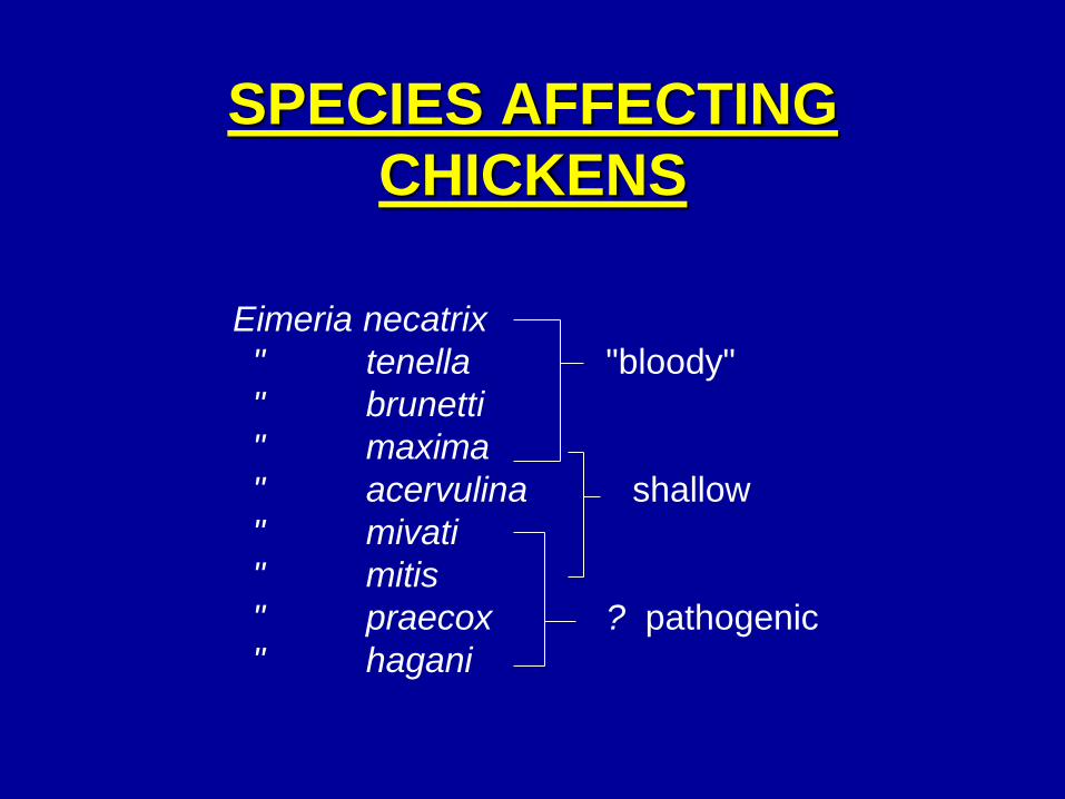
SPECIES AFFECTING
CHICKENS
Eimeria necatrix
" tenella "bloody"
" brunetti
" maxima
" acervulina shallow
" mivati
" mitis
" praecox ? pathogenic
" hagani
INCUBATION PERIOD
• Depend on species of coccidia 7-8 days to complete life
cycle.
• Mortality - depends on coccidia species and dosage.
Usually 5 to 8 days after infection.
• Blood appears - 4-5 days depending on coccidia species.
COURSE OF DISEASE
• 1-3 wk. on a flock basis.
• Depends on species of coccidia.
• Immunity develops.
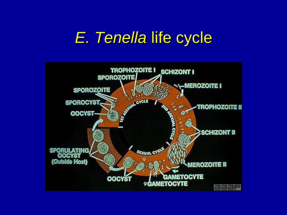
E. Tenella life cycle
ECONOMIC EFFECT
• Mortality - variable.
• Poor feed conversion and weight gains.
• Reduction of desired pigmentation.
• The lack of uniformity in pullets.
• Above depends on species of coccidia,
dosage, condition of overall health and
genetics of the chicken.
• Every ton of feed has $5/ton of coccidiostat.
METHOD OF SPREAD
• The natural behavioral traits are conducive
to ingestion of sporulated oocyst.
• Starts in a few birds and dosage is built up
over one or two passages and whole flock
may be exposed.
SIGNS
Typical "sick bird" - depressed and ruffled
feathers.
May or may not have bloody diarrhea
depending on species of Coccidia.
Mortality - may be first thing noticed. Species
dependent.
Loss in egg production - rare because usually
exposed and immune before the start of lay.
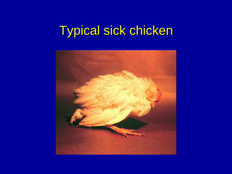
Typical sick chicken
POSTMORTEM LESIONS
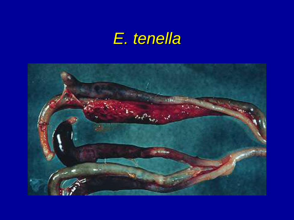
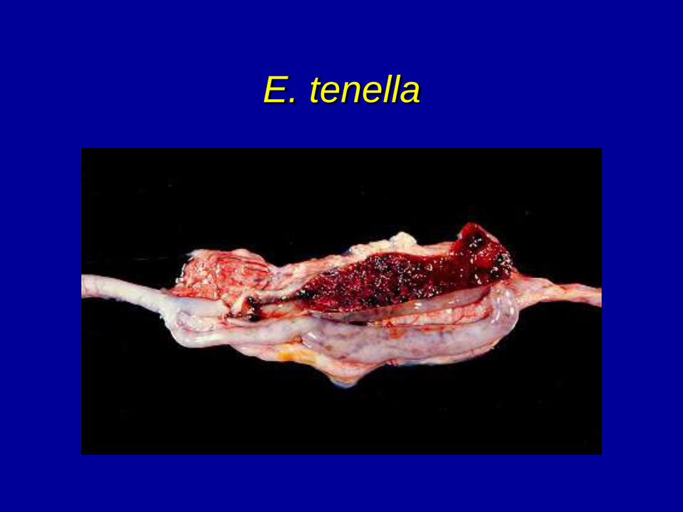
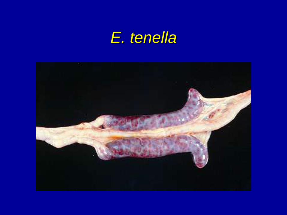
• Eimeria tenella - cecal coccidiosis.
• Erosions of cecal wall with free blood and
bloody cores in ceca.
• Caseous cores (old cases).
• Micro - presence of ovoid oocyst in cecal
scraping from sub-epithelium.
• Oocysts are double walled.
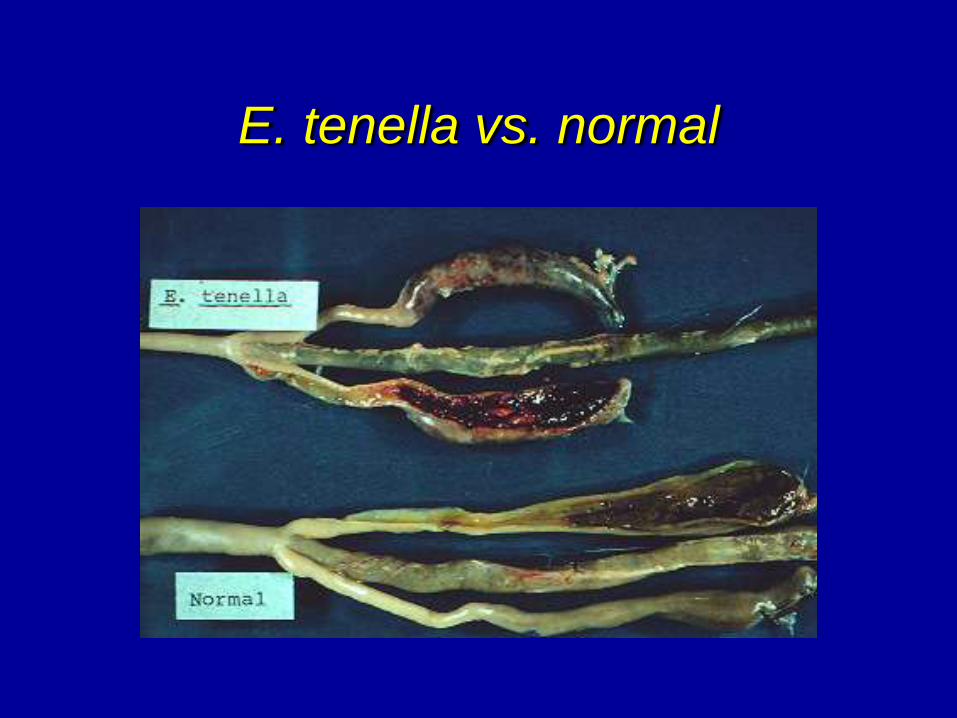
E. tenella vs. normal
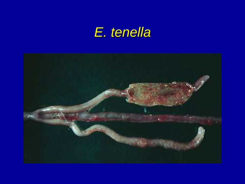
E. tenella
E. tenella
E. tenella
E. tenella
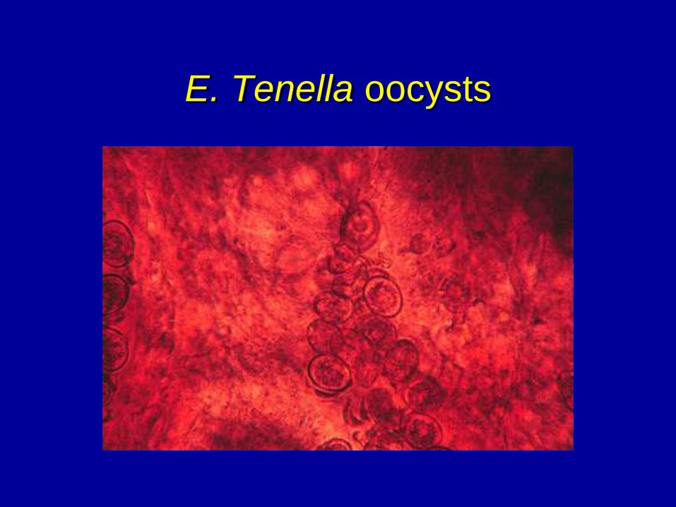
E. Tenella oocysts
POSTMORTEM LESIONS
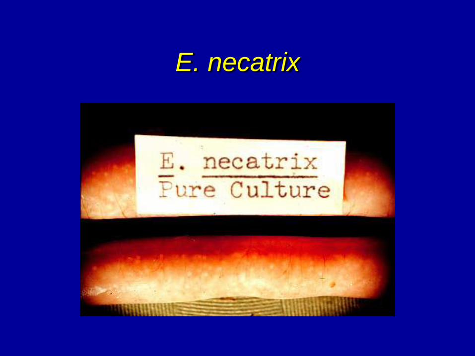
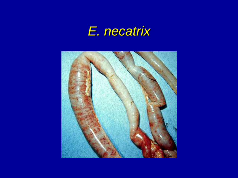
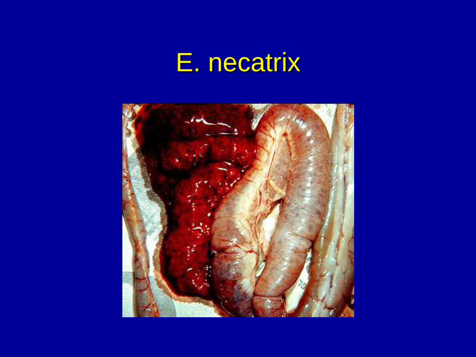
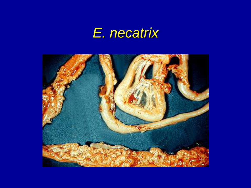
• Eimeria necatrix - mid gut.
• Ballooning in mid gut. Parasite in sub-epithelium.
• Serosa - white spots and petechial hemorrhages
• Open gut - mucoid blood-filled exudate.
• Micro - gut has large schizonts (single wall with
little definition) but no oocyst. Life stages moves
to ceca where oocysts are found.
• Ceca-oblong ovoid oocyst with no wall erosion.
• Less likely to be a mixed infection.
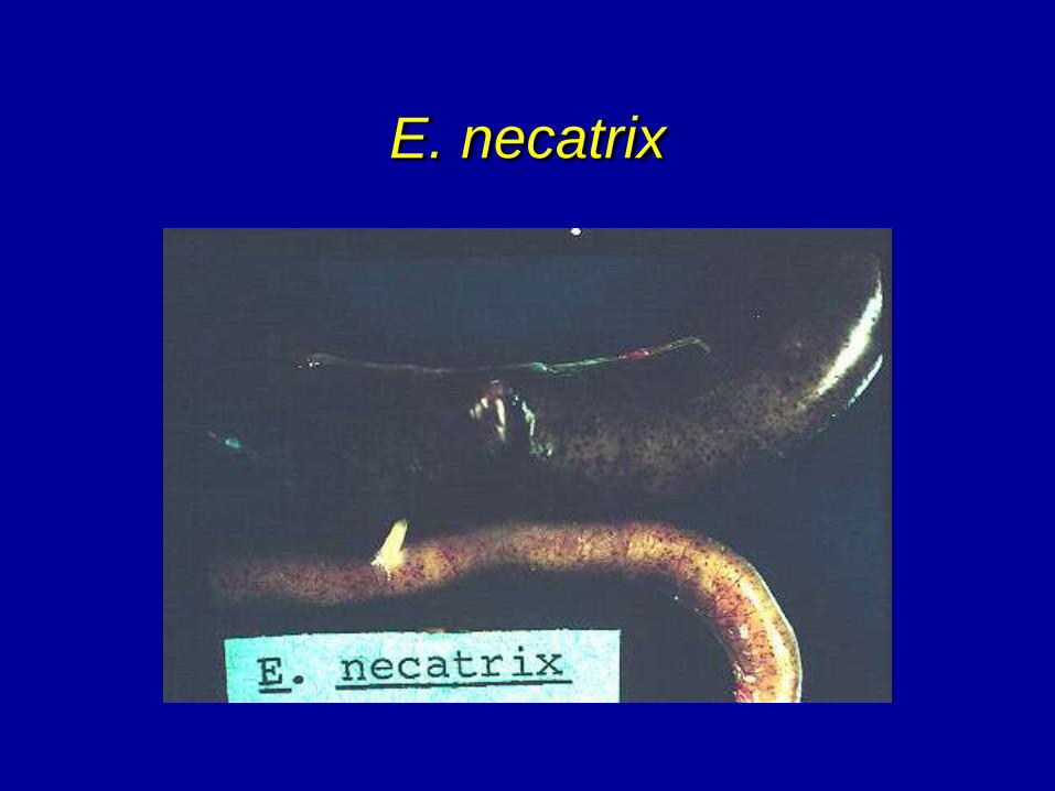
E. necatrix
E. necatrix
E. necatrix
E. necatrix
E. necatrix
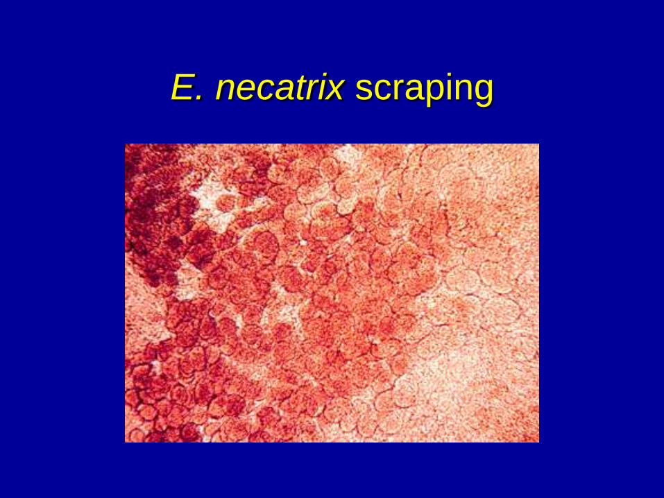
E. necatrix scraping
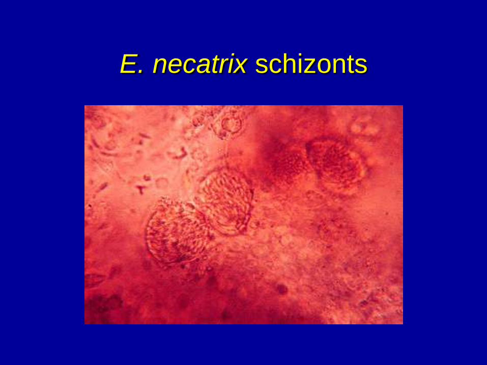
E. necatrix schizonts
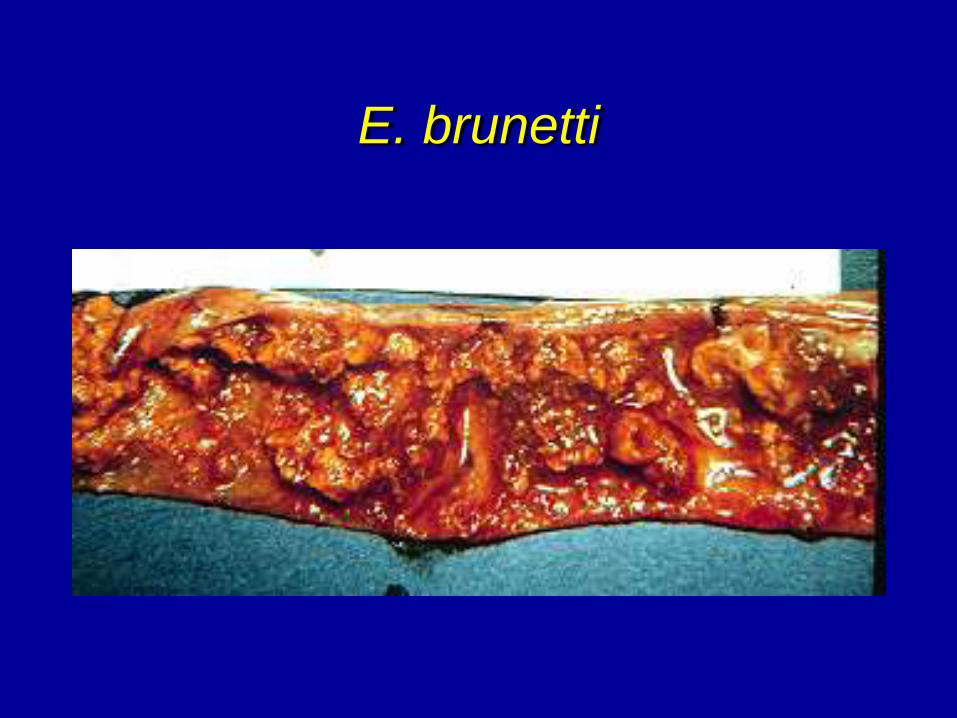
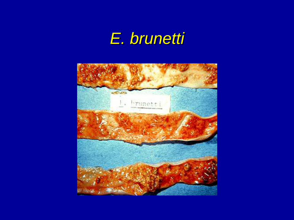
POSTMORTEM LESIONS
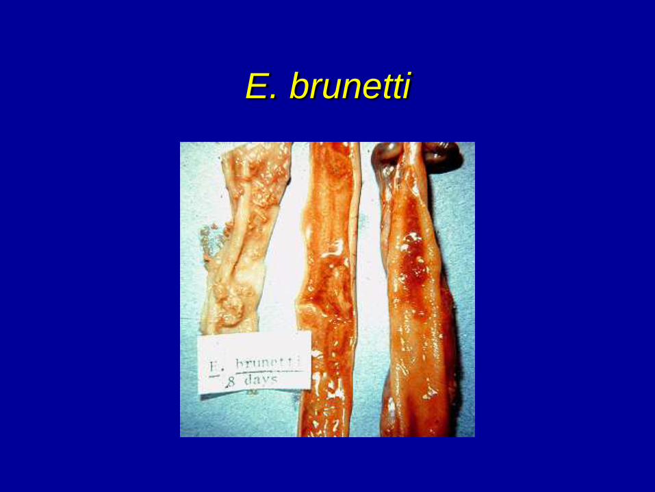
• Eimeria brunetti - mainly in lower SI and rectum.
Some infection in mid SI and ceca.
• Serosa - ecchymotic white areas with thickening of
lower SI and rectal walls.
• Open gut - coagulative necrosis, mucoid bloody
enteritis in lower gut.
• Micro - large ovoid oocyst in coagulative material.
Infection in sub-epithelium.
E. brunetti
E. brunetti
E. brunetti
POSTMORTEM LESIONS
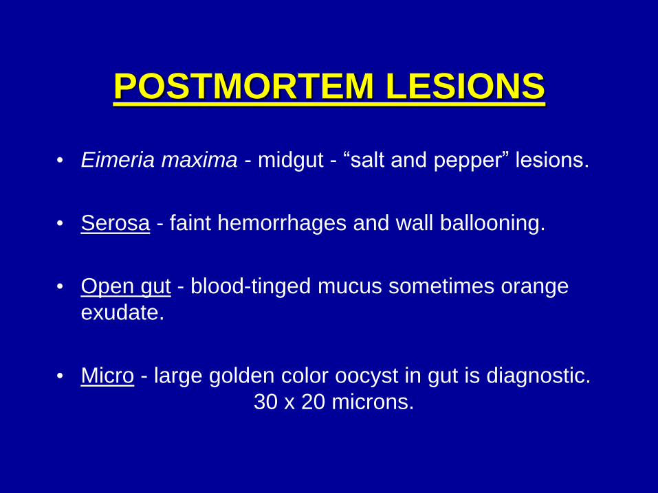
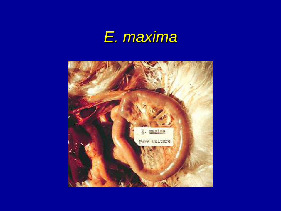
• Eimeria maxima - midgut - “salt and pepper” lesions.
• Serosa - faint hemorrhages and wall ballooning.
• Open gut - blood-tinged mucus sometimes orange
exudate.
• Micro - large golden color oocyst in gut is diagnostic.
30 x 20 microns.
E. maxima
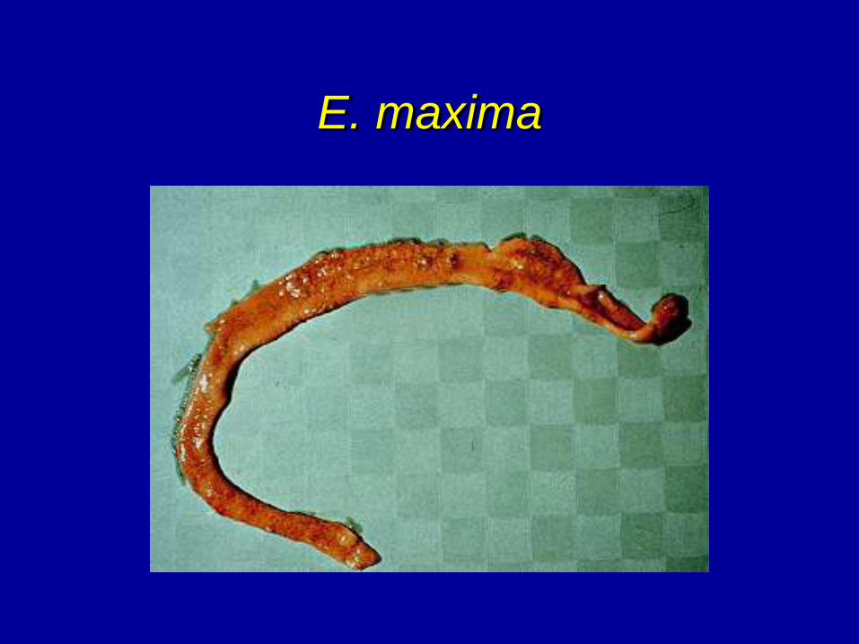
E. maxima
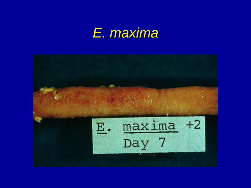
E. maxima
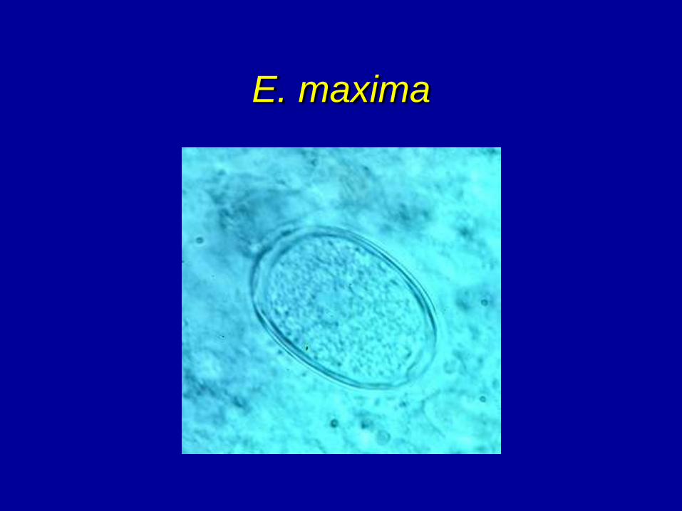
E. maxima
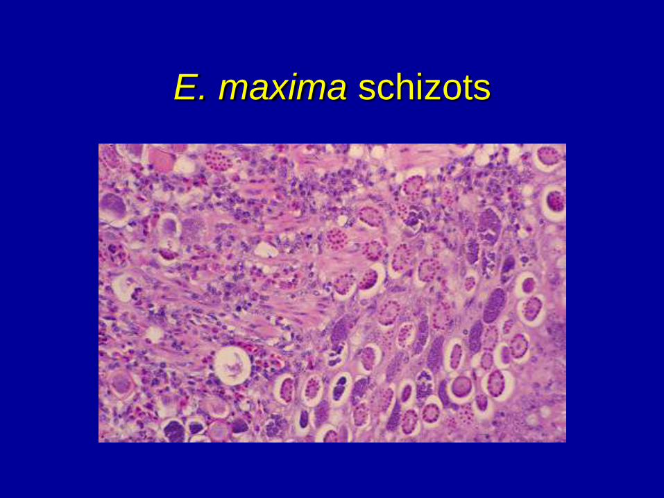
E. maxima schizots
POSTMORTEM LESIONS
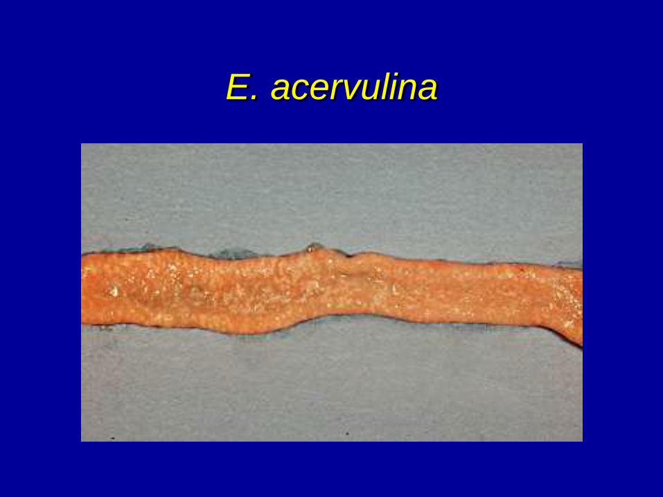
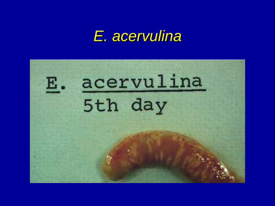
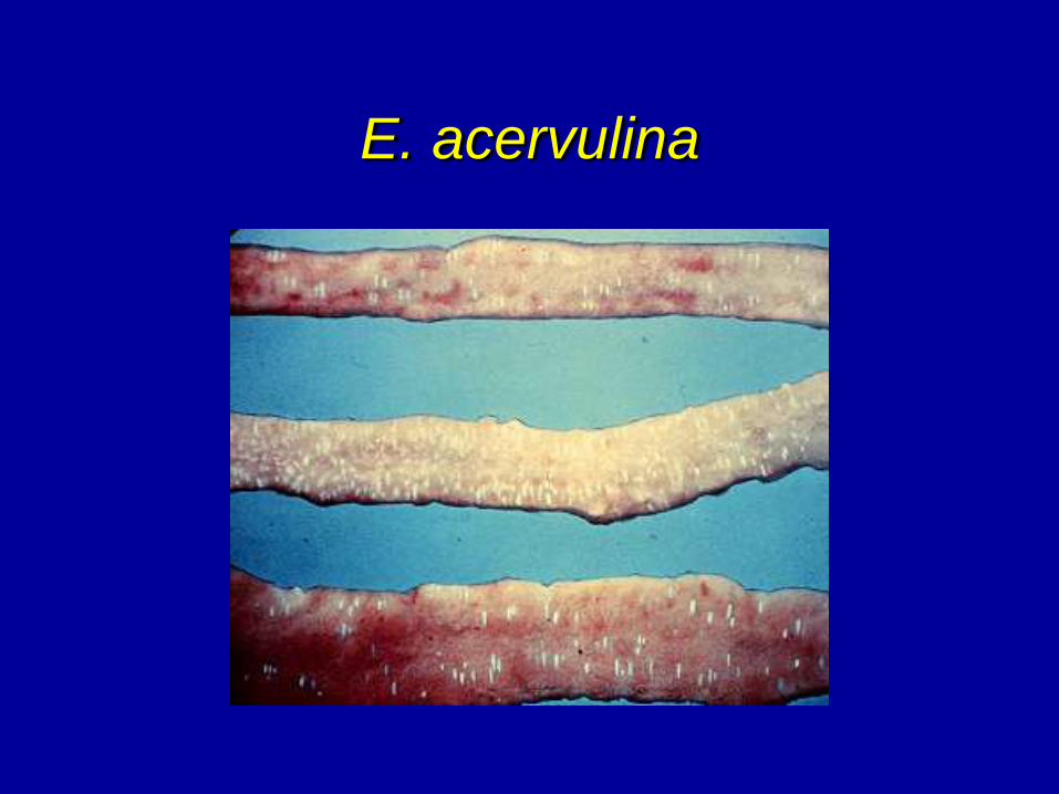
• Eimeria acervulina - usually in duodenal area.
• Serosa - white plaques that may be elongated and have a tendency to be "Ladder like" (horizontally) or in severe infections coalesced plaques and wall thickening. The ladder lesions are becoming less common.
• Open gut - only the epithelium is affected. May be whitish petechiae to coalesced lesions with a milky appearance (oocyst) in severe infection. Pathology depends on dosage.
• Micro - small (18 x 14 microns) ovoid oocyst from gut epithelium.
E. acervulina
E. acervulina
E. acervulina
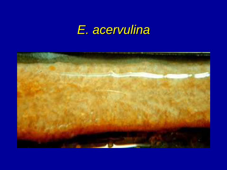
E. acervulina

POSTMORTEM LESIONS
• Eimeria mivati - some disagreement over the actual authenticity of this being a separate species from E. acervulina.
• Occurs in the epithelium of the upper gut.
• Serosa - similar to E. acervulina but round lesions and more gut thickening.
• Open gut - depending on dosage from individual plaques to coalesced large infected areas that appear milky.
• Micro - similar to E. acervulina with more macrogametocytes present mixed with oocyst.
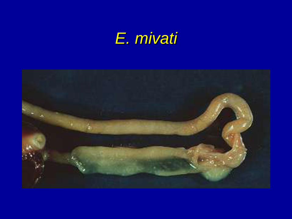
E. mivati
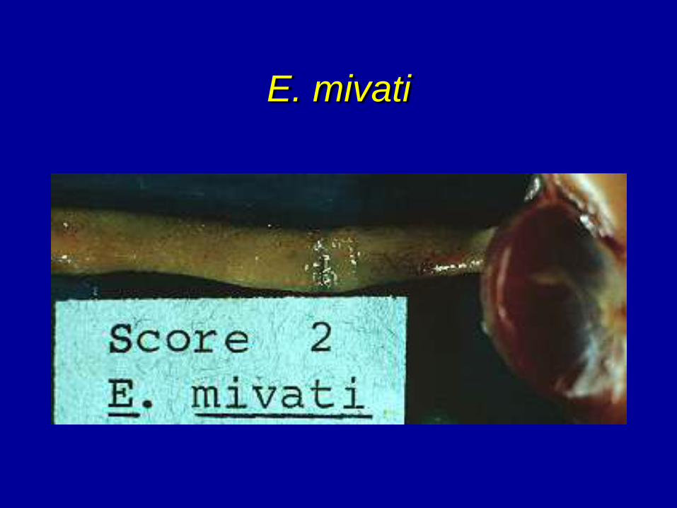
E. mivati
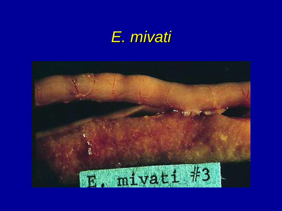
E. mivati
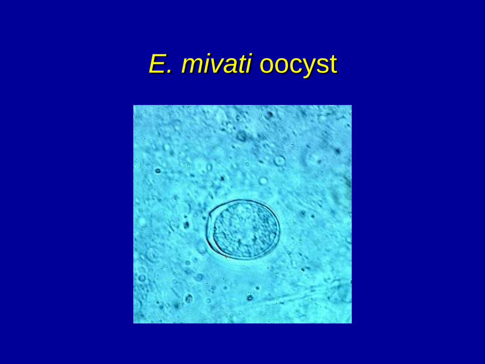
E. mivati oocyst
POSTMORTEM LESIONS
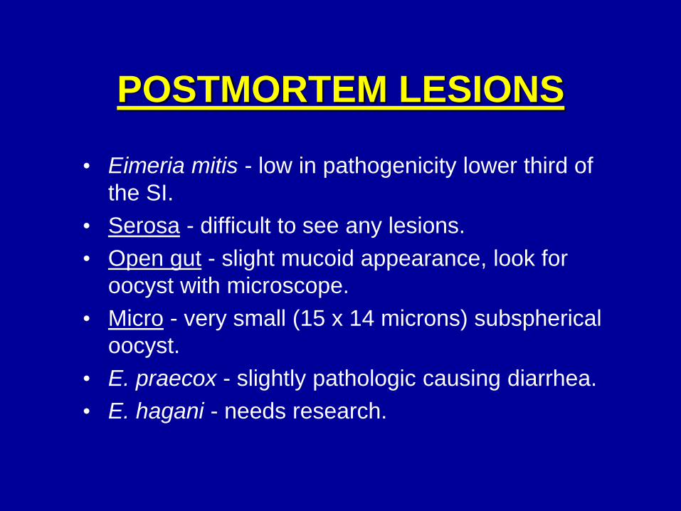
• Eimeria mitis - low in pathogenicity lower third of
the SI.
• Serosa - difficult to see any lesions.
• Open gut - slight mucoid appearance, look for
oocyst with microscope.
• Micro - very small (15 x 14 microns) subspherical
oocyst.
• E. praecox - slightly pathologic causing diarrhea.
• E. hagani - needs research.
DIAGNOSIS
• The presence of lesions and some stage of
the life cycle of coccidia (usually oocyst).
• Need to look at live and dead birds.
• Select birds typical of the flock, not culls.
Culls may be off feed and not ingesting
coccidiostat so are very likely to have cocci
even if it is not a flock problem.
• Must use light microscope to confirm.
• Can speciate by location of lesions and type
and size of oocyst.
DIAGNOSIS (CONT.)
• The serosal surface is examined for white plaques and hemorrhagic petechiae. The area of the gut affected is considered.
• The gut is opened and the mucosa of the affected area is scraped off with a spatula and placed on a microscopic slide.
• A cover-slip is applied to the scraping and mashed to produce a thin smear.
• The slide is then examined with a light microscope. Low power (100x) is used for scanning and high dry power (430x) for detail examination and measurements.
DIAGNOSIS (CONT.)
• Many times coccidiosis will be caused by
a mixture of species. Usually if birds are
dying, there is a predominance of one
deep invading species.
• Coccidiasis-presence of stage of life cycle
(usually oocyst) without lesions in the gut
or effect on production.
• Disease has a tendency to be over-
diagnosed by servicemen.
COMMENT
Other enteritis problems may appear
similar to coccidiosis. However, a
microscopic examination should clarify the
diagnosis. Don’t treat for coccidiosis
unless you know for sure.
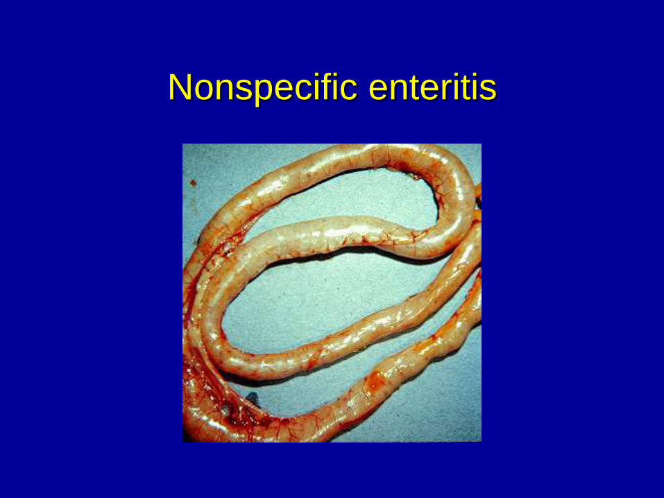
Nonspecific enteritis
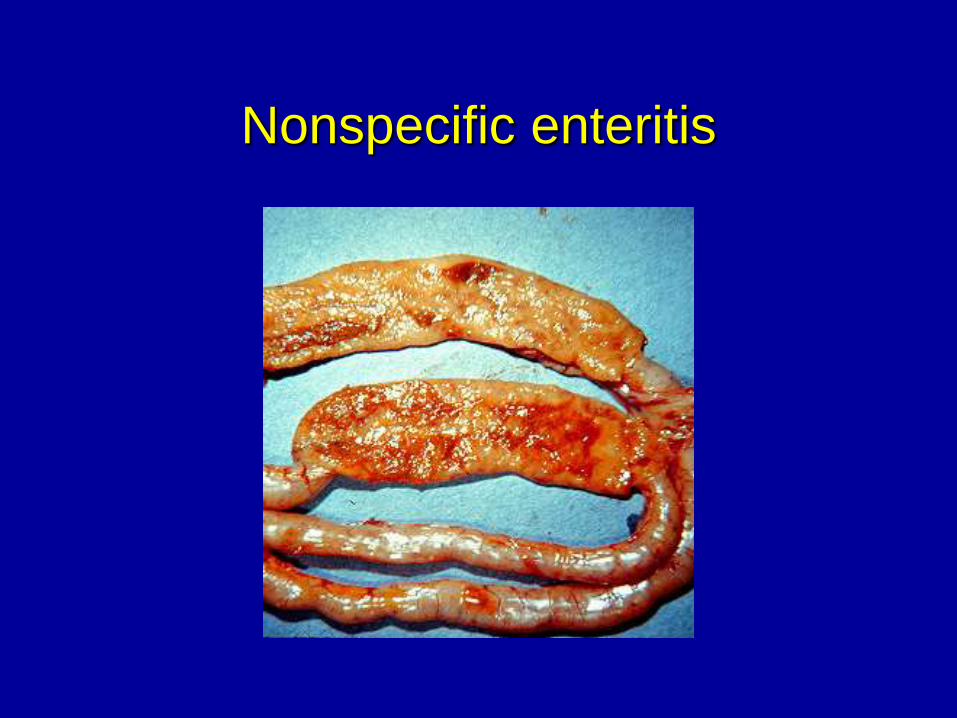
Nonspecific enteritis
TREATMENT
• Follow directions closely.
• Treatment - usually in water.
• Overtreatment can cause drug toxicity (Sulfas).
• Take into consideration the season of the year.
Sulfa drugs can be toxic if birds change their
drinking habits based on temperature.
• Some drugs can be used for both treatment and
control.
• Treatment - really prevention.
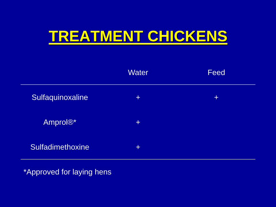
TREATMENT CHICKENS
Water Feed
Sulfaquinoxaline + +
Amprol®* +
Sulfadimethoxine +
*Approved for laying hens
PREVENTION
Genetics - not developed.
Nutritionally - certain vitamins help but not
reliable.
Quarantine & depopulation - not practical -
can make things worse.
Sanitation - not practical - may produce more
susceptible birds. Very resistant to
chemicals.
PREVENTION (CONT.)
Reduced exposure - shift from floor to cage
rearing but bird must remain in cages or on wire
for life.
Immunization - requires exposure (natural or
planned) to live coccidia. This is used in broiler
breeders or leghorns. Vaccine available.
Chemotherapy - coccidiostats - presently most
practical and most used.
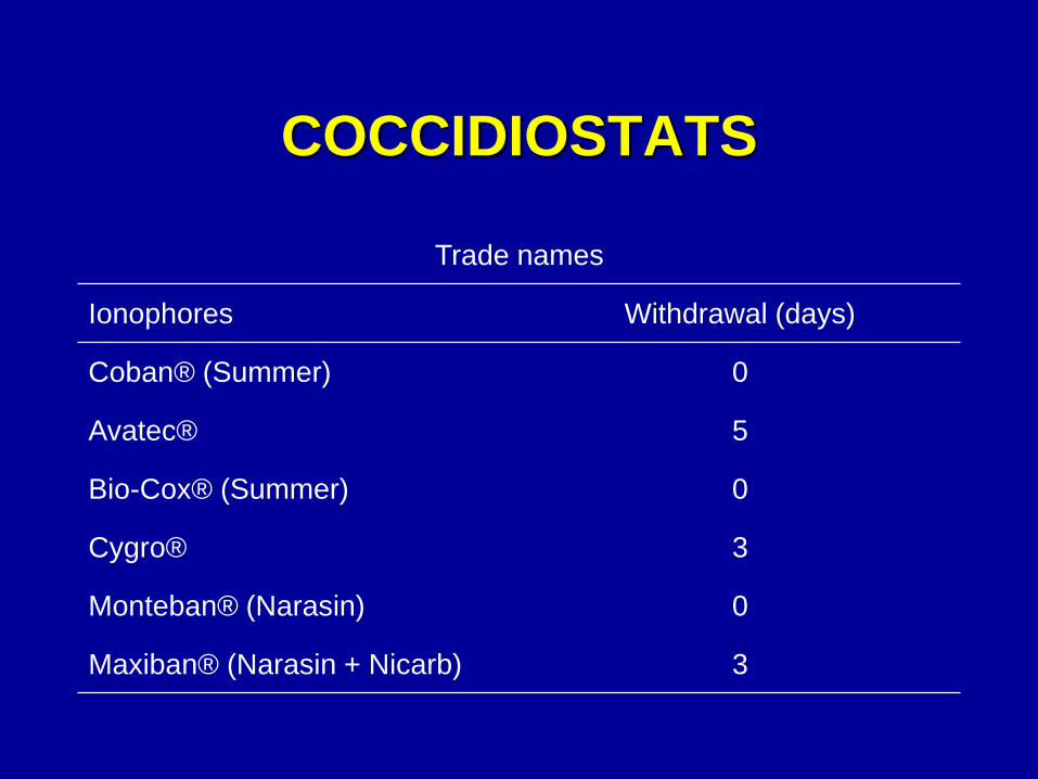
COCCIDIOSTATS
Trade names
Ionophores Withdrawal (days)
Coban® (Summer) 0
Avatec® 5
Bio-Cox® (Summer) 0
Cygro® 3
Monteban® (Narasin) 0
Maxiban® (Narasin + Nicarb) 3
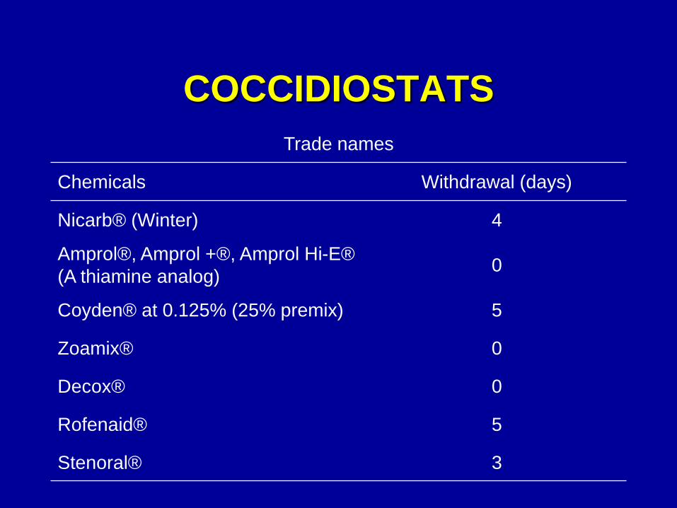
COCCIDIOSTATS
Trade names
Chemicals Withdrawal (days)
Nicarb® (Winter) 4
Amprol®, Amprol +®, Amprol Hi-E®
(A thiamine analog) 0
Coyden® at 0.125% (25% premix) 5
Zoamix® 0
Decox® 0
Rofenaid® 5
Stenoral® 3
SHUTTLE AND ROTATION
PROGRAMS
• These programs have been used to stop strain
resistance to specific drugs.
• Rotation involves complete change of drugs for
several months or years hoping resistance will
disappear.
• Shuttle programs involve the change of
Coccidiostats within a grow-out period of a
single flock.
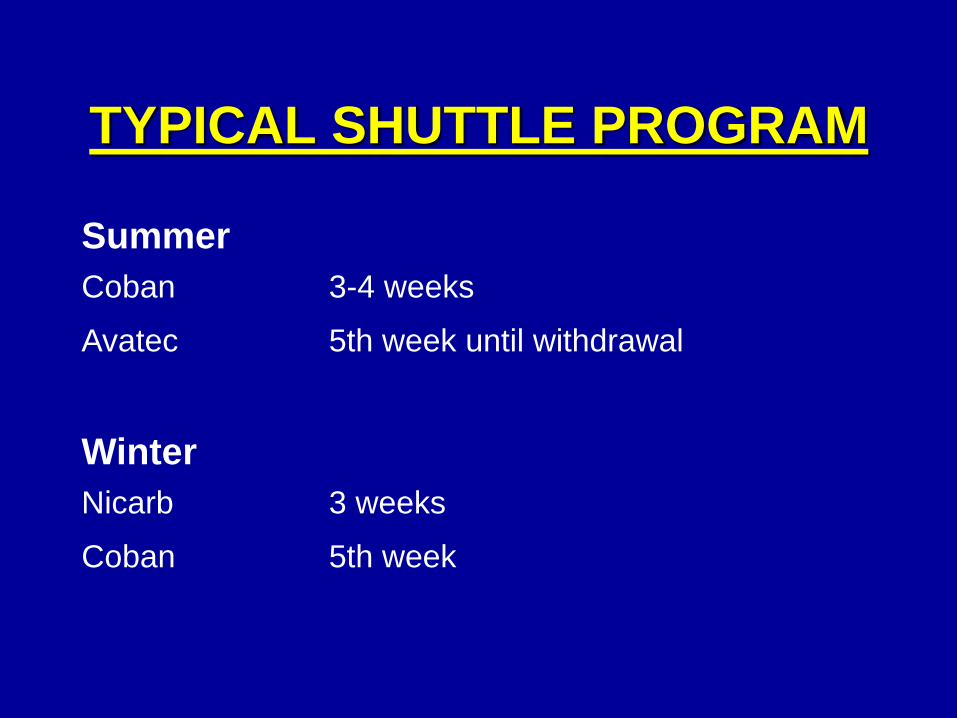
TYPICAL SHUTTLE PROGRAM
Summer
Coban 3-4 weeks
Avatec 5th week until withdrawal
Winter
Nicarb 3 weeks
Coban 5th week
IMMUNITY
• 2-3 cycles of infection to develop sufficient immunity
(depending on species of coccidia).
• Coccivac® - must give in first 7-10-days and proper
management of coccidia shed - need 20% moisture
in litter.
• 4-6 species used in the vaccine depending on
geography.
• Coccivac - mainly used in breeders, layer pullets,
and roasters.
• Coccivac - now given as an eyedrop in the hatchery
or sprayed on the feed at 3 days
IMMUNITY (CONT.)
• Important to develop immunity in breeders and
floor layers.
• Amprol - step down program.
• Research being done on new immunizing agents.
TURKEY COCCIDIOSIS
• Etiology - same genus; Eimeria
• Incubation period - same as chicken
• Course of disease - usually about (4-5 days)
• Mortality - low 5% except E. adenoides
• Method of spread - same as chicken
• Symptoms - Not as severe, more like "Shallow" invaders,
weight loss. No bloody diarrhea like in
chickens.
• Eimeria meleagrimitis
" adenoides pathogenic
" gallopavonis
• 4 other species - non-pathogenic
• Eimeria dispersa – affects quail also
This one is not host specific.
IMPORTANT SPECIES AFFECTING
TURKEYS
POSTMORTEM LESIONS
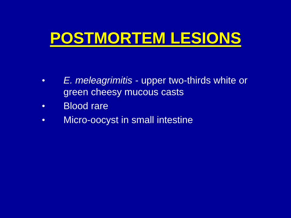
• E. meleagrimitis - upper two-thirds white or
green cheesy mucous casts
• Blood rare
• Micro-oocyst in small intestine
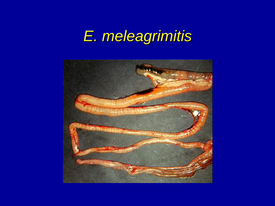
E. meleagrimitis
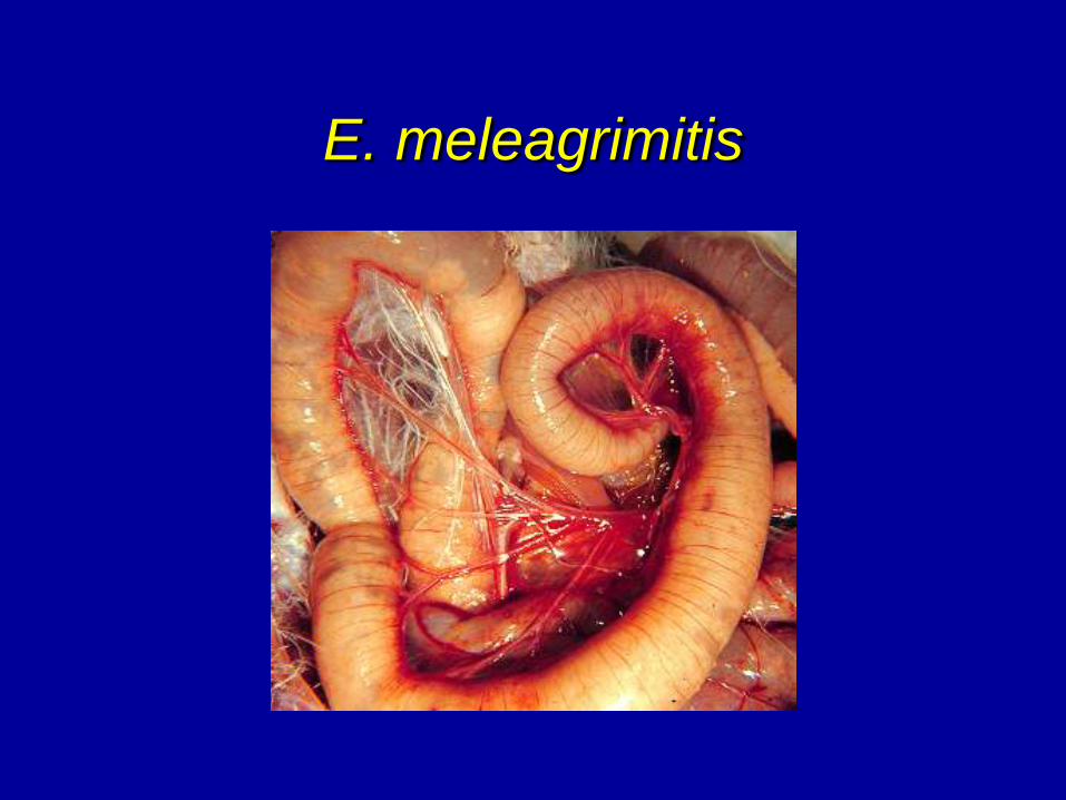
E. meleagrimitis
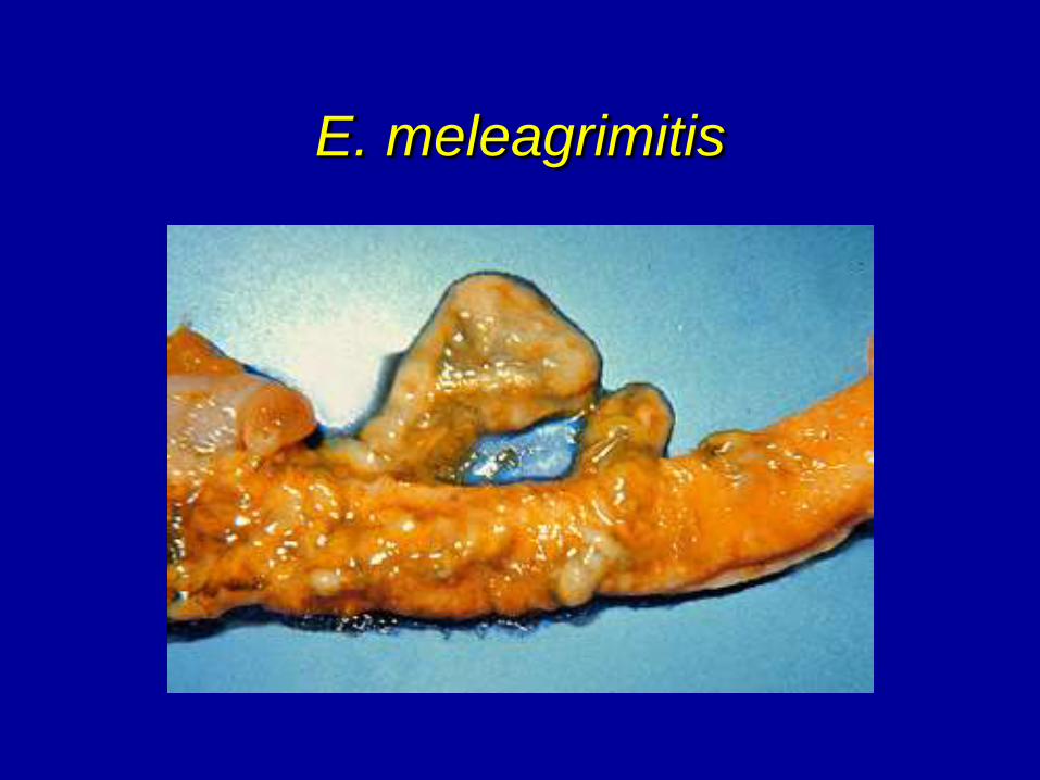
E. meleagrimitis
POSTMORTEM LESIONS
• E. adenoides - lower third small intestine,
ceca, and large intestine
• Dilation, edema and caseous exudate
"cottage cheese"
• Mortality may reach 100% in young
• This doesn’t occur often and is not seen in
Georgia.

E. adenoides
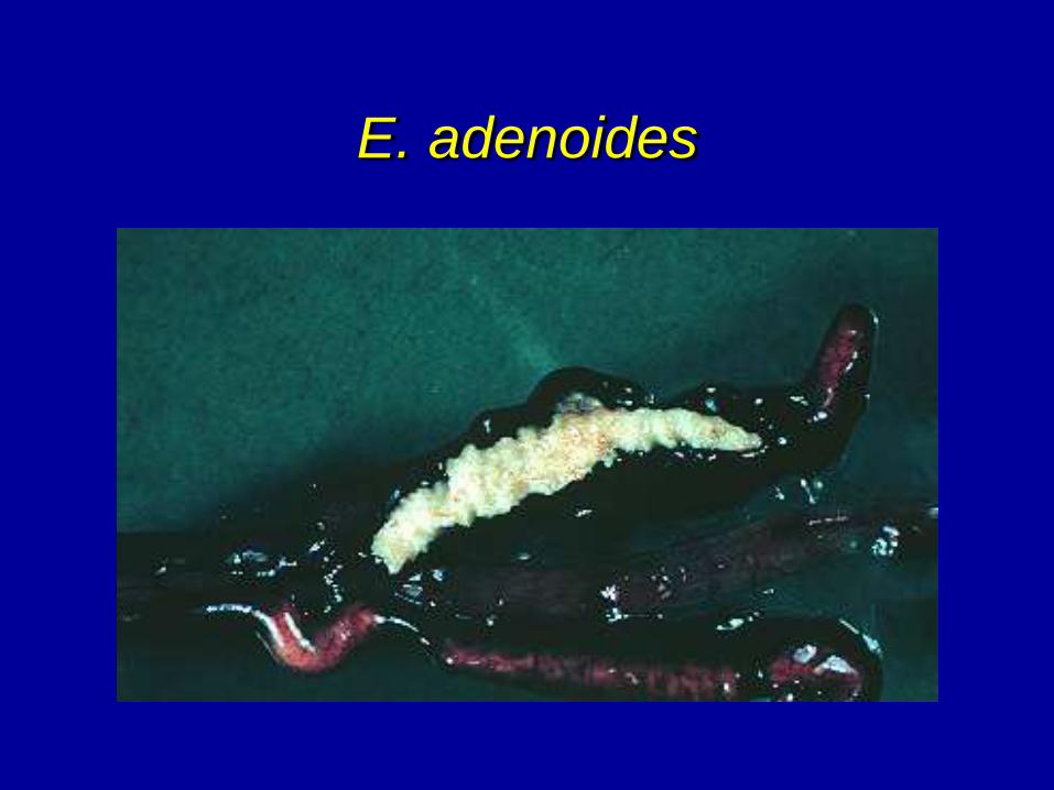
E. adenoides
POSTMORTEM LESIONS
• E. gallopavonis - lower third & ceca.
• Same as E. meleagrimitis, but in lower
intestines and ceca, in between the
cecal pouches.
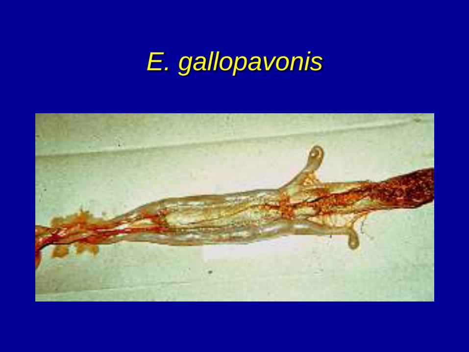
E. gallopavonis
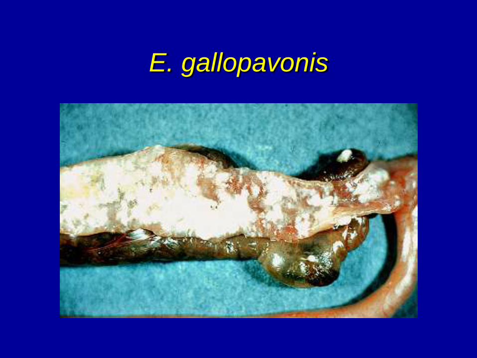
E. gallopavonis
TURKEY TREATMENT
• Sulfaquinoxaline (SQ) - toxic
• Amprol
• Sulfadimethoxine (Agribon@) in water
CONTROL
Try to develop some immunity in the poults. Feed coccidiostats during brooding
– Sulfaquinoxaline (SQ)
– Amprol
– Zoalene
– Coban - need to start birds on this or can get toxicity characterized by a “knockdown” where birds are recumbent with feet stretched out behind
– Stenoral
– Avatec