Autophagic Cell Death Is Induced by Acetone and Ethyl Acetate ...
10
The Scientific World Journal Volume 2012, Article ID 439479, 9 pages doi:10.1100/2012/439479 The cientificWorldJOURNAL Research Article Autophagic Cell Death Is Induced by Acetone and Ethyl Acetate Extracts from Eupatorium odoratum In Vitro : Effects on MCF-7 and Vero Cell Lines Faizah Bt. Harun, 1 Syed Mohsin Syed Sahil Jamalullail, 2 Khoo Boon Yin, 2 Zulkhairi Othman, 2 Anita Tilwari, 1 and Prabha Balaram 1 1 Institute for Research in Molecular Medicine (INFORMM), Universiti Sains Malaysia, Health Campus, 16150 Kubang Kerian, Kelantan, Malaysia 2 School of Health Sciences, Universiti Sains Malaysia, Health Campus, 16150 Kubang Kerian, Kelantan, Malaysia Correspondence should be addressed to Prabha Balaram, [email protected] Received 23 October 2011; Accepted 19 December 2011 Academic Editor: Julie Gavard Copyright © 2012 Faizah Bt. Harun et al. This is an open access article distributed under the Creative Commons Attribution License, which permits unrestricted use, distribution, and reproduction in any medium, provided the original work is properly cited. Eupatorium odoratum (EO) contains many biologically active compounds, the anticancer effects of which are not well documented. This study evaluates the cytotoxic effects and mechanism of action of EO extracts on MCF-7 and Vero cell lines. Evaluation of the cytotoxic activity using MTT assay, morphological alterations, and apoptosis were carried out. Autophagy was evaluated by LC3-A protein expression. Cytotoxic activity, membrane blebbing and ballooning at 24 hours, replacement by mass vacuolation, and dou- ble membrane vesicles mimicking autophagy and cell death were observed in the cancer cells. No apoptosis was observed by DNA fragmentation assay. Overexpression of LC3-A protein indicated autophagic cell death. Cell cycle analysis showed G0 and G2/M arrest. The Vero cells did not show significant cell death at concentrations <100 μg/mL. These results thus suggest that acetone and ethyl acetate extracts of EO induce cell death through induction of autophagy and hold potential for development as potential anticancer drugs. 1. Introduction The urge to find new compounds among plants to fight can- cer has become a matter of great interest among researchers. Plants harbor many chemical compounds with known or unknown potential activities against diseases. Eupatorium odoratum, also known as Chromolaena odor- ata (L.) King and Robinson, is a wildly growing free standing shrub from the family of Asteracea. Growing in a wet habitat, this plant has three-nerved leaves opposing each other, shaped into deltoid to oval lanceolate margin, and has long pointed tips. The white-colored flower of this plant is tubu- lar. Previous reports observed it to posses various biological activities including antimicrobial and anticancer effects. Local name for this plant include “pokok kapal terbang,” rumput putih (Indonesia), Siam weed, and others. Tradi- tional use of the leaves from this plant to reduce aches and pains has been practiced by villagers [1]. People in Machang, Kelantan, and Malaysia use this plant to treat wounds, uter- us-related problems, and to stop bleeding [2]. Hence it would be interesting to study the anticancer effects of its extracts which would help in evaluating the active principles as anticancer drugs. Breast cancer is one of the most debilitating disease of modern times [3]. The main modes of therapy used for con- trol of this disease are surgery and chemotherapy with vary- ing degrees of failure due to the high resistance to chemother- apy [4]. The treatment responses are especially dismal for hormone-independent cancers [4]. Hence, research into novel therapeutic compounds for these cancers is warranted. Plant products induce cell death by a variety of mech- anisms. The most common mechanisms reported are pro- grammed cell death (PCD)-type I (apoptosis), PCD type II autophagic cell death, and necrosis [5]. PCD type I is a mode
Transcript of Autophagic Cell Death Is Induced by Acetone and Ethyl Acetate ...

The Scientific World JournalVolume 2012, Article ID 439479, 9 pagesdoi:10.1100/2012/439479
The cientificWorldJOURNAL
Research Article
Autophagic Cell Death Is Induced by Acetone andEthyl Acetate Extracts from Eupatorium odoratum In Vitro:Effects on MCF-7 and Vero Cell Lines
Faizah Bt. Harun,1 Syed Mohsin Syed Sahil Jamalullail,2 Khoo Boon Yin,2
Zulkhairi Othman,2 Anita Tilwari,1 and Prabha Balaram1
1 Institute for Research in Molecular Medicine (INFORMM), Universiti Sains Malaysia, Health Campus,16150 Kubang Kerian, Kelantan, Malaysia
2 School of Health Sciences, Universiti Sains Malaysia, Health Campus, 16150 Kubang Kerian, Kelantan, Malaysia
Correspondence should be addressed to Prabha Balaram, [email protected]
Received 23 October 2011; Accepted 19 December 2011
Academic Editor: Julie Gavard
Copyright © 2012 Faizah Bt. Harun et al. This is an open access article distributed under the Creative Commons AttributionLicense, which permits unrestricted use, distribution, and reproduction in any medium, provided the original work is properlycited.
Eupatorium odoratum (EO) contains many biologically active compounds, the anticancer effects of which are not well documented.This study evaluates the cytotoxic effects and mechanism of action of EO extracts on MCF-7 and Vero cell lines. Evaluation of thecytotoxic activity using MTT assay, morphological alterations, and apoptosis were carried out. Autophagy was evaluated by LC3-Aprotein expression. Cytotoxic activity, membrane blebbing and ballooning at 24 hours, replacement by mass vacuolation, and dou-ble membrane vesicles mimicking autophagy and cell death were observed in the cancer cells. No apoptosis was observed by DNAfragmentation assay. Overexpression of LC3-A protein indicated autophagic cell death. Cell cycle analysis showed G0 and G2/Marrest. The Vero cells did not show significant cell death at concentrations <100 µg/mL. These results thus suggest that acetoneand ethyl acetate extracts of EO induce cell death through induction of autophagy and hold potential for development as potentialanticancer drugs.
1. Introduction
The urge to find new compounds among plants to fight can-cer has become a matter of great interest among researchers.Plants harbor many chemical compounds with known orunknown potential activities against diseases.
Eupatorium odoratum, also known as Chromolaena odor-ata (L.) King and Robinson, is a wildly growing free standingshrub from the family of Asteracea. Growing in a wet habitat,this plant has three-nerved leaves opposing each other,shaped into deltoid to oval lanceolate margin, and has longpointed tips. The white-colored flower of this plant is tubu-lar. Previous reports observed it to posses various biologicalactivities including antimicrobial and anticancer effects.Local name for this plant include “pokok kapal terbang,”rumput putih (Indonesia), Siam weed, and others. Tradi-tional use of the leaves from this plant to reduce aches and
pains has been practiced by villagers [1]. People in Machang,Kelantan, and Malaysia use this plant to treat wounds, uter-us-related problems, and to stop bleeding [2]. Hence itwould be interesting to study the anticancer effects of itsextracts which would help in evaluating the active principlesas anticancer drugs.
Breast cancer is one of the most debilitating disease ofmodern times [3]. The main modes of therapy used for con-trol of this disease are surgery and chemotherapy with vary-ing degrees of failure due to the high resistance to chemother-apy [4]. The treatment responses are especially dismal forhormone-independent cancers [4]. Hence, research intonovel therapeutic compounds for these cancers is warranted.
Plant products induce cell death by a variety of mech-anisms. The most common mechanisms reported are pro-grammed cell death (PCD)-type I (apoptosis), PCD type IIautophagic cell death, and necrosis [5]. PCD type I is a mode

2 The Scientific World Journal
of self cannibalism which involves individual cells [6] doesnot cause inflammation to the neighboring cells, and hasbecome a mechanism of interest in drug research. However,for certain cells in which the mechanism of cell death iscaspase independent like in MCF-7 cells, other mechanismswhich might differ from PCD type I could be in place suchas PCD type II autophagy. Autophagic cell death is dividedinto three types: macroautophagy, microautophagy, andchaperone-mediated autophagy. Main features of this PCDare that it involves formation of double membrane vesiclesand also autolysosomes, and can be visualized under phasecontrast microscope as double membrane vacuole like struc-tures inside the cytoplasm surrounding the organelles. Thesevesicles have been shown to fuse with lysosomes (containinglysozyme) to form autolysosomes and the organelles insideare processed and converted into ATP to be further usedfor cell metabolism [7, 8]. In certain treatments with plantextracts inducing excessive autophagic mechanism, a situa-tion arises when no organelles are left inside the cells to beused as energy. This leads to cell death, and DNA ladderingmight also be seen at later phases during the digestion of thenucleus by the endonuclease.
A number of drug compounds such as Tamoxifen,Rapamycin, sodium butyrate, and SAHA induce autophagyin cancer cells. Compounds, isolated from plants such as res-veratrol, oleandrin, and triterpenoid saponins have also beenshown to exhibit autophagy in a variety of cancer cells[9–11]. The mode of action of the extracted compoundsdepends on the parts of the plants used and the mode ofextraction. Using Acetone as the solvent, compounds belong-ing to lipid soluble or phenolic compound classes are ex-tracted, while ethyl acetate extracts more of phenols andflavonoids which are heat stable compounds. Many of thecompounds extracted by these solvents are reported to havepotent anticancer activity. Luteolin and phenolic compoundspresent in plants of this species have shown evidence ofmoderate toxicity against NCI-H187 cells and weak toxicityagainst human breast cancer cells [12, 13]. However, little isknown about its effects on different tumor cell lines, espe-cially caspase-3 deficient cell lines. In addition, the mecha-nisms of cell death induced by this plant are not well des-cribed or reported.
In this study, the elucidation of cytotoxicity effects onepithelial carcinoma cell lines and the mechanism of deathusing evaluation of morphological alterations, DNA-ladder-ing assay and LC3-A protein expression, showed the acetoneand ethyl acetate extracts of Eupatorium odoratum, to inducecell death predominantly through autophagy.
2. Materials and Methods
2.1. Cell Culture Preparation. Caspase-3-deficient humanmetastatic mammary carcinoma cells (MCF-7) and Vero cellswere maintained in RPMI-1640 (Hyclone, USA) with glu-tamine and supplemented with 10% fetal bovine serum(FBS) (Hyclone, USA) and Penicillin-Streptomycin (Gibco,Invitrogen) at 37◦C in 5% CO2 humidified atmosphere in-cubator (Thermo Forma, USA).
2.2. Plant Material. The plant Eupatorium odoratum wascollected by means of universal sampling. The leaves wereseparated from the fresh plant and used for preparation ofthe extracts.
2.3. Preparation of E. odoratum Extracts. Extracts were pre-pared using Soxhlet extraction method. Briefly, leaves ofE. odoratum were rinsed with tap water, washed with 5%ethanol, followed by rinsing with distilled water. Leaves werethen dried at 37◦C in dry oven and ground into powder andextracted using five different solvents following the nonpolarto polar solvent regime, namely, petroleum ether, ethyl ace-tate, acetone, methanol, and water. Solvents were diminishedfrom the extracts by reduced-pressure evaporation usingrotary evaporator (Heidolph, Germany). Aqueous extractwas dried using freeze drying method and kept at −20◦C.
Excluding aqueous extract, other plant extracts were dis-solved in DMSO to obtain final concentration of 50 mg/mL,filter sterilized with 0.20 µM PVDF membrane and stored at−20◦C.
Working solution was prepared by diluting the stocksolution with RPMI-1640 containing 10% FBS into nine dif-ferent concentrations (5 µg/mL, 10 µg/mL, 20 µg/mL, 25 µg/mL, 50 µg/mL, 100 µg/mL, 250 µg/mL, 500 µg/mL, and1000 µg/mL) just before use.
2.4. Cytotoxicity Assay. Cytotoxicity of plant extracts onMCF-7 cell line was tested using MTT (3-(4,5-dimethyl-thiazol-2-yl)-2,5-diphenyltetrazolium bromide, a tetrazole)cytotoxicity assay. Briefly, cells (1.25 × 10⁴ cells/mL) wereseeded into sterile 96-well flat bottom culture plates (Nunc,Germany). After 24 hours of incubation, plant extracts wereadded into respective culture wells (final concentration rang-ing from 2.5 to 500 µg/mL) and cultured for three timeintervals: 24, 48, and 72 hours. Tamoxifen at the same con-centrations as the plant extract including the IC50 valuewas added as the control drug to assess cell death. 4 hoursbefore the end of the culture period, media from each wellwas removed, and cell surface was washed with phosphate-buffered saline (PBS) pH 7.4 two times. 15 µL MTT reagentwas added, and the plate was incubated at 37◦C for 4 hours.Formazan crystals were then solubilized with 100 µL DMSO,and the plate was incubated in dark for 2 hours. Finally,absorbance was measured at 595 nm wavelength using ELISAreader (Thermo Scientific, USA). Vero cells grown andtreated in the same manner were used as the control for theMTT assays. IC50 was calculated using the Graph Pad Prismsoftware. All assays were carried out in triplicate, and themean values were recorded.
2.5. Morphological Study. MCF-7 cells at 1.25× 10⁵ cells/mLwere cultured in 96 well cell culture plates and incubated in5% CO2 incubator at 37◦C for 24 hours to obtain cells in thelog phase of its growth. Subsequently, 100 µL plant extractsat the IC50 concentrations was added into the culture wells intriplicate. No extract was added in the wells adjacent to theones in which the extract was added. These wells served ascontrols for each period of incubation. Cells were incubated

The Scientific World Journal 3
in incubator for respective culture periods (24, 48, or 72hours). After each treatment, culture media from the wellwas removed and transferred into separate tubes. Cells re-maining inside the wells were observed under a phase con-trast microscope and the picture was captured. For fluores-cent microscopic analysis, 25 µL of cell suspension was mixedwith 25 µL of acridine orange/ethidium bromide workingsolution (1x) and left for 2 minutes at room temperature toallow absorption of dye into cells. Cells were then observedunder white light of fluorescent microscope, and finallyunder blue light (450 nm). Assessment was carried out forfeatures such as membrane blebbing, nuclear condensation,and other features of apoptosis and cytoplasmic and nuclearchanges. Pictures observed were captured and edited usingQ-Capture software.
2.6. DNA Extraction. Control and treated cells seeded intriplicate inside 96-well cell culture plates were harvestedusing trypsinization method. Cell suspensions were thencentrifuged at 2000 rpm for 5 minutes to pellet the cells.Supernatant was removed, and the pellet was resuspendedin 1 mL cell lysis buffer; 10% SDS, 2 µL RNase (100 µg/mL),and incubated at 56◦C for 2 hours with brief vortexingfor 10 seconds each for 15 minutes. 10 µL of Proteinase K(20 mg/mL) was added to the tubes and incubated at 37◦Cfor 2 hours with brief vortexing for 10 seconds each for 15minutes followed by two volumes of cold isopropanol addedinto the mixture to precipitate the DNA. The tubes were leftfor 10 minutes at room temperature. Tubes containing DNAwas then centrifuged at 13000 rpm for 20 minutes in refrig-erated centrifuge at 4◦C. DNA pellet formed was washedwith 70% ethanol and centrifuged. Supernatant was totallyremoved and DNA pellet was air dried at room temperatureand finally resuspended in 20–50 µL tris-EDTA (TE) buffer.The prepared DNA was checked for purity and concentrationusing a nanodrop. A ratio of 1.6 and above was consideredpure enough to carry out the experiments. DNA was storedat −20◦C until use.
2.7. DNA-Laddering Assay. The DNA isolated from cellstreated with the plant extracts at different time intervals, anduntreated cells were electrophoresed on 1.5% agarose gel at100 mV for 1 hour 30 minutes. 100 bp DNA marker was runin the first lane to act as a guide for assessing ladder forma-tion.
2.8. Cell Cycle Analysis. Cells after treatment with the ex-tracts were trypsinized and collected using method similarto subculturing of the cells. Cell number was adjusted to 2 ×10⁶ cells/mL suspension. The cells were then transferred into5 mL polystyrene round bottom tubes and spun at 1000 rpmfor 5 minutes. Supernatant was discarded, and pellet wasresuspended in cold PBS, pH 7.2. Cells were fixed by adding4 mL cold absolute ethanol. 100 µL of 200 µg/mL RNase wasadded, and the tube was incubated at 37◦C for about 30minutes to remove RNase. 100 µL of 1 mg/mL Propidiumiodide (PI) (Sigma Aldrich, USA) was added, and tubes wereincubated in the dark at room temperature (25◦C) for 5 to 10
minutes. The cells were analysed using FACS-SCAN (BD, BioSciences) within 3 hours of PI addition. Data was analyzedusing FACSDiva software Version 6.1.2.
2.9. Immunocytochemistry. Cells after treatment were har-vested via trypsinization. Cell smears were made by spread-ing the cells evenly on the microscopic slides. The smearswere dried at room temperature for 10 minutes and im-mersed into cold acetone (−20◦C) for 20 minutes to fix thecells. Slides with cell smears were kept at −80◦C until use.The slides were brought to room temperature before start ofimmunostaining. In order to block endogenous peroxidaseactivity, which might interfere with the staining, slides wereincubated in 3% H2O2 in TBS for 10 minutes and thenrinsed with TBS-T20 2-3 times. 200 µL of 1 : 100 diluted LC3-A antibody (Abcam, USA) was added, and the slides wereincubated for 1 hour followed by rinsing with TBS-20 andincubated with biotinylated universal anti-immunoglobulinin phosphate-buffered saline (PBS) containing stabilizingprotein with 0.015 mol/L sodium azide (Dako, USA). After30 minutes of incubation, slides were washed with TBS-20 toremove nonspecific binding of the antibody. 2 drops of strep-tavidin conjugated with horseradish peroxidase (HRP) inPBS (Dako, USA) were added onto the slides and incubatedfor another 30 minutes. Positive cells showing LC3-A proteinexpression were detected by developing the slides using 3,3′-diaminobenzidine (DAB) chromogen containing hydrogenperoxide. Slides were left for 10 minutes to allow color de-velopment and then counterstained with hematoxylin andrinsed with running tap water to remove excess dye. Slideswere subjected to dehydration process by immersing inascending grades of absolute ethanol and finally in xylenefor two minutes and mounted using Cytosealer. Expressionof LC3A was evaluated under a light microscope and gradedpositive/negative in comparison to the respective controlslides.
3. Results
3.1. Cytotoxicity Assay. MTT cytotoxicity assay was done toassess the cytotoxic potential of EOea and EOace on MCF-7cancer cells and Vero cells. On treatment with EOea, theMCF-7 cells showed an increased rate of cell death at a lowerconcentration of the extracts when compared to that in theVero cells (Figure 1 and Table 1). For MCF-7 cells, the IC50
recorded for ethyl acetate extracts of EO were 65.72 µg/mL,83.88 µg/mL and 92.84 µg/mL and that for the acetoneextracts were 133.9 µg/mL at 24 hours, but increased to163.0 µg/mL at 48 hours and 147.8 µg/mL at 72 hours (Figure1 and Table 1). The IC50 values for both the extracts werelower than that for tamoxifen. Cell death could be observedin Vero cells mainly at concentrations >100 µg/mL.
3.2. Morphological Observations. Morphological alterationsin the treated cells were observed in both EOea- and EOace-treated MCF-7 cells.The observations revealed the effects ofthe EOace to be more prominent in treated MCF-7 cellswhen compared to untreated cells (Figure 2). At 24 hours of
4 The Scientific World Journal
0
20
40
60
80
100
120
2.5 5 10 12.5 25 50 100 125 250 500
EoeaEoace
Tamoxifen
Concentration (µg/mL)
MCF-7 : 24 hrs
Via
ble
cells
(%
)
(a)
2.5 5 10 12.5 25 50 100 125 250 500
Concentration (µg/mL)
0
20
40
60
80
100
120
EoeaEoace
Tamoxifen
Via
ble
cells
(%
)
VERO : 24 hrs
(b)
0
20
40
60
80
100
120
2.5 5 10 12.5 25 50 100 125 250 500
EoeaEoace
Tamoxifen
Concentration (µg/mL)
Via
ble
cells
(%
)
MCF-7 : 48 hrs
(c)
Via
ble
cells
(%
)
VERO : 48 hrs
2.5 5 10 12.5 25 50 100 125 250 500
EoeaEoace
Tamoxifen
Concentration (µg/mL)
0
20
40
60
80
100
120
(d)
Via
ble
cells
(%
)
MCF-7 : 72 hrs
2.5 5 10 12.5 25 50 100 125 250 500
Concentration (µg/mL)
0
20
40
60
80
100
120
EoeaEoace
Tamoxifen
(e)
2.5 5 10 12.5 25 50 100 125 250 500
Concentration (µg/mL)
0
20
40
60
80
100
120
EoeaEoace
Tamoxifen
Via
ble
cells
(%
)
VERO : 72 hrs
(f)
Figure 1: Graph showing cell death induced by acetone and ethyl acetate extracts of E. odoratum on MCF-7 and Vero cell culture in vitro atvarious time periods: 24 hours, 48 hours, and 72 hours.

The Scientific World Journal 5
Table 1: Cytotoxic concentrations (IC50 values) of EOea and EOace extracts against MCF-7 and Vero cells at different periods of treatment.
IC50 value (µg/mL)
Cells Time (hours)
24 48 72
EOea EOace TAM EOea EOace TAM EOea EOace TAM
Vero cells 115.74 121.96 >500 105.04 115.49 91.78 114.49 125.94 102.42
MCF-7 cells 65.72 133.9 138.7 83.88 163.03 85.77 92.84 147.8 45.88
(a)
(b)
(c)
(d)
(e)
(f)
(g)
(h)
Figure 2: Phase contrast and fluorescent microscopic analysis of MCF-7 cells treated with EOea and EOace at different time periods. (a) and(e) untreated MCF-7 cells, (b), (c), (d) MCF-7 cells treated with EOea at 24, 48, and 72 hours, (f), (g), (h) cells treated with EOace at 24, 48,and 72 hours.
treatment, enlargement of the cells which was more promi-nent in EOace (Figures 2(b) and 2(f)) was observed. 50–60%of the cells showed membrane blebbing (indicated by smallprotrusions of the membrane) at this period. At 48 hours,blebbing and ballooning of the membrane were prominent inalmost all the cells. Cells showed extensive vacuolation in thecell cytoplasm (Figures 2(c) and 2(g)), indicating autophagylike mechanism of cell death. Autophagosome like structureswere clearly seen in the cells at 24–48 hours of treatment inboth EOea- and EOace-treated cells (Figures 2(b), 2(f), 2(c),and 2(g)). Similar observations were also seen in the acridine
orange-ethidium bromide staining where the blebbing couldbe seen distinctly at 24–48 hours of incubation with the ex-tract, and shrinkage and cell death were observed at 72 hours(Figures 2(d) and 2(h)). The cells were rounded, shrunk, andshowed signs of detachment from the surface of the wells andcell death. No presence of apoptotic bodies could be observedat any time point. No apoptotic changes or vacuolation wereobserved in the Vero cells at the IC50 concentration.
3.3. DNA Laddering and Immunocytochemistry Assays. DNAladdering assay showed the genomic DNA to be localized

6 The Scientific World Journal
near the wells and did not show any laddering (Hallmarkof apoptosis) or smearing indicative of DNA shearing andnecrosis (Figure 3) when compared to the positive control.
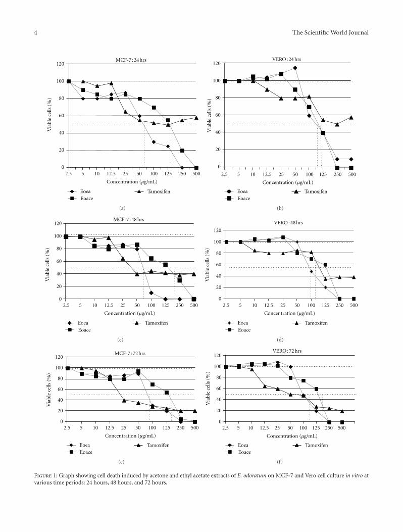
3.4. Cell Cycle Analysis. To test if EOea and EOace causea stage-dependant mechanism of inhibition, asynchronisedVero cells and MCF-7 cells were analysed by flow cytometryof stained DNA following treatment with the extracts. Theproportion of cell population in the three phases of the cellcycle are shown in Figures 4(a) and 4(b). A significant in-crease was observed in the G2/M phase accompanied by areduction in the G0/G1 phase in the MCF-7 cells on treat-ment with EOea (Figure 4(a)). Treatment with EOaceshowed an increase in the G0/G1 phase accompanied by adecrease in the G2/M phase (Figure 4(b)). No significantdifference was observed in the Vero cells treated for thesame period. The effect of EOea on G0/G1 was significant(P < 0.05) as early as 24 hours of treatment while the EOacewas slower affecting the cell cycle significantly (P < 0.05)only around 48 hours of treatment.
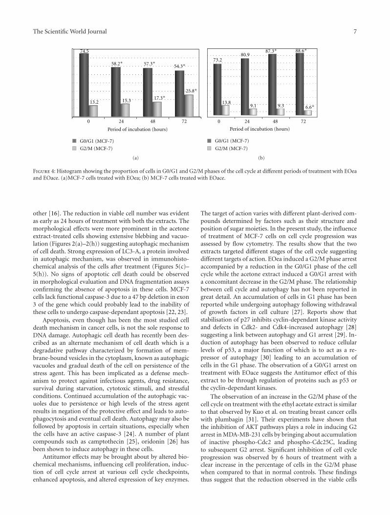
3.5. Expression of LC3-A Protein. The absence of apoptoticchanges, positive cytotoxic effect, and morphological appear-ance resembling autophagy prompted us to evaluate theexpression, using immunohistochemical analysis, of LC3-Aprotein which is involved during the process of autophagy.Human brain tissue was used as the positive control forexpression of LC3-A as directed in the manufacturer’s bro-chure.
Treatment of MCF-7 cells with EOea and EOace showedpositive expression of LC3-A protein, which was localizedin the cytoplasm of the cells. As seen in the morphologicalevaluation, the staining was more prominent in the EOace-treated cells when compared to the EOea-treated cells. Noexpression was observed in the untreated MCF-7 cells, andthe cells had clear cytoplasm (Figure 5(b)). At 24 hours,many cells showed the expression of LC3-A, but with mildstaining of the cytoplasm. In the case of EOea-treated cells,the number of cells showing positive staining for LC3-A wasmuch less in number when compared to the EOace-treatedcells (Figures 5(c) and 5(f)). After 48 hours of treatment,almost all the cells showed positive staining of the cells in theEOace-treated cells while 75% of the cells were positive in theEOea-treated cells (Figures 5(d) and 5(g)). At 72 hours, theexpression was much higher with both extracts and displayedintense staining for the protein (Figures 5(e) and 5(h)).
4. Discussion
Plants used in folk and traditional medicines have beenaccepted as leads for therapeutic drug development inmodern medicine. Eupatorium odoratum was chosen for thisstudy due to its use as an anticancer and wound-healingagent among the natives of Malaysia and in other parts of theworld [1, 2]. No documentation of its mechanism of actionwas found in literature, and hence this study evaluated thecytotoxic potential, the mechanism of cell death, and effects
1 2 3 4 5 6 7 8 9 10 11 12 13 14 15
1000 bp500 bp
100 bp
24 48 72
(hours)
Figure 3: DNA laddering assay showing no DNA fragmentation ornecrosis in MCF-7 cells treated with EOace and EOea at varioustime intervals., Lanes 1: DNA 100 bp ladder; 2: positive control; 3:negative control (untreated cells); 4 and 5: EOea-treated MCF-7cells—24 hours; 6 and 7: EOace-treated MCF-7 cells—24 hours;8 and 9: EOea-treated MCF-7 cells—48 hours; 10 and 11: EOace-treated MCF-7 cells—48 hours; 12 and 13: EOea-treated MCF-7cells—72 hours; 14 and 15: EOace-treated MCF-7 cells—72 hours.
on cell cycle of acetone and ethyl acetate extracts of this planton MCF-7 cells in vitro. Studies have observed the presenceof a large number of bioactive compounds in the acetone andethyl acetate extracts of plants including flavanoids, phenoliccompounds, triterpenoids, flavonoids, [14–16] which canbe extracted out by these solvents. These compounds arepresent in a number of food items and hold great potentialas drug candidates due to their safety, low toxicity, and wideacceptance.
Previous studies have isolated flavonoids, chalcones, fla-vones, essential oils, and other biological compounds fromdifferent parts of the plants of this family [12, 17–19], eventhough not Eupatorium odoratum per se. It was also observedthat different compounds differed in their target of actionin bringing about cell death [20, 21]. Antibacterial activity,antioxidant activity, and anticancer activity have been doc-umented in the extracts of these plants [12, 13] with nodetails of the mechanisms involved. Eupallinin A, a naturallyoccurring phytoalexin from Eupatorium Chinense L., wasfound to inhibit growth of Leukemia HL60 cells in vitro [17].
Caspase-3-deficient breast cancer cell line MCF-7 wasused as the test system in this study which was prompted bythe requirement of more effective treatment for the increas-ing incidence of breast cancers worldwide. The results of thepresent study showed potent cytotoxic effects on MCF-7 cellswith ethyl acetate and acetone extracts of this plant. The ethylacetate extract was active at a lower concentration (minimumIC50—65.72 µg/mL) when compared to the acetone extract ata minimum IC50 of 133.9 µg/mL as is evident from Table 1,and Figure 1. The IC50 value was found to be higher thanthat specified by NCI, USA for categorization of a pure com-pound as anticancer agent but lower than that of the standarddrug tamoxifen. This could be due to the fact that crude ex-tracts were used in this study due to the exploratory natureof the study, and inappropriate combinations of componentsin the extract would result in nullifying effects towards each
The Scientific World Journal 7
0 24 48 72
74.5
58.2∗ 57.3∗ 54.3∗
13.2 15.3 17.3∗25.8∗
G0/G1 (MCF-7)
G2/M (MCF-7)
Period of incubation (hours)
(a)
Period of incubation (hours)
0 24 48 72
73.280.9
87.3∗ 88.6∗
13.89.1 9.3 6.6∗
G0/G1 (MCF-7)
G2/M (MCF-7)
(b)
Figure 4: Histogram showing the proportion of cells in G0/G1 and G2/M phases of the cell cycle at different periods of treatment with EOeaand EOace. (a)MCF-7 cells treated with EOea; (b) MCF-7 cells treated with EOace.
other [16]. The reduction in viable cell number was evidentas early as 24 hours of treatment with both the extracts. Themorphological effects were more prominent in the acetoneextract-treated cells showing extensive blebbing and vacuo-lation (Figures 2(a)–2(h)) suggesting autophagic mechanismof cell death. Strong expression of LC3-A, a protein involvedin autophagic mechanism, was observed in immunohisto-chemical analysis of the cells after treatment (Figures 5(c)–5(h)). No signs of apoptotic cell death could be observedin morphological evaluation and DNA fragmentation assaysconfirming the absence of apoptosis in these cells. MCF-7cells lack functional caspase-3 due to a 47 bp deletion in exon3 of the gene which could probably lead to the inability ofthese cells to undergo caspase-dependant apoptosis [22, 23].
Apoptosis, even though has been the most studied celldeath mechanism in cancer cells, is not the sole response toDNA damage. Autophagic cell death has recently been des-cribed as an alternate mechanism of cell death which is adegradative pathway characterized by formation of mem-brane-bound vesicles in the cytoplasm, known as autophagicvacuoles and gradual death of the cell on persistence of thestress agent. This has been implicated as a defense mech-anism to protect against infectious agents, drug resistance,survival during starvation, cytotoxic stimuli, and stressfulconditions. Continued accumulation of the autophagic vac-uoles due to persistence or high levels of the stress agentresults in negation of the protective effect and leads to auto-phagocytosis and eventual cell death. Autophagy may also befollowed by apoptosis in certain situations, especially whenthe cells have an active caspase-3 [24]. A number of plantcompounds such as camptothecin [25], oridonin [26] hasbeen shown to induce autophagy in these cells.
Antitumor effects may be brought about by altered bio-chemical mechanisms, influencing cell proliferation, induc-tion of cell cycle arrest at various cell cycle checkpoints,enhanced apoptosis, and altered expression of key enzymes.
The target of action varies with different plant-derived com-pounds determined by factors such as their structure andposition of sugar moieties. In the present study, the influenceof treatment of MCF-7 cells on cell cycle progression wasassessed by flow cytometry. The results show that the twoextracts targeted different stages of the cell cycle suggestingdifferent targets of action. EOea induced a G2/M phase arrestaccompanied by a reduction in the G0/G1 phase of the cellcycle while the acetone extract induced a G0/G1 arrest witha concomitant decrease in the G2/M phase. The relationshipbetween cell cycle and autophagy has not been reported ingreat detail. An accumulation of cells in G1 phase has beenreported while undergoing autophagy following withdrawalof growth factors in cell culture [27]. Reports show thatstabilisation of p27 inhibits cyclin-dependant kinase activityand defects in Cdk2- and Cdk4-increased autophagy [28]suggesting a link between autophagy and G1 arrest [29]. In-duction of autophagy has been observed to reduce cellularlevels of p53, a major function of which is to act as a re-pressor of autophagy [30] leading to an accumulation ofcells in the G1 phase. The observation of a G0/G1 arrest ontreatment with EOace suggests the Antitumor effect of thisextract to be through regulation of proteins such as p53 orthe cyclin-dependant kinases.
The observation of an increase in the G2/M phase of thecell cycle on treatment with the ethyl acetate extract is similarto that observed by Kuo et al. on treating breast cancer cellswith plumbagin [31]. Their experiments have shown thatthe inhibition of AKT pathways plays a role in inducing G2arrest in MDA-MB-231 cells by bringing about accumulationof inactive phospho-Cdc2 and phospho-Cdc25C, leadingto subsequent G2 arrest. Significant inhibition of cell cycleprogression was observed by 6 hours of treatment with aclear increase in the percentage of cells in the G2/M phasewhen compared to that in normal controls. These findingsthus suggest that the reduction observed in the viable cells
8 The Scientific World Journal
(a) (b)
(c)
(d)
(e)
(f)
(g)
(h)
Figure 5: Immunocytochemical staining of MCF-7 cells treated with EOace and EOea at various time periods. Positive control: (a) Humanbrain tissue, (b)Untreated MCF-7 cells, (c) EOace treated MCF-7 cells at 24 hours, (d) 48 hours and (e) 72 hours. (f) EOea treated MCF-7cells at 24 hours, (g) 48 hours and (h) 72 hours. Positive expression of LC3-A is indicated by the reddish brown staining.
following treatment with EOea is due to autophagic celldeath and is associated with cell cycle arrest in the G2/Mphase.
In conclusion, the present observations provide prelimi-nary data to show that acetone and ethyl acetate extracts of
Eupatorium odoratum have potent cytotoxic activity againstMCF-7 cells. The data further emphasize that the mechanismunderlying cell death by these extracts is due to autophagyand cell cycle arrest. The data also suggest that the target ofaction of the active compound in the two extracts is different.

The Scientific World Journal 9
This calls for further studies on the active components forproper assessment of their chemotherapeutic properties andpossible development as promising anticancer drugs.
Conflict of Interests
The authors have no conflict of interests on the above work.
Acknowledgment
Authors would like to thank INFORMM and School ofHealth Sciences, Universiti Sains Malaysia for supporting re-search facilities to carry out the studies, Research universitygrant for financial support (RU Grant: 1001/CIPPM/811061)and USM Fellowship awarded to Ms. Faizah Harun.
References
[1] H. C. Ong and J. Norzalina, “Malay herbal medicine in Ge-mencheh, Negri Sembilan, Malaysia,” Fitoterapia, vol. 70, no.1, pp. 10–14, 1999.
[2] H. C. Ong and M. Nordiana, “Malay ethno-medico botany inMachang, Kelantan, Malaysia,” Fitoterapia, vol. 70, no. 5, pp.502–513, 1999.
[3] A. N. Hisham and C. H. Yip, “Overview of breast cancer inMalaysian women: a problem with late diagnosis,” Asian Jour-nal of Surgery, vol. 27, no. 2, pp. 130–133, 2004.
[4] J. Bange, E. Zwick, and A. Ullrich, “Molecular targets for breastcancer therapy and prevention,” Nature Medicine, vol. 7, no. 5,pp. 548–552, 2001.
[5] L. F. Dı́az, M. Chiong, A. F. G. Quest, S. Lavandero, and A.Stutzin, “Mechanisms of cell death: molecular insights andtherapeutic perspectives,” Cell Death and Differentiation, vol.12, no. 11, pp. 1449–1456, 2005.
[6] R. J. Bold, P. M. Termuhlen, and D. J. McConkey, “Apoptosis,cancer and cancer therapy,” Surgical Oncology, vol. 6, no. 3, pp.133–142, 1997.
[7] C. C. Hung, E. J. Davison, P. A. Robinson, and H. C. Ardley,“The aggravating role of the ubiquitin-proteasome system inneurodegenerative disease,” Biochemical Society Transactions,vol. 34, no. 5, pp. 743–745, 2006.
[8] B. Levine and G. Kroemer, “Autophagy in the pathogenesis ofdisease,” Cell, vol. 132, no. 1, pp. 27–42, 2008.
[9] Y. Kondo, T. Kanzawa, R. Sawaya, and S. Kondo, “The role ofautophagy in cancer development and response to therapy,”Nature Reviews Cancer, vol. 5, no. 9, pp. 726–734, 2005.
[10] W. O. Anthony Jr., L. Tan, A. E. Boitano, D. R. Sorenson, A.Aurora, and J. R. Liu, “Resveratrol-induced autophagocytosisin ovarian cancer cells,” Cancer Research, vol. 64, no. 2, pp.696–703, 2004.
[11] S. Meschini, M. Condello, M. Marra, G. Formisano, E. Fed-erici, and G. Arancia, “Autophagy-mediated chemosensitizingeffect of the plant alkaloid voacamine on multidrug resistantcells,” Toxicology in Vitro, vol. 21, no. 2, pp. 197–203, 2007.
[12] A. Suksamrarn, A. Chotipong, T. Suavansri et al., “Antimy-cobacterial activity and cytotoxicity of flavonoids from theflowers of Chromolaena odorata,” Archives of Pharmacal Re-search, vol. 27, no. 5, pp. 507–511, 2004.
[13] K. Srinivasa Rao, P. K. Chaudhury, and A. Pradhan, “Evalu-ation of anti-oxidant activities and total phenolic content ofChromolaena odorata,” Food and Chemical Toxicology, vol. 48,no. 2, pp. 729–732, 2010.
[14] S. M. Hassan and H. R. Ghareib, “Bioactivity of Ulva lactucaL. acetone extract on germination and growth of lettuce andtomato plants,” African Journal of Biotechnology, vol. 8, no. 16,pp. 3832–3838, 2009.
[15] A. H. Gilani and Atta-ur-Rahman, “Trends in ethnopharma-cology,” Journal of Ethnopharmacology, vol. 100, no. 1-2, pp.43–49, 2005.
[16] J. Dai and R. J. Mumper, “Plant phenolics: extraction, analysisand their antioxidant and anticancer properties,” Molecules,vol. 15, no. 10, pp. 7313–7352, 2010.
[17] J. Yuan, J. Yang, and J. Miao, “Chemical constituents of Eupa-torium odoratum,” Chinese Traditional and Herbal Drugs, vol.36, no. 12, article 1771, 2005.
[18] P. K. Bose, P. Chakrabarti, S. Chakravarti, S. P. Dutta, and A.K. Barua, “Flavonoid constituents of Eupatorium odoratum,”Phytochemistry, vol. 12, no. 3, pp. 667–668, 1973.
[19] M. S. Owolabi, A. Ogundajo, K. O. Yusuf et al., “Chemicalcomposition and bioactivity of the essential oil of Chromo-laena odorata from Nigeria,” Records of Natural Products, vol.4, no. 1, pp. 72–78, 2010.
[20] M. Salucci, L. A. Stivala, G. Maiani, R. Bugianesi, and V.Vannini, “Flavonoids uptake and their effect on cell cycle ofhuman colon adenocarcinoma cells (Caco2),” British Journalof Cancer, vol. 86, no. 10, pp. 1645–1651, 2002.
[21] K. Ishiguro, T. Ando, O. Maeda et al., “Ginger ingredientsreduce viability of gastric cancer cells via distinct mecha-nisms,” Biochemical and Biophysical Research Communications,vol. 362, no. 1, pp. 218–223, 2007.
[22] X. H. Yang, T. L. Sladek, X. Liu, B. R. Butler, C. J. Froelich,and A. D. Thor, “Reconstitution of caspase 3 sensitizes MCF-7 breast cancer cells to doxorubicin- and etoposide-inducedapoptosis,” Cancer Research, vol. 61, no. 1, pp. 348–354, 2001.
[23] B. Fazi, W. Bursch, G. M. Fimia et al., “Fenretinide inducesautophagic cell death in caspase-defective breast cancer cells,”Autophagy, vol. 4, no. 4, pp. 435–441, 2008.
[24] H. Rodriguez-Rocha, A. Garcia-Garcia, M. I. Panayiotidis, andR. Franco, “DNA damage and autophagy,” Mutation Research,vol. 711, no. 1-2, pp. 158–166, 2011.
[25] M. J. Abedin, D. Wang, M. A. McDonnell, U. Lehmann, andA. Kelekar, “Autophagy delays apoptotic death in breast cancercells following DNA damage,” Cell Death and Differentiation,vol. 14, no. 3, pp. 500–510, 2007.
[26] Q. Cui, S.-I. Tashiro, S. Onodera, M. Minami, and T. Ikejima,“Autophagy preceded apoptosis in oridonin-treated humanbreast cancer MCF-7 cells,” Biological and PharmaceuticalBulletin, vol. 30, no. 5, pp. 859–864, 2007.
[27] J. J. Lum, D. E. Bauer, M. Kong et al., “Growth factor regulationof autophagy and cell survival in the absence of apoptosis,”Cell, vol. 120, no. 2, pp. 237–248, 2005.
[28] J. Liang, S. H. Shao, Z. X. Xu et al., “The energy sensing LKB1-AMPK pathway regulates p27kip1 phosphorylation mediatingthe decision to enter autophagy or apoptosis,” Nature CellBiology, vol. 9, no. 2, pp. 218–224, 2007.
[29] E. Tasdemir, M. C. Maiuri, N. Tajeddine et al., “Cell cycle-dependent induction of autophagy, mitophagy and reticu-lophagy,” Cell Cycle, vol. 6, no. 18, pp. 2263–2267, 2007.
[30] E. Tasdemir, M. C. Maiuri, L. Galluzzi et al., “Regulation ofautophagy by cytoplasmic p53,” Nature Cell Biology, vol. 10,no. 6, pp. 676–687, 2008.
[31] P. L. Kuo, Y. L. Hsu, and C. Y. Cho, “Plumbagin induces G2-M arrest and autophagy by inhibiting the AKT/mammaliantarget of rapamycin pathway in breast cancer cells,” MolecularCancer Therapeutics, vol. 5, no. 12, pp. 3209–3221, 2006.

Submit your manuscripts athttp://www.hindawi.com
Hindawi Publishing Corporationhttp://www.hindawi.com Volume 2014
Anatomy Research International
PeptidesInternational Journal of
Hindawi Publishing Corporationhttp://www.hindawi.com Volume 2014
Hindawi Publishing Corporation http://www.hindawi.com
International Journal of
Volume 2014
Zoology
Hindawi Publishing Corporationhttp://www.hindawi.com Volume 2014
Molecular Biology International
GenomicsInternational Journal of
Hindawi Publishing Corporationhttp://www.hindawi.com Volume 2014
The Scientific World JournalHindawi Publishing Corporation http://www.hindawi.com Volume 2014
Hindawi Publishing Corporationhttp://www.hindawi.com Volume 2014
BioinformaticsAdvances in
Marine BiologyJournal of
Hindawi Publishing Corporationhttp://www.hindawi.com Volume 2014
Hindawi Publishing Corporationhttp://www.hindawi.com Volume 2014
Signal TransductionJournal of
Hindawi Publishing Corporationhttp://www.hindawi.com Volume 2014
BioMed Research International
Evolutionary BiologyInternational Journal of
Hindawi Publishing Corporationhttp://www.hindawi.com Volume 2014
Hindawi Publishing Corporationhttp://www.hindawi.com Volume 2014
Biochemistry Research International
ArchaeaHindawi Publishing Corporationhttp://www.hindawi.com Volume 2014
Hindawi Publishing Corporationhttp://www.hindawi.com Volume 2014
Genetics Research International
Hindawi Publishing Corporationhttp://www.hindawi.com Volume 2014
Advances in
Virolog y
Hindawi Publishing Corporationhttp://www.hindawi.com
Nucleic AcidsJournal of
Volume 2014
Stem CellsInternational
Hindawi Publishing Corporationhttp://www.hindawi.com Volume 2014
Hindawi Publishing Corporationhttp://www.hindawi.com Volume 2014
Enzyme Research
Hindawi Publishing Corporationhttp://www.hindawi.com Volume 2014
International Journal of
Microbiology


![Antimicrobial Screening of Scoparia dulcis and Eclipta ...assembly with extremity expanding solvents [1]began with n-hexane followed by ethyl acetic acetate, chloroform, acetone and](https://static.fdocuments.us/doc/165x107/607ea835bf04be5eba345cc5/antimicrobial-screening-of-scoparia-dulcis-and-eclipta-assembly-with-extremity.jpg)