ATROPHY CAUSED -...
7
REVERSIBILITY OF RENAL ATROPHY CAUSED BY UNILATERAL REDUCTION OF RENAL BLOOD SUPPLY * BY T. OMAE AND G. M. C. MASSON (From the Research Division of the Cleveland Clinic Foundation, and the Frank E. Bunts Educational Institute, Cleveland, Ohio) (Submitted for publication June 30, 1959; accepted September 4, 1959) Experimental unilateral reduction of renal blood flow results in atrophy of one kidney and hyper- trophy of the other (1-4). The atrophic process consists of involution of the glomeruli and trans- formation of the tubules into thick compact cords along with failure of excretory function. Hy- pertension is generally, but not necessarily, as- sociated with unilateral partial atrophy (5); when it is present and severe, only the nonatrophic kidney shows nephrosclerosis (14). Remission of hy- pertension following restoration of renal blood flow or uninephrectomy depends on the extent of the secondary hypertensive changes in the contra- lateral kidney (6). These manifestations constitute the experimental counterpart of those seen in clinical unilateral re- nal artery disease. Removal of the atrophic kid- ney is the treatment commonly advocated (7, 8). It occurred to us that if we could demonstate that a kidney atrophic, but otherwise devoid of lesions, could regain its normal excretory function, then it might be desirable to re-establish surgically its normal blood supply and if necessary to remove the contralateral kidney. Two previous observations suggest that re- covery of function may occur. In rats, unilateral curtailment of renal blood flow associated with simultaneous contralateral nephrectomy prevented renal atrophy in some of the animals (9). In dogs, a kidney anuric as a result of a clamp on the re- nal artery resumed urine formation after contra- lateral nephrectomy (10). The purpose of the present investigations was to extend these studies to see whether renal atrophy can be prevented regularly by simultane- ous contralateral nephrectomy and whether kid- neys with various degrees of atrophy can recover equally well when the clamp is removed or left * This investigation was supported in part by a grant (H-96) from the National Heart Institute, National In- stitutes of Health, Bethesda, Md. in place. We also observed the effects of these various manipulations on blood pressure. MATERIAL AND METHODS The procedure best suited for our purpose would have been one which regularly elicited a predictable degree of renal atrophy without necrosis and permitted easy re- moval of the constriction. Four methods were assessed: a loose nylon ligature around the aorta in very young rats (5); aortic constriction by silk thread in adult rats (1); use of silver clips on the aorta or on the renal ar- tery (2, 6). We found that thread ligature gave a more reproducible degree of constriction but had the disad- vantage of unavoidable fatal hemorrhages when release of the constriction was attempted; silver clips on aortae gave less predictable results but could be opened easily; constriction of an artery as small as the renal artery with clips was too capricious, although it permitted blood pressure measurements by tail plethysmography. For most of our experiments we adopted the procedure of aortic stenosis with silver clips. Sprague-Dawley rats, weighing 40 to 80 Gm. were used; they were males or females since sex has no in- fluence on the degree of renal atrophy (1). They were given tap water and fed Purina@ Fox Chow. Silver clips were prepared according to the method of Wilson and Byrom (6). From silver ribbon (0.8 mm. wide and 0.13 mm thick) which was annealed by careful heating over a gas burner, strips about 1 cm. long were cut and bent carefully over the mandrel of a No. 18 gauge needle, which gave an opening with a diameter of 0.55 mm. Following ether anesthesia and a midline incision, the aorta between the origins of the two renal arteries was dissected free with cotton pads; a clip was then hooked in this space and closed by twisting both ends together. In one experiment "endocrine" kidneys were prepared according to the technique of Selye and Stone (1). Aortic constriction was produced in adult rats (130 to 160 Gm.) by a silk ligature which enclosed the artery and the mandrel of a No. 22 gauge needle (0.25 mm. diameter) which was subsequently withdrawn; ligature of the left ureter was done to ascertain absence of excretory func- tion. Rats showing hydronephrosis at autopsy were dis- carded. Whether "endocrine" kidneys secrete excessive amounts of pressor substances (1) or, from morpho- logic observations (11), are deficient in antihypertensive principle is irrelevant; the fact remains that such atrophic kidneys cause an acute type of hypertension and vascu- 21
Transcript of ATROPHY CAUSED -...
REVERSIBILITY OF RENALATROPHYCAUSEDBY UNILATERALREDUCTIONOF RENALBLOODSUPPLY*
BY T. OMAEAND G. M. C. MASSON
(From the Research Division of the Cleveland Clinic Foundation, and the Frank E. BuntsEducational Institute, Cleveland, Ohio)
(Submitted for publication June 30, 1959; accepted September 4, 1959)
Experimental unilateral reduction of renal bloodflow results in atrophy of one kidney and hyper-trophy of the other (1-4). The atrophic processconsists of involution of the glomeruli and trans-formation of the tubules into thick compact cordsalong with failure of excretory function. Hy-pertension is generally, but not necessarily, as-sociated with unilateral partial atrophy (5); whenit is present and severe, only the nonatrophic kidneyshows nephrosclerosis (14). Remission of hy-pertension following restoration of renal blood flowor uninephrectomy depends on the extent of thesecondary hypertensive changes in the contra-lateral kidney (6).
These manifestations constitute the experimentalcounterpart of those seen in clinical unilateral re-nal artery disease. Removal of the atrophic kid-ney is the treatment commonly advocated (7, 8).It occurred to us that if we could demonstate thata kidney atrophic, but otherwise devoid of lesions,could regain its normal excretory function, thenit might be desirable to re-establish surgically itsnormal blood supply and if necessary to removethe contralateral kidney.
Two previous observations suggest that re-covery of function may occur. In rats, unilateralcurtailment of renal blood flow associated withsimultaneous contralateral nephrectomy preventedrenal atrophy in some of the animals (9). In dogs,a kidney anuric as a result of a clamp on the re-nal artery resumed urine formation after contra-lateral nephrectomy (10).
The purpose of the present investigations wasto extend these studies to see whether renalatrophy can be prevented regularly by simultane-ous contralateral nephrectomy and whether kid-neys with various degrees of atrophy can recoverequally well when the clamp is removed or left
* This investigation was supported in part by a grant(H-96) from the National Heart Institute, National In-stitutes of Health, Bethesda, Md.
in place. Wealso observed the effects of thesevarious manipulations on blood pressure.
MATERIAL AND METHODS
The procedure best suited for our purpose would havebeen one which regularly elicited a predictable degree ofrenal atrophy without necrosis and permitted easy re-moval of the constriction. Four methods were assessed:a loose nylon ligature around the aorta in very youngrats (5); aortic constriction by silk thread in adult rats(1); use of silver clips on the aorta or on the renal ar-tery (2, 6). Wefound that thread ligature gave a morereproducible degree of constriction but had the disad-vantage of unavoidable fatal hemorrhages when release ofthe constriction was attempted; silver clips on aortaegave less predictable results but could be opened easily;constriction of an artery as small as the renal arterywith clips was too capricious, although it permitted bloodpressure measurements by tail plethysmography. Formost of our experiments we adopted the procedure ofaortic stenosis with silver clips.
Sprague-Dawley rats, weighing 40 to 80 Gm. wereused; they were males or females since sex has no in-fluence on the degree of renal atrophy (1). They weregiven tap water and fed Purina@ Fox Chow. Silverclips were prepared according to the method of Wilsonand Byrom (6). From silver ribbon (0.8 mm. wide and0.13 mm thick) which was annealed by careful heatingover a gas burner, strips about 1 cm. long were cut andbent carefully over the mandrel of a No. 18 gauge needle,which gave an opening with a diameter of 0.55 mm.Following ether anesthesia and a midline incision, theaorta between the origins of the two renal arteries wasdissected free with cotton pads; a clip was then hookedin this space and closed by twisting both ends together.
In one experiment "endocrine" kidneys were preparedaccording to the technique of Selye and Stone (1).Aortic constriction was produced in adult rats (130 to 160Gm.) by a silk ligature which enclosed the artery andthe mandrel of a No. 22 gauge needle (0.25 mm. diameter)which was subsequently withdrawn; ligature of the leftureter was done to ascertain absence of excretory func-tion. Rats showing hydronephrosis at autopsy were dis-carded. Whether "endocrine" kidneys secrete excessiveamounts of pressor substances (1) or, from morpho-logic observations (11), are deficient in antihypertensiveprinciple is irrelevant; the fact remains that such atrophickidneys cause an acute type of hypertension and vascu-
21

T. OMAEAND G. M. C. MASSON
lar disease, associated with cachexia and high mortality.Animals were kept after operation for periods up to 54
days. At the end of the experiment, blood was taken forblood urea nitrogen (BUN) determinations. Blood pres-
sure was measured by tail plethysmography in rats inwhich the aortic clip was removed (12) ; in the others,hypertension was ascertained at the end of the experi-ment either by direct measurement in the carotid arteryunder light ether anesthesia or on the basis of cardiacventricular hypertrophy. The results from these threemethods are sufficiently accurate to provide comparativeinformation on normotensive or hypertensive states: tailpressure correlates closely with carotid pressure (12) andchanges in cardiac ventricular weights expressed in per-
centage of body weight parallel those in tail pressure
(13).At autopsy, kidneys and hearts were removed, fixed
in Susa fluid, dissected and weighed; hearts were weighedafter removal of auricles; sections were stained with peri-odic acid-fuchsin (PAS). Weights of organs were ex-
pressed in percentage of body weight at the time of re-
moval. Since both kidneys from the same animal hadnearly the same weight, we used the ratio left kidney/right kidney as an index of differential weight betweenthe two kidneys; this ratio will be referred to as thekidney ratio. Keeping in mind that renal atrophy was
always elicited on the left side, a decrease in ratio willreflect atrophy of the left kidney and/or hypertrophy ofthe right kidney; conversely an increase in ratio will in-dicate hypertrophy of the left kidney and/or atrophy ofthe right kidney. Interpretation of the ratio will dependon comparison with suitable controls.
EXPERIMENTALPROCEDUREANDRESULTS
1. Effects of uninephrectomy and unilateral "en-docrine kidney" on the contralateral kidneyThese preliminary experiments were carried
out for the following purposes: first, to ascertainthe variability of the normal kidney ratio in our
strain of rats; secondly, to determine the degreeof compensatory hypertrophy resulting from uni-lateral nephrectomy and lastly, using rats with"endocrine kidney" to determine whether renalhypertrophy is solely dependent on excretoryfunction of the contralateral kidney.
Thirty rats weighing 130 to 160 Gm. were di-vided into three groups: animals of Group I were
controls, those of Group II were uninephrecto-mized, and those of Group III were operated uponas described in the "endocrine kidney" technique.Groups II and III were sacrificed seven dayslater. The results (Table I) show that in nor-
mal rats there is a high degree of uniformity in theweight of each kidney which is reflected in thevalue of the kidney ratio of 0.99. Uninephrectomyof seven days' duration elicits a statistically sig-nificant compensatory hypertrophy; the weight ofthe right kidney increased from 0.47 to 0.56 Gm.giving a kidney ratio of 0.83. Heart weight was
unchanged. The 12 rats subjected to the "endo-crine kidney" procedure lost weight from a mean
value of 147 to 114 Gm. At autopsy, nine ratsshowed typical "endocrine" left kidneys withouthydronephrosis or necrosis. Macroscopic nephro-sclerosis was seen in four rats, but only in theright kidney; cardiac necrosis and hemorrhagesappeared in three animals; all the rats showedmicroscopic nephrosclerosis. There was about 33per cent atrophy of the left kidney (0.31 to com-
pare with 0.47 Gm. in controls) and a correspond-ing hypertrophy of the right kidney; this inverserelationship gave a kidney ratio of 0.49. Hyper-trophy on the right side was significantly greater
TABLE I
Effects of uninephrectomy and unilateral "endocrine kidney" on contralateral renal hypertrophy
KidneyKidney* ratioj
left/rightGroups Procedure Right Left - S.E. Heart wt.t 41 S.E.
Gm./100 Gm. body wt.I 0.47 0.46 0.99 0.36
(0.40-0.55) (0.40-0.54) +0.009 -4-0.007
II Uninephrectomy 0.56 0.47 0.83 0.35left (0.49-0.62) (0.44-0.53) :4=0.024 410.009
III "Endocrine kidney" 0.63 0.31 0.49 0.45left (0.58-0.70) (0.27-0.37) 410.029 :10.022
* Mean and range.t Mean and standard error of the mean.
22
REVERSIBILITY OF RENALATROPHY
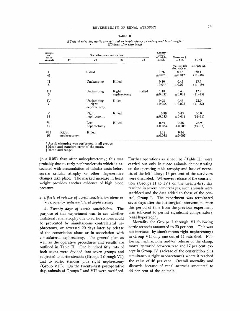
TABLE II
Effects of releasing aortic stenosis and uninephrectomy on kidney and heart weights(20 days after clamping)
Groups Kidneyand Operative procedure on day ratiot
no. of left/right Heart wt.tanimals 1* 20 27 34 i S.E. i S.E. BUNt
Gm. per 100 mg./100 ml.Gm. body wt.
I Killed 0.76 0.45 20.141 ±0.023 ±0.012 (11-38)
II Unclamping Killed 0.80 0.43 13.95 ±0.046 ±0.02 (11-19)
III Unclamping Right Killed 1.10 0.43 12.95 nephrectomy ±0.052 ±0.031 (11-15)
IV Unclamping Killed 0.98 0.43 22.07 + right ±0.056 ±0.023 (11-53)
nephrectomy
V Right Killed 0.99 0.43 30.012 nephrectomy ±0.035 ±0.011 (24-41)
VI Left Killed 0.59 0.36 25.912 nephrectomy +0.033 ±0.009 (19-33)
VII Right Killed 1.12 0.4410 nephrectomy ±0.038 +0.007
* Aortic clamping was performed in all groups.t Mean and standard error of the mean.4 Mean and range.
(p < 0.05) than after uninephrectomy; this wasprobably due to early nephrosclerosis which is as-sociated with accumulation of tubular casts beforesevere cellular atrophy or other degenerativechanges take place. The marked increase in heartweight provides another evidence of high bloodpressure.
2. Effects of release of aortic constriction alone orin association with unilateral nephrectomyA. Twenty days of aortic constriction. The
purpose of this experiment was to see whetherunilateral renal atrophy due to aortic stenosis couldbe prevented by simultaneous contralateral ne-phrectomy, or reversed 20 days later by releaseof the constriction alone or in association withcontralateral nephrectomy. The general plan aswell as the operative procedures and results areoutlined in Table II. One hundred fifty rats ofboth sexes were divided into seven groups andsubjected to aortic stenosis (Groups I through VI)and to aortic stenosis plus right nephrectomy(Group VII). On the twenty-first postoperativeday, animals of Groups I and VII were sacrificed.
Further operations as scheduled (Table II) werecarried out only in those animals demonstratingon the operating table atrophy and lack of necro-sis of the left kidney; 13 per cent of the survivorswere discarded. Whenever release of the constric-tion (Groups II to IV) on the twenty-first dayresulted in severe hemorrhages, such animals weresacrificed and the data added to those of the con-trol, Group I. The experiment was terminatedseven days after the last surgical intervention, sincethis period of time from the previous experimentwas sufficient to permit significant compensatoryrenal hypertrophy.
Mortality for Groups I through VI followingaortic stenosis amounted to 20 per cent. This wasnot increased by simultaneous right nephrectomy:in Group VII only one out of 11 rats died. Fol-lowing nephrectomy and/or release of the clamp,mortality varied between zero and 17 per cent, ex-cept in Group IV (release of the constriction plussimultaneous right nephrectomy) where it reachedthe value of 46 per cent. Overall mortality anddiscards because of renal necrosis amounted to46 per cent of the animals.
23
T. OMAEAND G. M. C. MASSON
Comparison of normal BUNvalues (mean 15.9,range 13 to 19.4 mg. per 100 ml.) with those ob-tained in various groups showed a slight butdefinite increase in Groups I, IV, V and VI.Opening of the clamp was sufficient to bring BUNvalues to normal (13.9 mg. to compare with 20.1mg. per 100 ml. in Group I) but not when associ-ated with simultaneous right nephrectomy (GroupIV). However, when right nephrectomy waspostponed by seven days, BUN values remainednormal.
Aortic constriction (Group I) resulted, as in thepreceding experiment with the "endocrine kidney,"in a kidney ratio of less than one which was notaltered by release of the constriction (Group II).When right nephrectomy was performed at thetime of unclamping (Group IV) or seven dayslater (Group III), the atrophic kidney enlargedand kidney ratios became equal to or greater thanone. There was no statistical difference betweenGroups III and IV. When right nephrectomywas performed without touching the clamp (GroupV), the kidney ratio was almost equal to one, in-dicating that kidney hypertrophy can occur inspite of partial obstruction to renal blood supply.This conclusion was confirmed by the results inGroup VII in which right nephrectomy wasperformed at the time of aortic constriction; theleft kidney not only did not atrophy but actuallyincreased its size, with a resulting kidney ratioof 1.12. The ratio of 0.56 obtained in Group VImerely indicates that removal of a kidney madeatrophic by aortic clamping can elicit further com-pensatory hypertrophy, presumably because someexcretory function remained. Incidentally, in afew rats of Group I, the left kidney was largerthan the right giving individual ratios greater thanone (one of them was 1.3) in comparison with amean ratio of 0.76 for the group; in these instancesthe right kidney was severely nephrosclerotic.
Heart weight values (Table II) were greaterthan 0.43 in all the groups except in Group VIwhere it was normal and equal to 0.36. This isin agreement with results of blood pressure de-terminations. Normal values were obtained inGroup VI (mean 110, range 105 to 120 mm. Hg);in the other groups values were mostly over 150mm. Hg and as high as 200 mm. Hg.
The localization and nature of the lesions havebeen amply documented by studies on rats with
"endocrine kidneys" (1, 2). Except for evidenceof hypertensive vascular disease in all groups, his-tologic examinations were not informative. Wecould not detect any change suggesting healing inGroup VI where hypertension remitted, probablybecause of the short period of observation. Inthis group, lesions of arteritis or arteriolar necrosiswere present in the heart or right kidney of sixrats; however, there was none in the pancreaticor mesenteric blood vessels. It is worth notingthat these lesions did not prevent the fall in pres-sure subsequent to left nephrectomy.
Since the period of observation after unclampingor uninephrectomy may have been too short toevaluate the complete effects of these procedures,we have, in the following experiments, extendedthe period from 7 to 20 days. Aortic stenosis wasperformed in 60 rats which were divided into fivegroups. Groups I and II were controls and weresacrificed, respectively, on the twentieth andfortieth days. The other groups were subjectedon the twentieth day to the following operativeprocedures: unclamping (Group III), unclampingplus right nephrectomy (Group IV), and unclamp-ing plus left nephrectomy (Group V). The ex-periment was terminated on the fortieth day. Theresults obtained were in agreement with the previ-ous ones. In short, renal atrophy was more pro-nounced after 40 than after 20 days, as indicatedby the respective values of kidney ratios of 0.65and 0.76; unclamping did not alter the ratio(0.63), while simultaneous right nephrectomy in-creased its value to 1.16. The only other signifi-cant observation was the high heart weight (mean0.41) following unclamping as compared with thenormal value (mean 0.34) following removal ofthe atrophic kidney. Histologic examinationshowed that after unclamping and right nephrec-tomy, there was complete restoration of renalmorphology so that the left kidney could not bedistinguished from a normal kidney which hadundergone compensatory hypertrophy.
B. Forty days of aortic constriction. The pur-pose of this experiment was similar to that of theprevious one; the difference was in extension from20 to 40 days of the period following aortic steno-sis, in order to produce chronic renal atrophy(Table III). One hundred forty-seven rats weredivided into five groups and subjected to aorticstenosis alone (Groups I to IV) or in association
24
REVERSIBILITY OF RENAL ATROPHY
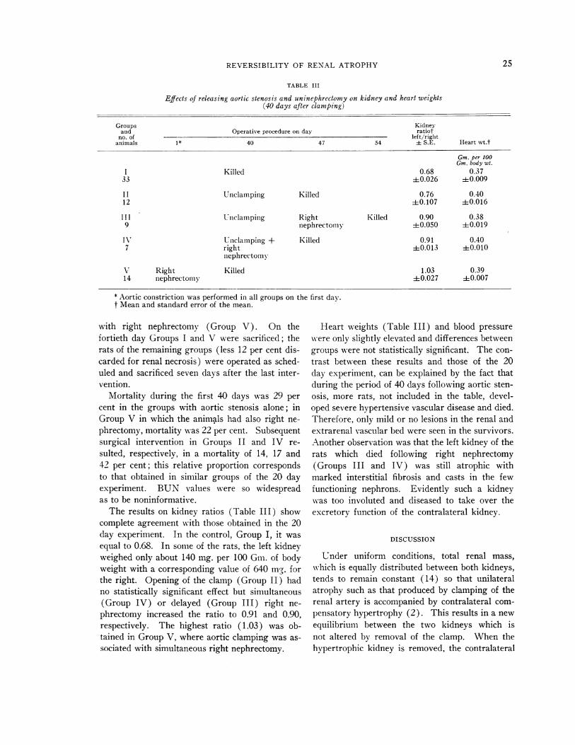
TABLE III
Effects of releasing aortic stenosis and uninephrectomy on kidney and heart weights(40 days after clamping)
Groups Kidneyand Operative procedure on day ratiot
no. of left/rightanimals 1* 40 47 54 i S.E. Heart wt.t
Gm. per 100Gm. body wt.
I Killed 0.68 0.3733 +0.026 ±0.009I I Unclamping Killed 0.76 0.4012 ±0.107 ±0.016
I I I Lf nclamping Right Killed 0.90 0.389 nephrectomy ±4-0.050 ±0.019
IX' Unclamping + Killed 0.91 0.407 right ±t0.013 +0.010
nephrectomy
V Right Killed 1.03 0.3914 nephrectomy ±0.027 ±0.007
* Aortic constriction was performed in all groups on the first day.t Mean and standard error of the mean.
with right nephrectomy (Group V). On thefortieth day Groups I and V were sacrificed; therats of the remaining groups (less 12 per cent dis-carded for renal necrosis) were operated as sched-uled and sacrificed seven days after the last inter-vention.
Mortality during the first 40 days was 29 percent in the groups with aortic stenosis alone; inGroup V in which the animals had also right ne-phrectomy, mortality was 22 per cent. Subsequentsurgical intervention in Groups II and IV re-sulted, respectively, in a mortality of 14, 17 and42 per cent; this relative proportion correspondsto that obtained in similar groups of the 20 dayexperiment. BUN values were so widespreadas to be noninformative.
The results on kidney ratios (Table III) showcomplete agreement with those obtained in the 20day experiment. In the control, Group I, it wasequal to 0.68. In some of the rats, the left kidneyweighed only about 140 mg. per 100 Gm. of bodyweight with a corresponding value of 640 mg. forthe right. Opening of the clamp (Group II) hadno statistically significant effect but simultaneous(Group IV) or delayed (Group III) right ne-phrectomy increased the ratio to 0.91 and 0.90,respectively. The highest ratio (1.03) was ob-tained in Group V, where aortic clamping was as-sociated with simultaneous right nephrectomy.
Heart weights (Table III) and blood pressurewere only slightly elevated and differences betweengroups were not statistically significant. The con-trast between these results and those of the 20day experiment, can be explained by the fact thatduring the period of 40 days following aortic sten-osis, more rats, not included in the table, devel-oped severe hypertensive vascular disease and died.Therefore, only mild or no lesions in the renal andextrarenal vascular bed were seen in the survivors.Another observation was that the left kidney of therats which died following right nephrectomy(Groups III and IV) was still atrophic withmarked interstitial fibrosis and casts in the fewfunctioning nephrons. Evidently such a kidneywas too involuted and diseased to take over theexcretory function of the contralateral kidney.
DISCUSSION
Under uniform conditions, total renal mass,which is equally distributed between both kidneys,tends to remain constant (14) so that unilateralatrophy such as that produced by clamping of therenal artery is accompanied by contralateral com-pensatory hypertrophy (2). This results in a newequilibrium between the two kidneys which isnot altered by removal of the clamp. When thehypertrophic kidney is removed, the contralateral
25
T. OMAEAND G. M. C. MASSON
atrophic kidney increases in size and regains itsfunction, even when the clamp is left in place.This is demonstrated by the present experimentsin which uninephrectomy was performed at thesame time as, or 20 to 40 days after, contralateralreduction of renal blood flow, and is in accord withprevious findings in dogs (10) and rats (9).However, absence of compensatory hypertrophyhas also been reported in 5 of 14 rats (9) and inyoung rabbits (15) in which thread was used asconstricting material. These discrepancies maybe explained by the observation (5) that a loosenylon ligature around the aorta in young rats re-sults during body growth in a progressive and al-most complete involution of the left kidney with-out evidence of hypertension. Presumably theblood supply to this kidney, being gradually de-creased, is barely enough to maintain the integrityof renal tissue and is therefore insufficient to per-mit hypersecretion of pressor substances or com-pensatory hypertrophy. Since gradual and com-plete renal involution was not observed with silverclips, one could assume that the physical natureof the constricting material (such as elasticity,width) is of critical importance.
The nature of the stimulus responsible for com-pensatory hypertrophy is unknown. The hypothe-sis which takes into account most of the pertinentobservations has been proposed by Braun-Menen-dez (16). In short, kidney growth is regulatedby protein metabolites (renotrophins) which arenormally destroyed by the kidney; following re-duction in renal mass, they accumulate and stimu-late renal growth until a new equilibrium isreached. The mitosis-stimulating factor foundin blood following uninephrectomy in rats may berenotrophin (17). That the kidney could inacti-vate its own secretions has been demonstrated withrenin (18). According to this hypothesis, hyper-tension develops whenever a kidney cannot in-crease its size in order to maintain renotrophinsat their normal levels. From this it is assumedthat an atrophic kidney, resulting from renal ar-tery clamping cannot hypertrophy. The presentexperiments disprove this assumption, but sug-gest that an unclamped kidney is more responsivethan a clamped kidney to the same renotrophicstimulus.
Although aortic stenosis resulted generally in akidney ratio left/right smaller than one, we found
a few cases in which the left kidney was largerthan the right. In such animals, the left had anormal appearance while the right was contractedand nephrosclerotic. Having eliminated anatomi-cal anomalies, we assumed that the followingevents took place: aortic clamping elicited atrophyof the left kidney and severe hypertension whichin turn produced nephrosclerosis of the unpro-tected right kidney; the ensuing loss of functioningtissue resulted in contraction of this kidney andsecondarily in hypertrophy of the atrophic kidney.Since the left hypertrophied kidney remainedstructurally normal, the factor responsible for hy-pertrophy is different from the one causing nephro-sclerosis. These observations also demonstratethat renal artery clamping protects the kidneyagainst high blood pressure without interferingwith renotrophic effects.
Release of the aortic constriction did not usuallybring back blood pressure to normal levels as evi-denced by heart weight. The lack of reliability ofplethysmographic values in this situation was dem-onstrated by the impossibility, particularly in the40 day experiment, in registering heart beats fromthe tail of some animals, suggesting persistent par-tial obstruction due to tissue proliferation (6).Considering heart weights greater than 0.40Gm. per 100 Gm. of body weight as indexes ofhypertension, we found that between 40 and 75per cent of the rats were still hypertensive afterrelease of aortic constriction. On the other hand,following removal of the atrophic kidney, the per-centage of animals remaining hypertensive wasless than 10 per cent. The most obvious explana-tions for the persistence of hypertension are pres-ence of vascular lesions in the contralateral kidneyand in the extrarenal arterial bed, and persistenceof mechanical obstructions in the aorta (19).Pieces of aorta removed for histologic examinationwere unfortunately lost. Indeed, a slight obstruc-tion may be sufficient to maintain hypertensionwithout marked interference with normal renalfunction as demonstrated in dogs by Corcoran andPage (20).
SUMMARY
1. Unilateral renal atrophy was elicited inyoung rats by partial constriction of the aorta be-tween the origins of the two renal arteries withsilver clips. This was associated with uninephrec-
26
REVERSIBILITY OF RENAL ATROPHY
tomy, unclamping. or a combination of the twoprocedures. Effects on kidney weight were ex-pressed by the kidney ratio, left kidney/right kid-ney, which in normal rats is equal to one.
2. Aortic stenosis alone resulted 20 and 40 dayslater in a kidney ratio of 0.76 and 0.68, respec-tively.
3. Right nephrectomy performed simultaneouslywith aortic stenosis did not increase the mortalityover aortic stenosis alone. Left renal atrophy wasprevented as shown by kidney ratios greater thanone.
4. Removal of the clamp, 20 to 40 days afteraortic stenosis, did not alter the kidney ratio.However, right nephrectomy caused hypertrophyof the left kidney, even when the clamp was leftin place. The degree of atrophy did not influencerecoverability as long as there were no degenera-tive lesions.
5. Unilateral renal atrophy was usually but notnecessarily accompanied by hypertension. Leftnephrectomy was more effective in remitting hy-pertension than removal of the clamp.
REFERENCES1. Selye, H., and Stone, H. Pathogenesis of the cardio-
vascular and renal changes which usually accom-pany malignant hypertension. J. Urol. (Baltimore)1946, 56, 399.
2. Pellegrini, G. The experimental ischemia of thekidney. Sci. med. ital. 1951, 2, 404.
3. Pickering, G. W., and Prinzmental, M. Experimentalhypertension of renal origin in the rabbit. Clin.Sci. 1938, 3, 357.
4. Bohle, A. Kritischer Beitrag zur Morphologie einerendokrinen Nierenfunktion und deren Bedeutungfur den Hockdruck. Arch. Kreisl.-Forsch. 1953,20, 193.
5. Robert, A. Production d'une atrophie progressivedu rein. Rev. canad. Biol. 1954, 13, 3.
6. Wilson, C., and Byrom, F. B. The vicious circle inchronic Bright's disease. Experimental evidencefrom hypertensive rat. Quart. J. Med. 1941, 10, 65.
7. Poutasse, E. F., and Dustan, H. P. Urologic causesof hypertension; hypertension due to renal arterylesions. Cleveland Clin. Quart. 1956, 23, 3.
8. Gellman, D. D. Reversible hypertension and uni-lateral renal artery disease. Quart. J. Med. 1958,27, 103.
9. Block, M. A., Wakim, K. G., and Mann, F. C. Ap-praisal of certain factors influencing compensatoryrenal hypertrophy. Amer. J. Physiol. 1953, 172,60.
10. Surtshin, A., and Latorre, G. Effects of contra-lateral nephrectomy on renal function depressed byprevious unilateral renal arterial constriction.Amer. J. Physiol. 1955, 182, 524.
11. Simonsen, M. The endocrine kidney. Acta path.microbiol. scand. 1950, 27, 520.
12. Del Greco, F., Olmsted, F., Masson, G. M., and Cor-coran, A. C. Graphic measurement of arterial pres-sure in the unanesthetized rat; an improved method.J. Lab. clin. Med. 1953, 41, 729.
13. Hall, O., Hall, C. E. and Ogden, E. Cardiac hyper-trophy in experimental hypertension and its regres-sion following reestablishment of normal bloodpressure. Amer. J. Physiol. 1953, 174, 175.
14. Braun-Menendez, E. Hypertension and relation be-tween body weight and kidney weight. Acta phys-iol. lat.-amer. 1952, 2, 2.
15'. Drury, D. R. Production of renal insufficiency bysurgical procedure. Proc. Soc. exp. Biol. (N. Y.)1932, 29, 856.
16. Braun-Menendez, E. Evidence for renotrophin asa causal factor in renal hypertension. Circulation1958, 17, 696.
17. Ogawa, K., and Nowinski, W. W. Mitosis stimu-lating factor in serum of unilaterally nephrecto-mized rats. Proc. Soc. exp. Biol. (N. Y.) 1958,99, 350.
18. Friedman, M., Selzer, A., and Sampson, J. J. Ob-servations concerning the pressor substance presentin the ischemic kidney blood of the dog. Amer. J.Physiol. 1941, 131, 799.
19. Floyer, M. A. The effect of nephrectomy and adrenal-ectomy upon the blood pressure in hypertensive andnormotensive rats. Clin. Sci. 1951, 10, 405.
20. Corcoran, A. C., and Page, I. H. Renal blood flowin experimental hypertension. Amer. J. Physiol.1942, 135, 361.
27