Atrioventricular Block-Induced Torsades de Pointes With ...
10
Circulation Journal Vol.74, December 2010 Circulation Journal Official Journal of the Japanese Circulation Society http://www.j-circ.or.jp he acquired form of long QT syndrome (LQTS) is a major cause of torsades de pointes (TdP), 1,2 which results from various factors, including drugs, brady- cardia or hypokalemia. Regarding bradycardia, Kurita et al demonstrated that patients with bradycardia-induced TdP dis- play abnormally prolonged QT intervals at slower heart rates (<60 beats/min) than those without TdP. 3 Some groups have reported the genetic background of bradycardia-induced TdP, as well as of congenital LQTS. In 2001, we reported a female with 2:1 atrioventricular block (AVB) and TdP, in whom the KCNH2 A490T mutant was identified as heterozygous. 4 Sub- sequently, Lupoglazoff et al demonstrated that, in neonates, LQTS with 2:1 AVB is associated with KCNH2 mutations whereas sinus bradycardia-related LQTS is associated with KCNQ1 mutations. 5 Chevalier et al reported that among 29 patients with complete AVB and a QT interval >600 ms, 5 (17%) had mutations on genes encoding K + channels, and the expression test of these mutations showed functional changes compared with the wild-type (WT) K + current. 6 Editorial p 2546 In Japan, some papers on congenital LQTS have been published, 2,7–9 but the molecular pathogenesis of AVB- related TdP has not been fully examined, particularly with respect to the relationship between genotype and cellular electrophysiology. The aim of this study was to investigate gene mutations and clarify their functional outcome in con- T Received May 31, 2010; revised manuscript received July 30, 2010; accepted August 2, 2010; released online October 21, 2010 Time for primary review: 16 days Department of Respiratory and Cardiovascular Medicine (Y.O., H.I., T.S., A.M., M.K., M.H.), Department of Physiology (W.-G.D., H.M.), Shiga University of Medical Science, Otsu; Division of Cardiology, Division of Arrhythmia and Electrophysiology, Department of Cardiovascular Medicine, National Cerebral and Cardiovascular Center, Suita (W.S.); and Department of Cardiovascular Medicine, Kyoto University Graduate School of Medicine, Kyoto (T.M., S.O., Y.N.), Japan Mailing address: Minoru Horie, MD, PhD, Department of Respiratory and Cardiovascular Medicine, Shiga University of Medical Science, Seta Tsukinowa-cho, Otsu 520-2192, Japan. E-mail: [email protected] ISSN-1346-9843 doi: 10.1253/circj.CJ-10-0498 All rights are reserved to the Japanese Circulation Society. For permissions, please e-mail: [email protected] Atrioventricular Block-Induced Torsades de Pointes With Clinical and Molecular Backgrounds Similar to Congenital Long QT Syndrome Yuko Oka, MD; Hideki Itoh, MD, PhD; Wei-Guang Ding, MD, PhD; Wataru Shimizu, MD, PhD; Takeru Makiyama, MD, PhD; Seiko Ohno, MD, PhD; Yukiko Nishio, MD; Tomoko Sakaguchi, MD, PhD; Akashi Miyamoto, MD; Mihoko Kawamura, MD; Hiroshi Matsuura, MD, PhD; Minoru Horie, MD, PhD Background: Atrioventricular block (AVB) sometimes complicates QT prolongation and torsades de pointes (TdP). Methods and Results: The clinical and genetic background of 14 AVB patients (57±21 years, 13 females) who developed QT prolongation and TdP was analyzed. Electrophysiological characteristics of mutations were ana- lyzed using heterologous expression in Chinese hamster ovary cells, together with computer simulation models. Every patient received a pacemaker or implantable cardioverter defibrillator; 3 patients had recurrence of TdP during follow-up because of pacing failure. Among the ECG parameters, QTc interval was prolonged to 561±76 ms in the presence of AVB, but shortened to 495±42 ms in the absence of AVB. Genetic screening for KCNQ1, KCNH2, SCN5A, KCNE1, and KCNE2 revealed four heterozygous missense mutations of KCNQ1 or KCNH2 in 4 patients (28.6%). Functional analyses showed that all mutations had loss of functions and various gating dysfunc- tions of IKs or IKr. Finally, action potential simulation based on the Luo-Rudy model demonstrated that most mutant channels induced bradycardia-related early afterdepolarizations. Conclusions: Incidental AVB, as a trigger of TdP, can manifest as clinical phenotypes of long QT syndrome (LQTS), and that some patients with AVB-induced TdP share a genetic background with those with congenital LQTS. (Circ J 2010; 74: 2562 – 2571) Key Words: Atrioventricular block; Ion channels; Long QT syndrome; Torsades de pointes ORIGINAL ARTICLE Arrhythmia/Electrophysiology
Transcript of Atrioventricular Block-Induced Torsades de Pointes With ...

Circulation Journal Vol.74, December 2010
Circulation JournalOfficial Journal of the Japanese Circulation Societyhttp://www.j-circ.or.jp
he acquired form of long QT syndrome (LQTS) is a major cause of torsades de pointes (TdP),1,2 which results from various factors, including drugs, brady-
cardia or hypokalemia. Regarding bradycardia, Kurita et al demonstrated that patients with bradycardia-induced TdP dis-play abnormally prolonged QT intervals at slower heart rates (<60 beats/min) than those without TdP.3 Some groups have reported the genetic background of bradycardia-induced TdP, as well as of congenital LQTS. In 2001, we reported a female with 2:1 atrioventricular block (AVB) and TdP, in whom the KCNH2 A490T mutant was identified as heterozygous.4 Sub-sequently, Lupoglazoff et al demonstrated that, in neonates, LQTS with 2:1 AVB is associated with KCNH2 mutations whereas sinus bradycardia-related LQTS is associated with
KCNQ1 mutations.5 Chevalier et al reported that among 29 patients with complete AVB and a QT interval >600 ms, 5 (17%) had mutations on genes encoding K+ channels, and the expression test of these mutations showed functional changes compared with the wild-type (WT) K+ current.6
Editorial p 2546
In Japan, some papers on congenital LQTS have been published,2,7–9 but the molecular pathogenesis of AVB-related TdP has not been fully examined, particularly with respect to the relationship between genotype and cellular electrophysiology. The aim of this study was to investigate gene mutations and clarify their functional outcome in con-
T
Received May 31, 2010; revised manuscript received July 30, 2010; accepted August 2, 2010; released online October 21, 2010 Time for primary review: 16 days
Department of Respiratory and Cardiovascular Medicine (Y.O., H.I., T.S., A.M., M.K., M.H.), Department of Physiology (W.-G.D., H.M.), Shiga University of Medical Science, Otsu; Division of Cardiology, Division of Arrhythmia and Electrophysiology, Department of Cardiovascular Medicine, National Cerebral and Cardiovascular Center, Suita (W.S.); and Department of Cardiovascular Medicine, Kyoto University Graduate School of Medicine, Kyoto (T.M., S.O., Y.N.), Japan
Mailing address: Minoru Horie, MD, PhD, Department of Respiratory and Cardiovascular Medicine, Shiga University of Medical Science, Seta Tsukinowa-cho, Otsu 520-2192, Japan. E-mail: [email protected]
ISSN-1346-9843 doi: 10.1253/circj.CJ-10-0498All rights are reserved to the Japanese Circulation Society. For permissions, please e-mail: [email protected]
Atrioventricular Block-Induced Torsades de Pointes With Clinical and Molecular Backgrounds Similar
to Congenital Long QT SyndromeYuko Oka, MD; Hideki Itoh, MD, PhD; Wei-Guang Ding, MD, PhD;
Wataru Shimizu, MD, PhD; Takeru Makiyama, MD, PhD; Seiko Ohno, MD, PhD; Yukiko Nishio, MD; Tomoko Sakaguchi, MD, PhD; Akashi Miyamoto, MD;
Mihoko Kawamura, MD; Hiroshi Matsuura, MD, PhD; Minoru Horie, MD, PhD
Background: Atrioventricular block (AVB) sometimes complicates QT prolongation and torsades de pointes (TdP).
Methods and Results: The clinical and genetic background of 14 AVB patients (57±21 years, 13 females) who developed QT prolongation and TdP was analyzed. Electrophysiological characteristics of mutations were ana-lyzed using heterologous expression in Chinese hamster ovary cells, together with computer simulation models. Every patient received a pacemaker or implantable cardioverter defibrillator; 3 patients had recurrence of TdP during follow-up because of pacing failure. Among the ECG parameters, QTc interval was prolonged to 561±76 ms in the presence of AVB, but shortened to 495±42 ms in the absence of AVB. Genetic screening for KCNQ1, KCNH2, SCN5A, KCNE1, and KCNE2 revealed four heterozygous missense mutations of KCNQ1 or KCNH2 in 4 patients (28.6%). Functional analyses showed that all mutations had loss of functions and various gating dysfunc-tions of IKs or IKr. Finally, action potential simulation based on the Luo-Rudy model demonstrated that most mutant channels induced bradycardia-related early afterdepolarizations.
Conclusions: Incidental AVB, as a trigger of TdP, can manifest as clinical phenotypes of long QT syndrome (LQTS), and that some patients with AVB-induced TdP share a genetic background with those with congenital LQTS. (Circ J 2010; 74: 2562 – 2571)
Key Words: Atrioventricular block; Ion channels; Long QT syndrome; Torsades de pointes
ORIGINAL ARTICLEArrhythmia/Electrophysiology
Circulation Journal Vol.74, December 2010
2563AVB-Induced TdP
secutive AVB patients complicated with TdP.
MethodsStudy PopulationThe study cohort contained 14 consecutive probands, from unrelated families, who showed a prolonged QT interval and TdP associated with AVB. They were referred to 3 institutes in Japan; Shiga University of Medical Science (Otsu), National Cardiovascular Center (Suita), and Kyoto Univer-sity Graduate School of Medicine (Kyoto) for LQTS genetic testing between 1996 and 2008.
Clinical CharacterizationIn each case, we recorded 12-lead electrocardiograms (ECGs) before and after AVB episodes, as well as gathering the results from other cardiovascular examinations and detailed clinical evaluations. Prolonged QT interval was diagnosed by the presence of prolongation of ventricular repolarization (corrected QT interval [QTc] >460 ms in lead V5, according to Bazett’s formula).10 We excluded cases of TdP caused by AVB with drugs associated with QT prolongation, as well as those with active ischemia detected by noninvasive or inva-sive tests, including coronary angiography. We also investi-gated cardiac events in all 14 probands and their family members. Cardiac events were syncope, TdP, ventricular fibrillation (VF), aborted cardiac arrest (requiring defibrilla-tion) or sudden cardiac death. We also followed the therapies and clinical prognoses of these patients.
Genetic AnalysisGenomic DNA was isolated from venous blood by QIAamp DNA blood midikit (Qiagen, Hilden, Germany). Established primer settings were used to amplify the entire coding regions of the known LQTS genes (KCNQ1, KCNH2, SCN5A, KCNE1, and KCNE2). Denaturing high-perfor-mance liquid chromatography (WAVE system Model 3500, Transgenomic, Omaha, NE, USA) was performed as described elsewhere, and abnormal conformers were ampli-fied by polymerase chain reaction (PCR), and sequenced with an ABI PRISM-3130 sequencer (Perkin-Elmer Applied Biosystems, Wellesley, MA, USA). If we detected mutations in these genes, family members associated with the probands were also genetically analyzed. Formal informed consent was obtained from each patient or their guardians according to standards approved by local institutional review boards.
Expression PlasmidsThe expression plasmids, pIRES2-EGFP/KCNQ1(wild-type; WT/KCNQ1) and pRc-CMV/KCNH2 (WT/KCNH2) were kindly provided by Dr Barhanin (Université de Nice, Sophia Antipolis, Valbonne, France) and Dr Sanguinetti (University of Utah, Salt Lake City, UT, USA), respectively. The muta-tions were introduced using overlap PCR. The mutant plas-mids were constructed by substituting the 838-bp XhoI-BglII for the G272V mutant, 464-bp HindIII-BstXI for the D111V mutant, 1458-bp BstXI-BglII for the A490T mutant, or 592-bp FseI-SbfI fragments for the P846T mutant for the corre-sponding fragments of WT/KCNQ1 or WT/KCNH2. The nucleotide sequence of the construct was confirmed prior to the expression studies.
Expression in Chinese Hamster Ovary (CHO) CellsCHO cells were maintained in Dulbecco’s modified Eagle’s medium and Ham’s F12 nutritional mixture (Gibco-BRL,
Rockville, MD, USA) supplemented with 10% fetal bovine serum (Gibco-BRL) and antibiotics (100 U/ml penicillin and 100 μg/ml streptomycin) in a humidified incubator gassed with 5% CO2 and 95% air at 37°C. CHO cells were tran-siently transfected using 1 μg of WT/KCNQ1 or mutant/KCNQ1, and 1 μg of pIRES-CD8/KCNE1 per 35-mm dish, using the LipofectAMINE method according to the manu-facturer’s instructions (Invitrogen, Carlsbad, CA, USA). In some experiments, 0.5 μg of WT/KCNQ1 was transfected with or without mutant/KCNQ1, instead of 1 μg of WT/KCNQ1. Cells successfully transfected with both KCNQ1 and KCNE1 cDNA were selected by green fluorescent pro-tein (GFP) and decoration with anti-CD8 antibody-coated beads (Dynabeads CD8; Dynal Biotech, Oslo, Norway). The cells were transiently transfected with either WT/KCNH2 or mutant/KCNH2, using the LipofectAMINE method accord-ing to the manufacturer’s instructions. For a 35-mm dish the amount of plasmid was 2 μg and 0.175 μg of GFP; only GFP-positive cells were used for the patch-clamp study.
Electrophysiological ExperimentsWhole-cell patch-clamp recordings were conducted at 37.0±1.0°C using an EPC-8 patch-clamp amplifier (HEKA, Lambrecht, Germany) 48–72 h after transfection. No leak subtraction was used. The normal Tyrode solution contained (in mmol/L): NaCl 140, KCl 5.4, CaCl2 1.8, MgCl2 0.5, NaH2PO4 0.33, glucose 5.5, and HEPES 5 (pH adjusted to 7.4 with NaOH). The pipette solution contained (in mmol/L): potassium aspartate 70, KCl 40, KH2PO4 10, EGTA 5, MgSO4 1, Na2-ATP (Sigma, St Louis, MO, USA) 3, Li2-GTP 0.1, and HEPES 5 (pH adjusted to 7.4 with KOH). A coverslip with adherent CHO cells was placed on the bottom of a glass recording chamber (0.5 ml in volume) mounted on the stage of an inverted microscope (TE2000-U, Nikon, Tokyo, Japan). Pipette resistance was 3–5 MΩ when filled with internal solution. Currents and voltages were digitized and voltage commands were generated through an LIH-1600 AD/DA interface (HEKA) controlled by PatchMaster soft-ware (HEKA). Current amplitude was divided by membrane capacitance (Cm) to obtain current densities (pA/pF) in each cell. The voltage-dependence of current activation was deter-mined by fitting the normalized tail current (Itail) vs test potential (Vtest) to a Boltzmann function:
Itail = 1 / (1 + exp[(V0.5 − Vt) / k]),
where V0.5 indicates the voltage at which the current is half-maximally activated and k is the slope factor.
Computer Simulation of Action Potential Duration (APD)Ventricular action potentials were simulated by using the dynamic Luo-Rudy model with recent modifications.11,12 The ratio of IKr and IKs conductance was set at 23:1, 17:1, and 19:1 in the epicardium, endocardium, and M cell layer, respectively. Based on the experimental data of voltage-clamp recordings of KCNH2 channels heterologously expressed in CHO cells, we constructed Markov or Hodg-kin-Huxley models for simulated mutant channels as com-pared with mutants associated with congenital LQTS. In order to construct mutant channel models, we decreased the conductance of each channel as appropriate for the decreased current density, and looked for adequate changes in mutant channels by changing each coefficient value, in turn, for gat-ing states associated with impaired gating defects. The simu-lation for voltage-clamp experiments was calculated using the 4th-order Runge-Kutta method with a fixed-time step of

Circulation Journal Vol.74, December 2010
2564 OKA Y et al.
0.02 ms. The simulation programs were coded in C++ and implemented for personal computers.12
Statistical AnalysisNumerical data are presented as mean ± standard error of the mean. Student’s t-test was used to compare the data between different groups for electrophysiological measurement, and differences were considered significant at P<0.05.
ResultsClinical Characteristics of the Patients in This StudyThe clinical characteristics of the 14 patients enrolled in the study are presented in Table 1. The mean age at the onset of AVB was 57±21 years, and 13 patients (92.8%) were females. All patients showed TdP with AVB: 10 had com-plete AVB, 3 had 2:1 AVB, and 1 had Wenckebach AVB. No patient had experienced syncope or ventricular arrhyth-mias prior to the appearance of TdP. One patient (case 6 in Table 1) with Wenckebach AVB had 2 family members who had suddenly died at the age of 1 year and 3 months, respectively. The mother of case 12 (Table 1) had atrial fibrillation, mitral regurgitation, and complete AVB with prolonged QT interval, but no TdP.
In most patients with AVB-related TdP, the tachyarrhyth-mia started from premature ventricular contractions after a long-pause interval following ventricular arrhythmias, so-called “TdP from short–long–short pattern” (Figure 1D).13 The ECGs available at the time of AVB showed severely prolonged QT interval (heart rate 44±11 beats/min and QTc 561±76 ms). On the other hand, the ECGs without AVB available in 7 cases also showed a prolonged QT interval (heart rate 64±27 beats/min, P<0.05, and QTc 495±42 ms, P=NS, vs those in AVB). ECGs in sinus rhythm were obtained in 4 and 3 patients before and after AVB, respec-tively.
All patients underwent implantation of either an implant-able cardioverter-defibrillator (ICD) or permanent pace-maker (PM), together with the administration of several drugs, including β-blockers (ICD n=3; PM n=11). Mean clinical follow-up during advanced therapy was 102±71 months. After the placement of a PM or ICD, 2 patients maintained own ventricular beats but the other 12 depended on ventricular pacing during the follow-up period. Three patients had recurrence of TdP even while receiving treat-ment. Patient no. 11 suddenly experienced repetitive TdP because of pacing failure and no. 13 also experienced TdP when her own ventricular beats had been set faster than the basal pacing rate. In patient 6, the reappearance of Wencke-bach AVB without ventricular pacing caused ventricular tachycardia. In all 3 cases, no gene mutations were detected.
Molecular Genetics and Clinical Characteristics of Patients With Gene MutationsThe genetic analysis revealed different heterozygous muta-tions in 4 (28.6%) of 14 AVB-related TdP cases (Table 1): 1 KCNQ1 mutation, G272V, and 3 KCNH2 mutations, D111V, A490T and P846T (Figure 1A). All were located in the non-pore regions; G272V is located in the S5 domain for the KCNQ1 channel; D111V, A490T, and P846T are located in the N-terminus, S2–S3 inner loop, and C-terminal domains for the KCNH2 channel, respectively (Figure 1B). In the remaining 10 patients, we were unable to detect any muta-tions associated with the 5 major LQTS-related genes.
G272V in KCNQ1 (Case 7 in Table 1) The G272V muta-
Tabl
e 1.
Clin
ical
Ch
arac
teri
stic
s an
d G
ene
Mu
tati
on
s o
f P
rob
and
s W
ith
Bra
dyc
ard
ia-I
nd
uce
d T
ors
ades
de
Po
inte
s
Cas
e n
o.
Ag
e (y
ears
)S
ex
(M/F
)D
iag
no
sis
Car
dia
c ev
ents
Fam
ily
his
tory
EC
G a
t A
VB
EC
G w
ith
ou
t A
VB
Th
erap
yF
ollo
w-u
pM
uta
tio
n/G
ene
QT
c (m
s)H
R
(bea
ts/m
in)
QT
c (m
s)H
R
(bea
ts/m
in)
Per
iod
(m
on
ths)
Arr
hyt
hm
ic
even
ts
127
F2:
1 A
VB
TdP
–60
050
545
71P
M 4
1N
one
A49
0T/K
CN
H2
274
FC
AV
BT
dP–
NA
NA
NA
NA
PM
22
Non
e 3
73M
CA
VB
TdP
–63
539
NA
NA
PM
104
Non
e 4
57F
CA
VB
TdP
–52
543
545
61IC
D, B
B, I
b 9
6N
one
D11
1V/K
CN
H2
569
F2:
1 A
VB
TdP
–45
245
476
86P
M, B
B, I
b 8
4N
one
621
FW
enck
ebac
h A
VB
TdP
Sud
den
deat
h62
565
489
75IC
D, I
b 7
9V
F b
ecau
se o
f W
enck
ebac
h A
VB
776
F2:
1 A
VB
TdP
–57
850
424
83P
M, B
B 5
9N
one
G27
2V/K
CN
Q1
871
FC
AV
BT
dP–
729
5748
972
ICD
46
Non
eP
846T
/KC
NH
2 9
73F
CA
VB
TdP
–56
733
NA
NA
PM
129
Non
e10
62F
CA
VB
TdP
–47
329
NA
NA
PM
277
Non
e11
68F
CA
VB
TdP
–55
239
NA
NA
PM
207
TdP
bec
ause
of V
pa
cing
failu
re12
38F
CA
VB
TdP
Mot
her
with
LQ
TS
493
2850
065
PM
, BB
NA
NA
1376
FC
AV
BT
dP–
500
46N
AN
AP
M 5
7T
dP b
ecau
se o
f low
ba
ck-u
p ra
te14
18F
CA
VB
TdP
–57
047
NA
NA
PM
130
Non
eA
ve ±
SD
57±
2156
1±76
44±
1149
5±42
64±
2710
2±71
AV
B,
atrio
vent
ricul
ar b
lock
; T
dP,
tors
ades
de
poin
tes;
PM
, pa
cem
aker
; C
AV
B,
com
plet
e at
riove
ntric
ular
blo
ck;
NA
, no
t av
aila
ble;
IC
D,
impl
anta
ble
card
iove
rter
defi
brill
ator
; B
B, β
-blo
cker
; Ib
, cl
ass
Ib
antia
rrhy
thm
ic d
rugs
; VF
, ven
tric
ular
fibr
illat
ion.

Circulation Journal Vol.74, December 2010
2565AVB-Induced TdP
Figure 1. Molecular discovery and clinical data associated with KCNQ1 and KCNH2 mutations, and the initiation of atrioven-tricular block -related torsades de pointes (TdP). (A) Denaturing high-performance liquid chromatography patterns and DNA sequence data in normal controls and patients with G272V for KCNQ1 (Left), D111V for KCNH2 (Middle), and P846T for KCNH2 (Right). (B) Schemes showing the topology of cardiac ion channel proteins for KCNQ1 and KCNH2 and the location of mutations identified in this study. (C) Two pedigrees of G272V and D111V families. Circles and squares represent female and male family members, respectively; probands are indicated by arrows. Heterozygous carriers are represented as half-filled symbols, family members in whom no genetic data were available are shown by open symbols, and non-carriers by open sym-bols with N. QTc intervals corrected by Bazzet’s formula in lead V5 are given for each available family member. (D) Represen-tative ECG recordings from case 7 (76-year-old female with G272V-KCNQ1 mutation). TdP during 2:1 AV block started with so-called “short (!)–long (*)–short (#) pattern” which resulted in a long pause (*).13
D Case 7: 76-year-old female, G272V/KCNQ1 *
!
!
* # ! * #
#

Circulation Journal Vol.74, December 2010
2566 OKA Y et al.
tion was identified in a 76-year-old female who did not have a particularly relevant family history (Figure 1A Left panel). For approximately 10 years, she had taken nilvadip-ine and gliclazide because of hypertension and diabetes mel-litus. Approximately 1 year before hospitalization, her QTc interval was within normal range (424 ms). When she was admitted to hospital because of syncope, her monitoring ECGs displayed 2:1 AVB (50 beats/min), prolonged QTc interval (578 ms), and repetitive TdP (Figure 1D). Her serum K+ level was low (2.5 mEq/L). Because AVB persisted, she underwent DDD PM implantation. After correction of the serum K+ level and PM therapy, her QTc interval shortened and TdP disappeared. She was free from cardiac events for the following 59 months. The genetic analysis revealed 3 children as non-mutation carriers (Figure 1C Left panel).
D111V in KCNH2 (Case 4 in Table 1) The D111V muta-tion was identified in a 57-year-old female who did not have a particularly relevant family history (Figure 1A Middle panel). She experienced syncope after eating breakfast, and the monitoring ECG in the ambulance documented complete AVB (43 beats/min), prolonged QTc interval (525 ms) and TdP. After external PM therapy was initiated, TdP disap-peared. She then underwent ICD implantation and started oral mexiletine hydrochloride (300 mg/day) and propranolol hydrochloride (30 mg/day); she has had no cardiac events over a follow-up period of 96 months. However, her QTc
interval has remained prolonged even in the absence of AVB (545 ms, 4 years later). The genetic tests in her 3 relatives showed 2 mutation carriers (Figure 1C Right panel): a 51-year-old sister and 29-year-old daughter. Both these rela-tives were asymptomatic. Her daughter’s QTc interval was within normal range (414 ms), but the sister’s was prolonged (461 ms).
P846T in KCNH2 (Case 8 in Table 1) The P846T muta-tion was found in a 71-year-old female who did not have a particularly relevant family history (Figure 1A Right panel). She experienced syncope after breakfast, and the monitoring ECG in the ambulance displayed complete AVB (45 beats/min) and repetitive TdP with prolonged QT interval. On admission, her AV conduction resumed at 57 beats/min, but her QTc interval remained prolonged (729 ms). After ICD implanta-tion, she was free from cardiac events for 46 months, but her QTc interval remained prolonged (489 ms). We did not con-duct a genetic analysis in this family.
A490T in KCNH2 (Case 1 in Table 1) We have previously reported the clinical features of a A490T mutation identified in a 27-year-old female.3 Briefly, her 12-lead ECG showed severe bradycardia because of 2:1 AVB (50 beats/min) with complete left bundle branch block and remarkable prolonga-tion of QTc interval (600 ms). She fainted and collapsed while talking on the telephone, and the Holter ECG showed TdP associated with 2:1 AVB.
Figure 2. G272V KCNQ1 mutant channel shows loss of function associated with decreased density on IKs. Functional analyses on wild-type (WT) and G272V mutant channels expressed in Chinese Hamster ovary cells. (A) Representative current record-ings for each channel. Currents were elicited from a holding potential of –80 mV, by depolarizing pulses (4-s duration) from –50 to +50 mV (with a 10-mV step increment) and subsequent repolarization to –50 mV for a 4-s duration (inset). Concentrations of cDNAs used for transfection are indicated near each graph. (B) Tail current-voltage relationships for 1 μg WT (closed circles; n=18), 0.5 μg WT (indicated by dotted line; n=14), 0.5 μg WT plus 0.5 μg G272V (green squares; n=19), 1 μg G272V (red squares; n=11). (C) Normalized activation curves by fitting to Boltzmann function.
Circulation Journal Vol.74, December 2010
2567AVB-Induced TdP
Expression StudyIn order to clarify the functional consequences of the G272V mutation of KCNQ1 and the D111V, A490T, and P846T mutations of KCNH2, we assessed the electrophysiological properties of the WT and mutant clones by using CHO cells.
Biophysical Assay of KCNQ1 Mutant Channel Figure 2A shows representative examples of whole-cell currents recorded from CHO cells transfected with WT/KCNQ1, G272V/KCNQ1 alone or WT co-expressed G272V/KCNQ1 (WT/G272V) plus KCNE1. CHO cells transfected with WT/KCNQ1 (1 or 0.5 μg) displayed outward currents with slow activation/deactivation kinetics on depolarization, which are typical of IKs currents, as previously reported.14,15 In contrast, a cell transfected with G272V/KCNQ1 (1 μg) displayed smaller IKs currents compared with that of the WT (1 μg). WT/G272V at an equimolar ratio (0.5 μg) also showed smaller IKs currents.
In Figure 2B, the tail current densities at –50 mV mea-
sured in multiple cells are plotted as a function of test pulse voltages (between –50 and +50 mV). The tail current densi-ties at −50 mV after depolarizing test pulses to +40 mV were 77.0±11 pA/pF for 1 μg WT (n=18), 49.5±7.9 pA/pF for 0.5 μg WT (n=14), 25.4±4.5 pA/pF for 0.5 μg WT/G272V (n=19) (vs WT 1 μg, P<0.001), 26.7±4.9 pA/pF for 1 μg G272V (n=11) (vs WT 1 μg, P<0.01). Thus, compared with the WT IKs current, co-transfection of the mutant affected the expressed current densities.
Figure 2C represents the voltage-dependence of current activation. Tail current densities after each test potential were fitted to a Boltzmann function (see Methods). The parameters were V0.5=−5.2±3.0 mV, k=11.1±0.6 for 1 μg WT, V0.5=−1.0±3.7 mV, k=11.7±1.1 for 0.5 μg WT/G272V, V0.5=5.7±5.4 mV, k=14.3±1.6 (vs WT 1 μg; P<0.05) for 1 μg G272V. Regarding half-activation voltages, WT plus G272V and G272V tended to shift to the depolarization side com-pared with WT but there was no statistical significance. In
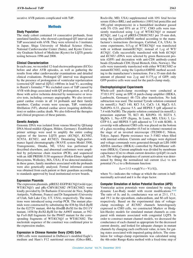
Figure 3. All KCNH2 mutant channels show loss of function associated with decreased density on IKr. Electrophysiological characteristics of KCNH2 mutant channels in Chinese Hamster ovary cells. (A) Families of representative current traces. Ex-perimental protocol is shown in the inset. Concentrations of cDNAs used for transfection are indicated above each graph. (B) Tail current-voltage relationships for 2 μg wild-type (WT: closed circles; n=20), 1 μg WT (dotted line; n=14), 1 μg WT plus 1 μg mutant (green squares; n=10–13), 2 μg mutant (red squares; n=8–12). (C) Normalized activation curves by fitting to Boltzmann function.
Table 2. Parameters of Inactivation in WT and Mutant KCNH2
WT (n=16)
WT/D111V (n=16)
D111V (n=15)
WT/A490T (n=17)
A490T (n=15)
WT/P846T (n=15)
P846T (n=16)
V0.5 (mV) –58.3±4.7 –40.1±4.1** –47.4±7.0 –32.5±3.9* –44.2±3.3 –38.7±2.4** –55.5±3.5
Slope factor 29.2±1.4 33.9±1.3 35.3±1.7** 30.6±1.4† 34.9±1.1** 33.0±0.6† 37.5±1.7*
*P<0.001 vs WT, **P<0.01 vs WT, †P<0.05 vs WT.WT, wild-type.
Circulation Journal Vol.74, December 2010
2568 OKA Y et al.
slope factors, G272V alone channel was larger than WT (P<0.05). Overall, the most important finding was the domi-nant-negative effect for the G272V channel.
Biophysical Assay of 3 KCNH2 Mutant Channels Figure 3A shows representative examples of whole-cell currents re-corded from CHO cells transfected with WT/KCNH2 (2 and 1 μg), mutant/KCNH2 (2 μg), or WT co-expressed mutant/ KCNH2 (WT/mutant) (1 μg each). CHO cells transfected with WT/KCNH2 (2 or 1 μg, Figure 3A Upper 2 panels) displayed outward currents with inward rectifying properties, which are typical of IKr currents.16 In contrast, the magnitude of currents from cells expressing all of the WT/mutants and mutant only were remarkably reduced (Figure 3A Lower 6 panels).
In Figure 3B, the tail current densities at –60 mV are plot-ted as a function of test pulse voltages (between –60 and +50 mV). The mean current densities after depolarizing test pulse to +20 mV in WT channels were 66.2±11 pA/pF for 2 μg (n=20) and 45.0±9.3 pA/pF for 1 μg (n=14). In contrast, those in the WT/mutant and mutant channels were 25.1±2.9 pA/pF in WT/D111V (n=13), 15.8±6.0 pA/pF in WT/A490T (n=10), 20.5±3.9 pA/pF in WT/P846T (n=12), 18.8±3.6 pA/pF for D111V (n=9), 15.2±3.4 pA/pF for A490T (n=12), 6.1±2.3 pA/pF for P846T (n=8), respectively. They were all significantly smaller than those of the 2-μg WT channels (vs WT 2 μg; P<0.01). Figure 3C shows that all WT/mutant and mutant channels tended to shift to the depolarization side
compared with the WT. Overall, all mutant channels showed loss of function associated with a dominant-negative effect and shift of the activation curve to depolarization.
We then examined whether the mutations affected the inactivation kinetics of mutant channels using a double-pulse protocol. V0.5 and the slope factor of steady-state inactiva-tion differed between WT and WT plus mutant or mutant. All mutant KCNH2 channels showed the shift of inactivation curves to depolarizing direction, and the differences were statistically significant (Table 2). Therefore, we also changed the parameters associated with inactivation states in the fol-lowing simulation study.
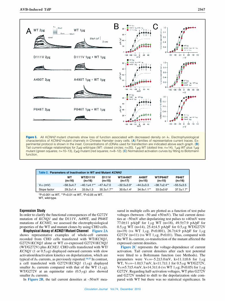
Figure 4A depicts original current traces showing deacti-vation at 4 different repolarization potentials (from −70 to −40 mV) of WT and/or mutant/KCNH2. Deactivating currents were best fit with a double-exponential function, and are sum-marized in Figure 4B. At 4 different potentials, both time constants (Tau-fast and Tau-slow) for D111V and WT/D111V were smaller than those of the WT. Tau-slow of WT/P846T was also smaller than those of the WT. We could not assess that of P846T (2 μg), because it was too small to measure. In contrast, there were no significant changes between the WT and WT/A490T or A490T in the deactivation process.
Computer Simulation of APDIn order to compare how functional changes caused by muta-tions affect ventricular action potentials, a simulation study
Figure 4. KCNH2 mutant channels show a faster deactivation. (A) Representative tail currents elicited after depolarization to +40 mV (inset protocol). (B) Deactivation process was fitted to 2 components having different time constants, tau-fast and tau-slow. They were calculated in a pooled data from multiple cells transfected by 3 mutants (from top of panel B, D111V, A490T and P846T, respectively) and are plotted against membrane potentials (−70 to −40), for 2 μg WT (closed circles; n=15), 1 μg WT plus 1 μg mutant (green squares; n=7–14 for each channel), 2 μg mutant (red squares; n=6–8).

Circulation Journal Vol.74, December 2010
2569AVB-Induced TdP
was conducted using the Luo-Rudy model, which incorpo-rated the Markov13 or Hodgkin-Huxley17 process gating for the mutant channels (Figure 5). Table 3 shows the parame-ters of simulation that were changed to fit to experimental results. We simulated action potentials in all myocardial layers at 3 different basic cycle lengths (BCL 600, 1,000, 2,000 ms) (Figures 5A–C). In the endocardium and epicar-dium, APD of all mutant models was prolonged, but did not produce early afterdepolarizations (EAD) (data not shown in Figure 5). In contrast, in the simulated M cell layer, APD was lengthened significantly at a slower heart rate. In the lower half of Figure 5, below each simulated action poten-tial, the corresponding bar graphs show APDs at 90% repo-larization. Three APD models with D111V, A490T, and P846T displayed EADs at BCL of 2,000 ms, whereas G272V displayed it at all BCLs.
DiscussionThere are 3 major findings in the present study. (1) In 4 of 14 consecutive AVB-associated TdP patients, 3 KCNH2 and 1 KCNQ1 heterozygous missense mutations were identified. (2) Electrophysiological analyses revealed loss of function associated with decreased current densities and various dys-functions on IKs or IKr in 4 mutants. (3) Functional changes reconstituted by the computer simulation resulted in a pro-longed APD and EAD under condition of bradycardia.
During AVB, our 14 patients showed a prolonged QT interval and TdP. Based on a comparison of ECGs available before and after AVB, we found the QT intervals were lengthened even in the absence of AVB. These clinical char-acteristics indicate that AVB-related TdP might share a simi-lar genetic background with congenital LQTS: mutations on cardiac ion channel genes could be partially causative. Lupo-
Figure 5. Computer simulations with Luo-Rudy myocardial model show bradycardia-induced early afterdepolarization (EAD). Each figure presents (A) action potential duration (APD) at basic cycle length (BCL) of 600 ms, (B) APD at BCL of 1,000 ms, and (C) APD at BCL of 2,000 ms. The longer the BCL in each model was, the more the APD was prolonged. EADs appeared in the models of D111V/WT, A490T/WT, and P846T/WT only at BCL of 2,000 ms, but appeared at all BCLs in the case of G272V/WT.
Table 3. Parameters of Simulation Data in Bradycardia-Induced Long QT Syndrome
Gene Mutation WT basal parameters Mutant changed parameters
KCNQ1 G272V gsk=0.202*(1+0.6/(1+pow(0.000038/cai),1.4))) gsk=0.067*(1+0.6/(1+pow(0.000038/cai),1.4)))
xs1 ss=1/(1+exp(-(v-1.5)/16.7)) xs1 ss=1/(1+exp(-(v-6.5)/16.7))
KCNH2 D111V gherg=0.0135*pow(Kout,0.59) gherg=0.331*0.0135pow(Kout,0.59)
αα=65.5e-3*exp(0.05547153*(v-36)) αα=65.5e-3*exp(0.05547153*(v-69))
αi=0.439*exp(–0.02352*(v+25))*4.5/Kout αi=0.439*exp(–0.02352*(v+3))*4.5/Kout
ββ=2.9375e-3*exp(–0.02158*v) ββ=2*2.9375e-3*exp(–0.02158*v)
KCNH2 A490T gherg=0.0135*pow(Kout,0.59) gherg=0.1887*0.0135pow(Kout,0.59)
αi=0.439*exp(–0.02352*(v+25))*4.5/Kout αi=0.439*exp(–0.02352*(v-6))*4.5/Kout
KCNH2 P846T gherg=0.0135*pow(Kout,0.59) gherg=0.265*0.0135pow(Kout,0.59)
αα=65.5e-3*exp(0.05547153*(v-36)) αα=65.5e-3*exp(0.05547153*(v-80))
αi=0.439*exp(–0.02352*(v+25))*4.5/Kout αi=0.439*exp(–0.02352*(v+3))*4.5/Kout
ββ=2.9375e-3*exp(–0.02158*v) ββ=1.3*2.9375e-3*exp(–0.02158*v)

Circulation Journal Vol.74, December 2010
2570 OKA Y et al.
glazoff et al5 demonstrated that in neonates that, while LQTS with 2:1 AVB is associated with KCNH2 mutations, sinus bradycardia-related LQTS is associated with KCNQ1 muta-tions. In 9 of 10 cases, 2:1 AVB-induced LQTS could be caused by LQTS-related gene mutations. In contrast, Cheva-lier et al found 4 K+ channel gene mutations in 5 of 29 adult patients with AVB-induced LQTS (17.3%).6 Our cohort also consisted of adult LQTS patients, with a mutation rate of 28.6%. This prevalence rate was similar to Chevalier’s report, but lower than that in the 2:1 AVB-related LQTS in neonates. These studies have shown that AVB-induced LQTS in neonates has a stronger genetic association than AVB-induced LQTS in adults. Regarding the diagnostic rate of genetic testing in general, no candidate mutations could be detected in 30–40% of congenital LQTS cases. In contrast, it has been shown recently that genetic polymorphisms modify the QT interval.18–24 Although we did not check polymor-phisms in the present study, it is possible that our subjects might have some modifier-gene mutations. Thus, it remains possible that the remaining 10 patients in our study without apparent genetic variants may have as yet unknown variants.
In our cohort, it was difficult to prove the efficacy of β-blockers because very few patients were taking these drugs. In order to investigate the efficacy of β-blockers it will be necessary to study more cases with AVB-induced TdP. The first step in the treatment of all patients with AVB-induced TdP is the implantation of a device. Although PM implanta-tion as first-line therapy for AVB-induced TdP is not dis-puted, 3 of our patients had a recurrence of TdP after the device was implanted, because of inadequate ventricular pacing, suggesting that AVB patients with TdP require strict PM management. In cases of persistent QT prolongation, even after PM therapy, it might become necessary to con-sider ICD implantation.
Several AVB-related gene mutations have been function-ally assayed:6 3 KCNH2 mutations, R328C, R696C and R1047L, were shown to have no strong dominant-negative effects on IKr. Another KCNE2 mutation (R77 W), which was identified in an AVB patient while taking flecainide, exerted no effects on IKr. Overall, previous analyses of mutations have shown them to cause only mild functional change. Our study showed similar results; all 4 mutants displayed loss of function associated with decreased densities on IKs or IKr, which were basically similar to those in congenital LQTS. On average, our patients experienced TdP at 57 years of age, which is older than the mean age of onset reported for those with congenital LQTS. Mutation carriers, who remain asymp-tomatic well into adulthood, may incidentally have fatal events in the presence of additional triggers, such as AVB.25
Several mutations of SCN5A, coding the α-subunit of Na+ channels, have been found in newborn and infant cases of long QT.26–28 They showed functional 2:1 AVB caused by profound QT prolongation. Therefore, the pathological basis differs between those cases and ours. Irrespective of genetic testing results, our patients who developed TdP in the pres-ence of AVB showed QT prolongation, even in sinus rhythm. Thus, AVB may not be directly associated with QT prolon-gation, but the bradycardia caused by AVB enhances it and eventually leads to TdP. Our computer simulation study showed that, at a slower heart rate, APD lengthened signifi-cantly, suggesting that AVB-related bradycardia could exac-erbate QT prolongation.
Study LimitationFemale sex is a predisposing factor for the development of
cardiac arrhythmic events in patients with congenital and acquired LQTS, as previous reports have demonstrated.29–31 In our study, almost all patients (93%) were also female, and therefore it would be possible that not only AVB but female sex affected cardiac repolarization and ventricular irritability in our cohort.
ConclusionThis study showed that incidental AVB as a trigger of TdP could manifest as clinical phenotypes of LQTS, and that some patients with AVB-induced TdP could have genetic backgrounds associated with congenital LQTS-related genes.
AcknowledgmentsWe thank Ms Arisa Ikeda for excellent technical assistance. This work was supported by a Grant-in-Aid for Scientific Research from the Japan Society for the Promotion of Science and the Biosimulation and Health Sciences Research Grant from the Ministry of Health, Labor, and Wel-fare of Japan, and grants from the Uehara Memorial Foundation (M.H.) and the Grant-in-Aid for Young Scientists from the Ministry of Educa-tion, Culture and Technology of Japan (H.I.).
References 1. Moss A, Kass R. Long QT syndrome: From channels to cardiac
arrhythmias. J Clin Invest 2005; 115: 2018 – 2024. 2. Shimizu W. Clinical Impact of Genetic Studies in Lethal Inherited
Cardiac Arrhythmias. Circ J 2008; 72: 1926 – 1936. 3. Kurita T, Ohe T, Marui N, Aihara N, Takaki H, Kamakura S, et al.
Bradycardia-induced abnormal QT prolongation in patients with complete atrioventricular block with torsades de pointes. Am J Cardiol 1992; 69: 628 – 633.
4. Yoshida H, Horie M, Otani H, Kawashima T, Onishi Y, Sasayama S. Bradycardia-induced long QT syndrome caused by a de novo missense mutation in the S2-S3 inner loop of HERG. Am J Med Genet 2001; 98: 348 – 352.
5. Lupoglazoff JM, Denjoy I, Villain E, Fressart V, Simon F, Bozio A, et al. Long QT syndrome in neonates: Conduction disorders associated with HERG mutations and sinus bradycardia with KCNQ1 mutations. J Am Coll Cardiol 2004; 43: 826 – 830.
6. Chevalier P, Bellocq C, Millat G, Piqueras E, Potet F, Schott JJ, et al. Torsades de pointes complicating atrioventricular block: Evi-dence for a genetic predisposition. Heart Rhythm 2007; 4: 170 – 174.
7. Nagaoka I, Shimizu W, Itoh H, Yamamoto S, Sakaguchi T, Oka Y, et al. Mutation site dependent variability of cardiac events in Japa-nese LQT2 form of congenital long-QT syndrome. Circ J 2008; 72: 694 – 699.
8. Makita N. Phenotypic overlap of cardiac sodium channelopathies. Circ J 2009; 73: 810 – 817.
9. Ogawa K, Nakamura Y, Terano K, Ando T, Hishitani T, Hoshino K. Isolated non-compaction of the ventricular myocardium associ-ated with long QT syndrome. Circ J 2009; 73: 2169 – 2172.
10. Bazett HC. An analysis of the time relations of electrocardiograms. Heart 1920; 7: 353 – 370.
11. Clancy CE, Rudy Y. Na+ channel mutation that causes both Bru-gada and long-QT syndrome phenotypes: A simulation study of mechanism. Circulation 2002; 105: 1208 – 1213.
12. Itoh H, Sakaguchi T, Ding WG, Watanabe E, Watanabe I, Nishio Y, et al. Latent genetic background and molecular pathogenesis of drug-induced long QT syndrome. Circ Arrhythmia Electrophysiol 2009; 2: 511 – 523.
13. Tan HL, Bardai A, Shimizu W, Moss AJ, Schulze-Bahr E, Noda T, et al. Genotype-specific onset of arrhythmias in congenital long-QT syndrome. Circulation 2006; 114: 2096 – 2103.
14. Barhanin J, Lesage F, Guillemare E, Fink M, Lazdunski M, Romey G. KvLQT1 and lsK (minK) proteins associate to form the IKs car-diac potassium current. Nature 1996; 384: 78 – 80.
15. Sanguinetti MC, Curran ME, Zou A, Shen J, Spector PS, Atkinson DL, et al. Coassembly of KvLQT1 and minK (IsK) proteins to form cardiac IKs potassium channel. Nature 1996; 384: 80 – 83.
16. Sanguinetti MC, Jiang C, Curran ME, Keating MT. A mechanistic link between an inherited and an acquired cardiac arrhythmia: HERG encodes the IKr potassium channels. Cell 1995; 81: 299 – 307.

Circulation Journal Vol.74, December 2010
2571AVB-Induced TdP
17. Hodgkin AL, Huxley AF. A quantitative description of membrane current and its application to conduction and excitation in nerve. J Physiol 1952; 117: 500 – 544.
18. Newton-Cheh C, Guo CY, Larson MG, Musone SL, Surti A, Camargo AL, et al. Common genetic variant in KCNH2 is associ-ated with QT interval duration: The Framingham Heart Study. Cir-culation 2007; 116: 1128 – 1136.
19. Plant LD, Bowers PN, Liu Q, Morgan T, Zhang T, State MW, et al. A common cardiac sodium channel variant associated with sudden infant death in African Americans, SCN5A S1103Y. J Clin Invest 2006; 116: 430 – 435.
20. Nishio Y, Makiyama T, Itoh H, Sakaguchi T, Ohno S, Gong YZ, et al. D85N, a KCNE1 polymorphism, is a disease-causing gene vari-ant in long QT syndrome. J Am Coll Cardiol 2009; 54: 812 – 819.
21. Newton-Cheh C, Eijgelsheim M, Rice KM, de Bakker PI, Yin X, Estrada K, et al. Common variants at ten loci influence QT interval duration in the QTGEN study. Nat Genet 2009; 41: 399 – 406.
22. Pfeufer A, Sanna S, Arking DE, Müller M, Gateva V, Fuchsberger C, et al. Common variants at ten loci modulate the QT interval duration in the QTSCD study. Nat Genet 2009; 41: 407 – 414.
23. Arking DE, Pfeufer A, Post W, Kao WH, Newton-Cheh C, Ikeda M, et al. A common genetic variant in the NOS1 regulator NOS1AP modulate cardiac repolarization. Nat Genet 2006; 38: 644 – 651.
24. Crotti L, Monti MC, Insolia R, Peljto A, Goosen A, Brink PA, et al. NOS1AP is a genetic modifier of the long-QT syndrome. Circu-lation 2009; 120: 1657 – 1663.
25. Sakaguchi T, Shimizu W, Itoh H, Noda T, Miyamoto Y, Nagaoka
I, et al. Hydroxyzine, a first generation H(1)-receptor antagonist, inhibits human ether-a-go-go-related gene (HERG) current and causes syncope in a patient with the HERG mutation. J Pharmacol Sci 2008; 108: 462 – 471.
26. Lupoglazoff JM, Cheav T, Baroudi G, Berthet M, Denjoy I, Cauchemez B, et al. Homozygous SCN5A mutation in long-QT syndrome with functional two-to-one atrioventricular block. Circ Res 2001; 89: E16 – E21.
27. Chang CC, Acharfi S, Wu MH, Chiang FT, Wang JK, Sung TC, et al. A novel SCN5A mutation manifests as a malignant form of long QT syndrome with perinatal onset of tachycardia/bradycardia. Cardiovasc Res 2004; 64: 268 – 278.
28. Miura M, Yamagishi H, Morikawa Y, Matsuoka R. Congenital long QT syndrome and 2:1 atrioventricular block with a mutation of the SCN5A gene. Pediatr Cardiol 2003; 24: 70 – 72.
29. Nakamura H, Kurokawa J, Bai CX, Asada K, Xu J, Oren RV, et al. Progesterone regulates cardiac repolarization through a nonge-nomic pathway: An in vitro patch-clamp and computational mod-eling study. Circulation 2007; 116: 2913 – 2922.
30. Makkar RR, Fromm BS, Steinman RT, Meissner MD, Lehmann MH. Female gender as a risk factor for torsades de pointes associ-ated with cardiovascular drugs. JAMA 1993; 270: 2590 – 2597.
31. Locati EH, Zareba W, Moss AJ, Schwartz PJ, Vincent GM, Lehmann MH, et al. Age- and sex-related differences in clinical manifestations in patients with congenital long-QT syndrome: Findings from the International LQTS Registry. Circulation 1998; 97: 2237 – 2244.