Association of Generalized Joint Hypermobility and ...
54
University of North Dakota UND Scholarly Commons Physical erapy Scholarly Projects Department of Physical erapy 2015 Association of Generalized Joint Hypermobility and Occurrence of Musculoskeletal Injury in Physical and Occupational erapy Students Patricia Bisek University of North Dakota Hannah Owen University of North Dakota Maleeka Rozeboom University of North Dakota Leah Tunseth University of North Dakota Follow this and additional works at: hps://commons.und.edu/pt-grad Part of the Physical erapy Commons is Scholarly Project is brought to you for free and open access by the Department of Physical erapy at UND Scholarly Commons. It has been accepted for inclusion in Physical erapy Scholarly Projects by an authorized administrator of UND Scholarly Commons. For more information, please contact [email protected]. Recommended Citation Bisek, Patricia; Owen, Hannah; Rozeboom, Maleeka; and Tunseth, Leah, "Association of Generalized Joint Hypermobility and Occurrence of Musculoskeletal Injury in Physical and Occupational erapy Students" (2015). Physical erapy Scholarly Projects. 606. hps://commons.und.edu/pt-grad/606
Transcript of Association of Generalized Joint Hypermobility and ...

University of North DakotaUND Scholarly Commons
Physical Therapy Scholarly Projects Department of Physical Therapy
2015
Association of Generalized Joint Hypermobilityand Occurrence of Musculoskeletal Injury inPhysical and Occupational Therapy StudentsPatricia BisekUniversity of North Dakota
Hannah OwenUniversity of North Dakota
Maleeka RozeboomUniversity of North Dakota
Leah TunsethUniversity of North Dakota
Follow this and additional works at: https://commons.und.edu/pt-grad
Part of the Physical Therapy Commons
This Scholarly Project is brought to you for free and open access by the Department of Physical Therapy at UND Scholarly Commons. It has beenaccepted for inclusion in Physical Therapy Scholarly Projects by an authorized administrator of UND Scholarly Commons. For more information,please contact [email protected].
Recommended CitationBisek, Patricia; Owen, Hannah; Rozeboom, Maleeka; and Tunseth, Leah, "Association of Generalized Joint Hypermobility andOccurrence of Musculoskeletal Injury in Physical and Occupational Therapy Students" (2015). Physical Therapy Scholarly Projects. 606.https://commons.und.edu/pt-grad/606

ASSOCIATION OF GENERALIZED JOINT HYPERMOBILITY AND OCCURRENCE OF MUSCULOSKELETAL INJURY IN PHYSICAL AND
OCCUPATIONAL THERAPY STUDENTS
by
Patricia Bisek Bachelor of Arts in Exercise Science
Concordia College, 2012
Hannah Owen Bachelor of Arts in Biology with Emphasis in Health and Medical Sciences
Minnesota State University Moorhead, 2011
Maleeka Rozeboom
Leah Tunseth
A Scholarly Project
Submitted to the Graduate Faculty ofthe
Department of Physical Therapy
School of Medicine and Health Science
University of North Dakota
in partial fulfillment of the requirements
for the degree of
Doctor of Physical Therapy
Grand Forks, North Dakota May 2015


This Scholarly Project, submitted by Patricia Bisek, Hannab Owen, Maleeka Rozeboom, and Leah Tunseth in partial fulfillment of the requirements for the Degree of Doctor of Physical Therapy from the University of North Dakota, has been read by the Advisor and Chairperson of Physical Therapy under whom the work has been done and is hereby approved.
ZGraduate School Advis
ii

Title
Department
Degree
PERMISSION
Association of Generalized Joint Hypermobility and Occurrence of Musculoskeletal Injury in Physical and Occupational Therapy Students
Physical Therapy
Doctor of Physical Therapy
In presenting this Scholarly Project in partial fulfillment of the reqillrements for a graduate degree from the University of North Dakota, we agree that the Department of Physical Therapy shall make it freely available for inspection. We further agree that permission for extensive copying for scholarly purposes may be granted by the professor who supervised our work or, in her absence, by the Chairperson of the Department. It is understood that any copying or publication or other use of this Scholarly Project or part thereof for financial gain shall not be allowed without our written permission. It is also understood that due recognition shall be given to us and the University of North Dakota in any scholarly use which may be made of any material in this Scholarly Project.
Signature
Date
iii
~ tyJ\Jl)vL.,
/v1~ ~.wem ~-
}\)\~ 11, 'lDIL{

TABLE OF CONTENTS
LIST OF FIGURES .................................................................................. V
LIST OF TABLES .................................................................................. VI
ACKNOWLEDGEMENTS ....................................................................... VII
ABSTRACT .............................................................................................. VIII
CHAPTER 1. INTRODUCTION ........................................................ .1
II. LITERATURE REVIEW ................................................. 5
III. METHODS ................................................................ 13
IV. RESULTS ................................................................. 21
V. DISCUSSION and CONCLUSION ................................... 23
APPENDIX A ........................................................................................ 28
APPENDIX B ........................................................................................ 30
APPENDIX C ....................................................................................... .34
APPENDIX D ....................................................................................... .38
REFERENCES ..................................................................................... .40
iv

LIST OF FIGURES
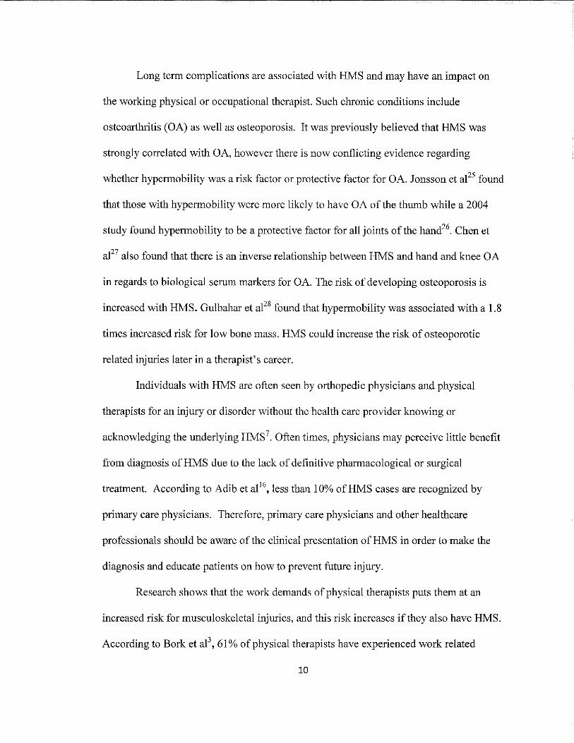
1. Measurement of elbow hyperextension greater than 10° .............................. 18
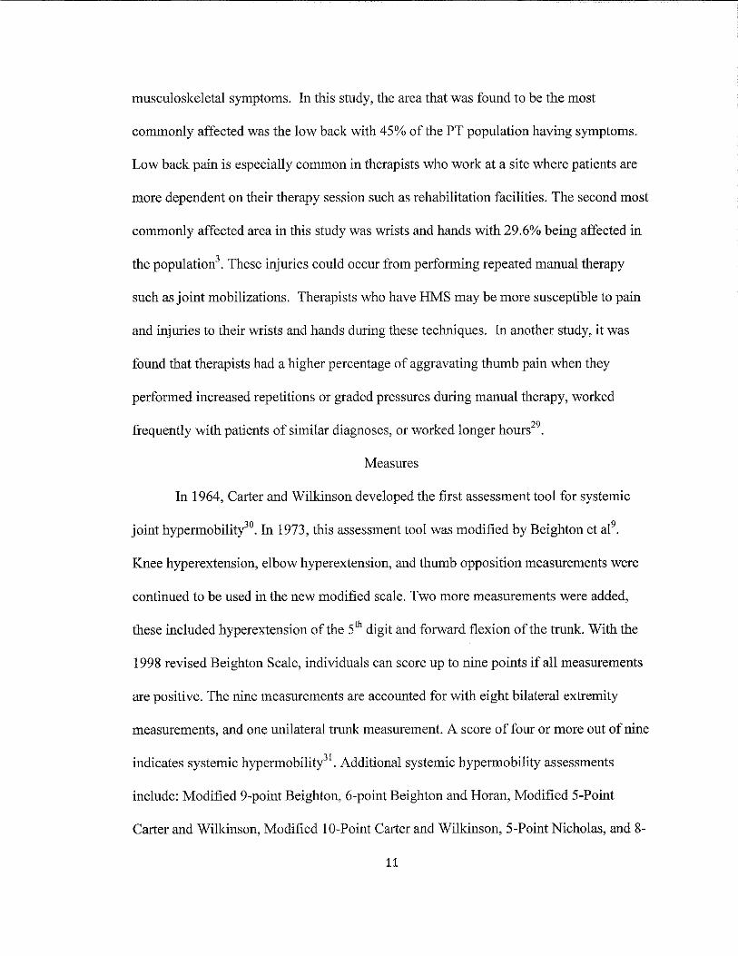
2. Measurement of 5th digit extension greater than 90° ................................... 18
3. Measurement of knee hyperextension greater than 10° ............................... 19
4. Apposition of the thumb to forearm ..................................................... .19
5. Trunk flexion with palms flat on floor. .................................................. 20
6. The percentage of participants in each group that have experienced at least one of the respective injuries ..................................................... 22
v
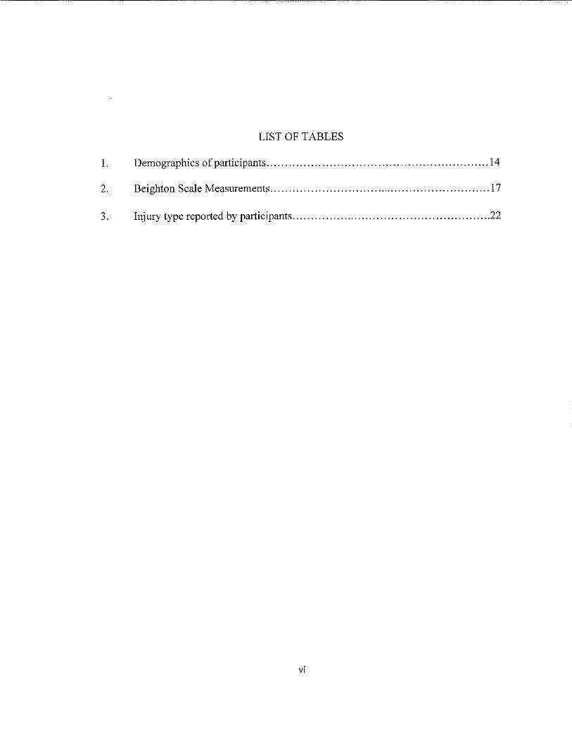
LIST OF TABLES
I. Demographics of participants ............................................................ 14
2. Beighton Scale Measurements ............................................................ 17
3. Injury type reported by participants ...................................................... 22
vi

ACKNOWLEDGEMENTS
We would like to recognize the University of North Dakota Department of
Physical Therapy for the use of the research room and necessary equipment, as well as
the Department of Occupational Therapy for allowing us to utilize their facility to
conduct our research.
We would also like to thank our advisor, Sue Jeno PT, PhD, for the guidance and
opportunity to be involved in research supporting our future profession. Your
encouragement and availability to act as a mentor has been greatly appreciated.
A special thanks to Renee Mabey, PT, PhD, for advising and instructing us in data
input procedures as well as preforming comprehensive statistical analysis. Your expertise
and invaluable logistical support has allowed us to gain a thorough understanding of the
significance of our results.
We would like to recognize the physical therapy students who participated in our
preliminary reliability study for their considerable time commitment and patience.
Without their participation we would not have gained the reliability necessary to conduct
this study.
Finally, we would like to thank each PT and OT student who volunteered their
time to support our research. It was greatly appreciated that they recognize the
importance of research development for our professions and personally invested in the
expansion of our current professional knowledge.
vii

ABSTRACT
Background: A prior research study showed that the prevalence ofhypermobility was
higher among Physical and Occupational Therapy students as compared to the general
population. The literature shows that certain injury rates are higher among those who are
hypermobile. This has led to the question of whether or not hypermobility is directly
related to injury and recurrence of injury.
Purpose: The purpose of this study was to assess Physical Therapy (PT) and
Occupational Therapy (OT) students for hypermobility as well as survey previous injury
history. This study analyzed the prevalence ofhypermobility with types of injuries in
order to determine if a relationship exists.
Methods: Eighty-six subjects (24 male, 62 female) were assessed for hypermobility
using the nine point Beighton Scale of Hypermobility. A score offour or higher out of
nine indicated the presence of joint hypermobility. Participants filled out a survey
regarding current activity level, previous and current athletic participation, injury history
regarding type and mechanism of injury.
Results: The prevalence ofhypermobility among PT and OT students was found to be
39.5%, a rate five times greater than the general population. Reported injuries were
grouped into the following classifications prior to statistical analysis: sprains, ligament
rupture, strain/contusion, fracture, and dislocation. Non-hypermobile participants were
more likely to have experienced a strain type injury (l(1, N=86) =5.059, p=0.024). No
viii

other statistically significant results were found, although fracture rates showed a trend of
occurring more frequently in non-hypermobile participants (p=O.167).
Conclusion: The prevalence of joint hypermobility is higher among Physical and
Occupational Therapy students than the general population. Injury rates are high among
both PT and OT populations, with strains occurring more frequently in non-hypermobile
subjects. In the future, increased sample size, as well as inclusion of the general student
population may lead to a greater significance in research results. Further research is
needed to determine the extent of such correlation.
Keywords: hypermobility; injury; prevalence; occurrence; recurrence; physical therapy;
occupational therapy
ix
CHAPTER I
INTRODUCTION
Scope of Study
The focus of this study was to determine the prevalence ofhypermobility and
associated injury rates among physical therapy (PT) and occupational therapy (OT)
students at the University of North Dakota. This study expanded upon two previous
studies by Hestekinl and Selinger, Newman, and Jensen-Bak2• The initial study by
Hestekin I showed that 21 % of physical therapy students exhibited signs of systematic
hypermobility, nearly 3 times that of the general population. The follow up study by
Selinger et al2 attempted to determine if there was a relationship between hypermobility
and type of injuries sustained by students in PT and OT professional education programs.
The reported hypermobility of the population was 32.6%, with dislocations being the
most frequent type of injury associated with hypermobility status. This study replicated
the study by Selinger et ae but also included the re-occurrence rates of injuries.
University of North Dakota PT and OT students participated in this study to assess the
hypermobility rate in this population.
Therapists are more prone to work injuries due to the physical demands of the job
according to Bork et al3 The study found the anatomical area that was most commonly
affected in PTs was the low back with 45% of the population having symptoms, second
were wrists and hands with 29.6% of the population. The presence ofhypermobility, in
addition to the demands of the profession, may have the potential to further increase
1

injury rates among therapists. Once hypermobility is recognized, preventative measures
should be taken to ensure that professionals can continue to work in their field safely and
successfully.
Problem Statement
This study focused on the prevalence ofhypermobility and how it correlates to
types of musculoskeletal injuries among PT and OT students. Inconsistencies have been
noted in the literature regarding the types of injuries that are more likely to occur as a
result of the increased laxity in the joints. Little to no research has been conducted
regarding re-occurrence rates associated with hypermobility status. Therefore, it was
important to develop consistent information regarding this issue.
Purpose of Study
The purpose of this study was to assess PT and OT students for hypermobility.
This study was designed to determine if there is a difference in the type and frequency of
injuries between hypermobile and non-hypermobile PT and OT students. Hypermobility
status was determined by scoring 4 or more on the Beighton Hypermobility Scale. The
scores were compared to the type and frequency of previous injuries to see if there was a
relationship. There was minimal correlation between soft tissue injury rate and systemic
hypermobility in this population. The clinical application of this study was to increase
awareness ofhypermoblity and its associated risks. Ifhypermobility is determined,
measures can be taken to prevent work related injuries by understanding the associated
risks and practicing proper body mechanics.
2

Significance of the Study
Previous research studies have indicated significantly higher prevalence of
hypermobility in PT and OT students using the Beighton Hypermobility Scalel,2. IfPT
and OT students tend to have a higher prevalence of systemic hypermobility, this may
lead to increased risk of soft tissue injuries. Individuals in these professions need to be
aware of their hypermobility and how to protect themselves from injury by using proper
body mechanics and other joint protection techniques.
Research Question
What is the hypermobility rate among PT and OT students? Do PT and OT
students who display systemic hypermobility have a greater incidence of soft tissue
injuries as compared to non-hypermobile PT and OT students?
Hypotheses and Alternative Hypotheses
Null Hypothesis: There is no significant difference in prevalence ofhypennobility among
PT and OT students as compared to the general population.
Alternative Hypothesis: There is a significant difference in prevalence ofhypermobility
among PT and OT students as compared to the general population. Physical and
Occupational Therapy students are more hypermo bile.
Null Hypothesis: There is no significant relationship in the incidence of a soft tissue
injury or injury types among PT and OT students who are hypermobile as compared with
those who are not hypermobile.
Alternative Hypothesis: There is a significant relationship in the incidence of a soft tissue
injury or injury types among PT and OT students who are hypennobile as compared with
those who are not hypermobile.
3

Null Hypothesis: There is no significant difference in the recurrence rate of injuries
among PT and OT students who are hypermobile as compared with those who are not
hypermobile.
Alternative hypothesis: There is a significant difference in the recurrence rate of injuries
among PT and OT students who are hypermobile as compared with those who are not
hypermo bile.
4

CHAPTER II
LITERATURE REVIEW
People have been intrigued with how hypermobility can affect individuals for
thousands of years dating back to the time of Hippocrates according to Grahame 4 . In the
4th century BC, Hippocrates described the Scythians as being, "so loose-limbed that they
were unable to draw a bow-string or hurl a j avelin. ,,4 (p.692) Joint hypermobility was
recognized as being clinically significant in the 19th century. Tschemogonas determined
that there was an association between characteristics of connective tissue including
"hyperextensibility ofthe skin and the hypermobility and luxation of the joints" 4 (p.32-33)
in individuals with Ehlers-Danlos syndrome.
In 1967, Kirk et al5 researched the association between j oint laxity and
musculoskeletal complaints, which they called hypermobility syndrome, however, the
cause of this hypermobility was not known. Presently, hypermobility is diagnosed when
an individual has range of motion (ROM) in synovial joints that is beyond normallimits6•
It is important to note that individuals with genetic diseases that affect joint
hypermobility such as Ehlers-Danlos Syndrome, Osteogenesis Imperfecta, and Marfan
Syndrome are not included in this category ofhypermobility syndrome.
Hypermobility syndrome is defined as generalized joint laxity with an association
of musculoskeletal symptoms, "where the joints are unduly lax and the range of motion is
in excess of the accepted normal in most of the joints examined" according to Kirk et a15•
A variety of terms are used interchangeably to describe hypermobility syndrome.
S

According to Russee, there are currently four names commonly used: hypermobility
syndrome (HMS), joint hypermobility syndrome, hypermobile joint syndrome, and
benign hypermobile joint syndrome. In this study the term hypermobility syndrome
(HMS) is used to consistently refer to individuals who have widespread hypermobility of
the joints.
The prevalence ofHMS in the adult population was found to be 7.6% by Dfaz et
al8 with a higher prevalence of HMS in females as compared to males9. The prevalence in
adolescents was found to be 11.7% by Seckin et al lO• Hypermobility Syndrome is more
prevalent in Asians Indians and Africans than English Caucasians9• II. The difference
between races alludes to the fact that genetics may be an important factor in the
probability of being hypermobile. Simpsonl2 found that there is a strong genetic
component with an autosomal dominant pattern with the identification of HMS in as
many as 50% of first degree relative cases. Sabin et al ll found that variations or
mutations of genes that code elastin, collagen, fibrillin, and tenascin lead to the biological
component ofHMS.
Connective tissue is primarily composed of collagen, which gives tendons,
ligaments, and joint capsules their ability to stabilize joints. The most prevalent collagen
in the human body is Type I, which is found in all ligaments, tendons, joint capsules,
skin, demineralized bone, and nerve receptors. It appears that individuals with HMS
have a decreased amount of Type I collagen when compared to the non-hypermobile
population. In a study by Child 14, it was found that individuals with HMS have an
abnormally small proportion of the stronger Type I collagen and an increase in the more
extensible Type III collagen. Type III collagen is typically found in the vascular system,
6
skin, and lungs. The increased proportion of Type III to Type I collagen may likely be
the reason for the increased tissue extensibility in individuals with HMS.
Hypermobility is not always the result of genetic and biological changes but can
be acquired through external means such as excessive stresses placed on the body; this is
known as adaptive hypermobility. An example of adaptive hypermobility is when
individuals such as dancers and gynmasts may acquire hypermobility through years of
training and stretching. A key feature of adaptive hypermobility is the absence of impact
on the physiological composition ofthe connective tissue in the body. However, it is
impossible to differentiate between adaptive and genetic hypermobility through gross
physical evaluations, such as the Beighton Scale. Whether the result of hypermobility be
due to genetics or lifestyle, individuals who are hypermobile are more highly associated
with injury than non-hypermobile counterparts.
Diagnosis of an individual with HMS, whether it is of systemic and/or adaptive
origin, can occur at any age. Symptoms can vary, but common characteristics include
increased laxity in multiple joints and joint pain. Hypermobility can be found in various
joints and can be present unilaterally or bilaterally. The most common joints that are
found to be hypermobile are the knee and ankle12. Additional joints that are commonly
hypermobile include joints of the fingers and hands6 However, with HMS, any other
joint could be hypermobile as well.
Individuals with HMS have a higher frequency of musculoligamentous lesions
than those with normal joint laxity according to Diaz et a18. Beighton et ae found a
positive correlation between joint laxity and musculoskeletal symptoms as well as
between joint laxitY and arthralgic complaints. Individuals with HMS may experience a
7
variety of intra-articular symptoms including ligament rupture, tendon rupture, hip
dysplasia, temporomandibular joint dysfunction, scoliosis, pes planus, increased lordosis,
and genu valguml5. These intra-articular symptoms may be related to a lack of
proprioception surrounding the joint.
Proprioception refers to the sensation of position and movement of joints under
dynamic conditions. An individual's ability to maintain joint stability is highly
connected with joint proprioception. Hypermobility has been linked to a significant
decrease in proprioceptive system function. This may further predispose an individual to
increased rates of injury as compared to those who have appropriately functioning
proprioceptive systems. In a study done by Sahin et al l3, researchers compared
proprioception between patients with HMS and non-hypermobile individuals. Subjects
with HMS had significantly higher number of errors with performing the proprioception
tests as compared to non-hypermobile subjects. The study then looked at the effects of
exercise and joint proprioception in those with HMS. Following a series of
proprioceptive exercises, subjects showed a significant increase in proprioceptive senses.
Although exercise will not reduce joint laxity, it is shown to improve function of the
surrounding musculature by increasing j oint proprioception. Therefore, individuals with
HMS who are made aware of the condition and regularly exercise may improve their
overall joint stability which could potentially decrease their risk for injury.
Individuals with hypermobility who do not take appropriate precautions have an
increased risk of injury, including dislocations, subluxations, and sprainsl6. The athletic
population in particular has been extensively studied regarding the relationship between
HMS and injury prevalence. A recent study found that elite soccer players with HMS
8
experienced a higher incidence of having an injury, a re-injury, or a severe injury than
those without HMS17. Furthennore, a 2010 meta-analysis found that athletes with HMS
experienced increased rates of lower extremity injuries than their non-hypennobile
counterparts. Knee joint injuries in particular were more common in the hypennobile
athletes. However, it was also found that there was no significant increase in ankle
injuries in athletes with HMSlS.ln fact, a 2006 review established that ankle
hypomobility, rather than hypermobility, may be a predictor for ankle sprains19•
Considering that students and therapists in the physical and occupational therapy fields
have high rates of athletic participation, it is important to understand the associations with
increased injuries. Furthermore, as practicing professionals, PTs and OTs need to
understand the existing relationship between HMS and musculoskeletal injures in order
to prevent them20.
One common location of upper extremities injuries for individuals with HMS
occurs at the glenohumeral joint21. Multidirectional glenohumeral instability (MDI) has
long been associated with hypennobility. Neer and Foster22 found that 47% of those with
MDI had generalized ligamentous laxity, while Cooper and Brems23 noted that 76% of
MDI surgical patients had generalized hypermobility. Instability in the glenohumeral
joint often leads to dislocation injuries. A 2013 study which assessed the risk of recurrent
shoulder dislocations in individuals with hypermobility found that the hypennobile
individuals had a 60% incidence of recurrent dislocations while non-hypermobile only
had 39% 24. There are no other recent studies available at this time, which address the
recurrence rates of injuries in association with hypermobility.
9
Long term complications are associated with HMS and may have an impact on
the working physical or occupational therapist. Such chronic conditions include
osteoarthritis eOA) as well as osteoporosis. It was previously believed that HMS was
strongly correlated with OA, however there is now conflicting evidence regarding
whether hypermobility was a risk factor or protective factor for OA. Jonsson et al2S found
that those with hypermobility were more likely to have OA of the thumb while a 2004
study found hypermobility to be a protective factor for all joints of the hand26 Chen et
af7 also found that there is an inverse relationship between HMS and hand and knee OA
in regards to biological serum markers for OA. The risk of developing osteoporosis is
increased with HMS. Gulbahar et al2s found that hypermobility was associated with a 1.8
times increased risk for low bone mass. HMS could increase the risk of osteoporotic
related injuries later in a therapist's career.
Individuals with HMS are often seen by orthopedic physicians and physical
therapists for an injury or disorder without the health care provider knowing or
acknowledging the underlying HMS 7 Often times, physicians may perceive little benefit
from diagnosis of HMS due to the lack of definitive pharmacological or surgical
treatment. According to Adib et a1 16, less than 10% ofHMS cases are recognized by
primary care physicians. Therefore, primary care physicians and other healthcare
professionals should be aware of the clinical presentation of HMS in order to make the
diagnosis and educate patients on how to prevent future injury.
Research shows that the work demands of physical therapists puts them at an
increased risk for musculoskeletal injuries, and this risk increases if they also have HMS.
According to Bork et ae, 61 % of physical therapists have experienced work related
10
musculoskeletal symptoms. In this study, the area that was found to be the most
commonly affected was the low back with 45% of the PT population having symptoms.
Low back pain is especially common in therapists who work at a site where patients are
more dependent on their therapy session such as rehabilitation facilities. The second most
commonly affected area in this study was wrists and hands with 29.6% being affected in
the population3. These injuries could occur from performing repeated manual therapy
such as joint mobilizations. Therapists who have HMS may be more susceptible to pain
and injuries to their wrists and hands during these techniques. In another study, it was
found that therapists had a higher percentage of aggravating thumb pain when they
performed increased repetitions or graded pressures during manual therapy, worked
frequently with patients of similar diagnoses, or worked longer hours29.
Measures
In 1964, Carter and Wilkinson developed the first assessment tool for systemic
joint hypermobility3o. In 1973, this assessment tool was modified by Beighton et a19.
Knee hyperextension, elbow hyperextension, and thumb opposition measurements were
continued to be used in the new modified scale. Two more measurements were added,
these included hyperextension of the 5th digit and forward flexion of the trunk. With the
1998 revised Beighton Scale, individuals can score up to nine points if all measurements
are positive. The nine measurements are accounted for with eight bilateral extremity
measurements, and one unilateral trunk measurement. A score of four or more out of nine
indicates systemic hypermobilitl l. Additional systemic hypermobility assessments
include: Modified 9-point Beighton, 6-point Beighton and Horan, Modified 5-Point
Carter and Wilkinson, Modified IO-Point Carter and Wilkinson, 5-Point Nicholas, and 8-
11

Point Wynne and Davies l8. The Beighton Scale was used for this study because it is
currently the most common one used for research32•
12

CHAPTER III
METHODS
Subjects
A total of89 participants, 25 males and 64 females between the ages of20-37
years, voluntarily participated in this research study which was approved by University of
North Dakota IRB-201202-291 (Appendix A) All involved participants were currently
emolled in either PT or OT professional curriculum. Exclusion criteria included: women
who were pregnant, subjects who were under the care of physician in regards to a
musculoskeletal injury, or subjects who had a known connective tissue disorder. Two
female participants were excluded from participating in hypermobility measurements as
they were being seen by a physician for a musculoskeletal injury. One male participant
was considered an outlier due to excessive injury rates and was not included in the
statistical analysis. The final subject inclusion was n=86 (male=24, female=62). See
Table 1 for more demographic information for the participants.
Instrumentation
The Beighton Hypermobility Scale was utilized to assess systemic hypermobility
in all participants. This scale measures hyperextension of the elbow, 5th metacarpal
phalangeal joint, and knee through goniometric measurements, as well as measures
ability to achieve passive thumb apposition to forearm and forward trunk flexion (see
Figures 1-5).
13

T bl 1 D a e emograp h' f ICS 0 : participants Characteristic Mean Range
Age (years) 23 20-37 Height (inches) 67.1 60-74
Weight (pounds) 150.4 11 0-235 Physical Activity 3.7 0-7
(days/week)
Characteristic N Percentage
Gender Female N=62 72.1% Male N=24 27.9%
Hand Dominance Left N=9 10.5%
Right N=77 89.5%
Instrumentation
The Beighton Hypermobility Scale was utilized to assess systemic hypermobility
in all participants. This scale measures hyperextension of the elbow, 5th metacarpal
phalangeal joint, and knee through goniometric measurements, as well as measures
ability to achieve passive thumb apposition to forearm and forward trunk flexion. (See
Figures 1-5)
Goniometric measurements for the knee and elbow were assessed using a 12 inch
360 degree goniometer with I degree increments. Fifth digit hypermobility was assessed
using a 6 inch 180 degree goniometer with 2 degree increments. The same goniometers
were used throughout the entire study to reduce measurement error.
Intra-rater reliability was established prior to data collection to confirm
goniometric consistency within each researcher. According to Portney and Watkins33,
"poor to moderate" reliability is defined as having an interclass correlation coefficient of
below .75, while above .75 is considered "good". To ensure reasonable reliability, .90 is
14

recommended for clinical measurements. Following the reliability study, one researcher
had reliability of .942 for the 5th digit extension. A second researcher had a reliability of
.961 for elbow extension. A third researcher had a reliability of .966 for knee extension.
The researchers with the highest intra-rater reliability measured that specific joint
throughout the entire study for all subjects.
Procedure
Subjects first read and signed an informed consent form. (Appendix B) Each
subject completed a survey pertaining to demographic data, activity and injury history
(Appendix C), and was informed that they could bypass any questions that they did not
wish to answer. Any subjects who met exclusion criteria did not participate in the study.
Following completion of the survey, researchers completed the Beighton
Hypermobility Assessment with each participant (Table 2). The measurements were
taken in a private room to ensure subject confidentiality. The order of joint
measurements was 5th metacarpal extension, thumb apposition, elbow extension, knee
extension, and lastly trunk-flexion. Limb measurements were performed on the right side
frrst. The participants received a score from zero to nine. A point was received for each
measurement that was deemed hypermobile (Table 1). If the subject scored a 4 or higher,
they were considered hypermobile31.
All measurements were recorded on the data collection form. (Appendix D) The
elbow, knee, and 5th digit were recorded to the nearest 10. Trunk flexion and apposition
of the thumb was recorded as a yes if they were able to complete the test, and no if they
were unable. The data collection form did not contain any identifiable information other
than the identification number that correlated with the survey.
15

Data Analysis
Data extracted from the survey by the 4 authors included participants age, gender,
height, hand dominance, weight, inclusion criteria (not pregnant or nursing, care of
physician for a musculoskeletal injury, or connective tissue disorder), athletics/sports
participation, physical activity level, injury history, injury mechanism, medical attention
for injury, received PT or OT, required surgery, and had any lasting disability. Data was
recorded and organized using IBM SPSS statistics 21.034. Pearson chi-square statistical
analysis was used to determine ifthere was a significant relationship between
hypermobility and the type or number of injuries. The statistical significance was set at
a=O.OS.
16

Table 2' Beighton Scale Measurements Measurements Position Directions Goniometer Point Gained
alignment Elbow Supine with Subject was Axis: Lateral 100 or more of extension shoulder in 150 relaxed with epicondyle hyperextension,
abduction, 00 proximal to the Stationary arm: one point for flexion, neutral olecranon on 12 Acromion each side rotation, and inch towel roll Movable arm: wrist fully Radial head and supinated styloid process
Fifth Sitting with Subject pulled Axis: 5th MCP 90 0 or more of metacarpal shoulder at 90 0 proximal phalanx joint extension, extension flexion, elbow into extension Stationary arm: one point for
& wrist in until feeling a 5th metacarpal each side neutral stretch that was Movable arm: 5th
slightly proximal phalanx uncomfortable without producing pain
Knee Supine with Subject was Axis: Joint line 100 or more of extension neutral hip relaxed with heel Stationary arm: hyperextension,
rotation on 32 inch pillow Lateral one point for roll epicondyle and each side
greater trochanter Movable arm: Fibular head and lateral malleolus
Thumb Sitting Examiner first N/A Able to oppose apposition demonstrated, thumb to
then performed forearm, one passively by point for each subject side
Trunk-flexion Standing with Examiner first N/A Could touch test feet shoulder demonstrated, their palms flat
width apart and then completed to the floor knees extended by subject
17
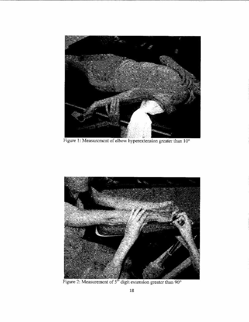
Figure 1: Measurement of elbow hyperextension greater than 10°
18
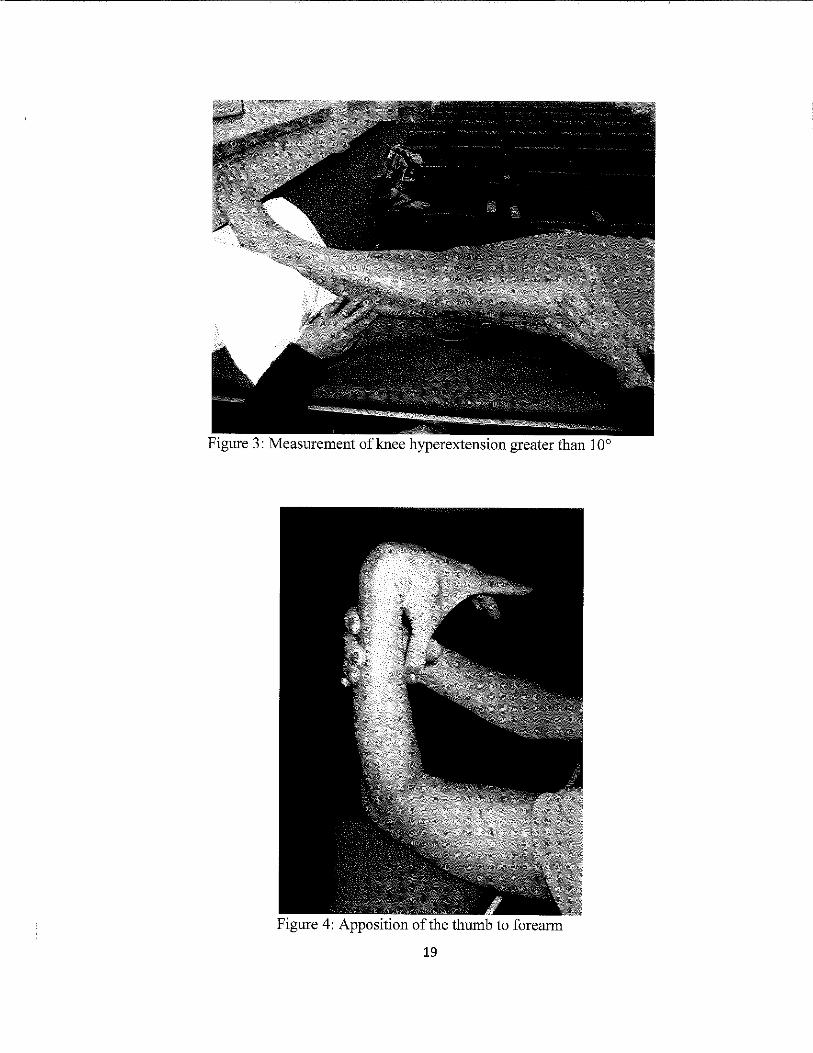
Figure 3: Measurement of knee hyperextension greater than 10°
Figure 4: Apposition of the thumb to forearm
19

20

CHAPTER IV
RESULTS
Eighty-nine PT and OT students (25 male, 64 female) voluntarily participated in
this research study, three of which were excluded (I male and 2 females). Of these 86,
there were 54 PT participants and 32 OT participants. The prevalence ofhypennobility
was found to be 39.5% (n=34) overall in the subject population, with a prevalence of
33.3% and 50% in PT and OT student participants, respectively. Ofthe 34 students with
hypennobility, 25 (71.4%) were female and 9 (26.5%) were male.
The questionnaire revealed that a majority of participants were active with a mean
of3.7 ± 2.08 days per week of exercise participation. All of the subjects reported that
they participated in at least one athletic activity during either pre-high school, high
school, college, intramural, or non-organized (independent) athletics. The most
commonly listed athletic activities which subjects participated in were basketball (49
subjects), volleyball (41), track and field (38), softball (23), and soccer (22).
The injuries reported on the questionnaire included: sprains, strains/contusion,
dislocation, fractures, ligament ruptures, and "other injuries". There was a statistically
significant difference in strain/contusion injuries between non-hypermobile and
hypennobile individuals with non-hypermobile participants being twice as likely to have
had a strain type injury compared to those who were hypennobile l (1, n=86)=5.06,
p=0.024 (see Table 2 and Figure 6). There was no statistical significance between groups
regarding the number of sprains, ligament ruptures, fractures, and dislocations (see Table
21

3). Though there was no statistical siguificance, fractures were reported more frequently
in individuals who were non-hypermobile than those who were hypermobile with 44.2%
and 29.4% having a fracture respectively.
Table 3' Injury type reported by participants Type of Injury Hypermobile Non-hypermobile "l p value
(N=35) (N=52) Sprain 70.6% (n-33) 63.5% (n-24) 0.467 0.494
Ligament rupture 8.8% (n=3) 5.8% (n=3) 0.296 0.587 Strain/Contusion 20.6% (n=7) 44.2% (n=23) 5.059 0.024
Fracture 29.4% (n-l0) 44.2% (n-23) 1.909 0.167 Dislocation 29.4% (n=lO) 21.2% (n=ll) 0.760 0.383
80.00% ,----------------------------
II Hypermobile 70.00%
II Non-hypermobile
60.00%
50.00%
40.00%
30.00%
20.00%
10.00%
0.00%
Sprain Ligament Rupture Strain/Contusion Fracture Dislocation
Figure 6: The percentage of participants in each group that have experienced at least one of the respective injuries.
22

CHAPTER V
DISCUSSION AND CONCLUSION
Discussion
The results showed that 39.5% ofPT and OT students presented with
hypennobility, and there was no significant difference in hypennobility rates between PT
and OT students. This rate is greater than five times the rate ofhypennobility found in
the general population, which is 7.6%8 Our findings also support the literature that
females have a higher prevalence ofhypennobility than males. In this study, of the 34
students with hypennobility, 25 (73.5%) were female and 9 (26.4%) were male.
Literature has shown that hypeilliobility increases musculoskeletal symptoms and
injuries; however our results did not support these findings 17. While the overall reported
number of injuries in those with hypeilliobility was elevated, it was not significantly
different from those without hypennobility. Strains were the only injury that was
significantly associated with hypennobility status, specifically that they were increased in
the non-hypennobile participants. Fractures were more common in the non-hypennobile
population while dislocations, sprains, and ligament ruptures were reported more often in
the hypennobile population. Although not significant, there was a trend that intra
articular injuries, which included sprains, ligament ruptures, and dislocations were more
common in individuals with HMS (see Figure 6). The reason for this could be that when
joints are hypennobile, they could have increased instability due to the laxitlo. This
23
possible instability could be why intra-articular injuries were found to be more prevalent
in the hypermobile group.
Throughout the data collection process several subjects indicated that their past
participation in various activities such as gymnastics, cheerleading, and dance which
require significant flexibility, likely contributed to their hypermobility status. From the
information collected on the questionnaires, 6 out of 8 dancers and 5 out of 9 gymnasts
were found to be hypermobile. This finding agrees with research that these athletes tend
to have a higher prevalence of hypermobility. Further research needs to be performed to
determine ifhypermobility in dancers and gymnasts is due to genetics or lifestyle.
The increased number of injuries reported by the participants may be related to
the increased activity levels of the entire subject population. There was not a significant
difference between activity levels between the hypermobile and non-hypermobile
individuals. Both groups indicated that they were active in sport participation when they
were younger, and currently there was a median of 4 days of exercise per week. Because
both populations are equally active, they are both at a high risk of injuries, making it
difficult to detect differences in injury rates associate with hypermobility.
It has been found that individuals with HMS have a significant decrease in
proprioceptive feedback. This could lead to an increased risk of injury. However,
someone with HMS can improve his or her proprioceptive senses with proprioception
exercise training13. No proprioceptive testing was performed in this study so it is
unknown if there is a difference in proprioception between participants with and without
HMS. However, the majority of participants in this study indicated that they have been
active in sports tInoughout their lives. Therefore, participants with HMS could potentially
24
have comparable proprioceptive feedback to participants who are non-hypermobile if
regular exercise in general could be shown to have an impact on improving
proprioception. This may account for the lack of difference in injury rates among the two
populations. Future studies would need to be conducted in order to confirm this
possibility.
There is conflicting research regarding how HMS may contribute to future disease
processes such as osteoarthritis. However. it has been shown that the risk of developing
other diseases, such as osteoporosis, is increased in individuals who have HMS.
Therefore, it is important that participants who were found to have HMS be educated on
potential future risks in order to take preventative measures.
The results of this study did not confirm findings of previous studies that showed
an increased prevalence of injuries in subjects with HMS. Flaws within the survey,
specifically questions regarding the recurrence of joints injured did not allow analysis of
injury recurrence rates. There is currently minimal research on injury recurrence rates and
hypermobility status. It remains necessary that future studies continue to pursue this
topic.
Limitations of the Study
The sample size for this study was larger than other studies, but still quite small.
Intentions were to pool the data with those from previous studies, however, modifications
to the data intake form prevented data pooling. Future studies should be able to utilize the
data from this study to create a larger sample size. The sample size was also limited
because only PT and OT students attending the University of North Dakota during spring
and summer semesters were included as participants.
25

Although the data form was improved from the study two years ago to include
more information on number of injuries experienced, a majority of participants found it to
be difficult to understand. Therefore, some data was inconsistent. It was also difficult for
participants to recall their past injuries and the age of occurrence. Participants were
instructed to recall their injury history to the best of their ability, so there was subjectivity
in the provided data. For future studies, the survey should be modified to ensure that all
injuries are accounted for in terms of injury type and mode of injury.
Improvements to this study could include a more detailed data analysis to reveal if
there is a relationship between hypermobility of a particular joint and injury occurrence
of that joint. It is also recommended that future studies look at mechanism of injuries
compared to hypermobility status. The current study had too small of a sample size to be
able to analyze this data.
Conclusion
This research study investigated the prevalence of systemic hypermobility among
PT and OT students as well as the correlation with previous injury history. There was a
significant finding that non-hypermobile participants were more likely to experience a
strain that those with HMS. Although not significant, trends in the data demonstrated
that non-hypermobile group had a greater rate of fractures while the participants with
HMS had a greater prevalence of sprains, ligament ruptures, and dislocations. This study
found that PT and OT students have a higher prevalence of HMS compared to the general
populationS. Therefore, it is important for PTs and OTs to be aware of their
hypermobility status and the associated injury risks when working with patients. Extra
26

precautions should be taken by both physical and occupational therapists who have HMS
.. . 29 to prevent lUJunes .
27

APPENDIX A

~'.
•
, .'
Date: 2/16/2012
REPORT OF ACTION: EXEMPT/EXPEDITED REVIEW University of North Dakota Institutional Review Board
Project Number: __ ,.J.RB-~01202~--,2",9=-1 __ _
Susan Principal Investigator: --~~-----...... -------~~---~----------.-------------
Department: Physical Therapy
Project Title: Association of Generalized Joint Hypermobility and Occurrence of Musculoskeletal Injury Among Physical end Occupationa.l Therapy Students _________ _
The a e eferenced roject was reviewed by a designa"ted member for the University's Institutional Review Board .72Yi1:L!d~ci"_:?-,f."'LL2c.. ___ and the fOllowing action was taken: . on
I .. 00 Project appr . d. Expedited Review Category No, 01/ tt'YIJ '1.
Next scheduled review must be before: Eebrllar:(2:l.,..J,2"O .... 13.L. ____________ , ____ _
IX! Copies of the attached consent form with the IRS approval stamp dated February 2.4> ... .!,2"'O""12"-__ must be used in obtainrng consent for this study.
Project approved, Exempt Review Category No, _" o This approvat is valid until ~_.__ __'"._ as I-o-ng-as-a-pp-ro-ve-d"--pr-o-cedures are followed, No
periodic review scheduled unless so stated In the Remarks Section.
o Copies of the .ttac~.d consent form with the IRS approval stamp dated must be used in obtaining consent for this study.
o Minor modifications required. The required corrections/additions must be submitted to ROC for review and approval, This study may NOT be started UNTIL final IRS approval has been received.
o Project approval deferred, This study may'not be started until finallRB approva1 has been received.
(See Remarks SectIon for further information.) .
o Disapproved claTm of exemption, This project requires Expedited or Full Board review. The Human Subjects Review Fonn must be filled out and submitted to the IRB for review.
o Proposed project is not human subject research and does not require IRB review.
o Not Research 0 Not HUman Subject
, P.LEASE NOTE: Requested revistons for student proposals MUST include adviser's signature. All revisions 'L-f MUST be highlighted .
. rEducation Requirements Completed, (Project cannot be started untillRB education requirements are met)
cc: Chair F Physical Therapy Signature of Designat IRB Member UND's Institutional R view Board
. 2D L. . I
If the proposed project (el1nicel medIcal) [s to be part of a research activity funded by a Federa! Agency, a special assurance statement or a completed 310 Form may be required. Contact RDC to obtain the required documents.
(Revised 10/2006)
29

APPENDIXB

INFORMED CONSENT
TIThE:
PROJECT DmF£TOR:
PHONE 1#
DEP ARTl\fENT:
STATEMENT OF RESEARCH
Association o/Generalized Joint Hypermobillty and Occurrence 0/ Musculoskeletal Injury in Physical and OCClipaliol1ai Tiu!rapy Students
Susan H N Jeno, PT. PhD
777.283J
Physical Therapy
A peI1Ion who is to participate in the research must give his or her informed consent to such participation. This consent must be based on IIIl understanding of the nature and risks of the research. This document provides informati,on th!tt is important for this understllllding. Research projects include only SUbjects who choose to take part. Please take your time in making your decision as to whether 10 participate. If you have questions atany pme, please ask.
You are inviled to be in a research study comparing generalized joint hypermobilit)- IIIld injury rates because you are a student in the professional program of either Physical or Occupational Therapy at the University of North Dakolll.
'J'hiOpiirpose ofihis study is to determine if individuaIs identified with generalized joint hypen\lobility (excessive joint mobility) are at a higher risk ofineurring musculoskeletal injury. The findings oft)Us study will help determine ifprevenllltive steps need to be taken to prevent injury in individuals with hypermobility during the academic preparation and future professional practice. You will be made aware if you are identified as being hypermobile. Resullll of the study will be available to you to assess the need of a prevenwive program. Approximately 200 people will take part in this study at the University of North Dakota Your partiCipation in the study will last approximately 20 minutes. You .... il1 need to visit the Department of Physical Therapy One time.
WHA.T WILL HAPPEN DURING TillS STUDY? Each subject will be asked to complete II questionnaire pertail)ing to demographic data, i\Ctivity, and injury history. The subject is free to Skip any questions ,that helshe would prefer ~t to answer. The Beighton method of testing joint laxity and criteria will be used to as !be measure of generalized joint bypermobility. Subjects will be assessed on their ability 10 do the following tests: Hyperextendthe little finger beyond 90 degrees, hyperextend the elbows beyond 10 degrees, hyperextend the knees beyond 10 degrees, apposition of the thumb to the flexor aspect of the forearm. and forward flex the trunk so the pnlms easily touch the floor with the knees fully extellded. A seoring system ofzero to nine is utilized with one point given for each extremity bilaterally and one point for the trunk if the test is positive for the aforementioned criteria. A subject with a score of 4 or more will be considered hypermobile. II is expected th!tt Iheenlire procedure will take approximately 20 minutes to complete,
Approval Date: ___ --'3'-0"-___ _ Expiration Date: "g ",": University of North Dakota IRS
Datc~_~ __ Subject Initials: __ _
31

WHAT ARE THE RISKS OF THE STUDY? There may be some risk from being in this study, though the risks to the subjects are anticipated to be minimal and unlikely in this study. The only riskthe subject may experience is a momentary slight discomfort if excessive force is used to move their joint into position for the tests. The subjects will be asked to move their joints only within their available range. Ifinjury should occur, medical treatment will be available, including first aid, emergency treatment, and follow-up care lIS it is to a member of the general public in similar situations. payment for such treatment must be provided by the subject and their third party payer, if any.
WHAT ARE THE BENEFITS OF THIS STUDY? By assessing if individuals with generalized joint hypermobility are at a greater risk of injury during normal daily activities compared to individuals who are not hypermobile, therapeutic methods can be developed to prevent injury. With this knowledge, hypermobiJe individuals may be able to avoid injury. The subjects in this study will be made aware tfthey have generalized joint hypermobility arnot. Following the study, the results will be made available to the subjects to allow them to assess whether a preventative program would be beneficial to them. The findings of this study will be directly applicable to iJijury predietion and the need for preventative intervention. To society as a .... itole, recognition ofinjucyrates and takin~ preventative me$ures to limit the those injuries will help 10 control health care cOst!rforthe professionals and hopefully help them lead longer, injury free careers. You will riot have any costs for being in thls research study nor will you will not he paidforheing ill thlsresearch Study; .
WHO IS FUNDING THE STUDY? The University of North Dakota and the research tealn are receiving no payments from other agencies.organiiatioIis, or companies to conduct this research study.
CONFIDENTIALITY The records oftliis study will be kept private to the extent permitted by law. In any report about this study that might be published, you will nol be identified. Your study record may ber.viewed by persons thaI audit IRE procedures at the University of North Dakota. Any information that is obtained in this'study and that can be identified with you will remain confidential and will be disclosed only with your permission or lIS required by law. Confidentiality will be maintained as each panicipant.\Vill be assigned a randomly selected idcotillcation number at the beginning of the study, which will be known by the researchers only. All information involving the research study will be secured in a locked. cabinet inside the Department of Physical Therapy at the University of North Dakota. A bar:d copy of the Slatistically analyze<l.data along with \he data collection sheets from the study will be secured in a locked cabinet inside the Department of Physical Therapy located at the University of North Dakota. Unless the data is required for future studies. the information will be destroyed via shredding three years aller the study has been completed.
If we write a report or amele abollt thill study, we will describe the study results in a summarized marmer so that you cannot be identified.
Approval Date: ___ ·-,3",0"n'V'~ __ Expiration Date: ____ '_9_)<_.,· __ _ University of North Dakota IRS
2 DaI<c---__ SohjCCllnitials; __ _
32

COMPENSATION FOR INJURY In the event that this research activity results in an injwy, treatment will be available including first aid, emergency treatment and follow-up care as needed. Payment for any such treatment is 10 be provided by you (you will be billed) or your third-party payer, if any (such as health inslll"lIIlI:e, Medicare, etc.) No funds have been sel aside to compensate you 10 the event ofinjwy. Also, the study staff cannot be responsible if you knowingly and willingly disregard the direetio!1.!l they give you.
IS TIllS STI.lDY VOLUNTARY? Your participation is voluntary. You may choose not to participate or you may discontinue your participation al any lime without penalty or loss of benefits In which you are otherwise entitled. Your decision whether or not to participate will no! affect your current or future relations with the University of North Dakota.
CONTACTS AND QUESTIONS? The researchers conducting this study are Susan H. N. Jeno, PT, PhD and Year 2 Graduate PhySical Tlterapy Students. You may ask any questions you have now. !fyou later have queStl6ns, concerns, or complaints about the research please con1il<:1 SIlSlIn Jeoo' 31777'2831 ' dutiog the day. If you have questions regarding your rights as a research s!lbject;orif'yuubave any'conceths or complaiols about the researcb, you may conblct llie University of'NotthDakota InStifutional ~eview BOl!rd III (701) 777-4279. Please call this nwnberifyou'cahh<iiteach' .':.' reiearcb}aff, on'ou Wish 10 talk with someone else. "
Your signature indicates that this research study has been explained to you, that your questions Jiave beelianswered; and that you agree to lake part in this study, You will receive a copy ofthL. form. . ' . . . , ,. ' ,'"
Subjects Name:
Signature of Subject
I Approval Date: 3 0 " Expiration Dale: 'i. 9, '" .
University of North Dakota IRS
33
Date ..
3
.~' \-
DOl< SuhjCi'1lnitialsiS::===

APPENDIXC

Parliclpant Survey
Age: Height (in ft. and in.): ____ _
Gender. M F Dominant hand: L R Weighl in pounds: _____ _
Iff.male, are you pregn!ll1t or nursing? Yes No
Are you currently under the care of a physician for a musculoskeletal injury? Yos No
Do you have a diagnosed connective tissue disorder? Yes No
Athletic Activity Did/do you compele in (Circle all that apply): pre-high school, high school, college, intramural, or non-<)rganized (independent) alhletics?
lfyes, list Sporl(s), _______________________ _
How many days/week do you currently participate in athletic aclivities during an average week? 01234567
What type of physical activity do you participate in? List all that apply _______ _
Injury History
The remaining questions concern your injury history. Please complete the following charts and questions so that we can gain an understanding of the types and numbers of injuries you may have sustained in the past
35
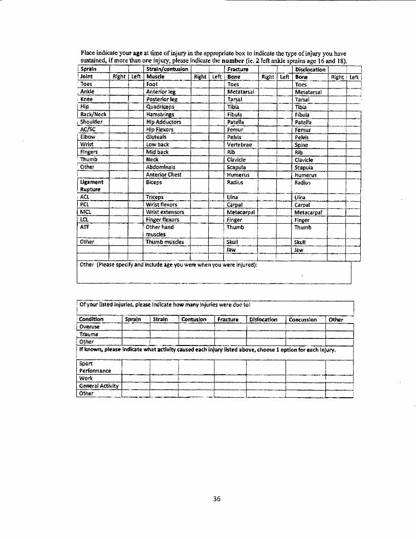

Please Indicate which, if any, Injuries for which you sought medical attention.
Please Indicate whkh, If any, injuries for which you received Physical or Occupational Therapy.
Please indicate which, Ifany, injuries required surgery.
Please Indicate which, if any, Injuries resulted In lasting disability.
Thank you for your time with this research study.
37

--- ------------------,
APPENDIXD

ID # '10 DATA cOLLECfION FORM
JOlNT TESTED
I
YES
I NO
5"' FINGER LEFT I - -
RIGHT I TIlUMB LEFT
RIGHT
LEFT I
ELBOW I I
RIGHT
KNEE LEFT
RIGHT I
~ TOTAL SCORE
."
39
REFERENCES
1. Hestekin B. The association of generalized joint hyperlaxity and occurrence of
musculoskeletal injury;l998.
2. Selinger K, Newman A, Jensen-Bale R. Association of generalized joint
hypennobility and occurrence of musculoskeletal injury in physical and
occupational therapy students; 2013.
3. Bork BE, Cook TM, Rosecrance JC, et al. Work-related musculoskeletal disorders
among physical therapists. Phys Ther. 1996;76:827-835.
4. Grahame R. Joint hypermobility: Clinical aspects. Pro Roy Soc Med.
1971 ;64:32-34.
5. Kirk JA, Ansell BM, Bywaters EGL. The hypermobility syndrome: Musculoskeletal complaints associated with generalized joint hypennobility. Ann
Rheu Dis. 1967;26:419-425.
6. Smith R, Damodaran AK, Swaminathan S, Campbell R, Barnsley L.
Hypermobility and sports injuries in junior netball players. Br J Sports Med.
2005;39:628-631.
7. Russek L. Hypermobility Syndrome. Phys Ther. 1999;79:591-599.
8. Diaz M, Estevez E, Guijo P. Joint hyperlaxity and musculoligamentous lesions:
study of a population of homogeneous age, sex and physical exertion. Br J Rheu.
1993;32: 120-122.
9. Beighton P, Solomon L, Soskolne C.L. Articular mobility in an African
population Ann Rheu Dis. 1973;32:413-418.
10. Seckin U, Sone! Tur B, Yilmaz 0, Yagci I, Bodur H, Arasil T. The prevalence of
joint hypennobility among high school students. Rheumatol Int. 2005;25:260-
263.
40

11. Wordsworth P, Ogilvie D, Smith R, Sykes B. Joint mobility with particular reference to racial variation and inherited connecti ve tissue disorders. Br J Rheu. 1987;26:9-12.
12. Simpson MR. Benign joint hypcrmobility syndrome: evaluation, diagnosis, and management. JAm Osteopath Assoc. 2006; 106(9): 531-536.
13. Sahin N, Baskent A, Cakmak A, Salli A, U gurlu H, Berker E. Evaluation of knee proprioception and effects of proprioception exercise in patients with benign joint hypennobility syndrome. Rheumatol Int. 2008;8:995-1000.
14. Child. Joint hypermobility syndrome: inherited disorder of collagen. J Rheu. 1986; 13(2):239-243.
15. Beighton PH, Grahame R, Bird H. Chapter 5: Musculoskeletal features of hypennobility and their management. In Beighton PH, Grahame R, Bird H, eds. Hypermobility of Joints. 4th ed. Springer London; 2012
16. Adib N, Davies K, Grahame R, Woo P, Murray KJ. Joint hypennobility syndrome in childhood. A not so benign multisystem disorder? Rheumatology (Oxford). 2005;44:744-750.
17. Konopinski MD, Jones GJ, Johnson MI. The effect of hyper mobility on the incidence of injuries in elite-level professional soccer players: a cohort study. Am J Sport Med. 2012;40(4):763-9.
18. Pacey V, Nicholson LL, Adams RD, Munn J, Munns CF. Generalizedjoint hypennobility and risk oflower limb joint injury during sport: a systematic review with meta-analysis. Am J Sport Med. 2010;38(7): 1487-1497.
19. De Noronha M, Refshauge KM, Herbert RD, Kilbreath S1. Do voluntary
strength, proprioception, range of motion, or postural sway predict occurrence of lateral ankle sprain? Br J Sport Med. 2006;40:824-828.
20. Wolf JM, Cameron KL, Owens BD. Impact of joint laxity and hypennobility on the musculoskeletal system. JAm Acad Orthop Surg. 2011; 19(8):463-71.
41
21. Cameron KL, Duffey ML, DeBerardino TM. Stoneman PD, Jones CJ, Owens BD.
Association of generalized joint hypermo bility with a history of glenohumeral joint instability. J Athl Train. 2010;45(3):253-258.
22. Neer CS II, Foster CR. Inferior capsular shift for involuntary inferior and
multidirectional instability of the shoulder: a preliminary report. J Bone Joint Surg. 1980;62(6):897-908.
23. Cooper RA, Brems JJ. The inferior capsular-shift procedure for multidirectional instability of the shoulder. J Bone Joint Surg. 1992;74(10):1516-1521.
24. Muhammad AA, Jenkins P, Ashton F, Robinson Christopher M. Hypermobility:
A risk factor for recurrent shoulder dislocations. Br J Sport Med. [serial online]. July 20i3;47(10):e3.
25. Jonsson H, Valtysdottir S, Kjartansson 0, Breldcan A. Hypermobilityassociated
with osteoarthritis of the thumb base: a clinical and radiological subset of hand
osteoarthritis. Ann Rheu Dis. 1996;55:540-543.
26. Kraus VB, Li YJ, Martin ER, Jordan .TM, Renner JB, Doherty M, Wilson AG,
Moskowitz R, Hochberg M, Loeser R, Hooper M, Sundseth S. Articular hypermobility is a protective factor for hand osteoarthritis. Arthritis Rheum.
2004;50(7):2178-2183.
27. Chen H, Shah S, Li Y, Stabler T, Jordan J, Kraus V. Inverse association of
general joint hypermobility with hand and knee osteoarthritis and serum cartilage
oligomeric matrix protein levels. Arthritis Rheum. 2008;58(12):3854-3864.
28. Gulbahar S, Sahin E, Baydar M, Bircan C, Kizil R, Manisali M, Akalin E, Peker
O. Hypermobility syndrome increases the risk for low bone mass. Clin Rheumatol.2006;25(4):511-4.
29. Wajon A, Ada L. Prevalence ofthurnb pain in physical therapists practicing spinal manipulative therapy. J Hand Ther. 2003;16(3):237-244.
30. Beighton P, Horan F. Orthopaedic aspects of the Ehlers-Danlos syndrome. J Bone
Joint Surg Br. 1969;51B(3):444-452.
42
31. Grahame R, Bird HA, Child A. The revised (Brighton 1998) criteria for the diagnosis of benign joint hypennobility syndrome (BJHS). J Rheu.
2000.27(7):1777-9.
32. Boyle KL, Philip W, Riegger-Krugh C. Intrarater and interrater reliability of the Beighton and Horan joint mobility index. J Athl Train. 2003;38(4):281-285.
33. Portney L, Watkins M. Foundations of clinical research: applications to practice.
Norwalk CT: Appleton & Lance; 1993:514.
34. IBM Corp. Released 2012. IBM SPSS Statistics for Windows, Version 21.0. Annonk, NY: IBM Corp.
43