Assessment of the Renal/Urinary System
71
Professor Minodora Mazur Chisinau 2019 Assessment of the Renal/Urinary System Why person has 2 eyes or 2 ears? And only one nose?
Transcript of Assessment of the Renal/Urinary System

Professor Minodora MazurChisinau 2019
Assessment of the Renal/Urinary System
Why person has 2 eyes or 2 ears? And only one nose?

Urinary system
• Kidneys
• Ureters
• Urinary bladder
• Urethra
How many kidneys does the person have?

Kidneys
• Paired organs
• Located retroperitoneallyon the posterior wall of the abdomen from T12-L3
• The average adult kidney weighs 4.5oz = 125-150 gr
• The right kidney sits islower in the abdomen due to liver placement
• An adrenal gland sits are on top of each kidney

Kidney Anatomy
Each kidney has two parts
• The renal medulla is the inner portion
– consists of renal pyramids which are collecting ducts that drain into renal pelvis
– Once urine leaves the renal pelvis the composition or amount of urine does not change
• The Cortex is the outer portion
– contains nephrons

Nephron
• Each kidney has approximately 1 million nephrons
• If the function is less than 20% replacement therapy is usually initiated
• The nephron is responsible for the initial formation of urine
Glomerulus

Kidney functions
• Urine formation• Excretion of waste products • Regulation of electrolytes• Regulation of acid-base balance• Control of water balance• Control BP• Regulation of RBC production• Synthesis of vitamin D to active form• Secretion of prostaglandins• Regulation of calcium and phosphorus balance

Urine Formation
• Urine is formed in the nephrons in a three step process
– Glomerular filtration– Tubular reabsorption– Tubular secretion
• Glomerular Filtration produces ultrafiltrate which enters the tubules
• Selective reabsorption of H2O & solutes occurs in tubules• Selective secretion of solutes occurs in tubules• 99% of ultrafiltrate is reabsorbed into the bloodstream• 1000-1500mL of urine is produced each day

Excretion of Waste Products
• The kidney is the body’s main excretory organ
• The major waste product of protein metabolism is urea– 25-30 g are produced and excreted daily
• Other waste products include:– Creatinine
– Phosphates
– Sulfates
– Uric acid
– Drug metabolites

Regulation of Electrolytes• In normally functioning kidneys the amount of electrolytes
excreted per day is equal to the amount ingested
• Sodium
– Linked to blood volume and pressure
– Significant effects on osmotic pressure
– 90% of Na in ultrafiltrate is reabsorbed in the proximal tubules and loops of Henle
– Aldosterone causes kidneys to reabsorb sodium• Potassium
– The kidneys excrete more than 90% of K intake to maintain a normal serum balance
– Aldosterone causes the kidneys to excrete potassium

Regulation of acid-base balance
• Normal serum pH is 7.35-7.45
• Normal urine pH is 4.6-8
• Kidneys 3rd line of defense in acid-base balance– respiratory & other buffer systems respond more rapidly
– kidneys require several hours to a day or more to readjust balance
• Reabsorb bicarbonate from ultrafiltrate
• Excrete large quantities of acid in the urine (phosphoric and sulfuric acids) by buffering with ammonia

Control of water balance
• The human body is made up of 60% water• Regulated by Antidiuretic hormone (ADH) or
vasopressin• Secreted by the posterior pituitary in response to
serum osmolality• ADH increases reabsorption of water to return
serum osmolality to normal• Decreased water intake stimulates ADH release• ADH controls volume & concentration of urine by
regulating permeability of distal tubule to H2O

Control blood pressure
• The kidney secrets the hormone renin when there is a decrease in BP
• Renin converts angiotensinogen to angiotensin I• Angiotensin I converts to angiotensin II• Angiotensin II is a powerful vasoconstrictor and causes
BP to increase• Increase in BP stops the excretion of renin• The adrenal cortex also releases aldosterone in response
to increasing serum osmolality or poor perfusion to increase BP

Regulation of Red Blood Cells production
• The kidneys release erythropoietin when they sense a decrease in oxygen in the blood
• Erythropoietin stimulates the bone marrow to produce (Red Blood Cells) RBCs

Vitamin D Synthesis
• The kidneys convert inactive vitamin D to 1,25-dihydroxycholecalciferol
• Vitamin D is necessary for calcium balance

Urination
• approximately 150-350 mL of urine triggers stretch receptors & stimulates afferent nerves sending signal to CNS
• Functional Capacity of bladder is 350mL• The efferent pelvic nerve stimulates the bladder to
contract, relaxing the urethral sphincter. The decrease in urethral pressure and contractions of the detrusor muscle opens the proximal urethra and leads to flow of urine
• reflex action however external sphincter can be contracted with voluntary control

Ghilon was asked what is more difficult:
Keep a secret?
Make good use of your free time?
Can you endure an injustice?
And for you what is more difficult?
Ilan Ghilon (b. May 12, 1956, Galați, Romania) is an Israeli
socialist politician originally from Romania, who is currently
serving as a deputy in the Knesset (Parliament) of the
State of Israel

Tulburări ale diurezei
Urinary volume normally varies in physiological conditions between 800-2000 ml / 24 hours, during intake and fluid losses.
Urinary Quantitative Changes:
• Polyuria - 2000 ml
• Oliguria - Reduction of urinary volume <500 ml/24h
• Anuria - Decreased diuresis <100 ml
• Nicturia - Equalization or inversion of the ratio between nocturnal and diurnal diuresis

Terminology
• Micturation—Urination • Frequency—Frequent voiding >every 3hours• Urgency—Strong desire to void• Hesitancy—Delay or difficulty in initiating voiding• Nocturia—Excessive urination at night• Oliguria—Output <400mL/day• Anuria—Output <50mL/day• Polyuria—Increased volume of urine voided• Hematuria—RBCs in urine• Dysuria—Painful or difficult voiding• Enuresis—Involuntary voiding during sleep• Incontinence—Involuntary loss of urine

Renal/Urinary System Changes with Aging
• Reduced renal blood flow causing kidney loss of cortical tissue by 80 years of age
• Thickened glomerular and tubular basement membranes, reducing filtrating ability
• Decreased tubule length
• Nocturnal polyuria and risk for dehydration
• Decreased glomerular filtration rate

Diagnostic Tests
• Blood Tests– Serum Creatinine (0.5 – 1.2 mg/dl)– Blood Urea Nitrogen (10-20 mg/dl)– BUN/Creatinine Ratio (12:1 to 20:1 mass)
• Urine Tests– Urinalysis – Urine for C&S– Composite (e.g., 24hr) urine collections– Creatinine Clearance Test– Urine Electrolytes– Osmolality (plasma; urine)

Diagnostic Tests (cont)
• Bedside sonography; Bladder scanners
• Radiographic Examinations– Kidneys, Ureter, and Bladder X-ray
– Intravenous Urography
– Computed Tomography
– Cystography and Cystourethrography
• Other Renal Diagnostic Tests– Renal Arteriography (Angiography)
– Renal Biopsy
– Renography (Kidney Scan)
– Ultrasonography

Assessment
• Family history and genetic risk assessment
• Demographic data and personal history
• Diet history
• Socioeconomic status
• Current health problems

Physical Assessment
• Inspection
• Auscultation
• Palpation
• Percussion
• Assessment of the urethra

Local objective examination
Inspection of the abdomen
increase in volume (diffuse bombardment) -meteorism from R-U colic
lower abdomen inspection - pumping of the hypogastric region into the bladder
inspection of external genital organs
- women - cistorectocel (RVP with parent-urinary prolapse)
- men - inflammation of the urinary meatus (erythema, swelling) in acute anterior urethritis

Palpation
Palpation of the kidney
Normally the kidney is not accessible to palpation, except:
pregnant women,
of asthenic constitution at which one can feel the lower pole of the kidney in deep inspiration.
The kidneys become accessible to palpation in case of:
renal ptosis,
increase in volume due to different causes (Hydronephrosis.

Techniques of palpationPalpation in the dorsal decubitus (Guyon)
The patient seated in the dorsal decubitus, with the thighs flexed on the pelvis in slight abduction, with the relaxed abdominal muscles.
The examiner is placed on the examined kidney side (right, left). Apply the hand on the same side as the kidney examined in the lumbar region with the fingers towards the costovertebral angle, and the other hand is applied to the level of the right hypochondrium with the fingers pointing towards the costal rim and outside the right abdomen (hands are applied "cross")
By pushing down the hand on the abdominal wall, the "kidney ball" is performed, the hand placed posteriorly feeling the lumbar contact of the kidney, the kidney is palpated thus by approaching the two hands (anterior and posterior)

Methods of palpation
Regardless of the method used, the diagnosticcriteria for palpation of the kidney are:posterior position, deeplumbar contactmobile with inspirationThe kidney has:an elastic-remittent, smooth consistency,
sliding under the hands like "cherry kernels";"bean bean" form when not distorted by a
pathological process

Palpation of painful points
– Previous painful points
– Upper pain point (BAZY basin, Pasteau upper ureteral)
– It is located at the intersection of the horizontal line passing through the
navel, with the vertical line passing through the McBurney point and
corresponds to the tip of the XII coast.
– It is painful to affect the kidney basin. The pain at this point is associated
with the urge to urinate due to a skin-reflex reflex

Palpation of the bladder
The bladder balloon resulting from acute or chronic retention of urine is palpated in the form of a formation:
hypogastric, located in the middle, of hemispherical, suprapubian form, shallow clearly delimited, elastic, sensible.
Palpation of the bladder gives patients a feeling of imperious urination.
Old forms of bladder or neurogenic bladder do not show palpatory sensitivity
Differential diagnosis in women (pregnant uterus, uterine tumors)
- Bladder tumor formation can sometimes palpate in advanced stages (hard, irregular formation)

The objective exam
Endorectal palpation for prostate exploration and urethral palpation - performed by urologist

Percutionlumbar - Giordano maneuver = Costovertebral angle tenderness (CVAT) (Murphy's punch sign, Pasternacki's sign, or Goldflam's sign (Latin: succusio renalis) is a medical test in which pain is elicited by percussion of the area of costovertebral angle, an angle made by the vertebral column and the costal margin). The test is positive in people with an infection around the kidney (perinephric abscess), pyelonephritis, hemorrhagic fever with renal syndrome or renal stone. Because the kidney is directly anterior to this area, known as the costovertebral angle, tapping disturbs the inflamed tissue, causing pain.of the bladder - hypogastric, round-oval shape with inferior concavity

Auscultation
• abnormalities of the renal artery (stenoses, aneurysms)
• It can be heard obliquely in the lumbar region or previously paraombilical, subcostal a breath
• with trill character
• synchronous with the
pulse

Urinalysis
• Color, odor, and turbidity
• Specific gravity
• pH
• Glucose
• Ketone bodies
• Protein
• Leukoesterase
• Cells, casts, crystals, and bacteria

Other Urine Tests
• Urine for culture and sensitivity
• Composite urine collections
• Creatinine clearance—best indication of overall kidney function
• Urine electrolytes
• Osmolarity, blood/plasma osmolarity, urine osmolarity

Macroscopic examination
Appreciation of urinary volume / 24h - diuresis variation between 800-2000 ml; ¼ diuresis - nocturnal, yellowish color
transparency - clear, odor - characteristic
-ammoniacal (urinary tract infections),
-acre apples (diabetes with ketonuria),
-pour (urinary tract infections with anaerobic flora)

Microscopic examination
Highlight 3 categories of elements:
-celule,
-cilindrii
-cristale
-simple sediment
-minute sediment (Addis – Hamburger)leucocite

RBC

Hematii dismorfe-leziune glomerulară

Celulele sternheimer-malbin


Blood casts in the tubes histological appearance

Erytrocytes casts

Leukocyte cylinders

Epithelial cylinders - tubular injury

Cilindrii hialini

Granular cylinders

WAXI CAST

Hyaline casts in a patient with hyperbilirubinuria

Greasy casts

Fatty cylinders look like a Maltese cross in polarized light

Cristals
Precipitated urine salts in the form of crystalsthey also occur in the urinary tract of normal peoplemeans a pathological condition only if they are in large quantitiesDepending on the type of diet, urinary pH, genetic predisposition, the
following types are found (more frequently):Urates (crystals, amorphous urates) - after uricozurics: Probenecid,sausage foods, urinary pH <Oxalates (oxaluria)Ammonia-Mg phosphates (alkaline, infected urine)Cholesterol (SN)the presence of germs - smears stained with methylene blue, Gram,
special collor (Ziehl – Nielsen)

Oxalate-looking envelopes

Rectangular-looking phosphates or coffins

Others Diagnostic Tests
• Bedside sonography/bladder scanners
• Computed tomography
• Kidney, ureter, and bladder x-rays
• Intravenous urography– Bowel preparation
– Allergy information
– Fluids

Renal Arteriography (Angiography)
• Possible bowel preparation
• Light meal evening before, then nothing by mouth
• Injection of radiopaque dye into renal arteries
• Assessment for bleeding
• Monitoring of vital signs
• Absolute bedrest for 4 to 6 hours
• Serum creatinine measured for several days to assess effects of test

Para-clinical investigation
USG organelor interne (rinichi)

CT kidney
GN

MRI kidney

Renal biopsy

Renal artery angiography

Adult kidney

Horseshoe kidneys

Hydronephrosis

Rinichi atrofic unilateral ischemic

Polycystic kidneys

Renal Biopsy
• Percutaneous kidney biopsy
• Clotting studies
• Preprocedure care
• Follow-up care– Assessment for bleeding for 24 hours
– Strict bedrest
– Monitoring for hematuria
– Comfort measures
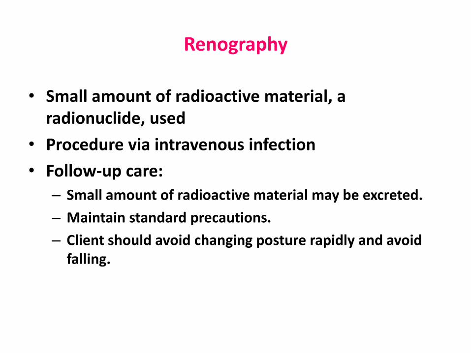
Renography
• Small amount of radioactive material, a radionuclide, used
• Procedure via intravenous infection
• Follow-up care:
– Small amount of radioactive material may be excreted.
– Maintain standard precautions.
– Client should avoid changing posture rapidly and avoid falling.

Other Diagnostic Tests
• Ultrasonography
• Cystoscopy and cystourethroscopy– Procedure is invasive.
– Consent is required.
– Postprocedure care includes monitoring for airway patency, vital signs, and urine output.
– Monitor for bleeding and infection.
– Encourage client to take oral fluids.

Retrograde Procedures
• Retrograde procedures go against the normal flow of urine.
• Procedure identifies obstruction or structural abnormalities with the instillation of dye into lower urinary tract.
• Monitor for infection.
• Follow-up care is the same as for a cystoscopic examination.

Urodynamic Studies
• Studies that examine the process of voiding include:
– Cystometrography
– Urethral pressure profile
– Electromyography
– Urine stream test

Thanks