Assessment of foetal heart rate monitoring devices in ...
150
Assessment of foetal heart rate monitoring devices in referral hospitals in Tanzania Towards improved quality of intrapartum care by Benjamin Anathory Kamala Thesis submitted in fulfilment of the requirements for the degree of PHILOSOPHIAE DOCTOR (PhD) Faculty of Health Sciences 2019
Transcript of Assessment of foetal heart rate monitoring devices in ...

Assessment of foetal heart rate monitoring devices in referral hospitals
in Tanzania
Towards improved quality of intrapartum care
by
Benjamin Anathory Kamala
Thesis submitted in fulfilment ofthe requirements for the degree of
PHILOSOPHIAE DOCTOR(PhD)
Faculty of Health Sciences
2019

2
University of Stavanger NO-4036 Stavanger NORWAY www.uis.no
©2019 Benjamin Anathory Kamala
I
PhD: Thesis UiS No.

iii
Acknowledgements
I would like to accord my deepest gratitude to Associate Professor Hege Ersdal, my main supervisor, for her tireless efforts in making sure this doctoral degree mission was successfully accomplished. Also, to my co-supervisors, Associate Professor Hussein Kidanto and Dr Muzdalifat Abeid, for granting me an opportunity to engage with the Safer Births Project and by creating a friendly research environment. To key personnel in my doctoral degree, Professor Jeffrey Perlman, Dr Ingvild Dalen and Dr Matilda Ngarina; you made me proud and smile all the way. I thank everybody mentioned above for their scientific guidance, mentorship and the enormous support provided throughout. The technical guidance delivered in the development of this course was provided with great humanity, resilience, ownership and perseverance. I am proud that our product delivery has provided the scientific community with valuable and referable information.
Dedicated acknowledgement goes to Dr. Muzdalifat, who believed in my epidemiology capability and introduced me to the Safer Births Project. Heartedly, I still cherish our interchangeable teacher-student relationship which has developed since 2005.
I thank Muhimbili National Hospital administration, particularly the Directorate of Research and Consultancy led by Dr Faraja Chiwanga, for tirelessly providing technical guidance. My sincere appreciations go to Temeke Referral Hospital administration for granting me the permission to conduct the studies at their facility. I strongly recognize all midwives and doctors working in the labour wards at both hospitals for their hard work and passionate care accorded to the mothers and new-borns. I appreciate the dedicated support of the coordination of logistics provided by Gilbert Kilonzo at Muhimbili and Temeke. I extend the same appreciation to the data clerks, Felister, Zuwena, Anna, Pricilla and Mchome, who tirelessly processed the data throughout the project.

iv
I humbly extend my arm of cordial gratitude to all expectant mothers and their new-borns who voluntarily consented to participate in these studies without any incentives. It is from them that the value of the knowledge that this research project has generated is now being appreciated by the scientific community.
Acknowledgements go to my fellow doctoral degree candidates in Safer Births, specifically, Robert, Paschal, Esto and Sara for their unconditional cooperation, support and lovely relationship. I acknowledge my former colleagues at MUHAS, MD07 group and “wana”-EPL group for their tireless, symbiotic support and encouragement provided to me throughout. The brotherly and sisterly peer jokes and laughter we shared provided a lot of psychological relief that anybody would wish to have at stressful moments. I sincerely appreciate the valuable support from T-MARC Tanzania under the leadership of Diana, Tumaini, Prisca, and Doris. Special thanks to my colleagues at B&B Healthcare under the leadership of Bhavin, Irene, and Diana for providing support that was always available when needed.
Special thanks to the Laerdal Global Health, Laerdal Medical and SAFER teams for the readiness to help in technical issues throughout the project. To Aileen Ireland, for editorial review of all write-ups to ensure that language proficiency is properly aligned. To my hosts in Stavanger, Thomas, Corinna and Ersdal Sr. who provided me with a feel of homely stay away from home. To Mrs. Kidanto, who constantly gave me a warm welcome while receiving supervision from Professor Kidanto.
Special thanks go to the Kamala family as a whole for their tireless support and for taking on family responsibilities in my absence. To myfather, Mwalimu Anathory Kamala, for his inspirational guidance and unselfishly offering his sons and daughters opportunities to exhaust their potential. To my mother, Ma Theonestina, for giving her endless and passionate love to me; being the 12th and last born, I feel it everywhere I am. I still cherish that “mama” decided to breast-feed me up to the age

v
of 5 years having contracted measles when I was one year old. She believed that only breastfeeding would provide me with the much-needed immunity to keep me in good health. What else would I request, “mama”!! Special gratitude to my brothers, Onesmo and Adonis Kamala, for their motivational, material and moral support in my life. Words cannot express it all.
I am grateful to the people who guided and supported me to my public health career; Joseph Rutabingwa, James Kajuna, Lazaro Peter, and Mama Kiriaki. To my spiritual guardians, Sister Rita Kokulamuuka, Pastors Mugisha and his wife Pastor Rebecca. Their prayers and encouraging spiritual words strengthened my journey. It is by God’s grace.
Finally, but importantly, is to my wife, the best half of me “malkia wa nguvu”, Dr. Diana K. Damian. Thank you for your love, resilience, patience and taking on the dual parental responsibilities while I was away. I understand it has not been easy, but I am finally here for you. To my sons, Fredrick Matungwa (who always wanted to know if my score was 100%) and Giovanni Mutashobya (my “twin”). You gave me strength in our video calls. Thank you for believing in your daddy that he will soon be back with ‘fruits’ for you.

vi
Dedication
To my late grandfather, Petro Kamala ‘Kabalitoija’, the cornerstone of our family education achievement

vii
Financial Support
I would sincerely like to thank the Laerdal Foundation for awarding mea scholarship to undertake my doctoral studies. On the same measure, my appreciation goes to the Norwegian Research for Global Health organization for granting me a scholarship to attend doctoral courses at different Norwegian institutions and scientific conferences. Thanks also to GLOBVAC, the Norwegian Research Council, for funding the Safer Births project, of which this PhD is a part.
The funding bodies had no role in the study design, data collection, data analysis, interpretation, writing of the articles, or the decision to submit for publication of any part of this research.

viii
Abbreviations
BEmONC Basic Emergency Obstetric and New-born Care
CEmONC Comprehensive Emergency Obstetric and New-born Care
CONSORT Consolidated Standards of Reporting Trials
CRF Case Report Forms
CTG Cardiotocograph
DHS Demographic and Health Survey
EFM Electronic Foetal Heart Rate Monitor
ENAP Every New-born Action Plan
END Early Neonatal Death
FHR Foetal Heart Rate
FSB Fresh Stillbirth
GA Gestation age
GBD Global Burden of Diseases
HIC High Income Countries
LIC Low Income Countries
LMIC Low Middle Income countries
MDGs Millennium Development Goals
MHIC Middle- and High-Income Countries
NPV Negative Predictive Value
MOHCDGEC Ministry of Health Community Development Gender Elderly and Children
PHSDP Primary Healthcare Service Delivery Program
PO-RALG Presidents Officer Regional Authority and Local Government
PPV Positive predictive Value
RCH Reproductive and Child Health
RCHS Reproductive and Child Health Section
RMNCAH Reproductive Maternal Newborn Child Adolescent Health
SBR Stillbirth Rates
SDG Sustainable Development Goals
STROBE Strengthening the Reporting of Observational Studies in Epidemiology
UNICEF United Nations International Children’s Emergency Fund
WHO World Health Organisation

ix
Definitions of key terms
Neonatal period: period from birth to 28 days of life
Perinatal period: period immediately before and after birth includingthe 1st week of life
Birth asphyxia: failure to initiate and sustain breathing at birthsecondary to intrauterine oxygen deprivation
Intrapartum period: period from the onset of labour to the end of the third stage of labour.
Stillbirth: a foetal death at or after 28 weeks of gestation but before birth
Fresh stillbirth: a baby born dead without signs of skin disintegration (death occurs mostly less than 12hrs prior to birth)
Macerated stillbirth: a baby born dead with skin disintegration (death assumed to occur more than 12hrs prior to birth)
Quality of care: the extent to which health care services provided to individual and patient populations improve desired health outcome
Preterm: baby born before 36 complete weeks of gestation

x
Summary
Background: There are 2.5 million neonatal deaths that occur globallyeach year: 25% of them secondary to labour complications (intrapartum related). In addition, 2.6 million stillbirths occur globally each year, 40% of them intrapartum related termed as fresh stillbirth (FSB). Moreover,more than 80% of neonatal deaths occur in low-income countries (LICs).Almost 50% occur in Sub-Saharan Africa, where the supply of service does not match with the demand of service provision. These deaths can be substantially reduced by improving quality of care around time of labour and childbirth. Intrapartum foetal heart rate (FHR) monitoring and partograph use form an important component of quality of careprovision. In LICs, where most births occur, FHR assessment is mostly done intermittently and mainly using Pinard stethoscope and rarely with hand-held Doppler devices. The effectiveness of assessment devices to detect FHR abnormalities in relation to the improvement of quality of intrapartum care is scarcely documented. An FHR monitor called Moyo, was designed for low-resource settings. The monitor can be used intermittently or strapped on for continuous FHR monitoring during labour.
Aims: The overall aim of this PhD project was to compare the effectiveness of different FHR monitoring devices and the associated improvement in quality of intrapartum care at two urban referral hospitals in Dar es Salaam, Tanzania.
Methods: We conducted three studies from 2013 to 2017. The studiesincluded singleton women in the active phase of the first stage of labourwho had normal baseline FHR on admission. Study I applied arandomized controlled study design between April 2013 through September 2015 at Muhimbili National Hospital. Hand-held Doppler(n=1,421) and Pinard stethoscope (n=1,423) arms were compared in theireffectiveness to detect abnormal baseline FHR. Secondary outcomes

xi
were time to childbirth, mode of childbirth, and perinatal outcomes, including Apgar score, bag mask ventilation, admission to neonatal unit, FSB and 24-hour neonatal deaths. Study II was a follow-up of Study I and applied a randomized controlled design at Muhimbili between February 2016 through September 2017. Strap-on automatic FHR monitor, Moyo (n=1,479) and intermittent Hand-held Doppler (n=1,494) arms were compared in their effectiveness to detect abnormal baseline FHR. Secondary outcomes were time intervals in labour, mode of childbirth and perinatal outcomes (as in Study I). Study III was a pre- and post-implementation study design, conducted at Temeke Regional Referral Hospital, concurrently with Study II. In the pre-implementation period, intermittent monitoring was applied with a Pinard stethoscope (March through June 2016, n=1,640), whereas in the post-implementation period, strap-on automatic Moyo was applied (July to mid-December 2016, n=2,442). The primary outcome was detection of abnormal baseline FHR. Secondary outcomes included frequency of assessment of FHR, partograph documentation, time intervals, intrauterine resuscitations and perinatal outcomes.
Results: In Study I, the Hand-held Doppler was found to be superior to the Pinard stethoscope (6.0% vs 3.9%, p=0.008) in the detection of abnormal baseline FHR during labour. Overall, perinatal outcomes did not differ between the two arms. However, post-hoc analysis showed that, for new-borns with abnormal FHR whose mothers had given birth vaginally, the composite adverse outcomes (neonatal unit admissions and perinatal deaths) were less prevalent in the Doppler arm (7 of 43 births, 16.3%) than in the Pinard arm (10 of 23 births, 43.5%), p=0.021.
Study II, building on the results of Study I, showed that the Moyo was superior to the Doppler (13.3% vs 9.8%) (p=0.002) in the detection of baseline FHR abnormalities. The results for the time from admission to detection of abnormal FHR and from last FHR to birth were shorter in the Moyo arm than in the Doppler. However, the time from detection of abnormal FHR to birth was approximately 31% longer in the Moyo

xii
compared to the Doppler. Caesarean section rates were higher with the Moyo (19%) arm compared to the Doppler (13%) (p=0.031). Overall,perinatal outcomes did not differ between the two arms.
In Study III, the Moyo was found to be superior to the Pinard (8.0% vs 1.6%) (p<0.0001) in the detection of baseline FHR abnormalities. Time from admission to birth and from last FHR to birth was shorter in the Moyo arm than the Pinard. The quality of intrapartum care provision improved significantly post-implementation in the Moyo arm. These included partograph use/documentation (98% vs 54%), and frequency of FHR monitoring (every 60 min vs 150 min). Other improvements included intrauterine resuscitations, and vacuum extraction of 5.8% vs2.2% post- versus pre-implementation, respectively.
Conclusions: This PhD project shows that the Hand-held Doppler was superior to the Pinard in the detection of abnormal baseline FHR. Further, it showed that implementation of strap-on automatic FHR assessment with the Moyo led to earlier and more frequent detection of abnormal FHR at the referral hospitals. This implementation was associated with improved quality of intrapartum care and partograph use. Moreover, the time from detection of abnormal FHR to birth was longer in the Moyo groups. Implementation of studies coupled with timely obstetric responses and powered to detect differences in perinatal outcomes are recommend.

xiii
Publications included
This thesis is based on the following research articles, which will be referred to throughout the text by Roman numerals:
Paper I:
Kamala BA, Kidanto H, Wangwe P, Dalen I, Mduma E, Perlman J,Ersdal HL. Intrapartum foetal heart rate monitoring using a handheld Doppler versus Pinard stethoscope: a randomizedcontrolled study in Dar es Salaam. Int J Women’s Health. Dove Press; 2018; Volume 10: 341–348. doi:10.2147/IJWH.S160675
Paper II:
Kamala B, Kidanto H, Dalen I, Ngarina M, Abeid M, Perlman J, Ersdal HL. Effectiveness of a Novel Continuous Doppler (Moyo) Versus Intermittent Doppler in Intrapartum Detection of Abnormal Foetal Heart Rate: A Randomised Controlled Study in Tanzania. Int J Environ Res Public Health. Multidisciplinary Digital Publishing Institute; 2019;16: 315. doi:10.3390/ijerph16030315
Paper III:
Kamala BA, Ersdal HL, Dalen I, Abeid MS, Ngarina MM, Perlman JM, Kidanto H. Implementation of a novel continuous foetalDoppler (Moyo) improves quality of intrapartum foetal heart rate monitoring in a resource-limited tertiary hospital in Tanzania: An observational study. PLoS One. Public Library of Science; 2018;13: e0205698. doi: 10.1371/journal.pone.0205698

xiv
Contents
Acknowledgements..........................................................................................iii
Financial Support............................................................................................vii
Abbreviations.................................................................................................viii
Summary........................................................................................................... x
Publications included.....................................................................................xiii
1 Introduction............................................................................................... 1 1.1 Background ............................................................................................... 1 1.2 Intrapartum FHR monitoring .................................................................... 5 1.3 Tanzania-Setting and Context ................................................................. 15 1.4 Conceptual framework............................................................................ 20 1.5 Summary................................................................................................. 23
2 Aims of the PhD project.......................................................................... 25 2.1 The specific aims of the studies were: .................................................... 25 2.2 Research questions.................................................................................. 25 2.3 Hypotheses:............................................................................................. 26
3 Methods and participants ........................................................................ 27 3.1 Study settings .......................................................................................... 27 3.2 Study design............................................................................................ 29 3.3 Participants.............................................................................................. 32 3.4 Training of relevant clinical staff ............................................................ 32 3.5 Training of research assistants ................................................................ 33 3.6 Study procedures..................................................................................... 34 3.7 Data collection and management ............................................................ 36 3.8 Measures/Variables................................................................................. 36 3.9 Statistical analysis ................................................................................... 39 3.10 Ethical considerations ............................................................................. 40
4 Summary of results ................................................................................. 42 4.1 Study I..................................................................................................... 42

xv
4.2 Study II ................................................................................................... 43 4.3 Study III .................................................................................................. 44
5 General discussion of results ................................................................... 47 5.1 Abnormal FHR detection ........................................................................ 47 5.2 Quality of care improvement .................................................................. 49 5.3 Time intervals ......................................................................................... 52 5.4 Perinatal outcomes .................................................................................. 52 5.5 Caesarean section rates ........................................................................... 53
6 Discussion of the methods ...................................................................... 55 6.1 Study design and internal validity ........................................................... 55 6.2 Biases in a pre- and post-implementation study (Study III) .................... 59 6.3 Statistical analysis ................................................................................... 60 6.4 External validity ...................................................................................... 61 6.5 Ethical Issues .......................................................................................... 61
7 Conclusions: ............................................................................................ 65 7.1 Recommendations ................................................................................... 66 7.2 Future studies .......................................................................................... 67
References ....................................................................................................... 68
Appendices ..................................................................................................... 80 Appendix 1 – Case Report Form Muhimbili (Study I and II) ............................ 80 Appendix 2- Case Report Form (Temeke Study III) .......................................... 84 Appendix 3-Consent Forms ............................................................................... 87 Appendix 4- Ethical clearance certificate ........................................................... 89

xvi
Introduction
1
1 Introduction
1.1 Background
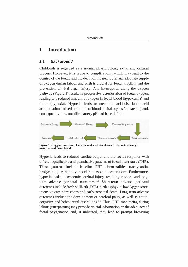
Childbirth is regarded as a normal physiological, social and cultural process. However, it is prone to complications, which may lead to the demise of the foetus and the death of the new-born. An adequate supply of oxygen during labour and birth is crucial for foetal viability and the prevention of vital organ injury. Any interruption along the oxygen pathway (Figure 1) results in progressive deterioration of foetal oxygen, leading to a reduced amount of oxygen in foetal blood (hypoxemia) and tissue (hypoxia). Hypoxia leads to metabolic acidosis, lactic acid accumulation and redistribution of blood to vital organs (acidaemia) and,consequently, low umbilical artery pH and base deficit.
Figure 1: Oxygen transferred from the maternal circulation to the foetus through maternal and foetal blood
Hypoxia leads to reduced cardiac output and the foetus responds with different qualitative and quantitative patterns of foetal heart rates (FHR).These patterns include baseline FHR abnormalities (tachycardia, bradycardia), variability, decelerations and accelerations. Furthermore,hypoxia leads to ischaemic cerebral injury, resulting in short- and long-term adverse perinatal outcomes.1,2 Short-term adverse perinatal outcomes include fresh stillbirth (FSB), birth asphyxia, low Apgar score, intensive care admissions and early neonatal death. Long-term adverse outcomes include the development of cerebral palsy, as well as neuro-cognitive and behavioural disabilities.3–5 Thus, FHR monitoring duringlabour (intrapartum) may provide crucial information on the adequacy of foetal oxygenation and, if indicated, may lead to prompt lifesaving

Introduction
2
intervention/s to prevent brain injury.6,7 In low-resource settings, the risk of adverse events related to reduced oxygen delivery to tissues is high,largely due to inadequate labour monitoring.8,9 Placing a focus on providing quality care during labour and at the time of birth, timely identification and protecting the foetus from hypoxia with subsequent ischemia, may save the lives of many new-borns.5,10
1.1.1 Global burden of under-5, neonatal mortality and stillbirthsGlobally, a total of 5.4 million occurred in 2017. The highest risk of death is during the first month of life amounting to 17 deaths per 1,000 live births. These neonatal deaths translated to 46% (2.5 million in 2017)of the under-5 deaths; an increase from 40% in 2000.2,11,12 One million (equivalent to 38%) of these 2.5 million neonatal deaths occurred within 24 hours of birth (termed early neonatal deaths). Moreover, 25% of deaths are intrapartum-related (asphyxia), with the other main causes related to prematurity and infections (Figure 2). In 2017, intrapartum-related events caused nearly 11% of under-5 mortality.8,9,11
Figure 2: Causes of neonatal deaths (Source: WHO - Maternal and Child Epidemiology Estimation; Methods and data sources for child cause of deaths, 2017)
34
2521
11 9
05
10152025303540
Premature Intrapartumrelated
Infections Congenitalanomalies
Others
Perc
enta
ge

Introduction
3
The progress observed in the reduction of mortality is uneven by age. It has declined by 60% among infants aged 1–5 years, 50% among 1–11 month-olds and by 40% among neonates over the past two decades.12 The disparity in the decline has been attributed to a global shift to perform interventions in the post-neonatal period with less emphasis on the neonatal period.13 With no accelerated interventions in place, 28 million neonates will die between 2018 and 2030; more than a quarter of them due to asphyxia.14
In 2017, almost 2.6 million stillbirths occurred, with FSB accounting for 40% (1.1 million) of the total number.15,16 The majority of FSB are intrapartum-related stillbirths. The burden of these FSB figures may be higher than reported, as nearly half of the world’s new-born babies have no birth certificate, and the majority of neonatal deaths and almost all stillbirths are not documented.9,11 Overall, a total of 5.1 million stillbirths and neonatal deaths occur every year, of which 33% (1.7 million) are estimated to be intrapartum-related.
1.1.2 Perinatal mortality epidemiology In 2017, Sub-Saharan Africa and South Asia had the highest neonatal mortality rate, both at 27 deaths per 1,000 live births, which is 9 times higher than in high-income countries (HIC). These two regions account for nearly 80% of global new-born deaths. Intrapartum-related neonatal mortality rates were 25 times higher in the LIC.8 However, these estimates are higher than documented, as there is lack of registry data on these vital statistics.11 The leading causes of neonatal deaths in sub-Saharan Africa and South Asia in a population-based survey were asphyxia (40% and 34%), infections (35% and 37%) and prematurity (19% and 24%), respectively.17 These causes of deaths have all declined markedly in HIC, primarily because of improved quality of obstetric care compared to LIC. There is a need for improved intrapartum management and continuum of care through the postnatal period in LIC.

Introduction
4
Moreover, globally, the proportion of women giving birth with a skilled birth attendant increased to 73%, but only half of the births in sub-Saharan Africa are covered by skilled birth attendants, showing strikingdisparities in quality of care provision around labour and birth.12,14 The burden of stillbirths in sub-Saharan Africa and South Asia is estimated at 17 and 35 per 1,000 live births, respectively.17 These FSB rates are 50-fold higher in LIC compared to HIC.10
1.1.3 Global strategies to reduce perinatal mortalityIn the Sustainable Development Goals (SGD) era, the discourse has shifted from health for development, to development being a necessary component of health improvement. Target 3.2 of the SDG3 calls for theend of preventable deaths of new-borns with a target mortality rate below 12 per 1,000 live births by 2030.18 Most countries have already achieved this target, but mostly in HIC. Accelerated efforts are needed to allowthe remaining countries, predominantly LIC, to achieve this target and,as a consequence, save the lives of children.12,18–20
Although FSB is an important marker of quality of intrapartum care, the measurement of the global burden of disease, as contained in the SDGs,only counts deaths that occur after a live birth. Moreover, analyses of development aid have shown that stillbirth studies and interventionswere rarely funded. 5,21
In 2016, the WHO responded by launching a perinatal mortality audit toidentify specific stillborn causes and improve the quality of perinatal care.22 Also, the Every New-born Action Plan (ENAP), developed by WHO/UNICEF, addresses the importance of the accountability of stillbirths in supporting the United Nations’ Every Woman Every Childmovement.5,23 ENAP supports countries in reaching the target of no more than 12 new-born deaths per 1,000 live births, and less than 12 stillbirths per 1,000 births, by 2030.5,21

Introduction
5
Although most high- and middle-income countries have achieved the ENAP target, more than 56 LICs, mostly in Africa, have double the burden, necessitating accelerated efforts. ENAP, UNICEF and the WHO have endorsed interventions during labour, birth and immediately after birth as they have proven to save the lives of many new-borns. A 5-yearnetwork on improving the quality of care and to prevent maternal, new-born and child health deaths was launched in 2017 targeting a 50% reduction in these deaths.24
1.1.4 Global research priorities on perinatal mortalityWhile new-born survival has gained rapid attention in recent years, the corresponding actions are still inadequate.25 The WHO, ENAP, Disease Control Priorities and the World Bank recommend research and testing implementation studies that focus on the time of labour and birth withessential monitoring tools, including FHR monitoring devices and admission ultrasound.5,26–29 The Every New-born Study group recommended innovative and context-specific implementation researcharound the intrapartum period to reduce perinatal mortality.30 Quality improvement research in relation to the reduction of intrapartum stillbirths, neonatal mortality and disability was also recommended by experts.31 The development and testing of simple, innovative, user-friendly, robust, low-cost FHR monitors in labour is a priority, callingfor research on quality of intrapartum care to reduce perinataldeaths.1,11,13
1.2 Intrapartum FHR monitoring
FHR monitoring is widely used for foetal surveillance. However, there exists an incongruity among scientists about its use to predict foetal well-being. Most abnormal FHR tracings by electronic monitors have high sensitivity but low specificity to adverse foetal outcomes.32 The intrinsic positive predictive value (PPV) of abnormal FHR tracing, i.e., the probability of a positive test resulting into an adverse outcome, is verylow.33 This is largely due to the large number of false-positive results.

Introduction
6
Some studies reported the low sensitivity of 27% for foetal academia and 5-min Apgar score and PPV as low as 2% for most adverse new-bornoutcomes.34–37 Studies using FIGO criteria reported sensitivity of up to95% and PPV as low as 5% for different FHR abnormalities.38
However, despite these low specificity and PPV results, other studies have documented that detected FHR abnormalities had a more than 2-fold odds of being associated with foetal and new-born morbidity and mortality. 32,35 Specifically, detected tachycardia had a 1.8-fold odds of neonatal admission.37 Moreover, these studies have shown high negative predictive values (NPV) at more than 90%.
Despite low PPV, baseline FHR monitoring during labour remains the mainstay midwifery aspect, being central providing quality intrapartum care in most LIC. Numerous studies show that intrapartum abnormal FHR detection is associated with adverse perinatal outcomes.32,37 FSB and early neonatal deaths (markers of preventable deaths during labour),low Apgar scores, seizures, encephalopathy, and respiratory failure are some of the short-term adverse outcomes. Many presumably healthy foetuses may die unnoticed due to inadequate FHR monitoring. This underpins the importance of adequate FHR monitoring during labour.
In order to improve the quality of labour management and perinatal outcomes, the WHO developed a partograph.39 This resource is basically a graphic representation of the progress of labour events and foetal statusplotted against time. Its use is one of the important components of quality of care provision during labor.40 FHR monitoring should be combined with partograph documentation to increase the regularity of observations, identify early warning signs, and effect timely decision making.26,39 The use of a partograph during labour is fundamental toimproving the quality of intrapartum care and subsequent perinatal outcomes in LIC.41 If FHR and partograph use is implemented properly, this may result in a significant reduction of perinatal deaths and theassociated long-term morbidities.9
Introduction
7
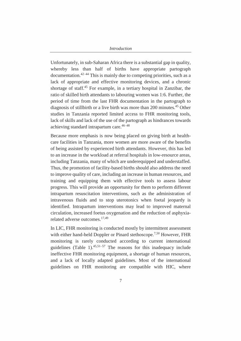
Unfortunately, in sub-Saharan Africa there is a substantial gap in quality, whereby less than half of births have appropriate partograph documentation.42–44 This is mainly due to competing priorities, such as a lack of appropriate and effective monitoring devices, and a chronic shortage of staff.45 For example, in a tertiary hospital in Zanzibar, the ratio of skilled birth attendants to labouring women was 1:6. Further, the period of time from the last FHR documentation in the partograph to diagnosis of stillbirth or a live birth was more than 200 minutes.45 Other studies in Tanzania reported limited access to FHR monitoring tools, lack of skills and lack of the use of the partograph as hindrances towards achieving standard intrapartum care.46–48
Because more emphasis is now being placed on giving birth at health-care facilities in Tanzania, more women are more aware of the benefits of being assisted by experienced birth attendants. However, this has led to an increase in the workload at referral hospitals in low-resource areas, including Tanzania, many of which are underequipped and understaffed. Thus, the promotion of facility-based births should also address the need to improve quality of care, including an increase in human resources, and training and equipping them with effective tools to assess labour progress. This will provide an opportunity for them to perform different intrapartum resuscitation interventions, such as the administration of intravenous fluids and to stop uterotonics when foetal jeopardy is identified. Intrapartum interventions may lead to improved maternal circulation, increased foetus oxygenation and the reduction of asphyxia-related adverse outcomes.17,49
In LIC, FHR monitoring is conducted mostly by intermittent assessment with either hand-held Doppler or Pinard stethoscope.7,50 However, FHR monitoring is rarely conducted according to current international guidelines (Table 1).45,51–57 The reasons for this inadequacy include ineffective FHR monitoring equipment, a shortage of human resources, and a lack of locally adapted guidelines. Most of the international guidelines on FHR monitoring are compatible with HIC, where
Introduction
8
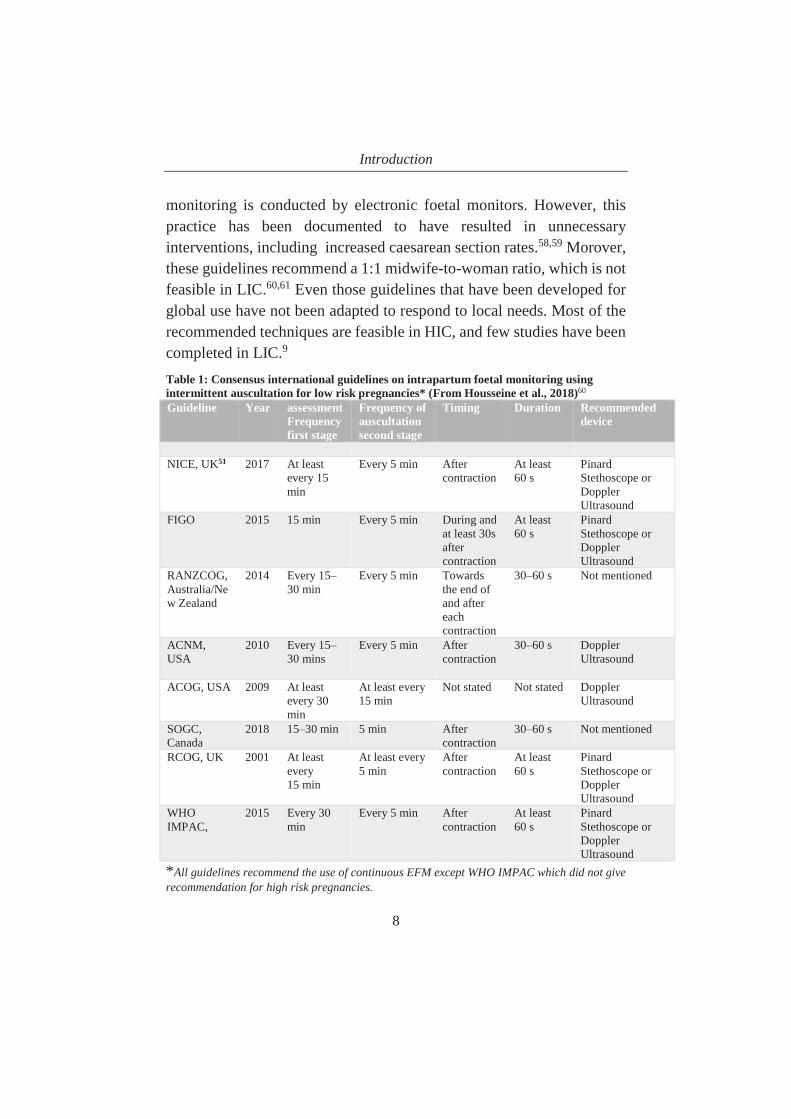
monitoring is conducted by electronic foetal monitors. However, this practice has been documented to have resulted in unnecessary interventions, including increased caesarean section rates.58,59 Morover, these guidelines recommend a 1:1 midwife-to-woman ratio, which is not feasible in LIC.60,61 Even those guidelines that have been developed for global use have not been adapted to respond to local needs. Most of therecommended techniques are feasible in HIC, and few studies have been completed in LIC.9
Table 1: Consensus international guidelines on intrapartum foetal monitoring using intermittent auscultation for low risk pregnancies* (From Housseine et al., 2018)60
Guideline Year assessment Frequency first stage
Frequency of auscultation second stage
Timing Duration Recommended device
NICE, UK51 2017 At least every 15 min
Every 5 min After contraction
At least 60 s
Pinard Stethoscope or Doppler Ultrasound
FIGO 2015 15 min Every 5 min During and at least 30s aftercontraction
At least 60 s
Pinard Stethoscope or Doppler Ultrasound
RANZCOG, Australia/New Zealand
2014 Every 15–30 min
Every 5 min Towards the end of and after each contraction
30–60 s Not mentioned
ACNM, USA
2010 Every 15–30 mins
Every 5 min After contraction
30–60 s Doppler Ultrasound
ACOG, USA 2009 At least every 30 min
At least every15 min
Not stated Not stated Doppler Ultrasound
SOGC, Canada
2018 15–30 min 5 min Aftercontraction
30–60 s Not mentioned
RCOG, UK 2001 At least every15 min
At least every5 min
After contraction
At least 60 s
PinardStethoscope or Doppler Ultrasound
WHO IMPAC,
2015 Every 30 min
Every 5 min After contraction
At least 60 s
Pinard Stethoscope or Doppler Ultrasound
*All guidelines recommend the use of continuous EFM except WHO IMPAC which did not giverecommendation for high risk pregnancies.

Introduction
9
1.2.1 History of FHR monitoring methods Foetal heart auscultation was given little attention until it was discussed for the first time by Mayor and Kergaradec in 1818, when they needed to assess whether the foetus was alive or dead.62 Its popularity was later accelerated by Kennedy’s publication about obstetric auscultation in 1833. In the early 1800s, Laënnec rolled a sheet of paper into a tube and listened through the device, which was later replicated in wood for foetalheart auscultation; a method that has continued to be used to date.45 The following sections describes the various different developments in FHR monitoring tools.
The Pinard stethoscope was developed by a French physician, Dr A. Pinard, in the 1880s, and was in wide use by the 1950s.62 It is the most common instrument, using Laënnec’s technique of sound amplification,transmitting it from the foetal heart to the examiner’s ear. It is currently used in mostly in LIC to intermittently detect abnormal FHR and to facilitate obstetric intervention. However, there is a need for a significant degree of skills and experience to use it accurately. One must count heartbeats while watching a clock and perform multiple calculations to obtain accurate records. Auscultation with this foetal stethoscope has been reported to be uncomfortable for both the patient and the midwife.The DeLee stethoscope is an alternative to the Pinard stethoscope butusing the same technology (Photo 1).

Introduction
10
Photo 1: Pinard and DeLee stethoscope foetal heart rate monitors (copyright-free internet images)
The Handheld Doppler was developed in the 1960s using a technology developed by Austrian physicist, Christian Doppler.61,62 It is an electronic device used for intermittent auscultation and relies on a single-crystal doppler effect. The Doppler uses ultrasound-detected movements of foetal cardiac structures and subjects them to signal modification. Handheld Doppler devices are simple to use and cause less maternal discomfort than the Pinard foetal stethoscope. The readings can be objectively recorded but the device requires electricity or batteries.
Another type of Doppler is called the Freeplay wind-up handheld foetal Doppler. It has rechargeable batteries and can also be hand-cranked,providing rapid recharging with only a minute of winding, and can be used for to up to 10 minutes. Its readings are objective, and the device (Photo 2) is well accepted by mothers and health care providers in LIC.63,64
Photo 2: hand-held Doppler (Source: Muhimbili National Hospital Labour Ward)
Continuous electronic foetal heart rate monitoring was introduced into hospitals in the 1970s using electronic foetal monitoring (EFM) byCardiotocography (CTG) for continuous monitoring. The device recordsthe FHR parameters, including variabilities, decelerations, accelerations,tachycardia and bradycardia as well as the uterine contractions in labour.It has two transducers placed on the mother’s abdomen to detect FHR and uterine muscle activity.45 Records may be captured externally via an

Introduction
11
ultrasound transducer attached to the mother’s abdomen, or internally,via a foetal scalp electrode placed directly on the baby’s head.65 It needs a continuous supply of electricity, specific storage environment, and continuous staff training for accurate interpretation. Readings are printed on paper and are sometimes stored on a computer for later reference.
Photo 3: Electronic Foetal heart rate monitor (Cardiotocograph) (Source: copyright-free internet image)
1.2.2 Safer Births project and the development of an automatic strap-on continuous FHR monitoring device termed MoyoSafer Births is a research, development and implementation project designed to improve foetal heart rate monitoring, new-born resuscitation and perinatal outcomes worldwide. The project was aimed at developinginnovative products and training materials to better equip and train healthcare workers and at establishing new knowledge related to labour and births in LIC. It is a collaborative project involving variousNorwegian and Tanzanian institutions. Safer Births implementation activities were conducted in conjunction with the Helping Babies Breathe program in Tanzania.
As one of the strategies to improve FHR monitoring to facilitate awareness of foetal distress and to inform decision-making, an automatic strap-on FHR monitor called Moyo was developed. This device was designed to facilitate early identification of foetuses at risk of asphyxia(Photo 4).

Introduction
12
As part of the Safer Births project, a randomized controlled study, carried out in rural settings in Tanzania, revealed that the use of Moyo increased the identification of baseline abnormal FHR and subsequent intrauterine resuscitations.66 Further, qualitative studies on the preferences and acceptability of the strap-on Moyo device among mothers and clinical staff have been conducted. The findings of these studies show that preference and acceptance was high compared to other devices. Also, the use of Moyo was reported to positively affect the women’s birth experience, whereby an audio-visual monitor reassured them of the wellbeing of the foetus.
Photo 4: Part 1: Moyo sensor strapped onto the mother’s abdomen for prolonged monitoring. Lightweight and portable, it allows the mother to move around freely. Part 2: Metal pads to detect maternal heart rate (Photo reprinted with permission from Laerdal Global Health)
In 2008, Wyatt recommended a number of necessary features for developing appropriate technology for use in low resource settings.67
Table 2 summarizes a comparison of different FHR monitoring devicesby these recommended parameters.

Introduction
13
Table 2: comparison of different FHR monitoring methods available by different parameters.
Parameter Pinard stethoscope
Hand-held Doppler
Wind-up Hand-held Doppler
CTG Moyo
Pinard stethoscope
Availability for LIC
Available Available Available Limited availability
Available in some countries
Cost Inexpensive Relatively inexpensive
Relatively inexpensive
Expensive Relatively inexpensive
Power and consumables
Not needed Continuous supply of replacement batteries
Built-in rechargeable batterie, can be hand cranked
Continuous power supply
Built-in rechargeable battery from multiple mains of electricity
Use Intermittent Intermittent Intermittent Continuous Both intermittent and continuous
Maternal FHR comparison
No No No Yes
FHR display No Yes Yes Yes YesFHR records No No No Yes
continuousYes, for 30 min
Acceptability by mothers
Low High High Low due to limited mobility
High
Acceptability by clinical staff
Low High High Low High
Operation in harsh environment
Yes Yes Yes No Yes
Mode of operation
Skilledlistening and arithmetic
Simple to use with minimal training
Simple to use with minimal training
Skilled use and interpretationneeded
Simple to use with minimal training
Life span >5 years 5 Years 5 Years 5 yearsAvailability for LIC
Available Available Available Not available Available in some countries
Cost US$) ~3–5 ~200 ~200 Expensive ~198Mobility of the women
Allows mobility
Allows mobility
Allows mobility
Does not Allow mobility
Allows mobility

Introduction
14
1.2.3 Current FHR monitoring practices The main methods for intrapartum FHR in LIC are intermittentmonitoring using mostly the Pinard and, to a lesser extent, the hand-held Doppler. However, a research gap exists in the effectiveness of theseFHR assessment techniques in these settings, including Tanzania. In sub-Saharan Africa, two documented randomized studies had investigatedFHR devices; specifically, the intermittent Doppler and the Pinard method, by 2016. One study in Uganda showed increased FHR detection in the Doppler compared to the Pinard arm, however, with no difference in perinatal outcomes.7 The second study, conducted in Harare, showed that abnormalities in foetal heart rate were detected more often by the Doppler than with the Pinard method. This resulted in less hypoxic ischemic encephalopathy, seizures and deaths.68 Recently, two additional studies have been conducted in rural settings in Tanzania as part of the Safer Births program. The first study, which compared the Pinard and hand-held Doppler, showed no difference in abnormal FHR detection and subsequent perinatal outcomes.69 The second study compared the Moyo with the Pinard method and showed that the use of Moyo increased FHR detection and intrapartum resuscitations compared to the Pinard with similar perinatal outcomes.66
1.2.4 FHR monitoring techniques in HIC
Most of the RCTs and systematic reviews in the use of FHR monitoring techniques have been conducted in HIC.58,65 A systematic review of 12 trials compared continuous monitoring with CTG versus intermittentmonitoring with the Doppler or Pinard. In the intermittent monitoring arms of the studies, women received one-to-one care. The findings showed that there was no difference in the numbers of intrapartum-related deaths between the groups, but there was a reduction in incidence of neonatal seizure.58,70 A cohort study conducted in the US showed that continuous CTG was associated with lower intrapartum-related deaths and less rates of low Apgar score incidence at 5 minutes.71,72 However,

Introduction
15
in both reports, continuous monitoring was associated with significantly more births by caesarean section and by instrumental vaginal births with no differences in new-born morbidity and mortality.
A recent systematic review analysis of 36 studies, six from LIC, found improved outcomes with the use of partograph during labour. Using a CTG increased the odds of caesarean section by approximately 30% with no benefits on perinatal outcomes observed.41 The review recommended the use of intermittent FHR monitoring combined with partograph a feasible technique to improve new-born outcome. Implementation studies on these methods were also recommended.
The effectiveness of a novel strap-on automatic Moyo monitoring device has not been evaluated in an urban setting. Moreover, there is no evidence to date on the implementation of FHR monitoring in relation to partograph use, nor on the quality of health care provision during labour. Because there is an uncertainty regarding the appropriate FHR device to use during labour and its relationship to adverse outcomes, there is a need to identify the most effective and scalable FHR technique.
1.3 Tanzania-Setting and Context
1.3.1 The country and the people Tanzania (Figure 3) is situated in Eastern Africa within the African Great Lakes region, occupying an area of 947,300 km² (land: 885,800 km2, water: 61,500 km2). Important landmarks of Tanzania include Mount Kilimanjaro, Africa’s highest mountain in north-eastern Tanzania, the Ngorongoro crater, the Serengeti National Park, and many lakes, including Lake Victoria. Administratively, Tanzania has a total of 31 regions; 26 in Tanzania Mainland, and 5 in the Zanzibar islets.73
Tanzania has a population of 55 million, with an average annual growth rate of 2.8%. Tanzania has the largest population in East Africa, and almost a third of the population is urban. Tanzania’s youthful population – about two-thirds of the population is aged under 25 – is growing rapidly

Introduction
16
because of the high total fertility rate of 4.8 children per woman.74 The economy depends on agriculture, providing 85% of exports, and employs 65% of the work force. Over 28% of the population live below the Basic Needs Poverty Line ($1.90 per day) and 10% below the Food Poverty Line ($0.50 per day). Table 3 illustrates selected economic and heath indicators.
Figure 3: Map showing Tanzania and Dar es Salaam (Source: free internet image)
Table 3: selected economic and health indicators (Source: 2012 Census survey, Demographic Health Survey 2010 and 2015, Tanzania in Figures 2016)
Indicator 2015/2016 estimates
Population growth rate 2.75%
Crude birth rate (births/1,000 population) 35.6
Crude death rate (deaths/1,000 population) 7.6
Infant mortality rate (deaths/1,000 live births) 39.9
Total fertility rate: (children born/woman) 4.77
Life expectancy at birth: total population (years) 62.6
Male (years) 61.2
Female (years) 64.1
Contraceptive prevalence rate (currently married women, mCPR) 38.40%
Health expenditures: percentage of GDP (2014 5.60%
Physicians density: physicians/100,000 population (2012) 3
Unemployment rate: 10.30%
Population below poverty line: $1.90 a day (2015) 22.80%

Introduction
17
GDP - per capita (PPP): $3,300
1.3.2 Tanzanian health systemTanzania has a hierarchical health structure running parallel with an administrative hierarchy. Primary health care facilities, includingdispensaries, are at the bottom, with health centres at ward level, district hospitals at district level, regional referral hospital at regional level,zonal hospitals, and one national hospital. Due to the inaccessibility of facilities offering maternal and new-born care, some communities have established maternity waiting homes located near health facilities to facilitate access.
Dispensaries conduct normal births. These facilities are usually equipped with few beds for medical treatment or observation before referral. Women first treated in dispensaries are referred to the health centres that admit patients. In recent years, some of the health centres have been upgraded to hospitals to cater for the high demand for advanced care. District hospitals act as referral facilities for health centres. At these hospitals, specialized care is provided, depending on the available specialist. Referrals from districts are made to the regional referral hospitals, which provide more advanced care. However, in Dar es Salam,the main commercial city, three district hospitals (Temeke, Amana and Mwananyamala) have been upgraded to regional referral hospitals due to an increased specialized care demand. Zonal hospitals are positioned at the highest hierarchical level, and are staffed with specialized doctors, super-specialists and specialized equipment and care. Muhimbili National Hospital occupies the highest level of all facilities and receivesreferrals from multiple referral hospitals. A total of 7,685 (70% public and 30% private) health facilities were established in Tanzania by 2017.Under Public Private Partnership (PPP), some private hospitals have signed service agreements with the government to provide health services as designated hospitals. These include the exemption ofpregnant women and under-5 children from out-of-pocket payments.

Introduction
18
Tanzania is among one of the sub-Saharan African countries of those thatrecord a serious shortage (54%) of Human Resources for Health (HRH); a key element for the delivery of quality health care.75 Efforts to mitigate the shortage include the expansion of training institutions, increased enrolment, transformation to a competence-based curriculum and task sharing among care providers.
1.3.3 National health policies and programs With the health system conforming to a pyramid structure, from the community at the lowest level to the Muhimbili Hospital at national level, the coordination and management of health-care functions are shared by two ministries. The first is the Ministry of Health Community Development Gender Elderly and Child (MOHCDGEC), which isresponsible for the formulation of policies and technical guidelines andoverseeing service delivery from the regional referral hospitals and consultant hospitals. The second is the President’s Office-Regional Administration and Local government (PO-RALG) Directorate of Health, Social Welfare & Nutrition Services (DHSWNS), whichimplements the policies, standards and strategic plan, and oversees thedistrict hospitals, health centres, dispensaries and various community-based services.
The Reproductive and Child Health Section (RCHS), under MOHCDGEC, is responsible for the preparation and review of policies, guidelines, and manuals for maternal and child health. The Section alsocoordinates activities and programs with other ministries and organizations dealing with RCH issues and conducts a review of standards of quality maternal and childcare.
Some health policies that target the improvement of perinatal care include the health payment exemption policy, cost sharing and health insurance. Pregnant women and children under the age of 5 are among those exempted from paying health insurance. Cost share covers include all Tanzanians, whereby the government have subsidized medical care
Introduction
19
costs. Contributing to the National Health Insurance fund is mandatory for all government employees and is optional for those employed in the private sector, as well as for groups and individuals.
MOHCDGEC has programs and frameworks for the provision, monitoring and evaluation of RCH services. However, program implementation is largely under-budgeted and the fund disbursement mechanisms to the districts are poor.76,77 In 2007, the Primary Healthcare Service Delivery Program (PHSDP 2007–2017) was established, aimed at accelerating the provision of primary health care. Activities includedstrengthening health systems, financing, medicine provision, equipment and supplies.78 This led to only partial success, as the maternal mortality rate (MMR) and the perinatal mortality rate increased between 2010 and 2015 due to substandard care during labour and births.79 As a part of theimprovement of the quality of care, Direct Health Facility Financing (DHFF) was introduced. Also, some health facilities are linked to Results-Based Financing (RBF) and the Community Health Fund, giving them some degree of financial autonomy.80
1.3.4 Current strategies for new-born careProgress towards the prevention of neonatal deaths has been slower compared to improvements in the overall under-5 mortality rate (Figure 4). The decline was recorded as being reduced from 40 to 25 deaths per 1,000 live births for neonatal deaths from 1999 to 2016. The neonatal contribution to under-5 mortality rates increased from 27% to 37%during the same period.81–83
To improve perinatal health with a specific focus on perinatal mortality, a cross-cutting strategy was formulated. This was the National Road Map Strategic Plan to Improve RMNCAH: The One Plan II (2016–2020),which was built on the Health Sector Strategic Plan IV (HSSP 2015–2020). One of the aims of the RMNCAH is to reduce perinatal mortality by 20% by 2020.

Introduction
20
Figure 4: Trends in early childhood mortality (Source: DHS-1999, 2004, 2010, 2016) *Computed as the difference between the neonatal and infant mortality rates IMR=Infant mortality rate Care during childbirth, Emergency Obstetric and New-born Care (EmONC) guidelines form the major elements of the strategy. EmONC is a set of evidence-based packages of interventions and services that serve to identify obstetric and new-born complications and to make timely and appropriate management decisions for improving the quality of care. The basic components of EmONC (BEmONC) are supposed to be provided at all health facilities. At hospital level, comprehensive (CEmONC) services are provided. Some of the health centres have been upgraded to provide CEmONC services. However, according to an assessment of EmONC in 2015, only 13% of dispensaries, 28% of all health centres and 62% of hospitals were capable of performing all functions.79,84 This shows that there is still inadequate quality of care provision during labour and birth.
1.4 Conceptual framework
Modified WHO vision 2015 framework for quality of care in labour The World Health Organization (WHO) defines quality of care as ‘the extent to which health care services provided to individuals and patient
020406080
100120140160
1999 2004 2010 2016
Deat
hs p
er 1
000
live
birt
hs
Under 5 IMR Post neonatal* Neonatal

Introduction
21
populations improve desired health outcomes. In order to achieve this, health care needs to be safe, effective, timely, efficient, equitable, andpeople-centred.”85 Quality of care in most of the health facilities in LIC is complex and needs multidisciplinary interplay. Post-2015, the WHO envisioned a world where all pregnant women and new-borns wouldobtain access to quality care around the perinatal period, in line with the WHO global ENAP and the Ending Preventable Maternal Mortality (EPMM) agenda.5,14
The WHO framework was used in this thesis to conceptualize quality of care for maternal and new-born health. Important components of the framework, including its policies, strategies and guidelines (Figure 5),have been identified.
There has been an increased number of births occurring at tertiary facilities due to the available expertise and facilities for operative birthscompared to those available at lower facility levels. This compromises the quality of care provision due to inadequate levels of human resources,lack of physical infrastructure, and supplies not matching demand. Hence, the importance of the health system is recognised in theimprovement of the skills of the workforce, the availability of the medical products, and the provision of continuous medical education, finance, leadership and governance, which will cascade down to the quality of care provision.86
In order to provide quality care during labour, Tanzania needs to have competent, skilled, midwives who are equipped with effective tools. This should be coupled with readily available and accessible elements of infrastructure, such as adequate operating theatres. Also, skilled providers should be supplied with locally developed or adapted guidelines and EmONC services.
In the provision of care (Figure 5), a safe, effective and efficient intrapartum FHR monitoring device, forms a critical element of improved and safe care. When FHR monitoring with appropriate tools is

Introduction
22
combined with appropriate partograph use, this should provide an actionable information system to assist in the review and audit of the labour.26,39
A secondary element is a need for effective communication about the labour progress with the mother, as well as promoting the woman’s dignity and respect. All these elements should lead to improved quality of intrapartum care, which forms the causal pathways that lead towardsbetter perinatal outcomes, as shown in the framework.
Figure 5: Quality of care framework to improve perinatal outcomes (Modified from the WHO Vision 2015)

Introduction
23
1.5 Summary The rates of stillbirths and neonatal deaths are high in Tanzania and significantly contribute to the burden of disease. There are geographical and income disparities associated with this burden, with the highest rates being found in sub-Saharan Africa (including Tanzania). Moreover, there is an increased volume of mothers giving births at health facilities, which, when coupled with a shortage of skilled birth attendants and inappropriate tools to monitor labour, increases the risk of adverse perinatal outcomes, including FSB and early neonatal deaths. Several international guidelines and documents, including those developed by the WHO and UNICEF, recommend interventions focusing on the improvement of quality of care around labour and births, because they provide a triple return on investment; i.e., the wellbeing of the mother, foetus and new-born.5,11,57,87 Intrapartum FHR monitoring and partograph use in labour are considered important quality strategies that may facilitate improvement in the provision of care during labour and birth.
However, in LIC (including Tanzania), where most births occur, there are uncertainties about the kinds of devices that are effective in FHR monitoring during labour. It is hypothesized that studies evaluating effective FHR monitoring devices, and their subsequent implementation, will improve the quality of care, and, by proxy, improve perinatal outcomes. This thesis responds by contributing to a better understanding of effective FHR monitoring devices during labour. Further, it assesses the implementation strategies for FHR monitoring in relation to partograph use, and intrapartum-related interventions where appropriate.

Introduction
24

Aims of the PhD project
25
2 Aims of the PhD project
The overall aim of this PhD project was to compare the effectiveness of different FHR monitoring devices and the associated improvement in quality of intrapartum care at two urban referral hospitals in Dar es Salaam, Tanzania.
2.1 The specific aims of the studies were:
1. To compare the effectiveness of intermittent monitoring with a hand-held Doppler versus the Pinard stethoscope in the detection of baseline abnormal FHR in labour (Study I).
2. To compare the effectiveness of continuous monitoring with the strap-on automatic Moyo versus intermittent monitoring with the hand-held Doppler in the detection of baseline abnormal FHR in labour (Study II).
3. To describe time intervals for different events in labour, mode of giving births and perinatal outcomes for the different FHR monitoring methods (all studies).
4. To assess the quality of midwifery practices related to FHR monitoring (including partograph documentation) pre-implementation using the Pinard stethoscope compared to post-implementation of the automatic strap-on Moyo (Study III).
2.2 Research questions
In urban referral hospitals in Tanzania:
1. Does intrapartum intermittent FHR assessment with a hand-held Doppler differ in detection of baseline abnormal FHR compared to a Pinard stethoscope among low-risk parturient women? (Study I)
2. Does intrapartum FHR assessment with a strap-on automatic Moyo differ in the detection of baseline abnormal FHR compared

Aims of the PhD project
26
to intermittent assessment with hand-held Doppler among parturient women? (Study II)
3. Which FHR monitoring method is associated with earlier detection of baseline abnormal FHR? (All studies)
4. Does the implementation of strap-on automatic Moyo compared to intermittent assessment with Pinard stethoscope affect partograph documentation and the quality of midwifery practices in labour? (Study III)
2.3 Hypotheses:
1. Intermittent FHR assessment with hand-held Doppler will detect more baseline abnormal FHR compared to Pinard stethoscope among parturient women (Study I).
2. FHR assessment with strap-on automatic Moyo will detect more baseline abnormal FHR compared to intermittent assessment with hand-held Doppler among parturient women (Study II).
3. FHR assessment with strap-on automatic Moyo will detect baseline FHR abnormalities earlier compared to intermittent assessment with hand-held Doppler and Pinard stethoscope among parturient women (Studies II & III)
4. FHR assessment with strap-on automatic Moyo will improve quality of intrapartum care (including the use of partograph) as compared to intermittent assessment with Pinard stethoscope (Study III).

Methods and participants
27
3 Methods and participants
3.1 Study settings
All three studies were conducted in Dar es Salaam; the major commercial city and former capital of Tanzania, located on the Eastern coast and facing the Indian Ocean. It has total area of 1393 km2 with five municipalities. In 2019, the projected population of the city is approximately 6.3 million and it has the highest growth rate of any region in the country (5.6% year). Roughly 35% of the Dar es Salaam population comprises children under the age of 14.74
3.1.1 Muhimbili National Hospital
Studies I and II were conducted at Muhimbili, a teaching hospital and the largest consultant hospital in Tanzania, situated in Ilala municipality. About 10,000 births are facilitated annually, corresponding to about 35 births per day. The hospital serves as a tertiary referral hospital for the whole country. It deals with many complicated obstetric cases with more than 50% of them by caesarean section (the highest rate in the country). The high rate of caesarean sections is due to increased referral of complicated cases from the lower-level facilities, maternal requests and inappropriate indications. By 2016, approximately 7% of births were stillborn and 2% died within 24 hours.88 Births are conducted by nurse-midwives and doctors, assisted by medical and midwifery students.
The labour ward at Muhmbili has 20 birthing beds (Figure 6). There are approximately 25 nurse midwives, which is far below the WHO benchmark for the supply of a minimum of 20 skilled birth attendants and 60 beds per 3,600 births per year, respectively.89 The ward is managed by 5 nurse-midwives and 2 nursing assistants in each shift of 12 hours. The doctors-on-call team comprises 1 consultant, 1 obstetrician, 2 obstetric residents, and 1 intern doctor on 24-hour call. There is an obstetric operating theatre located in a separate building adjacent to the maternity block equipped with two operating beds.

Methods and participants
28
Figure 6: Schematic drawing of the labour ward at Muhimbili and Temeke (Illustration by Darja and Karl-Otto)
On admission, a nurse-midwife screens all women for vital signs registration, initial FHR assessment, and vaginal examination before they enter the labour ward. A brief obstetric history and vital signs are taken, and the required information is entered in the labour ward register. In cases of uncertainties, the midwives and on-call doctor review the partograph and make collective decisions. After a normal vaginal birth,mothers and babies are observed in the hospital for up to 24 hours,whereas caesarean births entail 48 to 72 hours of observation. Babies in need of medical attention are admitted to the neonatal unit.90,91

Methods and participants
29
3.1.2 Temeke Regional Referral Hospital Study III was conducted at Temeke Regional Referral Hospital, located within the Temeke municipality. The municipality is the industrial district of the city and has the largest concentration of low-income residents in Tanzania at about 2 million people. It has about 135 healthfacilities that refer complicated cases to Temeke Hospital for advanced care.
The hospital has about 30–50 births per day (more than 15,000 per year). Its labour room (Figure 6) has 18 beds (far below the WHO benchmark) and a general operating theatre in a separate building is used for obstetric and other surgical cases. The obstetrics unit has two qualified obstetricians, 12 general doctors, 25 nurse-midwives, five medical attendants and a varying number of rotating intern medical doctors and nurses who perform assist births. Nurses have three shifts per day with an average of three nurses and one medical attendant per shift. Doctors have two shifts per day with one medical doctor and two interns during the day and night shifts, respectively. Some emergency cases are referred to Muhimbili.
3.2 Study design
This thesis comprised three interlinked quantitative studies (Table 4). Avariety of quantitative study designs were used to achieve the overall aims stated above. The two RCTs (Studies I and II) were a sequentialdesign and were carried out at Muhimbili, and one pre/post observationalstudy (Study III) was conducted at Temeke (Table 4). Study I was conducted from 1st April 2013 to 30th September 2015. Study II was conducted from 1st March 2016 to 30th September 2017. Study II built on the results of Study I and used the most effective device to comparethis with the novel strap-on automatic Moyo. Study III was conducted from 7th March 2016 to 15th December 2016, concurrent with Study II. The study methods are summarized in Table 5.

Methods and participants
30
Table 4: Timelines of three PhD studies at Muhimbili and Temeke
2013 2014 2015 2016 2017 Quarter of the year 1 2 3 4 1 2 3 4 1 2 3 4 1 2 3 4 1 2 3 4
Study I
Study II
Study III Table 5: Summary of research design, methods and participants for three studies
Study I Study II Study III Study design
Randomized controlled study (Intermittent Doppler vs intermittent Pinard stethoscope)
Randomized controlled study (Continuous Moyo vs Intermittent Doppler)
Pre and post implementation study (Continuous Moyo vs Intermittent Doppler)
Sample size Doppler (1,421) vs Pinard (1,423)
Moyo (1,479) vs Doppler (1494)
Post/Moyo (2,442) vs pre/Pinard (1640)
Study focus Effectiveness in detection
Effectiveness in detection and timeliness
Improvement in midwifery practices and quality of care
Recruitment period
April 2013 to September 2015
March 2016 to September 2017
Pre: March to June 2016 Post: July-December 2016
Study setting
Muhimbili National Hospital
Muhimbili National Hospital
Temeke Regional Referral Hospital
Participants Pregnant singleton women in true labour with normal baseline FHR on admission
Pregnant singleton women in true labour with normal baseline FHR on admission
Pregnant singleton women in true labour with normal baseline FHR on admission
Data Collection method
Structured case report form (CRF)
Structured case report form (CRF)
Structured case report form (CRF)
Data analysis methods
χ2 and Fisher’s exact test Mann–Whitney test, Independent samples t-test, Binary logistic regression
χ2 and Fisher’s exact test Mann–Whitney test, Independent samples t-tests, Binary and multinomial logistic regression, Linear regression (with a natural log-transformation)
χ2 and Fisher’s exact test, Mann–Whitney test, Independent samples t-test Binary and multinomial logistic regression

Methods and participants
31
3.2.1 Randomized controlled studies (Studies I and II)
Studies I and II were randomized controlled trials using a superior,parallel, open label design and conducted at Muhimbili Hospital. In both studies, the randomization sequence was computer-generated by an independent statistician. Details of the allocated group were given to the study coordinator, who supervised data clerks to write unique identifiers on cards and put them in sequentially numbered opaque sealed envelopes (SNOSE). The allocation sequence was concealed from investigators,participants and clinical staff implementing and assessing outcomes.However, due to the nature of the interventions (medical devices), it was not possible to blind the participants and clinical staff. Errors were minimized to 5% significance level, and probability to detect existing effect (power) was set at 80% for two-sided comparison of proportions.A minimum of 10% more women were included to account for potentially missing data in order to keep the sample large enough to detect meaningful difference.
3.2.2 Pre- and post-interventional study (Study III)The third study (Study III) was conducted at Temeke in a pre- (3 months using intermittent Pinard) and post- (4.5 months using continuous Moyo) intervention design. Records at Temeke showed that abnormal FHR was detected in approximately 2.0% of all low-risk births using Pinard. Anincrease in the detection rate of 5% was the smallest change that we considered to clinically meaningful. Assuming an increase in detection rate of at least 5% with the strap-on Moyo monitor, we planned the study to include a minimum of 890 (total 1,780) women pre- and post-implementation, which would give a 90% power effect with an alpha level of 0.05 (two-sided comparison to detect such a change). This sample size was assumed to be reached within a study period of 4 monthsin total (2 months pre- and 2 months post-implementation). However, due to delays in implementation and to account for missing data, the study period was extended to a total of 7.5 months (3 months before and 4.5 months after).

Methods and participants
32
3.3 Participants
The inclusion and exclusion criteria are presented in Table 6. Study I included term low-risk pregnant women in labour. Studies II and III included women with gestation age (GA) above 28 weeks as they were considered viable with current National guidelines.
Table 6: Inclusion and exclusion criteria for participants in three studies Inclusion criteria GA >28* complete weeks Cervical dilatation >3cm**
Normal FHR on admission
Singleton
Written consent obtained
Exclusion criteria Elective Caesarean section
Severely sick client, e.g., eclampsia, pre-eclampsia
Multiple pregnancy
Study I; GA >37 weeks, ** Study I; cervical dilatation >3cm and <7cm
3.4 Training of relevant clinical staff
In Study I, before the start of the implementation of the intervention, all labour ward staff (midwives and doctors) at both hospitals were trained for a full day on FHR management protocols. On-the-job short and frequent refresher training sessions were conducted intermittently to increase protocol adherence and accommodate incoming staff who did not receive the initial training. Training included theoretical information about FHR monitoring during labour, the management of an abnormal FHR and partograph documentation. The criteria for FHR monitoring were emphasized and included monitoring and recording of FHR this every 30 minutes in the first stage of labour, and every 5–15 minutes in the second stage, as per the WHO guidelines and other international guidelines.45 For intermittent assessment (i.e., using Pinard and Doppler), midwives were trained to assess the FHR during the last 10 minutes of every half hour, particularly before, during, and immediately after a contraction. Any FHR abnormalities were to be reported to the doctor on call for consideration and potential actions.

Methods and participants
33
In Studies II and III, in addition to the training described above, training sessions using a Moyo standard operating procedures were provided (Photo 5). The labour ward staff were also told that abnormal FHR detections should be reported to the doctor on call, who then acted according to hospital protocols.
Photo 5: Drs Muzdalifat, Matilda, Sisters Amina and Anna (Part 1 at Muhimbili) and Drs Muzidalifat and Kamala (Part 2 at Temeke) training midwives and doctors on intrapartum FHR monitoring and standard operating procedures for different methods (Photo taken by Benjamin Kamala and Gilbert Kilonzo, permission to use the photo was obtained from trained participants)
3.5 Training of research assistants
For all studies, research nurses were trained for one additional day on the research protocol and data collection methods (Photo 6). This ensured the accuracy and completeness of the data recorded on the paper-based case report form (CRF) (see Appendices 1 and 2 for entries relating to the three studies, respectively). Data were collected from mothers’ antenatal cards, partograph, obstetric register, and, when needed, from routine neonatal morbidity and mortality records in the neonatal unit.

Methods and participants
34
Photo 6: Dr Kamala Training research nurses on data collection process using care report format Temeke (Photo taken by Gilbert Kilonzo, permission to use the photo was obtained from research nurses)
3.6 Study procedures
In all studies (Studies I, II & III), for intermittent auscultation, women were monitored using the standard protocol with a either a Pinard stethoscope or hand-held Doppler (Power-free Education Technology). The midwives then continued with their routine activities and periodically revisited the women to check and record FHR readings in the partograph and to perform other management as indicated.
For Studies II and III, women monitored with continuous Moyo received information on how the device was to be used by the enrolling midwife.Important information was provided to women randomized to the Moyoarm for its proper use. Moyo, equipped with a rechargeable battery, has a 9-crystal Doppler ultrasound sensor, which facilitates the rapid identification of FHR. It can be used continuously (strapped-on) or intermittently. The detection area is increased, necessitating less palpation. It has a 30-minute histogram display of the FHR (in continuous mode) and an audio-visual alarm if abnormal FHR is

Methods and participants
35
detected. Monitor readings are coded with colours. A green reading indicates normal FHR. Yellow is a warning indicator for FHR outside the normal range (Figure 7). A question mark meant that no FHR is detected or that the sensor is displaced. A red reading with an alarm indicates when the records are outside the range for more than three minutes or when no FHR is detected, which may be an indicator of foetal death or a displaced sensor. The mothers would call the labour ward staff when there were abnormalities in the readings that had not been spotted by the attendant. A set of dry electrodes assist in the differentiation ofmaternal heart rate from FHR. All the above-mentioned features are meant to facilitate interventions and promote greater control of the management of multiple expectant women by the birth attendant.
For those mothers who consented. Moyo was strapped on for continuous FHR monitoring. The midwife continued with her routine activities, but periodically revisited the women to check and record the FHR reading or when an alarm sounded from the Moyo. The Moyo remained strapped on until the end of the second stage of labour, or immediately prior to the start of a caesarean section.

Methods and participants
36
Figure 7: Moyo monitor with different colour codes for different FHR readings (Source: Laerdal Global Health)
3.7 Data collection and management Data collection for the three studies was conducted by trained research nurses. CRFs were cross-checked by investigators for quality and completeness before entry. CRFs with queries were returned to the research nurse for verification and correction before data entry. A data entry template was generated in EpiData by the investigators and a statistician. Verified data were double entered by trained data clerks. Then, data were transferred to SPSS for analysis (IBM SPSS Statistics for Windows, Version 25.0. Armonk, NY: IBM Corp.). Patient information was recorded using confidential codes for each woman and was kept in a secure place.
3.8 Measures/Variables
Table 7 summarises the primary, secondary and background variables and values for the different studies included in this thesis. Details of each variable is appended (Appendix 1 and 2)

Methods and participants
37
Table 7: Primary, secondary and background variables registered and analysed for the three studies
Variable Values/units of measure Study I
Study II
Study III
Primary outcome FHR during labour Normal, Abnormal √ √ √ Secondary outcomes FHR documentation in labour Yes, No √ Frequency of FHR documentation <30min, 30–60 min, 60–120 min, >120
min √
Mode of birth Normal (SVD), Assisted breech, caesarean section, Vacuum extraction
√ √ √
Indications for caesarean section Obstructed labour, Abnormal FHR, Prolonged labour, Others
√ √
Received resuscitation Stimulation, suction, ventilation √ √ Apgar score at 5 minutes Low (<7), Normal (≥7) √ √ √ Birth outcomes Normal, Admitted to neonatal unit,
Fresh stillbirths √ √ √
Intrauterine resuscitation Stop oxytocin, change mother’s position, Give IV fluids, Give Oxygen
√ √
Neonatal outcomes 24-hours Normal, still admitted to neonatal unit, early neonatal death
√ √ √
Time from Admission to birth Minutes √ √ Time from Admission to Abnormal FHR detection
Minutes √ √
Time from Last FHR to birth Minutes √ √ √ Time Between FHR assessment Minutes √ Time from abnormal FHR to birth Minutes √ √ Duration of stages of labour Minutes √ √ √ Baseline characteristics Age (years) <20, 20–35, >35 Birth weight Low (<2500gm), normal (≥2500gm) √ √ √ GA (weeks) <37, 37–42, >42 √ √ √ Cervical dilatation on admission Centimetres √ √ Parity 1, 2–4, >4 √ √ Education No formal education, primary,
secondary, above secondary √ √
Presentation Cephalic, breech √ √ Source of admission Home, inpatient, Referrals √ √ √ HCW assisting birth Doctor, nurse midwife √ √ Marital status Married, Single, cohabiting √ √ Antenatal visits None, 1–3, >3 √ √ Maternal infection Yes, No √ ANC problem Yes, no √ √ √ Health care worker attending birth Doctor, nurse-midwife, nurse attendant,
intern nurse, intern doctor √ √
Obstetric complications Yes, No √ √

Methods and participants
38
3.8.1 Primary outcomes An abnormal baseline FHR was used as the primary outcome in all three studies. There is no global consensus on the range of normal baseline FHR. Some guidelines recommend a normal baseline range between 100˗180 beats/min.51,60 FIGO recommends a normal baseline FHR from 110-160 beats/min, thus abnormal FHR is considered to be <110 or >160 beats/min.45 In this thesis, we used the range as stated in the national guidelines, which is defined as normal (120 to160 beats/min throughout labour and birth) or abnormal (absent, <120 or >160 beats/min). The classification of an abnormal FHR was reached after at least three abnormal assessments at different abdominal sites with the Pinard Stethoscope or the Handheld Doppler for intermittent assessment. For Moyo, a persisting abnormal FHR lasting for at least 3 minutes was classified as abnormal.
3.8.2 Secondary outcomes In Study I, secondary outcomes included mode of birth (vaginal birth, caesarean section, assisted breech, and vacuum extraction), time intervals (admission to birth, detection of abnormal FHR to birth), perinatal outcomes, including Apgar scores at 5 minutes (abnormal was defined as an Apgar score <7) a surrogate measure of birth asphyxia,4,92 bag mask ventilation, admission to neonatal unit for treatment, FSB, and death within 24 hours postpartum. Mode of birth was dichotomized into two categories (i.e., vaginal, including vacuum extraction, and caesarean section) due to there being relatively fewer cases in the vacuum extraction category. FSB was defined as an Apgar score of zero at both 1 and 5 minutes, with intact skin and suspected death during labour/birth. A composite perinatal outcome measure included FSB, admission to the neonatal area, and death within 24 hours.
In Study II, secondary outcomes included the Apgar score at 1 and 5 minutes; mode of birth, perinatal outcome at birth, and composite perinatal outcomes at birth and 24h, as defined in Study I above. Time

Methods and participants
39
intervals included admission to abnormal FHR detection, admission to birth, abnormal FHR to birth, and last FHR to birth. After detection of abnormal FHR, the recorded intrauterine resuscitation procedures included discontinuing oxytocin, changing maternal position and administering intravenous fluids. Additionally, in Study III, partograph use was recorded, including frequency of documentation and intervals between FHR monitoring.
3.8.3 Background variables Background variables for all studies included age of the mother (<20, 20–35, and >35 years), education level (no formal education, primary, secondary and tertiary), marital status (married/cohabiting and single), ANC visits (<4, and ≥4) and parity (1, 2–4,>4). GA (in complete weeks) was based on first trimester ultrasound (if available) or self-report of the last normal menstrual period. Preterm was defined as a GA <37 weeks; term pregnancy was defined as ≥37 and <42 weeks; and post-term as a GA >42 weeks. Antenatal maternal complications were recorded from Antenatal Cards or if the mother had any history of infection during her pregnancy. Birth weight in grams was recorded immediately after birth using a calibrated scale in the labour ward and was dichotomized as low birth weight if <2,500 g and normal if ≥2,500g.
3.9 Statistical analysis
In Studies I, II and III, descriptive statistics were presented as means (standard deviation, SD) or medians (inter quartile range, IQR) for continuous variables and as counts and proportions for categorical variables. Proportions were compared by Pearson chi-square tests. Odds ratios (OR) with 95% confidence intervals (CI) were calculated as estimates of the effect measure for categorical variables. Adjusted OR (AOR) values, using both logistic and multinomial regression, were estimated to account for imbalances in baseline characteristics and for an increase in subject-specific precision. The Shapiro–Wilk test was used to test for normality of continuous variables. Symmetrically distributed

Methods and participants
40
continuous variables were compared by t-test, and the Mann-Whitney Utest was used for skewed data. A p-value of <5% was considered statistically significant.
In Study II, in addition to above analyses, when comparing skewed time variables, we used linear regression analysis with a natural log-transformed outcome. Log-transformation was completed to improve model fit.93 Due to this transformation, this effect is reported as relative change in median time in percentages.94 In Studies I and II, CONSORT reporting guidelines were used to report the findings of these studies to provide transparency and reliable evidence on the effect of the interventions. STROBE was used as the reporting guideline for StudyIII.
3.10 Ethical considerations
3.10.1 Patient and public involvement The need for the development of user-friendly FHR monitoring wasinitially evaluated at Haydom, a rural-based hospital in Northern Tanzania, and at Muhimbili Hospital. The Moyo was then designed and developed, in collaboration with clinical staff at these hospitals, at Laerdal Medical and Stavanger University Hospital in Norway. Thisdesign of Moyo was in response to the needs of the clinical staff and expectant mothers in these resource-limited settings to improve thequality of care during labour. In addition, focus was placed on the increased use of the partograph during labour and birth, with the ultimate goal of reducing FSB and END. Patients were informed of the design,advantages and disadvantage of their participation being recruited as participants Appendix 3.
3.10.2 Ethical clearanceAll studies were conducted in accordance with the Declaration of Helsinki.95 For Study I, Ethical clearance to conduct and publish the study was granted by the Publication and Ethical Committee of the

Methods and participants
41
MUHAS (Ref: MU/DRP/AEC/Vol.XVIII/105). The protocols for Studies II and III were approved by both the National Institute of Medical Research in Tanzania (NIMR/HQ/R.8c/Vol. I/388, Appendix 4) and the Regional Committee for Medical and Health Research Ethics, Western Norway (REK Vest). Approvals to publish the studies were sought from NIMR (Study II: NIMR/HQ/P.12 VOL. XXIV/15, Study III: NIMR/HQ/P.12 VOL. XXV/57). All participants gave their written informed consent for inclusion before they participated in any of the studies. For parturient women who were in severe pain, randomization was conducted and differed consent to use the data was later sought, after birth and when the mother was in a more comfortable situation. Localpermission to conduct each study was sought from the Muhimbili Hospital Directorate of Research and Consultancy and the Temeke Municipal Council. The protocols for Studies I and II were registered at ClinicalTrials.gov Identifier: NCT01869582 and NCT02790554,respectively.
3.10.3 Data and safety monitoring committeeThe randomized studies were monitored by an independent data monitoring committee comprising one statistician and one paediatricianand aimed at protecting participant exposure to unreasonable risks and to determine whether the trial should be stopped before the scheduled completion date. Discontinuation was planned in case of imbalances in serious adverse effects (FSB and END). Blinded data analysis was conducted mid-way through the trial and the committee recommended the continuation of the study based on the results of this analysis.

Summary of results
42
4 Summary of results
This thesis aimed at comparing the effectiveness of FHR monitoring methods and associated improvement in quality of intrapartum care. The main findings of these three studies are summarized in this section and detailed results can be found in the individual papers, as appended.
4.1 Study I
In total, 2,844 eligible women were assigned to FHR monitoring with either Pinard (n=1,423) or Doppler (n=1,421) at Muhimbili. Abnormal FHRs were more often detected in the Doppler (6.0%) versus the Pinard (3.9%) arm (AOR = 1.59, 95% CI: 1.13–2.26). Secondary outcomes (Apgar score < 7, delivery of bag mask ventilation, mode of birth,perinatal admissions and deaths, and time intervals between events)revealed no significant differences (Table 8).
Table 8: Comparison of labour and perinatal outcomes in the Doppler versus Pinardarms among low-risk parturient women at MNH
Primary/secondary outcomes
Unadjusted OR p-value AOR* p-value
Abnormality of FHR 1.56 (1.12–2.21) 0.012 1.59 (1.13–2.26) 0.008Mode of birth: Caesareansection
0.98 (0.79–1.23) 0.89 0.96 (0.77–1.21) 0.76
Apgar 5 minutes <7 1.31 (0.76–2.26) 0.40 1.38 (0.79–2.24) 0.25Bag Mask ventilation attempted
1.19 (0.80–1.57) 0.51 1.18 (0.84–1.65) 0.35
Admissions to neonatal unit at birth
1.36 (0.82–2.25) 0.24 1.42 (0.86–2.33) 0.17
Fresh stillbirths 0.63 (0.20–1.92) 0.41 0.67 (0.22–2.07) 0.49Still admitted at 24 hours 1.22 (0.65–2.28) 0.63 1.25 (0.66–2.34) 0.49Perinatal deaths (FSB + deaths within 24 hours)
0.59 (0.22–1.65) 0.32 0.62 (0.26–1.73) 0.36
Composite outcomes (Perinatal deaths and admissions)
0.99 (0.59–1.69) 0.98 0.73 (0.34–1.47) 0.35
*Logistic and multinomial regression analysis adjusted for maternal infection and sources of admission.Abbreviations: AOR, adjusted odds ratio; FHR, foetal heart rate; FSB, fresh stillbirth

Summary of results
43
4.2 Study II
In total, 2,973 eligible women were assigned to FHR monitoring with either Moyo (n=1,479) or Doppler (n = 1,494) at Muhimbili. Abnormal FHRs were more often detected in the Moyo arm (13.3%) versus the Doppler (9.8%) arm (adjusted odds ratio = 1.46, 95% CI: 1.16–1.76) (p=0.002), as shown in Table 9. There were higher rates of caesarean section in the Moyo arm as compared to the Doppler arm; i.e., 18.9% versus 12.9%, respectively (p=0.03). Overall, perinatal outcomes did not differ between the two arms (Table 9). Table 9: Comparison of labour and perinatal outcomes between strap-on Moyo versus intermittent Doppler
Primary/secondary outcomes
Unadjusted OR (95% CI)
P-value AOR (95% CI) P-Value
FHR during labour Abnormal 1.41 (1.12–1.77) 0.003 1.46 (1.16–1.76) 0.002 Mode of childbirth Caesarean section 1.58 (1.29–1.93) 0.001 1.26 (1.01–1.53) 0.031 Apgar score at 1st minute Abnormal (<7) 0.79 (0.61–1.03) 0.082 0.92 (0.69–1.22) 0.56 Apgar score at 5th minute Abnormal (<7) 0.83 (0.55–1.25) 0.38 0.95 (0.69–1.63) 0.80 Birth outcomes Admitted for treatment 0.73 (0.56–0.94) 0.017 0.88 (0.69–1.17) 0.39 FSB 0.98 (0.39–2.48) 0.97 1.43 (0.55–1.19) 0.46 Admitted + FSB 0.74 (0.58–0.96) 0.021 0.91 (0.69–1.19) 0.50 24–hours outcome Still admitted for treatment 0.75 (0.57–0.98) 0.036 0.93 (0.69–1.24) 0.52 FSB+END 1.09 (0.46–2.57) 0.85 1.59 (0.65–3.90) 0.35 Admitted + FSB + END 0.77 (0.59–1.00) 0.051 0.97 (0.72–1.26) 0.71
Table 10 shows that time from admission to birth was comparable between study arms (p=0.40). Time interval from admission to abnormal FHR detection was 14% sooner in the Moyo arm as compared to the Doppler arm. Time from last FHR measurement to birth was significantly shorter in the Moyo arm by 12% compared to the Doppler arm, which showed an increased frequency of FHR monitoring. Among births with abnormal FHR, time from detection to birth was 36% longer in the Moyo arm as compared to the Doppler arm. Time from detection

Summary of results
44
to birth was significantly longer in those giving birth by caesarean section (nearly 2 hours) compared to vaginal birth. Table 10: Comparison of time intervals between intermittent Doppler and strap-on automatic Moyo
Time intervals Unadjusted effect size (%)
P-value
Adjusted effect size
(%)
P-value
Admission to abnormal FHR Detection
1 (-17–22) 0.96 -14 (-29–4) 0.12
Admission to birth 15 (8–20) <0.001 -2 (-7–31) 0.40 Last FHR to birth -8 (-16–1) 0.082 -12 (-19–4) 0.006 Abnormal FHR to birth (All births) 52 (22–90) 0.001 36 (9–70) 0.007 Abnormal FHR to birth (vaginal birth)
52 (19–95) 0.001 36 (5–77) 0.018
Abnormal FHR to birth (caesarean section)
8 (-21–48) 0.50 8 (-21–48) 0.68
4.3 Study III
A total of 4,082 eligible women were provided with FHR intermittent monitoring with Pinard (n=1,640) in the pre-intervention and with strap-on Moyo (n=2,442) in the post-implementation. Abnormal FHRs were more often detected post-implementation with Moyo (8.0%) versus pre-implementation with Pinard (1.6%) (AOR = 6.9, 95% CI: 3.9–12.2) (p<0.001) (Table 11). Higher rates of caesarean section and vacuum birth were recorded post-implementation with Moyo as compared to pre-implementation with Pinard (5.4 vs 2.6% and 5.8 vs 2.2%, respectively) (p<0.001).

Summary of results
45
Table 11: Unadjusted and adjusted comparison of FHR documentation practices and outcomes post vs pre-implementation of strap-on automatic Moyo
Variable Unadjusted OR (Moyo vs Pinard)
p-value
Adjusted OR (Moyo vs Pinard)**
p-value
Intrapartum FHR monitoring
Yes 38.5 (28.57–50.0) <0.001 45.5 (34.4–62.5) <0.001 FHR during labour* Abnormal 5.44 (3.14–9.41) <0.001 6.9 (3.9–12.2) <0.001 Mode of birth Caesarean Section 2.23 (1.57–3.16) <0.001 5.8 (3.3–10.0) <0.001 Vacuum 2.84 (1.96–4.12) <0.001 3.85 (2.5–5.8) <0.001 Received resuscitation 0.57 (0.48–0.68) <0.001 0.63 (0.52–0.75) <0.001 Stimulation 0.74 (0.61–0.89) 0.001 0.86 (0.71–1.06) 0.14 Suction 0.95 (0.78–1.14) 0.57 0.99 (0.81–1.22) 0.96 Ventilation attempted 1.95 (1.37–2.78) <0.001 2.28 (1.57–3.30) <0.001 Low Apgar score at 5 minutes (<7)
1.58 (0.95–2.64) 0.19 1.58 (0.95–2.64) 0.19
Birth outcomes Admitted to neonatal unit
1.55 (1.09–2.19) 0.014 1.71 (1.18–2.47) 0.005
Fresh Stillbirths 0.78 (0.28–2.15) 0.63 0.90 (0.30–2.63) 0.85 Neonatal outcomes 24-hours
Admitted to neonatal unit
1.87 (1.19–2.91) 0.006 2.11 (1.33–3.38) 0.002
END 0.95 (0.30–3.01) 0.94 0.99 (0.29–3.30) 0.97 (FSB+END) 0.98 (0.43–2.17) 0.96 0.73 (0.31–1.72) 0.47
*Only those who were monitored are included in the denominator, **Adjusted for baseline imbalances; SVD= Spontaneous vaginal birth, FSB= Fresh Stillbirths, END=Early neonatal deaths, AOR = Adjusted Odds Ratio An increased proportion of women received intrauterine resuscitation post-implementation in the Moyo group versus the Pinard group. Specifically, oxytocin was discontinued in 2.4% of women as compared to 0.4%; changing position and initiating IV fluids increased to 5.5% from 0.1%, and to 6.5% from 0.5%, respectively, post versus pre-implementation. Figure 8 shows that, during the post-implementation period, 2% of the women had the FHR undocumented in the partograph compared to the pre-implementation period, which was 46%. The frequency of the FHR monitoring/documentation in the partographs was higher post-implementation.

Summary of results
46
Figure 8: Frequency of foetal heart rate monitoring and documentation pre-implementation (Pinard) vs post-implementation of the Moyo Table 12 shows that the median time interval from admission to birth was 13 min shorter post-implementation (p=0.002), the median time interval from the last FHR assessment to birth was 45 versus 60 minutes (p<0.001), and the median time interval between FHR documentation in the partograph was every 60 versus every 150 minutes (p<0.001), post- versus pre-implementation, respectively.
Table 12: Observed time intervals during labour and comparison pre versus post-implementation periods
Pre-implementation
Pinard
Post-implementation Moyo
p-value
Admission to birth (n=1640) (n=2442) 225 (130, 387) 212 (117, 355) 0.002 Admission to Abnormal FHR detection n=14 n=191 230 (120, 630) 138 (65, 302) 0.184 Last FHR to birth n=890 n=2389 60 (30, 100) 45 (21, 85) <0.001 Between FHR monitoring n=890 n=890 150 (86, 299) 60 (41, 86) <0.001 Abnormal FHR to birth n=14 n=191 28 (19, 57) 43 (23, 80) 0.255
Data is presented as median (IQR)

General discussion of results
47
5 General discussion of results
This thesis found that FHR monitoring using the strap-on automaticMoyo monitor was superior to using a Hand-held Doppler (Study II),which was in turn superior to using a Pinard stethoscope in the detection of baseline FHR abnormalities in labour (Study I). The strap-on Moyomonitor detected abnormalities significantly earlier as compared to the Doppler and the Pinard methods of FHR monitoring. Time from detection of abnormal FHR to birth was found to be longer in the strap-on Moyo arm compared to the Doppler and Pinard monitoring methods (Studies II and III). Midwifery practices improved significantly after the implementation of the strap-on Moyo device. These practices included improved frequency of FHR assessments, partograph use/documentation in labour, and intrauterine resuscitation (changing of mother’s position, stopping oxytocin and administration of intravenous fluids). Also, the use of vacuum birth and caesarean section increased in both Studies IIand III. The use of continuous Moyo was not associated with anystatistically significant difference in overall perinatal outcomes, because the studies were not powered to do so.
5.1 Abnormal FHR detection
These findings on baseline FHR abnormalities are comparable to prior studies in LIC, where the Doppler technologies detected more FHR abnormalities.7,68 There are several reasons for an increase in thedetection of abnormal FHR using the Moyo and Doppler monitors compared to the Pinard. The Moyo and Doppler devices use Doppler technology, which is more sensitive and provides digital sound and readings, which do not require much skill to interpret and can easily be confirmed by peers. The reading is displayed within a short period of time and involves less targeting for the foetal heart, unlike the Pinard, which requires the attendant to perform a mental arithmetic calculation while listening and while watching the clock. Further, the Pinard is

General discussion of results
48
difficult to use in certain labouring and birthing positions and requires acomplete minute of counting to calculate FHR.45 Additionally, due to the subjective nature of the Pinard stethoscope readings, it is difficult to use in a noisy labour ward. Midwives may feel unsure about its reliabilityand hence are less likely to use it compared to the two electronic devices.67,96 In prior studies, mothers further reported that the two techniques were more comfortable compared to the Pinardstethoscope.7,64
However, acid-base tests were not available to confirm of foetal hypoxia.Furthermore, studies in this thesis included a narrow range of normal baseline FHR (120–160 beats/min) as compared to other studies and international guidelines. These may have resulted in high false positiverates due to high sensitivity and the low PPV, as documented previously.29,36–38 These FHR positivity rates need to be carefully interpreted and supplemented with adjunct tests, such acid-base tests and intrapartum ultrasound coupled with appropriate clinical judgement, to make appropriate decisions.
This thesis also found that continuous Moyo monitoring detected abnormal FHR more frequently than did the intermittent Dopplermonitoring technique (Study II). The reasons may be due to increased sensitivity and continuous strap-on monitoring of the Moyo device,which is equipped with a 9-crystal sensor as compared to the single-crystal sensor in the Doppler. Additionally, Moyo has an increased detection area, and is equipped with an audio-visual alarm, whereabnormal FHR is detected, and a 30-minute histogram review of the tracing. Furthermore, on admission, mothers were instructed on how the Moyo was being used and were told to call the midwives or any clinical staff when the colour changed on the monitor readings and/or if there was an alarm. Moreover, mothers continuously heard the foetal heart sounds, providing reassurance of their babies’ viability. Given the interactive nature of the midwife-mother relationship, the monitor may

General discussion of results
49
have influenced the increased detection of abnormal FHR which could have been missed otherwise.97
The observed difference in FHR detection using the hand-held Doppler between Study I (6.0%) and Study II (9.8%) can be explained by two main reasons. First, in Study I, we included only term parturient women (>36 weeks), who have less risk of having distressed foetuses, as compared to Study II, which included parturient women from 28 weeks. Secondly, due to time differences in conducting the studies, there may be changes in the risk for the population of women who attended Muhimbili compared to a prior population. For example, historical data showed a relative increase in caesarean section rates from about 30% in 2013 to more than 50% in 2016, showing a significant change in population dynamics of the women who were attended at Muhimbili.
5.2 Quality of care improvement
There had been an improvement in the level of skilled birth attendance,which resulted in an increase in the number of facility-based births; toalmost 63% in Tanzania in 201581 and 73% globally by 2017.15
However, this demand has not been matched by a parallel improvement in the quality of care provision. Tanzania reported a 54% shortage of health care workers in 2015.75 A significant number of deaths may thus be attributed to inadequate care provision during labour.5,11,17,98
An ENAP report documented that the use of partograph is a marker of quality improvement measures in labour. This tool, developed and recommended by the WHO, has been evaluated in different countries and it has been shown that, if used properly, it is a prerequisite for significant improvement in intrapartum care, thus resulting in the reduction of maternal and foetal deaths and early neonatal morbidity and mortality.1,39,42,99–102 Unfortunately, its use in LIC is low, due to the absence of user-friendly intrapartum FHR monitoring devices where the most frequently used device is the Pinard stethoscope. The finding of

General discussion of results
50
low FHR monitoring documentation (54%) in the pre-implementation period of Study III is consistent with previous studies conducted in Sub-Saharan Africa and South Asia.42,43,81,103 Another study, carried out in a tertiary facility in Zanzibar, found that 80% of FSB have no documented FHR, partly due to a shortage of human resources and a lack of appropriate devices to assess labour.100 Health care workers with demanding workloads are more likely to miss important changes in the foetal condition without access to an appropriate assessment device.45,67
The improvements shown in the quality of intrapartum FHR monitoring, i.e., partograph documentation, increased FHR monitoring frequencies and increased intrapartum resuscitation, in Study III are probably due to the recognised user-friendly features of the Moyo device. These features enable the midwife to attend several patients concurrently, with minimal interruption to their routine duties, which is one major benefit of this device when responding to increased numbers of facility birth.15,81 Communication forms an important component of the experience of care in the WHO quality of care framework (Figure 5). The use of the Moyo device improves effective communication between the midwife and expectant mothers and reassures and empowers the mother.97
As documented in the quality of care framework (Figure 5), this improved quality of intrapartum care provision is integral to the causal pathway towards a reduction of adverse perinatal outcomes. If coupled with continuum of care, i.e., locally developed guidelines and equipped birth attendants, and timely response to operative births and resuscitation of new-born, improved quality of care provision may improve the outcomes.26,49,104
Despite improved rates, the documentation of FHR monitoring frequencies of <30 min and <60 min were still low (13% and 51%, respectively) with continuous monitoring as compared to the available recommended guidelines (Table 1).45,51,105 This relatively low frequency of documentation indicates that other factors in the continuum of care
General discussion of results
51
contribute to this suboptimal documentation. As documented earlier, the use of this WHO partograph should be coupled with simple, timely and realistic management guidelines to meet the demand and achieve the desired effect. For example, in the newly developed locally-tailored document for intrapartum care, the PartoMa guidelines, developed in atertiary facility in Zanzibar, documentation of FHR every 30 minutes is recommended, but less than every 60 minutes was accepted.10,106 The implementation of these tailored guidelines has resulted in improved perinatal outcomes.10
In addition, the midwife was able to respond to the abnormal FHR by implementing intrauterine resuscitation attempts more frequently in an effort to reduce intrapartum hypoxia, preventing vital organ demage.1,2,107 These resuscitation methods included changing themother’s position, stopping oxytocin, and giving intravenous fluids interventions, which increases body perfusion. Furthermore, in Study III,we observed more than double an increase in the number of vacuum extractions. Vacuum births are immediate measures made in response to persistently abnormal FHR with full cervical dilatation. Given the fact that the infrastructure for performing caesarean sections may not be readily available in all settings, properly conducted vacuum extraction may improve perinatal outcomes.
These cumulative findings indicate that monitoring with the strap-onautomatic Moyo, a device developed and tailored to respond to local needs in LIC, significantly improved midwifery standards and the quality of care provided during the intrapartum period, as recommended by local and international guidelines. The device was developed with multiple consultative meetings with clinical staff and patients in the low-resource setting, responding to local needs on FHR monitoring. This improved quality of intrapartum care is the proxy indication towards improved perinatal outcomes, as described in the conceptual framework and the WHO quality of care guidelines (Figure 5).26
General discussion of results
52
5.3 Time intervals
In both Studies II and III, it was observed that the Moyo detected abnormal FHR earlier than did the Doppler and Pinard. An ability to detect earlier FHR abnormalities earlier is a good intrinsic property in a screening tool as it provides chances of improving outcomes, through associated timely response.33 Moreover, the time intervals from detection of abnormal FHR to birth were found to be longer in the continuous Moyo arm than in the Doppler and Pinard arms. As observed in Study III, there was increased intrauterine resuscitation in response to FHR abnormalities, which may have resulted in longer time to birth in the Moyo groups.
However, the median time to caesarean section was too long (>2 hours in both arms of Study II). These delays to birth among the caesarean section group may have potentiated more instances of foetal compromise and hence adverse perinatal outcomes. Timely caesarean sections are needed if improvements in perinatal morbidity and mortality are expected. For example, a study conducted in Zanzibar revealed that an hour’s delay led to a 20% increased odds of FSB.100 These delays in timely response may be due to multiple factors. Firstly, both health facilities are tertiary referral level with a high volume of clients and are understaffed (midwife–patient ratio averaging more than 1:5). The ability to respond to every decided caesarean section may have been highly compromised. Secondly, caesarean sections were not always readily available due to high demand, leading to queuing for this intervention or a lack of skills in conducting instrumental vaginal births. These might have led to delayed time from detection to birth.
5.4 Perinatal outcomes
In all three studies, despite the increased detection of an abnormal FHR, there were no statistically significant differences in overall perinatal outcomes (i.e., Apgar score, FSB, END, neonatal admission). There are several potential reasons for this lack of difference.

General discussion of results
53
Firstly, the studies were performed among relatively low-risk pregnantwomen, who have a lower chance of having distressed babies, and hence fewer adverse perinatal outcomes. In order to detect such small proportion differences, a very large sample would have been needed,65,72,108,109 and the studies were not powered to do so.
Secondly, while an abnormal FHR was detected earlier using continuous rather than intermittent monitoring, the time to birth was longer in the Moyo arm. While it is possible that after the detection of FHR abnormalities resuscitation was instituted, leading to continuation of normal labour, the delay could also be due to overwhelmed staff. Such a delay may lead to lack of improvements in perinatal outcomes that could have resulted from early detection. Recent studies conducted as part of the Safer Births project in rural Tanzania documented that adverse perinatal outcomes were associated with delayed birth of babies with detected FHR abnormalities.110,111 Timely birth of these babies may have improved perinatal outcomes in the continuous Moyo groups.Remarkably, in the studies in this thesis, the median time from abnormal FHR detection to birth by caesarean section was much higher (almost 2 hours) than recommended (30 min).112 Potential reasons for this delay may relate to the fact that some of the women scheduled for caesarean section were held back due to other more urgent caesarean section cases. Additionally, the labour ward and obstetric theatre are situated indifferent buildings at both hospitals, hence increasing the time lag from decision to incision. It is believed that, if the early detection was coupled with timely responses, this would have resulted in the statistically significant differences that were found in the perinatal outcomes in Studies II and III.59,72
5.5 Caesarean section rates
In Studies II and III, the Moyo arm had higher rates (18%) compared to the Doppler (13%) arm, results that are consistent with previous studieson continuous FHR monitoring.108 These studies, conducted in HIC,
General discussion of results
54
showed that continuous electronic foetal monitoring was associated with higher detection of abnormal FHR and more operative births. Most tests with high sensitivity have shown to have low PPV, which may lead to unnecessary interventions, such as caesarean sections.32,34 In HIC, misinterpretation of the CTGs readings and the use of CTG as a predictive rather than a screening tool for caesarean section and cerebral palsy-related litigation issues, were reported as the reasons for increased the number of unnecessary caesarean sections.58,70,71 The same reasons may have caused increased rates of caesarean section seen in the continuous monitoring groups in these studies. Adverse events during labour are multifactorial and should not entirely rely on FHR rather than on the general status of the mother and foetus. Moreover, intrapartum interventions such caesarean section and resuscitation need to be rational and executed in a timely manner to avoid adverse perinatal events.71 For example, large cohort studies in the Netherlands and the United States reported better perinatal outcomes in the use of continuous FHR monitoring, showing that the use of continuous monitoring coupled with rational and timely operative birth may not be entirely detrimental.72,113 Moreover, in this study, clinicians and midwives may not have undertaken timely and appropriate interventions once the decision was made to perform a caesarean section.114

Discussion of the methods
55
6 Discussion of the methods
In the philosophy of science, knowing the causes of things helps us to understand how things in the world work and how we can make use of this knowledge to improve social systems to change the world in which we live.115 Although there have been multiple debates across the world on causation as to whether science should rely on the existence of causal routes, it remains important in the knowledge produced in both the social and natural sciences that we describe the relationship between events and to make inferences based on a convincing body of evidence.
In this thesis we employed a probabilistic and interventionism accountof a difference-making view of causation philosophy.116 In a probabilistic account of causal inference, the probability of an event occurring or not occurring is higher in the presence of the cause than when it is absent.117 On the other hand, the interventionism/manipulation theory suggests the cause happens before the event/outcome and hence there is mostly some time lag between the two, as is the case inexperimental studies.115,117,118
Our experimental (randomized and pre and post) study designs were based on these two theories of causal relationships. Both designs aimed at ascertaining the causal relationship between the interventions (FHR monitoring) and several outcomes (quality of care provision and perinatal outcomes). The strengths and limitations of these methodologies are discussed in the coming sections.
6.1 Study design and internal validity
This thesis employed a prospective study design, which is the preferred and best design for establishing relationships between the outcome of interest and exposure variables. In this model, the investigators observethe developments of the outcome as per the exposure of interest and hence establishing associations between different factors in relation to

Discussion of the methods
56
time.33 In this thesis, a variety of designs were used to achieve the overall aims stated above.
Randomized controlled trials (RCT) are among the most credible (gold standard) and sit at the top of the hierarchy of study designs in terms of quality of evidence with respect to their ability to establish causal associations between an intervention and an outcome.119,120 RCTs provide evidence with unbiased comparison between groups.121–123 Randomization ensures that all other potential confounders are allocated randomly between the study arms and differences that will be observed occur by chance rather than by bias. The detection rates of FHR abnormalities between either of the two study arms would be similarly equal if the FHR monitoring devices were switched between the arms. The advantages of using RCT include the elimination of selection bias.119,124
Conversely, accurate estimation of the effect measured in RCT methodology relies on measurements that have very little error. There are two types of potential errors; namely, systematic, and random errors. Systematic errors can occur because of errors in the data collection equipment or in the study design. An estimate that has little systematic errors, i.e., less biases, is described as being ‘valid’, whereas that with few random errors is termed as ‘precise’.125,126 The validity of inferences concluded as they refer to members of the target population is named ‘internal validity’, whereas that pertaining to external population is termed as ‘external validity’ or ‘generalizability’.124
Biases that result from errors in the design and conduct of studies lead to a reduction in internal validity of certain studies and can lead to the underestimation or overestimation of the true intervention effect (effect size). In the following sections, different sources of biases are discussed. Also presented is how these were overcome in this thesis to increase the reliability of the results presented here.

Discussion of the methods
57
6.1.1 Selection bias Selection bias refers to systematic error in which there exists differences between the baseline characteristics of the groups that are being compared. In order to prevent selection bias in allocating interventions to participants, the random sequence generation and concealment rules were employed
If random sequence generation is perfectly implemented, prognostic factors will be balanced evenly across the intervention groups on average. Some systematic reviews in studies that have had inadequate sequence generation have resulted in exaggerated estimated measures of intervention.127 In Studies I and II, the two comparison groups were computer generated using simple randomization with an equal allocation ratio by an independent statistician to ensure equal distribution of the potential risks.
To counter the knowledge of the next sequence, efforts were made to conceal the allocation of the sequence to avoid systemic enrolment into a certain treatment group. Analyses of studies with poor allocation concealment found an increase of 18–40% beneficial effect of intervention as compared to studies with proper concealment.123,128
In Studies I and II, details of the allocated group were given to the study coordinator, who used sequentially numbered opaque sealed envelopes. The allocation sequence was concealed from the clinical staff who enrolled participants and assessed the outcomes. Envelopes were opened only after the enrolled participants completed. It is believed that this reduced the effect of the perceived effectiveness of certain FHR monitoring devices among some clinical staff.123
6.1.2 Masking, performance and detection bias Masking refers to making either participants and/or implementors unaware of the interventions in which participants are allocated. In double masking, both participants and implementors are masked. A lack of masking may systematically affect performance and how outcomes

Discussion of the methods
58
are determined (detection bias), leading to the exaggeration of the intervention by up to 9%.128 This lack of masking increases the chances of the Hawthorne effect occurring, i.e., the participants are more likely to perform to the investigator’s expectations if their allocation is not masked.93 However, a double-masking strategy was not possible in these two RCTs, and there may have been systematic preference of devices, leading to biased assessments. However, the effect may have been minimal, as the Hawthorn effect is more prevalent in studies with subjective outcomes and fewer participants, contrary to the studies in this thesis, which had hard outcomes and large sample.129
6.1.3 Attrition bias Sometimes referred to as incomplete outcome measure is attrition bias; a systematic difference between randomized groups in withdrawals or drop-out from a study. Dropouts may have certain characteristics, leading to systematic differences. In our studies, all women who consented and were randomized were followed until birth and the babies were followed either until discharge or until 24 hours maximum with no loss to follow-up, reducing the incidence of attrition biases.
6.1.4 Selective outcome reporting Statistically non-significant results are more likely to be selectively withheld from publication with the purpose of concealing an ineffective intervention. In this thesis, the studies were registered at ClinicalTrials.gov with a full description of their primary and secondary outcomes prior to commencement. All outcomes, including non-significant results, were reported in published papers.
6.1.5 Baseline imbalance and confounder Baseline imbalance in background characteristics strongly related to outcome measures can cause bias in the intervention effect estimate. This can occur by chance and should be taken into consideration. In Study II, significant imbalances were found in baseline factors that confounded secondary outcomes. GA, which influences perinatal outcomes and

Discussion of the methods
59
cervical dilatation with an effect on time intervals, was imbalanced atbaseline despite efforts to sequence their generation and concealment. To mitigate the confounding effect of these variables, multivariate regression was conducted for perinatal outcomes. However, potential rest-confounding factors, either not reported or unknown, may have affected the interventional outcomes. In this study, we used a simple randomization scheme, which may have caused imbalances in treatment groups within confounding factors and which may lead to type 1 error,i.e., false positive rates. However, we believe that the sample size waslarge enough to counterpoise this potential bias.120
6.2 Biases in a pre- and post-implementation study(Study III)
Pre- and post-intervention studies, a type of quasi-experimental study,are studies that aim to evaluate interventions but that do not use randomization. The main weakness of a quasi-experimental design is alack of randomization, which may affect the internal validity of the research findings.
This study design was chosen for two main reasons. Firstly, as documented earlier, when an intervention is to be introduced in a clinical setting with multiple outcomes to be observed, a pre- and post-intervention (quasi-experimental designs) is the preferred design.130 In this study, multiple quality-of-care-related outcomes were being investigated. Due to these multiple outcomes, this was the preferred design. Secondly, previous studies, including Study I in this thesis, have documented an increased effectiveness of FHR abnormalities detectionusing Doppler technology.7,50 The Moyo device uses Doppler technology with increased sensors, and it was therefore believed that its implementation would be beneficial in terms of quality of FHR monitoring and, consequently, perinatal outcomes.
Discussion of the methods
60
However, a pre- post-implementation design is threatened by several factors that may affect its internal validity.131 First, difficulties may be encountered in controlling for important confounding variables due to lack of randomization. Secondly, the observed effects may be related to natural changes that the midwives experience with the passage of time,i.e., their maturation. Thirdly, there may be other events happeningconcurrently with an intervention which may affect the outcomes,leading into false conclusions. Fourthly, there may be some interactionswith other concurrent interventions, and lastly, cyclical seasonal trendsmay threaten the attribution of an observed outcome to anintervention.131–133
To increase the internal validity of the findings, most of the potential confounders were identified and controlled for in the multivariate regression models. Secondly, the time from pre- and post-intervention was too short for maturation to have made many significant changes in the quality of care provision. Thirdly, to the best of our knowledge there was no concurrent intervention, events or systemic changes that might have improved these midwifery practices, and an identical complement of staff was available during both time periods. Fourthly, the follow-uptime was short; up to 24 hours post-birth, hence the number of participants lost to follow-up bias was minimized. Moreover, the Hawthorne effect may have little influence on the differences in quality of care observed as, in both time periods, clinical staff were aware of the study that was going on and the sample size was too large for this effect to influence the findings.
6.3 Statistical analysis
Before the commencement of these studies, we performed sample-sizecalculations using estimate measures from previous studies (Studies Iand II) and hospital records (Study III).

Discussion of the methods
61
Intention to treat (ITT) analyses were used. This is the recommendedanalysis as it is accepted as the least biased method of estimating the interventional effects of experimental studies investigating causality.134
For births with normal outcomes who were discharged before 24 hours, the last observation was carried forward and an imputation approach was assumed.135 However, ITT may be limited by the fact that, where more participants do not receive the allocated interventions, a dilution of treatment effect can occur.
6.4 External validity
External validity or generalizability refers to how the results are applicable to or inferred by the general population. In our studies we had clearly defined inclusion and exclusion criteria. We included singleton pregnant women, with GA above 28 weeks admitted with normal FHR in an active phase of labour. Because both Muhimbili and Temeke are referral hospitals, a significant number of women were excluded in this study as they mostly arrived in either the second stage of labour, with abnormal FHR, multiple pregnancies or had elective caesarean sections. Hence the average intervention effects on primary outcomes (FHR abnormalities) and secondary outcomes, such as perinatal morbidity and mortality, caesarean section rates are more likely to be differ (underestimated) compared to the general population of parturientwomen.124 These strict inclusion criteria posed low external validity. However, it is anticipated that the rates of detection of abnormal FHR would be proportionally increased, as found in these studies.
6.5 Ethical Issues
Both biomedical and social science research, when performed withhuman participants, are bound to ethical principles. The Declaration of Helsinki, decreed in 1964, emphasized the principles of autonomy, beneficence, non-maleficence and justice and has since been amended several times.95 These principles are aimed at safeguarding the interest

Discussion of the methods
62
of the research participants and the community at large. Research should be conducted in the way that the study participants, and the communities and environments in which they are based, are safeguarded againstserious adverse events.136 These considerations have been carefully attended to for each of the studies presented in this thesis, and have followed each of the basic ethical principles discussed below.
6.5.1 The consent processesThe concept of informed consent originated from the Nuremberg Trials,parts of which were held to try Nazi doctors for their participation in crimes against humanity in the 1940s. The concept was expanded by the World Medial Association and first mandated in 1964 under the Declaration of Helsinki.137 A person should be made aware of all possible benefits and any potential harm which may result from their participation in the research and they must be free from coercive pressures or harms, especially those amongst the vulnerable population.95
In this PhD research project, the participants’ consent form was designed to include the necessary components and information in Kiswahili language for easy comprehension (Appendix 3). The participants were informed that their refusal to participate would not deny them being provided standard care in the management of their treatment. They were also informed that they had the right to withdraw from participation
However, there were some issues which raised ethical dilemmas. Firstly, some labouring women might have been in such severe pain that, on admission, the consent process may not have been conducted in an effective, free and comfortable manner. To deal with this ethical issue, we requested amendments to the protocol to include a “differed consent” process from the institutional review boards; both in Tanzania and Norway. In this amendment, an eligible labouring woman who was notable to provide consent at the time of admission was randomized per protocol. The consent was then sought after she had given birth to the

Discussion of the methods
63
baby and was in a comfortable state and then able to better understand the study objectives and intention of the results.
Secondly, nurse-midwives working in the labour ward screened andrecruited the mothers in the study and took them through the consent process. Labouring women may have been afraid to refuse to consent due to a perceived authoritative power possessed by the nurses, which may be perceived as provision of inappropriate care. To address this issue, we conducted training with the research nurses to emphasize theimportance of adhering to ethical principles for ensuring free and informed consent.
6.5.2 Vulnerable population, consent process and child protection A vulnerable population includes people who lack an ability to make personal life choices, decisions to maintain independence and self-determine necessitating requirement of potential safeguard against real or potential harms for the protection of their welfare and rights.138 They include pregnant women, foetuses, children, mentally disabled persons and the poor.
According to Tanzanian law, a child is anyone aged below 18 complete years, but females can be married at as young as 14 years. Within this contradiction we assumed the positioning of an emancipated minor concept. An “emancipated” minor signifies that the individual has assumed most adult responsibilities before reaching the adult age (usually 18). Those participants who were aged under 18 years were assigned the designation of emancipated minor in our studies, as they were already pregnant (a role assumed but adults) and/or either married, taking care of children and holding other roles assumed to have the ability to make an informed decision.
6.5.3 Responsibility for avoiding harm
It has been earlier documented by consequentialism theorists that research activities should provide good outcomes only. However, currentdebates recommend weighting the types of risks and damages versus the

Discussion of the methods
64
positive values of research as these may be just as appropriate and satisfactory as the whole.
In this project involving foetal wellbeing monitoring, mothers and their expected babies may be subjected to serious harm, such as the distress of the foetus, the death of the new-born and/or babies born with various degrees of impairments due to inadequate monitoring. To control for these adverse serious events, an independent data monitoring committee was established, comprising a paediatrician and statisticians. Their roles were to conduct analysis and perform a midway evaluation.
6.5.4 Norms, value and integrity of research Scientific dishonesty is defined as actions or omissions that lead to false or distorted research results or that give misleading information related to specific research findings.137,139 For scientific integrity in this thesis, the protocols were registered on the ClinicalTrial.gov database. Data were analysed and interpreted by multiple investigators. All authors had a role in making sure that they critically reviewed the results objectively and were in consensus with the results, their interpretation, and the related conclusions and recommendations. Further, the research funding received for this project was independent. Before accepting the funding, it was clarified that the funders had no role in the study design, data collection, analysis, or the decision to publish the manuscript.

Conclusions:
65
7 Conclusions:
This PhD project contributes to the scientific body of evidence on effective FHR monitoring devices during labour in referral hospitals in a low-resource setting. The continuous monitoring Moyo device was found to be the most effective in the detection of abnormal FHR, followed by the handheld Doppler, when both were compared to the Pinard stethoscope. The use of continuous monitoring should help birth attendants to recognise distressed foetuses complementing their midwifery skills. Adjunct tests, such as acid-base tests may facilitate decision making, however such tests are rarely available in LICs.
Moreover, the longer time from abnormal FHR detection to birth in the continuous Moyo arms may have been due to concurrent intrapartum resuscitation or due to overwhelmed clinical staff and inadequate infrastructure for timely interventions. Overall perinatal outcomes did not differ; likely because of relatively low power to detect small differences.
The findings in this PhD project suggest that implementation of a continuous FHR monitoring contributed to better partograph documentation. It further improved the quality of intrapartum care (timely detection and frequent resuscitation). Continuous FHR monitoring using an appropriate device in LICs may be considered in the continuum of care provision in tertiary hospitals, as the shortfall in human resources continues to be an addressed.

Conclusions:
66
7.1 Recommendations
7.1.1 Health systemTrained skilled birth attendants need to be provided with appropriateworking tools, and facilities be staffed with those who possess thenecessary skills to provide quality improvement services.Low level health facilities should be strengthened so that women areattended at those settings and to relieve the tertiary facilities of thehigh volume of births. Services that need to be strengthened includeoperating theatres, blood transfusion services, emergency drugs, andresuscitation equipment, so that complications can be managedlocally.The development of appropriate technologies suitable for LIC needto be participatory responding to the local needs of the clinical staffand patients. This will increase the acceptability and usefulness ofsuch innovations.
7.1.2 Institutional/Hospital levelAppropriate FHR Monitoring devices need to be made available inthe labour ward so that surveillance of FHR can be structured andrecorded appropriately in the partograph, quality of care can beimproved, and intrapartum-related morbidity and mortality can bereduced.Adjunct tests, such as admission and intrapartum ultrasound andblood gas tests, need to be made available at tertiary hospitals tocomplement screening tests.There should be locally adopted guidelines developed byparticipatory and consultative meetings with clinical staff on theappropriate management of any identified intrapartum abnormalities.Continuous medical education must be provided to existing andincoming clinical staff in low doses and at high frequency so thatskills can be propagated successfully.

Conclusions:
67
Also, an emphasis needs to be placed on providing training to labourward clinical staff (doctors and midwives) on instrumental birth suchas the use of vacuum. Caesarean section should be conducted onlywhen optimally indicated and not substituted for vacuum births.The hospital administration needs to endeavour to increase and retainstaff who are already working in the labour ward. A high turn-overof staff was evident during the studies included in this thesis, whichproved to be a huge challenge as well as revealing a need to providefrequent training sessions for new members of staff.Labour wards and obstetric theatres should be strategically situatedto facilitate the transfer of parturient women for caesarean section.This will facilitate timely responses when caesarean sections arenecessary and will reduce the decision-to-incision time lag.
7.2 Future studies
Large multicentre studies powered to detect differences in perinataloutcomes are recommended.Conducting systematic review and meta-analysis of similar studiesmay generate findings with enough power.The findings of improved quality of care provision are obtained fromone health facility; whether the conclusions can be generalized needsfurther study. Conducting further implementation research withsimilar methods but in different settings to determine theeffectiveness of the implementation and to increase external validityof the findings is recommended.Qualitative studies at tertiary facilities to assess barriers tofacilitating timely interventions by the clinical staff, including lowusage of instrumental births, are recommended.

References
68
References
1. Hofmeyr, G. J. et al. Obstetric care in low-resource settings:what, who, and how to overcome challenges to scale up? Int.J. Gynaecol. Obstet. 107 Suppl, S21–45 (2009).
2. United Nations. The Millennium Development Goals Report.New York: United Nations (2014). Available at:http://www.un.org/millenniumgoals/2014 MDG report/MDG2014 English web.pdf. (Accessed: 29th November 2015)
3. Spencer-Jones J. Make every mother and child count. S. Afr.Med. J. 95, 3824 (2005).
4. van Handel, M., Swaab, H., de Vries, L. S. & Jongmans, M. J.Long-term cognitive and behavioral consequences of neonatalencephalopathy following perinatal asphyxia: a review. Eur.J. Pediatr. 166, 645–54 (2007).
5. WHO & UNICEF. Every Newborn: An Action Plan to EndPreventable Deaths. Geneva and New York: WHO andUNICEF (2014).
6. King, T. & Parer, J. The physiology of fetal heart rate patternsand perinatal asphyxia. J. Perinat. Neonatal Nurs. 14, 19-39;quiz 102–3 (2000).
7. Byaruhanga, R. et al. Use of wind-up fetal Doppler versusPinard for fetal heart rate intermittent monitoring in labour: arandomised clinical trial. BMJ Open 5, e006867 (2015).
8. Lawn, J. E. et al. Two million intrapartum-related stillbirthsand neonatal deaths: where, why, and what can be done? Int.J. Gynaecol. Obstet. S5-18, S19 (2009). doi: 10.1016/j.ijgo.2009.07.016
9. Lawn, J. E. et al. Every Newborn: progress, priorities, andpotential beyond survival. Lancet 384, 189 (2014).
10. Maaløe, N. et al. Effect of locally tailored labour managementguidelines on intrahospital stillbirths and birth asphyxia at thereferral hospital of Zanzibar: a quasi-experimental pre-poststudy (The PartoMa study). BJOG 125, 235–45 (2018).doi:10.1111/1471-0528.14933
11. UNICEF. Every Child Alive: The urgent need to end newborndeaths. New York: UNICEF (2018).

References
69
12. UNICEF, WHO, World Bank Group and United Nations (UNIGME). Levels & Trends in Child Mortality. Report 2015.Estimates developed by the UN Inter-agency Group for ChildMortality Estimation. New York, Geneva & Washington:UNICEF, WHO, World Bank Group & UN (2018).
13. Lawn, J. E. et al. Setting research priorities to reduce almostone million deaths from birth asphyxia by 2015. PLoS Med. 8,e1000389 (2011).
14. WHO. World health statistics 2017: Monitoring health for theSDGs Geneva: World Health Organization (2017).
15. UNICEF, WHO, World Bank Group & United Nations. Levelsand Trends in Child Mortality Child Mortality. New York,Geneva & Washington: UNICEF, WHO, World Bank Group& UN (2017).
16. Saleem, S. et al. A prospective study of maternal, fetal andneonatal deaths in low- and middle-income countries. Bull.World Health Organ. 92, 605–12 (2014).
17. Alliance for Maternal and Newborn Health Improvement(AMANHI) Mortality Study Group, et al. Population-basedrates, timing, and causes of maternal deaths, stillbirths, andneonatal deaths in south Asia and sub-Saharan Africa: a multi-country prospective cohort study. Lancet. Glob. Heal. 6,e1297–1308 (2018).
18. United Nations. Transforming our world: The 2030 agendafor sustainable development. New York: United Nations(2015). doi:10.1007/s13398-014-0173-7.2
19. WHO. World health statistics 2018: Monitoring health for theSDGs. Geneva: World Health Organization (2018).
20. WHO. Under-five mortality Geneva: World HealthOrganization (2018).
21. Moxon, S. G. et al. Count every newborn: a measurementimprovement roadmap for coverage data. BMC PregnancyChildbirth 15 Suppl 2, S8 (2015).
22. Moxon, S. G. et al. Count every newborn: a measurementimprovement roadmap for coverage data. BMC PregnancyChildbirth 15 Suppl 2, S8 (2015).
23. WHO. The global strategy for women’s, children’s andadolescents’ health (2016-2030): Every woman every child

References
70
(2015).24. WHO. What is the Quality of Care Network? (2017).25. Shiffman, J. Issue attention in global health: the case of
newborn survival. Lancet 375, 2045–9 (2010).26. Tunçalp, Ӧ et al. Quality of care for pregnant women and
newborns-the WHO vision. BJOG 122, 1045–9 (2015).27. Black, R. E. et al. Reproductive, maternal, newborn, and child
health: key messages from Disease Control Priorities 3rdEdition. Lancet 388, 2811–24 (2016).
28. Greenwold, N., Wallace, S., Prost, A. & Jauniaux, E.Implementing an obstetric ultrasound training program inrural Africa. Int. J. Gynecol. Obstet. 124, 274–277 (2014).
29. Grytten, J., Skau, I., Sørensen, R. & Eskild, A. Does the Useof Diagnostic Technology Reduce Fetal Mortality? HealthServ. Res. 53, 4437–4459 (2018).
30. Darmstadt, G. L. et al. Who has been caring for the baby?Lancet 384, 174–88 (2014).
31. Yoshida, S. et al. Setting research priorities to improve globalnewborn health and prevent stillbirths by 2025. J. Glob.Health 6, 010508 (2016).
32. Hadar, A. et al. Abnormal fetal heart rate tracing patternsduring the first stage of labor: Effect on perinatal outcome.Am. J. Obstet. Gynecol. 185, 863–868 (2001)
33. Rothman, K. J., Greenland, S. & Lash, T. L. Modernepidemiology. Philadelphia, PA: Lippincott Williams &Wilkins (2008).
34. Umstad, M. P. The predictive value of abnormal fetal heartrate patterns in early labour. Aust. N. Z. J. Obstet. Gynaecol.33, 145–9 (1993).
35. Xu, H., Mas-Calvet, M., Wei, S.-Q., Luo, Z.-C. & Fraser, W.D. Abnormal fetal heart rate tracing patterns in patients withthick meconium staining of the amniotic fluid: associationwith perinatal outcomes. Am. J. Obstet. Gynecol. 200, 283.e1-283.e7 (2009).
36. Low, J. A., Victory, R. & Derrick, E. J. Predictive value ofelectronic fetal monitoring for intrapartum fetal asphyxia withmetabolic acidosis. Obstet. Gynecol. 93, 285–91 (1999)
37. Taylor, R., Burk, C., Sugiyama, N., Friedman, P. &

References
71
Ogunyemi, D. Utility of Intrapartum Fetal Heart Rate Monitoring in a Contemporary Labor and Delivery Unit [4Q]. Obstet. Gynecol. 129, S174 (2017)
38. Schiermeier, S. et al. Sensitivity and specificity of intrapartum computerised FIGO criteria for cardiotocography and fetal scalp pH during labour: multicentre, observational study. BJOG An Int. J. Obstet. Gynaecol. 115, 1557–1563 (2008).
39. World Health Organization partograph in management of labour. World Health Organization Maternal Health and Safe Motherhood Programme. Lancet 343, 1399–404 (1994).
40. Kruk, M. E. et al. Quality of basic maternal care functions in health facilities of five African countries: an analysis of national health system surveys. Lancet Glob. Heal. 4, e845–e855 (2016).
41. Housseine, N. et al. Strategies for intrapartum foetal surveillance in low- and middle-income countries: A systematic review. PLoS One 13, e0206295 (2018).
42. Yisma, E., Dessalegn, B., Astatkie, A. & Fesseha, N. Completion of the modified World Health Organization (WHO) partograph during labour in public health institutions of Addis Ababa, Ethiopia. Reprod. Health 10, 23 (2013).
43. Kc, A., Wrammert, J., Clark, R. B., Ewald, U. & Målqvist, M. Inadequate fetal heart rate monitoring and poor use of partogram associated with intrapartum stillbirth: a case-referent study in Nepal. BMC Pregnancy Childbirth 16, (2016).
44. Adusi-Poku, Y. et al. Quality of care: a review of maternal deaths in a regional hospital in Ghana. Afr. J. Reprod. Health 19, 68–76 (2015).
45. Lewis, D. & Downe, S. FIGO consensus guidelines on intrapartum fetal monitoring: Intermittent auscultation. Int. J. Gynecol. Obstet. 131, 9–12 (2015).
46. Schmiegelow, C. et al. Factors associated with and causes of perinatal mortality in northeastern Tanzania. Acta Obstet. Gynecol. Scand. 91, 1061–8 (2012).
47. Kidanto, H. et al. Predisposing factors associated with stillbirth in Tanzania. Int. J. Gynecol. Obstet. 130, 70–73 (2015).

References
72
48. Heemelaar, S. et al. Criteria-based audit of caesarean sectionin a referral hospital in rural Tanzania. Trop. Med. Int. Heal.21, 525–34 (2016).
49. Buchmann, E. J. & Velaphi, S. C. Confidential enquiries intohypoxic ischaemic encephalopathy. Best Pract. Res. Clin.Obstet. Gynaecol. 23, 357–68 (2009).
50. Kamala, B. A. et al. Intrapartum fetal heart rate monitoringusing a handheld Doppler versus Pinard stethoscope: arandomized controlled study in Dar es Salaam. Int. J. Womens.Health 10, 341–348 (2018).
51. NICE. Intrapartum care for healthy women and babies:Guidance and guidelines. London: NICE (2017).
52. American College of Obstetricians and Gynecologists. ACOGPractice Bulletin No. 106: Intrapartum fetal heart ratemonitoring: nomenclature, interpretation, and generalmanagement principles. Obstet. Gynecol. 114, 192–202(2009).
53. The Royal Australian and New Zealand College ofObstetricians and Gynaecologists. Intrapartum FetalSurveillance Clinical Guideline – Third Edition (2014).
54. Liston, R., Sawchuck, D. & Young, D. No. 197b-Fetal HealthSurveillance: Intrapartum Consensus Guideline. J. Obstet.Gynaecol. Can. 40, e298–322 (2018).
55. Martis, R., Emilia, O., Nurdiati, D. S. & Brown, J. Intermittentauscultation (IA) of fetal heart rate in labour for fetal well‐being. Cochrane Database Syst. Rev. Issue 2, Art. No.:CD008680 (2017). doi: 10.1002/14651858.CD008680.pub2
56. American College of Obstetricians and Gynecologists. ACOGPractice Bulletin No. 106: Intrapartum Fetal Heart RateMonitoring: Nomenclature, Interpretation, and GeneralManagement Principles. Obstet. Gynecol. 114, 192–202(2009).
57. Pregnancy, Childbirth, Postpartum and Newborn Care: Aguide for essential practice Third Edition. (2015).
58. Alfirevic, Z., Devane, D., Gyte, G. M. & Cuthbert, A.Continuous cardiotocography (CTG) as a form of electronicfetal monitoring (EFM) for fetal assessment during labour.Cochrane Database Syst. Rev. Issue 2. Art. No.: CD006066

References
73
(2017). doi: 10.1002/14651858.CD006066.pub359. Haws, R. A. et al. Reducing stillbirths: screening and
monitoring during pregnancy and labour. BMC PregnancyChildbirth 9 Suppl 1, S5 (2009).
60. Housseine, N. et al. Delphi consensus statement onintrapartum fetal monitoring in low-resource settings. Int. J.Gynecol. Obstet. (2018). doi:10.1002/ijgo.12724
61. Heelan, L. Fetal monitoring: creating a culture of safety withinformed choice. J. Perinat. Educ. 22, 156–65 (2013).
62. Hale, R. Monitoring fetal and maternal wellbeing. Br. J.Midwifery 15, 107–9 (2007).
63. Woods, D. Appropriate technology and education forimproved intrapartum care in underresourced countries. S. Afr.J. Obstet. Gynaecol. 15, (2009).
64. Bezuidenhout, H., Woods, D., Wyatt, J. & Lawn, J. Does fetalheart rate count? Developing a low cost, alternative poweredDoppler fetal heart monitor for use in low resource highmortality settings. In 4th IET Seminar on AppropriateHealthcare Technologies for Developing Countries 155–159.IET (2006). doi:10.1049/ic.2006.0673
65. Devane, D. et al. Cardiotocography versus intermittentauscultation of fetal heart on admission to labour ward forassessment of fetal wellbeing. Cochrane Database Syst. Rev.Issue 1. Art. No.: CD005122 (2017). doi:10.1002/14651858.CD005122.pub5
66. Mdoe, P. F. et al. Randomized controlled trial of continuousDoppler versus intermittent fetoscope fetal heart ratemonitoring in a low-resource setting. Int. J. Gynecol. Obstet.143, 344–50 (2018).
67. Wyatt, J. Appropriate medical technology for perinatal care inlow-resource countries. Ann. Trop. Paediatr. 28, 243–51(2008).
68. Mahomed, K., Nyoni, R., Mulambo, T., Kasule, J. & Jacobus,E. Randomised controlled trial of intrapartum fetal heart ratemonitoring. BMJ 308, 497–500 (1994).
69. Mdoe, P. F. et al. Intermittent fetal heart rate monitoring usinga fetoscope or handheld Doppler in rural Tanzania: arandomized controlled trial. BMC Pregnancy Childbirth 18,

References
74
134 (2018).70. Nelson, K. B., Sartwelle, T. P. & Rouse, D. J. Electronic fetal
monitoring, cerebral palsy, and caesarean section:assumptions versus evidence. BMJ 355, i6405 (2016).
71. Mullins, E., Lees, C. & Brocklehurst, P. Is continuouselectronic fetal monitoring useful for all women in labour?BMJ 359, j5423 (2017).
72. Chen, H.-Y., Chauhan, S. P., Ananth, C. V., Vintzileos, A. M.& Abuhamad, A. Z. Electronic fetal heart rate monitoring andits relationship to neonatal and infant mortality in the UnitedStates. Am. J. Obstet. Gynecol. 204, 491.e1–10 (2011).
73. National Bureau of Statistics (NBS) and Office of ChiefGovernment Statistician (OCGS), Zanzibar. 2014. The 2012Population and Housing Census: Basic Demographic andSocio- Economic Profile; Key Findings. Dar es Salaam,Tanzania: NBS and OCGS (2012).
74. NBS and OCGS. National Population Projections. Dar esSalaam, Tanzania: NBS and OCGS (2018).
75. 81. Human Resource for Health and Social Welfare StrategicPlan 2014–2019. (2014).
76. Mkoka, D. A., Kiwara, A., Goicolea, I. & Hurtig, A.-K.Governing the implementation of emergency obstetric care:experiences of rural district health managers, Tanzania. BMCHealth Serv. Res. 14, 333 (2014).
77. Frumence, G., Nyamhanga, T., Mwangu, M. & Hurtig, A.-K.The dependency on central government funding ofdecentralised health systems: experiences of the challengesand coping strategies in the Kongwa District, Tanzania. BMCHealth Serv. Res. 14, 39 (2014).
78. Ministry of Health and Social Welfare. Primary HealthServices Development Programme--MMAM 2007-2017.Ministry of Health and Social Welfare (2007).
79. Muganyizi, P. et al. Availability, coverage and geographicaldistribution of emergency obstetric and neonatal care servicesin Tanzania Mainland. J. Gynecol. Obstet. 5, 1–8 (2017).
80. Kapologwe, N. A. et al. Understanding the implementation ofDirect Health Facility Financing and its effect on healthsystem performance in Tanzania: a non-controlled before and

References
75
after mixed method study protocol. Heal. Res. Policy Syst. 17, 11 (2019).
81. Ministry of Health and Social Welfare (MoHSW) [Tanzania Mainland], Ministry of Health (MoH) [Zanzibar], National Bureau of Statistics (NBS), Office of the Chief Government Statistician (OCGS), & ICF International. Tanzania Demographic and Health Survey and Malaria Indicator Survey (TDHSMIS) 2015-16. (2016).
82. UNDP. The Millennium Development Goals Report 2015. New York: UNDP (2015).
83. The National Road Map Strategic Plan to Improve Reproductive, Maternal, Newborn, Child and Adolescent Health in Tanzania (2016 – 2020): One Plan. (2016).
84. Availability, Utilisation and Quality of Emergency Obstetric and New-born Care (EmONC) Services in Tanzania Mainland. (2015).
85. WHO. Quality of care: a process for making strategic choices in health systems. Geneva: World Health Organization (2006).
86. Alliance for Health Policy and Systems Research & WHO. Systems Thinking for Health Systems Strengthening. Geneva: World Health Organization (2009).
87. UNICEF. Child Mortality 2017. New York: UNICEF (2017). 88. Kamala, B. A., Mgaya, A. H., Ngarina, M. M. & Kidanto, H.
L. Predictors of low birth weight and 24-hour perinatal outcomes at Muhimbili National Hospital in Dar es Salaam, Tanzania: a five-year retrospective analysis of obstetric records. Pan Afr. Med. J. 29, 220 (2018).
89. Matthews, Z. World health report 2005: make every mother and child count. World Health 33, 409–11 (2005).
90. Mgaya, A. H., Massawe, S. N., Kidanto, H. L. & Mgaya, H. N. Grand multiparity: is it still a risk in pregnancy? BMC Pregnancy Childbirth 13, 241 (2013).
91. Kidanto, H. L. et al. Introduction of a qualitative perinatal audit at Muhimbili National Hospital, Dar es Salaam, Tanzania. BMC Pregnancy Childbirth 9, 45 (2009).
92. Lie, K. K., Groholt, E.-K. & Eskild, A. Association of cerebral palsy with Apgar score in low and normal birthweight infants: population-based cohort study. BMJ 341, c4990 (2010).

References
76
93. Clark, G. T. & Mulligan, R. Fifteen common mistakes encountered in clinical research. J. Prosthodont. Res. 55, 1–6 (2011).
94. Barrera-Gómez, J. & Basagaña, X. Models with transformed variables. Epidemiology 26, e16–17 (2015).
95. The World Medical Association. WMA Declaration of Helsinki – Ethical Principles for Medical Research Involving Human Subjects. Ferney-Voltaire: The World Medical Association (2013).
96. Mgaya, A. H., Litorp, H., Kidanto, H. L., Nyström, L. & Essén, B. Criteria-based audit to improve quality of care of foetal distress: standardising obstetric care at a national referral hospital in a low resource setting, Tanzania. BMC Pregnancy Childbirth 16, 343 (2016).
97. Rivenes Lafontan, S. et al. “I was relieved to know that my baby was safe”: women’s attitudes and perceptions on using a new electronic fetal heart rate monitor during labor in Tanzania. Int. J. Environ. Res. Public Health 15, 302 (2018).
98. Ahmed, I. et al. Population-based rates, timing, and causes of maternal deaths, stillbirths, and neonatal deaths in south Asia and sub-Saharan Africa: a multi-country prospective cohort study. Lancet Glob. Heal. 6, e1297–308 (2018).
99. Wall, S. N. et al. Reducing intrapartum-related neonatal deaths in low- and middle-income countries—what works? Semin. Perinatol. 34, 395–407 (2010).
100. Maaløe, N. et al. Stillbirths and quality of care during labour at the low resource referral hospital of Zanzibar: a case-control study. BMC Pregnancy Childbirth 16, 351 (2016).
101. WHO. Preventing Prolonged Labour: A practicle Guide; The partograph. Geneva: World Health Organization (1994).
102. Opoku, B. K. & Nguah, S. B. Utilization of the modified WHO partograph in assessing the progress of labour in a metropolitan area in Ghana. Res. J. Womens. Health 2 (2015).
103. Yisma, E. et al. Knowledge and utilization of partograph among obstetric care givers in public health institutions of Addis Ababa, Ethiopia. BMC Pregnancy Childbirth 13, 17 (2013).
104. Buchmann, E. J. & Pattinson, R. C. Babies who die from
References
77
labour-related intrapartum hypoxia: a confidential enquiry in South African public hospitals. Trop. Doct. 36, 8–10 (2006).
105. ACOG Committee on Practice Bulletins. ACOG PracticeBulletin. No. 62: Intrapartum fetal heart rate monitoring.Obstet. Gynecol. 105, 1161–9 (2005).
106. Maaløe, N. et al. Labour management guidelines for aTanzanian referral hospital: the participatory developmentprocess and birth attendants’ perceptions. BMC PregnancyChildbirth 17, 175 (2017).
107. Mdoe, P. F. et al. Randomized controlled trial of continuousDoppler versus intermittent fetoscope fetal heart ratemonitoring in a low-resource setting. Int. J. Gynaecol. Obstet.143, 344–50 (2018). doi:10.1002/ijgo.12648
108. Alfirevic, Z., Devane, D. & Gyte, G. M. L. Continuouscardiotocography (CTG) as a form of electronic fetalmonitoring (EFM) for fetal assessment during labour.Cochrane database Syst. Rev. CD006066 (2006).doi:10.1002/14651858.CD006066
109. Haws, R. A. et al. Reducing stillbirths: screening andmonitoring during pregnancy and labour. BMC PregnancyChildbirth 9 Suppl 1, S5 (2009
110. Moshiro, R. et al. Predictors of death including quality ofpositive pressure ventilation during newborn resuscitation andthe relationship to outcome at seven days in a rural Tanzanianhospital. PLoS One 13, e0202641 (2018).
111. Ersdal, H. L. et al. Fresh stillborn and severely asphyxiatedneonates share a common hypoxic-ischemic pathway. Int. J.Gynecol. Obstet. 141, 171–80 (2018). doi:10.1002/ijgo.12430
112. Royal College of Obstetrics and Gynaecology. Caesareansection: NICE clinical guideline 132. London: RCOG (2011).
113. Evers, A. C. C. et al. Perinatal mortality and severe morbidityin low and high-risk term pregnancies in the Netherlands:prospective cohort study. BMJ 341, c5639 (2010).
114. Brocklehurst, P. et al. Computerised interpretation of fetalheart rate during labour (INFANT): a randomised controlledtrial. Lancet 389, 1719–29 (2017).
115. Cartwright, N. & Montuschi, E. Philosophy of social science:a new introduction. Oxford: Oxford University Press (2014).

References
78
116. 76.117. Williamson, J. & Illari, P. M. In Encyclopedia of Philosophy
and the Social Sciences London: SAGE Publications, Inc.(2013). doi:10.4135/9781452276052.n29
118. Steel, D. Social mechanisms and causal inference. Philos. Soc.Sci. 34, 55–78 (2004).
119. Moher, D. et al. CONSORT 2010 explanation andelaboration: updated guidelines for reporting parallel grouprandomised trials. BMJ 340, c869 (2010).
120. Efird, J. Blocked randomization with randomly selected blocksizes. Int. J. Environ. Res. Public Health 8, 15–20 (2010).
121. Kabisch, M., Ruckes, C., Seibert-Grafe, M. & Blettner, M.Randomized controlled trials: part 17 of a series on evaluationof scientific publications. Dtsch. Arztebl. Int. 108, 663–8(2011).
122. Kendall, J. M. Designing a research project: randomisedcontrolled trials and their principles. Emerg. Med. J. 20, 164–8 (2003).
123. Schulz, K. F., Chalmers, I., Hayes, R. J. & Altman, D. G.Empirical evidence of bias. Dimensions of methodologicalquality associated with estimates of treatment effects incontrolled trials. JAMA 273, 408–12 (1995).
124. Rothman, K. J., Greenland, S. & Lash, T. L. Modernepidemiology. Philadelphia, PA: Lippincott Williams &Wilkins (2008).
125. Suresh, K. An overview of randomization techniques: anunbiased assessment of outcome in clinical research. J. Hum.Reprod. Sci. 4, 8–11 (2011).
126. Chan, A.-W. & Altman, D. G. Identifying outcome reportingbias in randomised trials on PubMed: review of publicationsand survey of authors. BMJ 330, 753 (2005).
127. Als-Nielsen, B., Gluud, L. & Gluud, C. Methodologicalquality and treatment effects in randomised trials: a review ofsix empirical studies. In: Bridging the gaps. Abstracts of the12th Cochrane Colloquium; Abstracts of the 12th CochraneColloquium; Ottawa, ON. (2004).
128. Pildal, J. et al. Impact of allocation concealment onconclusions drawn from meta-analyses of randomized trials.

References
79
Int. J. Epidemiol. 36, 847–857 (2007).129. Wood, L. et al. Empirical evidence of bias in treatment effect
estimates in controlled trials with different interventions andoutcomes: meta-epidemiological study. BMJ 336, 601–5(2008).
130. Harris, A. D. et al. The use and interpretation of quasi-experimental studies in medical informatics. J. Am. Med.Inform. Assoc. 13, 16–23 (2006).
131. Shadish, W. R., Cook, T. D. & Campbell, D. T. Experimentaland quasi-experimental designs for generalized causalinference. Boston, MA: Houghton Mifflin (2002).
132. Eliopoulos, G. M. et al. The use and interpretation of quasi-experimental studies in infectious diseases. Clin. Infect. Dis.38, 1586–1591 (2004).
133. Shadish, W. R. & Heinsman, D. T. Experiments versus quasi-experiments: do they yield the same answer? NIDA Res.Monogr. 170, 147–64 (1997).
134. Polit, D. F. & Gillespie, B. M. Intention-to-treat in randomizedcontrolled trials: recommendations for a total trial strategy.Res. Nurs. Health 33, 355–68 (2010).
135. Streiner, D. L. The case of the missing data: methods ofdealing with dropouts and other research vagaries. Can. J.Psychiatry. 47, 68–75 (2002).
136. Israel, M. & Hay, I. in Research Ethics for Social Scientists129–44. London: SAGE Publications, Ltd. (2006).doi:10.4135/9781849209779.n9
137. All European Academics (ALLEA). The European Code ofConduct for Research Integrity. Berlin: ALLEA (2017).
138. Liamputtong, P. Researching the vulnerable: a guide tosensitive research methods. London: SAGE Publications Inc.(2007).
139. Hofmann, B., Myhr, A. I. & Holm, S. Scientific dishonesty--a nationwide survey of doctoral students in Norway. BMCMed. Ethics 14, 3 (2013).

80
Appendices
Appendix 1 – Case Report Form Muhimbili (Study I and II)1 Study Station 1 MNH 2 HLH
2 Mother Hospital ID (HLH) orCase note number (MNH)
3 Delivery Number (MNH)
4 Mother's age (in complete years) Years
5 Gravida
6 Parity (number of children)Born alive Born dead
7 Marital status 1Married 2 Single 3Cohabiting 4 Others (Specify)______
8 Maternal education No formal education 2 Primary education 3 Secondary education 4 College and above
9 Antenatal care attendance 1 YES No. of visits __________ 2 NO
10 Antenatal problem (PIH, Anaemia, PROM, Infection, etc)
1 YES 2 NO
11 Source of admission 1 Referral: _______________ 2 Home 3Maternity home (waiting area) : Hours since start of labour
12 During admission to labour ward (>3 cm)
Date: (ddmmyy) Time :
13 Cervical dilatation (on admission) CM 99 Not measured14 Number of Foetus (e.g. 1, 2, 3, 4…..) number of foetuses 15 Gestational age 1 Term 2 Pre-term WEEKS16 Foetal Heart Rate (FHR) on
admission1 Normal (120-160 BPM) Rate: BPM2 Abnormal; BPM3 Not detectable 9 Not measured
17 Presentation 1Cephalic 2 Breech 3 Others (specify)_________
SCREENINGNot Eligible if (for FHR)
Abnormal or undetected FHR at admission, Unable/refused to give differed consent, came in second stage (full dilatation), multiplies (twins and more)
18 RANDOMISATION 1 Pinard 2 Doppler 3 Moyo;number of Moyo (device)
Labour/delivery information (Take all the records and this section should be filled after delivery)
19 Maternal fever 1 YES 2 NO20 Maternal Infection (>1 is possible)
1 Reproductive tract 2 UTI3 malaria 4 HIV 5 Others; mention -
_____________
1 YES 0 NO1 YES 0 NO1 YES 0 NO 1 YES 0 NO1 YES 0 NO

81
21 Equipment checked 1 YES 2 NO 3 NA (MNH)22 Delivery kit present 1 YES 2 NO 3 NA (MNH)23 Resuscitation kit present 1 YES 2 NO 3 NA (MNH)24 Bag mask present 1YES 2 NO 3 NA (MNH)25 Foetal heart rate
(Every 30 minutes in 1. Stage and every 15 minutes in 2. Stage)
1 Normal (120-160 BPM) (Skip to Q28)2 Abnormal: Rate: BPM Time :
3 Not detectable 9 Not measured26 Those in Doppler with abnormal
FHR, was it confirmed by Moyo?1 Yes ; If yes what rate BPM 2 No
27 If abnormal FHR, What was done? 1 Stop oxytocin 2 change mother's position
3 IV fluid given 4 Oxygen given 5 Others
(Specify)................................
1 YES 0 NO1 YES 0 NO1 YES 0 NO 1 YES 0 NO1 YES 0 NO
28 Duration of labour1st. stage2nd. stage3rd. stage
: hrs:min: hrs:min: hrs:min
29 Last FHR measurement before delivery
BPM; Time :
30 Amniotic Fluid colour 1 Clear 2 Slight Meconium3 Thick Meconium 4 Blood stained
31 Labour complicationsObstructed laborUterine rupturePre-EclampsiaEclampsiaCord ProlapseBleeding/PlacentaPraeviaShoulder dystocia
1 YES 0 NO1 YES 0 NO1 YES 0 NO 1 YES 0 NO1 YES 0 NO1 YES 0 NO1 YES 0 NO
32 Mode of delivery (If 1,3,4 or 5 skip to HCW attending delivery)
1 VD (SVD) 2 C/S 3 VD ( ABD) 4 VD (Vacuum) 5 Others; mention __
33 Date of delivery YEAR34 Time of delivery (24 HOURS)35 Category of CS 1 Emergency CS 2 Elective CS 36 If CS; what indication 1 Obstructed labour 2 Fetal distress 3
Previous CS 4 Malpresentation 5 Others;
mention____________37 HCW attending the delivery 1 Midwife 2 Ward attendant 5 Doctor
3 Student 4 Clinical officer 6 None
Neonatal information
38 Birth weight GRAM
39 Sex of new-born 1 MALE 2 FEMALE 3 Ambiguous
40 Apgar score (range 0-10) 1 MIN 5 MIN

82
41 RESUSCITATION ATTEMPTED 1 YES; Fill in this section 2 NO; go to next section
42 Use of Newborn Resuscitation Monitor (NRM)
1 YES 2 NO 3 NAIf Yes; name of monitor ______________________If No; mention reason ________________________
43 Who took care of the Newborn 1 Midwife 2 Obstetrician 3 Clinical Officer
4 Paediatrician 5 Other MD 6 AMO 7Others ____
44 StimulationSuctionBag mask ventilationDevice used for bag mask ventilation
1 YES 2 NO 1 YES; by use of Penguin 3 YES; not
Penguin 2 NO1 YES 2 NO1 Upright bag 2 Standard bag 4
Upright bag with PEEP45 Did the attending HCW/midwife call
for help to resuscitate?Who provided resuscitation
1 YES 2 NO
1 Midwife 2 Operating Nurse 3Clinical Officer 4 Doctor
5 Other; __________________ 6 AMO46 Last HBB full course attended? MONTH YEAR 2 Never
Attended 3. Not Sure 4 On job training
47 Ever practiced with NeoNatalie in past 7 days?
1 YES 2 NO
48 NEONATAL OUTCOMEwithin 30 min
1 NORMAL2 Admitted neonatal unit for treatment (36 in
MNH)3 Death 4 Stillbirth (fresh) 5 Stillbirth
(macerated)6 Admitted to Neonatal unit for observation
(If 3,4, or 5 skip neonatal outcome)49 Neonatal outcome at 24 hours
postpartum /at discharge _________ hours postpartum
1 NORMAL 2 Still in neonatal unit3 Death 6 Seizures
50 Neonatal outcome of admitted baby at ________ days (max 7 days)
1 NORMAL 2 Still in neonatal unit 3 Death 6 Seizures
For premature (GA <35 weeks)51 Antenatal Dexamethasone 1 YES 2 NO 3 NA 52 Number of dosages Enter the actual number 53 Maternal antibiotics 1 YES 2 NO 54 Neonatal antibiotics (Amp/Genta) 1 YES 2 NO 55 Initial temperature Place the value in degrees of centigrade
56 Maternal outcome (Complications)
PPH1 YES 0 NO1 YES 0 NO1 YES 0 NO

83
Perineal tear (>/=3rd
degree)Cervical tearRetained placentaOthers specify ---------------------
Status at discharge
1 YES 0 NO1 YES 0 NO
1Alive 2 Near miss 3 Dead

84
Appendix 2- Case Report Form (Temeke Study III)SN MOTHERS HOSPITAL ID1 Delivery Number2 Gravida3 Parity (number of children) Born alive
Born dead4 Number of foetuses number of foetuses 5 Source of admission 1 Referral 2 Home 3 Inpatient
4 others (Specify) ___________6 Mother's age (in complete years) in complete years7 Marital status 1Married 2 Single 3 Cohabiting 4
Others (Specify)_______ 8 Maternal education 1 No formal education 2 Primary
education 3 Secondary education 4 College and
above9 Antenatal care attendance 1 YES: ______________________ 2 NO 10 Pregnancy complication 1 YES: Specify) ___________________ 2
NO 11 ON ADMISSION to Labour
ward >/=3cmDate: (DD/MM/YY); TIME
: hr/min12 Gestational age WEEKS13 Foetal heart rate 1 Normal (120-160) BPM 2
Abnormal BPM 3 Not detectable 9 Not measured
14 Cervical dilatation (on admission)
CM
15 Presentation 1Cephalic 2 Breech 3 Others (specify)__________
16 Device used to monitor FHR 1 Pinard 2 Doppler 3 Moyo; # of Moyo (device)
LABOUR AND DELIVERY17 Maternal fever 1 YES 2 NO18 Maternal Infection (more than 1
is possible)Reproductive tractUTIMalariaHIVOthers; mention -_____________
1 YES 0 NO1 YES 0 NO1 YES 0 NO 1 YES 0 NO1 YES 0 NO
19 Foetal heart rate (Every 30 minutes in 1. Stage and every 15 minutes in 2. Stage)
1 Normal (120-160) 2 Abnormal: BPM; TIME : hr/min 3 Not detectable 9 Not measured

85
20 Those in Pinard with abnormal FHR, was it confirmed by Moyo?
1 Yes ; If yes what rate BPM 2No
21 Device mostly used for taking FHR
1Pinard 2 Handheld Doppler 3 None 4 Moyo
22 If abnormal FHR, what was done?
1 Stop oxytocin 2 change mother's position
3 IV fluid given 4 Oxygen given 5 Others
(Specify)................................
1 YES 0 NO1 YES 0 NO1 YES 0 NO 1 YES 0 NO1 YES 0 NO
23 Duration of labour: 1st. stage 2nd. stage
3rd. stage
: hrs:min: hrs:min: hrs:min
24 Labour complicationsObstructed laborUterine rupturePre-EclampsiaEclampsiaCord ProlapseBleeding/PlacentaPraeviaShoulder dystociaOthers(specify)__________
1 YES 0 NO1 YES 0 NO1 YES 0 NO 1 YES 0 NO1 YES 0 NO1 YES 0 NO1 YES 0 NO1 YES 0 NO
25 Last foetal heart rate before delivery How many times was FHR taken in labor
BPM TIME : hr/min 9Not measured
(Enter the number)
26 Mode of delivery 1 VD (SVD) 2 C/S 3 VD ( ABD) 4VD (Vacuum)
5 Others; mention ______________6 Referred; Date: ; Time
: hr/min27 Referral 1 Muhimbili 2 Other: Specify__________28 Date and time of birth Date Time :
hr/min 29 If CS; what indication 1 Obstructed labour 2 Foetal distress
3 Previous CS 4 Malpresentation 5 Others;
mention_________________ 30 HCW attending the delivery 1 Midwife 2 Ward attendant 5 Doctor
3 Student 4 Clinical officer 6 NoneNEWBORN
INFORMATION31 Birth weight GRAM32 Sex of newborn 1 MALE 2 FEMALE

86
33 Apgar score (range 0-10) 1 MIN 5 MINRESUSCITATION ATTEMPTED
1 YES; Fill in this section 2 NO; go to next section
34 StimulationSuctionBag mask ventilation
1 YES 2 NO 1 YES; 2 NO1 YES 2 NO
35 NEONATAL OUTCOMEwithin 30 min
1 NORMAL 2 referred neonatal unit 6 KMC ward
3 Death 4 Stillbirth (fresh) 5 Stillbirth (macerated)
36 Neonatal outcome at 24 hours postpartum /at discharge _________ hours postpartum
1 normal 2 Still KMC 5 Still in neonatal unit
3 Death 4 Referred 6 Seizures37 Neonatal outcome of admitted
baby at ________ days (max 7 days)
1 NORMAL 2 Still in KMC 5 Still in neonatal unit
3 Death 4Referred 6 Seizures
38 Maternal outcome (Complications)
PPHPerineal tear (>/=3rd
degree)Cervical tearRetained placentaOthers specify ---------------------
Status at discharge
1 YES 0 NO1 YES 0 NO1 YES 0 NO 1 YES 0 NO1 YES 0 NO
1Alive 2 Near miss 3 Dead

87
Appendix 3-Consent Forms
RIDHAA YA KUTUMIA TAARIFA KWA AJILI YA UTAFITI WA UZAZI SALAMA (SAFERBIRTH)
Mimi ninaitwa ................................... ni muuguzi/mkunga katika hospitali ya Taifa ya Muhimbili.
Wakati wa uchungu tutatumia kifaa kimojawapo kati ya tulivyonavyo kwa ajili ya kupima na kufuatilia mapigo ya moyo ya mtoto wako. Vifaa hivi vimeruhusiwa kutumika na vyote vinatoa majibu yanayotuwezesha kujua mtoto wako anaendeleaje tumboni. Lakini hatuna hakika kipi ni bora zaidi ya kingine. Hivyo tunahitaji kujua ili tuweze kusaidia kugundua mapema watoto wenye shida na kuweza kuokoa watoto wengi zaidi. Tunaomba ruhusa yako kutumia taarifa zako za wakati wa uchungu na kujifungua na za mtoto wako kwa ajili ya utafiti. Hatutakuhoji wala kuchukua taarifa Zaidi. Jina lako halitaonekana popote katika taarifa za kiutafiti. Una haki ya kukataa au kukubali kutoa ruhusa ya taarifa zako kutumika kwa ajili ya utafiti.
Je unaruhusu taarifa zako kutumika kwa ajili ya utafiti?a. Ndiyo (Endelea na Utafiti) sahihi ya mgonjwa/dole gumba………………………………b. Hapana (sitisha) sahihi ya mgonjwa/dole gumba………………………………………..c. Sahihi ya muuguzi…………………………………………………………………….d. Tarehe …………………………………………….
Ahsante kwa muda wako na kwa uamuzi wako.

88
CONSENT FORM -English Version
SAFER BIRTH RESEARCH PROJECT
My name is ………………………………………. I am a midwife at this hospital (Muhimbili National Hospital).
Together with my colleagues we are conducting research on fetal heart rate monitoring during labor. In the labor process we are going to use either of the two devices to monitor the heart rate of your expected baby. These fetal heart rate monitoring devices a have been approved and certified by responsible authorities. These devices help us understand the wellbeing of your expected baby. We are unsure which device is more effective in detecting abnormal fetal heart rates.We want to know which one is more effective in identifying babies with problems so that we can save their lives. We re requesting for your consent to use the information gathered from the labor process for research purposes. We will not take any other information from apart from what is written in your partogram. You name will not appear anywhere in our records. We will use unique identification numbers only. You have the right to accept or reject to participate in this research. Your decision to rejection or to accept participation will not in any way affect the line of your management. You will be managed according to the hospital protocol.Do you allow us to you the information collected to be used for research purpose?e. Yes (Continue with research) Signature/Fingerprint………………………………f. No (stop)g. Midwifesignature…………………………………………………………………….h. Date …………………………………………….
Thank you for your time

89
Appendix 4- Ethical clearance certificate

90

Paper


© 2018 Kamala et al. This work is published and licensed by Dove Medical Press Limited. The full terms of this license are available at https://www.dovepress.com/terms.php and incorporate the Creative Commons Attribution – Non Commercial (unported, v3.0) License (http://creativecommons.org/licenses/by-nc/3.0/). By accessing the work you
hereby accept the Terms. Non-commercial uses of the work are permitted without any further permission from Dove Medical Press Limited, provided the work is properly attributed. For permission for commercial use of this work, please see paragraphs 4.2 and 5 of our Terms (https://www.dovepress.com/terms.php).
International Journal of Women’s Health 2018:10 341–348
International Journal of Women’s Health Dovepress
submit your manuscript | www.dovepress.com
Dovepress 341
O R I G I N A L R E S E A R C H
open access to scientific and medical research
Open Access Full Text Article
http://dx.doi.org/10.2147/IJWH.S160675
Intrapartum fetal heart rate monitoring using a handheld Doppler versus Pinard stethoscope: a randomized controlled study in Dar es Salaam
Benjamin A Kamala1,2
Hussen L Kidanto3,4
Peter J Wangwe2,5
Ingvild Dalen3
Estomih R Mduma3,6
Jeffrey M Perlman7
Hege L Ersdal1,8
1Faculty of Health Sciences, University of Stavanger, Stavanger, Norway; 2Department of Obstetrics and Gynecology, Muhimbili National Hospital, Dar es Salaam, Tanzania; 3Department of Research, Stavanger University Hospital, Stavanger, Norway; 4School of Medicine, Aga Khan University, Dar es Salaam, Tanzania; 5Department of Obstetrics and Gynecology, Muhimbili University of Health and Allied Science, Dar es Salaam, Tanzania; 6Department of Research, Haydon Lutheran Hospital, Manyara, Tanzania; 7Department of Pediatrics, Weill Medical College, New York, NY, USA; 8Department of Anesthesiology and Intensive Care, Stavanger University Hospital, Stavanger, Norway
Background: Fetal stethoscopes are mainly used for intermittent monitoring of fetal heart rate
(FHR) during labor in low-income countries, where perinatal mortality is still high. Handheld
Dopplers are rarely available and are dependent on batteries or electricity. The objective was
to compare the Pinard stethoscope versus a new wind-up handheld Doppler in the detection
of abnormal FHR.
Materials and methods: We conducted a randomized controlled study at Muhimbili National
Hospital, Tanzania, from April 2013 to September 2015. Women with gestational age 37 weeks,
cephalic presentation, normal FHR on admission, and cervical dilatation 7 cm were included.
Primary outcome was abnormal FHR detection ( 120 or 160 beats/min). Secondary endpoints
were time to delivery, mode of delivery, and perinatal outcomes. 2, Fisher’s exact test, Mann–
Whitney test, and logistic regression were conducted. Unadjusted and adjusted odds ratios were
calculated with respective 95% confidence interval.
Results: In total, 2,844 eligible women were assigned to FHR monitoring with Pinard (n 1,423)
or Doppler (n 1,421). Abnormal FHRs were more often detected in the Doppler (6.0%) versus
the Pinard (3.9%) arm (adjusted odds ratio 1.59, 95% confidence interval: 1.13–2.26, p 0.008).
Median (interquartile range) time from abnormal FHR detection to delivery was comparable
between Doppler and Pinard, ie, 80 (60,161) and 89 (52,165) minutes, respectively, as was the
incidence of cesarean delivery (12.0% versus 12.2%). The incidence of adverse perinatal out-
comes (fresh stillbirths, 24-hour neonatal admissions, and deaths) was similar overall; however,
among newborns with abnormal FHR delivered vaginally, adverse outcomes were less incident
in Doppler (7 of 43 births, 16.3%) than in the Pinard arm (10 of 23 births, 43.5%), p 0.021.
Conclusion: Intermittent FHR monitoring using Doppler was associated with an increased
detection of abnormal FHR compared to Pinard in a low-risk population. Time intervals from
abnormal FHR detection to delivery were longer than recommended in both arms. Perinatal
outcomes were better among vaginally delivered newborns with detected abnormal FHR in
the Doppler arm.
Keywords: fetal heart rate, perinatal outcomes, Pinard stethoscope, Doppler
IntroductionChildbirth is regarded as a normal physiological process; however, in low-income
countries (LIC) there is an increased risk of mortality for both the mother and her
newborn.1 More than 99% of all newborn deaths occur in LIC, with important causes
including lack of skilled personnel, essential technology, and supplies, including
medicines.2 Annually, 1.02 million fresh stillbirths (FSB) occur,1,3,4 and intrapartum-
related neonatal deaths account for almost 40% of 2.6 million neonatal deaths.5
Correspondence: Benjamin A KamalaFaculty of Health Sciences, University of Stavanger, Stavanger, NorwayTel 47 4644 1263Email [email protected]

International Journal of Women’s Health 2018:10submit your manuscript | www.dovepress.com
Dovepress
Dovepress
342
Kamala et al
Intrapartum interruption of placental blood flow to the
fetus has both short-term and long-term adverse conse-
quences. Short-term outcomes include stillbirth, low Apgar
score, need for resuscitations, neonatal intensive care unit
admissions, and early neonatal deaths.6 Long-term outcomes
include cognitive and behavioral disabilities – affecting per-
haps as much as one million children each year.7,8 A focus on
high coverage of good quality care during birth, including
timely identification and rescue of the fetus from intrapartum
hypoxia,9 will save the lives of many newborns.10 Effective
fetal heart rate (FHR) monitoring tools for early detection of
FHR abnormalities should facilitate appropriate obstetrical
interventions, and hence contribute to the reduction of FSB
and early deaths.11
Auscultation with the fetal stethoscope may be uncom-
fortable to the patient and midwives,6,12 but it is often the
only method of fetal monitoring available in many units
in LIC.6 Handheld Doppler devices are simple to use and
relatively cheap, compared to electronic fetal monitors,
and cause less maternal discomfort than the Pinard fetal
stethoscope.13 On the other hand, they require electricity
or batteries.2 The Freeplay wind-up handheld fetal Doppler
has rechargeable batteries and can also be hand-cranked to
provide rapid recharging with 1 minute of winding, pro-
viding 10 minutes of use. Its readings are reliable, and the
device is well accepted by mothers and health care providers
in LIC.14,15
A recent Cochrane Systematic review reported on a
paucity of studies (trials) comparing intermittent ausculta-
tion of fetal heart rate in labor for fetal well-being using the
methods described in this manuscript which are frequently
used in low-income settings.16 Only 2 studies were identified
in the subject area, and several important outcomes were not
reported, indicating presence of uncertainties regarding the
use of intermittent auscultation of FHR in labor. The review
recommended more randomized trials in low-income set-
tings comparing different monitoring tools and timing for
intermittent auscultation.
The aim of this study was to compare the effectiveness
of 2 devices for intermittent FHR monitoring during labor,
ie, the Pinard fetal stethoscope (Pinard) versus the FreePlay
wind-up handheld Doppler (Doppler) (Power-free Education
and Technology, Cape Town, South Africa) regarding their
ability to detect FHR abnormalities. Secondary outcomes
were time intervals from abnormal FHR detection to delivery,
mode of delivery, and perinatal outcomes (FSBs, 24-hour
neonatal admissions, and deaths).
Materials and methodsDesignThis is a prospective nonblinded randomized controlled
study comparing Pinard and Doppler for intermittent FHR
monitoring. Women were randomly allocated to one of the
2 study arms by choosing Sequentially Numbered Opaque
Sealed Envelopes scheme.
Study settingThis study was conducted at Muhimbili National Hospital
(MNH) in Dar es Salaam from April 2013 to September 2015.
MNH is a teaching hospital for the Muhimbili University of
Health and Allied Sciences and is the largest consultant hospital
in the United Republic of Tanzania. It is situated in Dar es
Salaam, which has a population of nearly 5 million and an
annual population growth rate of 4.3%.17 About 10,000 deliver-
ies are conducted annually, corresponding to about 35 deliver-
ies per day. The hospital serves as a tertiary referral hospital
of the city and the neighboring regions. It deals with many
complicated obstetric cases, 50% of these ending in cesarean
section (the highest in the country). The high rate of cesarean
sections is due to increased referral of complicated cases from
the lower-level facilities and suboptimal indications.18,19 Deliv-
eries are conducted by nurse-midwives and doctors, assisted by
medical and midwifery students from the university.
Study populationThis study involved low-risk pregnancies that met the fol-
lowing eligibility criteria: gestation age 37 weeks, cephalic
presentation, normal FHR on admission, and cervical dilata-
tion 7 cm. Exclusion criteria included women presenting
with placental abruption, ruptured uterus, elective cesarean
section, and multiple pregnancies. In addition, women admit-
ted without FHR measure, or severely ill patients who could
not give consent, were excluded from the study, but were
managed according to the hospital protocol.
Training and FHR monitoringBefore start of the study, a 1-day workshop was conducted
to train midwives and the doctors on all aspects of the
research protocol as well as the detection and interpreta-
tion of FHR abnormalities, using both devices. They were
trained to follow the World Health Organization guideline
of monitoring FHR every 30 minutes during the first stage,
and every 5–15 minutes during the second stage of labor.
Midwives were trained to listen to the FHR during the last
10 minutes of every half hour, particularly before, during,
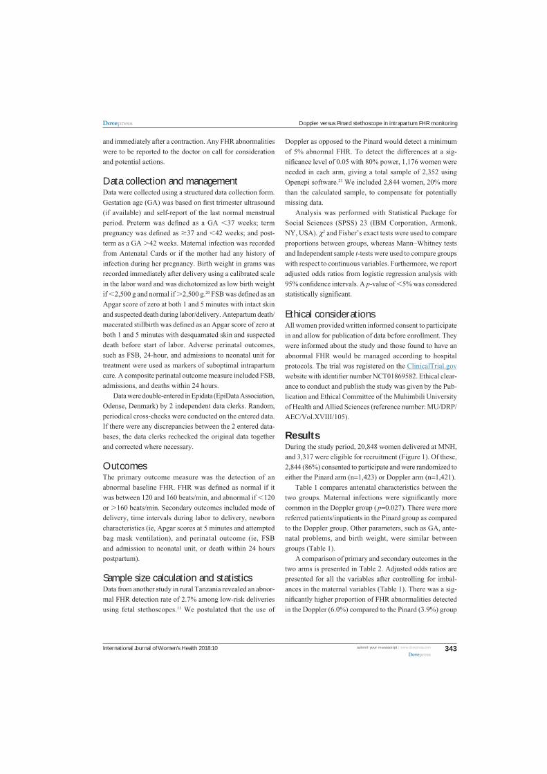
International Journal of Women’s Health 2018:10 submit your manuscript | www.dovepress.com
Dovepress
Dovepress
343
Doppler versus Pinard stethoscope in intrapartum FHR monitoring
and immediately after a contraction. Any FHR abnormalities
were to be reported to the doctor on call for consideration
and potential actions.
Data collection and managementData were collected using a structured data collection form.
Gestation age (GA) was based on first trimester ultrasound
(if available) and self-report of the last normal menstrual
period. Preterm was defined as a GA 37 weeks; term
pregnancy was defined as 37 and 42 weeks; and post-
term as a GA 42 weeks. Maternal infection was recorded
from Antenatal Cards or if the mother had any history of
infection during her pregnancy. Birth weight in grams was
recorded immediately after delivery using a calibrated scale
in the labor ward and was dichotomized as low birth weight
if 2,500 g and normal if 2,500 g.20 FSB was defined as an
Apgar score of zero at both 1 and 5 minutes with intact skin
and suspected death during labor/delivery. Antepartum death/
macerated stillbirth was defined as an Apgar score of zero at
both 1 and 5 minutes with desquamated skin and suspected
death before start of labor. Adverse perinatal outcomes,
such as FSB, 24-hour, and admissions to neonatal unit for
treatment were used as markers of suboptimal intrapartum
care. A composite perinatal outcome measure included FSB,
admissions, and deaths within 24 hours.
Data were double-entered in Epidata (EpiData Association,
Odense, Denmark) by 2 independent data clerks. Random,
periodical cross-checks were conducted on the entered data.
If there were any discrepancies between the 2 entered data-
bases, the data clerks rechecked the original data together
and corrected where necessary.
OutcomesThe primary outcome measure was the detection of an
abnormal baseline FHR. FHR was defined as normal if it
was between 120 and 160 beats/min, and abnormal if 120
or 160 beats/min. Secondary outcomes included mode of
delivery, time intervals during labor to delivery, newborn
characteristics (ie, Apgar scores at 5 minutes and attempted
bag mask ventilation), and perinatal outcome (ie, FSB
and admission to neonatal unit, or death within 24 hours
postpartum).
Sample size calculation and statisticsData from another study in rural Tanzania revealed an abnor-
mal FHR detection rate of 2.7% among low-risk deliveries
using fetal stethoscopes.11 We postulated that the use of
Doppler as opposed to the Pinard would detect a minimum
of 5% abnormal FHR. To detect the differences at a sig-
nificance level of 0.05 with 80% power, 1,176 women were
needed in each arm, giving a total sample of 2,352 using
Openepi software.21 We included 2,844 women, 20% more
than the calculated sample, to compensate for potentially
missing data.
Analysis was performed with Statistical Package for
Social Sciences (SPSS) 23 (IBM Corporation, Armonk,
NY, USA). 2 and Fisher’s exact tests were used to compare
proportions between groups, whereas Mann–Whitney tests
and Independent sample t-tests were used to compare groups
with respect to continuous variables. Furthermore, we report
adjusted odds ratios from logistic regression analysis with
95% confidence intervals. A p-value of 5% was considered
statistically significant.
Ethical considerationsAll women provided written informed consent to participate
in and allow for publication of data before enrollment. They
were informed about the study and those found to have an
abnormal FHR would be managed according to hospital
protocols. The trial was registered on the ClinicalTrial.gov
website with identifier number NCT01869582. Ethical clear-
ance to conduct and publish the study was given by the Pub-
lication and Ethical Committee of the Muhimbili University
of Health and Allied Sciences (reference number: MU/DRP/
AEC/Vol.XVIII/105).
ResultsDuring the study period, 20,848 women delivered at MNH,
and 3,317 were eligible for recruitment (Figure 1). Of these,
2,844 (86%) consented to participate and were randomized to
either the Pinard arm (n 1,423) or Doppler arm (n 1,421).
Table 1 compares antenatal characteristics between the
two groups. Maternal infections were significantly more
common in the Doppler group ( p 0.027). There were more
referred patients/inpatients in the Pinard group as compared
to the Doppler group. Other parameters, such as GA, ante-
natal problems, and birth weight, were similar between
groups (Table 1).
A comparison of primary and secondary outcomes in the
two arms is presented in Table 2. Adjusted odds ratios are
presented for all the variables after controlling for imbal-
ances in the maternal variables (Table 1). There was a sig-
nificantly higher proportion of FHR abnormalities detected
in the Doppler (6.0%) compared to the Pinard (3.9%) group

International Journal of Women’s Health 2018:10submit your manuscript | www.dovepress.com
Dovepress
Dovepress
344
Kamala et al
(adjusted odds ratio 1.59, p 0.008). Overall, there were
no significant differences in any of the secondary outcomes
(Apgar score 7, delivery of bag mask ventilation, mode of
delivery, perinatal admissions, and deaths).
The mean ( SD) duration of first stage of labor was simi-
lar in both groups (ie, nearly 11 hours 30 minutes 2 hours
40 minutes, p 0.83). The mean duration of second stage was
slightly longer in the Doppler (34 14 minutes) compared
to the Pinard (32 14 minutes) group (t-test, p 0.039). The
median (interquartile range) time intervals from detection of
an abnormal FHR to delivery were almost comparable, ie,
80 (60, 161) minutes in the Doppler group, and 89 (52, 165)
minutes in the Pinard group (Mann–Whitney test, p 0.88)
for all modes of deliveries.
Figure 2 shows that, overall, 142 (5.0%) cases of abnor-
mal FHR were detected in this study. Among the 16 perinatal
deaths, 8 (50%) were noted to have an abnormal FHR pattern.
One perinatal death was recorded among newborns delivered
by cesarean section. Subgroup analysis of the composite peri-
natal outcomes (ie, FSB and 24-hour deaths and admissions
to a neonatal area) revealed that newborns with abnormal
FHR delivered vaginally had a more unfavorable outcome in
the Pinard group (10 of 23; 43.5%) compared to the Doppler
group (7 of 43; 16.3%) (Fisher’s exact test, p 0.021). There
was no time difference in this subgroup analysis between the
study arms (p 0.305).
DiscussionWe found a higher likelihood of detecting abnormal FHR
by intermittent monitoring using the Doppler technique as
opposed to the Pinard. However, overall, perinatal outcomes
were similar, although subgroup analysis revealed that new-
borns with abnormal FHR delivered vaginally had better
perinatal outcomes in the Doppler compared to the Pinard
group. The time intervals from detection of an abnormal FHR
to delivery were long in both groups.
Our findings on FHR abnormalities are comparable to
prior studies completed in Kampala and Harare, where the
Doppler detected more FHR abnormalities than the Pinard
fetal stethoscope.22,23 The Kampala study reported that despite
a higher detection of FHR abnormalities with the Doppler
technique, no improvement in perinatal outcome was seen,
Figure 1 Trial pro le.
Table 1 Comparison of maternal characteristics in the Pinard and Doppler groups among low-risk parturient women at MNH
Antenatal characteristics Pinard n 1,423 (%) Doppler n 1,421 (%) Total (%) n 2,844 (%) p-value*
Maternal infection 28 (2.0) 48 (3.3) 76 (2.7) 0.027Low birth weight 65 (4.6) 62 (4.3) 127 (4.4) 0.790GA 42 weeks 6 (0.4) 8 (0.6) 14 (0.5) 0.790Antenatal problem 128 (9.0) 111 (7.7) 239 (8.4) 0.250Referred patient/inpatient 205 (14.4) 158 (11.0) 363 (12.7) 0.008
Note: * 2 tests.Abbreviations: GA, gestational age; MNH, Muhimbili National Hospital.

International Journal of Women’s Health 2018:10 submit your manuscript | www.dovepress.com
Dovepress
Dovepress
345
Doppler versus Pinard stethoscope in intrapartum FHR monitoring
which is similar to our overall finding. A suggested reason for
this relates to several contextual constraints in low-resource
settings leading to lack of timely interventions to deliver the
baby.9 These constraints include, among others, a delay in
decision-making due to a high patient to staff ratio in the labor
ward, and often multiple simultaneous patients waiting for an
emergency cesarean section.18,22 A longer than recommended
decision-to-delivery time interval may in part explain the lack
of difference in perinatal outcomes between the groups in the
present study. One would have anticipated that the higher
detection rate of abnormal FHR by Doppler would lead to
a timelier intervention such as a cesarean section. However,
the frequency of a cesarean section was unaffected, and we
speculate that the striking imbalance between available health
resources (staff, access to theater) and large volume of patients
likely play a crucial role.24,25 Therefore, in order to effectively
manage critical cases and improve perinatal outcomes,
improved FHR monitoring techniques coupled with better
staffing, as well as improved equipment and theater facilities,
ie, an overall increased capacity and improved systems, are
necessary in order to affect perinatal outcomes.
A subgroup analysis of those newborns with a detected
abnormal FHR, delivered vaginally, revealed improved
composite perinatal outcomes in the Doppler compared to
the Pinard group. These findings are similar to those found
in the Harare study,21 which involved dedicated research
midwives, and where improved perinatal outcomes were
reported in the Doppler arm. These findings may also
indicate that midwives can detect FHR abnormalities more
frequently and earlier when using the Doppler technique
compared to a fetal stethoscope, thereby recognizing
signs of intrapartum hypoxia more often and at an
earlier stage.
There might be several reasons for more and/or earlier
detection of abnormal FHR in the Doppler group. The
Doppler technique provides digital sound and readings,
which do not require much skill to interpret and can easily
be confirmed by peers, as opposed to the Pinard, which
requires a complete minute of counting.15 Additionally,
midwives may feel unsure about the reliability of the Pinard
assessments.18 A qualitative study (in-depth interviews and
focus group discussions) performed among labor ward staff
at MNH reports that the Doppler was the preferred device
for improving FHR monitoring.26
LimitationsThis study was not powered to detect overall differences in
perinatal outcomes. A study involving an appropriate sample
size, perhaps in a more high-risk population and coupled with
timely obstetric intervention, might be able to show differ-
ences in overall perinatal outcomes. Second, the documented
fear of blame from peers and hospital management at MNH,
as suggested in the qualitative study previously conducted in
the same hospital,26 might have led to a defensive practice
with overreporting of abnormal FHR. Third, in this study, we
have not been able to perform other tests, such as fetal scalp
pH levels, to confirm possible fetal hypoxic state. Fourth, the
study involved 2 different medical devices in measuring FHR,
and it was not possible to blind the patients and providers.
Conclusion and recommendationMonitoring of FHR using a wind-up Doppler was associ-
ated with an increased detection of abnormal FHR. Overall
perinatal outcomes were comparable between groups, but
there were better perinatal outcomes for newborns with
detected abnormal FHR delivered vaginally in the Doppler
Table 2 Frequencies of abnormal FHR detections, newborn characteristics, and perinatal outcomes in the Pinard and Doppler arms among low-risk parturient women at MNH
Primary/secondary outcomes Pinard n 1,423 (%)
Doppler n 1,421 (%)
Unadjusted OR p-value* AOR** p-value*
Abnormality of FHR 56 (3.9) 86 (6.0) 1.56 (1.12–2.21) 0.012 1.59 (1.13–2.26) 0.008Mode of delivery: cesarean section 174 (12.2) 172 (12.0) 0.98 (0.79–1.23) 0.89 0.96 (0.77–1.21) 0.76Apgar 5 minutes 7 23 (1.6) 30 (2.1) 1.31 (0.76–2.26) 0.40 1.38 (0.79–2.24) 0.25Bag mask ventilation attempted 68 (4.8) 76 (5.3) 1.19 (0.80–1.57) 0.51 1.18 (0.84–1.65) 0.35Admissions to neonatal unit at birth 28 (2.0) 38 (2.7) 1.36 (0.82–2.25) 0.24 1.42 (0.86–2.33) 0.17Fresh stillbirths 8 (0.6) 5 (0.3) 0.63 (0.20–1.92) 0.41 0.67 (0.22–2.07) 0.49Still admitted at 24 hours 18 (1.3) 22 (1.5) 1.22 (0.65–2.28) 0.63 1.25 (0.66–2.34) 0.49Perinatal deaths (FSB deaths within 24 hours)
10 (0.7) 6 (0.4) 0.59 (0.22–1.65) 0.32 0.62 (0.26–1.73) 0.36
Composite outcomes (perinatal deaths and admissions)
28 (2.0) 28 (2.0) 0.99 (0.59–1.69) 0.98 0.73 (0.34–1.47) 0.35
Notes: Data are presented as n (%) and AOR. *Wald test. 2 test. **Multiple logistic regression analysis adjusted for maternal infection and sources of admission.Abbreviations: AOR, adjusted odds ratio; FHR, fetal heart rate; FSB, fresh stillbirth; MNH, Muhimbili National Hospital.

International Journal of Women’s Health 2018:10submit your manuscript | www.dovepress.com
Dovepress
Dovepress
346
Kamala et al
group. A study powered for perinatal outcomes, coupled with
timely interventions, may be able to demonstrate differences
in overall perinatal outcome.
Data sharing statementData can be made available to editors and/or reviewers upon
request from Research, Teaching and Consultancy Unit of
Muhimbili National Hospital (address: PO Box 65000, Dar
es Salaam, Tanzania; phone: 255 22 215 1599; fax: 255
22 215 0534; email: [email protected])
AcknowledgmentWe thank the hospital administration for allowing us to use
the hospital facilities to collect data for this study. We also
wish to acknowledge the entire midwife team for the excellent
job of keeping records of all deliveries in the midwifery book
that was then computerized. We thank the well-motivated
data clerks of the Safer Births project for data entry and clean-
ing. The Laerdal Foundation funded the study. The funder
had no role in the study design, data collection, analysis, or
decision to publish or preparation of the manuscript.
Figure 2 Flow diagrams of FHR detections, mode of delivery, and perinatal outcomes in both arms, Pinard and Doppler.Abbreviation: FHR, fetal heart rate.

International Journal of Women’s Health 2018:10 submit your manuscript | www.dovepress.com
Dovepress
Dovepress
347
Doppler versus Pinard stethoscope in intrapartum FHR monitoring
Author contributionsBAK conducted the initial statistical data analysis and pre-
pared all sections of the first draft and compiled the final
manuscript. HLK conceived and designed the study and
supervised the training of research midwives and the data
collection process. He was also involved in data analysis and
the interpretation of the results. PJW and ERM participated
in the design of the study and the review of the manuscript.
ID reviewed the statistical analysis and the final draft of
the manuscript. HLE and JMP were involved in design of
the study, interpretation of data, and critically reviewed the
manuscript. All authors contributed toward data analysis,
drafting and critically revising the paper and agree to be
accountable for all aspects of the work. All authors agreed
on the final submitted manuscript.
DisclosureBAK received unconditional funding for studies from Laerdal
Foundation. The other authors report no conflicts of interest
in this work.
References1. Lawn JE, Lee AC, Kinney M, et al. Two million intrapartum-related
stillbirths and neonatal deaths: where, why, and what can be done?Int J Gynaecol Obstet. 2009;107(Suppl 1):S5–S18, S19.
2. Wyatt J. Appropriate medical technology for perinatal care in low-resource countries. Ann Trop Paediatr. 2008;28(4):243–251.
3. Lawn J, Shibuya K, Stein C. No cry at birth: global estimates of intra-partum stillbirths and intrapartum-related neonatal deaths. Bull World Health Organ. 2005;83:409–417.
4. Hill K, Thomas K, AbouZahr C, et al. Estimates of maternal mortality worldwide between 1990 and 2005: an assessment of available data. Lancet. 2007;370(9595):1311–1319.
5. UNICEF, WHO, World Bank Group and United Nations. Levels and Trends in Child Mortality Child Mortality. New York, NY: UNICEF; 2017.
6. Martis R, Emilia O, Nurdiati DS, Brown J. Cochrane Database ofSystematic Reviews. Martis R, editor. Vol 2. Hoboken, NJ: John Wiley & Sons, Ltd; 2017:CD008680.
7. Spencer-Jones J. Make every mother and child count. S Afr Med J. 2005;95(6):382, 384.
8. van Handel M, Swaab H, de Vries LS, Jongmans MJ. Long-term cogni-tive and behavioral consequences of neonatal encephalopathy followingperinatal asphyxia: a review. Eur J Pediatr. 2007;166(7):645–654.
9. Maaløe N, Housseine N, Meguid T, et al. Effect of locally tailoredlabour management guidelines on intrahospital stillbirths and birthasphyxia at the referral hospital of Zanzibar: a quasi-experimentalpre-post study (The PartoMa study). BJOG An Int J Obstet Gynaecol. 2018;125(2):235–245.
10. WHO and UNICEF. Every Newborn: An Action Plan to End Prevent-able Deaths. Geneva: World Health Organization; 2014.
11. Langli Ersdal H, Mduma E, Svensen E, Sundby J, Perlman J. Intermit-tent detection of fetal heart rate abnormalities identify infants at greatest risk for fresh stillbirths, birth asphyxia, neonatal resuscitation, and early neonatal deaths in a limited-resource setting: a prospective descrip-tive observational study at Haydom Lutheran Hospital. Neonatology. 2012;102:235–242.
12. Wyatt J. Appropriate medical technology for perinatal care in low-resource countries. Ann Trop Paediatr. 2008;28(4):243–251.
13. Vintzileos AM, Antsaklis A, Varvarigos I, Papas C, Sofatzis I,Montgomery JT. A randomized trial of intrapartum electronic fetalheart rate monitoring versus intermittent auscultation. Obstet Gynecol. 1993;81(6):899–907.
14. Woods D. Appropriate technology and education for improved intra-partum care in underresourced countries. S Afr J Obstet Gynaecol. 2009;15(3):78–79.
15. Bezuidenhout H, Woods D, Wyatt J, Lawn J. Does fetal heart rate count? Developing a low cost, alternative powered Doppler fetal heart monitor for use in low resource high mortality settings. In: 4th IET Seminaron Appropriate Healthcare Technologies for Developing Countries;2006:155–159; London.
16. Martis R, Emilia O, Nurdiati DS, Brown J. Cochrane Database ofSystematic Reviews. Martis R, editor. Hoboken, NJ: John Wiley &Sons, Ltd; 2017.
17. National Bureau of Statistics (NBS) and Office of Chief Government Statistician (OCGS), Zanzibar. The 2012 Population and Housing Cen-sus: Basic Demographic and Socio-Economic Profile; Key Findings. Dar es Salaam, Tanzania: NBS and OCGS; 2014. Available from: http://nbs.go.tz/nbs/takwimu/census2012/Basic_Demographic_and_Socio-Economic_Profile_PopularVersion-KeyFindings_2012_PHC_Eng-lishVersion.pdf. Accessed November 11, 2015.
18. Mgaya AH, Litorp H, Kidanto HL, Nyström L, Essén B. Criteria-based audit to improve quality of care of foetal distress: standardising obstetric care at a national referral hospital in a low resource setting, Tanzania. BMC Pregnancy Childbirth. 2016;16(1):343.
19. Litorp H, Kidanto HL, Nystrom L, Darj E, Essén B. Increasing cae-sarean section rates among low-risk groups: a panel study classifying deliveries according to Robson at a university hospital in Tanzania.BMC Pregnancy Childbirth. 2013;13(1):107.
20. World Health Organization. International Statistical Classification of Diseases and Related Health Problems, Tenth Revision. Geneva: World Health Organization; 2011;18:39.
21. Kelsey J, Whittemore A, Evans A, Thompson D. Methods in Observa-tional Epidemiology. Oxford: Oxford University Press; 1996.
22. Byaruhanga R, Bassani DG, Jagau A, Muwanguzi P, Montgomery AL, Lawn JE. Use of wind-up fetal Doppler versus Pinard for fetal heart rate intermittent monitoring in labour: a randomised clinical trial. BMJ Open. 2015;5(1):e006867.
23. Mahomed K, Nyoni R, Mulambo T, Kasule J, Jacobus E. Randomised controlled trial of intrapartum fetal heart rate monitoring. BMJ. 1994;308(6927):497–500.
24. Shemdoe A, Mbaruku G, Dilip A, et al. Explaining retention of health-care workers in Tanzania: moving on, coming to “look, see and go,” or stay? Hum Resour Health. 2016;14:2.
25. The United republic of Tanzania Ministry of Health and Social Welfare. Human Resource for Health and Social Welfare Strategic Plan 2014–2019. Dar es Salaam, Tanzania: Ministry of Health and Social Welfare; 2014.
26. Litorp H, Mgaya A, Mbekenga CK, et al. Fear, blame and transpar-ency: obstetric caregivers’ rationales for high caesarean section rates in a low-resource setting. Soc Sci Med. 2015;143:232–240.

International Journal of Women’s Health
Publish your work in this journal
Submit your manuscript here: http://www.dovepress.com/international-journal-of-womens-health-journal
The International Journal of Women’s Health is an international, peer-reviewed open-access journal publishing original research, reports, editorials, reviews and commentaries on all aspects of women’s healthcare including gynecology, obstetrics, and breast cancer. The manuscript management system is completely online and includes
a very quick and fair peer-review system, which is all easy to use. Visit http://www.dovepress.com/testimonials.php to read real quotes from published authors.
International Journal of Women’s Health 2018:10submit your manuscript | www.dovepress.com
Dovepress
Dovepress
Dovepress
348
Kamala et al

Paper


International Journal of
Environmental Research
and Public Health
Article
Effectiveness of a Novel Continuous Doppler (Moyo)Versus Intermittent Doppler in Intrapartum Detectionof Abnormal Foetal Heart Rate: A RandomisedControlled Study in Tanzania
Benjamin Kamala 1,2,* , Hussein Kidanto 3,4, Ingvild Dalen 3, Matilda Ngarina 2,
Muzdalifat Abeid 4, Jeffrey Perlman 5 and Hege Ersdal 1,6
1 Faculty of Health Sciences, University of Stavanger, 4036 Stavanger, Norway; [email protected] Muhimbili National Hospital, P.O. Box 65000, Dar es Salaam, Tanzania; [email protected] Department of Research, Stavanger University Hospital, 4011 Stavanger, Norway;
[email protected] (H.K.); [email protected] (I.D.)4 School of Medicine, Aga Khan University, P.O. Box 38129, Dar es Salaam, Tanzania;
[email protected] Department of Paediatrics, Weill Cornell Medicine, New York, NY 10065, USA; [email protected] Department of Anaesthesiology and Intensive Care, Stavanger University Hospital, 4011 Stavanger, Norway* Correspondence: [email protected] or [email protected]; Tel.: +47-46441263 or +25-5713691949
Received: 20 November 2018; Accepted: 22 January 2019; Published: 24 January 2019���������������
Abstract: Background: Intrapartum foetal heart rate (FHR) monitoring is crucial for identificationof hypoxic foetuses and subsequent interventions. We compared continuous monitoring usinga novel nine-crystal FHR monitor (Moyo) versus intermittent single crystal Doppler (Doppler) for thedetection of abnormal FHR. Methods: An unmasked randomised controlled study was conducted ina tertiary hospital in Tanzania (ClinicalTrials.gov Identifier: NCT02790554). A total of 2973 low-risksingleton pregnant women in the first stage of labour admitted with normal FHR were randomisedto either Moyo (n = 1479) or Doppler (1494) arms. The primary outcome was the proportion ofabnormal FHR detection. Secondary outcomes were time intervals in labour, delivery mode, Apgarscores, and perinatal outcomes. Results: Moyo detected abnormal FHR more often (13.3%) comparedto Doppler (9.8%) (p = 0.002). Time intervals from admission to detection of abnormal FHR were15% shorter in Moyo (p = 0.12) and from the detection of abnormal FHR to delivery was 36%longer in Moyo (p = 0.007) compared to the Doppler arm. Time from last FHR to delivery was 12%shorter with Moyo (p = 0.006) compared to Doppler. Caesarean section rates were higher with theMoyo device compared to Doppler (p = 0.001). Low Apgar scores (<7) at the 1st and 5th min werecomparable between groups (p = 0.555 and p = 0.800). Perinatal outcomes (fresh stillbirths and24-h neonatal deaths) were comparable at delivery (p = 0.497) and 24-h post-delivery (p = 0.345).Conclusions: Abnormal FHR detection rates were higher with Moyo compared to Doppler. Moyodetected abnormal FHR earlier than Doppler, but time from detection to delivery was longer. Studiespowered to detect differences in perinatal outcomes with timely responses are recommended.
Keywords: foetal heart rate; Moyo; Doppler; perinatal outcomes
1. Introduction
The intrapartum period poses a great risk for both baby and mother. Globally, 2.6 million neonatesdie annually during the neonatal period, accounting for approximately 46% of all under-five deaths [1].Approximately 36% of these neonates die on the first day [1,2], and 25% are intrapartum related [1,3].
Int. J. Environ. Res. Public Health 2019, 16, 315; doi:10.3390/ijerph16030315 www.mdpi.com/journal/ijerph

Int. J. Environ. Res. Public Health 2019, 16, 315 2 of 16
Moreover, 40% of 2.6 million stillbirths are intrapartum related and termed fresh stillbirths (FSB) [4].Most of these perinatal deaths occur in low income countries (LIC) where effective emergency obstetriccare provision is low [3].
Prolonged intrapartum foetal hypoxia, invariably because of interruption of placental blood flow,may result in a FSB or a severely asphyxiated neonate [5,6]. Following delivery, such neonates may die,or survive with variable degrees of hypoxic-ischemic brain injury, leading to long-term neurocognitiveand behavioural impairment [7,8].
Intrapartum foetal heart rate (FHR) monitoring is an important strategy in providing a moretargeted and appropriate management of foetal well-being [9]. Intermittent assessment with eithera hand-held Doppler or Pinard Stethoscope is the main method of intrapartum FHR monitoring inLIC [10,11]. However, studies document that intrapartum FHR monitoring is not conducted accordingto current international guidelines [12–14], due to a shortage of human resource and appropriatemonitoring equipment [12,15], leading to perinatal morbidity and mortality [16].
Our previous studies using a novel continuous Doppler (Moyo) showed that accurate FHRmonitoring enhanced early detection of the at-risk foetus [17,18]. The Moyo device has features thatmay facilitate early identification of foetuses at risk of intra-partum hypoxia and improve the qualityof midwifery practices [17]. Continuous intrapartum FHR monitoring with cardiotocograph (CTG)coupled with timely interventions, such as caesarean sections in high income countries, has beenassociated with improved perinatal outcomes [9]. There is a paucity of studies on FHR monitoring inLIC, where most births occur, posing a critical need to implement and test new monitoring strategiesin these settings [19]. An ideal device for LIC settings should be low-cost, simple to operate, possibleto operate on a range of power sources, and without a need for continuous power supply, which theMoyo device represents [20].
We hypothesized that continuous intrapartum monitoring with Moyo as compared tointermittent Doppler assessment would lead to more timely and frequent detection of FHRabnormalities. The objective of the study was to compare the effectiveness of continuous monitoring(Moyo-intervention) versus intermittent hand-held (Doppler-standard of care) in the intrapartumdetection of abnormal FHR.
2. Materials and Methods
2.1. Study Design
We conducted a parallel-arms, unmasked randomised controlled study from March 2016 toSeptember 2017 at Muhimbili National Hospital (MNH) in Dar es Salaam, Tanzania.
2.2. Study Settings
MNH is the national referral hospital and a teaching hospital for Muhimbili University. Thehospital provides both basic and comprehensive emergency obstetric care and has approximately10,000 annual deliveries; 50% by Caesarean section (CS) [10]. The labour ward at MNH has 20 deliverybeds and approximately 25 nurse midwives. The ward is managed by 5 nurse-midwives and 2 nursingassistants in each shift of 12 h. The doctors-on-call team comprises 1 consultant, 1 obstetrician,2 obstetric residents, and 1 intern doctor on 24-h call. There are two obstetric operating theatres ina separate building adjacent to the maternity block.
On admission, a nurse midwife screens all women for vital signs registration, initial FHRassessment, and vaginal examination before entering the labour ward. A brief history and vitalsigns are taken and required information entered in the labour ward register. The on-call doctorreviews the partograph and undertakes the initial and subsequent obstetric examination until delivery.After a normal vaginal delivery, mothers and babies are observed in the hospital for 6–10 h. Babieswith respiratory distress and others in need of medical attention are admitted to the neonatal unit.Management protocols for mothers and babies in this setting have been described previously [21].

Int. J. Environ. Res. Public Health 2019, 16, 315 3 of 16
2.3. Study Participants
The study participants included mothers in labour with an estimated gestational age above28 weeks and with ≥3 cm cervical dilatation. Exclusions included scheduled elective CS, multiplepregnancies, cases with abnormal or undetectable FHR on admission, admission in the second stagewith full cervical dilation, precipitous delivery, and critically ill patients with no measurements of FHR.
2.4. Patient and Public Involvement
The need for development of the Moyo device started at Haydom, a rural based hospital inNorthern Tanzania, and MNH responding to increased intrapartum related perinatal morbidity andmortality [5,6,22]. The device was developed in collaboration with clinical staff at these hospitals,Laerdal Medical, and Stavanger University Hospital in Norway. It was in response to the needs of theclinical staff and mothers in these resource limited settings to reduce FSB and END (early neonataldeaths). Patients were told of the design of the study before being recruited to participate. Qualitativestudies on preferences and acceptability of the continuous FHR monitoring with the Moyo deviceamong mothers and clinical staff have been conducted in these settings. Positive responses on thisdevice compared to the traditional Pinard stethoscope and Doppler were obtained and documented inour previous studies [23,24].
2.5. Randomisation, Concealment, and Masking
A randomisation sequence was computer-generated by an independent statistician. Details of theallocated group were given to the study coordinator, who supervised data clerks to write on cardsand put them in sequentially numbered opaque sealed envelopes and sealed them. The allocationsequence was concealed from investigators and nurses enrolling participants and assessing outcomes.Envelopes were prepared and stored in a locked cabinet. Consecutively numbered envelopes wereopened only after the enrolled participants completed assessments. The women and enrolling nurseswere unaware of the allocation group until after eligible women were informed about the study anda written consent was obtained. Women, nurses, and doctors were unmasked.
2.6. Training
Pre-study trainings using a Moyo training package focusing on standard operating proceduresfor Moyo and international FHR monitoring standards were conducted in January and February 2016by study investigators. All labour ward staff were trained for a full day on these FHR managementprotocols before starting the study. Continuous on-job refresher trainings were conducted (everytwo months) to increase protocol adherence and accommodate incoming staff who did not receivethe initial training. Training included theoretical information about FHR monitoring during labourand management of an abnormal FHR. Criteria for FHR monitoring were established and includedmonitoring recording every 30 min in the first stage of labour, and every 5–15 min in the secondstage [12–14]. The labour ward staff were also told that abnormal FHR detections should be reportedto the doctor on call, who should act according to hospital protocols. Research nurses (at least 2 pershift) were trained for one additional day on research protocol and data collection to ensure accuracyand completeness of the data in the paper-based case report form (CRF). Data were collected frommothers’ antenatal cards, partograph, obstetric register, and, when needed, from routine neonatalmorbidity and mortality records in the neonatal unit.
2.7. The Intervention (The Moyo Device)
Moyo (Figures 1 and 2) (Moyo, Laerdal Global Health, Stavanger, Norway) is a novel strap-onFHR monitor equipped with a rechargeable battery, containing a nine-crystal Doppler ultrasoundsensor, which facilitates the rapid identification of FHR within 5 s. Additional features of the Moyodevice have been described in our previous studies [17,18,23].

Int. J. Environ. Res. Public Health 2019, 16, 315 4 of 16
Women randomised to the Moyo arm received information on how the device was to be used bythe enrolling midwife before the device was strapped on. The midwife continued with her routineactivities, but periodically revisited the women to check and record the FHR reading or in case of anabnormal FHR alarm from Moyo [23]. Moyo continued to be strapped on until the end of the secondstage or immediately prior to the start of a CS.
Figure 1. Moyo—the novel continuous FHR (foetal heart rate) monitor (Laerdal Global Health).* patient applied part.

Int. J. Environ. Res. Public Health 2019, 16, 315 5 of 16
Figure 2. The Moyo FHR monitor with a 30-minutes historical display (Laerdal Global Health).
2.8. Control (Hand-Held Doppler)
In the control arm, women were monitored intermittently with the standard protocol of FHRmonitoring every 30 min in the first stage and 5–15 min in the second stage using a hand-held Doppler(Power-free Education Technology, Pet.og.za, Cape Town, South Africa). Doppler detects FHR andprovides a steady state number per min on a display, as well as an audible sound of the FHR [11].It permits the midwife to locate the FHR while allowing others, including the mother, to hear the FHR.The midwife would continue with her routine activities and periodically revisit the women to checkand record FHR readings in the partograph and perform other management as indicated.
2.9. Outcomes
The primary outcome measure was FHR defined as normal (120 to160 beats/min throughoutlabour and delivery) or abnormal (absent, <120 or >160 beats/min lasting for at least two min) in thecontinuous Moyo despite repositioning of the Moyo sensor, and with three abnormal assessments atdifferent sites in the intermittent Doppler arm.
Secondary outcomes included the Apgar score at one and five minutes (abnormal was defined asan Apgar score <7); mode of delivery (vaginal delivery, CS, assisted breech, and vacuum extraction);perinatal outcome at birth (i.e., normal, admission to the neonatal unit, or FSB), outcome at 24-h (i.e.,normal, still admitted to the neonatal unit, or END); and composite perinatal outcomes at birth and24-h (normal, admission in neonatal unit, FSB, and END). Apgar score <7 at five minutes was used asa surrogate measure of birth asphyxia [25]. Mode of delivery was dichotomized into two categories(i.e., vaginal, including vacuum delivery, and CS) due to relatively fewer cases in the vacuum deliverycategory. Time intervals included admission to abnormal FHR detection, admission to delivery, fromabnormal FHR detection to delivery, and last FHR assessment to delivery. After detection of abnormalFHR, recorded intrauterine resuscitation included discontinuing oxytocin, changing maternal position,administering intravenous fluids, and provision of oxygen.

Int. J. Environ. Res. Public Health 2019, 16, 315 6 of 16
2.10. Trial Monitoring and Stopping Rules
The trial was monitored by an independent data monitoring committee comprising one statisticianand one paediatrician aimed at protecting participant exposure to unreasonable risks. Discontinuationwas planned in case of imbalances in serious adverse effects (FSB and END). Blinded data analysiswas conducted mid-way through the trial and the committee recommended continuation of the study.
2.11. Sample Size Estimation
Historical data showed that when using the hand-held Doppler, abnormal FHR was detected in4.5% of low-risk deliveries. We postulated that continuous assessment of FHR using Moyo woulddetect a minimum of 7% of abnormal FHR. To detect these differences at a significance level of 0.05with 80% power, a minimum of 1350 cases would be needed in each arm. An additional 10% wasadded to the sample size to allow for missing data. The final sample size was 2970.
2.12. Data Management
Data collection was conducted by trained research nurses (at least 2 per shift) filling the CRF.CRFs was cross-checked by the investigators for quality and completeness before entry. All CRFs withqueries were returned to the research nurse for verification and correction before data entry. A dataentry template was generated in Epi Data by investigators and statistician. All verified data weredouble-entered by trained data clerks. Then, data was transferred to SPSS for analysis (IBM SPSSStatistics for Windows, Version 23.0, IBM Corp, Armonk, NY, US). Patient information were recordedusing confidential codes and kept in a secured place.
2.13. Statistical Analysis
Descriptive statistics were expressed as means (standard deviation, SD) or medians (interquartile range, IQR) for continuous variables and as counts and proportions for categorical variables.Proportions were compared by a Pearson chi-square test. Odds ratios (OR) with respective 95%confidence intervals were calculated as estimates of the effect for categorical variables. Adjusted OR(AOR) using both logistics and multinomial regressions were estimated to account for imbalances inbaseline characteristics and for an increase in subject-specific precision. Symmetrically distributedcontinuous variables were compared by t-test, and the Mann-Whitney U was used for skeweddata. To adjust for baseline imbalances when comparing skewed time variables, we used linearregression analysis with a natural log-transformed outcome to calculate beta-coefficients. Due to thistransformation, we used beta coefficients to estimate the effect size (ES), i.e., relative change in mediantime in percentages as documented before [26]. A p-value < 0.05 was considered significant.
2.14. Ethical Clearance
The study was registered at ClinicalTrials.gov Identifier: NCT02790554. All subjects gave theirwritten informed consent for inclusion before they participated in the study. The study was conductedin accordance with the Declaration of Helsinki, and the protocol was approved by both the NationalInstitute of Medical Research in Tanzania (NIMR/HQ/R.8a/Vol. IX/1434) and the Regional Committeefor Medical and Health Research Ethics, Western Norway (REK Vest). Local permission was soughtfrom MNH Directorate of Research and Consultancy. Permission to publish was granted by NIMR(NIMR/HQ/P.12 VOL. XXV/57).
3. Results
From March 2016 to September 2017, a total of 3547 admitted women were eligible. Of these,438 were not randomised due to precipitous labour and 136 did not consent to participate in the study.In total, 2973 women were enrolled, 1479 assigned to Moyo and 1494 to Doppler as shown in the studyprofile (Figure 3).

Int. J. Environ. Res. Public Health 2019, 16, 315 7 of 16
Figure 3. Trial profile.
3.1. Maternal, Antenatal, and Perinatal Characteristics
Maternal, antenatal, and perinatal characteristics of the study subjects are shown in Table 1.Maternal mean age was comparable between study arms. The Moyo arm had a lower proportion ofpreterm deliveries compared to Doppler (12% vs. 17%, p ≤ 0.001). Women in the Moyo arm wereadmitted earlier in labour with a mean cervical dilatation of 4.4 ± 1.5 cm compared to 5.0 ± 1.7 cm inthe Doppler arm, p ≤ 0.001.
Table 1. Baseline demographic, clinical, and perinatal characteristics of randomized low risk pregnantwomen in labour.
Variables Intermittent Doppler (n = 1494) Continuous Moyo (n = 1479)
Age (years)Mean (SD) 28.3 (5.6) 27.8 (5.3)
<20 84 (5.6) 66 (4.5)20–35 1223 (81.9) 1260 (85.2)>35 187 (12.5) 153 (10.3)
EducationNo/Primary 557 (37.3) 424 (28.7)Secondary 375 (25.1) 366 (24.7)
College/University 562 (37.6) 689 (46.6)Marital status
Married/Cohabiting 1370 (91.7) 1384 (93.6)Single 124 (8.3) 95 (6.4)
Antenatal care visits<4 466 (31.2) 402 (27.2)≥4 1028 (68.8) 1077 (72.8)
ParityMedian (IQR) 2 (1, 3) 2 (1, 3)
Prime 576 (38.6) 697 (54.8)2–4 805 (53.9) 709 (47.9)>4 113 (7.6) 73 (4.9)
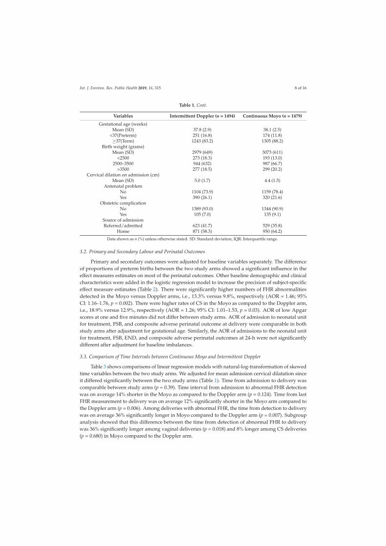
Int. J. Environ. Res. Public Health 2019, 16, 315 8 of 16
Table 1. Cont.
Variables Intermittent Doppler (n = 1494) Continuous Moyo (n = 1479)
Gestational age (weeks)Mean (SD) 37.8 (2.9) 38.1 (2.5)
<37(Preterm) 251 (16.8) 174 (11.8)≥37(Term) 1243 (83.2) 1305 (88.2)
Birth weight (grams)Mean (SD) 2979 (649) 3073 (611)
<2500 273 (18.3) 193 (13.0)2500–3500 944 (632) 987 (66.7)
>3500 277 (18.5) 299 (20.2)Cervical dilation on admission (cm)
Mean (SD) 5.0 (1.7) 4.4 (1.5)Antenatal problem
No 1104 (73.9) 1159 (78.4)Yes 390 (26.1) 320 (21.6)
Obstetric complicationNo 1389 (93.0) 1344 (90.9)Yes 105 (7.0) 135 (9.1)
Source of admissionReferred/admitted 623 (41.7) 529 (35.8)
Home 871 (58.3) 950 (64.2)
Data shown as n (%) unless otherwise stated. SD: Standard deviation, IQR: Interquartile range.
3.2. Primary and Secondary Labour and Perinatal Outcomes
Primary and secondary outcomes were adjusted for baseline variables separately. The differenceof proportions of preterm births between the two study arms showed a significant influence in theeffect measures estimates on most of the perinatal outcomes. Other baseline demographic and clinicalcharacteristics were added in the logistic regression model to increase the precision of subject-specificeffect measure estimates (Table 2). There were significantly higher numbers of FHR abnormalitiesdetected in the Moyo versus Doppler arms, i.e., 13.3% versus 9.8%, respectively (AOR = 1.46; 95%CI: 1.16–1.76, p = 0.002). There were higher rates of CS in the Moyo as compared to the Doppler arm,i.e., 18.9% versus 12.9%, respectively (AOR = 1.26; 95% CI: 1.01–1.53, p = 0.03). AOR of low Apgarscores at one and five minutes did not differ between study arms. AOR of admission to neonatal unitfor treatment, FSB, and composite adverse perinatal outcome at delivery were comparable in bothstudy arms after adjustment for gestational age. Similarly, the AOR of admissions to the neonatal unitfor treatment, FSB, END, and composite adverse perinatal outcomes at 24-h were not significantlydifferent after adjustment for baseline imbalances.
3.3. Comparison of Time Intervals between Continuous Moyo and Intermittent Doppler
Table 3 shows comparisons of linear regression models with natural-log-transformation of skewedtime variables between the two study arms. We adjusted for mean admission cervical dilatation sinceit differed significantly between the two study arms (Table 1). Time from admission to delivery wascomparable between study arms (p = 0.39). Time interval from admission to abnormal FHR detectionwas on average 14% shorter in the Moyo as compared to the Doppler arm (p = 0.124). Time from lastFHR measurement to delivery was on average 12% significantly shorter in the Moyo arm compared tothe Doppler arm (p = 0.006). Among deliveries with abnormal FHR, the time from detection to deliverywas on average 36% significantly longer in Moyo compared to the Doppler arm (p = 0.007). Subgroupanalysis showed that this difference between the time from detection of abnormal FHR to deliverywas 36% significantly longer among vaginal deliveries (p = 0.018) and 8% longer among CS deliveries(p = 0.680) in Moyo compared to the Doppler arm.

Int.
J.En
viro
n.R
es.P
ublic
Hea
lth2019,1
6,31
59
of16
Table
2.C
ompa
riso
nof
labo
uran
dpe
rina
talo
utco
mes
betw
een
inte
rmitte
ntD
oppl
eran
dco
ntin
uous
Moy
o.
Labourand
Perinata
lO
utcom
es
Inte
rmitte
ntD
opple
r(n
=1494)
ContinuousM
oyo
(n=
1479)
Unadju
sted
OR
*(9
5%
CI)
p-Valu
eA
OR
(95%
CI)
**p-
Valu
e
FHR
duri
ngla
bour
Nor
mal
1347
(90.
2)12
82(8
8.4)
Abn
orm
al14
7(9
.8)
197
(13.
3)1.
41(1
.12–
1.77
)0.
003
1.46
(1.1
6–1.
76)
0.00
2M
ode
ofde
liver
yVag
inal
1302
(87.
1)12
00(8
1.1)
CS
192
(12.
9)27
9(1
8.9)
1.58
(1.2
9–1.
93)
0.00
11.
26(1
.01–
1.53
)0.
031
Apg
arsc
ore
at1s
tmin
ute
Nor
mal
(≥7)
1361
(91.
1)13
73(9
2.8)
Abn
orm
al(<
7)13
3(8
.9)
106
(7.2
)0.
79(0
.61–
1.03
)0.
082
0.92
(0.6
9–1.
22)
0.55
5A
pgar
scor
eat
5th
min
ute
Nor
mal
(≥7)
1442
(96.
2)14
36(9
7.1)
Abn
orm
al(<
7)52
(3.5
)43
(2.9
)0.
83(0
.55–
1.25
)0.
375
0.95
(0.6
9–1.
63)
0.80
0D
eliv
ery
outc
omes
Nor
mal
1338
(89.
6)13
61(9
2.0)
Adm
itte
dfo
rtr
eatm
ent
147
(9.8
)10
9(7
.4)
0.73
(0.5
6–0.
94)
0.01
70.
88(0
.69–
1.17
)0.
387
FSB
9(0
.6)
9(0
.6)
0.98
(0.3
9–2.
48)
0.97
11.
43(0
.55–
1.19
)0.
464
Adm
itte
d+
FSB
156
(10.
4)11
8(8
.0)
0.74
(0.5
8–0.
96)
0.02
10.
91(0
.69–
1.19
)0.
497
24-h
outc
ome
Nor
mal
1352
(90.
5)13
68(9
2.5)
Still
adm
itte
dfo
rtr
eatm
ent
132
(8.8
)10
0(6
.8)
0.75
(0.5
7–0.
98)
0.03
60.
93(0
.69–
1.24
)0.
523
FSB
+EN
D10
(0.7
)11
(0.7
)1.
09(0
.46–
2.57
)0.
849
1.59
(0.6
5–3.
90)
0.34
5A
dmitte
d+
FSB
+EN
D14
2(9
.5)
111
(7.5
)0.
77(0
.59–
1.00
)0.
051
0.97
(0.7
2–1.
26)
0.70
6
*O
ddsra
tio
forab
norm
ality/
poor
outc
ome
forM
oyo
vs.D
oppl
er**
Adju
sted
forba
selin
ech
arac
teri
stic
sim
bala
nces
.Dat
ash
own
asn
(%)u
nles
sot
herw
ise
stat
ed.O
R:O
ddsra
tio,
CI:
confi
denc
ein
terv
als,
FHR:F
oeta
lHea
rtRat
e,C
S:C
aesa
rean
Sect
ion,
END
:Ear
lyne
onat
alde
aths
;FSB
:fre
shst
illbi
rths
.

Int.
J.En
viro
n.R
es.P
ublic
Hea
lth2019,1
6,31
510
of16
Table
3.C
ompa
riso
nof
tim
ein
terv
als
(in
min
utes
)bet
wee
nin
term
itte
ntD
oppl
eran
dco
ntin
uous
Moy
o.
Tim
eIn
terv
als
Inte
rmitte
ntD
opple
r(M
edia
n(IQ
R))
ContinuousM
oyo
(Media
n(IQ
R))
Unadju
sted
β-C
oeffi
cient(9
5%
CI)
Unadju
sted
Effect
Siz
e(%
)p-
Valu
eA
dju
stedβ-C
oeffi
cient
(95%
CI)
*A
dju
sted
Effect
Siz
e(%
)p-
Valu
e
Adm
issi
onto
abno
rmal
FHR
Det
ection
(min
ute)
n=
147
n=
197
197
(108
,330
)19
2(1
10,3
30)
0.00
( −0.
19–0
.20)
1(−
17–2
2)0.
962
−0.1
5(−
0.34
–0.0
4)−1
4(−
29–4
)0.
124
Adm
issi
onto
deliv
ery
n=
1494
n=
1479
240
(150
,390
)28
8(1
71,2
88)
0.14
(0.0
8–0.
19)
15(8
–20)
<0.0
01−0
.02
(−0.
07–0
.27)
−2(−
7–31
)0.
399
Last
FHR
tode
liver
yn
=14
94n
=14
7915
(9,3
0)13
(6,3
0)−0
.08
(−0.
17–0
.01)
−8(−
16–1
)0.
082
−0.1
3(−
0.21
–0.0
4)−1
2(−
19–4
)0.
006
Abn
orm
alFH
Rto
deliv
ery
(All
deliv
erie
s)n
=14
7n
=19
740
(25,
98)
73(4
0,13
0)0.
42(0
.20–
0.64
)52
(22–
90)
0.00
10.
31(0
.09–
0.53
)36
(9–7
0)0.
007
Abn
orm
alFH
Rto
deliv
ery
(VD
)n
=11
4n
=13
330
(20,
52)
54(3
0,94
)0.
42(0
.17–
0.67
)52
(19–
95)
0.00
10.
31(0
.05–
0.57
)36
(5–7
7)0.
018
Abn
orm
alFH
Rto
deliv
ery
(CS)
n=
33n
=64
110
(89,
162)
122
(78,
141)
0.08
( −0.
23–0
.39)
8(−
21–4
8)0.
496
0.08
(−0.
24–0
.39)
8(−
21–4
8)0.
680
*A
dju
sted
force
rvic
aldila
tation
(by
linea
rre
gres
sion
ofna
tura
l-lo
g-tr
ansf
orm
edtim
ein
terv
als)
;IQ
R:i
nter
quar
tile
rang
e,C
I:C
onfid
ence
Inte
rval
s;FH
R:f
oeta
lhea
rtra
te;V
D:v
agin
alde
liver
y,C
S:C
aesa
rean
Sect
ion;
All
tim
ein
terv
alsar
ein
min
utes
.

Int. J. Environ. Res. Public Health 2019, 16, 315 11 of 16
3.4. Indications for CS and Intrauterine Resuscitation
Table 4 shows the indications for CS in relation to FHR detection in the two groups. Overall,there was no difference in the proportion of FHR abnormalities in the Moyo compared to Dopplerarms (22.9% vs. 17.2%, respectively, p = 0.129). There were no differences in FHR abnormalities forthe different indications except for obstructed labour group, where FHR abnormalities were detectedmore often in the Moyo versus the Doppler group (17.3 vs. 7.7%, respectively, p = 0.052).
Table 4. Comparison of indications for Caesarean section (CS) by foetal heart rate (FHR) abnormalitiesbetween Doppler and Moyo.
Indication for CSIntermittent Doppler
n = 192Continuous Moyo
n = 279p-Value
Normal FHRn = 159 (82.8)
Abnormal FHRn = 33 (17.2)
Normal FHRn = 215 (77.1)
Abnormal FHRn = 64 (22.9)
Obstructed labour 72 (92.3) 6 (7.7) 100 (82.6) 21 (17.3) 0.052Persistently abnormal FHR 0 (0) 21 (100) 0 (0) 39 (100) NA
Prolonged labour 53 (100) 0 (0) 85 (98.8) 1 (1.2) NAOthers 34 (85.0) 6 (15.0) 30 (90.9) 3 (9.1) 0.584
Data is shown as n (%), NA: Not applicable because one of the cells contains a zero value.
Overall, 85.3% of all foetuses with an abnormal FHR detected received at least one intrauterineresuscitation (87.0% vs. 84.0% for Moyo vs. Doppler, respectively, p = 0.281). These interventionsincluded discontinuing oxytocin (38.8% vs. 30.6%, p = 0.117), changing maternal position (57.5% vs.45.5%, p = 0.859), and administering intravenous fluids (77.7% vs. 82.9%, p = 0.234) for the Moyoversus the Doppler arms, respectively.
3.5. Abnormal Foetal Heart Rate Detection, Mode of Delivery, and 24-Hour Perinatal Outcomes in theContinuous Moyo Versus Intermittent Doppler Arms
Figure 4 shows subgroup comparisons of abnormal FHR detection, mode of delivery, and perinataloutcomes between the two arms. Of the 21 perinatal deaths that occurred within 24 h (i.e., 10 FSBand 11 END), 16 were associated with an abnormal FHR detection, equally proportioned in both arms.In cases with abnormal FHR detection, nearly equal proportions of deaths occurred with vaginaldeliveries (5.3% vs. 4.4%, p = 0.749) in both arms, whereas it was lower in the Moyo compared to theDoppler arm (i.e., 1.6% vs. 9.1%, p = 0.077) in CS deliveries, respectively. With a normal FHR, in CSdeliveries, there were no deaths in the Moyo arm while there were two deaths in the Doppler arm. Forvaginal deliveries, two of the three deaths in the Moyo arm were due to congenital malformation andone due to foetal distress.

Int. J. Environ. Res. Public Health 2019, 16, 315 12 of 16
Figure 4. Flow diagrams of foetal heart rate (FHR) detections, mode of delivery, and perinatal outcomesin continuous Moyo and intermittent Doppler.
4. Discussion
This is the first randomised controlled study comparing a robust continuous FHR monitoringdevice (Moyo), developed for LIC settings to intermittent monitoring (Doppler) in an urban resourcelimited hospital. Use of the Moyo device identified 46% more foetuses with an abnormal FHRcompared to Doppler assessments. An abnormal FHR was detected earlier when using the Moyo ascompared to Doppler, however, the time from detection to delivery was longer in the Moyo arm. TheCS rates were 26% higher in Moyo compared to Doppler although the difference was due to primarycauses rather than an abnormal FHR. There were no differences in perinatal outcomes between thetwo groups after adjustment for baseline imbalances.
The findings from this study are similar to a recent study by our group comparing the Moyo devicewith a Pinard fetoscope in a rural setting in Tanzania [18]. Thus, there was an increased detection ofabnormal FHR and intrauterine resuscitation in the Moyo arm, however, no differences were noted inperinatal outcomes [18]. One potential explanation for the increased and earlier detection of abnormalFHR when using Moyo is likely due to the increased sensitivity and continuous monitoring of thedevice. Thus, Moyo has an increased detection area, can detect FHR within 5 s, and has a 9-crystalsensor as compared to the single-crystal sensor in the Doppler machine. In addition, the Moyo isequipped with an automatic alarm which beeps in case of sustained abnormal FHR (>3 min), enablingthe midwife to record the abnormalities, which are likely missed by intermittent auscultation [17].Furthermore, the device provides 30-min FHR recording for review, enabling midwives to monitorlabour progress accurately, as we recently documented [17]. In addition, we have recently published

Int. J. Environ. Res. Public Health 2019, 16, 315 13 of 16
qualitative assessment among mothers randomised to continuous monitoring with Moyo versusintermittent Doppler assessment, and reported that Moyo was the preferred device [23]. This wasdue to an interactive maternal-midwife component of Moyo, related to the fact that mothers couldcontinuously hear the foetal heart sounds. This provided reassurance of their babies’ viability [23].
Despite the increased detection of an abnormal FHR, there were no significant differences inperinatal outcomes (Figure 4). There are several potential reasons for this finding. Firstly, the studywas performed among relatively low-risk labouring women, who are less likely to have distressedbabies, and hence fewer adverse perinatal outcomes. As noted previously, a very large sample wouldhave been needed to detect such small differences in proportions [9,27–29], and the study was notpowered to do so. Secondly, while an abnormal FHR was detected earlier using continuous ratherthan intermittent monitoring, there was a significant overall delay to delivery in both the Moyo andDoppler arms, i.e., 73 min vs. 40 min, respectively, potentially leading to more foetal compromise.Recent studies in rural Tanzania have documented adverse perinatal outcomes associated with delayeddelivery of babies with detected FHR abnormalities [5,6]. Timely delivery of these babies may haveimproved perinatal outcomes in both groups. Notably, in this study, the median time from abnormalFHR detection to delivery by CS was as high as 112 and 100 min in the Moyo and Doppler arms,respectively. The recommended time from decision to Caesarean delivery of the distressed baby isless than 30 min as per current international guidelines [30,31]. Potential reasons for this delay mayrelate to the fact that some of the women scheduled for CS were held back due to other more urgentCS cases [32]. The overall CS rate at MNH is above 50%, and most of these are done on an emergencybasis [32]. Additionally, the labour ward and obstetric theatre are situated in two different buildings,hence increasing the time lag from decision to actual CS (Table 3) [21]. Importantly, evidence fromhigh income countries indicates that the use of advanced FHR monitors coupled with timely CS forfoetal distress is associated with reduced neonatal hypoxia, seizures, and perinatal deaths [7,27,28].
The higher rates of CS in the Moyo (26%) compared to the Doppler arm is consistent withprevious studies and systematic reviews [7,9,28,33]. However, in this study, the higher CS rates weredue to primary obstetric causes (such as obstructed labour) rather than the abnormal FHR (Table 4).Furthermore, previous studies have reported that clinicians and midwives may not undertake timelyand appropriate interventions once a decision to perform a CS is taken, leading to the foetus beingcompromised [33,34]. This could have been a challenge in our study as well (with the obstetric theatrelocated in a different building), especially in the Moyo arm, with higher rates of CS.
Limitations
In this resource limited setting, the technology to conduct scalp foetal blood gas sampling, andthus, the ability to identify co-existent hypoxia/acidosis, was not available to support the significanceof the FHR abnormalities. Moreover, there was an imbalance in the distribution of preterm infants andcervical dilatation on admission between the two randomisation arms; however, these were adjustedfor in the regression analysis. Thirdly, due to the nature of the intervention (medical device), it was notpossible to blind the health care workers who implemented and assessed the outcomes. In this study,we used simple randomization instead of a randomised block design with different block sizes, whichwould have minimized any unmasked bias. Fourth, some women were not randomized in this studydue to precipitous labour and few were missed due to concurrent multiple admission, which mayhave made the findings less generalizable. Moreover, this study was designed to detect an abnormal<120 or >160 beats/min or absent FHR. Thus, the degree or persistence of bradycardia or the degree ofthe FHR variability were not recorded, which may have influenced the outcome. Finally, the studyinvolved low-risk pregnancies with fewer adverse perinatal outcomes than would have been expectedin the overall population.

Int. J. Environ. Res. Public Health 2019, 16, 315 14 of 16
5. Conclusions
An abnormal FHR was detected more frequently and earlier when using continuous monitoringwith Moyo as compared to intermittent assessments using Doppler. There were no differences inadverse perinatal outcomes; the latter was likely related to the small sample size, a delayed responseto delivery, and the low-risk nature of the study population. Studies designed and powered todetect differences in perinatal outcomes among high risk foetuses with timely obstetric responses arerecommended [28].
Author Contributions: B.K. participated in the designing of the study, training, overall data curation, formalanalysis and original draft preparation. H.K. participated in conceptualization, design, methodology, superviseddata collection, review and editing and critically reviewed the manuscript. M.A. and M.N. participated in themethodology, training of midwives, supervision and review of the manuscript. I.D. reviewed the statisticalanalysis and the final draft of the manuscript. J.P. was involved in the design of the study and critically reviewedthe manuscript. H.E. was involved in design of the study, interpretation of data, and critically reviewed themanuscript. All authors agreed on authorship and the final submitted manuscript.
Funding: Research Council of Norway through the Global Health and Vaccination Program (GLOBVACproject number 228203) and United States Agency for International Development (USAID grant number #AID-OAA-F-15-00017) supported the study. BAK received an unconditional study grant from Laerdal Foundation.The funders had no role in the study design, data collection, analysis, or decision to publish or preparation of themanuscript. The corresponding author had full access to all the data in the study and had final responsibility forthe decision to submit for publication.
Acknowledgments: The authors wish to thank the women who consented to participate in the study. Weacknowledge the hospital administration and labour ward staff for allowing and cooperating with us to usethe hospital facilities to collected data for this study. We also wish to acknowledge the research nurses for thewell-done job of keeping records of all deliveries in the data collection form. We thank the well-motivated dataclerks and study coordinator of the Safer Births project for data entry and cleaning.
Data Availability: Raw data is available only on request. One can contact the Head of Research, teachingand Consultancy unit of Muhimbili National Hospital: Address, P.O. Box 65000. City, Dar es Salaam. Phone,+25-5222151599. Fax, +25-5222150534. Email: [email protected]. No additional data available.
Conflicts of Interest: The authors have no conflicts of interest.
References
1. UNICEF; WHO; World Bank Group; United Nations. Levels and Trends in Child Mortality Child Mortality;UNICEF: New York, NY, USA, 2017.
2. Oza, S.; Cousens, S.N.; Lawn, J.E. Estimation of daily risk of neonatal death, including the day of birth, in186 countries in 2013: A vital-registration and modelling-based study. Lancet Glob. Heal. 2014, 2, e635–e644.[CrossRef]
3. Lawn, J.E.; Lee, A.C.C.; Kinney, M.; Sibley, L.; Carlo, W.A.; Paul, V.K.; Pattinson, R.; Darmstadt, G.L. Twomillion intrapartum-related stillbirths and neonatal deaths: Where, why, and what can be done? Int. J.Gynaecol. Obstet. 2009. [CrossRef] [PubMed]
4. Lawn, J.E.; Blencowe, H.; Oza, S.; You, D.; Lee, A.C.C.; Waiswa, P.; Lalli, M.; Bhutta, Z.; Barros, A.J.D.;Christian, P.; et al. Every Newborn: Progress, priorities, and potential beyond survival. Lancet 2014, 384,189–205. [CrossRef]
5. Moshiro, R.; Perlman, J.M.; Kidanto, H.; Kvaløy, J.T.; Mdoe, P.; Ersdal, H.L. Predictors of death includingquality of positive pressure ventilation during newborn resuscitation and the relationship to outcome atseven days in a rural Tanzanian hospital. PLoS ONE 2018, 13, e0202641. [CrossRef]
6. Ersdal, H.L.; Eilevstjønn, J.; Linde, J.E.; Yeconia, A.; Mduma, E.R.; Kidanto, H.; Perlman, J. Fresh stillbornand severely asphyxiated neonates share a common hypoxic-ischemic pathway. Int. J. Gynecol. Obstet. 2018.[CrossRef] [PubMed]
7. Nelson, K.B.; Sartwelle, T.P.; Rouse, D.J. Electronic fetal monitoring, cerebral palsy, and caesarean section:Assumptions versus evidence. BMJ 2016, 355, i6405. [CrossRef] [PubMed]
8. Van Handel, M.; Swaab, H.; de Vries, L.S.; Jongmans, M.J. Behavioral Outcome in Children with a History ofNeonatal Encephalopathy following Perinatal Asphyxia. J. Pediatr. Psychol. 2010, 35, 286–295. [CrossRef][PubMed]

Int. J. Environ. Res. Public Health 2019, 16, 315 15 of 16
9. Alfirevic, Z.; Devane, D.; Gyte, G.M.; Cuthbert, A. Continuous cardiotocography (CTG) as a form of electronicfetal monitoring (EFM) for fetal assessment during labour. Cochrane Database Syst. Rev. 2017. [CrossRef][PubMed]
10. Kamala, B.A.; Kidanto, H.L.; Wangwe, P.J.; Dalen, I.; Mduma, E.R.; Perlman, J.M.; Ersdal, H.L. Intrapartumfetal heart rate monitoring using a handheld Doppler versus Pinard stethoscope: A randomized controlledstudy in Dar es Salaam. Int. J. Womens Health 2018, 10, 341–348. [CrossRef]
11. Byaruhanga, R.; Bassani, D.G.; Jagau, A.; Muwanguzi, P.; Montgomery, A.L.; Lawn, J.E. Use of wind-up fetalDoppler versus Pinard for fetal heart rate intermittent monitoring in labour: A randomised clinical trial.BMJ Open 2015, 5, e006867. [CrossRef]
12. NICE. Intrapartum Care for Healthy Women and Babies | Guidance and Guidelines | NICE; NICE: London,UK, 2017.
13. American College of Obstetricians and Gynecologists. ACOG Practice Bulletin No. 106: Intrapartum fetalheart rate monitoring: Nomenclature, interpretation, and general management principles. Obstet. Gynecol.2009, 114, 192–202. [CrossRef] [PubMed]
14. The Royal Australian and New Zealand College of Obstetricians and Gynaecologists. The Royal Australian andNew Zealand College of Obstetricians and Gynaecologists Intrapartum Fetal Surveillance Clinical Guideline—ThirdEdition 2014, 3rd ed.; The Royal Australian and New Zealand College of Obstetricians and Gynaecologists:Victoria, Australia, 2014; ISBN 978-0-646-92056-6.
15. Kc, A.; Wrammert, J.; Clark, R.B.; Ewald, U.; Målqvist, M. Inadequate fetal heart rate monitoring and pooruse of partogram associated with intrapartum stillbirth: A case-referent study in Nepal. BMC PregnancyChildbirth 2016, 16. [CrossRef] [PubMed]
16. Buchmann, E.J.; Velaphi, S.C. Confidential enquiries into hypoxic ischaemic encephalopathy. Best Pract. Res.Clin. Obstet. Gynaecol. 2009, 23, 357–368. [CrossRef] [PubMed]
17. Kamala, B.A.; Ersdal, H.L.; Dalen, I.; Abeid, M.S.; Ngarina, M.M.; Perlman, J.M.; Kidanto, H.L.Implementation of a novel continuous fetal Doppler (Moyo) improves quality of intrapartum fetal heart ratemonitoring in a resource-limited tertiary hospital in Tanzania: An observational study. PLoS ONE 2018, 13,e0205698. [CrossRef] [PubMed]
18. Mdoe, P.F.; Ersdal, H.L.; Mduma, E.; Moshiro, R.; Dalen, I.; Perlman, J.M.; Kidanto, H. Randomized controlledtrial of continuous Doppler versus intermittent fetoscope fetal heart rate monitoring in a low-resource setting.Int. J. Gynaecol. Obstet. 2018. [CrossRef]
19. Hofmeyr, G.J.; Haws, R.A.; Bergström, S.; Lee, A.C.; Okong, P.; Darmstadt, G.L.; Mullany, L.C.; Oo, E.K.S.;Lawn, J.E.; Lawn, J.E.; et al. Obstetric care in low-resource settings: What, who, and how to overcomechallenges to scale up? Int. J. Gynecol. Obstet. 2009, 107, S21–S45. [CrossRef]
20. Wyatt, J. Appropriate medical technology for perinatal care in low-resource countries. Ann. Trop. Paediatr.2008, 28, 243–251. [CrossRef]
21. Mgaya, A.H.; Massawe, S.N.; Kidanto, H.L.; Mgaya, H.N. Grand multiparity: Is it still a risk in pregnancy?BMC Pregnancy Childbirth 2013, 13, 241. [CrossRef]
22. Ersdal, H.L.; Mduma, E.; Svensen, E.; Perlman, J. Birth asphyxia: A major cause of early neonatal mortalityin a Tanzanian rural hospital. Pediatrics 2012, 129, e1238–e1243. [CrossRef]
23. Rivenes Lafontan, S.; Sundby, J.; Ersdal, H.; Abeid, M.; Kidanto, H.; Mbekenga, C. “I Was Relieved to KnowThat My Baby Was Safe”: Women’s Attitudes and Perceptions on Using a New Electronic Fetal Heart RateMonitor during Labor in Tanzania. Int. J. Environ. Res. Public Health 2018, 15, 302. [CrossRef]
24. Mdoe, P.F.; Ersdal, H.L.; Mduma, E.; Moshiro, R.; Kidanto, H.; Mbekenga, C. Midwives’ perceptions on usinga fetoscope and Doppler for fetal heart rate assessments during labor: A qualitative study in rural Tanzania.BMC Pregnancy Childbirth 2018, 18, 103. [CrossRef] [PubMed]
25. Lie, K.K.; Grøholt, E.-K.; Eskild, A. Association of cerebral palsy with Apgar score in low and normalbirthweight infants: Population based cohort study. BMJ 2010, 341, c4990. [CrossRef] [PubMed]
26. Barrera-Gómez, J.; Basagaña, X. Models with Transformed Variables. Epidemiology 2015, 26, e16–e17.[CrossRef] [PubMed]
27. Chen, H.-Y.; Chauhan, S.P.; Ananth, C.V.; Vintzileos, A.M.; Abuhamad, A.Z. Electronic fetal heart ratemonitoring and its relationship to neonatal and infant mortality in the United States. Am. J. Obstet. Gynecol.2011, 204, 491. [CrossRef] [PubMed]

Int. J. Environ. Res. Public Health 2019, 16, 315 16 of 16
28. Haws, R.A.; Yakoob, M.Y.; Soomro, T.; Menezes, E.V.; Darmstadt, G.L.; Bhutta, Z.A. Reducing stillbirths:Screening and monitoring during pregnancy and labour. BMC Pregnancy Childbirth 2009, 9 (Suppl. 1), S5.[CrossRef]
29. Devane, D.; Lalor, J.G.; Daly, S.; McGuire, W.; Cuthbert, A.; Smith, V. Cardiotocography versus intermittentauscultation of fetal heart on admission to labour ward for assessment of fetal wellbeing. Cochrane DatabaseSyst. Rev. 2017. [CrossRef]
30. American Academy of Pediatrics and American College of Obstetricians and Gynecologists. Guidelinesfor Perinatal Services, 8th ed.; American Academy of Pediatrics: Elk Grove Village, IL, USA, 2013; ISBN978-1-61002-087-9.
31. Royal College of Obstetrics and Gynaecology. Caesarean section: NICE clinical guideline 132; Royal College ofObstetricians and Gynaecologists: London, UK, 2011; ISBN 978-1-4731-2992-4.
32. Litorp, H.; Kidanto, H.L.; Nystrom, L.; Darj, E.; Essén, B. Increasing caesarean section rates among low-riskgroups: A panel study classifying deliveries according to Robson at a university hospital in Tanzania.BMC Pregnancy Childbirth 2013, 13, 107. [CrossRef]
33. Martis, R.; Emilia, O.; Nurdiati, D.S.; Brown, J. Intermittent auscultation (IA) of fetal heart rate in labour forfetal well-being. Cochrane Database Syst. Rev. 2017, 2, CD008680. [CrossRef]
34. Brocklehurst, P.; Field, D.; Greene, K.; Juszczak, E.; Keith, R.; Kenyon, S.; Linsell, L.; Mabey, C.; Newburn, M.;Plachcinski, R.; et al. Computerised interpretation of fetal heart rate during labour (INFANT): A randomisedcontrolled trial. Lancet 2017, 389, 1719–1729. [CrossRef]
© 2019 by the authors. Licensee MDPI, Basel, Switzerland. This article is an open accessarticle distributed under the terms and conditions of the Creative Commons Attribution(CC BY) license (http://creativecommons.org/licenses/by/4.0/).

Paper


RESEARCH ARTICLE
Implementation of a novel continuous fetalDoppler (Moyo) improves quality ofintrapartum fetal heart ratemonitoring in aresource-limited tertiary hospital in Tanzania:An observational study
Benjamin A. KamalaID1,2 , Hege L. Ersdal1,3, Ingvild Dalen4, Muzdalifat S. Abeid5,6, MatildaM. Ngarina2, Jeffrey M. Perlman7, Hussein L. Kidanto2,3
1 Faculty of Health Sciences, University of Stavanger, Stavanger, Norway, 2 Department of Obstetrics andGynecology, Muhimbili National Hospital, Dar es Salaam, Tanzania, 3 Department of Anaesthesiology andIntensive Care, Stavanger University Hospital, Stavanger, Norway, 4 Department of Research, StavangerUniversity Hospital, Stavanger, Norway, 5 Department of Obstetrics and Gynecology, Temeke RegionalReferral Hospital, Dar es Salaam, Tanzania, 6 School of Medicine, Aga Khan University, Dar es Salaam,Tanzania, 7 Department of Pediatrics, Weill Medical College, New York, New York, United States of America
* [email protected], [email protected]
Abstract
BackgroundIntrapartum Fetal Heart Rate (FHR) monitoring is crucial for the early detection of abnormal
FHR, facilitating timely obstetric interventions and thus the potential reduction of adverse
perinatal outcomes. We explored midwifery practices of intrapartum FHRmonitoring pre
and post implementation of a novel continuous automatic Doppler device (the Moyo).
MethodologyA pre/post observational study among low-risk pregnancies at a tertiary hospital was con-
ducted fromMarch to December 2016. In the pre-implementation period, intermittent moni-
toring was conducted with a Pinard stethoscope (March to June 2016, n = 1640 women). In
the post-implementation period, Moyo was used for continuous FHRmonitoring (July-
December 2016, n = 2442 women). The primary outcome was detection of abnormal FHR
defined as absent, FHR 120or FHR 160bpm. The secondary outcomes were rates of
assessment/documentation of FHR, obstetric time intervals and intrauterine resuscitations.
Chi-square test, Fishers exact test, t-test and Mann-Whitney U test were used in bivariate
analysis whereas binary and multinomial logistic regression were used for multivariate.
ResultsMoyo use was associated with greater detection of abnormal FHR (8.0%) compared with
Pinard (1.6%) (p 0.001). There were higher rates of non-assessment/documentation
of FHR pre- (45.7%) compared to post-implementation (2.2%) (p 0.001). At pre-
PLOSONE | https://doi.org/10.1371/journal.pone.0205698 October 11, 2018 1 / 14
a1111111111a1111111111a1111111111a1111111111a1111111111
Citation: Kamala BA, Ersdal HL, Dalen I, Abeid MS,Ngarina MM, Perlman JM, et al. (2018)Implementation of a novel continuous fetal Doppler(Moyo) improves quality of intrapartum fetal heartrate monitoring in a resource-limited tertiaryhospital in Tanzania: An observational study. PLoSONE 13(10): e0205698. https://doi.org/10.1371/journal.pone.0205698
Editor: Antoniya Georgieva, University of Oxford,UNITED KINGDOM
Received: April 26, 2018
Accepted: September 28, 2018
Published: October 11, 2018
Copyright: 2018 Kamala et al. This is an openaccess article distributed under the terms of theCreative Commons Attribution License, whichpermits unrestricted use, distribution, andreproduction in any medium, provided the originalauthor and source are credited.
Data Availability Statement:Data are availableonly upon request, due to ethical restrictions onsharing the dataset because it contains potentiallyidentifying participant information. One can contactthe Directorate of Research Temeke MunicipalCouncil: Address, PO Box 46343, Dar es Salaam,Tanzania. Phone, +255 22 2928132. Email:[email protected] on data request.

implementation, 8% of deliveries had FHR documented as often as� 60minutes, compared
to 51% post-implementation (p 0.001). Implementation of continuous FHRmonitoring was
associated with a shorter time interval from the last FHR assessment to delivery i.e. median
(IQR) of 60 (30,100) to 45 (21,85) minutes (p 0.001); and shorter time interval between
each FHR assessment i.e. from 150 (86,299) minutes to 60 (41,86) minutes (p 0.001). Cae-
sarean section rates increased from 2.6 to 5.4%, and vacuum deliveries from 2.2 to 5.8%
(both p 0.001). Perinatal outcomes i.e. fresh stillbirths and early neonatal deaths were simi-
lar between time periods. The study was limited by both lack of randomization and involve-
ment of low-risk pregnant women with fewer adverse perinatal outcomes than would be
expected in a high-risk population.
ConclusionImplementation of the Moyo device, which continuously measures FHR, was associated
with improved quality in FHRmonitoring practices and the detection of abnormal FHR.
These improvements led to more frequent and timely obstetric responses. Follow-up studies
in a high-risk population focused on a more targeted description of the FHR abnormalities
and the impact of intrauterine resuscitation is a critical next step in determining the effect on
reducing perinatal mortality.
IntroductionWorldwide 40% of 1.2 million stillbirths are intrapartum-related, i.e., termed fresh stillbirths
(FSB) [1]. Some of these deaths are sometimes misclassified as early neonatal deaths (END)
[2]. The identification and potential prevention of these FSB has not been addressed in the Sus-
tainable Development Goals (SDG) [3,4]. About 2.6 million babies die annually during the
neonatal period, of whom approximately 36% die on the first day, and 73% during the first
week of life [5,6]. These neonatal deaths account for approximately 46% of all under-five
deaths [6], an increase from the approximately 40% noted in 2000 [7,8] due to decline in mor-
tality in other ages. Nearly a quarter of these neonatal deaths are intrapartum-related and
occur mostly in low-resource settings [6,9–11].
Fetal heart rate (FHR) monitoring is crucial for the early screening and identification of
existing or impending asphyxia. Studies show that an abnormal FHR detected during labor is
associated with intrapartum fetal hypoxia, which may lead to an FSB, END or a live-born
infant with variable degrees of hypoxic-ischemic brain injury [2,9,12–15]. Hence, early detec-
tion of a hypoxic state is a first step in potentially preventing these important problems. It is
estimated that approximately 3 million deaths related to FSB and END could potentially be
prevented by equipping and training health workers with tools, i.e., enhanced FHRmonitoring
capability to enhance the quality of care around the time of birth [12].
Indeed, improved FHR monitoring, coupled with the use of partogram documentation, has
the potential to reduce intrapartum-related perinatal deaths [15–17]. Several reports show that
appropriate documentation is completed in less than half of all deliveries [16–18], due to com-
peting priorities and shortages of staff [19]. For example, in a tertiary hospital in Zanzibar, the
ratio of birth attendant to laboring women was 1:6 [10], far less than the recommended 1:1
ratio for high-risk deliveries [20,21].
Continuous intrapartum fetal heart rate monitoring improves detection of FHR abnormalities
PLOSONE | https://doi.org/10.1371/journal.pone.0205698 October 11, 2018 2 / 14
Funding: The Laerdal Foundation, the ResearchCouncil of Norway through the Global Health andVaccination Program (GLOBVAC project number228203) and United States Agency for Internationaldevelopment (USAID grant number # AID-OAA-F-15-00017) supported the study. BAK has receivedunrestricted PhD grant from Laerdal Foundation.The funding sources had no role in the design andconduct of the study, collection, management,analysis, and interpretation of the data; preparation,review, or approval of the manuscript; and decisionto submit the manuscript for publication. Thecorresponding author had full access to all the datain the study and had final responsibility for thedecision to submit for publication
Competing interests: The authors have declaredthat no competing interests exist

Auscultation with a fetal stethoscope, and occasionally with a fetal Doppler, are often the
only method of fetal monitoring available in many low-resource settings [22,23]. In high-
resource countries, cardiotocograph (CTG) is used, but complexities including high cost and
need to continuous electricity supply limit use in low-resource settings [24]. Wyatt et al. theo-
rized that the ideal device for these settings should be affordable and simple to operate [24].
The recent development of a novel strap-on FHR monitor, called Moyo, has facilitated a
more rapid identification of the FHR and may be a breakthrough in identifying fetuses at
high-risk of intrapartum hypoxia-ischemia. Although the reliability of the device is difficult to
ascertain, it is noteworthy that in a recent qualitative assessment among mothers in Tanzania,
it was noted that Moyo was the preferred device to use. This likely reflects the maternal-mid-
wife interactive nature of the device, as well as the ability of the mother to hear fetal heart
sounds providing “reassurance” of her fetus wellbeing [25].
We hypothesized that continuous FHR monitoring device will facilitate detection of abnor-
mal FHR and timely interventions. The primary objective of the present study was to compare
continuous FHR monitoring during labor using the Moyo device with prior intermittent FHR
monitoring using a Pinard stethoscope for the detection of FHR abnormalities defined as
absent, FHR<120 or FHR>160bpm in a resource-constrained tertiary hospital. Secondary
outcomes were subsequent obstetric interventions, partogram documentation, frequency of
newborn resuscitations, and the effect on perinatal outcomes.
Methods
Study design
A pre/post observational analytical study among low-risk pregnancies was conducted from
March through December 2016 at Temeke Regional Referral Hospital in Dar es Salaam. In the
pre-intervention period of 3 months (March to June), Pinard stethoscopes were used intermit-
tently, and in the post-intervention period of 5 months (July to December), Moyo devices
were used for continuous monitoring of FHR during labor.
The intervention
TheMoyo (Laerdal Global Health, Stavanger, Norway) device is a novel strap-on FHRmonitor
equipped with a battery, containing a nine-crystal Doppler ultrasound sensor which facilitates
the rapid identification of FHR (Fig 1). It can be used in either continuous or intermittent
mode. The detection area reaches about 15 cm in radius, which makes palpation and aiming
for heart beats less critical. Using a set of dry electrodes, maternal heart rate can be differenti-
ated from FHR. The Moyo displays a 30-minute historical graph of FHR, as well as an audio-
visual alarm which alerts the midwife every time there is an abnormal FHR or undetected FHR
lasting for more than three minutes and continued alarming until something was done to cor-
rect the abnormality. We collected data on an intervention by the midwife following the initial
Moyo alarm but not on subsequent alerts. A training flowchart is also provided to facilitate
decision-making and timely responses.
Study setting
This study was conducted at Temeke, a referral hospital located in Dar Es Salaam, Tanzania.
Temeke municipality has a population of about 2 million people [26]. The municipality has
about 135 health facilities referring complicated cases to Temeke for advanced care. The hospi-
tal has about 30–60 deliveries per day (more than 12 000 per year). Its labor room has 18 beds
and a general operating theatre is used for obstetric and other surgical cases. The obstetrics
Continuous intrapartum fetal heart rate monitoring improves detection of FHR abnormalities
PLOSONE | https://doi.org/10.1371/journal.pone.0205698 October 11, 2018 3 / 14

unit has two qualified obstetricians, 12 general doctors, 25 nurse-midwives, five medical atten-
dants and a varying number of rotating intern medical doctors and nurses who perform deliv-
eries. Nurses have three shifts per day with an average of three nurses and one medical
attendant per shift. Doctors have two shifts with one medical doctor and two interns during
the day and night shifts, respectively. Some emergency cases are referred to Muhimbili
National Hospital.
Training
Study training, focusing on the standard operating procedures for using the Moyo, was con-
ducted in February 2016. The study investigators (MAS, MMN, BAK) trained midwives
(n = 25) and doctors (n = 12) from the labor ward for one day. For both study periods i.e. pre-
and post-implementation, training included FHR monitoring during labor (normal FHR
range, i.e., 120 to 160 bpm) and the management of an abnormal FHR defined as absent,
FHR<120 or FHR>160bpm). The criteria for monitoring were established and included the
monitoring and documentation of the FHR every 30 minutes in the first stage of labor, every
15 minutes in the second stage, and immediately after every contraction, as per WHO and
other international guidelines [27,28]. Refresher training sessions were conducted monthly to
accommodate incoming healthcare workers. Research nurses (2 per shift) were trained sepa-
rately on the research protocol and data collection to ensure the accuracy and completeness of
the data. They observed deliveries in the labor room and followed admissions into neonatal
units in shifts.
Study procedures
During the pre-implementation period, upon admission of the eligible women in the labor
ward, a written consent was sought. FHR was to be monitored intermittently by auscultation
using a Pinard stethoscope every 30 minutes in the first stage of labor, every 15 minutes in the
second stage, and immediately after every contraction. A midwife auscultated the FHR for a
complete minute with the Pinard stethoscope. Only baseline FHR was recorded in this study.
Fig 1. Moyo–The new continuous fetal heart rate monitor (Laerdal Global Health). FHR abnormalities defined asabsent, FHR<120 or FHR>160bpm).
https://doi.org/10.1371/journal.pone.0205698.g001
Continuous intrapartum fetal heart rate monitoring improves detection of FHR abnormalities
PLOSONE | https://doi.org/10.1371/journal.pone.0205698 October 11, 2018 4 / 14

The Pinard is unable to delineate either decelerations or accelerations. FHR was recorded as
abnormal if the FHR was absent or FHR<120 or FHR>160bpm.
During the implementation period, eligible woman admitted to the labor ward was given
initial information about the Moyo by the nurse midwife. For those who consented, a Moyo
was strapped on for continuous FHR monitoring. The midwife would then continue with her
routine activities but also periodically (every 30 minutes) revisit the woman to check and
record FHR reading from the Moyo monitor, or when the alarm for abnormal FHR was acti-
vated. Similarly, FHR was recorded as abnormal if there was absent heart rate or FHR<120 or
FHR>160bpm from the Moyo monitor. The Moyo device was strapped on to the mother until
the end of the second stage of labor or just before the commencement of a caesarean section.
During both periods, the midwives were supposed to document the FHR in the partogram.
Research nurses recorded the frequency of the partogram and FHR documentation, the
intrapartum management of different events (stopping oxytocin, giving intravenous fluids,
changing mother’s position) and perinatal outcomes on the data collection form.
Study population
The study population included every low-risk woman admitted in labor. Exclusions included
those scheduled for elective cesarean section, twin pregnancies, women with abnormal FHR
on admission i.e. absent; FHR<120 or FHR>160bpm, critically ill patients or with no mea-
surements of FHR on admission, and admission in the second stage of labor coupled with full
cervical dilatation.
Sample size
At Temeke, historical data showed that, when using available fetal auscultation, i.e., a Pinard
stethoscope, abnormal FHR was detected in approximately 2.0% of all low-risk deliveries.
Assuming an increase in detection rate to at least 5% with the Moyo device, we planned the
study to include a minimum of 890 (total 1780) cases pre- and post-implementation, which
would give us 90% power with alpha level of 0.05. This sample size was assumed to be reached
within a study period of totally 4 months (2 months pre- and 2 months post-implementation),
however, due to delays in implementation and to account for missing data, the study period
was extended to totally 7.5 months.
Data collection
Data were collected using a data collection form, containing background characteristics which
included maternal age (categorized as< 20, 20–35 and> 35 years), education level (primary,
secondary and post-secondary training), marital status (married or single), antenatal care
(ANC) attendance (none, 1–3 and> 3 visits), parity (nulliparous, 1–3 and> 3 deliveries), and
gestational age (GA, in weeks) which was later dichotomized into preterm and term; all of
these variables were extracted from the women’s ANC cards on admission. The recorded labor
and delivery variables included source of admission (home, referral or inpatient), presentation
of the baby (cephalic or breach), and mode of delivery (normal vaginal, vacuum delivery and
caesarean section). Intrauterine resuscitation performed after the detection of abnormal FHR
were recorded, and included change of maternal position, discontinuing oxytocin, giving
intravenous fluids and oxygen administration. Time intervals included labor ward admission
to delivery, last FHR to delivery, and intervals between FHR monitoring.
Continuous intrapartum fetal heart rate monitoring improves detection of FHR abnormalities
PLOSONE | https://doi.org/10.1371/journal.pone.0205698 October 11, 2018 5 / 14

Outcome measures
Outcome measures included abnormal FHR detection i.e. absent, FHR<120 or FHR>
160bpm, mode of delivery, Apgar score at 5 minutes (low if the score was< 7), resuscitation
(stimulation, suction and ventilation), FSB, admissions to neonatal unit, END at 24 hours, and
composite perinatal mortality (FSB and END).
Data management and analysis
The collected data were crosschecked for accuracy and completeness by the investigators
before entry. Trained data clerks conducted double-entry of the verified data. Data consistency
was checked, and mismatched cases were retrieved and corrected accordingly before analysis.
Data analysis was conducted using SPSS (IBM SPSS Statistics for Windows, Version 23.0.
Armonk, NY: IBM Corp).
Mean (SD), median (IQR) and proportion were used for descriptive statistics of back-
ground variables and outcomes. Pearson’s Chi-square and Fisher’s exact tests were used to test
for proportion differences. T-test and Mann-Whitney U test were used to compare group
mean and median respectively. Binary and multinomial logistic regression analyses were used
to compare outcome variables pre- and post-implementation of the Moyo. We present unad-
justed and adjusted odds ratio (AOR) with 95% confidence intervals (95%CI). STROBE was
used as reporting guideline of this study.
Ethical clearance
This study was part of the Safer Births project, certified by both the National Institute of Medi-
cal Research in Tanzania (NIMR/HQ/R.8a/Vol. IX/1434) and the Regional Committee for
Medical and Health Research Ethics, Western Norway (REK Vest). Permission to publish was
granted by NIMR (NIMR/HQ/P.12 VOL. XXIV/15). Local permission was sought from
Temeke Municipal Council. In the labor ward, participants were informed about the study
and provided written consent if they agreed to participate. Routine clinical performance and
patient information was recorded using confidential codes and these were kept in a safe and
secure place by the investigators. Research staff were trained on maintaining confidentiality
and signed a confidentiality agreement.
ResultsDuring the study period, 7777 deliveries were recorded at the hospital (Fig 2), 3053 pre- and
4724 post-implementations of Moyo. Pre-implementation 1781 women were eligible and 1640
(92%) consented to participate. Post-implementation 2673 women were eligible and 2442 con-
sented to participate (91.3%). Main reasons for exclusion included an abnormal FHR on
admission, mothers scheduled for elective caesarean section, and those who presented with
full cervical dilatation on admission.
The maternal and obstetric characteristics of the women included in both study periods are
shown in Table 1. There were more primigravid mothers, more married women, mothers with
a higher educational level, infants of a higher gestational age, and less premature infants (all
p<0.001) post- versus pre-implementation.
Fig 3 shows the frequency of FHR monitoring pre- and post-implementation of the Moyo.
During the post-implementation period, 2389/2442 (98%) of the women had the FHR moni-
tored and documented in the partogram compared with the pre-implementation period,
which was 890/1640 (54%) of the women (p<0.001). Overall, the frequency of the FHR moni-
toring was higher in post-implementation compared to pre-implementation (p<0.001). Post-
Continuous intrapartum fetal heart rate monitoring improves detection of FHR abnormalities
PLOSONE | https://doi.org/10.1371/journal.pone.0205698 October 11, 2018 6 / 14

implementation, 13% of the women were documented every< 30 minutes compared to 2%
pre-implementation. Approximately 38% of the mothers had their FHR documented every
30–60 minutes in the post-implementation period compared to 6% pre-implementation. Fur-
thermore, post-implementation, 37% and 10% of the mothers had FHR documented every
61–120 and>120 minutes compared to 14% and 32% pre-implementation, respectively (all
p<0.001).
An increased proportion of women received intrauterine resuscitation (i.e., change of
mother’s position, stopping oxytocin, starting IV fluids and oxygen administration) post-
implementation. Specifically, oxytocin was discontinued in 2.4% as compared to 0.42%; chang-
ing position and initiating IV fluids increased to 5.5% from 0.06% and to 6.5% from 0.5%,
respectively (all p<0.001).
Table 2 shows the proportions, unadjusted and adjusted comparisons of different labor,
delivery and perinatal outcomes between the two time-periods. Women had 45 times higher
odds of having the FHR monitored and documented post-implementation (AOR 45; 95% CI
34.4–62.5) (p< 0.001). An abnormal FHR detection had almost 7 times higher odds of being
detected post-implementation (AOR 6.90, 95%CI 3.89–12.24). A caesarean delivery was 5.7
times and a vacuum extraction 3.8 times higher odds post- versus pre-implementation (both
p< 0.001). Overall, infants had higher odds of receiving any form of resuscitation post-imple-
mentation (p< 0.001). More specifically, a lower proportion of babies were stimulated post-
versus pre-implementation (11.3% vs 14.8%, p = 0.001), whereas a higher proportion received
bag mask ventilation post- compared to pre-implementation (5.0% vs 2.6%, p<0.001). More
babies were admitted to a neonatal area following birth and at 24-hours post-delivery during
the post- compared to pre-implementation period (p = 0.001). Perinatal mortality did not dif-
fer between the two time periods.
Fig 2. Flow chart of the study population.
https://doi.org/10.1371/journal.pone.0205698.g002
Continuous intrapartum fetal heart rate monitoring improves detection of FHR abnormalities
PLOSONE | https://doi.org/10.1371/journal.pone.0205698 October 11, 2018 7 / 14

There were more caesarean sections (5.4%) post- compared to pre-implementation (2.6%)
(p<0.001). The primary indications for caesarean section were: fetal distress, 48% vs 35%;
obstructed labour, 9% vs 14%; prolonged labor, 23% vs 40%; and previous CS, 12% vs 9%, in
post- versus pre-implementation periods, respectively (p = 0.349).
Table 3 shows the median (IQR) time intervals comparing those who had at least one FHR
assessed and documented post- versus pre-implementation of the Moyo. The median time
interval from admission to delivery was 212 compared to 225 minutes (p = 0.002), the median
time interval from the last FHR assessment to delivery was 45 versus 60 minutes (p<0.001),
and the median time interval between FHR documentation in the partogram was every 60 ver-
sus every 150 minutes (p<0.001), post- versus pre-implementation, respectively. There was no
significant difference in time interval from either admission to detection of abnormal FHR or
from abnormal FHR detection to delivery.
DiscussionThe findings in this study demonstrate that implementation of continuous FHR monitoring
using a novel Moyo device, was associated with a 6.90-fold increased detection of abnormal
FHR i.e. absent, FHR<120 or FHR>160bpm, markedly improved intrapartum FHR
Table 1. Baseline maternal and obstetric characteristics of women admitted in the labor ward at temeke hospital pre- and post-implementation of a continuousautomatic Doppler (Moyo) fromMarch to December 2016.
Maternal/Obstetrics characteristics Pre-implementation;Pinard
(N = 1640)
Post-implementation;Moyo
(N = 2442)
Age (years) (Mean ± SD) 25.7±6.1 25.4±6.0<20 267 (16.3) 394 (16.1)
20–35 1234 (75.2) 1859 (76.1)
>35 134 (8.5) 189 (7.7)
Parity Primigravida 622 (37.9) 1104 (46.9)
2–4 877 (53.5) 1145 (46.9)
Grand multiparity 141 (8.6) 193 (7.9)
Source of admission Home 1050 (64.0) 2025 (82.9)
Inpatient/Referrals 590 (36.0) 417 (17.1)
Marital status Married 1243 (75.8) 2074 (84.9)
Single/cohabiting 397 (24.8) 368 (15.1)
Antenatal visits None 37 (2.3) 37 (1.5)
1–3 652 (39.8) 941 (38.5)
>3 951 (58.0) 1464 (60.0)
Education Primary 1131 (69.0) 1908 (78.1)
Secondary and above 509 (31.0) 534 (21.9)
Gestation age (weeks) (Mean ± SD) 38.4±2.0 38.9±1.7Preterm 45 (2.7) 39 (1.6)
Term 1595 (97.3) 2403 (98.4)
Cervical dilatation (cm) (Mean ± SD) 6.3±1.5 6.2±1.5Presentation Cephalic 1630 (99.4) 2413 (98.8)
Breech 10 (0.6) 29 (1.2)�HCW attending delivery Doctor 86 (5.2) 135 (5.5)
Nurse/midwife 1554 (94.8) 2307(94.5)
�HCW: Healthcare worker
https://doi.org/10.1371/journal.pone.0205698.t001
Continuous intrapartum fetal heart rate monitoring improves detection of FHR abnormalities
PLOSONE | https://doi.org/10.1371/journal.pone.0205698 October 11, 2018 8 / 14

monitoring practices, enhanced documentation on the partogram, a reduced time interval
from the last FHR assessment to delivery and was coupled with more intrauterine resuscita-
tions. A cesarean section was 5.7-fold higher odds, and a vacuum extraction delivery 3.8-fold
higher odds post-implementation. Overall, the need for resuscitation interventions was less
post-implementation, however, more babies received bag mask ventilation during the latter
period. There were no differences in FSB and END, but there were more admissions to the
neonatal unit following delivery and at 24-hours during the post-implementation period.
Adherence to standard clinical practice of FHR monitoring, especially in low income coun-
tries, has been persistently inadequate, which likely has contributed to the unchanged rates of
FSB and END over time. In order to facilitate FHR monitoring as well as partograph docu-
mentation in accordance with international guidelines, strategies have focused on augmenting
human resources, pre- and in-service continued training, as well as enhancing supportive
supervision [29]. Further studies have addressed poor midwives attitudes, as well as lack of
confidence and skills, as additional important factors contributing to suboptimal FHRmoni-
toring [29–31]. Using the Moyo device, the ability of the midwife to identify an abnormal FHR
was improved in two ways. First, by visually documenting the details of abnormal FHR in real-
time, via the 30-minutes histogram review of the tracing. Second, via activation of an alarm, if
the FHR abnormality was of a three-minutes duration. This latter feature allowed the midwife
to monitor several mothers simultaneously, which is a major benefit of this device. This trans-
lated into improved FHR monitoring practices including timely responses, such as reduced
time to the detection of an abnormal FHR following admission, shorter times from the last
FHR measurement to delivery as well as shorter overall duration of labor. This is consistent
with previous studies showing that improved fetal surveillance was associated with reduction
of labor time [32]. In addition, the midwife was then able to respond to the abnormal FHR, by
implementing intrauterine resuscitation attempts more frequently, in efforts to reduce intra-
partum hypoxia/ischemia as documented earlier [33]. These cumulative findings indicate that
by providing continuous FHR monitoring, coupled with an audible alarm system, a significant
Fig 3. Frequency of Fetal Heart Rate monitoring and documentation post-implementation vs pre-implementation of the Moyo.
https://doi.org/10.1371/journal.pone.0205698.g003
Continuous intrapartum fetal heart rate monitoring improves detection of FHR abnormalities
PLOSONE | https://doi.org/10.1371/journal.pone.0205698 October 11, 2018 9 / 14
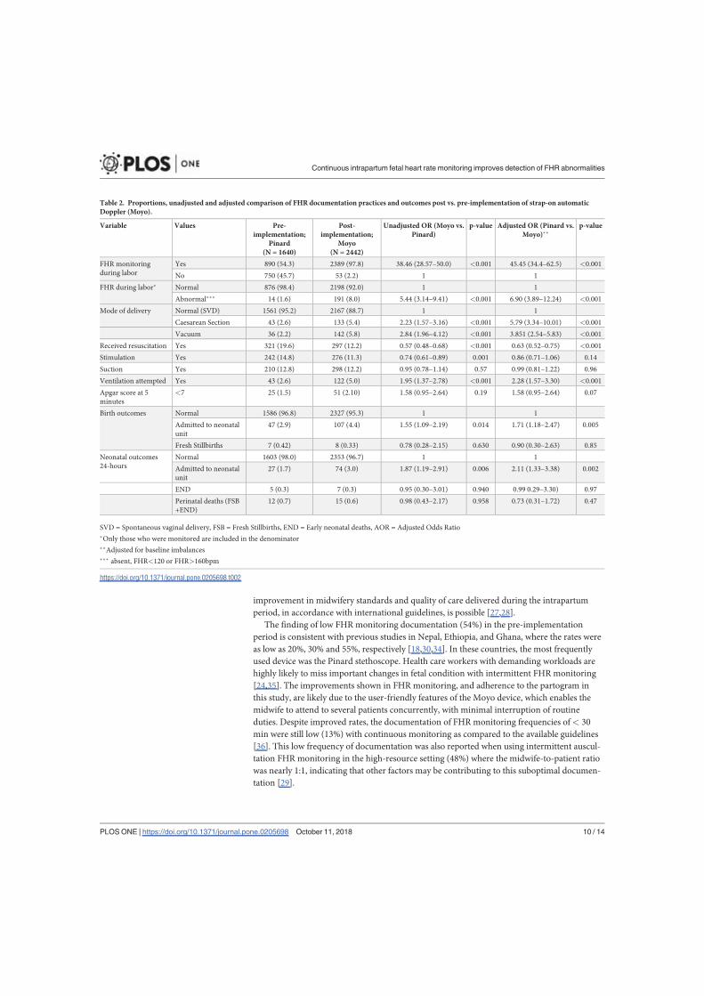
improvement in midwifery standards and quality of care delivered during the intrapartum
period, in accordance with international guidelines, is possible [27,28].
The finding of low FHRmonitoring documentation (54%) in the pre-implementation
period is consistent with previous studies in Nepal, Ethiopia, and Ghana, where the rates were
as low as 20%, 30% and 55%, respectively [18,30,34]. In these countries, the most frequently
used device was the Pinard stethoscope. Health care workers with demanding workloads are
highly likely to miss important changes in fetal condition with intermittent FHR monitoring
[24,35]. The improvements shown in FHR monitoring, and adherence to the partogram in
this study, are likely due to the user-friendly features of the Moyo device, which enables the
midwife to attend to several patients concurrently, with minimal interruption of routine
duties. Despite improved rates, the documentation of FHR monitoring frequencies of< 30
min were still low (13%) with continuous monitoring as compared to the available guidelines
[36]. This low frequency of documentation was also reported when using intermittent auscul-
tation FHR monitoring in the high-resource setting (48%) where the midwife-to-patient ratio
was nearly 1:1, indicating that other factors may be contributing to this suboptimal documen-
tation [29].
Table 2. Proportions, unadjusted and adjusted comparison of FHR documentation practices and outcomes post vs. pre-implementation of strap-on automaticDoppler (Moyo).
Variable Values Pre-implementation;
Pinard(N = 1640)
Post-implementation;
Moyo(N = 2442)
Unadjusted OR (Moyo vs.Pinard)
p-value Adjusted OR (Pinard vs.Moyo)��
p-value
FHR monitoringduring labor
Yes 890 (54.3) 2389 (97.8) 38.46 (28.57–50.0) <0.001 45.45 (34.4–62.5) <0.001
No 750 (45.7) 53 (2.2) 1 1
FHR during labor� Normal 876 (98.4) 2198 (92.0) 1 1
Abnormal��� 14 (1.6) 191 (8.0) 5.44 (3.14–9.41) <0.001 6.90 (3.89–12.24) <0.001
Mode of delivery Normal (SVD) 1561 (95.2) 2167 (88.7) 1 1
Caesarean Section 43 (2.6) 133 (5.4) 2.23 (1.57–3.16) <0.001 5.79 (3.34–10.01) <0.001
Vacuum 36 (2.2) 142 (5.8) 2.84 (1.96–4.12) <0.001 3.851 (2.54–5.83) <0.001
Received resuscitation Yes 321 (19.6) 297 (12.2) 0.57 (0.48–0.68) <0.001 0.63 (0.52–0.75) <0.001
Stimulation Yes 242 (14.8) 276 (11.3) 0.74 (0.61–0.89) 0.001 0.86 (0.71–1.06) 0.14
Suction Yes 210 (12.8) 298 (12.2) 0.95 (0.78–1.14) 0.57 0.99 (0.81–1.22) 0.96
Ventilation attempted Yes 43 (2.6) 122 (5.0) 1.95 (1.37–2.78) <0.001 2.28 (1.57–3.30) <0.001
Apgar score at 5minutes
<7 25 (1.5) 51 (2.10) 1.58 (0.95–2.64) 0.19 1.58 (0.95–2.64) 0.07
Birth outcomes Normal 1586 (96.8) 2327 (95.3) 1 1
Admitted to neonatalunit
47 (2.9) 107 (4.4) 1.55 (1.09–2.19) 0.014 1.71 (1.18–2.47) 0.005
Fresh Stillbirths 7 (0.42) 8 (0.33) 0.78 (0.28–2.15) 0.630 0.90 (0.30–2.63) 0.85
Neonatal outcomes24-hours
Normal 1603 (98.0) 2353 (96.7) 1 1
Admitted to neonatalunit
27 (1.7) 74 (3.0) 1.87 (1.19–2.91) 0.006 2.11 (1.33–3.38) 0.002
END 5 (0.3) 7 (0.3) 0.95 (0.30–3.01) 0.940 0.99 0.29–3.30) 0.97
Perinatal deaths (FSB+END)
12 (0.7) 15 (0.6) 0.98 (0.43–2.17) 0.958 0.73 (0.31–1.72) 0.47
SVD = Spontaneous vaginal delivery, FSB = Fresh Stillbirths, END = Early neonatal deaths, AOR = Adjusted Odds Ratio�Only those who were monitored are included in the denominator��Adjusted for baseline imbalances��� absent, FHR<120 or FHR>160bpm
https://doi.org/10.1371/journal.pone.0205698.t002
Continuous intrapartum fetal heart rate monitoring improves detection of FHR abnormalities
PLOSONE | https://doi.org/10.1371/journal.pone.0205698 October 11, 2018 10 / 14

In this study, we noted an increased rate of caesarean section deliveries from 2.6 to 5.4%,
presumably in response to the abnormal FHR. This rate is similar with a worldwide popula-
tion-based ecological study (2012) that reported an overall caesarean rate of 5.2% in low
income countries [37]. The World Health Organization (WHO) suggests that a rate of between
10 and 15% at a population level, may reflect optimal intrapartum care, provided decisions are
based on balancing the risks and benefits of this intervention [38,39].
It is noteworthy that while the overall need for resuscitation decreased post continuous
monitoring, the number of babies receiving bag mask ventilation increased. This incongruous
finding may be due to several interrelated factors. First, the higher incidence of an abnormal
FHR may have reflected an intrapartum hypoxia/ischemia state, with resultant respiratory
depression upon delivery. Second, the caesarean deliveries were invariably performed under
general anesthesia and depending on the duration between initiation of anesthesia and deliv-
ery, this may have resulted in the initial respiratory depression, particularly in the setting of an
abnormal FHR. There were no differences in FSB and END likely reflecting a low occurrence
of these morbidities in this low-risk population. An explanation for the increased number of
admissions post-implementation to a newborn area is not entirely clear. However, more moth-
ers underwent caesarean section, invariably under general anesthesia post-implementation. In
this setting, mothers are usually separated from their newborns for 24 hours after the caesarean
section. In addition, some of these neonates were admitted for observation following bag mask
ventilation, and/or were waiting for their mothers to recover from surgery.
Limitations
There were several limitations. First, this was a pre- and post-implementation study design,
hence there was no randomization. However, we consider the time difference between the
pre- and post-implementation period of too short a duration for factors other than the inter-
vention to cause the improvements. Furthermore, there were no observed systemic changes
that might have led to improved FHR monitoring. Second, although imbalances in baseline
characteristics were observed, these were adjusted in the regression analysis to remove poten-
tial confounding effects and improve the precision of the effect measures estimates. Third, the
study involved only low-risk pregnancies with fewer adverse perinatal outcomes than would
Table 3. Comparison of different median time intervals pre- and post-implementation of the Moyo at Temeke�.
Time intervals (q1, q3) Pre-implementationPinard
Post-implementationMoyo
P-value�
Admission to delivery (n = 1640) (n = 2442)
225 (130, 387) 212 (117, 355) 0.002
Admission to Abnormal FHR�� detection n = 14 n = 191
230 (120, 630) 138 (65, 302) 0.184
Last FHR assessment/ documentation to delivery n = 890 n = 2389
60 (30, 100) 45 (21, 85) <0.001
Between FHR assessment/ documentation n = 890 n = 890
150 (86, 299) 60 (41, 86) <0.001
Abnormal FHR to delivery n = 14 n = 191
28 (19, 57) 43 (23, 80) 0.255
q1 25th percentile and q3 75th percentile�Mann-Whitney U test, FHR = Fetal Heart Rate��absent, FHR<120or FHR>160bpm
https://doi.org/10.1371/journal.pone.0205698.t003
Continuous intrapartum fetal heart rate monitoring improves detection of FHR abnormalities
PLOSONE | https://doi.org/10.1371/journal.pone.0205698 October 11, 2018 11 / 14

have been expected in the overall population. Fourth, some health workers might have failed
to complete the partogram, even if FHRmeasurements were taken, leading to a misclassifica-
tion as being non-documented. Fifth, only baseline FHR abnormalities i.e. absent,
FHR<120bpm or FHR>160bpm were recorded while early, late decelerations or rapid accel-
erations were not addressed in this study. Unfortunately, the time from alert to response was
not collected in this study. Moreover, we did not collect data on when there was an alert with-
out a response. This important question will be included in future prospective studies in the
high-risk population.
Conclusion
Implementation of the Moyo device, which continuously measures FHR, was associated with
improvement in the quality of FHR monitoring practices, and the detection of abnormal FHR
(absent; FHR<120bpm or FHR>160bpm) in the resource-constrained setting. These
improvements led to more frequent and timely obstetric responses. Follow-up studies in the
high-risk population, focused on a more targeted description of the FHR abnormality, includ-
ing the duration, recurrence, and the relation to uterine contractions, as well as the impact of
intrauterine resuscitation on the FHR abnormalities, is a critical next step in determining the
impact on reducing perinatal mortality.
AcknowledgmentsWe acknowledge the hospital administration and labor ward staff for cooperating with us and
allowing us to use the hospital facilities to collect data for this study. We also wish to acknowl-
edge the research nurses for the well-done job of keeping records of all deliveries in the data
collection form. We thank the well-motivated data clerks and study coordinator of the Safer
Births project for data entry and cleaning.
Author ContributionsConceptualization: Benjamin A. Kamala, Hege L. Ersdal, Muzdalifat S. Abeid, Jeffrey M. Perl-
man, Hussein L. Kidanto.
Data curation: Benjamin A. Kamala, Muzdalifat S. Abeid, Matilda M. Ngarina, Jeffrey M.
Perlman, Hussein L. Kidanto.
Formal analysis: Benjamin A. Kamala, Hege L. Ersdal, Ingvild Dalen, Hussein L. Kidanto.
Funding acquisition:Hege L. Ersdal, Jeffrey M. Perlman.
Methodology: Benjamin A. Kamala, Hege L. Ersdal, Ingvild Dalen, Muzdalifat S. Abeid,
Matilda M. Ngarina, Jeffrey M. Perlman, Hussein L. Kidanto.
Project administration:Hege L. Ersdal.
Supervision:Matilda M. Ngarina, Jeffrey M. Perlman, Hussein L. Kidanto.
Writing – original draft: Benjamin A. Kamala.
Writing – review & editing:Hege L. Ersdal, Ingvild Dalen, Muzdalifat S. Abeid, Matilda M.
Ngarina, Jeffrey M. Perlman, Hussein L. Kidanto.
References1. Lawn JE, Blencowe H, Oza S, You D, Lee ACC,Waiswa P, et al. Every Newborn: progress, priorities,
and potential beyond survival. Lancet (London, England). 2014; 384: 189–205. https://doi.org/10.1016/S0140-6736(14)60496-7
Continuous intrapartum fetal heart rate monitoring improves detection of FHR abnormalities
PLOSONE | https://doi.org/10.1371/journal.pone.0205698 October 11, 2018 12 / 14

2. Ersdal HL, Eilevstj nn J, Linde JE, Yeconia A, Mduma ER, Kidanto H, et al. Fresh stillborn and severelyasphyxiated neonates share a common hypoxic-ischemic pathway. Int J Gynecol Obstet. Wiley-Black-well; 2018; https://doi.org/10.1002/ijgo.12430
3. UNDP. The Millennium Development Goals Report 2015. New York; 2015.
4. UNDP. Transforming our world: The 2030 agenda for sustainable development. https://sustainabledevelopment.un.org/content/documents/7891Transforming%20Our%20World.pdf. NewYork; 2015. https://doi.org/10.1007/s13398-014-0173-7.2
5. Oza S, Cousens SN, Lawn JE. Estimation of daily risk of neonatal death, including the day of birth, in186 countries in 2013: a vital-registration and modelling-based study. Lancet Glob Heal. 2014; 2: e635–e644. https://doi.org/10.1016/S2214-109X(14)70309-2
6. UNICEF,WHO,World Bank Group, United Nations. Levels and Trends in Child Mortality Child Mortality[Internet]. New York; 2017. Available: https://www.unicef.org/publications/files/Child_Mortality_Report_2017.pdf
7. Dye C, Boerma T, Evans D, Harries A, Lienhardt C, McManus Joanne, et al. Research for UniversalHealth Coverage. In: World Health Report [Internet]. 2013 [cited 13 Nov 2015]. Available: http://www.searo.who.int/indonesia/documents/research-for-universal-health-coverage(9789240690837_eng).pdf
8. United Nations. The Millennium Development Goals Report [Internet]. 2014 [cited 29 Nov 2015]. Avail-able: http://www.un.org/millenniumgoals/2014MDG report/MDG 2014 English web.pdf
9. Lawn JE, Lee ACC, KinneyM, Sibley L, Carlo WA, Paul VK, et al. Two million intrapartum-related still-births and neonatal deaths: where, why, and what can be done? Int J Gynaecol Obstet. 2009;107Suppl: S5–18, S19. https://doi.org/10.1016/j.ijgo.2009.07.016 PMID: 19815202
10. Maal e N, Housseine N, Meguid T, Nielsen B, Jensen A, Khamis R, et al. Effect of locally tailored labourmanagement guidelines on intrahospital stillbirths and birth asphyxia at the referral hospital of Zanzibar:a quasi-experimental pre-post study (The PartoMa study). BJOG An Int J Obstet Gynaecol. 2017;https://doi.org/10.1111/1471-0528.14933 PMID: 28892306
11. Yoshida S, Martines J, Lawn JE,Wall S, Souza JP, Rudan I, et al. Setting research priorities to improveglobal newborn health and prevent stillbirths by 2025. J Glob Health. 2016; 6: 010508. https://doi.org/10.7189/jogh.06.010508 PMID: 26401272
12. WHO, UNICEF. Every Newborn: an action plan to end preventable deaths: Executive summary. In:Geneva: World Health Organization. [Internet]. 2014 [cited 12 Jan 2016]. Available: http://www.everynewborn.org/Documents/Executive-summary_EN.pdf
13. Hofmeyr GJ, Haws RA, Bergstrom S, Lee ACC, Okong P, Darmstadt GL, et al. Obstetric care in low-resource settings: what, who, and how to overcome challenges to scale up? Int J Gynaecol Obstet.2009;107 Suppl: S21–44, S44-5. https://doi.org/10.1016/j.ijgo.2009.07.017 PMID: 19815204
14. van Handel M, Swaab H, de Vries LS, JongmansMJ. Long-term cognitive and behavioral conse-quences of neonatal encephalopathy following perinatal asphyxia: a review. Eur J Pediatr. 2007; 166:645–54. https://doi.org/10.1007/s00431-007-0437-8 PMID: 17426984
15. Mahomed K, Nyoni R, Mulambo T, Kasule J, Jacobus E. Randomised controlled trial of intrapartumfetal heart rate monitoring. BMJ. 1994; 308: 497–500. Available: http://www.pubmedcentral.nih.gov/articlerender.fcgi?artid=2542781&tool=pmcentrez&rendertype=abstract PMID: 8136665
16. Kc A, Wrammert J, Clark RB, Ewald U, M lqvist M. Inadequate fetal heart rate monitoring and poor useof partogram associated with intrapartum stillbirth: a case-referent study in Nepal. BMC PregnancyChildbirth. 2016;16. https://doi.org/10.1186/s12884-016-0806-2
17. Yisma E, Dessalegn B, Astatkie A, Fesseha N. Completion of the modifiedWorld Health Organization(WHO) partograph during labour in public health institutions of Addis Ababa, Ethiopia. Reprod Health.BioMed Central; 2013; 10: 23. https://doi.org/10.1186/1742-4755-10-23 PMID: 23597239
18. Adusi-Poku Y, Antwil E, Osei-Kwakye K, Tetteh C, Detoh EK, Antwi P. Quality of Care: A Review ofMaternal Deaths in a Regional Hospital in Ghana. Afr J Reprod Health. 2015; 19: 68–76. Available:http://www.ncbi.nlm.nih.gov/pubmed/26897915
19. Nyamtema AS, Urassa DP, Massawe S, Massawe A, Lindmark G, Van Roosmalen J. Staffing needsfor quality perinatal care in Tanzania. Afr J Reprod Health. 2008; 12: 113–24. Available: http://www.ncbi.nlm.nih.gov/pubmed/19435016 PMID: 19435016
20. Lewis D, Downe S. FIGO consensus guidelines on intrapartum fetal monitoring: Intermittent ausculta-tion. Int J Gynecol Obstet. Elsevier; 2015; 131: 9–12. https://doi.org/10.1016/j.ijgo.2015.06.019 PMID:26433400
21. Morrison JC, Chez BF, Davis ID, Martin RW, RobertsWE, Martin JN, et al. Intrapartum fetal heart rateassessment: monitoring by auscultation or electronic means. Am J Obstet Gynecol. 1993; 168: 63–6.Available: http://www.ncbi.nlm.nih.gov/pubmed/8420351 PMID: 8420351
Continuous intrapartum fetal heart rate monitoring improves detection of FHR abnormalities
PLOSONE | https://doi.org/10.1371/journal.pone.0205698 October 11, 2018 13 / 14
22. Kamala BA, Kidanto H, Wangwe P, Dalen I, Mduma E, Perlman J, et al. Intrapartum fetal heart ratemonitoring using a handheld Doppler versus Pinard stethoscope: a randomized controlled study in Dares Salaam. Int J Womens Health. 2018;Volume 10: 341–348. https://doi.org/10.2147/IJWH.S160675PMID: 30022861
23. Martis R, Emilia O, Nurdiati DS, Brown J. Intermittent auscultation (IA) of fetal heart rate in labour forfetal well-being. In: Martis R, editor. Cochrane Database of Systematic Reviews. Chichester, UK: JohnWiley & Sons, Ltd; 2017. p. CD008680. https://doi.org/10.1002/14651858.CD008680.pub2
24. Wyatt J. Appropriate medical technology for perinatal care in low-resource countries. Ann Trop Pae-diatr. 2008; 28: 243–51. https://doi.org/10.1179/146532808X375396 PMID: 19021939
25. Rivenes Lafontan S, Sundby J, Ersdal H, Abeid M, Kidanto H, Mbekenga C. “I Was Relieved to KnowThat My BabyWas Safe”: Women’s Attitudes and Perceptions on Using a New Electronic Fetal HeartRate Monitor during Labor in Tanzania. Int J Environ Res Public Health. 2018; 15: 302. https://doi.org/10.3390/ijerph15020302 PMID: 29425167
26. NBS and OCGS. National Bureau of Statistics (NBS) and Office of Chief Government Statistician(OCGS), Zanzibar. 2014. The 2012 Population and Housing Census: Basic Demographic and Socio-Economic Profile; Key Findings. Dar es Salaam, Tanzania: [Internet]. 2012 [cited 11 Nov 2015]. Avail-able: http://nbs.go.tz/nbs/takwimu/census2012/Basic_Demographic_and_Socio-Economic_Profile_PopularVersion-KeyFindings_2012_PHC_EnglishVersion.pdf
27. American College of Nurse Midwives. Intermittent Auscultation for Intrapartum Fetal Heart Rate Surveil-lance. J MidwiferyWomens Health. 2007; 52: 314–319. https://doi.org/10.1016/j.jmwh.2007.03.021PMID: 17467602
28. The Royal Australian and New Zealand College of Obstetricians and Gynaecologists. Intrapartum FetalSurveillance Clinical Guideline–Third Edition 2014. 2014;Version 3. Available: https://www.ranzcog.edu.au/RANZCOG_SITE/media/RANZCOG-MEDIA/Women’sHealth/Statementandguidelines/Clinical-Obstetrics/Intrapartum-Fetal-Surveillance-Guideline-Third-edition-Aug-2014.pdf?ext=.pdf
29. Asibong U, Okokon IB, Agan TU, Oku A, Opiah M, Essien EJ, et al. The use of the partograph in labormonitoring: a cross-sectional study among obstetric caregivers in General Hospital, Calabar, CrossRiver State, Nigeria. Int J Womens Health. Dove Press; 2014; 6: 873–880. https://doi.org/10.2147/IJWH.S49188 PMID: 25342920
30. Yisma E, Dessalegn B, Astatkie A, Fesseha N, Fawole A, Hunyinbo K, et al. Knowledge and utilizationof partograph among obstetric care givers in public health institutions of Addis Ababa, Ethiopia. BMCPregnancy Childbirth. BioMed Central; 2013; 13: 17. https://doi.org/10.1186/1471-2482-13-17
31. Boeing J, Gatlin T, Hendler E, Hyde E, Sienko K. Fetal Heart Rate Monitor for Resource-Limited Set-tings. 2012;
32. Maude RM, Skinner JP, Foureur MJ. Intelligent Structured Intermittent Auscultation (ISIA): evaluationof a decision-making framework for fetal heart monitoring of low-risk women. BMC Pregnancy Child-birth. BioMed Central; 2014; 14: 184. https://doi.org/10.1186/1471-2393-14-184 PMID: 24884597
33. Mdoe PF, Ersdal HL, Mduma E, Moshiro R, Dalen I, Perlman JM, et al. Randomized controlled trial ofcontinuous Doppler versus intermittent fetoscope fetal heart rate monitoring in a low-resource setting.Int J Gynaecol Obstet. 2018; https://doi.org/10.1002/ijgo.12648 PMID: 30120775
34. KC A,Wrammert J, Clark RB, Ewald U, M lqvist M, Lawn J, et al. Inadequate fetal heart rate monitoringand poor use of partogram associated with intrapartum stillbirth: a case-referent study in Nepal. BMCPregnancy Childbirth. BioMed Central; 2016; 16: 233. https://doi.org/10.1186/s12884-016-1034-5PMID: 27542350
35. Lewis D, Downe S, FIGO Intrapartum Fetal Monitoring Expert Consensus Panel. FIGO consensusguidelines on intrapartum fetal monitoring: Intermittent auscultation. Int J Gynaecol Obstet. 2015; 131:9–12. https://doi.org/10.1016/j.ijgo.2015.06.019 PMID: 26433400
36. World Health Organization. Preventing Prolonged Labour: A practicle Guide; The partograph. Geneva;1994.
37. Ye J, Zhang J, Mikolajczyk R, Torloni M, Gulmezoglu A, Betran A. Association between rates of caesar-ean section and maternal and neonatal mortality in the 21st century: a worldwide population-based eco-logical study with longitudinal data. BJOGAn Int J Obstet Gynaecol. 2016; 123: 745–753. https://doi.org/10.1111/1471-0528.13592 PMID: 26331389
38. World Health Organization. WHOStatement on Caesarean Section Rates [Internet]. Geneva; 2015.Available: http://apps.who.int/iris/bitstream/10665/161442/1/WHO_RHR_15.02_eng.pdf?ua=1
39. Betran AP, Torloni MR, Zhang J, Ye J, Mikolajczyk R, Deneux-Tharaux C, et al. What is the optimal rateof caesarean section at population level? A systematic review of ecologic studies. Reprod Health.BioMed Central; 2015; 12: 57. https://doi.org/10.1186/s12978-015-0043-6 PMID: 26093498
Continuous intrapartum fetal heart rate monitoring improves detection of FHR abnormalities
PLOSONE | https://doi.org/10.1371/journal.pone.0205698 October 11, 2018 14 / 14