Asherman syndrome—one century later
21
Asherman syndrome—one century later Dan Yu, M.Med., a,b Yat-May Wong, MRCOG, c Ying Cheong, M.D., b Enlan Xia, M.B.B.S., a and Tin-Chiu Li, M.D., Ph.D. b a Hysteroscopic Center, Fu Xing Hospital, Capital Medical University, Beijing, People’s Republic of China; b Department of Obstetrics and Gynecology, Jessop Wing, Royal Hallamshire Hospital, Sheffield, United Kingdom; and c Private Practice, Kular Lumpar, Malaysia Objective: To provide an update on the current knowledge of Asherman syndrome. Design: Literature review. Setting: The worldwide reports of this disease. Patient(s): Patients with Asherman syndrome who presented with amenorrhea or hypomenorrhea, infertility, or recurrent pregnancy loss. Intervention(s): Hysteroscopy and hysteroscopic surgery have been the gold standard of diagnosis and treatment respectively for this condition. Main Outcome Measure(s): The etiology, pathology, symptomatology, diagnosis, treatment, and reproductive outcomes were analyzed. Result(s): This syndrome occurs mainly as a result of trauma to the gravid uterine cavity, which leads to the for- mation of intrauterine and/or intracervical adhesions. Despite the advances in hysteroscopic surgery, the treatment of moderate to severe Asherman syndrome still presents a challenge. Furthermore, pregnancy after treatment remains high risk with complications including spontaneous abortion, preterm delivery, intrauterine growth restric- tion, placenta accrete or praevia, or even uterine rupture. Conclusion(s): The management of moderate to severe disease still poses a challenge, and the prognosis of severe disease remains poor. Close antenatal surveillance and monitoring are necessary for women who conceive after treatment. (Fertil Steril Ò 2008;89:759–79. Ó2008 by American Society for Reproductive Medicine.) Key Words: Intrauterine adhesion, Asherman syndrome, hysteroscopic adhesiolysis, infertility, synechia, hystero- scopy, amenorrhea It has been more than a century since Heinrich Fritsch (1) first described a case of posttraumatic intrauterine adhesion. In 1927, Bass (2) reported 20 cases of cervical obstruction in a series of 1500 patients who had undergone induced abor- tions. In 1946, Stamer (3) reviewed 37 cases reported in the literature and added 24 cases of his own with intrauterine adhesions associated with gravid uterus. In 1948, Joseph G. Asherman published a series of papers (4–7) to describe the frequency, etiology, symptoms, and roentgenologic picture of this condition, and Asherman syndrome has been used to describe the disease ever since. There have been an increas- ing number of cases of this syndrome described worldwide. The focus of research in the initial 50 to 60 years has been on the prevalence, etiology, and the pathology of this disease. As the understanding of this condition improves, together with the advent of endoscopic technology, the focus of re- search has now been shifted more toward diagnosis, treat- ment, and reproductive outcomes. This review provides an update on the current knowledge of the etiology, pathology, symptomatology, diagnosis, treatment, and reproductive outcomes of Asherman syndrome. DEFINITION From Asherman’s original definition, the syndrome was a consequence of trauma to the endometrium, producing par- tial or complete obliteration in the uterine cavity and/or the cervical canal, resulting in conditions such as menstrual ab- normalities, infertility, and recurrent pregnancy loss. Although the original description of Asherman syndrome was primarily based on a series of cases of intrauterine adhe- sions produced after curettage of the gravid uterus, it is now understood that there are several possible underlying causes of intrauterine adhesions. Consequently, the term Asherman syndrome should not be confined to only cases following curettage of the gravid uterus (see the section on etiology). The term ‘‘syndrome’’ in Greek refers to ‘‘concurrence of symptoms,’’ a group of symptoms that collectively indicate or characterize a disease. In this respect, the diagnosis of Asherman syndrome should be made only in women with clinical symptoms. There are cases in whom the formation of intrauterine adhesions is not associated with any clinical symptoms. In these cases, one should avoid using the term ‘‘Asherman syndrome’’ because there are no symptoms at Received February 7, 2008; revised and accepted February 7, 2008. D.Y. has nothing to disclose. Y-M.W. has nothing to disclose. Y.C. has nothing to disclose. E.X. has nothing to disclose. T-C.L. has nothing to disclose. Reprint requests: Dan Yu, M.Med., Hysteroscopic Center, Fu Xing Hospital, Capital Medical University, Beijing, 100038, People’s Republic of China (FAX: 0086-10-63486922; E-mail: yudanny2006@yahoo. com.cn). 0015-0282/08/$34.00 Fertility and Sterility â Vol. 89, No. 4, April 2008 759 doi:10.1016/j.fertnstert.2008.02.096 Copyright ª2008 American Society for Reproductive Medicine, Published by Elsevier Inc.
-
Upload
nguyenthuan -
Category
Documents
-
view
228 -
download
1
Transcript of Asherman syndrome—one century later
Asherman syndrome—one century laterDan Yu, M.Med.,a,b Yat-May Wong, MRCOG,c Ying Cheong, M.D.,b Enlan Xia, M.B.B.S.,a andTin-Chiu Li, M.D., Ph.D.b
a Hysteroscopic Center, Fu Xing Hospital, Capital Medical University, Beijing, People’s Republic of China; b Department of
Obstetrics and Gynecology, Jessop Wing, Royal Hallamshire Hospital, Sheffield, United Kingdom; and c Private Practice,
Kular Lumpar, Malaysia
Objective: To provide an update on the current knowledge of Asherman syndrome.Design: Literature review.Setting: The worldwide reports of this disease.Patient(s): Patients with Asherman syndrome who presented with amenorrhea or hypomenorrhea, infertility, orrecurrent pregnancy loss.Intervention(s): Hysteroscopy and hysteroscopic surgery have been the gold standard of diagnosis and treatmentrespectively for this condition.Main Outcome Measure(s): The etiology, pathology, symptomatology, diagnosis, treatment, and reproductiveoutcomes were analyzed.Result(s): This syndrome occurs mainly as a result of trauma to the gravid uterine cavity, which leads to the for-mation of intrauterine and/or intracervical adhesions. Despite the advances in hysteroscopic surgery, the treatmentof moderate to severe Asherman syndrome still presents a challenge. Furthermore, pregnancy after treatmentremains high risk with complications including spontaneous abortion, preterm delivery, intrauterine growth restric-tion, placenta accrete or praevia, or even uterine rupture.Conclusion(s): The management of moderate to severe disease still poses a challenge, and the prognosis of severedisease remains poor. Close antenatal surveillance and monitoring are necessary for women who conceive aftertreatment. (Fertil Steril� 2008;89:759–79. �2008 by American Society for Reproductive Medicine.)
Key Words: Intrauterine adhesion, Asherman syndrome, hysteroscopic adhesiolysis, infertility, synechia, hystero-scopy, amenorrhea
It has been more than a century since Heinrich Fritsch (1) firstdescribed a case of posttraumatic intrauterine adhesion. In1927, Bass (2) reported 20 cases of cervical obstruction ina series of 1500 patients who had undergone induced abor-tions. In 1946, Stamer (3) reviewed 37 cases reported in theliterature and added 24 cases of his own with intrauterineadhesions associated with gravid uterus. In 1948, Joseph G.Asherman published a series of papers (4–7) to describe thefrequency, etiology, symptoms, and roentgenologic pictureof this condition, and Asherman syndrome has been used todescribe the disease ever since. There have been an increas-ing number of cases of this syndrome described worldwide.The focus of research in the initial 50 to 60 years has beenon the prevalence, etiology, and the pathology of this disease.As the understanding of this condition improves, togetherwith the advent of endoscopic technology, the focus of re-search has now been shifted more toward diagnosis, treat-ment, and reproductive outcomes. This review provides an
Received February 7, 2008; revised and accepted February 7, 2008.
D.Y. has nothing to disclose. Y-M.W. has nothing to disclose. Y.C. has
nothing to disclose. E.X. has nothing to disclose. T-C.L. has nothing
to disclose.
Reprint requests: Dan Yu, M.Med., Hysteroscopic Center, Fu Xing
Hospital, Capital Medical University, Beijing, 100038, People’s Republic
of China (FAX: 0086-10-63486922; E-mail: yudanny2006@yahoo.
com.cn).
0015-0282/08/$34.00doi:10.1016/j.fertnstert.2008.02.096 Copyright ª2008 American
update on the current knowledge of the etiology, pathology,symptomatology, diagnosis, treatment, and reproductiveoutcomes of Asherman syndrome.
DEFINITION
From Asherman’s original definition, the syndrome wasa consequence of trauma to the endometrium, producing par-tial or complete obliteration in the uterine cavity and/or thecervical canal, resulting in conditions such as menstrual ab-normalities, infertility, and recurrent pregnancy loss.
Although the original description of Asherman syndromewas primarily based on a series of cases of intrauterine adhe-sions produced after curettage of the gravid uterus, it is nowunderstood that there are several possible underlying causesof intrauterine adhesions. Consequently, the term Ashermansyndrome should not be confined to only cases followingcurettage of the gravid uterus (see the section on etiology).
The term ‘‘syndrome’’ in Greek refers to ‘‘concurrence ofsymptoms,’’ a group of symptoms that collectively indicateor characterize a disease. In this respect, the diagnosis ofAsherman syndrome should be made only in women withclinical symptoms. There are cases in whom the formationof intrauterine adhesions is not associated with any clinicalsymptoms. In these cases, one should avoid using the term‘‘Asherman syndrome’’ because there are no symptoms at
Fertility and Sterility� Vol. 89, No. 4, April 2008 759Society for Reproductive Medicine, Published by Elsevier Inc.
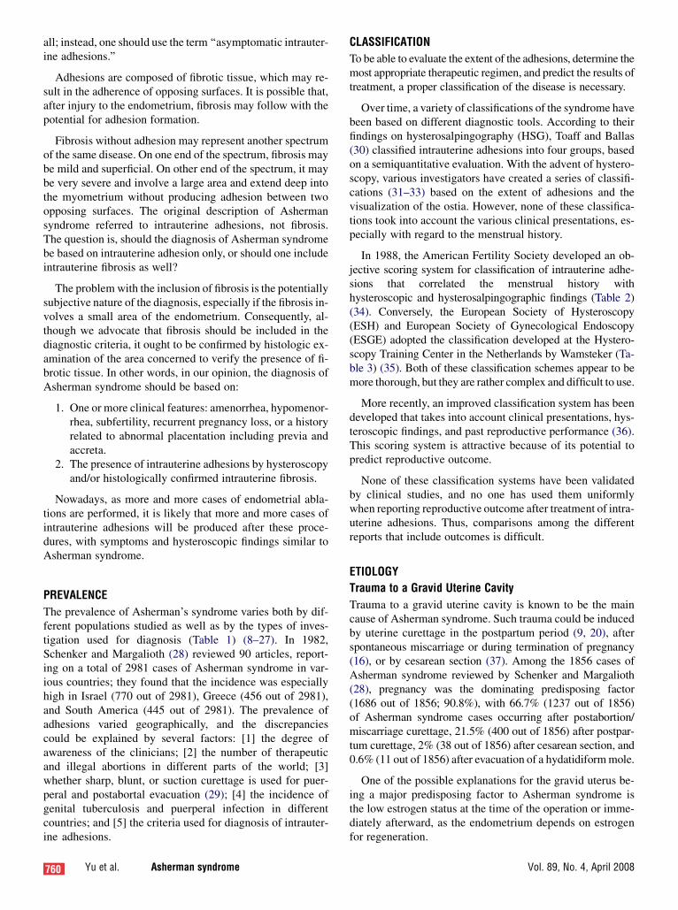
all; instead, one should use the term ‘‘asymptomatic intrauter-ine adhesions.’’
Adhesions are composed of fibrotic tissue, which may re-sult in the adherence of opposing surfaces. It is possible that,after injury to the endometrium, fibrosis may follow with thepotential for adhesion formation.
Fibrosis without adhesion may represent another spectrumof the same disease. On one end of the spectrum, fibrosis maybe mild and superficial. On other end of the spectrum, it maybe very severe and involve a large area and extend deep intothe myometrium without producing adhesion between twoopposing surfaces. The original description of Ashermansyndrome referred to intrauterine adhesions, not fibrosis.The question is, should the diagnosis of Asherman syndromebe based on intrauterine adhesion only, or should one includeintrauterine fibrosis as well?
The problem with the inclusion of fibrosis is the potentiallysubjective nature of the diagnosis, especially if the fibrosis in-volves a small area of the endometrium. Consequently, al-though we advocate that fibrosis should be included in thediagnostic criteria, it ought to be confirmed by histologic ex-amination of the area concerned to verify the presence of fi-brotic tissue. In other words, in our opinion, the diagnosis ofAsherman syndrome should be based on:
1. One or more clinical features: amenorrhea, hypomenor-rhea, subfertility, recurrent pregnancy loss, or a historyrelated to abnormal placentation including previa andaccreta.
2. The presence of intrauterine adhesions by hysteroscopyand/or histologically confirmed intrauterine fibrosis.
Nowadays, as more and more cases of endometrial abla-tions are performed, it is likely that more and more cases ofintrauterine adhesions will be produced after these proce-dures, with symptoms and hysteroscopic findings similar toAsherman syndrome.
PREVALENCE
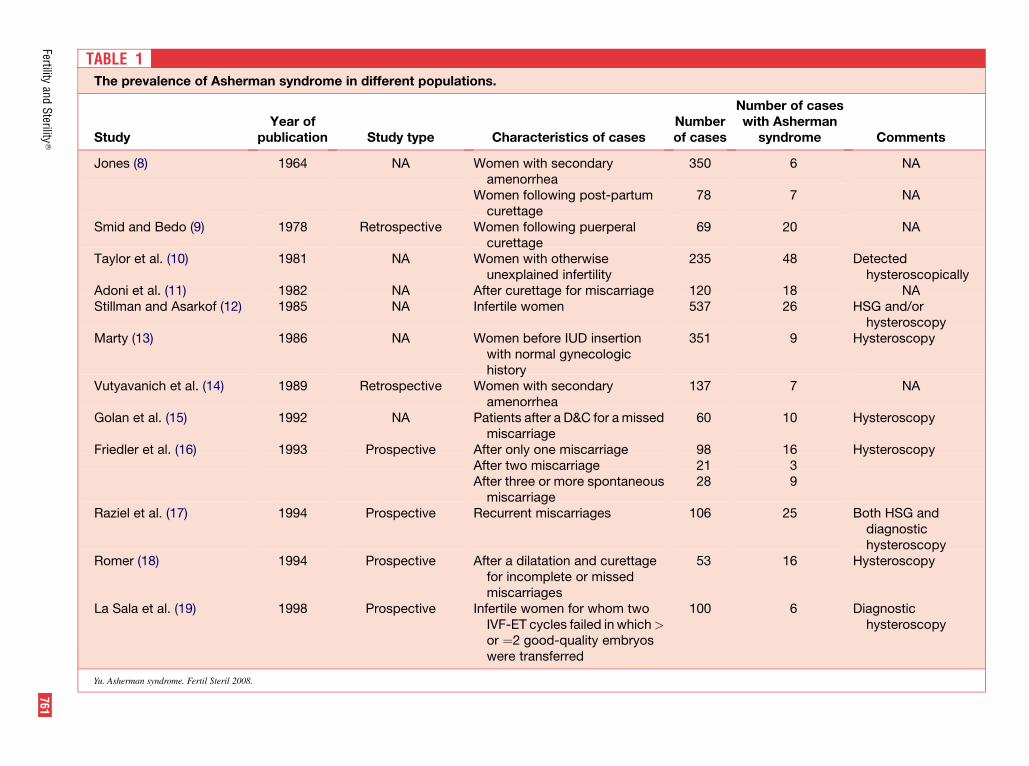
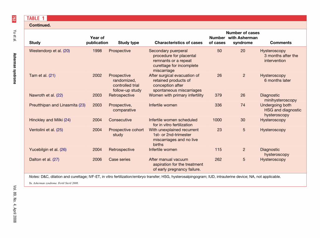
The prevalence of Asherman’s syndrome varies both by dif-ferent populations studied as well as by the types of inves-tigation used for diagnosis (Table 1) (8–27). In 1982,Schenker and Margalioth (28) reviewed 90 articles, report-ing on a total of 2981 cases of Asherman syndrome in var-ious countries; they found that the incidence was especiallyhigh in Israel (770 out of 2981), Greece (456 out of 2981),and South America (445 out of 2981). The prevalence ofadhesions varied geographically, and the discrepanciescould be explained by several factors: [1] the degree ofawareness of the clinicians; [2] the number of therapeuticand illegal abortions in different parts of the world; [3]whether sharp, blunt, or suction curettage is used for puer-peral and postabortal evacuation (29); [4] the incidence ofgenital tuberculosis and puerperal infection in differentcountries; and [5] the criteria used for diagnosis of intrauter-ine adhesions.
760 Yu et al. Asherman syndrome
CLASSIFICATION
To be able to evaluate the extent of the adhesions, determine themost appropriate therapeutic regimen, and predict the results oftreatment, a proper classification of the disease is necessary.
Over time, a variety of classifications of the syndrome havebeen based on different diagnostic tools. According to theirfindings on hysterosalpingography (HSG), Toaff and Ballas(30) classified intrauterine adhesions into four groups, basedon a semiquantitative evaluation. With the advent of hystero-scopy, various investigators have created a series of classifi-cations (31–33) based on the extent of adhesions and thevisualization of the ostia. However, none of these classifica-tions took into account the various clinical presentations, es-pecially with regard to the menstrual history.
In 1988, the American Fertility Society developed an ob-jective scoring system for classification of intrauterine adhe-sions that correlated the menstrual history withhysteroscopic and hysterosalpingographic findings (Table 2)(34). Conversely, the European Society of Hysteroscopy(ESH) and European Society of Gynecological Endoscopy(ESGE) adopted the classification developed at the Hystero-scopy Training Center in the Netherlands by Wamsteker (Ta-ble 3) (35). Both of these classification schemes appear to bemore thorough, but they are rather complex and difficult to use.
More recently, an improved classification system has beendeveloped that takes into account clinical presentations, hys-teroscopic findings, and past reproductive performance (36).This scoring system is attractive because of its potential topredict reproductive outcome.
None of these classification systems have been validatedby clinical studies, and no one has used them uniformlywhen reporting reproductive outcome after treatment of intra-uterine adhesions. Thus, comparisons among the differentreports that include outcomes is difficult.
ETIOLOGY
Trauma to a Gravid Uterine Cavity
Trauma to a gravid uterine cavity is known to be the maincause of Asherman syndrome. Such trauma could be inducedby uterine curettage in the postpartum period (9, 20), afterspontaneous miscarriage or during termination of pregnancy(16), or by cesarean section (37). Among the 1856 cases ofAsherman syndrome reviewed by Schenker and Margalioth(28), pregnancy was the dominating predisposing factor(1686 out of 1856; 90.8%), with 66.7% (1237 out of 1856)of Asherman syndrome cases occurring after postabortion/miscarriage curettage, 21.5% (400 out of 1856) after postpar-tum curettage, 2% (38 out of 1856) after cesarean section, and0.6% (11 out of 1856) after evacuation of a hydatidiform mole.
One of the possible explanations for the gravid uterus be-ing a major predisposing factor to Asherman syndrome isthe low estrogen status at the time of the operation or imme-diately afterward, as the endometrium depends on estrogenfor regeneration.
Vol. 89, No. 4, April 2008
rs
Number of caseswith Asherman
syndrome Comments
6 NA
7 NA
20 NA
48 Detectedhysteroscopically
18 NA26 HSG and/or
hysteroscopy9 Hysteroscopy
7 NA
10 Hysteroscopy
16 Hysteroscopy39
25 Both HSG anddiagnostichysteroscopy
16 Hysteroscopy
6 Diagnostichysteroscopy
TABLE 1The prevalence of Asherman syndrome in different populations.
StudyYear of
publication Study type Characteristics of casesNumbeof case
Jones (8) 1964 NA Women with secondaryamenorrhea
350
Women following post-partumcurettage
78
Smid and Bedo (9) 1978 Retrospective Women following puerperalcurettage
69
Taylor et al. (10) 1981 NA Women with otherwiseunexplained infertility
235
Adoni et al. (11) 1982 NA After curettage for miscarriage 120Stillman and Asarkof (12) 1985 NA Infertile women 537
Marty (13) 1986 NA Women before IUD insertionwith normal gynecologichistory
351
Vutyavanich et al. (14) 1989 Retrospective Women with secondaryamenorrhea
137
Golan et al. (15) 1992 NA Patients after a D&C for a missedmiscarriage
60
Friedler et al. (16) 1993 Prospective After only one miscarriage 98After two miscarriage 21After three or more spontaneous
miscarriage28
Raziel et al. (17) 1994 Prospective Recurrent miscarriages 106
Romer (18) 1994 Prospective After a dilatation and curettagefor incomplete or missedmiscarriages
53
La Sala et al. (19) 1998 Prospective Infertile women for whom twoIVF-ET cycles failed in which>or ¼2 good-quality embryoswere transferred
100
Yu. Asherman syndrome. Fertil Steril 2008.
Fertilityand
Sterility
�761
sNumberof cases
Number of caseswith Asherman
syndrome Comments
50 20 Hysteroscopy3 months after theintervention
f
s
26 2 Hysteroscopy6 months later
lity 379 26 Diagnosticminihysteroscopy
336 74 Undergoing bothHSG and diagnostichysteroscopy
1000 30 Hysteroscopy
23 5 Hysteroscopy
115 2 Diagnostichysteroscopy
nt.
262 5 Hysteroscopy
ram; IUD, intrauterine device; NA, not applicable.
TABLE 1Continued.
StudyYear of
publication Study type Characteristics of case
Westendorp et al. (20) 1998 Prospective Secondary puerperalprocedure for placentalremnants or a repeatcurettage for incompletemiscarriage
Tam et al. (21) 2002 Prospectiverandomized,controlled trialfollow-up study
After surgical evacuation oretained products ofconception afterspontaneous miscarriage
Nawroth et al. (22) 2003 Retrospective Women with primary inferti
Preutthipan and Linasmita (23) 2003 Prospective,comparative
Infertile women
Hinckley and Milki (24) 2004 Consecutive Infertile women scheduledfor in vitro fertilization
Ventolini et al. (25) 2004 Prospective cohortstudy
With unexplained recurrent1st- or 2nd-trimestermiscarriages and no livebirths
Yucebilgin et al. (26) 2004 Retrospective Infertile women
Dalton et al. (27) 2006 Case series After manual vacuumaspiration for the treatmeof early pregnancy failure
Notes: D&C, dilation and curettage; IVF-ET, in vitro fertilization/embryo transfer; HSG, hysterosalpingog
Yu. Asherman syndrome. Fertil Steril 2008.
762Y
uet
al.A
sherman
syndrome
Vol.
89,N
o.4,
April2008
TABLE 2The American Fertility Society classification of intrauterine adhesions, 1988.
<l/3 1/3–2/3 >2/3Extent of Cavity Involved
1 2 4
Filmy Filmy & Dense DenseType of Adhesions
1 2 4
Normal Hypomenorrhea AmenorrheaMenstrual Pattern
0 2 4
Prognostic classification HSGa score Hysteroscopy score
Stage l (Mild) 1-4Stage ll (Moderate) 5-8Stage lll (Severe) 9-12
Source: The American Fertility Society classifications of adnexal adhesions, distal tubal occlusion, tubal occlusion second-ary to tubal ligation, tubal pregnancies, mullerian anomalies and intrauterine adhesions. Fertil Steril 1988;49:944-55. (34)
a All adhesions should be considered dense.
Yu. Asherman syndrome. Fertil Steril 2008.
Another possible explanation could be the physiologicchanges that occur in a gravid uterus around the pregnancyperiod. The uterus could be in a vulnerable state after preg-nancy, making the basal layer of endometrium more easilydamaged by any trauma, especially curettage. Westendorpet al. (20) reported that, with ambulatory hysteroscopy, intra-uterine adhesions were found in 40% of the women undergo-ing either a secondary removal of placental remnants afterdelivery or a repeat curettage for incomplete abortions.
Trauma to Nongravid Endometrium
Trauma to a nongravid uterine cavity could also result in Asher-man syndrome. Schenker and Margalioth (28) reported thatAsherman syndrome may follow a diagnostic curettage (30out of 1856, 1.6%), abdominal myomectomy (24 out of1856, 1.3%), cervical biopsy or polylectomy (10 out of 1856,0.5%), insertion of an intrauterine device (3 out of 1856,0.2%), or the use of radium (1 out of 1856, 0.05%). The devel-opment of intrauterine adhesions can also result from variousforms of hysteroscopic surgery. In a prospective, randomizedstudy on 95 women who underwent hysteroscopic surgery,Taskin et al. (38) found that the frequency of Ashermansyndrome was 6.7% (1 out of 15) of patients after resectionof septa, and 31.3% (10 out of 32) and 45.5% (9 out of 20) ofpatients after hysteroscopic resection of solitary fibroids andmultiple fibroids, respectively. More recently, one case ofintrauterine adhesion was found after bilateral uterine artery em-bolization (UAE) (39); another was found after uterine devascu-larization because of severe postpartum hemorrhage (40).
Endometrial ablation is often followed by the formation ofintrauterine adhesions. The incidence of adhesion formation
Fertility and Sterility�
in women treated with thermal balloon ablation was reportedto be 36.4% (41). This is not surprising because the basallayer of the endometrium is often removed or destroyed, os-tensibly creating a local environment that is either noncondu-cive to, or insufficient for, regeneration of the endometrium,followed by the subsequent replacement of the luminal epi-thelial lining of the uterine cavity with fibrosis. There havebeen reports on the formation of intrauterine adhesions aftervarious forms of endometrial ablation (41–43).
Infection
There has been some controversy over the exact mechanismwhereby infection could result in Asherman syndrome. Severalinvestigators (10, 44, 45) maintained that the primary cause ofintrauterine adhesions has been infection, especially chronicor subacute endometritis; others (18, 46) have held the oppositeview, based on a study by Polishuk et al. (47) of 171 women whohad undergone cesarean sections. Among these 171 cases, 28patients developed significant endometritis. Afterward, HSGshowed no difference in the occurrence of intrauterine adhesionsbetween endometritis group and the control group (no infec-tion). They concluded that endometritis is unlikely to be a majorfactor in the pathogenesis of intrauterine or endocervical adhe-sion. Nevertheless, some investigators (48, 49) believe that in-flammatory processes do contribute to the damaging effect oftrauma and act synergistically to produce intrauterine adhesions.Uterine trauma and subsequent inflammation, in conjunctionwith a low estrogen status, may potentially lead to fibrosis.
Since the first report in 1956 (50), it has been recognizedthat endometrial tuberculosis may result in severe intrauter-ine adhesions, often with total obliteration of the uterine
763
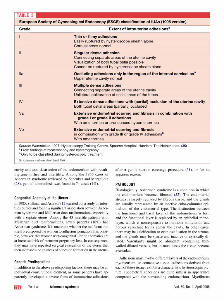
TABLE 3European Society of Gynecological Endoscopy (ESGE) classification of IUAs (1995 version).
Grade Extent of intrauterine adhesionsa
I Thin or filmy adhesionsEasily ruptured by hysteroscope sheath aloneCornual areas normal
II Singular dense adhesionConnecting separate areas of the uterine cavityVisualization of both tubal ostia possibleCannot be ruptured by hysteroscope sheath alone
IIa Occluding adhesions only in the region of the internal cervical osb
Upper uterine cavity normal
III Multiple dense adhesionsConnecting separate areas of the uterine cavityUnilateral obliteration of ostial areas of the tubes
IV Extensive dense adhesions with (partial) occlusion of the uterine cavityBoth tubal ostial areas (partially) occluded
Va Extensive endometrial scarring and fibrosis in combination withgrade I or grade II adhesions
With amenorrhea or pronounced hypomenorrhea
Vb Extensive endometrial scarring and fibrosisin combination with grade III or grade IV adhesionsb
With amenorrhea
Source: Wamsteker, 1997, Hysteroscopy Training Centre, Spaarne Hospital, Haarlem, The Netherlands. (35)a From findings at hysteroscopy and hysterography.b Only to be classified during hysteroscopic treatment.
Yu. Asherman syndrome. Fertil Steril 2008.
cavity and total destruction of the endometrium with result-ing amenorrhea and infertility. Among the 1856 cases ofAsherman syndrome reviewed by Schenker and Margalioth(28), genital tuberculosis was found in 74 cases (4%).
Congenital Anomaly of the Uterus
In 1985, Stillman and Asarkof (12) carried out a study on infer-tile couples and found a significant association between Asher-man syndrome and M€ullerian duct malformations, especiallywith a septate uterus. Among the 43 infertile patients withM€ullerian duct malformations, seven patients (16%) hadAsherman syndrome. It is uncertain whether the malformationitself predisposed the women to adhesion formation. It is possi-ble, however, that women with congenital uterine anomalies areat increased risk of recurrent pregnancy loss. In consequence,they may have repeated surgical evacuation of the uterus thatthen increases the chances of adhesion formation in the uterus.
Genetic Predisposition
In addition to the above predisposing factors, there may be anindividual constitutional element, as some patients have ap-parently developed a severe form of intrauterine adhesions
764 Yu et al. Asherman syndrome
after a gentle suction curettage procedure (51), or for noapparent reason.
PATHOLOGY
Histologically, Asherman syndrome is a condition in whichthe endometrium becomes fibrosed (52). The endometrialstroma is largely replaced by fibrous tissue, and the glandsare usually represented by an inactive cubo-columnar epi-thelium of the endometrial type. The distinction betweenthe functional and basal layer of the endometrium is lost,and the functional layer is replaced by an epithelial mono-layer, which is nonresponsive to hormone stimulation andfibrous synechiae forms across the cavity. In other cases,there may be calcification or even ossification in the stroma,and the glands may be sparse and inactive or cystically di-lated. Vascularity might be abundant, containing thin-walled dilated vessels, but in most cases the tissue becomeavascular.
Adhesions may involve different layers of the endometrium,myometrium, or connective tissue. Adhesions derived fromeach of these tissues exhibit a characteristic hysteroscopic pic-ture: endometrial adhesions are quite similar in appearancecompared with the surrounding endometrium. Myofibrous
Vol. 89, No. 4, April 2008
adhesions, which are most often encountered, are character-ized by the presence of a thin layer of overlying endometrium,the surface of which is furnished with many glandular ostia.The surface of connective tissue adhesions lack an endometriallining and contrast markedly with the adjacent endometrium.Fibrous adhesions that show dense connective tissue exhibitno lining in contrast to surrounding endometrium (53).
Using full-thickness myometrial biopsy specimens of pa-tients, Yaffe et al. (54) reported that the uterine wall contained50% to 80% of fibrous tissue compared with 13% to 20% ofthe control group. March (55) postulated that fibrosis limitsuterine myometrial activity and reduces the perfusion of sexsteroids, resulting in atrophy. Sometimes fibrosis involvingthe basal layers of the endometrium or the myometriummay occur without detectable intrauterine adhesions.
The extent of the endometrial damage may not directly cor-relate with the severity of the symptoms. For obstructive amen-orrhea, the lesion is often focal and limited to the uterineisthmus and cervical canal. A biopsy of the fundal part of theuterine cavity often reveals normal or inactive endometrium.
The histologic appearance after transcervical resection ofthe endometrium (TCRE) is similar to that of Asherman syn-drome (56). However, in addition to the fibrosis, otherchanges may be detrimental, including epithelioid or foreignbody granuloma, collections of hemosiderin, black-browncarbon, and metallic pigment (56, 57), or less commonly,necrotizing granulomatous inflammation (58).
SYMPTOMATOLOGY
The symptoms of Asherman syndrome include menstrual ab-normalities, infertility, recurrent pregnancy loss, and otherpregnancy complications.
Menstrual Abnormalities
Menstrual abnormalities, including hypomenorrhea andamenorrhea, remain the most common clinical features(1339 out of 1973, 68%) (28) associated with Asherman syn-drome. Amenorrhea may be caused by various etiologic fac-tors: [1] cervical adhesions blocking menstrual flow; [2]severe endometrial fibrosis leading to destruction of the en-tire basal layer of the endometrium (59). Dysmenorrhea is oc-casionally present (55 out of 1973, 3.5%) (28). In atreticamenorrhea, mechanical obstruction of the internal cervicalos could lead to secondary amenorrhea, periodic discomfortor pain, hematometra, and even hematosalpinx.
Infertility
Schenker and Margalioth (28) analyzed the symptoms of2151 cases of Asherman syndrome. They found that infertil-ity was present in 43% of women studied (802 out of 2151).One possible reason for infertility is the occlusion of the tubalostia, uterine cavity, or the cervical canal caused by adhe-sions. These synechiae could prevent the migration of spermor implantation of the embryo.
Fertility and Sterility�
Repeated Pregnancy Loss
Although a severe degree of intrauterine adhesions causes ob-struction of the cavity and infertility, a milder degree of adhe-sions may be associated with repeated pregnancy loss. Thepossible etiologic factors for recurrent pregnancy loss in-clude: [1] constriction of the uterine cavity caused by adhe-sions; [2] lack of a sufficient amount of normal endometrialtissue to support implantation and development of the pla-centa; and [3] defective vascularization of the residual endo-metrial tissue consequent upon fibrosis of endometrium (60).
Other Pregnancy Complications
The spontaneous conception rate with Asherman syndromewas reported as 45.5% (133 out of 292) (28). It was further re-ported by Schenker and Margalioth (28) that, among 165 preg-nancies in women with untreated Asherman syndrome, the rateof spontaneous miscarriage was 40% (66 out of 165), pretermdelivery was 23% (38 out of 165), term delivery was 30% (50out of 165), placenta accrete was 13% (21 out of 165), and ec-topic pregnancy was 12% (2 out of 165). The pregnancy com-plication rates in this group of patients appeared to be high,although there was no proper control group.
Theoretically, the defective placentation also may lead tointrauterine growth restriction (IUGR), though there havebeen only several cases of IUGR described in pregnantwomen with Asherman syndrome after endometrial ablations(61). The defective uterine endometrium and obliterated uter-ine cavity may also predispose women to ectopic tubal andcervical pregnancies (62–64). There has never been any re-port on fetal limb amputation associated with Asherman syn-drome, although limb amputation is associated with thepresence of amniotic bands after amniocentesis.
Endometrial Ablation
Endometrial ablation could lead to intrauterine adhesions,and those patients who conceived would be expected to de-velop pregnancy complications not dissimilar to those ofAsherman syndrome resulting from other reasons. In a recentliterature review of 70 pregnancies after endometrial abla-tions, Hare and Olah (61) found the following complicationsin 70 postablation pregnancies: ectopic pregnancy (1 out of70, 1.4%), spontaneous miscarriage (15 out of 70, 21%), pre-mature delivery (13 out of 70, 18.6%), IUGR (3 out of 70,4%), and morbidly adherent placenta (10 out of 70, 14.3%).In this study, there were only four normal pregnancies re-ported (4 out of 70, 5.7%). In 2005, Mukul and Linn (42) re-ported a case of significant fetal malformations, includingpositional deformities in the neck, an asymmetric chest, se-vere scoliosis, and limb abnormalities, associated with uter-ine synechiae resulting from previous endometrial ablation.In a retrospective study of 39 pregnancies after transcervicalresection of endometrium (TCRE) with cutting loop, Xiaet al. (65) observed five ectopic pregnancies (12.8%); 32cases with intrauterine pregnancy were terminated under ul-trasound guidance with two difficult procedures. Only one
765
pregnancy resulted in spontaneous miscarriage, which wasmanaged by suction curettage. One term pregnancy had pla-centa increta, resulting in a caesarean hysterectomy.
Endometrial Malignancy
In patients with intrauterine adhesions severe enough to pro-duce amenorrhea, the remaining biologically active endome-trium, in however small an amount, might undergo malignantchange, and the diagnosis in such a situation could potentiallybe very difficult. Sandridge et al. (66) first reported a case of en-dometrial carcinoma in a 71-year-old woman who developedpostmenopausal bleeding while receiving unopposed estrogen.Hysteroscopic examination showed extensive intrauterinesynechiae and a polypoid lesion adjacent to an adhesionband. Biopsy of the polypoid lesion confirmed endometrialadenocarcinoma. They concluded that Asherman syndromeand endometrial adenocarcinoma can exist simultaneously.In such cases, hysteroscopy is essential for target biopsy.
Clinical–Pathological Correlation
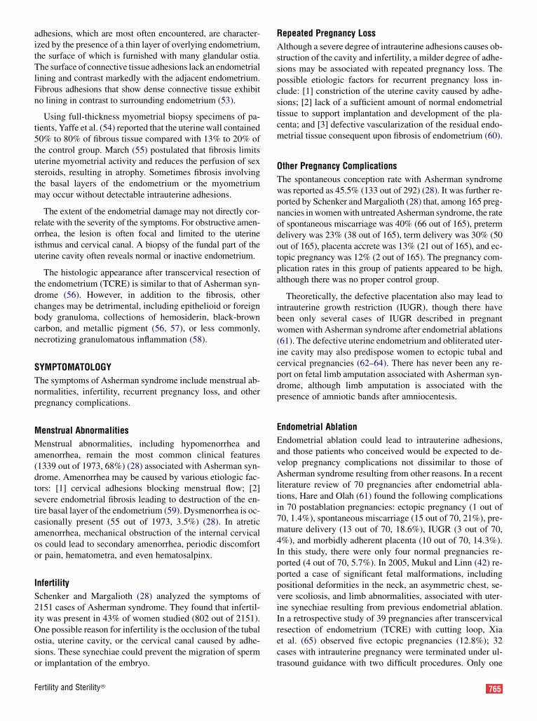
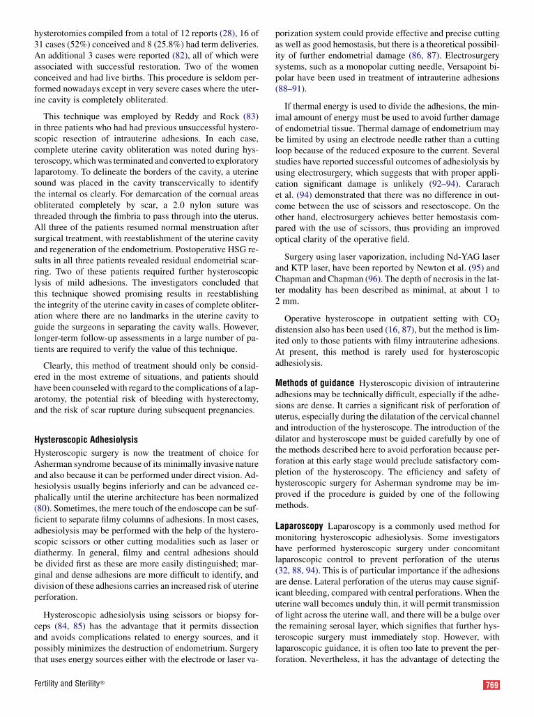
The clinical features are closely associated with pathologicfindings, such as the depth of fibrosis, location of the adhe-sions, and extent of the pathologic changes. The location ofthe adhesion can include the cervical canal, uterine cavity,or both cervical canal and uterine cavity (Fig. 1).
Obstructive amenorrhea is a consequence of intracervicaladhesions or stenosis, and patients often present with amen-orrhea with periodic abdominal discomfort. There may bea normal uterine cavity and a good prognosis after treatment.
Frequently, the adhesions are in the uterine cavity. It is themost common category and has several subcategories, which
FIGURE 1
Clinical pathology correlation of Ashermansyndrome.
1) Central adhesion withoutobliteration of cavity
3) Complete obliterate ofwhole uterus cavity
Location of the pathology of Asherman`s syndrome
2. Cervical canal adhesion(Atretic amenorrhea)
3. Uterine cavityadhesion
2) Partial obliterate andconstriction of cavity
4. Uterine cavity combined withcervical canal adhesion
1. Intrauterine fibrosis without visibleadhesion or obliteration of cavity
Yu. Asherman syndrome. Fertil Steril 2008.
766 Yu et al. Asherman syndrome
we characterize as central intrauterine adhesion without con-striction of the cavity, partial obliteration of the cavity withconstriction, and complete obliteration of the cavity. In allof these conditions, the patient may present with any symp-toms described in previous section, and the extent of diseasecan be confirmed by hysteroscopy. The extent of the diseasewill influence the prognosis after treatment. Patients withcentral intrauterine adhesions always have some normal en-dometrium and a relatively normal cavity. Therefore, theprognosis after treatment is usually good. Patients with par-tial obliteration of the cavity have a reduced and irregularcavity, and no cavity can be found in patients with completeobliteration of the cavity. Amenorrhea and infertility are thetwo most common accompanying clinical features in patientswith partial or complete obliterated cavity. The prognosis forpregnancy in these patients is often poor.
In some cases, the adhesions can be located in both cervi-cal canal and cavity of the uterus. The patient may presentwith any symptoms, including menstrual abnormalities, in-fertility, and pregnancy complications, and the prognosis(such as normal menses and fertility) depends very muchon the severity and extent of the adhesions.
INVESTIGATIONS
The following investigations may be considered in womenwith suspected Asherman syndrome.
Radiologic Diagnosis
Hysterosalpingography Before the invention of the hystero-scope, HSG was the first-line investigation to visualize theuterine cavity. Today, many gynecologists still consider it tobe a valid initial test for Asherman syndrome. Hysterosalpin-gography has the advantages of being able to outline the areasof occlusion or special filling defects and providing an ap-praisal of the cornual region, tubal contours, and tubal pa-tency. Wamsteker (67) described the HSG appearance ofintrauterine adhesions as defects that appear as sharply out-lined intrauterine structures, which are centrally and/or mar-ginally located. These filling defects are often irregular,angulated, and of homogenous opacity. In more severe cases,partial obliteration of the cavity may be observed; its bordersmay be irregular, and either one or both of the tubes may beoccluded. In severe intrauterine adhesions, the uterine cavityis completely distorted and narrowed, and both tubes may beoccluded. With complete occlusion of the lower part of theuterine cavity, HSG will fail to show any contrast filling ofthe uterine cavity and will provide no information on theextent of the adhesions. A short cannula should be used toprevent perforation in cases of suspected severe adhesions.
Several studies have examined the value of HSG in the di-agnosis of intrauterine adhesions. In a retrospective study of400 patients (68), HSG was found to be as accurate ashysteroscopy in diagnosis of normal and abnormal uterinecavities, although the nature of the intrauterine filling defectswas accurately revealed by hysteroscopy only. Another
Vol. 89, No. 4, April 2008
investigation showed that more than one third (38.3%) ofHSG examinations may have false-positive findings (17). Ina prospective study reported by Soares et al. (69), the diag-nostic accuracy of HSG in the detection of intrauterine adhe-sion was compared with hysteroscopic findings as the goldstandard. Among 65 women studied, five had hysteroscopicevidence of intrauterine adhesions; HSG had a sensitivityof 75% in the detection of intrauterine adhesions and a posi-tive predictive value of 50%.
Hysterosalpingography is a simple screening method forintrauterine adhesions, and it remains an important screeningprocedure for the diagnosis of intrauterine adhesions, espe-cially in the infertile patients because additional informationabout the fallopian tubes may be obtained. However, HSGhas a number of limitations. First, it may not detect endome-trial fibrosis per se, as in cases without adhesions. Second,HSG has a high rate of false-positive results and has limita-tions in defining the nature of identified intrauterine fillingdefects. Third, minor filmy adhesions might not consistentlyproduce abnormal shadows on HSG. Fourth, air bubbles, mu-cous, and debris may all mimic filling defects, and poorplacement of the cannula can cause intravasation. An exces-sive amount of contrast medium in the uterus also can oblit-erate shadows caused by intrauterine adhesions.
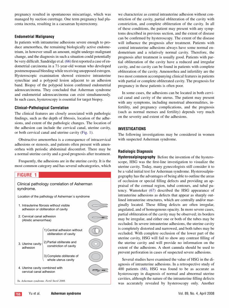
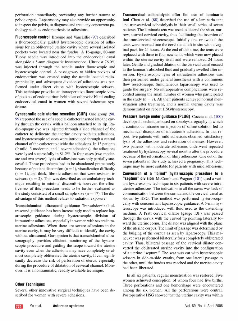
Ultrasonography Ultrasonography was used in the diagnosisof endometrial fibrosis or intrauterine adhesion by Confinoet al. (70) and Schlaff and Hurst (71). It is a noninvasive pro-cedure that allows visualization of the uterine cavity, whichmay not otherwise be possible during HSG or hysteroscopywhen the uterine cavity has been obliterated. Typically, adhe-sions appear as dense echoes within the cavity. In women withsevere intrauterine adhesions, ultrasonography may show thefollowing typical appearance: the endometrial echo becomesdifficult to visualize, with irregular thickness and one or moreinterruptions of the endometrium at the sites of fibrosis. In ad-dition, there may sometimes be one or more echolucent (cys-tic-like) areas interrupting the endometrium, representinglocalized collection of menstrual blood in an area where thefunctional layers of the endometrium are preserved (Fig. 2).
Both the sensitivity and the specificity of ultrasound in diag-nosis of the intrauterine adhesions have been reported to bequite low: the sensitivity of transvaginal ultrasonographywas reported as 52% (72) and the specificity as only 11%(73). Nevertheless, ultrasonography is useful when HSG isnot possible because of obliteration of lower uterine cavity.In this situation, ultrasonography may be used to evaluatethe upper uterine cavity, and the findings maybe of prognosticsignificance. Schlaff and Hurst (71) used transvaginal ultraso-nography to study the endometrial echo in seven patients withAsherman syndrome presenting with amenorrhea and com-plete obstruction of the uterine cavity before hysteroscopicsurgery. They found that patients with normal-appearing en-dometrium above the level of obstruction are likely to benefitsuccessful hysteroscopic treatment and can expect to resumenormal postoperative menstrual function. Patients who havelittle or no endometrium seen on transvaginal ultrasonography
Fertility and Sterility�
are extremely unlikely to profit by hysteroscopic creation ofa uterine cavity from within the myometrium.
Three-dimensional ultrasonography techniques have beenused by a few investigators (73, 74) to detect adhesions in theuterine cavity, with a specificity of 45% (73). In the study re-ported by Soares et al. (69), transvaginal ultrasonography didnot detect any of the cases of intrauterine adhesions andyielded three false-positive diagnoses of this disease (sensi-tivity and positive predictive value of 0).
Sonohysterography Sonohysterography (SHG), which com-bines transvaginal sonography with intrauterine injection ofisotonic saline, has been shown to be as accurate as HSGand to be superior to transvaginal ultrasonography in thedetection of intrauterine adhesions (72). In this technique,20 to 30 mL of saline may be introduced into the uterine cavitythrough a catheter. Intrauterine adhesions are suspected if thereare one or more echogenic areas between the anterior and pos-terior walls in the liquid-filled uterine cavity, or if the full dis-tension of the cavity was impeded by tethering of the walls bythin or thick bands of synechiae (72). In a study of 19 patientswith intrauterine adhesions, the sensitivities of HSG and SHGin the diagnosis of intrauterine adhesions were both 100%, andthe sensitivity of transvaginal ultrasonography was only 52%(72). In another study on diagnostic accuracy in 65 infertilewomen, SHG had both similar sensitivity (75%) and specific-ity (positive predictive values as 42.9%) to HSG (69).
FIGURE 2
Transvaginal ultrasonography of a case of Ashermansyndrome with typical interrupted endometrial echo.There were several hyperechoic bridges indicatingintrauterine adhesions, with several echolucentareas in between the bridges indicating collection ofmenstrual blood in areas where there are functioningendometrial tissues. þ, indicates the uterineadhesions.
Yu. Asherman syndrome. Fertil Steril 2008.
767
With SHG, it is possible to perform a complete ultrasoundexamination of the uterine structure including the uterinecavity and myometrium. However, like HSG, SHG is of valueonly in cases of partial intrauterine adhesions because normalsaline will not be able to enter into the uterine cavity when itis completely obstructed (75, 76). Sonohysterography is use-ful in situations where transvaginal ultrasonography yieldsnormal results but the clinical suspicion of intrauterine adhe-sions remains high.
Magnetic resonance imaging Magnetic resonance imaging(MRI) has been used as a tool to investigate patients withAsherman syndrome (77, 78), especially those with involve-ment of the cervical canal (79). The main advantage of MRIis its ability to image the uterine cavity above the adhesionsand assess the endometrial remnants in the upper part of theuterine cavity, which may influence the decision and outcomeof treatment. However, the MRI-signal characteristics of in-trauterine adhesions have not been examined in detail; it is an-ticipated that adhesions would produce low signal intensity onT2 images (79). Thus, MRI may have a supplementary role inthe diagnosis of complete obliteration of uterine cavity or cer-vical obstruction that cannot be visualized by hysteroscopy.
Hysteroscopy
Hysteroscopy, as compared with radiologic tests, can moreaccurately confirm the presence, extent, and degree of adhe-sions and the quality of the endometrium because the uterinecavity can be directly inspected. The hysteroscope may beguided through the endocervix under visual control or by ul-trasound guidance until the internal os is completely visibleor the adhesive obstruction is identified (67).
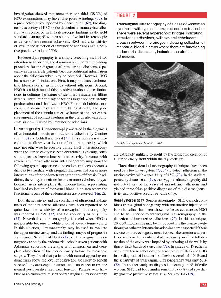
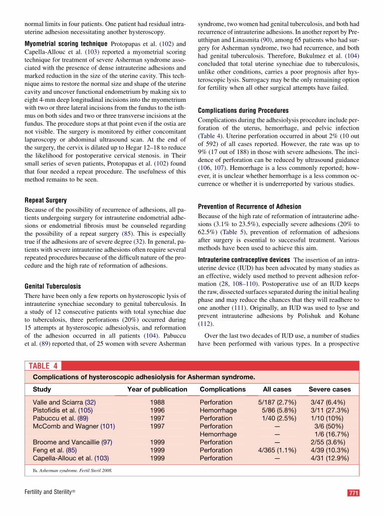
Al-Inany (80) described the different varieties of intrauter-ine adhesions as visualized via the hysteroscope: [1] the centraladhesion appears as a column with broadening ends connect-ing the opposite uterine walls; [2] marginal adhesion (whichmay be easily missed) appears like a crescent or half-drawncurtain, which can hide a cornu or sidewall and create an asym-metric aspect to the uterine cavity (Fig. 3), and [3] complexmarginal central scars, which divide the uterine cavity into sev-eral smaller chambers, can appear, some of which maybe con-cealed to visual examination. Superficial mucosal adhesionsusually have the same color as the surrounding endometriumand can be easily broken down by the pressure of the hystero-scope; the fibrous or myometrial bands appear white in color,and are often dense and difficult to divide. The diagnosis ofadhesion resulting from tuberculosis may be suggested bythe appearance of a network of small alveoli coating the uterinewalls. The fundus may look like a honeycomb. Endometrialfibrosis appears as pale patches on hysteroscopy (81).
If it is not possible to perform a hysteroscopy satisfactorilybecause of significant obliteration of the cavity, the otheralternative diagnostic modalities such as ultrasonography orMRI should be considered. Alternatively, hysteroscopic eval-uation may be facilitated by adhesiolysis under ultrasound orother methods of guidance. The methylene blue test may be
768 Yu et al. Asherman syndrome
used to differentiate between fibrosis and normal endometriallining (32). When methylene blue is injected through theinflow channel of the hysteroscope, the endometrium stainswell but connective (fibrotic) tissue and myometrium do not.
TREATMENT
Treatment of Asherman syndrome aims at restoring the sizeand shape of the uterine cavity, preventing recurrence ofthe adhesion, promoting the repair and regeneration of the de-stroyed endometrium, and restoring normal reproductivefunctions. Over the last century, many surgical techniqueshave been described.
Expectant Management
Schenker and Margalioth (28) gathered 23 amenorrheicwomen from the literature who had not undergone any surgi-cal intervention, of whom 18 regained regular menses after 1to 7 years. For fertility outcome, 292 women in whom treat-ment was withheld were collated, among whom 133 women(45.5%) conceived spontaneously.
Blind Dilation and Curettage
Before the advent of hysteroscopy, Asherman syndrome wastreated by dilation and curettage of the uterus. It is not sur-prising that this method resulted in a high incidence of uterineperforation and had a low success rate. This method is nowconsidered obsolete.
Hysterotomy
Transfundal separation of the walls of endometrial cavity byhysterotomy has been described. In an analysis of 31 cases of
FIGURE 3
Hysteroscopic view of a case of Ashermansyndrome with adhesions bands in the anterior andleft lateral side wall of the uterine cavity.
Yu. Asherman syndrome. Fertil Steril 2008.
Vol. 89, No. 4, April 2008
hysterotomies compiled from a total of 12 reports (28), 16 of31 cases (52%) conceived and 8 (25.8%) had term deliveries.An additional 3 cases were reported (82), all of which wereassociated with successful restoration. Two of the womenconceived and had live births. This procedure is seldom per-formed nowadays except in very severe cases where the uter-ine cavity is completely obliterated.
This technique was employed by Reddy and Rock (83)in three patients who had had previous unsuccessful hystero-scopic resection of intrauterine adhesions. In each case,complete uterine cavity obliteration was noted during hys-teroscopy, which was terminated and converted to exploratorylaparotomy. To delineate the borders of the cavity, a uterinesound was placed in the cavity transcervically to identifythe internal os clearly. For demarcation of the cornual areasobliterated completely by scar, a 2.0 nylon suture wasthreaded through the fimbria to pass through into the uterus.All three of the patients resumed normal menstruation aftersurgical treatment, with reestablishment of the uterine cavityand regeneration of the endometrium. Postoperative HSG re-sults in all three patients revealed residual endometrial scar-ring. Two of these patients required further hysteroscopiclysis of mild adhesions. The investigators concluded thatthis technique showed promising results in reestablishingthe integrity of the uterine cavity in cases of complete obliter-ation where there are no landmarks in the uterine cavity toguide the surgeons in separating the cavity walls. However,longer-term follow-up assessments in a large number of pa-tients are required to verify the value of this technique.
Clearly, this method of treatment should only be consid-ered in the most extreme of situations, and patients shouldhave been counseled with regard to the complications of a lap-arotomy, the potential risk of bleeding with hysterectomy,and the risk of scar rupture during subsequent pregnancies.
Hysteroscopic Adhesiolysis
Hysteroscopic surgery is now the treatment of choice forAsherman syndrome because of its minimally invasive natureand also because it can be performed under direct vision. Ad-hesiolysis usually begins inferiorly and can be advanced ce-phalically until the uterine architecture has been normalized(80). Sometimes, the mere touch of the endoscope can be suf-ficient to separate filmy columns of adhesions. In most cases,adhesiolysis may be performed with the help of the hystero-scopic scissors or other cutting modalities such as laser ordiathermy. In general, filmy and central adhesions shouldbe divided first as these are more easily distinguished; mar-ginal and dense adhesions are more difficult to identify, anddivision of these adhesions carries an increased risk of uterineperforation.
Hysteroscopic adhesiolysis using scissors or biopsy for-ceps (84, 85) has the advantage that it permits dissectionand avoids complications related to energy sources, and itpossibly minimizes the destruction of endometrium. Surgerythat uses energy sources either with the electrode or laser va-
Fertility and Sterility�
porization system could provide effective and precise cuttingas well as good hemostasis, but there is a theoretical possibil-ity of further endometrial damage (86, 87). Electrosurgerysystems, such as a monopolar cutting needle, Versapoint bi-polar have been used in treatment of intrauterine adhesions(88–91).
If thermal energy is used to divide the adhesions, the min-imal amount of energy must be used to avoid further damageof endometrial tissue. Thermal damage of endometrium maybe limited by using an electrode needle rather than a cuttingloop because of the reduced exposure to the current. Severalstudies have reported successful outcomes of adhesiolysis byusing electrosurgery, which suggests that with proper appli-cation significant damage is unlikely (92–94). Cararachet al. (94) demonstrated that there was no difference in out-come between the use of scissors and resectoscope. On theother hand, electrosurgery achieves better hemostasis com-pared with the use of scissors, thus providing an improvedoptical clarity of the operative field.
Surgery using laser vaporization, including Nd-YAG laserand KTP laser, have been reported by Newton et al. (95) andChapman and Chapman (96). The depth of necrosis in the lat-ter modality has been described as minimal, at about 1 to2 mm.
Operative hysteroscope in outpatient setting with CO2
distension also has been used (16, 87), but the method is lim-ited only to those patients with filmy intrauterine adhesions.At present, this method is rarely used for hysteroscopicadhesiolysis.
Methods of guidance Hysteroscopic division of intrauterineadhesions may be technically difficult, especially if the adhe-sions are dense. It carries a significant risk of perforation ofuterus, especially during the dilatation of the cervical channeland introduction of the hysteroscope. The introduction of thedilator and hysteroscope must be guided carefully by one ofthe methods described here to avoid perforation because per-foration at this early stage would preclude satisfactory com-pletion of the hysteroscopy. The efficiency and safety ofhysteroscopic surgery for Asherman syndrome may be im-proved if the procedure is guided by one of the followingmethods.
Laparoscopy Laparoscopy is a commonly used method formonitoring hysteroscopic adhesiolysis. Some investigatorshave performed hysteroscopic surgery under concomitantlaparoscopic control to prevent perforation of the uterus(32, 88, 94). This is of particular importance if the adhesionsare dense. Lateral perforation of the uterus may cause signif-icant bleeding, compared with central perforations. When theuterine wall becomes unduly thin, it will permit transmissionof light across the uterine wall, and there will be a bulge overthe remaining serosal layer, which signifies that further hys-teroscopic surgery must immediately stop. However, withlaparoscopic guidance, it is often too late to prevent the per-foration. Nevertheless, it has the advantage of detecting the
769
perforation immediately, preventing any further trauma topelvic organs. Laparoscopy may also provide an opportunityto inspect the pelvis, to diagnose and treat any concurrent pa-thology such as endometriosis or adhesions.
Fluoroscopic control Broome and Vancaillie (97) describeda fluoroscopically guided hysteroscopic division of adhe-sions for an obliterated uterine cavity where several isolatedpockets were located near the fundus. A 16-gauge, 80-mmTuohy needle was introduced into the endocervical canalalongside a 5-mm diagnostic hysteroscope. Ultravist 76.9%was injected through the needle under fluoroscopic andhysteroscopic control. A passageway to hidden pockets ofendometrium was created using the needle located radio-graphically, and subsequent division of adhesions was per-formed under direct vision with hysteroscopic scissors.This technique provides an intraoperative fluoroscopic viewof pockets of endometrium behind an otherwise blind-endingendocervical canal in women with severe Asherman syn-drome.
Gynecoradiologic uterine resection (GUR) One group (98,99) reported the use of a special catheter inserted into the cav-ity through the cervix with a balloon attached to its tip. Ra-dio-opaque dye was injected through a side channel of thecatheter to delineate the uterine cavity with its adhesions,and hysteroscopic scissors were introduced through a centralchannel of the catheter to divide the adhesions. In 13 patients(9 mild, 3 moderate, and 1 severe adhesions), the adhesionswere lysed successfully in 81.2%. In four cases (two moder-ate and two severe), lysis of adhesions was only partially suc-cessful. These procedures had to be abandoned prematurelybecause of patient discomfort (n¼ 1), visualization difficulty(n ¼ 1), and thick, fibrotic adhesions that were resistant toscissors (n ¼ 2). This was described as an ambulatory tech-nique resulting in minimal discomfort; however, the effec-tiveness of this procedure needs to be further evaluated asthe study consisted of a small sample size (n ¼ 17). The dis-advantage of this method relates to radiation exposure.
Transabdominal ultrasound guidance Transabdominal ul-trasound guidance has been increasingly used to replace lap-aroscopic guidance during hysteroscopic division ofintrauterine adhesions, especially in women with severe intra-uterine adhesions. When there are severe adhesions in theuterine cavity, it may be very difficult to identify the cavitywithout ultrasound. Our opinion is that transabdominal ultra-sonography provides efficient monitoring of the hystero-scopic procedure and guiding the scope toward the uterinecavity even when the adhesions may have completely or al-most completely obliterated the uterine cavity. It can signifi-cantly decrease the risk of perforation of uterus, especiallyduring the procedure of dilatation of cervical channel. More-over, it is a nontraumatic, readily available technique.
Other Techniques
Several other innovative surgical techniques have been de-scribed for women with severe adhesions.
770 Yu et al. Asherman syndrome
Transcervical adhesiolysis after the use of laminariatent Chen et al. (88) described the use of a laminaria tentand transcervical adhesiolysis in their small series of sevenpatients. The laminaria tent was used to distend the short, nar-row, scarred cervical cavity, thus facilitating the insertion ofthe transcervical resectoscope. Initially one or two of thetents were inserted into the cervix and left in situ with a vag-inal pack for 24 hours. At the end of this time, the tents werereplaced with three to four new tents, which were now placedwithin the uterine cavity itself and were removed 24 hourslater. Gentle and gradual dilation of the cervical canal ensuedas the laminaria absorbed fluid and gradually swelled after in-sertion. Hysteroscopic lysis of intrauterine adhesions wasthen performed under general anesthesia with a continuousflow resectoscope. Simultaneous laparoscopy was used toguide the surgery. No intraoperative complications were re-corded among the small number of women who participatedin the study (n ¼ 7). All their patients achieved normal men-struation after treatment, and a normal uterine cavity wasdemonstrated on repeat HSG/hysteroscopy.
Pressure lavage under guidance (PLUG) Coccia et al. (100)developed a technique based on sonohysterography in whicha continuous intrauterine injection of saline solution led tomechanical disruption of intrauterine adhesions. In that re-port, five patients with mild adhesions obtained satisfactorylysis of the adhesions and restoration of menses. However,two patients with moderate adhesions underwent repeatedtreatment by hysteroscopy several months after the procedurebecause of the reformation of filmy adhesions. One out of theseven patients in the study achieved a pregnancy. This tech-nique may be more suitable for patients with mild adhesions.
Conversion of a ‘‘blind’’ hysteroscopic procedure to a‘‘septum’’ division McComb and Wagner (101) used a vari-ant hysteroscopic technique in six patients with severe intra-uterine adhesions. The indication in all the cases was lack ofcommunication between the cornua and the cervical canal asshown by HSG. This method was performed hysteroscopi-cally with concomitant laparoscopic guidance. A 5-mm hys-teroscope was introduced with fluid used as the distendingmedium. A Pratt cervical dilator (gauge 13F) was passedthrough the cervix with the curved tip pointing laterally to-ward the uterine cornu. The dilator was aligned with the planeof the uterine corpus. The limit of passage was determined bythe bulging of the cornua as seen by laparoscopy. This ma-neuver was performed bilaterally for a completely obliteratedcavity. Thus, bilateral passage of the cervical dilator con-verted the obliterated uterine cavity into the configurationof a uterine ‘‘septum.’’ The scar was cut with hysteroscopicscissors in side-to-side swaths, from one lateral passage tothe other, until the fundus was reached and the uterine cavityhad been liberated.
In all six patients, regular menstruation was restored. Fivewomen achieved conception, of whom four had live births.Three perforations and one hemorrhage were encounteredamong the six women. All the perforations were central.Postoperative HSG showed that the uterine cavity was within
Vol. 89, No. 4, April 2008
normal limits in four patients. One patient had residual intra-uterine adhesion necessitating another hysteroscopy.
Myometrial scoring technique Protopapas et al. (102) andCapella-Allouc et al. (103) reported a myometrial scoringtechnique for treatment of severe Asherman syndrome asso-ciated with the presence of dense intrauterine adhesions andmarked reduction in the size of the uterine cavity. This tech-nique aims to restore the normal size and shape of the uterinecavity and uncover functional endometrium by making six toeight 4-mm deep longitudinal incisions into the myometriumwith two or three lateral incisions from the fundus to the isth-mus on both sides and two or three transverse incisions at thefundus. The procedure stops at that point even if the ostia arenot visible. The surgery is monitored by either concomitantlaparoscopy or abdominal ultrasound scan. At the end ofthe surgery, the cervix is dilated up to Hegar 12–18 to reducethe likelihood for postoperative cervical stenosis. in Theirsmall series of seven patients, Protopapas et al. (102) foundthat four needed a repeat procedure. The usefulness of thismethod remains to be seen.
Repeat Surgery
Because of the possibility of recurrence of adhesions, all pa-tients undergoing surgery for intrauterine endometrial adhe-sions or endometrial fibrosis must be counseled regardingthe possibility of a repeat surgery (85). This is especiallytrue if the adhesions are of severe degree (32). In general, pa-tients with severe intrauterine adhesions often require severalrepeated procedures because of the difficult nature of the pro-cedure and the high rate of reformation of adhesions.
Genital Tuberculosis
There have been only a few reports on hysteroscopic lysis ofintrauterine synechiae secondary to genital tuberculosis. Ina study of 12 consecutive patients with total synechiae dueto tuberculosis, three perforations (20%) occurred during15 attempts at hysteroscopic adhesiolysis, and reformationof the adhesion occurred in all patients (104). Pabuccuet al. (89) reported that, of 25 women with severe Asherman
syndrome, two women had genital tuberculosis, and both hadrecurrence of intrauterine adhesions. In another report by Pre-utthipan and Linasmita (90), among 65 patients who had sur-gery for Asherman syndrome, two had recurrence, and bothhad genital tuberculosis. Therefore, Bukulmez et al. (104)concluded that total uterine synechiae due to tuberculosis,unlike other conditions, carries a poor prognosis after hys-teroscopic lysis. Surrogacy may be the only remaining optionfor fertility when all other surgical attempts have failed.
Complications during Procedures
Complications during the adhesiolysis procedure include per-foration of the uterus, hemorrhage, and pelvic infection(Table 4). Uterine perforation occurred in about 2% (10 outof 592) of all cases reported. However, the rate was up to9% (17 out of 188) in those with severe adhesions. The inci-dence of perforation can be reduced by ultrasound guidance(106, 107). Hemorrhage is a less commonly reported; how-ever, it is unclear whether hemorrhage is a less common oc-currence or whether it is underreported by various studies.
Prevention of Recurrence of Adhesion
Because of the high rate of reformation of intrauterine adhe-sions (3.1% to 23.5%), especially severe adhesions (20% to62.5%) (Table 5), prevention of reformation of adhesionsafter surgery is essential to successful treatment. Variousmethods have been used to achieve this aim.
Intrauterine contraceptive devices The insertion of an intra-uterine device (IUD) has been advocated by many studies asan effective, widely used method to prevent adhesion refor-mation (28, 108–110). Postoperative use of an IUD keepsthe raw, dissected surfaces separated during the initial healingphase and may reduce the chances that they will readhere toone another (111). Originally, an IUD was used to lyse andprevent intrauterine adhesions by Polishuk and Kohane(112).
Over the last two decades of IUD use, a number of studieshave been performed with various types. In a prospective
TABLE 4Complications of hysteroscopic adhesiolysis for Asherman syndrome.
Study Year of publication Complications All cases Severe cases
Valle and Sciarra (32) 1988 Perforation 5/187 (2.7%) 3/47 (6.4%)Pistofidis et al. (105) 1996 Hemorrhage 5/86 (5.8%) 3/11 (27.3%)Pabuccu et al. (89) 1997 Perforation 1/40 (2.5%) 1/10 (10%)McComb and Wagner (101) 1997 Perforation — 3/6 (50%)
Hemorrhage — 1/6 (16.7%)Broome and Vancaillie (97) 1999 Perforation — 2/55 (3.6%)Feng et al. (85) 1999 Perforation 4/365 (1.1%) 4/39 (10.3%)Capella-Allouc et al. (103) 1999 Perforation — 4/31 (12.9%)
Yu. Asherman syndrome. Fertil Steril 2008.
Fertility and Sterility� 771
TABLE 5Outcome of hysteroscopic adhesiolysis for Asherman syndrome: restoration of menstruation inwomen presenting with amenorrhea or hypomenorrhea.
StudyYear of
publication
Normal mensesfollowing surgery,
number (%)
Reformation ofintrauterineadhesions
Reformation ofintrauterine adhesions
in severe cases
Fedele et al. (84) 1986 11/21 (52.4%) — —Valle and Sciarra (32) 1988 149/169 (88.2%) 44/187 (23.5%) 23/47 (48.9%)Pabuccu et al. (89) 1997 29/34 (85.3%) 8/40 (20%) 6/10 (60%)Feng et al. (85) 1999 294/351 (83.8%) — —Capella-Allouc et al. (103) 1999 — — 10/16 (62.5%)Preutthipan and Linasmita (90) 2000 45/50 (85%) 2a/65 (3.1%) 2a/10 (20%)
a Both of the patients who had reformation had tuberculosis of the genital tract.
Yu. Asherman syndrome. Fertil Steril 2008.
observational study by Vesce et al. (113), 48 women withfunctional secondary amenorrhea were treated with the inser-tion of a copper IUD. Forty patients had the regular menseswithin a few weeks after insertion of the device. The investi-gators attributed this effect to the inflammatory reaction stim-ulated by copper IUDs in the endometrium as a consequenceof the release of various types of prostaglandins and enzymes.In a literature review, March (111) discussed the use of IUDsand concluded that T-shaped IUDs may have too small a sur-face area to prevent adhesion reformation, and that IUDs con-taining copper may induce an excessive inflammatoryreaction. Therefore, their use is not advised in patients whohave had intrauterine adhesions. The loop IUD is consideredthe best choice for the prevention of reformation of intrauter-ine adhesions (49, 111), although it is no longer available inmany countries including the United States.
Up to now, there have been no randomized, controlled tri-als to confirm the usefulness of IUDs in preventing adhesionreformation after hysteroscopic lysis of intrauterine adhe-sions. The introduction of an IUD also may carry a smallrisk of perforation of the uterus.
Foley balloon catheter Some studies have reported on the useof a Foley catheter introduced into the uterine cavity with aninflated balloon for several days after lysis of adhesions to pre-vent recurrence (51, 114). In 2003, Orhue et al. (115) assessedthe use of an IUD or a Foley catheter balloon as an adjunctivetreatment of intrauterine adhesion in patients presenting withinfertility. In a 4-year initial period, patients with intrauterineadhesion were treated with the insertion of an IUD for 3months after adhesiolysis (n¼ 51). In the next 4 years, a pedi-atric Foley catheter balloon was used for 10 days afteradhesiolysis instead of the IUD. In the Foley catheter group(n¼ 59), 81.4% of the patients had restoration of normal men-struation compared with 62.7% in the IUD group (P<.05).Investigation by HSG showed that the need for repeated treat-ment was also significantly less in the Foley catheter group.The conception rate in the Foley group was 33.9% comparedwith 22.5% in the IUD group. They concluded that the Foley
772 Yu et al. Asherman syndrome
catheter was a safer, more effective method for preventingreformation of intrauterine adhesions after adhesiolysis.
In a prospective controlled study, Amer et al. (116) as-sessed the efficacy of an intrauterine balloon in preventing in-trauterine adhesions after operative hysteroscopy. A 10FFoley catheter balloon, inflated with 3.5 mL of saline andits stem cut above the cervix, was left intrauterine in 32 pa-tients. The balloon was removed 1 week after the operation.Diagnostic hysteroscopies were performed 6 to 8 weeks post-operatively to evaluate intrauterine adhesions in the group.Intrauterine adhesions were found in seven patients in thegroup with the balloon (7 out of 32; 21.9%) versus nine pa-tients in the group without a balloon (9 out of 18; 50%)(P¼.04). After hysteroscopic adhesiolysis, the reformationof intrauterine adhesions was found in four patients in the bal-loon group (4 out of 12¼ 33.3%) versus four patients withoutthe balloon (5 out of 8 ¼ 62.5%) (P¼.199). These results areof clinical and practical importance although they did notreach statistical significance in subgroup with intrauterine ad-hesions. The investigators concluded that intrauterine bal-loon application after operative hysteroscopy is of greatvalue in preventing intrauterine adhesions.
Amer and Abd-EI-Maeboud (117) had tried amnion graftsafter hysteroscopic lysis of intrauterine adhesions. In a pilotstudy involving 25 patients with moderate or severe intrauter-ine adhesions, hysteroscopic adhesiolysis was followed byintrauterine application of a fresh amnion graft over an in-flated Foley catheter balloon for 2 weeks. Second-look hys-teroscopy revealed adhesion reformation in 48% of thepatients who had had initial severe adhesions, but all had min-imal adhesions. Randomized comparative studies are neededto validate this method’s benefits, including the reproductiveoutcomes.
The use of a balloon to prevent adhesion formation after ad-hesiolysis maintains the freshly separated uterine cavity byseparating the opposing uterine walls. However, insertinga Foley catheter balloon and keeping its stem from coming
Vol. 89, No. 4, April 2008
out of the cervix invites ascending infection from the vagina.The overinflated balloon increases pressure on the uterinewalls, which may result in decreased blood flow to uterinewalls with potential effects on endometrial regeneration. Inaddition, this method can produce significant discomfort forthe patient.
Hyaluronic acid (HA) Recently, several investigators (118–120) have studied intrauterine application of modified hyalur-onic acid (HA). Hyaluronic acid, a natural component of theextracellular matrix, the vitreous humor, and synovial fluid ofthe joint, has been proposed as a barrier agent to prevent ad-hesion development after abdominal and pelvic surgery(121). The anti-adhesive effects depend on the preparation’smolecular weight as well as its concentration (122). Themodified HA, including Seprafilm (Genzyme Corporation,Cambridge, MA) and auto-cross-linked HA (ACP) gel (Hya-lobarrier gel; Baxter, Pisa, Italy), has been used to reduce theintrauterine adhesions after adhesiolysis.
Seprafilm, a bioresorbable membrane formulated fromchemically modified HA (sodium hyaluronate) and carboxy-methyl cellulose, has been shown to significantly reduce in-trauterine adhesions. Tsapanos et al. (118) reported ona randomized, controlled trial to evaluate the safety and effi-cacy of Seprafilm in preventing and reducing postoperativeendometrial synechiae formation after suction evacuationor curettage for incomplete, missed, and recurrent abortion.A standard commercial slide of Seprafilm membrane wascut into two pieces. Each piece was rolled into a thin cylinder.After dilation and suction evacuation, the first cylinder waspushed into the endometrial cavity; the second cylinder wasdetained in the endocervical canal, covering the exo-orificesand endo-orifices. Hysterosalpingography was performed inpatients who were still not pregnant 8 months after the inter-vention to evaluate the results of the study. Seprafilm turnsinto a hydrophilic gel approximately 24 hours after place-ment and provides a protective coating around traumatizedtissues for up to 7 days during re-epithelization. In the Sepra-film-treated group, only one patient (1 out of 10; 10%) devel-oped intrauterine adhesions; in the control group, sevenpatients (7 out of 14; 50%) developed intrauterine adhesions.
Auto-cross-linked HA (ACP) is obtained from modified HAand seems particularly suitable for preventing adhesion for-mation because of its higher adhesivity and more prolongedresidence time on the injured surface compared with unmod-ified HA (123). In a prospective randomized, controlled study(119), ACP gel (Hyalobarrier gel; Baxter) was introduced intothe uterine cavity at the end of adhesiolysis procedure in 43 pa-tients with Asherman syndrome. The procedure was consid-ered complete when, under hysteroscopic view, the gelappeared to completely fill the cavity from tubal ostia to inter-nal os. Ultrasonographic data showed that ACP gel was able tokeep uterine walls separated for at least 72 hours. With second-look hysteroscopy at the end of the 3-month follow-up period,a statistically significantly lower rate of postsurgical intrauter-ine adhesions was observed in the group using ACP treatment
Fertility and Sterility�
(6 out of 43) in comparison with the control group (13 out of41) (14.0% versus 31.7%; P<.05).
Hormone treatment Wood and Pena (124) proposed the useof estrogens to stimulate the regeneration of endometriumand promote reepithelialization of the scarred surfaces(125). In a randomized study of 60 women who underwentdilation and curettage (D&C) during the first trimester ofpregnancy (125), women who received estrogen–progestintherapy (n ¼ 30) after adhesiolysis had statistically signifi-cantly greater endometrial thickness (0.84 cm vs. 0.67 cm;P¼.02) and endometrial volume (3.85 cm2 vs. 1.97 cm2)than the control group (n ¼ 30). These findings suggest thatestrogen–progestin therapy does significantly increase endo-metrial thickness and volume. However, there has been noobjective evidence based on randomized, controlled trialsto confirm the efficacy of estrogen treatment on the reductionof reformation of intrauterine adhesions.
Many gynecologists do use estrogen therapy after lysis ofintrauterine adhesions, but its use has not been universallyadopted. In Beijing and Sheffield, we use estradiol valerate,4 mg/day for 4 weeks, with the addition of a progestogen(e.g., medroxyprogesterone acetate at a dose of 10 mg/day)in the last 2 weeks of the estrogen treatment.
Prevention of Asherman Syndrome
Prevention is always better than cure. To prevent the forma-tion of endometrial fibrosis and adhesions, it is essential thatany trauma to the uterus be avoided, especially in the preg-nant or postpartum state.
1. Avoid postpartum or postabortion curettage Diagnosis ofretained products of conception would present a clinical chal-lenge. Although ultrasonographic examination may be poten-tially useful, its accuracy has not yet been established. Salineinfusion sonohysterography has enhanced our ability to diag-nose retained products of conception (126). Alcazar (127)tried transvaginal B-mode ultrasonography combined withcolor velocity imaging and pulsed Doppler to detect retainedtrophoblastic tissue and concluded that this investigationcould be useful to detect retained trophoblastic tissue andto select patients suitable for conservative management.Transvaginal duplex Doppler ultrasonography is also an ef-fective noninvasive method for evaluating patients with per-sistent postpartum hemorrhage (128).
Curettage in the postpartum or postabortion period shouldbe avoided as far as possible.
Hysteroscopy should be considered an effective methodfor diagnosis and treatment of retained products of concep-tion. Goldenberg et al. (129) used operative hysteroscopywith a cutting loop working as a curette for selective removalof the adherent residual tissue while avoiding interferencewith the rest of the endometrial surface. They found that se-lective curettage of residual trophoblastic tissue directed byhysteroscopy is an easy, short procedure that may be prefer-able to conventional, nonselective, blind curettage.
773
2. Perform gentle curettage If surgical evacuation is indeedrequired in the puerperium or after miscarriage, suction evacu-ation of the uterus should be undertaken by a senior obstetrician.It should be performed gently, with the use of either suction ora blunt (not sharp) curette to avoid unnecessary trauma.
3. Select medical management of miscarriages When ter-mination of early pregnancy is necessary, medical treatmentshould be considered instead of surgical options. In a ran-domized controlled trial of 82 women who had been treatedwith conservative management, medical means, or surgicalevacuation after spontaneous miscarriage, Tam et al. (21)found two cases (2 out of 26; 7.7%) of intrauterine adhesionsdetected in those managed by surgical evacuation. However,there were no cases of adhesions in patients managed con-servatively or by medical means. Earlier regimens assessedthe systemic and intrauterine injection of prostaglandins.In a large randomized, multicenter study supported byWHO that compared intramuscularly administered prosta-glandin E2 (PGE2) analogue and vacuum aspiration inwomen with an early pregnancy up to 51 days (130), theoverall frequency of complete abortion was 91% for prosta-glandin treatment and 94% for vacuum aspirations. This wasfollowed by the introduction of the antiprogestogen mife-pristone in combination with prostaglandin (misoprostol)(131–133).
Since its introduction, the uptake of medical abortion hasbeen steadily increasing in countries where it has been avail-able for routine use. In a prospective, randomized, placebo-controlled trial, Tang et al. (134) compared the use of mife-pristone with sublingual or vaginal misoprostol for medicalabortions of less than 9 weeks’ gestation and found that thecombination of mifepristone and misoprostol is effectivefor medical abortion up to 63 days. Both sublingual and vag-inal are effective routes of administration.
Similarly, in the management of incomplete miscarriage ordelayed miscarriage, expectant or medical treatment shouldbe considered (135).
Outcomes of Treatment
Surgical success may be judged by the restoration of normalanatomy in the uterine cavity. The rate of successful ana-tomic restoration of a first procedure has been reported torange from 57.8% to 97.5% (136, 137). However, evenwhen the uterine cavity has been restored anatomically,the extent of endometrial fibrosis will determine the repro-ductive outcome. Hence, the restoration of both uterineanatomy and the function of the endometrium are equallyimportant.
Adhesion reformation has been a major limiting step to thesuccess of the operation. The reformation of intrauterine ad-hesions appears to be directly related to the severity of the ad-hesions. It has been reported that the recurrence rate forintrauterine adhesions ranges from 3.1% to 23.5% amongall cases of intrauterine adhesions and from 20% to 62.5%in those with severe adhesions (Table 5). Repeated surgery
774 Yu et al. Asherman syndrome
for those who have adhesion reformation may be worthwhileas there have been case reports of conception and deliveryafter repeated surgical adhesiolysis (101–103).
Another measure of the success of the procedure is resto-ration of normal menses. The return of menstruation hasbeen reported to range from 52.4% to 88.2% (Table 5).From five available studies, we can conclude that, of 625women who underwent surgical treatment of Asherman syn-drome, 528 (84.5%) regained normal menstruation.
Finally, in women who present with infertility or preg-nancy wastage, the outcome may be measured in terms ofpregnancy rate and live birth rate. Pace et al. (137) reportedthat, in women with Asherman syndrome, pregnancy ratevaried from 28.7% before surgery to 53.6% after hystero-scopic treatment. In a study of women with two or moreprevious unsuccessful pregnancies (136), the operative suc-cess as measured by live birth rate improved from 18.3%preoperatively to 68.6% postoperatively. In the literature(summarized in Table 6), the pregnancy rate after hystero-scopic lysis of intrauterine adhesions in women whowanted to have a child has been about 74% (468 out of632), which is much higher than found in untreated women(46%). The pregnancy rate after treatment in women withinfertility is about 45.6% (104 out of 228); the successfulpregnancy rate after treatment in severe cases is reportedto be consistently lower (18 out of 55 ¼ 33%). For womenwith previous pregnancy wastage, both the pregnancy rateand the live birth rate after treatment are reasonably high(121 out of 135 ¼ 89.6% and 104 out of 135 ¼ 77.0%, re-spectively).
PROGNOSIS
Before the use of hysteroscopy, the pregnancy rate after treat-ment was reported to be 51% (540 out of 1052), which wasonly slightly better than found in those who had not beentreated (133 out of 292; 46%) (28). Women with Ashermansyndrome who underwent hysteroscopic division of adhesionhad increased rates of conception (468 out of 632; 74%).Among those who conceived, the live birth per pregnancyachieved was 529 out of 666 (79.4%; see Table 6). Womenwho conceive after treatment of Asherman syndrome stillhave a high risk of pregnancy complications, including spon-taneous abortion, premature delivery, abnormal placentation,intrauterine growth restriction (IUGR), and uterine ruptureduring pregnancy or delivery.
Everett (140) reported that, in the general population, in550 women who conceived, bleeding occurred before the20th week in 117 patients (117 out of 550; 21%), and 67 preg-nancies (67 out of 550; 12%) ended in miscarriage. The spon-taneous miscarriage rate after treatment of intrauterineadhesions was around 20% (94 out of 477) (see Table 6). Itis unclear whether this represents an increase in the risk ofearly miscarriage after treatment of Asherman syndrome,as the likelihood of miscarriage in the general population(about 15% to 20%) is rather close to this figure.
Vol. 89, No. 4, April 2008
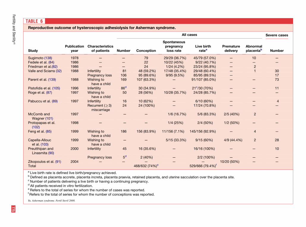
Severe cases
Prematuredelivery
Abnormalplacentab Number
) — 10 —) — — —) — 2) — 1 30) — 17) — — 73
— — 11) — — —
— — 4) — — —
) 2/5 (40%) 2 —
1/2 (50%) — —
) — 4 —
4/9 (44.4%) 2 28
— — 10
— — —10/20 (50%) — —
)f
lacenta site.
TABLE 6Reproductive outcome of hysteroscopic adhesiolysis for Asherman syndrome.
All cases
StudyPublication
yearCharacteristics
of patients Number Conception
Spontaneouspregnancyloss rate
Live birthratea
Sugimoto (138) 1978 — — 79 29/29 (36.7%) 45/79 (57.0%Fedele et al. (84) 1986 — — 22 10/22 (45%) 9/22 (40.7%Friedman et al.(82) 1986 — — 24 1/24 (4.2%) 23/24 (95.8%Valle and Sciarra (32) 1988 Infertility 81 48 (59.2%) 17/48 (35.4%) 29/48 (60.4%
Pregnancy loss 106 95 (89.6%) 9/95 (9.5%) 85/95 (89.5%Parent et al. (139) 1988 Wishing to
have a child169 107 (63.3%) — 91/107 (85.0%
Pistofidis et al. (105) 1996 Infertility 86d 30 (34.9%) — 21c/30 (70%)Roge et al. (87) 1997 Wishing to
have a child50 28 (56%) 10/28 (35.7%) 24/28 (85.7%
Pabuccu et al. (89) 1997 Infertility 16 10 (62%) — 6/10 (60%)Recurrent (R3)
miscarriage24 24 (100%) — 17/24 (70.8%
McComb andWagner (101)
1997 — — — 1/6 (16.7%) 5/6 (83.3%
Protopapas et al.(102)
1998 — — — 1/4 (25%) 2/4 (50%)
Feng et al. (85) 1999 Wishing tohave a child
186 156 (83.9%) 11/156 (7.1%) 145/156 (92.9%
Capella-Alloucet al. (103)
1999 Wishing tohave a child
— — 5/15 (33.3%) 9/15 (60%)
Preutthipan andLinasmita (90)
2000 Infertility 45 16 (35.6%) — 16/16 (100%)
Pregnancy loss 5d 2 (40%) — 2/2 (100%)Zikopoulos et al. (91) 2004 — — — — —Total 468/632 (74%)e 529/666 (79.4%
a Live birth rate is defined live birth/pregnancy achieved.b Defined as placenta accrete, placenta increta, placenta praevia, retained placenta, and uterine sacculation over the pc Number of patients delivering a live birth or having a continuing pregnancy.d All patients received in vitro fertilization.e Refers to the total of series for whom the number of cases was reported.f Refers to the total of series for whom the number of conceptions was reported.
Yu. Asherman syndrome. Fertil Steril 2008.
Fertilityand
Sterility
�775
Continued collection of data is required to determine if themiscarriage rate after treatment of Asherman syndrome is in-creased. This increased rate could be related to the presenceof fibrosed endometrium, which impairs successful implanta-tion. Premature delivery has also been reported to be morecommon in women with Asherman syndrome (see Table 6).Pregnancies after hysteroscopic lysis of adhesions for Asher-man syndrome may also be complicated by placenta accreta(111) and placenta increta (85). Other complications, such asretained placenta (85) and uterine sacculation over the pla-centa site (82), have also been reported. Yasmin and Adeghe(141) reported a case of severe early-onset IUGR at 29weeks’ pregnancy after hysteroscopic adhesiolysis; IUGRmay be a consequence suboptimal placentation and hencecompromised placenta perfusion during pregnancy. Uterinerupture has been described after hysteroscopic surgery forAsherman syndrome, and it is undoubtedly related to theweakened and scarred uterine wall after surgery (142–146),especially if perforation of the uterus was sustained duringthe procedure (147, 148).
Thus, pregnancies in women with a history of Ashermansyndrome should be considered to be high risk. Careful mon-itoring during the antenatal period, especially the third tri-mester, should be undertaken.
CONCLUSION
Asherman syndrome is a worldwide disease. One century af-ter the condition was first reported, a lot of progress has beenmade in the investigation and treatment of the condition. Hys-teroscopy has been the method of choice in the investigationand treatment of the condition. The management of moderateto severe disease still poses a challenge, and the prognosis ofsevere disease remains poor. Repeat surgery may be neces-sary in some cases but may not always produce the desiredoutcome. In those who succeed in achieving pregnancy aftertreatment of the condition, careful surveillance of the preg-nancy is essential because a number of obstetrics complica-tions may occur. Future research should focus on thecellular and molecular aspects of endometrial tissue regener-ation as well as the prevention of postsurgical adhesion for-mation and reformation.
REFERENCES1. Fritsch H. Ein Fall von volligen Schwund der Gebaumutterhohle nach
Auskratzung. Zentralbl Gynaekol 1894;18:1337–42.
2. Bass B. Ueber die Verwachsungen in der cervix uterinach curettagen.
Zentralbl Gynakol 1927;51:223.
3. Stamer S. Partial and total atresia of the uterus after excochleation. Acta
Obstet Gynecol Scand 1946;26:263–97.
4. Asherman JG. Amenorrhoea traumatica (atretica). J Obstet Gynaecol
Br Emp 1948;55:23–30.
5. Asherman JG. Traumatic intrauterine adhesions. J Obstet Gynaecol Br
Emp 1950;57:892–6.
6. Asherman JG. Traumatic intrauterine adhesions and their effects on fer-
tility. Int J Fertil 1957;2:49–54.
7. Asherman JG. Les adherences intrauterines. Rev Fr Gynecol Obstet
1961;56:311.
8. Jones WE. Traumatic intrauterine adhesions. A report of 8 cases with
emphasis on therapy. Am J Obstet Gynecol 1964;89:304–13.
776 Yu et al. Asherman syndrome
9. Smid I, Bedo T. Curettage during puerperium and its late consequences
[article in German]. Zentralbl Gynakol 1978;100:916–20.
10. Taylor PJ, Cumming DC, Hill PJ. Significance of intrauterine adhesions
detected hysteroscopically in eumenorrheic infertile women and role of
antecedent curettage in their formation. Am J Obstet Gynecol
1981;139:239–42.
11. Adoni A, Palti Z, Milwidsky A, Dolberg M. The incidence of intrauter-
ine adhesions following spontaneous abortion. Int J Fertil 1982;27:
117–8.
12. Stillman RJ, Asarkof N. Association between Mullerian duct malfor-
mations and Asherman syndrome in infertile women. Obstet Gynecol
1985;65:673–7.
13. Marty R. Hysteroscopy necessary before IUD? Acta Eur Fertil 1986;17:
449–50.
14. Vutyavanich T, Rugpao S, Uttavichai C, Tovanabutra S, Ittipankul W.
Secondary amenorrhea at Maharaj Nakhon Chiang Mai Hospital.
J Med Assoc Thai 1989;72:160–6.
15. Golan A, Schneider D, Avrech O, Raziel A, Bukovsky I, Caspi E. Hys-
teroscopic findings after missed abortion. Fertil Steril 1992;58:508–10.
16. Friedler S, Margalioth EJ, Kafka I, Yaffe H. Incidence of post-abortion
intra-uterine adhesions evaluated by hysteroscopy—a prospective
study. Hum Reprod 1993;8:442–4.
17. Raziel A, Arieli S, Bukovsky I, Caspi E, Golan A. Investigation of the
uterine cavity in recurrent aborters. Fertil Steril 1994;62:1080–2.
18. Romer T. Post-abortion-hysteroscopy—a method for early diagnosis of
congenital and acquired intrauterine causes of abortions. Eur J Obstet
Gynecol Reprod Biol 1994;57:171–3.
19. La Sala GB, Montanari R, Dessanti L, Cigarini C, Sartori F. The role of
diagnostic hysteroscopy and endometrial biopsy in assisted reproduc-
tive technologies. Fertil Steril 1998;70:378–80.
20. Westendorp IC, Ankum WM, Mol BW, Vonk J. Prevalence of Asher-
man’s syndrome after secondary removal of placental remnants or a re-
peat curettage for incomplete abortion. Hum Reprod 1998;13:3347–50.
21. Tam WH, Lau WC, Cheung LP, Yuen PM, Chung TK. Intrauterine ad-
hesions after conservative and surgical management of spontaneous
abortion. J Am Assoc Gynecol Laparosc 2002;9:182–5.
22. Nawroth F, Foth D, Schmidt T. Minihysteroscopy as routine diagnostic
procedure in women with primary infertility. J Am Assoc Gynecol Lap-
arosc 2003;10:396–8.
23. Preutthipan S, Linasmita V. A prospective comparative study between hys-
terosalpingography and hysteroscopy in the detection of intrauterine pa-
thology in patients with infertility. J Obstet Gynaecol Res 2003;29:33–7.
24. Hinckley MD, Milki AA. 1000 office-based hysteroscopies prior to in
vitro fertilization: feasibility and findings. JSLS 2004;8:103–7.
25. Ventolini G, Zhang M, Gruber J. Hysteroscopy in the evaluation of pa-
tients with recurrent pregnancy loss: a cohort study in a primary care
population. Surg Endosc 2004;18:1782–4.
26. Yucebilgin MS, Aktan E, Bozkurt K, Kazandi M, Akercan F, Mgoyi L,
Terek MC. Comparison of hydrosonography and diagnostic hystero-
scopy in the evaluation of infertile patients. Clin Exp Obstet Gynecol
2004;31:56–8.27. Dalton VK, Saunders NA, Harris LH, Williams JA, Lebovic DI. Intra-
uterine adhesions after manual vacuum aspiration for early pregnancy
failure. Fertil Steril 2006;85:1823.e1–3.28. Schenker JG, Margalioth EJ. Intrauterine adhesions: an updated ap-
praisal. Fertil Steril 1982;37:593–610.29. Chapman K, Chapman R. Asherman’s syndrome: a review of the liter-
ature, and a husband and wife’s 20-year world-wide experience. J R Soc
Med 1990;83:576–80.
30. Toaff R, Ballas S. Traumatic hypomenorrhea-amenorrhea (Asherman’s
syndrome). Fertil Steril 1978;30:379–87.
31. March CM, Israel R, March AD. Hysteroscopic management of intra-
uterine adhesions. Am J Obstet Gynecol 1978;130:653–7.
32. Valle RF, Sciarra JJ. Intrauterine adhesions: hysteroscopic diagnosis,
classification, treatment, and reproductive outcome. Am J Obstet Gyne-
col 1988;158:1459–70.
33. Donnez J, Nisolle M. Hysteroscopic lysis of intrauterine adhesions
(Asherman’s syndrome). In: Donnez J, ed. Atlas of laser operative lap-
aroscopy and hysteroscopy. New York: Press-Parthenon, 1994:305–22.
Vol. 89, No. 4, April 2008
34. The American Fertility Society classifications of adnexal adhesions,
distal tubal occlusion, tubal occlusion secondary to tubal ligation, tubal
pregnancies, M€ullerian anomalies and intrauterine adhesions. Fertil
Steril 1988;49:944–55.
35. Wamsteker K, De Block S. Diagnostic hysteroscopy: technique and
documentation. In: Sutton C, Diamond M, eds. Endoscopic surgery
for gynecologists. London: WB Saunders, 1998:511–24.
36. Nasr AL, Al-Inany HG, Thabet SM, Aboulghar M. A clinicohystero-
scopic scoring system of intrauterine adhesions. Gynecol Obstet Invest
2000;50:178–81.
37. Badawy SZ, Orbuch L, Khurana KK. Secondary amenorrhea with se-
vere intrauterine adhesions and chronic uterine torsion after cesarean
section in a teenage girl. J Pediatr Adolesc Gynecol 1998;11:93–6.
38. Taskin O, Sadik S, Onoglu A, Gokdeniz R, Erturan E, Burak F,
Wheeler JM. Role of endometrial suppression on the frequency of intra-
uterine adhesions after resectoscopic surgery. J Am Assoc Gynecol
Laparosc 2000;7:351–4.
39. Davies C, Gibson M, Holt EM, Torrie EP. Amenorrhoea secondary to
endometrial ablation and Asherman’s syndrome following uterine ar-
tery embolization. Clin Radiol 2002;57:317–8.
40. Roman H, Sentilhes L, Cingotti M, Verspyck E, Marpeau L. Uterine de-
vascularization and subsequent major intrauterine synechiae and ovar-
ian failure. Fertil Steril 2005;83:755–7.
41. Leung PL, Tam WH, Yuen PM. Hysteroscopic appearance of the endo-
metrial cavity following thermal balloon endometrial ablation. Fertil
Steril 2003;79:1226–8.
42. Mukul LV, Linn JG. Pregnancy complicated by uterine synechiae after
endometrial ablation. Obstet Gynecol 2005;105:1179–82.
43. Taskin O, Onoglu A, Inal M, Turan E, Sadik S, Vardar E, Postaci H,
Wheeler JM. Long-term histopathologic and morphologic changes after
thermal endometrial ablation. J Am Assoc Gynecol Laparosc 2002;9:
186–90.
44. Rabau E, David A. Intrauterine adhesions: etiology, prevention, and
treatment. Obstet Gynecol 1963;22:626–9.
45. Czernobilsky B. Endometritis and infertility. Fertil Steril 1978;30:
119–30.
46. Jensen PA, Stromme WB. Amenorrhea secondary to puerperal curet-
tage (Asherman’s syndrome). Am J Obstet Gynecol 1972;113:150–7.
47. Polishuk WZ, Anteby SO, Weinstein D. Puerperal endometritis and in-
trauterine adhesions. Int Surg 1975;60:418–20.
48. Smid A, Borsos A, Takacs I. Etiology of Asherman’s syndrome (intrauter-
ine synechia) [article in German]. Zentralbl Gynakol 1980;102:380–5.
49. Shaffer W. Role of uterine adhesions in the cause of multiple pregnancy
losses. Clin Obstet Gynecol 1986;29:912–24.
50. Netter AP, Musset R, Lambert A, Salomon Y. Traumatic uterine syne-
chiae: a common cause of menstrual insufficiency, sterility, and abor-
tion. Am J Obstet Gynecol 1956;71:368–75.
51. Klein SM, Garcia CR. Asherman’s syndrome: a critique and current re-
view. Fertil Steril 1973;24:722–35.52. Buckley CH. Normal endometrium and non-proliferative conditions of
the endometrium. In: Fox H, Wells M, eds. Obstetrical and gynaecolog-
ical pathology. 5th ed. London: Churchill Livingstone, 2002:391–442.53. Al-Inany H. Intrauterine adhesions; an update. Acta Obstet Gynecol
Scand 1998;80:986–93.
54. Yaffe H, Ron M, Polishuk WZ. Amenorrhoea, hypomenorrhoea and
uterine fibrosis. Am J Obstet Gynecol 1978;130:599–601.
55. March CM. Update: Intrauterine adhesions. Fertil News 1996;19(1):
Forum.
56. McCulloch TA, Wagner B, Duffy S, Barik S, Smith JH. The pathology
of hysterectomy specimens following trans-cervical resection of the en-
dometrium. Histopathology 1995;27:541–7.57. Silvernagel SW, Harshbarger KE, Shevlin DW. Postoperative granulo-
mas of the endometrium: histological features after endometrial abla-
tion. Ann Diagn Pathol 1997;1:82–90.
58. Clark IW. Necrotizing granulomatous inflammation of the uterine body
following diathermy ablation of the endometrium. Pathology 1992;24:
32–3.
59. Letteries GS. Structural abnormalities of the uterus: intrauterine adhe-
sions. In: Letterie GS, ed. Structural abnormalities and reproductive
Fertility and Sterility�
failure: effective techniques for diagnosis and management. Malden,
MA: Blackwell Science, 1998:251–73.
60. Polishuk WZ, Sadovsky E. A syndrome of recurrent intrauterine adhe-
sions. Am J Obstet Gynecol 1975;123:151–8.
61. Hare AA, Olah KS. Pregnancy following endometrial ablation: a review
article. J Obstet Gynaecol 2005;25:108–14.
62. Forssman L. Posttraumatic intrauterine synechiae and pregnancy. Ob-
stet Gynecol 1965;26:710–3.
63. Dicker D, Feldberg D, Samuel N, Goldman JA. Etiology of cervical
pregnancy. Association with abortion, pelvic pathology, IUDs and
Asherman’s syndrome. J Reprod Med 1985;30:25–7.
64. Klyszejko C, Bogucki J, Klyszejko D, Ilnicki W, Donotek S, Kozma J.
Cervical pregnancy in Asherman’s syndrome [article in Polish]. Gine-
kol Pol 1987;58:46–8.
65. Xia E, Li TC, Yu D, Huang X, Zheng J, Liu Y, Zhang M. The occurrence
and outcome of 39 pregnancies after 1621 cases of transcervical resec-
tion of endometrium. Hum Reprod 2006;21:3282–6.
66. Sandridge DA, Councell RB, Thorp JM. Endometrial carcinoma arising
within extensive intrauterine synechiae. Eur J Obstet Gynecol Reprod
Biol 1994;56:147–9.
67. Wamsteker K. Intrauterine adhesions (synechiae). In: Brosens I,
Wamsteker K, eds. Diagnostic imaging and endoscopy in gynecology:
a practical guide. London: WB Saunders, 1997:171–84.
68. Fayez JA, Mutie G, Schneider PJ. The diagnostic value of hysterosal-
pingography and hysteroscopy in infertility investigation. Am J Obstet
Gynecol 1987;156:558–60.
69. Soares SR, Barbosa dos Reis MM, Camargos AF. Diagnostic accuracy of
sonohysterography, transvaginal sonography, and hysterosalpingography
in patients with uterine cavity diseases. Fertil Steril 2000;73:406–11.
70. Confino E, Friberg J, Giglia RV, Gleicher N. Sonographic imaging of
intrauterine adhesions. Obstet Gynecol 1985;66:596–8.
71. Schlaff WD, Hurst BS. Preoperative sonographic measurement of endo-
metrial pattern predicts outcome of surgical repair in patients with se-
vere Asherman’s syndrome. Fertil Steril 1995;63:410–3.
72. Salle B, Gaucherand P, de Saint Hilaire P, Rudigoz RC. Transvaginal
sonohysterographic evaluation of intrauterine adhesions. J Clin Ultra-
sound 1999;27:131–4.
73. Sylvestre C, Child TJ, Tulandi T, Tan SL. A prospective study to eval-
uate the efficacy of two- and three-dimensional sonohysterography in
women with intrauterine lesions. Fertil Steril 2003;79:1222–5.
74. Pal A, Babinszki A, Vajda G, Kovacs L. Diagnosis of Asherman’s syn-
drome with three-dimensional ultrasound. Ultrasound Obstet Gynecol
2000;15:341–3.
75. Parsons AK, Lense JJ. Sonohysterography for endometrial abnormali-
ties: preliminary results. J Clin Ultrasound 1993;21:87–95.
76. Rudigoz RC, Salle B, Piacenza JM, de Saint-Hilaire P, Gaucherand P.
Hysterosonographic study of the uterine cavity [article in French]. J Gy-
necol Obstet Biol Reprod (Paris) 1995;24:697–704.
77. Dykes TA, Isler RJ, McLean AC. MR imaging of Asherman syndrome:
total endometrial obliteration. J Comput Assist Tomogr 1991;15:
858–60.
78. Letterie GS, Haggerty MF. Magnetic resonance imaging of intrauterine
synechiae. Gynecol Obstet Invest 1994;37:66–8.
79. Bacelar AC, Wilcock D, Powell M, Worthington BS. The value of MRI
in the assessment of traumatic intra-uterine adhesions (Asherman’s syn-
drome). Clin Radiol 1995;50:80–3.80. Al-Inany H. Intrauterine adhesions. An update. Acta Obstet Gynecol
Scand 2001;80:986–93.81. Taylor PJ, Hamou JE. Hysteroscopy in infertility and repeated preg-
nancy loss. In: Gordon AG, Lewis BV, DeCherney AH, eds. Atlas of
gynecologic endoscopy. London: Mosby-Wolfe, 1995:162.82. Friedman A, DeFazio J, DeCherney A. Severe obstetric complications
after aggressive treatment of Asherman syndrome. Obstet Gynecol
1986;67:864–7.83. Reddy S, Rock JA. Surgical management of complete obliteration of the
endometrial cavity. Fertil Steril 1997;67:172–4.
84. Fedele L, Vercellini P, Viezzoli T, Ricciardiello O, Zamberletti D. Intra-
uterine adhesions: current diagnostic and therapeutic trends. Acta Eur
Fertil 1986;17:31–7.
777

85. Feng ZC, Yang B, Shao J, Liu S. Diagnostic and therapeutic hystero-
scopy for traumatic intrauterine adhesions after induced abortions:
clinical analysis of 365 cases. Gynaecol Endosc 1999;8:95–8.
86. Duffy S, Reid PC, Sharp F. In-vivo studies of uterine electrosurgery. Br
J Obstet Gynaecol 1992;99:579–82.
87. Roge P, Cravello L, D’Ercole C, Broussse M, Boubli L, Blanc B. Intra-
uterine adhesions and fertility: results of hysteroscopic treatment.
Gynaecol Endosc 1997;6:225–8.
88. Chen FP, Soong YK, Hui YL. Successful treatment of severe uterine
synechiae with transcervical resectoscopy combined with laminaria
tent. Hum Reprod 1997;12:943–7.
89. Pabuccu R, Atay V, Orhon E, Urman B, Ergun A. Hysteroscopic treat-
ment of intrauterine adhesions is safe and effective in the restoration of
normal menstruation and fertility. Fertil Steril 1997;68:1141–3.
90. Preutthipan S, Linasmita V. Reproductive outcome following hystero-
scopic lysis of intrauterine adhesions: a result of 65 cases at Ramathi-
bodi Hospital. J Med Assoc Thai 2000;83:42–6.
91. Zikopoulos KA, Kolibianakis EM, Platteau P, de Munck L, Tournaye H,
Devroey P, Camus M. Live delivery rates in subfertile women with
Asherman’s syndrome after hysteroscopic adhesiolysis using the resec-
toscope or the Versapoint system. Reprod Biomed Online 2004;8:720–5.
92. Chervenak FA, Neuwirth RS. Hysteroscopic resection of the uterine
septum. Am J Obstet Gynecol 1981;141:351–3.
93. DeCherney A, Polan ML. Hysteroscopic management of intrauterine
lesions and intractable uterine bleeding. Obstet Gynecol 1983;61:
392–7.
94. Cararach M, Penella J, Ubeda A, Labastida R. Hysteroscopic incision of
the septate uterus: scissors versus resectoscope. Hum Reprod 1994;9:
87–9.
95. Newton JR, MacKenzie WE, Emens MJ, Jordan JA. Division of uterine
adhesions (Asherman’s syndrome) with the Nd-YAG laser. Br J Obstet
Gynaecol 1989;96:102–4.
96. Chapman R, Chapman K. The value of two stage laser treatment for se-
vere Asherman’s syndrome. Br J Obstet Gynaecol 1996;103:1256–8.
97. Broome JD, Vancaillie TG. Fluoroscopically guided hysteroscopic di-
vision of adhesions in severe Asherman syndrome. Obstet Gynecol
1999;93:1041–3.
98. Gleicher N, Pratt D, Levrant S, Rao R, Balin M, Karande V. Gynaecor-
adiological uterine resection. Hum Reprod 1995;10:1801–3.
99. Karande V, Levrant S, Hoxsey R, Rinehart J, Gleicher N. Lysis of intra-
uterine adhesions using gynecoradiologic techniques. Fertil Steril
1997;68:658–62.
100. Coccia ME, Becattini C, Bracco GL, Pampaloni F, Bargelli G, Scarselli G.
Pressure lavage under ultrasound guidance: a new approach for outpatient
treatment of intrauterine adhesions. Fertil Steril 2001;75:601–6.
101. McComb PF, Wagner BL. Simplified therapy for Asherman’s syn-
drome. Fertil Steril 1997;68:1047–50.
102. Protopapas A, Shushan A, Magos A. Myometrial scoring: a new tech-
nique for the management of severe Asherman’s syndrome. Fertil Steril
1998;69:860–4.
103. Capella-Allouc S, Morsad F, Rongieres-Bertrand C, Taylor S,
Fernandez H. Hysteroscopic treatment of severe Asherman’s syndrome
and subsequent fertility. Hum Reprod 1999;14:1230–3.
104. Bukulmez O, Yarali H, Gurgan T. Total corporal synechiae due to tuber-
culosis carry a very poor prognosis following hysteroscopic synechial-
ysis. Hum Reprod 1999;14:1960–1.
105. Pistofidis GA, Dimitropoulos K, Mastrominas M. Comparison of oper-
ative and fertility outcome between groups of women with intrauterine
adhesions after adhesiolysis. J Am Assoc Gynecol Laparosc
1996;3(Suppl):S40.
106. Shalev E, Shimoni Y, Peleg D. Ultrasound controlled operative hystero-
scopy. J Am Coll Surg 1994;179:70–1.
107. Coccia ME, Becattini C, Bracco GL, Bargelli G, Scarselli G. Intraoper-
ative ultrasound guidance for operative hysteroscopy. A prospective
study. J Reprod Med 2000;45:413–8.
108. Massouras HG. Intrauterine adhesions: a syndrome of the past with the
use of the Massouras duck’s foot no. 2 intrauterine contraceptive device.
Am J Obstet Gynecol 1973;116:576–8.
778 Yu et al. Asherman syndrome
109. Jewelewicz R, Khalaf S, Neuwirth RS, Vande Wiele RL. Obstetric com-
plications after treatment of intrauterine synechiae (Asherman’s
syndrome). Obstet Gynecol 1976;47:701–5.
110. Polishuk WZ, Weinstein D. The Soichet intrauterine device in the treat-
ment of intrauterine adhesions. Acta Eur Fertil 1976;7:215–8.
111. March CM. Intrauterine adhesions. Obstet Gynecol Clin North Am
1995;22:491–505.
112. Polishuk WZ, Kohane S. Intrauterine adhesions: diagnosis and therapy.
Obstet Gynecol Digest 1966;8:41.
113. Vesce F, Jorizzo G, Bianciotto A, Gotti G. Use of the copper intrauterine
device in the management of secondary amenorrhea. Fertil Steril
2000;73:162–5.
114. Comninos AC, Zourlas PA. Treatment of uterine adhesions (Asher-
man’s syndrome). Am J Obstet Gynecol 1969;105:862–8.
115. Orhue AA, Aziken ME, Igbefoh JO. A comparison of two adjunctive
treatments for intrauterine adhesions following lysis. Int J Gynaecol
Obstet 2003;82:49–56.
116. Amer MI, El Nadim A, Hassanein K. The role of intrauterine balloon
after operative hysteroscopy in the prevention of intrauterine adhesion:
a prospective controlled study. MEFS J 2005;10:125–9.
117. Amer MI, Abd-El-Maeboud KH. Amnion graft following hystero-
scopic lysis of intrauterine adhesions. J Obstet Gynaecol Res 2006;32:
559–66.
118. Tsapanos VS, Stathopoulou LP, Papathanassopoulou VS, Tzingounis VA.
The role of Seprafilm bioresorbable membrane in the prevention and
therapy of endometrial synechiae. J Biomed Mater Res 2002;63:
10–4.
119. Acunzo G, Guida M, Pellicano M, Tommaselli GA, Di Spiezio Sardo A,
Bifulco G, et al. Effectiveness of auto-cross-linked hyaluronic acid gel
in the prevention of intrauterine adhesions after hysteroscopic adhesiol-
ysis: a prospective, randomized, controlled study. Hum Reprod
2003;18:1918–21.
120. Guida M, Acunzo G, Di Spiezio Sardo A, Bifulco G, Piccoli R,
Pellicano M, et al. Effectiveness of auto-crosslinked hyaluronic acid
gel in the prevention of intrauterine adhesions after hysteroscopic sur-
gery: a prospective, randomized, controlled study. Hum Reprod
2004;19:1461–4.
121. Burns JW, Skinner K, Colt J, Sheidlin A, Bronson R, Yaacobi Y,
Goldberg EP. Prevention of tissue injury and postsurgical adhesions
by precoating tissues with hyaluronic acid solutions. J Surg Res
1995;59:644–52.
122. De Iaco PA, Stefanetti M, Pressato D, Piana S, Dona M, Pavesio A,
Bovicelli L. A novel hyaluronan-based gel in laparoscopic adhesion
prevention: preclinical evaluation in an animal model. Fertil Steril
1998;69:318–23.
123. Mensitieri M, Ambrosio L, Nicolais L, Bellini D, O’Regan M. Visco-
elastic properties modulation of a novel auto-cross-linked hyaluronic
acid polymer. J Mater Sci Mater Med 1996;7:695–8.
124. Wood J, Pena G. Treatment of traumatic uterine synechias. Int J Fertil
1964;9:405–10.
125. Farhi J, Bar-Hava I, Homburg R, Dicker D, Ben-Rafael Z. Induced re-
generation of endometrium following curettage for abortion: a compar-
ative study. Hum Reprod 1993;8:1143–4.
126. Wolman I, Gordon D, Yaron Y, Kupferminc M, Lessing JB, Jaffa AJ.
Transvaginal sonohysterography for the evaluation and treatment of
retained products of conception. Gynecol Obstet Invest 2000;50:
73–6.
127. Alcazar JL. Transvaginal ultrasonography combined with color veloc-
ity imaging and pulsed Doppler to detect residual trophoblastic tissue.
Ultrasound Obstet Gynecol 1998;11:54–8.
128. Achiron R, Goldenberg M, Lipitz S, Mashiach S. Transvaginal duplex
Doppler ultrasonography in bleeding patients suspected of having resid-
ual trophoblastic tissue. Obstet Gynecol 1993;81:507–11.
129. Goldenberg M, Schiff E, Achiron R, Lipitz S, Mashiach S. Managing
residual trophoblastic tissue. Hysteroscopy for directing curettage. J
Reprod Med 1997;42:26–8.
130. World Health Organisation. Menstrual regulation by intramuscular in-
jections of 16-phenoxy-tetranor PGE2 methyl sulfonylamide or vacuum
Vol. 89, No. 4, April 2008
aspiration. A randomized multicentre study. World Health Organisation
Task Force on Post-ovulatory Methods for Fertility Regulation. Br J Ob-
stet Gynaecol 1987;94:949–56.
131. Fonseca W, Misago C, Kanji N. Misoprostol plus mifepristone. Lancet
1991;338:1594.
132. Misoprostol and legal medical abortion. Lancet 1991;338:1241–2.
133. Baird DT, Norman JE, Thong KJ, Glasier AF. Misoprostol, mifepris-
tone, and abortion. Lancet 1992;339:313.
134. Tang OS, Chan CC, Ng EH, Lee SW, Ho PC. A prospective, random-
ized, placebo-controlled trial on the use of mifepristone with sublingual
or vaginal misoprostol for medical abortions of less than 9 weeks ges-
tation. Hum Reprod 2003;18:2315–8.
135. Trinder J, Brocklehurst P, Porter R, Read M, Vyas S, Smith L. Manage-
ment of miscarriage: expectant, medical, or surgical? Results of rando-
mised controlled trial (Miscarriage Treatment (MIST) trial). BMJ
2006;332:1235–40.
136. Katz Z, Ben-Arie A, Lurie S, Manor M, Insler V. Reproductive outcome
following hysteroscopic adhesiolysis in Asherman’s syndrome. Int J
Fertil Menopausal Stud 1996;41:462–5.
137. Pace S, Stentella P, Catania R, Palazzetti PL, Frega A. Endoscopic treat-
ment of intrauterine adhesions. Clin Exp Obstet Gynecol 2003;30:26–8.
138. Sugimoto O. Diagnostic and therapeutic hysteroscopy for traumatic in-
trauterine adhesions. Am J Obstet Gynecol 1978;131:539–47.
139. Parent B, Barbot J, Dubuisson JB. Syn�echies ut�erine encyclop�edie med-
ico-chirurgicale. Gyn�ecologie 1988;140A:10.
Fertility and Sterility�
140. Everett C. Incidence and outcome of bleeding before the 20th week of
pregnancy: prospective study from general practice. BMJ 1997;315:32–4.
141. Yasmin H, Adeghe JH. Severe early-onset intrauterine growth restric-
tion (IUGR) in a woman with Asherman’s syndrome. J Obstet Gynaecol
2004;24:312–4.
142. Deaton JL, Maier D, Andreoli J Jr. Spontaneous uterine rupture during
pregnancy after treatment of Asherman’s syndrome. Am J Obstet Gyne-
col 1989;160:1053–4.
143. Hulka JF. Uterine rupture after treatment of Asherman’s syndrome. Am
J Obstet Gynecol 1990;162:1352–3.
144. Shiau CS, Hsieh CC, Chiang CH, Hsieh TT, Chang MY. Intrapartum
spontaneous uterine rupture following uncomplicated resectoscopic
treatment of Asherman’s syndrome. Chang Gung Med J 2005;28:123–7.
145. Tannous W, Hamon J, Henry-Suchet J, Achard B, Lelaidier C, Belaisch-
Allart J. Ruptures ut�erines lors d’ accouchements apr�es hyst�eroscopy
op�eratoire. Press Med 1996;25:159–61.
146. ChokriA, Chekib M, FethiZ, Anis F, Sadok M. Rupture ut�erine autroisi�eme
apr�es m�etroplastie hyst�eroscopique. Tunis Med 2000;78:527–9.
147. Gurgan T, Yarali H, Urman B, Dagli V, Dogan L. Uterine rupture fol-
lowing hysteroscopic lysis of synechiae due to tuberculosis and uterine
perforation. Hum Reprod 1996;11:291–3.
148. Sentilhes L, Sergent F, Roman H, Verspyck E, Marpeau L. Late compli-
cations of operative hysteroscopy: predicting patients at risk of uterine
rupture during subsequent pregnancy. Eur J Obstet Gynecol Reprod
Biol 2005;120:134–8.
779