Accusandi necessitas incumbet domino servum suum accusatio ...
Upload
vuongthienCategory
view
225download
0
INFECTION AND IMMUNITY, Dec. 2006, p. 6632–6641 Vol. 74, No. 120019-9567/06/$08.00�0 doi:10.1128/IAI.00720-06
Ascaris suum-Derived Products Suppress Mucosal Allergic Inflammationin an Interleukin-10-Independent Manner via Interference with
Dendritic Cell Function�
Brittany W. McConchie,1* Hillary H. Norris,1 Virgilio G. Bundoc,1 Shweta Trivedi,1Agnieszka Boesen,1 Joseph F. Urban, Jr.2 and Andrea M. Keane-Myers1
Laboratory of Allergic Diseases, National Institute of Allergy and Infectious Diseases, National Institutes of Health, Rockville,Maryland 20852,1 and Nutrient Requirements and Functions Laboratory, Beltsville Human Nutrition Research Center,
Agricultural Research Service, United States Department of Agriculture, Beltsville, Maryland 207052
Received 4 May 2006/Returned for modification 7 June 2006/Accepted 2 September 2006
We have previously demonstrated that protection from allergic inflammation by Ascaris suum infection wascharacterized by a global increase in interleukin-10 (IL-10) and the development of protective CD4�/CD25�
T cells (L. Schopf, S. Luccioli, V. Bundoc, P. Justice, C. C. Chan, B. J. Wetzel, H. H. Norris, J. F. Urban, Jr.,and A. Keane-Myers, Investig. Ophthalmol. Vis. Sci. 46:2772–2780, 2005). Here, we used A. suum pseudocoe-lomic fluid (PCF) in lieu of infection to define molecular mechanisms of allergic protection in a mouse modelof allergic inflammation. Mice were sensitized with ragweed (RW) and PCF (RW/PCF), PCF alone, or RWalone and then challenged intratracheally, intranasally, and supraocularly with RW. Histological examinationof the eyes and lungs, analysis of the bronchoalveolar lavage fluid (BALF), and characterization of ex vivocytokine responses were performed to determine allergic inflammatory responses. RW/PCF-treated mice hadsuppressed allergic immune responses compared to mice given RW alone. To investigate whether IL-10 wasinvolved in PCF-mediated allergic protection, similar experiments were performed using mice geneticallydeficient for IL-10. Persistent protection from allergic disease was observed in the absence of IL-10, indicatingthe primary mechanism of PCF protection is IL-10 independent. Ex vivo and in vitro analysis of PCF-treateddendritic cells (DC) demonstrated reduced activation receptor expression and cytokine production in responseto either RW or lipopolysaccharide stimulation. These findings extend previous studies that showed infectionwith A. suum alters expression of allergic disease and suggest that PCF can contribute to this effect byinterference with DC function.
Helminth infection is a potent activator of Th2 responses inthe host (20) and is generally characterized by increased num-bers of mast cells, eosinophils, and goblet cells as well assecretion of Th2 cytokines and immunoglobulin E (IgE) (36).Allergies are characterized by a similar Th2 profile in atopicindividuals (19). In subjects from countries where helminthinfection is endemic, there is a paradoxically lower incidence ofallergic disease than in subjects from countries where the in-fections are less prevalent (34). This suggests that helminthinfection may provide some protection from the developmentof allergic inflammation.
Epidemiologic studies examining the influence of helminthinfection on allergic disease have demonstrated an inverserelationship between the longevity and magnitude of parasiteinfection and the development of the allergic response (3, 31).For example, asthmatic patients infected with Schistosomamansoni were shown to have lower levels of the proallergiccytokines interleukin-4 (IL-4) and IL-5 than their noninfectedcounterparts when challenged with house dust mite antigen(4). Separate studies have suggested that parasites might di-rectly suppress the allergic response, as anti-helminthic treat-ment of parasite-infected individuals correlates with increased
allergen-specific cutaneous anaphylaxis and IgE antibody pro-duction (21, 32). The mechanism by which parasite-derivedfactors can inhibit allergic inflammation, however, remainsspeculative.
These studies explore the interrelationship between Ascarisinfection and allergic disease. Ascaris infection affects over 1.4billion people worldwide, and infection with this parasite hasbeen implicated in altering the allergic response either posi-tively by acting as an adjuvant to increase disease or negativelyby acting to suppress the allergic response (5). We previouslyestablished, using a mouse model of allergic conjunctivitis, thatacute helminth infection with A. suum leads to enhanced al-lergic disease (27). Conversely, these studies also demon-strated that chronic infection with A. suum provided protectionfrom subsequent allergic exposure. Protection was character-ized by a global increase in IL-10, which has been associatedwith CD4�/CD25� T-regulatory cells. Transfer of CD4�/CD25� T cells from mice chronically infected with A. suum torecipient mice sensitized with either ragweed (RW) and acuteA. suum infection or RW alone resulted in a reduced responseto subsequent RW challenge, suggesting that A. suum-inducedCD4�/CD25� T cells could directly reduce allergic disease tononparasite allergens.
Infection with A. suum, as with most helminth infections,involves multiple stages of larval development in host organsduring the migratory phase. The complex life cycle of theparasite complicates the therapeutic use of infective A. suum,
* Corresponding author. Mailing address: Twinbrook II Room 125,12441 Parklawn Drive, Rockville, MD 20852. Phone: (301) 402-0992.Fax: (301) 480-1207. E-mail: [email protected].
� Published ahead of print on 11 September 2006.
6632
on March 30, 2019 by guest
http://iai.asm.org/
Dow
nloaded from
as pulmonary fibrosis can develop in the lung tissue traversedby the migratory phase of the larvae (10, 16). In order tocharacterize the molecular basis of helminth modulation ofallergic disease and to avoid the pathology associated withlarval migration, we evaluated the activity of A. suum-derivedproducts in a murine model of allergic inflammation.Pseudocoelomic fluid (PCF) was selected because it is a met-abolically active fluid that is easily obtained from the bodycavity of adult worms, and it contains a number of parasiteantigens that induce antibody responses in several differentmammalian species, including mice (15, 30). The main proteinconstituent of PCF is ABA-1, which represents the major IgE-binding component in patients with ascariasis (14, 25).
Recent studies have employed a variety of A. suum extractsto explore the relationship between infection and allergic in-flammation; however, there has been considerable variation inboth the preparation of the extracts and the inflammatorymodel used (17, 29). Souza et al. (28) compared Ascaris ex-tracts from adult worms and eggs and found similar inhibitoryprofiles for allergic inflammation, suggesting that allergic inhi-bition is not parasite stage specific and likely involves numer-ous parasite antigens. In agreement with our chronic A. suuminfection model (27), Paterson et al. found that an extract ofAscaris body fluid mixed with ovalbumin (OVA) (25) increasedIL-10 production and the development T-regulatory cells.These Ascaris extract-induced T-regulatory cells were found tosuppress delayed type hypersensitivity to nonparasite antigens(6, 25) but were not tested in type 1 allergic responses involvingmucosal sites as examined here.
In the present studies, we sensitized mice with either PCFconcurrent with the nonparasite antigens RW (RW/PCF) orOVA (OVA/PCF), PCF alone, RW alone, or OVA alone.Mice were subsequently challenged with the appropriate aller-gen (RW or OVA) and assessed for development of allergicconjunctivitis and/or asthma. PCF suppressed the developmentof allergic inflammation compared to control mice given aller-gen sensitization alone. Experiments using IL-10-deficientmice under similar conditions showed persistent protectionfrom the development of allergy, indicating that PCF couldprotect in the absence of IL-10. Additional ex vivo and in vitrostudies of PCF-treated dendritic cells (DC) demonstrated thatPCF was able to suppress DC activation marker expression andcytokine production following lipopolysaccharide (LPS) stim-ulation. These findings suggest that PCF contains potent DC-inhibitory molecules that represent some, but not all, of themechanisms of allergic suppression induced by infection withA. suum.
MATERIALS AND METHODS
Animals. A/J mice were obtained from Harlan Laboratories (Indianapolis,IN), and BALB/c and C57BL/6 mice were obtained from the National CancerInstitute. Mice were housed and maintained in the Comparative MedicineBranch at a National Institute of Allergy and Infectious Diseases (NIAID)/National Institutes of Health (NIH) animal facility (Rockville, MD). IL-10-deficient mice on a C57BL/6 background were obtained from the NIH Reposi-tory at Taconic (Germantown, NY) (11). The studies reported here conform tothe principles for laboratory animal research outlined by the Animal Welfare Act(NIH/Department of Health and Human Services) guidelines for the experimen-tal use of animals and were approved by the NIAID Animal Care and UseCommittee. Each group contained 5 to 10 mice, and experiments were per-formed a minimum of three separate times.
PCF sensitization and RW challenge protocol. On days 0 and 5, mice weresensitized systemically via a 200-�l intraperitoneal (i.p.) injection containingeither 50 �g RW extract (Greer Laboratories, Lenoir, N.C.) (RW), 100 �g PCF(obtained from adult A. suum worms that tested negative for endotoxin) (PCF),50 �g RW plus 100 �g PCF (RW/PCF), or phosphate-buffered saline (PBS)emulsified in an equal volume mixture with alum (Pierce Laboratories, Rock-ford, IL). The mice were subsequently challenged on days 14 and 15 with 1 mgRW extract in PBS dropwise in the eye (5 �l/eye) (18, 27). Control mice werechallenged with PBS. For assessment of pulmonary inflammation, mice were alsochallenged with 50 �g of RW or PBS control intratracheally (i.t.) on day 14 andintranasally (i.n.) on day 15. Mice were sacrificed 72 h after the final challenge toevaluate conjunctival eosinophil infiltration, cellular inflammation in the lung,and cytokine levels in the sera and bronchoalveolar lavage fluid (BALF). In allcases, the mice were deeply anesthetized with an i.p. injection of ketamine (FortDodge Animal Health, Fort Dodge, Iowa)-xylazine (Phoenix Pharmaceuticals,St. Joseph, MO) (100 mg/kg of body weight and 10 mg/kg, respectively) andexsanguinated.
PCF sensitization and OVA challenge protocol. On days 0 and 5, five to eightBALB/c mice per group were sensitized systemically via a 200-�l i.p. injectioncontaining either 100 �g ovalbumin (OVA) (Sigma-Aldrich), 100 �g PCF (PCF),100 �g OVA plus 100 �g PCF (OVA/PCF), or PBS emulsified in an equal-volume mixture with alum (Pierce Laboratories) to measure allergic lung inflam-matory responses to a model nonpollen allergen. Mice were challenged intra-tracheally and intranasally using OVA (50 �g) and were exsanguinated afteranesthesia 72 h after the final challenge to evaluate lung pathology and cellularinflammation.
BALF. Immediately after exsanguination, lungs were cannulated with a 20-gauge intravenous catheter and gently washed once with 500 �l 1% fetal bovineserum (FBS) (HyClone, Logan, UT) in PBS (for cytokine analysis) or twice with750 �l 1% FBS in PBS (for analysis of cellular infiltration). Samples for cytokineanalysis were stored at �80°C. Samples for cellular analysis were prepared as acytospin (Thermo-Shandon, Pittsburg, PA) for differential cellular analysis afterstaining with Kwik-diff (Thermo-Shandon), and a portion was used to determinetotal cell counts.
Histology. To assess cellular infiltration in the conjunctiva and surroundingtissue, the eyes and lids were removed intact and immediately fixed in 10%neutral buffered formalin (EMD Chemicals, Gibbstown, NJ). Fixed tissues weresent to American Histolabs (Gaithersburg, MD) for slide preparation with par-affin embedding and staining with either Giemsa or hematoxylin and eosin forvisualization of cellular inflammation. Quantitative cell counts were made on five400� nonoverlapping fields per eyelid section and five sections per treatmentgroup using slides that were masked to the reader (27). To assess pulmonaryinflammation, lungs were lavaged for BALF and removed 72 h after the finalallergen challenge. After removal, lungs were immediately placed in 10% neutralbuffered formalin. Lung tissues were sent to Histoserv (Germantown, MD),embedded in paraffin, and stained with hematoxylin and eosin for visualization ofcellular inflammation and periodic acid Schiff for visualization of mucus-con-taining goblet cells. Slides were masked to the reader, and a minimum of fivelungs per group were scored with a ranking of 0 to 4 based on the level ofperibronchial cuffing (PBC), perivascular cuffing (PVC), goblet cell hyperplasia,and interstitial inflammation. A score of 0 is normal lung with no inflammationor obvious increases in goblet cells in the bronchioles and alveolar spaces, 1 is alung with minor PVC, 2 is a lung with moderate PVC and PBC cuffing, 3 is a lungwith increased PVC and PBC with evidence of goblet cells in the smaller airways,and 4 is a lung with severe PVC and PBC, goblet cell hyperplasia, and interstitialinflammation (2).
Antigen-specific in vitro assays. Spleens were removed, cells were made intosingle-cell suspensions, and red blood cells were lysed by treatment with ACKlysis buffer per the manufacturer’s instructions (Biosource, Camarillo, CA). Theremaining cells were then cultured in one of the following: medium alone as anegative control, medium and 2.5 �g/ml concanavalin A (Sigma) as a positivecontrol, or medium and 50 �g/ml of RW extract. Medium used was composed ofRPMI 1640 with Glutamax, gentamicin (10 mg/ml), 1 M HEPES, �-mercapto-ethanol (Invitrogen, Carlsbad, CA), and 10% FBS (HyClone, Logan, UT). Atotal of 2 � 106 splenocytes/well were plated in a 24-well plate at a volume of 2ml/well. Supernatants were harvested after 48 h in culture and immediatelyfrozen at �80°C prior to analysis of cytokines
Quantitative measurement of cytokines by luminex technology. CytokinesIL-4, IL-5, IL-13, and gamma interferon (IFN-�) were measured using theBiosource Multiplex Assay kit (Biosource International, Camarillo, CA) per themanufacturer’s instructions. All samples, including standards, were assayed induplicate. Fluorescence was measured by using the Liquichip reader (QIAGEN,Valencia, CA).
VOL. 74, 2006 A. SUUM PRODUCTS SUPPRESS ALLERGIC INFLAMMATION 6633
on March 30, 2019 by guest
http://iai.asm.org/
Dow
nloaded from
BMDC assays. Bone marrow dendritic cells (BMDC) were prepared fromfemur and tibia after removing the bone marrow cells by flushing with Hank’sbalanced salt solution supplemented with 1% HEPES (Invitrogen, Grand Island,NY). To induce DC development, 1 � 106 bone marrow cells/ml were culturedin complete RPMI 1640 medium (10% FCS, 10 mg/ml gentamicin, 0.1% �-mer-captoethanol, 1% HEPES in the presence of 40 ng/ml of murine recombinantgranulocyte-macrophage colony-stimulating factor; PeproTech, Rocky Hill, NJ).On day 4, supernatants were gently removed and fresh medium was addedtogether with 40 ng/ml granulocyte-macrophage colony-stimulating factor and 10ng/ml of murine recombinant IL-4 (PeproTech) to the remaining semiadherentcells. The cells were harvested on day 6, seeded in 24-well plates, and stimulatedwith 100 �g/ml of PCF. After 6 h of stimulation with PCF, 20 ng/ml of LPS wasadded, and the cells were stimulated for additional 5 h. The cells and thesupernatants were subsequently harvested for fluorescent-activated cell sorter(FACS) staining and cytokine analysis, respectively.
Ex vivo analysis of CD11c� dendritic cells. Mice were sensitized systemicallyvia a 200-�l i.p. injection containing either 50 �g RW extract (Greer Laborato-ries) (RW), 100 �g PCF (PCF), 50 �g RW plus 100 �g PCF (RW/PCF), or PBS(PBS). All products and controls were emulsified 1:1 in an equal (100 �l)-volumemixture with alum (Pierce Laboratories). Mice were sacrificed at either 24 h or6 days postsensitization, injected i.p. with 6 ml PBS, and gently lavaged. Addi-tionally, a group of naı̈ve mice was sacrificed, injected i.p. with 6 ml PBS, andlavaged as controls for cellular infiltration in response to alum alone. Peritoneallavage cells were centrifuged (300 relative centrifugal force for 10 min); theresultant pellet was resuspended in 1 ml PBS with 2% FBS. A 100-�l aliquot ofeach sample for cellular analysis was prepared as a cytospin (Thermo-Shandon)for differential cellular analysis after staining with Kwik-diff (Thermo-Shandon),and an additional 50-�l portion was used to determine total cell counts. Theremaining cells were stained for CD11c as well as the costimulatory markers
CD86, CD40, and major histocompatibility complex (MHC) class II for analysisby FACS.
Flow-cytometric analysis (FACS). Peritoneal lavage cells and BMDC werestained for CD11c as well as the costimulatory markers CD86, CD40, and MHCclass II to determine cellular activation (clones HL-3, GL-1, HM40-3, and 39-10-8, respectively; BD Biosciences). FACS analysis was performed on CD11c�
gated cells to determine the expression of costimulatory markers on dendriticcells. FACS analysis was performed using a FACS Calibur (BDBiosciences)using CellQuest software. Data analysis was performed using FlowJo software5.7.2 (Tree Star, Ashland, OR).
Data analysis. Data are summarized as means � standard errors. One-wayanalysis of variance with Tukey’s multiple comparison posttest was performedusing GraphPad Prism version 4.00 for Windows (GraphPad Software, SanDiego, California). The statistical significance value was set at P � 0.05.
RESULTS
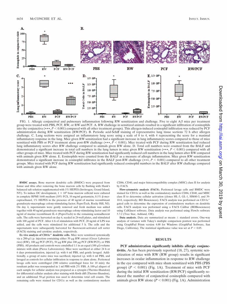
PCF administration significantly inhibits allergic conjunc-tivitis. As has been previously reported (18, 27), systemic sen-sitization of mice with RW (RW group) results in significantincreases in ocular inflammation in response to RW challengein the eye compared with mice sham sensitized with PBS (PBSgroup) (P � 0.001) (Fig. 1A). Treatment of mice with PCFduring the initial RW sensitization (RW/PCF) significantly re-duced the number of conjunctival eosinophils compared withanimals given RW alone (P � 0.001) (Fig. 1A). Administration
FIG. 1. Allergic conjunctival and pulmonary inflammation following RW sensitization and challenge. Five to eight A/J mice per treatmentgroup were treated with PBS, PCF, RW, or RW and PCF. A. RW challenge in sensitized animals resulted in a significant infiltration of eosinophilsinto the conjunctiva (***, P � 0.001) compared with all other treatment groups). This allergen-induced eosinophil infiltration was reduced by PCFadministration during RW sensitization (RW/PCF). B. Periodic acid-Schiff staining of representative lung tissue sections 72 h after allergenchallenge. C. Lung sections were assigned an inflammatory lung score using a scale of 0 to 4, with 4 representing the score for a maximalinflammatory response in the lung. Mice given RW sensitization had a significant increase in lung inflammatory scores compared to those of micesensitized with PBS or PCF treatment alone post-RW challenge (***, P � 0.001). Mice treated with PCF during RW sensitization had reducedlung inflammatory scores after RW challenge compared to animals given RW alone. D. Total cell numbers were counted from the BALF anddemonstrated a significant increase in total cell numbers in the lung lumen in mice given RW sensitization (***, P � 0.001) compared with allother groups of mice. Mice treated with PCF during RW sensitization had significantly reduced cell numbers in the lung lumen after RW comparedwith animals given RW alone. E. Eosinophils were counted from the BALF as a measure of allergic inflammation. Mice given RW sensitizationdemonstrated a significant increase in eosinophil infiltrates in the BALF post-RW challenge (***, P � 0.001) compared to all other treatmentgroups. Mice treated with PCF during RW sensitization had significantly reduced eosinophil numbers in the BALF after RW challenge comparedwith animals given RW alone.
6634 MCCONCHIE ET AL. INFECT. IMMUN.
on March 30, 2019 by guest
http://iai.asm.org/
Dow
nloaded from
of PCF alone did not significantly increase eosinophil numbersabove PBS controls (P 0.05).
PCF administration significantly inhibits allergen-inducedpulmonary eosinophilia. PBS (Fig. 1B, far-left panel)- andPCF (Fig. 1B, right-center panel)-sensitized control mice hadlittle response to RW challenge. Conversely, RW challenge insensitized mice resulted in significant amounts of goblet cellhyperplasia, interstitial inflammation, and perivascular andperibronchiolar cuffing with an infiltration of lymphocytesand eosinophils. RW/PCF-treated mice had significant re-ductions in pulmonary inflammation compared with animalsgiven RW alone (Fig. 1B, left-center panel), resulting insignificantly increased lung inflammatory scores over whatwas observed in PBS-sensitized controls (P � 0.001) (Fig.1C). Systemic administration of PCF during the initial RWsensitization significantly reduced overall airway pathology(Fig. 1B, far-right panel) compared with animals given RWalone (P � 0.05) (Fig. 1C).
Total cell numbers in the BALF were significantly increasedafter RW sensitization and challenge (P � 0.001) (Fig. 1D).Administration of PCF during RW sensitization, however, sig-nificantly reduced the total cellular number in the BALF afterRW challenge compared to untreated RW-sensitized and-challenged mice (P � 0.001) (Fig. 1D). PCF administrationalone did not alter the total cell number compared with PBS-treated control mice (P 0.05). Eosinophil numbers weremeasured in the BALF as an indicator of allergic inflammatoryresponses in the lungs. The numbers of eosinophils were sig-
nificantly increased in the BALF of the RW-sensitized and-challenged animals compared with PBS control mice (P �0.001) (Fig. 1E). This number was significantly reduced in micegiven systemic PCF during RW sensitization (P � 0.001) (Fig.1E). PCF administration alone did not significantly increasethe number of eosinophils in the BALF compared with PBScontrol animals (P 0.05). These findings combined with thestudies assessing the role of PCF in the prevention of allergiceye disease suggest that systemic administration of PCF mayprotect from allergic disease at multiple sites of allergen chal-lenge.
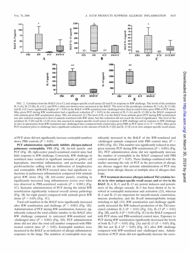
PCF treatment decreases allergen-induced Th2 cytokine lev-els in in vitro antigen-specific recall assays and ex vivo in theBALF. IL-4, IL-5, and IL-13 are potent inducers and potenti-ators of the allergic cascade. IL-5 has been shown to be in-volved in eosinophil maturation and activation (23), whereasIL-4 and IL-13 are important for smooth-muscle constriction,mucous production, and the B-cell immunoglobulin isotypeswitching to IgE (33). RW sensitization and challenge signifi-cantly increased the RW-induced production of the Th2-asso-ciated cytokines IL-5 (P � 0.01) (Fig. 2A), IL-13 (P � 0.01)(Fig. 2B), and IL-4 (P � 0.05) (Fig. 2C) in the BALF comparedwith PCF alone and PBS-sensitized control mice. Exposure toPCF during RW sensitization significantly decreased the levelsof both IL-5 (P � 0.05) (Fig. 2A) and IL-13 (P � 0.05) (Fig.2B) but not IL-4 (P 0.05) (Fig. 2C) after RW challengecompared with RW-sensitized and -challenged mice. Admin-istration of PCF alone did not significantly increase the secre-
FIG. 2. Cytokines from the BALF (A to C) and antigen-specific recall assays (D and E) in response to RW challenge. The levels of the cytokinesIL-5 (A), IL-13 (B), IL-4 (C), and IFN-� (data not shown) were measured in the BALF. The level of the proallergic cytokines IL-5 (A), IL-13 (B),and IL-4 (C) were significantly higher (P � 0.05) in the BALF of RW-sensitized and -challenged mice than in control mice given PBS or PCF alone.Mice given PCF during RW sensitization had a significant reduction (P � 0.05) in the amount of IL-5 (A) and IL-13 (B) in the BALF comparedwith animals given RW sensitization alone. ND, not detected. (C) The level of IL-4 in the BALF from animals given PCF during RW sensitizationwas also reduced compared to that of animals sensitized with RW alone, but this reduction did not reach the level of significance. The level of thecytokines IL-5 (D) and IL-13 (E) were also assessed in antigen-specific recall assays in response to RW challenge and in response to RW challengein vitro in splenocytes from RW-sensitized and -challenged mice compared with control mice given PBS or PCF alone (***, P � 0.001). Mice givenPCF treatment prior to challenge had a significant reduction in the amount of both IL-5 (D) and IL-13 (E) in in vitro antigen-specific recall assays.
VOL. 74, 2006 A. SUUM PRODUCTS SUPPRESS ALLERGIC INFLAMMATION 6635
on March 30, 2019 by guest
http://iai.asm.org/
Dow
nloaded from
tion of IL-5 (P 0.05) (Fig. 2A), IL-13 (P 0.05) (Fig. 2B),or IL-4 (P 0.05) (Fig. 2C in the BALF compared with controlmice treated with PBS.
In vitro antigen-specific recall assays were performed onsplenocytes taken from all four in vivo treatment groups (PBS,RW, PCF, and RW/PCF). Splenocyte cultures from mice inthe RW-alone group produced significantly more IL-5 (P �0.001) (Fig. 2D) and IL-13 (P � 0.001) (Fig. 2E) in response toRW stimulation in vitro than spleen cells from mice in thePCF-alone and PBS control groups. PCF treatment duringRW sensitization in vivo suppressed the production of IL-5(P � 0.001) (Fig. 2D) and IL-13 (P � 0.001) (Fig. 2E) inRW-induced spleen cell assays in vitro. In vivo treatment withPCF alone did not alter the in vitro production of either IL-5or IL-13 above PBS control animals.
In order to determine if the decrease in Th2-associatedcytokines was due to a simultaneous increase in Th2-blockingTh1 cytokines, the levels of IFN-� were also assessed in theBALF and in vitro antigen-specific splenocyte recall assays.The levels of IFN-� in response to RW challenge were belowthe level of detection for the multiplex assay in all cases. Allgroups of cells in the in vitro recall assays, however, did pro-duce IFN-� in response to the mitogen concanavalin A as apositive control (data not shown). Taken together, these re-sults suggest that the decrease in Th2 cytokines was not due toa concurrent increase in Th1 cytokines.
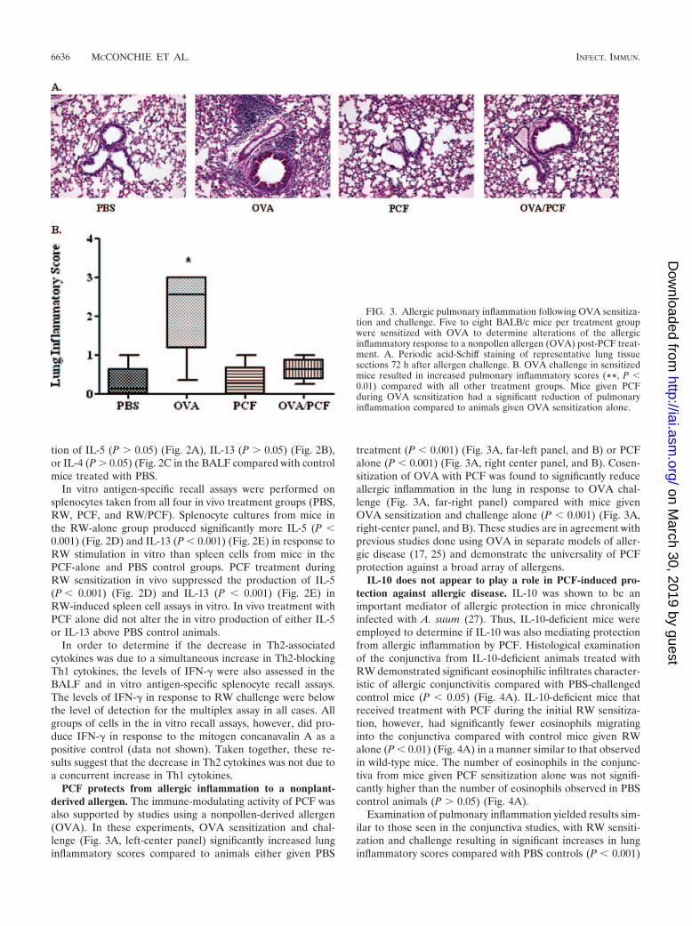
PCF protects from allergic inflammation to a nonplant-derived allergen. The immune-modulating activity of PCF wasalso supported by studies using a nonpollen-derived allergen(OVA). In these experiments, OVA sensitization and chal-lenge (Fig. 3A, left-center panel) significantly increased lunginflammatory scores compared to animals either given PBS
treatment (P � 0.001) (Fig. 3A, far-left panel, and B) or PCFalone (P � 0.001) (Fig. 3A, right center panel, and B). Cosen-sitization of OVA with PCF was found to significantly reduceallergic inflammation in the lung in response to OVA chal-lenge (Fig. 3A, far-right panel) compared with mice givenOVA sensitization and challenge alone (P � 0.001) (Fig. 3A,right-center panel, and B). These studies are in agreement withprevious studies done using OVA in separate models of aller-gic disease (17, 25) and demonstrate the universality of PCFprotection against a broad array of allergens.
IL-10 does not appear to play a role in PCF-induced pro-tection against allergic disease. IL-10 was shown to be animportant mediator of allergic protection in mice chronicallyinfected with A. suum (27). Thus, IL-10-deficient mice wereemployed to determine if IL-10 was also mediating protectionfrom allergic inflammation by PCF. Histological examinationof the conjunctiva from IL-10-deficient animals treated withRW demonstrated significant eosinophilic infiltrates character-istic of allergic conjunctivitis compared with PBS-challengedcontrol mice (P � 0.05) (Fig. 4A). IL-10-deficient mice thatreceived treatment with PCF during the initial RW sensitiza-tion, however, had significantly fewer eosinophils migratinginto the conjunctiva compared with control mice given RWalone (P � 0.01) (Fig. 4A) in a manner similar to that observedin wild-type mice. The number of eosinophils in the conjunc-tiva from mice given PCF sensitization alone was not signifi-cantly higher than the number of eosinophils observed in PBScontrol animals (P 0.05) (Fig. 4A).
Examination of pulmonary inflammation yielded results sim-ilar to those seen in the conjunctiva studies, with RW sensiti-zation and challenge resulting in significant increases in lunginflammatory scores compared with PBS controls (P � 0.001)
FIG. 3. Allergic pulmonary inflammation following OVA sensitiza-tion and challenge. Five to eight BALB/c mice per treatment groupwere sensitized with OVA to determine alterations of the allergicinflammatory response to a nonpollen allergen (OVA) post-PCF treat-ment. A. Periodic acid-Schiff staining of representative lung tissuesections 72 h after allergen challenge. B. OVA challenge in sensitizedmice resulted in increased pulmonary inflammatory scores (**, P �0.01) compared with all other treatment groups. Mice given PCFduring OVA sensitization had a significant reduction of pulmonaryinflammation compared to animals given OVA sensitization alone.
6636 MCCONCHIE ET AL. INFECT. IMMUN.
on March 30, 2019 by guest
http://iai.asm.org/
Dow
nloaded from
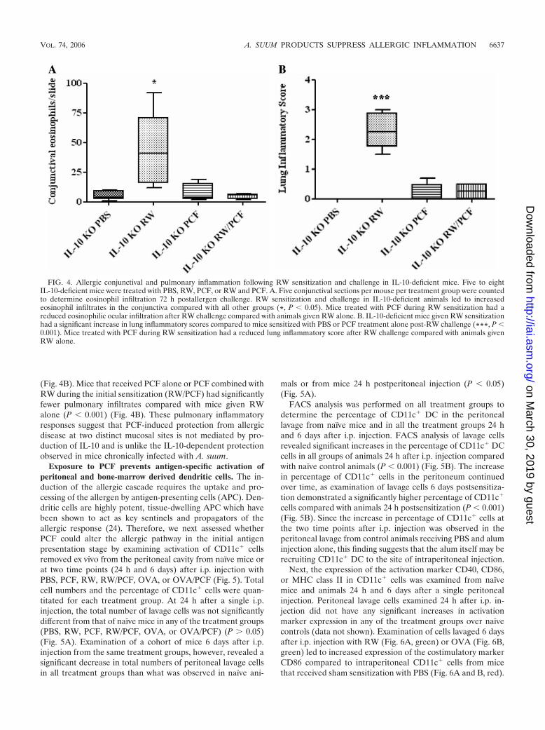
(Fig. 4B). Mice that received PCF alone or PCF combined withRW during the initial sensitization (RW/PCF) had significantlyfewer pulmonary infiltrates compared with mice given RWalone (P � 0.001) (Fig. 4B). These pulmonary inflammatoryresponses suggest that PCF-induced protection from allergicdisease at two distinct mucosal sites is not mediated by pro-duction of IL-10 and is unlike the IL-10-dependent protectionobserved in mice chronically infected with A. suum.
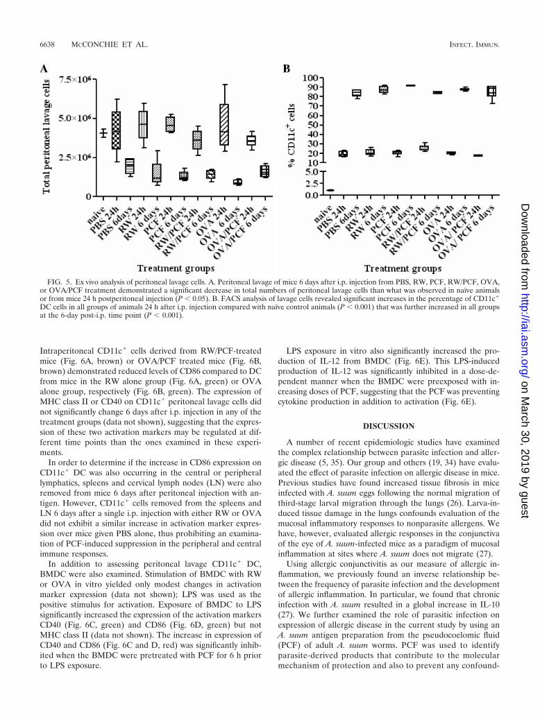
Exposure to PCF prevents antigen-specific activation ofperitoneal and bone-marrow derived dendritic cells. The in-duction of the allergic cascade requires the uptake and pro-cessing of the allergen by antigen-presenting cells (APC). Den-dritic cells are highly potent, tissue-dwelling APC which havebeen shown to act as key sentinels and propagators of theallergic response (24). Therefore, we next assessed whetherPCF could alter the allergic pathway in the initial antigenpresentation stage by examining activation of CD11c� cellsremoved ex vivo from the peritoneal cavity from naı̈ve mice orat two time points (24 h and 6 days) after i.p. injection withPBS, PCF, RW, RW/PCF, OVA, or OVA/PCF (Fig. 5). Totalcell numbers and the percentage of CD11c� cells were quan-titated for each treatment group. At 24 h after a single i.p.injection, the total number of lavage cells was not significantlydifferent from that of naı̈ve mice in any of the treatment groups(PBS, RW, PCF, RW/PCF, OVA, or OVA/PCF) (P 0.05)(Fig. 5A). Examination of a cohort of mice 6 days after i.p.injection from the same treatment groups, however, revealed asignificant decrease in total numbers of peritoneal lavage cellsin all treatment groups than what was observed in naı̈ve ani-
mals or from mice 24 h postperitoneal injection (P � 0.05)(Fig. 5A).
FACS analysis was performed on all treatment groups todetermine the percentage of CD11c� DC in the peritoneallavage from naı̈ve mice and in all the treatment groups 24 hand 6 days after i.p. injection. FACS analysis of lavage cellsrevealed significant increases in the percentage of CD11c� DCcells in all groups of animals 24 h after i.p. injection comparedwith naı̈ve control animals (P � 0.001) (Fig. 5B). The increasein percentage of CD11c� cells in the peritoneum continuedover time, as examination of lavage cells 6 days postsensitiza-tion demonstrated a significantly higher percentage of CD11c�
cells compared with animals 24 h postsensitization (P � 0.001)(Fig. 5B). Since the increase in percentage of CD11c� cells atthe two time points after i.p. injection was observed in theperitoneal lavage from control animals receiving PBS and aluminjection alone, this finding suggests that the alum itself may berecruiting CD11c� DC to the site of intraperitoneal injection.
Next, the expression of the activation marker CD40, CD86,or MHC class II in CD11c� cells was examined from naı̈vemice and animals 24 h and 6 days after a single peritonealinjection. Peritoneal lavage cells examined 24 h after i.p. in-jection did not have any significant increases in activationmarker expression in any of the treatment groups over naı̈vecontrols (data not shown). Examination of cells lavaged 6 daysafter i.p. injection with RW (Fig. 6A, green) or OVA (Fig. 6B,green) led to increased expression of the costimulatory markerCD86 compared to intraperitoneal CD11c� cells from micethat received sham sensitization with PBS (Fig. 6A and B, red).
FIG. 4. Allergic conjunctival and pulmonary inflammation following RW sensitization and challenge in IL-10-deficient mice. Five to eightIL-10-deficient mice were treated with PBS, RW, PCF, or RW and PCF. A. Five conjunctival sections per mouse per treatment group were countedto determine eosinophil infiltration 72 h postallergen challenge. RW sensitization and challenge in IL-10-deficient animals led to increasedeosinophil infiltrates in the conjunctiva compared with all other groups (*, P � 0.05). Mice treated with PCF during RW sensitization had areduced eosinophilic ocular infiltration after RW challenge compared with animals given RW alone. B. IL-10-deficient mice given RW sensitizationhad a significant increase in lung inflammatory scores compared to mice sensitized with PBS or PCF treatment alone post-RW challenge (***, P �0.001). Mice treated with PCF during RW sensitization had a reduced lung inflammatory score after RW challenge compared with animals givenRW alone.
VOL. 74, 2006 A. SUUM PRODUCTS SUPPRESS ALLERGIC INFLAMMATION 6637
on March 30, 2019 by guest
http://iai.asm.org/
Dow
nloaded from
Intraperitoneal CD11c� cells derived from RW/PCF-treatedmice (Fig. 6A, brown) or OVA/PCF treated mice (Fig. 6B,brown) demonstrated reduced levels of CD86 compared to DCfrom mice in the RW alone group (Fig. 6A, green) or OVAalone group, respectively (Fig. 6B, green). The expression ofMHC class II or CD40 on CD11c� peritoneal lavage cells didnot significantly change 6 days after i.p. injection in any of thetreatment groups (data not shown), suggesting that the expres-sion of these two activation markers may be regulated at dif-ferent time points than the ones examined in these experi-ments.
In order to determine if the increase in CD86 expression onCD11c� DC was also occurring in the central or peripherallymphatics, spleens and cervical lymph nodes (LN) were alsoremoved from mice 6 days after peritoneal injection with an-tigen. However, CD11c� cells removed from the spleens andLN 6 days after a single i.p. injection with either RW or OVAdid not exhibit a similar increase in activation marker expres-sion over mice given PBS alone, thus prohibiting an examina-tion of PCF-induced suppression in the peripheral and centralimmune responses.
In addition to assessing peritoneal lavage CD11c� DC,BMDC were also examined. Stimulation of BMDC with RWor OVA in vitro yielded only modest changes in activationmarker expression (data not shown); LPS was used as thepositive stimulus for activation. Exposure of BMDC to LPSsignificantly increased the expression of the activation markersCD40 (Fig. 6C, green) and CD86 (Fig. 6D, green) but notMHC class II (data not shown). The increase in expression ofCD40 and CD86 (Fig. 6C and D, red) was significantly inhib-ited when the BMDC were pretreated with PCF for 6 h priorto LPS exposure.
LPS exposure in vitro also significantly increased the pro-duction of IL-12 from BMDC (Fig. 6E). This LPS-inducedproduction of IL-12 was significantly inhibited in a dose-de-pendent manner when the BMDC were preexposed with in-creasing doses of PCF, suggesting that the PCF was preventingcytokine production in addition to activation (Fig. 6E).
DISCUSSION
A number of recent epidemiologic studies have examinedthe complex relationship between parasite infection and aller-gic disease (5, 35). Our group and others (19, 34) have evalu-ated the effect of parasite infection on allergic disease in mice.Previous studies have found increased tissue fibrosis in miceinfected with A. suum eggs following the normal migration ofthird-stage larval migration through the lungs (26). Larva-in-duced tissue damage in the lungs confounds evaluation of themucosal inflammatory responses to nonparasite allergens. Wehave, however, evaluated allergic responses in the conjunctivaof the eye of A. suum-infected mice as a paradigm of mucosalinflammation at sites where A. suum does not migrate (27).
Using allergic conjunctivitis as our measure of allergic in-flammation, we previously found an inverse relationship be-tween the frequency of parasite infection and the developmentof allergic inflammation. In particular, we found that chronicinfection with A. suum resulted in a global increase in IL-10(27). We further examined the role of parasitic infection onexpression of allergic disease in the current study by using anA. suum antigen preparation from the pseudocoelomic fluid(PCF) of adult A. suum worms. PCF was used to identifyparasite-derived products that contribute to the molecularmechanism of protection and also to prevent any confound-
FIG. 5. Ex vivo analysis of peritoneal lavage cells. A. Peritoneal lavage of mice 6 days after i.p. injection from PBS, RW, PCF, RW/PCF, OVA,or OVA/PCF treatment demonstrated a significant decrease in total numbers of peritoneal lavage cells than what was observed in naı̈ve animalsor from mice 24 h postperitoneal injection (P � 0.05). B. FACS analysis of lavage cells revealed significant increases in the percentage of CD11c�
DC cells in all groups of animals 24 h after i.p. injection compared with naı̈ve control animals (P � 0.001) that was further increased in all groupsat the 6-day post-i.p. time point (P � 0.001).
6638 MCCONCHIE ET AL. INFECT. IMMUN.
on March 30, 2019 by guest
http://iai.asm.org/
Dow
nloaded from
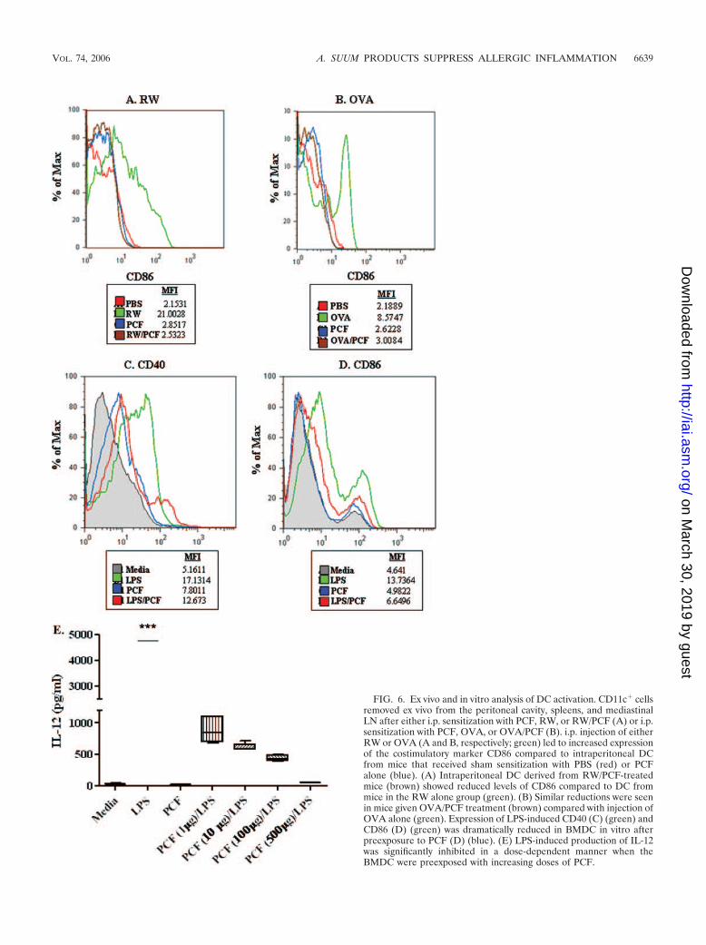
FIG. 6. Ex vivo and in vitro analysis of DC activation. CD11c� cellsremoved ex vivo from the peritoneal cavity, spleens, and mediastinalLN after either i.p. sensitization with PCF, RW, or RW/PCF (A) or i.p.sensitization with PCF, OVA, or OVA/PCF (B). i.p. injection of eitherRW or OVA (A and B, respectively; green) led to increased expressionof the costimulatory marker CD86 compared to intraperitoneal DCfrom mice that received sham sensitization with PBS (red) or PCFalone (blue). (A) Intraperitoneal DC derived from RW/PCF-treatedmice (brown) showed reduced levels of CD86 compared to DC frommice in the RW alone group (green). (B) Similar reductions were seenin mice given OVA/PCF treatment (brown) compared with injection ofOVA alone (green). Expression of LPS-induced CD40 (C) (green) andCD86 (D) (green) was dramatically reduced in BMDC in vitro afterpreexposure to PCF (D) (blue). (E) LPS-induced production of IL-12was significantly inhibited in a dose-dependent manner when theBMDC were preexposed with increasing doses of PCF.
VOL. 74, 2006 A. SUUM PRODUCTS SUPPRESS ALLERGIC INFLAMMATION 6639
on March 30, 2019 by guest
http://iai.asm.org/
Dow
nloaded from
ing effects from A. suum larva-induced tissue damage duringmigration. PCF is a metabolically active fluid that maintainshemostatic pressure in the worm and provides precursormolecules for membrane and cuticular synthesis and heme-containing proteins for oxidative metabolism (15). Some ofthese molecules are recognized as antigens by infected ro-dents, rabbits (15), and pigs (30) and include ABA-1, themajor IgE-binding antigen in patients that express resis-tance to ascariasis (25).
Administration of PCF into the peritoneum during the ini-tial allergen (RW) sensitization significantly reduced eosino-phil migration into the conjunctiva in response to allergenchallenge, which is similar to what was observed when micewere chronically infected with A. suum. In addition, a singlesystemic administration of PCF was able to significantly reducepulmonary eosinophilic inflammation and total lung pathologyin response to challenge with RW.
The immune-modulating activity of PCF was also supportedby studies using a nonpollen-derived allergen (OVA). A num-ber of researchers have begun exploring the relationship be-tween the coevolution of parasite infection and the develop-ment of allergic responses to common allergens (5, 19). Arecent study by Bielory et al. (personal communication) hasused epitope mapping technology to unmask conserved do-mains between a number of helminth-derived proteins andenvironmental allergens. These shared antigenic determi-nants could theoretically result in cross-reactivity betweenimmune responses of A. suum and RW. Therefore, we con-ducted a second series of experiments employing OVA sen-sitization and challenge as a representative model nonplantallergen in lieu of RW. We and others have found OVAdoes not easily elicit a response in mouse conjunctiva with-out additional manipulation (13); therefore, only pulmonaryinflammation was assessed in the OVA-induced allergy stud-ies. The combination of OVA and PCF was able to signifi-cantly suppress pulmonary responses to challenge with OVAcompared to mice sensitized and challenged with OVAalone. These findings suggest PCF is capable of suppressingthe allergic response to a wide range of traditional andatypical allergens at multiple tissue sites.
Ascaris infection significantly increases Th2 cytokine re-sponses in human and animal models (9, 22, 27). Conversely,PCF exposure during sensitization in RW-sensitized and -chal-lenged animals significantly decreases allergen-induced Th2cytokine responses in vivo. This decrease in Th2 cytokine pro-duction was likely not due to a concurrent increase in Th2-abrogating Th1 cytokines, as the levels of IFN-� were notincreased after PCF exposure compared to animals exposed toRW alone or PBS control.
Previous studies with an allergy protection model suggestedthat mice infected chronically with A. suum were protectedfrom nonparasite allergen-induced disease through IL-10 (25).IL-10 does not appear to contribute to the mechanism ofaction of PCF, since mice genetically deficient in IL-10 thatwere exposed to PCF during the initial allergen exposure wereprotected from allergic challenge. Transforming growth factor� (TGF-�) is an additional regulatory-associated cytokine ini-tially proposed to be protective in allergic disease (8, 24).However, studies in animals receiving PCF treatment (data notshown) did not detect increased TGF-� production after RW
challenge. Recent studies by Boxall et al. demonstrated thatTGF-� may contribute to airway remodeling in asthma (7),suggesting that TGF-� production may be detrimental ratherthan protective for allergic disease. These findings suggest thatthe role for TGF-� in allergic inflammation warrants furtherinvestigation.
Finally, ex vivo and in vitro studies were conducted todetermine if PCF was affecting APC activation by examiningDC activation marker expression. In these studies, the func-tional response of CD11c� DC removed ex vivo from theperitoneal cavity, spleen, and LN of PCF-exposed micepost-RW and -OVA exposure was examined. These studiesdemonstrated that although the total number of peritoneallavage cells decreased 6 days after peritoneal injection withantigen compared to either naı̈ve mice or mice examined24 h after i.p. injection, the percentage of CD11c� cellsincreased dramatically. These findings suggest either thatCD11c� cells were propagating in situ or that additional DCwere migrating to the site of antigen stimulation. In addi-tion, examination of activation markers on the CD11c� peri-toneal lavage cells examined 6 days after antigen stimulationrevealed that PCF suppressed the allergen-induced in-creased expression of the costimulatory molecule CD86 onCD11c� DC in the peritoneum. Further in vitro studiesusing BMDC demonstrated that PCF inhibited LPS-inducedexpression of the activation markers CD40 and CD86. Pre-exposure of BMDC with PCF also suppressed LPS-inducedproduction of IL-12 in a dose-dependent manner, suggestingthat PCF was inhibiting cytokine production in addition tocellular activation. Since LPS is a potent stimulator of DCactivation and cytokine production, this suppression wasespecially striking.
Whether PCF is inhibiting, down-regulating, or blockingToll-like receptors (TLR), or directly suppressing ligandprocessing by the DC is currently under investigation. Todate, there have been few clearly defined pathogen-acti-vated molecular patterns detected in A. suum. Other hel-minth parasites such as Schistosoma express molecules thathave been shown to act through TLR3, TLR4, TLR9, orTLR11 (1, 12). LPS-containing endotoxin is known as apotent activator of DC through TLR2 and TLR4 and is apossible contaminant of products derived from gastrointes-tinal parasites (24). Stimulation of these TLRs is known toinitiate a signal transduction cascade through the adaptormolecule MyD88 or TIRAP and TRIF, leading to an in-creased transcription of NF-B and the switching on of anumber of immune response genes (26). The PCF used inthese experiments, however, was determined by limulus as-says to have endotoxin levels below the level of detection,and exposure to PCF alone did not effect DC activation.This suggests that PCF may not be activating directlythrough either TLR2 or TLR4, although it does not rule outa role for PCF in inhibiting those receptors. We are plan-ning additional studies employing TLR-deficient mice to aidin the evaluation of TLR in our PCF-mediated protectionmodel.
These studies, in total, suggest that PCF may prevent thedevelopment of allergic disease by inhibiting appropriate APCactivation and initiation of the allergic cascade. The expressionof products by A. suum could potentially limit local allergic
6640 MCCONCHIE ET AL. INFECT. IMMUN.
on March 30, 2019 by guest
http://iai.asm.org/
Dow
nloaded from
reactions that contribute to a reduced host response to infec-tion, since migrating larvae express antigens found in PCF (13,28). We are currently conducting a number of experiments toverify these concepts and to better define the mechanisms ofPCF-induced regulation of APCs, influence on T-regulatorycells, and suppression of allergic inflammation.
ACKNOWLEDGMENTS
We thank Susan Pierce, Tom Wynn, and Marcus Hodges for theirreview of the manuscript and insightful suggestions. We also thankBrian Kelsall and Cecilia Johansson for their useful advice on dendriticcell assays.
Funding was provided by NIH/NIAID Intramural and USDA (CRIS1265-32000-064-00D).
REFERENCES
1. Aksoy, E., C. S. Zouain, F. Vanhoutte, J. Fontaine, N. Pavelka, N. Thieblemont,F. Willems, P. Ricciardi-Castagnoli, M. Goldman, M. Capron, B. Ryffel, and F.Trottein. 2005. Double-stranded RNAs from the helminth parasite Schistosomaactivate TLR3 in dendritic cells. J. Biol. Chem. 280:277–283.
2. Al-Shami, A., R. Spolski, J. Kelly, A. Keane-Myers, and W. J. Leonard. 2005.A role for TSLP in the development of inflammation in an asthma model. J.Exp. Med. 202:829–839.
3. Araujo, M. I., B. S. Hoppe, M. Medeiros, Jr., and E. M. Carvalho. 2004.Schistosoma mansoni infection modulates the immune response against al-lergic and auto-immune diseases. Mem. Inst. Oswaldo Cruz 99:27–32.
4. Araujo, M. I., A. A. Lopes, M. Medeiros, A. A. Cruz, L. Sousa-Atta, D. Sole,and E. M. Carvalho. 2000. Inverse association between skin response toaeroallergens and Schistosoma mansoni infection. Int. Arch. Allergy Immu-nol. 123:145–148.
5. Arruda, L. K., and A. B. Santos. 2005. Immunologic responses to commonantigens in helminthic infections and allergic disease. Curr. Opin. AllergyClin. Immunol. 5:399–402.
6. Boitelle, A., C. Di Lorenzo, H. E. Scales, E. Devaney, M. W. Kennedy, P.Garside, and C. E. Lawrence. 2005. Contrasting effects of acute and chronicgastro-intestinal helminth infections on a heterologous immune response ina transgenic adoptive transfer model. Int. J. Parasitol. 35:765–775.
7. Boxall, C., S. T. Holgate, and D. E. Davies. 2006. The contribution oftransforming growth factor-beta and epidermal growth factor signalling toairway remodelling in chronic asthma. Eur. Respir. J. 27:208–229.
8. Capron, A., D. Dombrowicz, and M. Capron. 2004. Helminth infections andallergic diseases: from the Th2 paradigm to regulatory networks. Clin. Rev.Allergy Immunol. 26:25–34.
9. Dawson, H. D., E. Beshah, S. Nishi, G. Solano-Aguilar, M. Morimoto, A.Zhao, K. B. Madden, T. K. Ledbetter, J. P. Dubey, T. Shea-Donohue, J. K.Lunney, and J. F. Urban, Jr. 2005. Localized multigene expression patternssupport an evolving Th1/Th2-like paradigm in response to infections withToxoplasma gondii and Ascaris suum. Infect. Immun. 73:1116–1128.
10. Eyre, P., D. H. Nymeyer, B. M. McCraw, and T. R. Deline. 1976. Protectionby acetylsalicylic acid and other agents in experimental acute interstitialpneumonia of calves. Vet. Rec. 98:64–66.
11. Hoffmann, K. F., S. L. James, A. W. Cheever, and T. A. Wynn. 1999. Studieswith double cytokine-deficient mice reveal that highly polarized Th1- andTh2-type cytokine and antibody responses contribute equally to vaccine-induced immunity to Schistosoma mansoni. J. Immunol. 163:927–938.
12. Jenkins, S. J., J. P. Hewitson, S. Ferret-Bernard, and A. P. Mountford.2005. Schistosome larvae stimulate macrophage cytokine productionthrough TLR4-dependent and -independent pathways. Int. Immunol. 17:1409–1418.
13. Kahn, M., N. P. Barney, R. M. Briggs, K. J. Bloch, and M. R. Allansmith.1990. Penetrating the conjunctival barrier. The role of molecular weight.Investig. Ophthalmol. Vis. Sci. 31:258–261.
14. Kennedy, M. W., E. M. Fraser, and J. F. Christie. 1991. MHC class II (I-A)region control of the IgE antibody repertoire to the ABA-1 allergen of thenematode Ascaris. Immunology 72:577–579.
15. Kennedy, M. W., and F. Qureshi. 1986. Stage-specific secreted antigens of
the parasitic larval stages of the nematode Ascaris. Immunology 58:515–522.
16. Liljegren, C. H., B. Aalbaek, O. L. Nielsen, and H. E. Jensen. 2003. Somenew aspects of the pathology, pathogenesis, and aetiology of disseminatedlung lesions in slaughter pigs. APMIS 111:531–538.
17. Lima, C., A. Perini, M. L. Garcia, M. A. Martins, M. M. Teixeira, and M. S.Macedo. 2002. Eosinophilic inflammation and airway hyper-responsivenessare profoundly inhibited by a helminth (Ascaris suum) extract in a murinemodel of asthma. Clin. Exp. Allergy 32:1659–1666.
18. Magone, M. T., S. M. Whitcup, A. Fukushima, C. C. Chan, P. B. Silver, andL. V. Rizzo. 2000. The role of IL-12 in the induction of late-phase cellularinfiltration in a murine model of allergic conjunctivitis. J. Allergy Clin.Immunol. 105:299–308.
19. Maizels, R. M. 2005. Infections and allergy - helminths, hygiene and hostimmune regulation. Curr. Opin. Immunol. 17:656–661.
20. Maizels, R. M., A. Balic, N. Gomez-Escobar, M. Nair, M. D. Taylor, and J. E.Allen. 2004. Helminth parasites–masters of regulation. Immunol. Rev. 201:89–116.
21. McKay, D. M. 2006. The beneficial helminth parasite? Parasitology 132:1–12.
22. McSharry, C., Y. Xia, C. V. Holland, and M. W. Kennedy. 1999. Naturalimmunity to Ascaris lumbricoides associated with immunoglobulin E anti-body to ABA-1 allergen and inflammation indicators in children. Infect.Immun. 67:484–489.
23. Mori, A., O. Kaminuma, K. Ogawa, A. Nakata, R. W. Egan, K. Akiyama, andH. Okudaira. 2000. Control of IL-5 production by human helper T cells as atreatment for eosinophilic inflammation: comparison of in vitro and in vivoeffects between selective and nonselective cytokine synthesis inhibitors. J.Allergy Clin. Immunol. 106:S58–S64.
24. O’Garra, A., L. Steinman, and K. Gijbels. 1997. CD4� T-cell subsets inautoimmunity. Curr. Opin. Immunol. 9:872–883.
25. Paterson, J. C., P. Garside, M. W. Kennedy, and C. E. Lawrence. 2002.Modulation of a heterologous immune response by the products of Ascarissuum. Infect. Immun. 70:6058–6067.
26. Pearce, E. J., C. M. Kane, and J. Sun. 2006. Regulation of dendritic cellfunction by pathogen-derived molecules plays a key role in dictating theoutcome of the adaptive immune response. Chem. Immunol. Allergy 90:82–90.
27. Schopf, L., S. Luccioli, V. Bundoc, P. Justice, C. C. Chan, B. J. Wetzel, H. H.Norris, J. F. Urban, Jr., and A. Keane-Myers. 2005. Differential modulationof allergic eye disease by chronic and acute ascaris infection. Investig. Oph-thalmol. Vis. Sci. 46:2772–2780.
28. Souza, V. M., E. L. Faquim-Mauro, and M. S. Macedo. 2002. Extracts ofAscaris suum egg and adult worm share similar immunosuppressive proper-ties. Braz. J. Med. Biol. Res. 35:81–89.
29. Souza, V. M., J. F. Jacysyn, and M. S. Macedo. 2004. IL-4 and IL-10 areessential for immunosuppression induced by high molecular weight proteinsfrom Ascaris suum. Cytokine 28:92–100.
30. Urban, J. F., Jr., H. Alizadeh, and R. D. Romanowski. 1988. Ascaris suum:development of intestinal immunity to infective second-stage larvae in swine.Exp. Parasitol. 66:66–77.
31. van den Biggelaar, A. H., C. Lopuhaa, R. van Ree, J. S. van der Zee, J. Jans,A. Hoek, B. Migombet, S. Borrmann, D. Luckner, P. G. Kremsner, and M.Yazdanbakhsh. 2001. The prevalence of parasite infestation and house dustmite sensitization in Gabonese schoolchildren. Int. Arch. Allergy Immunol.126:231–238.
32. van den Biggelaar, A. H., L. C. Rodrigues, R. van Ree, J. S. van der Zee, Y. C.Hoeksma-Kruize, J. H. Souverijn, M. A. Missinou, S. Borrmann, P. G.Kremsner, and M. Yazdanbakhsh. 2004. Long-term treatment of intestinalhelminths increases mite skin-test reactivity in Gabonese schoolchildren.J. Infect. Dis. 189:892–900.
33. Wills-Karp, M. 2004. Interleukin-13 in asthma pathogenesis. Immunol. Rev.202:175–190.
34. Wilson, M. S., and R. M. Maizels. 2006. Regulatory T cells induced byparasites and the modulation of allergic responses. Chem. Immunol. Allergy90:176–195.
35. Yazdanbakhsh, M., P. G. Kremsner, and R. van Ree. 2002. Allergy, parasites,and the hygiene hypothesis. Science 296:490–494.
36. Yazdanbakhsh, M., and L. C. Rodrigues. 2001. Allergy and the hygienehypothesis: the Th1/Th2 counterregulation can not provide an explanation.Wien Klin. Wochenschr. 113:899–902.
Editor: W. A. Petri, Jr.
VOL. 74, 2006 A. SUUM PRODUCTS SUPPRESS ALLERGIC INFLAMMATION 6641
on March 30, 2019 by guest
http://iai.asm.org/
Dow
nloaded from


![)JOEBXJ1VCMJTIJOH$PSQPSBUJPO ...con rmedinthemuscles,haemolymph,andreproductive system of A. suum []. TPS was isolated from muscles of A. suum and its properties have been detected.](https://static.fdocuments.us/doc/165x107/60e57c50dafc1611b11f9c61/joebxj1vcmjtijohpsqpsbujpo-con-rmedinthemuscleshaemolymphandreproductive.jpg)