Endoscopic mucosal resection - ASGE
12
TECHNOLOGY STATUS EVALUATION REPORT Endoscopic mucosal resection Prepared by: ASGE TECHNOLOGY COMMITTEE Joo Ha Hwang, MD, PhD, FASGE, Vani Konda, MD, Barham K. Abu Dayyeh, MD, MPH, Shailendra S. Chauhan, MD, FASGE, Brintha K. Enestvedt, MD, MBA, Larissa L. Fujii-Lau, MD, Sri Komanduri, MD, John T. Maple, DO, FASGE, Faris M. Murad, MD, Rahul Pannala, MD, MPH, Nirav C. Thosani, MD, Subhas Banerjee, MD, FASGE, Chair This document was reviewed and approved by the Governing Board of the American Society for Gastrointestinal Endoscopy. The ASGE Technology Committee provides reviews of existing, new, or emerging endoscopic technologies that have an impact on the practice of GI endoscopy. Evidence-based methodology is used, by using a MEDLINE literature search to identify pertinent clinical studies on the topic and a MAUDE (U.S. Food and Drug Administra- tion Center for Devices and Radiological Health) data- base search to identify the reported adverse events of a given technology. Both are supplemented by accessing the “related articles” feature of PubMed and by scruti- nizing pertinent references cited by the identified studies. Controlled clinical trials are emphasized, but in many cases, data from randomized, controlled trials are lack- ing. In such cases, large case series, preliminary clinical studies, and expert opinions are used. Technical data are gathered from traditional and Web-based publications, proprietary publications, and informal communications with pertinent vendors. For this review, the MEDLINE database was searched for publications in English through September 2014 by using the keywords “endo- scopic lesion removal,”“endoscopic resection,”“endo- scopic mucosal resection,” and “EMR.” Technology Status Evaluation Reports are drafted by 1 or 2 members of the ASGE Technology Committee, re- viewed and edited by the committee as a whole, and approved by the Governing Board of the ASGE. When financial guidance is indicated, the most recent coding data and list prices at the time of publication are pro- vided. Technology Status Evaluation Reports are scientific reviews provided solely for educational and informa- tional purposes. Technology Status Evaluation Reports are not rules and should not be construed as establishing a legal standard of care or as encouraging, advocating, requiring, or discouraging any particular treatment or payment for such treatment. BACKGROUND EMR was developed for minimally invasive, organ-sparing endoscopic removal of benign and early malignant lesions in the GI tract. This report focuses on instruments, injection solutions, and techniques currently used for EMR. This report is an update of a previous Technology Status Evalua- tion Report titled “Endoscopic Mucosal Resection and Endo- scopic Submucosal Dissection.” 1 The topic of endoscopic submucosal dissection (ESD) is now discussed in a separate Technology Status Report. 2 TECHNOLOGY UNDER REVIEW: EMR EMR is an endoscopic technique developed for the removal of sessile or flat neoplasms confined to the super- ficial layers (mucosa and submucosa) of the GI tract. The commonly used techniques can be categorized as injec- tion-, cap-, and ligation-assisted EMR. Underwater EMR is a newer technique that is useful, particularly for salvage EMR. Proper patient and lesion selection for EMR with endo- scopic and/or endosonographic evaluations is essential. Before the start of any EMR procedure, close visual inspec- tion to delineate the margins, particularly of flat lesions, is imperative because manipulation of the lesion may obscure landmarks. It may be helpful to mark the margins of the targeted lesion with superficial cautery marks with the tip of a snare or with argon plasma coagulation (APC). Electrosurgical unit settings for polypectomy and EMR are discussed in a previous Technology committee document. 3 A retrieval device may then be used to retrieve EMR specimens. Injection-assisted EMR Injection-assisted EMR is also often called saline solution lift–assisted polypectomy. This technique was introduced in 1955 for rigid sigmoidoscopy and then in 1973 for flex- ible colonoscopy. 4,5 The procedure starts with injection of Copyright ª 2015 by the American Society for Gastrointestinal Endoscopy 0016-5107/$36.00 http://dx.doi.org/10.1016/j.gie.2015.05.001 www.giejournal.org Volume 82, No. 2 : 2015 GASTROINTESTINAL ENDOSCOPY 215
Transcript of Endoscopic mucosal resection - ASGE
C00h
w
TECHNOLOGY STATUS EVALUATION REPORT
opyright ª 2015 by the16-5107/$36.00ttp://dx.doi.org/10.1016
ww.giejournal.org
Endoscopic mucosal resection
Prepared by: ASGE TECHNOLOGY COMMITTEE
Joo Ha Hwang, MD, PhD, FASGE, Vani Konda, MD, Barham K. Abu Dayyeh, MD, MPH,Shailendra S. Chauhan, MD, FASGE, Brintha K. Enestvedt, MD, MBA, Larissa L. Fujii-Lau, MD,Sri Komanduri, MD, John T. Maple, DO, FASGE, Faris M. Murad, MD, Rahul Pannala, MD, MPH,Nirav C. Thosani, MD, Subhas Banerjee, MD, FASGE, Chair
This document was reviewed and approved by the Governing Board of the American Society for GastrointestinalEndoscopy.
The ASGE Technology Committee provides reviews ofexisting, new, or emerging endoscopic technologies thathave an impact on the practice of GI endoscopy.Evidence-based methodology is used, by using a MEDLINEliterature search to identify pertinent clinical studies onthe topic and a MAUDE (U.S. Food and Drug Administra-tion Center for Devices and Radiological Health) data-base search to identify the reported adverse events of agiven technology. Both are supplemented by accessingthe “related articles” feature of PubMed and by scruti-nizing pertinent references cited by the identified studies.Controlled clinical trials are emphasized, but in manycases, data from randomized, controlled trials are lack-ing. In such cases, large case series, preliminary clinicalstudies, and expert opinions are used. Technical data aregathered from traditional and Web-based publications,proprietary publications, and informal communicationswith pertinent vendors. For this review, the MEDLINEdatabase was searched for publications in Englishthrough September 2014 by using the keywords “endo-scopic lesion removal,” “endoscopic resection,” “endo-scopic mucosal resection,” and “EMR.”
Technology Status Evaluation Reports are drafted by 1or 2 members of the ASGE Technology Committee, re-viewed and edited by the committee as a whole, andapproved by the Governing Board of the ASGE. Whenfinancial guidance is indicated, the most recent codingdata and list prices at the time of publication are pro-vided. Technology Status Evaluation Reports are scientificreviews provided solely for educational and informa-tional purposes. Technology Status Evaluation Reportsare not rules and should not be construed as establishinga legal standard of care or as encouraging, advocating,requiring, or discouraging any particular treatment orpayment for such treatment.
American Society for Gastrointestinal Endoscopy
/j.gie.2015.05.001
BACKGROUND
EMR was developed for minimally invasive, organ-sparingendoscopic removal of benign and early malignant lesions inthe GI tract. This report focuses on instruments, injectionsolutions, and techniques currently used for EMR. Thisreport is an update of a previous Technology Status Evalua-tion Report titled “Endoscopic Mucosal Resection and Endo-scopic Submucosal Dissection.”1 The topic of endoscopicsubmucosal dissection (ESD) is now discussed in aseparate Technology Status Report.2
TECHNOLOGY UNDER REVIEW: EMR
EMR is an endoscopic technique developed for theremoval of sessile or flat neoplasms confined to the super-ficial layers (mucosa and submucosa) of the GI tract. Thecommonly used techniques can be categorized as injec-tion-, cap-, and ligation-assisted EMR. Underwater EMR isa newer technique that is useful, particularly for salvageEMR.
Proper patient and lesion selection for EMR with endo-scopic and/or endosonographic evaluations is essential.Before the start of any EMR procedure, close visual inspec-tion to delineate the margins, particularly of flat lesions, isimperative because manipulation of the lesion mayobscure landmarks. It may be helpful to mark the marginsof the targeted lesion with superficial cautery marks withthe tip of a snare or with argon plasma coagulation(APC). Electrosurgical unit settings for polypectomy andEMR are discussed in a previous Technology committeedocument.3 A retrieval device may then be used toretrieve EMR specimens.
Injection-assisted EMRInjection-assisted EMR is also often called saline solution
lift–assisted polypectomy. This technique was introducedin 1955 for rigid sigmoidoscopy and then in 1973 for flex-ible colonoscopy.4,5 The procedure starts with injection of
Volume 82, No. 2 : 2015 GASTROINTESTINAL ENDOSCOPY 215
Endoscopic mucosal resection
a solution into the submucosal space under the lesioncreating a safety cushion. The cushion lifts the lesion, facil-itating capture and removal by using a snare while mini-mizing mechanical or electrocautery damage to thedeeper layers of the GI wall. The lesion may be removedin a single resection or a piecemeal fashion.
Cap-assisted EMRCap-assisted EMR also uses submucosal injection to lift
the target mucosal lesion. Dedicated mucosectomy deviceshave been developed that use a cap affixed to the tip of theendoscope (EMR Kit; Olympus America Inc, Center Valley,Pa) (Table 1).6 These single-use devices come equippedwith a specially designed crescent-shaped electrocauterysnare that must be opened and positioned on the internalcircumferential ridge at the tip of the cap (Fig. 1). Theendoscope is then immediately positioned over the targetlesion, and suction is used to retract the mucosa into thecap after which the snare is closed to capture the lesion.The lesion is then resected with standard snare excisiontechnique by using electrocautery. The available cap-assisted mucosectomy devices differ primarily in the charac-teristics of the cap. Caps are composed of clear plastic,which may be soft or hard. The caps are cylindrical and avail-able with a flat circular (straight)– or oval (oblique)–shapedtip, both with outer diameters ranging from 12.9 to 18 mm.
Ligation-assisted EMRIn ligation-assisted EMR, a band ligation device (Duette
Multi-Band Mucosectomy device, Cook Medical Inc.,Winston-Salem, NC) is attached to the endoscope, andthe banding cap is positioned over the target lesion withor without previous submucosal injection. Suction isapplied to retract the lesion into the banding cap, and aband is deployed to capture the lesion, thereby creatinga pseudopolyp. An electrocautery snare is then used toresect the pseudopolyp above or below the band.7,8 Thehandle of the EMR band ligator allows insertion andadvancement of a snare device through the endoscopeworking channel without requiring removal of the bandingapparatus. The kit also includes a 1.5 � 2.5-cm hexagonalbraided electrocautery snare available with a 5F (for diag-nostic endoscopes) or 7F (for therapeutic endoscopes)insertion sheath. In addition, the band ligation device in-corporates 6 bands, allowing potential resection at asmany as 6 mucosal sites without the need to change thedevice. Two sizes of ligating caps are available to fit endo-scopes with outer diameters of 9.5 to 13 mm and 11 to14 mm.
Underwater EMRIn the underwater EMR (UEMR) technique, luminal air is
suctioned, and water is instilled to fill the GI lumen andimmerse the target lesion. Water immersion allows lesionvisualization without over distention of the GI tract wall.It is postulated to “float” the mucosa and submucosa
216 GASTROINTESTINAL ENDOSCOPY Volume 82, No. 2 : 2015
away from the deeper muscularis propria layer and allowsEMR without requiring submucosal injection.9 Thistechnique has the theoretical advantages of eliminatingany risk of tracking neoplastic cells into deeper layers ofthe GI tract wall by the injection needle and makingcapture of flat lesions easier. This method has also beenreported to be effective in managing recurrences afterprevious EMR, as well as patients with previous partialresections and biopsies of lesions10 because theseinterventions may result in submucosal fibrosis, makinglifting of the lesion with submucosal injection difficult.11-18
Adjunctive techniquesAdditional ablative techniques are used in an organ-
specific manner in addition to EMR for the ablation ofresidual tissue. In the esophagus, radiofrequency or cryoa-blation is frequently used to ablate additional Barrett’sesophagus after EMR of the dysplastic lesions. Duringresection of flat adenomas in the GI tract, APC or theuse of hot biopsy forceps (also known as the hot avulsiontechnique) may be used to ablate residual adenomatoustissue at the base and edges of the resection site.13,19-22
However, the application of APC to ablate residual adeno-matous tissue was associated with a higher risk of adenomarecurrence.23 Use of the snare tip with soft coagulation forresidual tissue that cannot be removed by snare resectionin the colon is currently being evaluated in a randomized,controlled trial.
Specimen handlingBecause EMR specimens are larger than biopsy samples,
it is helpful for pathologic interpretation to orient andmount the specimen before submerging it in fixative.The specimen is often pinned onto a paraffin wax blockand fully submerged in fixative before transporting thespecimen to pathology. A paraffin wax block is beneficialbecause it will not float in fixative.
Submucosal injection solutionsAlthough submucosal injection is not essential for all
EMR procedures, it is an integral part of injection-assistedEMR. Various solutions have been used for submucosal in-jection (Table 2). The ideal agent should be inexpensive,readily available, nontoxic, easy to inject, and provide along-lasting submucosal cushion.24,25 Normal saline solutionis widely available and often used for injection-assisted EMR.However, a cushion made with normal saline solution oftendissipates within minutes. Multiple studies have demon-strated longer lasting cushions made with various agentsincluding hyaluronic acid (HA), hydroxypropyl methylcellu-lose (HPMC), succinylated gelatin, glycerol, and a fibrinogensolution.26-32 Currently, there are no injection solutions thatare specifically approved for EMR by the U.S. Food andDrug Administration; therefore, all solutions mentioned inthis document would be considered off-label. A 0.4% solu-tion of HA is approved as an injection solution for
www.giejournal.org
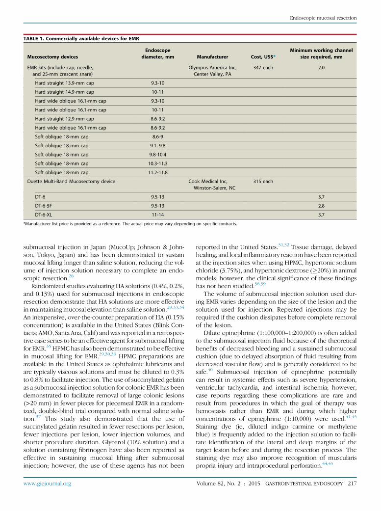
TABLE 1. Commercially available devices for EMR
Mucosectomy devicesEndoscope
diameter, mm Manufacturer Cost, US$*Minimum working channel
size required, mm
EMR kits (include cap, needle,and 25-mm crescent snare)
Olympus America Inc,Center Valley, PA
347 each 2.0
Hard straight 13.9-mm cap 9.3-10
Hard straight 14.9-mm cap 10-11
Hard wide oblique 16.1-mm cap 9.3-10
Hard wide oblique 16.1-mm cap 10-11
Hard straight 12.9-mm cap 8.6-9.2
Hard wide oblique 16.1-mm cap 8.6-9.2
Soft oblique 18-mm cap 8.6-9
Soft oblique 18-mm cap 9.1–9.8
Soft oblique 18-mm cap 9.8-10.4
Soft oblique 18-mm cap 10.3-11.3
Soft oblique 18-mm cap 11.2-11.8
Duette Multi-Band Mucosectomy device Cook Medical Inc,Winston-Salem, NC
315 each
DT-6 9.5-13 3.7
DT-6-5F 9.5-13 2.8
DT-6-XL 11-14 3.7
*Manufacturer list price is provided as a reference. The actual price may vary depending on specific contracts.
Endoscopic mucosal resection
submucosal injection in Japan (MucoUp; Johnson & John-son, Tokyo, Japan) and has been demonstrated to sustainmucosal lifting longer than saline solution, reducing the vol-ume of injection solution necessary to complete an endo-scopic resection.28
Randomized studies evaluatingHA solutions (0.4%, 0.2%,and 0.13%) used for submucosal injections in endoscopicresection demonstrate that HA solutions are more effectiveinmaintainingmucosal elevation than saline solution.28,33,34
An inexpensive, over-the-counter preparation of HA (0.15%concentration) is available in the United States (Blink Con-tacts; AMO, Santa Ana, Calif) andwas reported in a retrospec-tive case series to be an effective agent for submucosal liftingfor EMR.35 HPMChas also been demonstrated to be effectivein mucosal lifting for EMR.29,30,36 HPMC preparations areavailable in the United States as ophthalmic lubricants andare typically viscous solutions and must be diluted to 0.3%to 0.8% to facilitate injection. The use of succinylated gelatinas a submucosal injection solution for colonic EMR has beendemonstrated to facilitate removal of large colonic lesions(>20 mm) in fewer pieces for piecemeal EMR in a random-ized, double-blind trial compared with normal saline solu-tion.37 This study also demonstrated that the use ofsuccinylated gelatin resulted in fewer resections per lesion,fewer injections per lesion, lower injection volumes, andshorter procedure duration. Glycerol (10% solution) and asolution containing fibrinogen have also been reported aseffective in sustaining mucosal lifting after submucosalinjection; however, the use of these agents has not been
www.giejournal.org
reported in the United States.31,32 Tissue damage, delayedhealing, and local inflammatory reaction have been reportedat the injection sites when using HPMC, hypertonic sodiumchloride (3.75%), and hypertonic dextrose (�20%) in animalmodels; however, the clinical significance of these findingshas not been studied.38,39
The volume of submucosal injection solution used dur-ing EMR varies depending on the size of the lesion and thesolution used for injection. Repeated injections may berequired if the cushion dissipates before complete removalof the lesion.
Dilute epinephrine (1:100,000–1:200,000) is often addedto the submucosal injection fluid because of the theoreticalbenefits of decreased bleeding and a sustained submucosalcushion (due to delayed absorption of fluid resulting fromdecreased vascular flow) and is generally considered to besafe.40 Submucosal injection of epinephrine potentiallycan result in systemic effects such as severe hypertension,ventricular tachycardia, and intestinal ischemia; however,case reports regarding these complications are rare andresult from procedures in which the goal of therapy washemostasis rather than EMR and during which higherconcentrations of epinephrine (1:10,000) were used.41-43
Staining dye (ie, diluted indigo carmine or methyleneblue) is frequently added to the injection solution to facili-tate identification of the lateral and deep margins of thetarget lesion before and during the resection process. Thestaining dye may also improve recognition of muscularispropria injury and intraprocedural perforation.44,45
Volume 82, No. 2 : 2015 GASTROINTESTINAL ENDOSCOPY 217
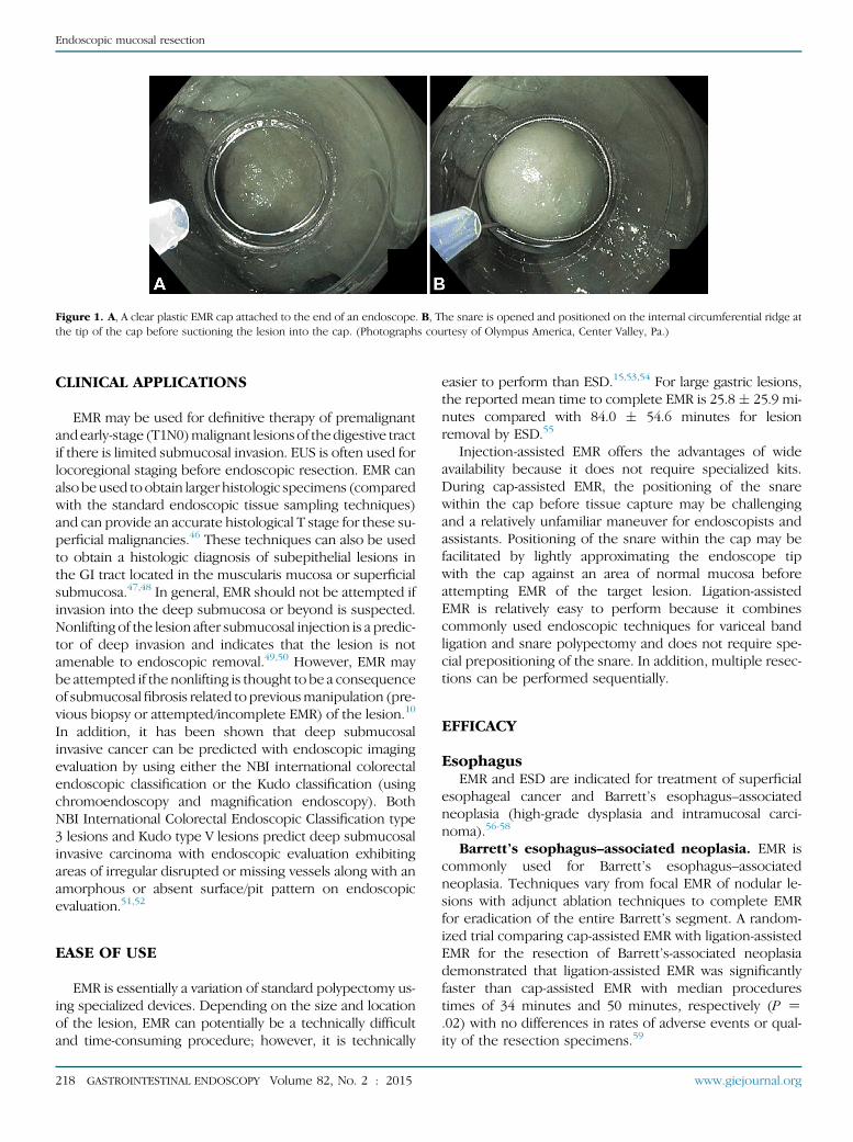
Figure 1. A, A clear plastic EMR cap attached to the end of an endoscope. B, The snare is opened and positioned on the internal circumferential ridge atthe tip of the cap before suctioning the lesion into the cap. (Photographs courtesy of Olympus America, Center Valley, Pa.)
Endoscopic mucosal resection
CLINICAL APPLICATIONS
EMR may be used for definitive therapy of premalignantandearly-stage (T1N0)malignant lesionsof thedigestive tractif there is limited submucosal invasion. EUS is often used forlocoregional staging before endoscopic resection. EMR canalso beused toobtain larger histologic specimens (comparedwith the standard endoscopic tissue sampling techniques)and can provide an accurate histological T stage for these su-perficial malignancies.46 These techniques can also be usedto obtain a histologic diagnosis of subepithelial lesions inthe GI tract located in the muscularis mucosa or superficialsubmucosa.47,48 In general, EMR should not be attempted ifinvasion into the deep submucosa or beyond is suspected.Nonlifting of the lesion after submucosal injection is a predic-tor of deep invasion and indicates that the lesion is notamenable to endoscopic removal.49,50 However, EMR maybe attempted if the nonlifting is thought to be a consequenceof submucosalfibrosis related to previousmanipulation (pre-vious biopsy or attempted/incomplete EMR) of the lesion.10
In addition, it has been shown that deep submucosalinvasive cancer can be predicted with endoscopic imagingevaluation by using either the NBI international colorectalendoscopic classification or the Kudo classification (usingchromoendoscopy and magnification endoscopy). BothNBI International Colorectal Endoscopic Classification type3 lesions and Kudo type V lesions predict deep submucosalinvasive carcinoma with endoscopic evaluation exhibitingareas of irregular disrupted or missing vessels along with anamorphous or absent surface/pit pattern on endoscopicevaluation.51,52
EASE OF USE
EMR is essentially a variation of standard polypectomy us-ing specialized devices. Depending on the size and locationof the lesion, EMR can potentially be a technically difficultand time-consuming procedure; however, it is technically
218 GASTROINTESTINAL ENDOSCOPY Volume 82, No. 2 : 2015
easier to perform than ESD.15,53,54 For large gastric lesions,the reported mean time to complete EMR is 25.8 � 25.9 mi-nutes compared with 84.0 � 54.6 minutes for lesionremoval by ESD.55
Injection-assisted EMR offers the advantages of wideavailability because it does not require specialized kits.During cap-assisted EMR, the positioning of the snarewithin the cap before tissue capture may be challengingand a relatively unfamiliar maneuver for endoscopists andassistants. Positioning of the snare within the cap may befacilitated by lightly approximating the endoscope tipwith the cap against an area of normal mucosa beforeattempting EMR of the target lesion. Ligation-assistedEMR is relatively easy to perform because it combinescommonly used endoscopic techniques for variceal bandligation and snare polypectomy and does not require spe-cial prepositioning of the snare. In addition, multiple resec-tions can be performed sequentially.
EFFICACY
EsophagusEMR and ESD are indicated for treatment of superficial
esophageal cancer and Barrett’s esophagus–associatedneoplasia (high-grade dysplasia and intramucosal carci-noma).56-58
Barrett’s esophagus–associated neoplasia. EMR iscommonly used for Barrett’s esophagus–associatedneoplasia. Techniques vary from focal EMR of nodular le-sions with adjunct ablation techniques to complete EMRfor eradication of the entire Barrett’s segment. A random-ized trial comparing cap-assisted EMR with ligation-assistedEMR for the resection of Barrett’s-associated neoplasiademonstrated that ligation-assisted EMR was significantlyfaster than cap-assisted EMR with median procedurestimes of 34 minutes and 50 minutes, respectively (P Z.02) with no differences in rates of adverse events or qual-ity of the resection specimens.59
www.giejournal.org
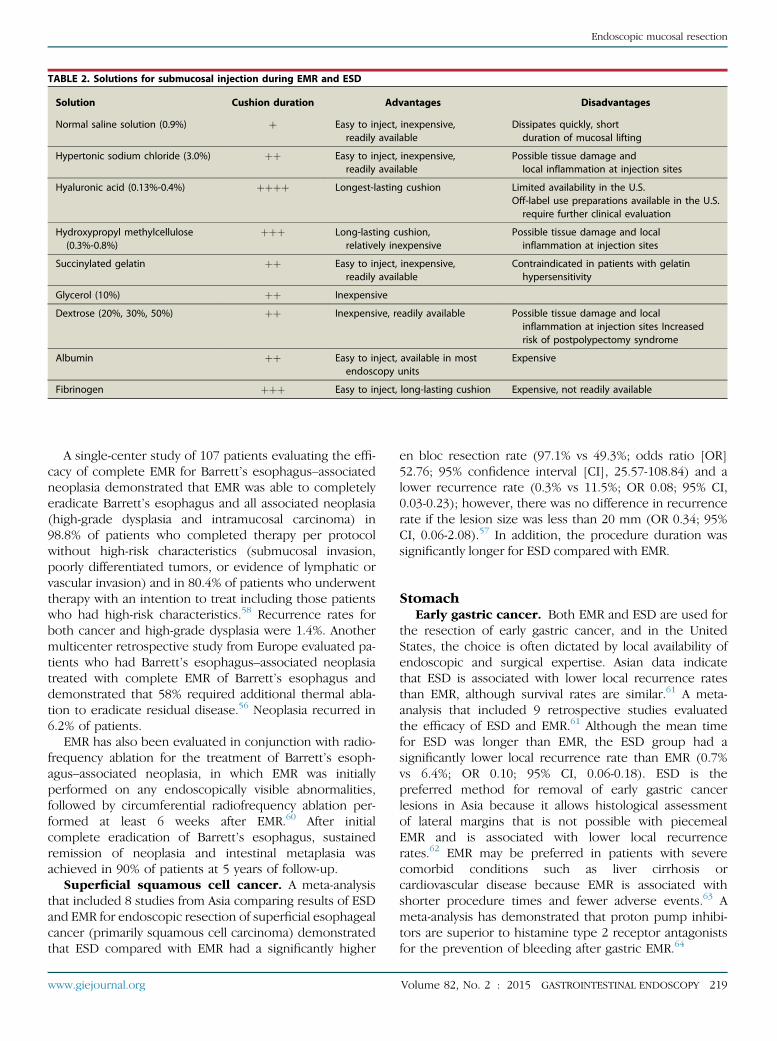
TABLE 2. Solutions for submucosal injection during EMR and ESD
Solution Cushion duration Advantages Disadvantages
Normal saline solution (0.9%) þ Easy to inject, inexpensive,readily available
Dissipates quickly, shortduration of mucosal lifting
Hypertonic sodium chloride (3.0%) þþ Easy to inject, inexpensive,readily available
Possible tissue damage andlocal inflammation at injection sites
Hyaluronic acid (0.13%-0.4%) þþþþ Longest-lasting cushion Limited availability in the U.S.Off-label use preparations available in the U.S.require further clinical evaluation
Hydroxypropyl methylcellulose(0.3%-0.8%)
þþþ Long-lasting cushion,relatively inexpensive
Possible tissue damage and localinflammation at injection sites
Succinylated gelatin þþ Easy to inject, inexpensive,readily available
Contraindicated in patients with gelatinhypersensitivity
Glycerol (10%) þþ Inexpensive
Dextrose (20%, 30%, 50%) þþ Inexpensive, readily available Possible tissue damage and localinflammation at injection sites Increasedrisk of postpolypectomy syndrome
Albumin þþ Easy to inject, available in mostendoscopy units
Expensive
Fibrinogen þþþ Easy to inject, long-lasting cushion Expensive, not readily available
Endoscopic mucosal resection
A single-center study of 107 patients evaluating the effi-cacy of complete EMR for Barrett’s esophagus–associatedneoplasia demonstrated that EMR was able to completelyeradicate Barrett’s esophagus and all associated neoplasia(high-grade dysplasia and intramucosal carcinoma) in98.8% of patients who completed therapy per protocolwithout high-risk characteristics (submucosal invasion,poorly differentiated tumors, or evidence of lymphatic orvascular invasion) and in 80.4% of patients who underwenttherapy with an intention to treat including those patientswho had high-risk characteristics.58 Recurrence rates forboth cancer and high-grade dysplasia were 1.4%. Anothermulticenter retrospective study from Europe evaluated pa-tients who had Barrett’s esophagus–associated neoplasiatreated with complete EMR of Barrett’s esophagus anddemonstrated that 58% required additional thermal abla-tion to eradicate residual disease.56 Neoplasia recurred in6.2% of patients.
EMR has also been evaluated in conjunction with radio-frequency ablation for the treatment of Barrett’s esoph-agus–associated neoplasia, in which EMR was initiallyperformed on any endoscopically visible abnormalities,followed by circumferential radiofrequency ablation per-formed at least 6 weeks after EMR.60 After initialcomplete eradication of Barrett’s esophagus, sustainedremission of neoplasia and intestinal metaplasia wasachieved in 90% of patients at 5 years of follow-up.
Superficial squamous cell cancer. A meta-analysisthat included 8 studies from Asia comparing results of ESDand EMR for endoscopic resection of superficial esophagealcancer (primarily squamous cell carcinoma) demonstratedthat ESD compared with EMR had a significantly higher
www.giejournal.org
en bloc resection rate (97.1% vs 49.3%; odds ratio [OR]52.76; 95% confidence interval [CI], 25.57-108.84) and alower recurrence rate (0.3% vs 11.5%; OR 0.08; 95% CI,0.03-0.23); however, there was no difference in recurrencerate if the lesion size was less than 20 mm (OR 0.34; 95%CI, 0.06-2.08).57 In addition, the procedure duration wassignificantly longer for ESD compared with EMR.
StomachEarly gastric cancer. Both EMR and ESD are used for
the resection of early gastric cancer, and in the UnitedStates, the choice is often dictated by local availability ofendoscopic and surgical expertise. Asian data indicatethat ESD is associated with lower local recurrence ratesthan EMR, although survival rates are similar.61 A meta-analysis that included 9 retrospective studies evaluatedthe efficacy of ESD and EMR.61 Although the mean timefor ESD was longer than EMR, the ESD group had asignificantly lower local recurrence rate than EMR (0.7%vs 6.4%; OR 0.10; 95% CI, 0.06-0.18). ESD is thepreferred method for removal of early gastric cancerlesions in Asia because it allows histological assessmentof lateral margins that is not possible with piecemealEMR and is associated with lower local recurrencerates.62 EMR may be preferred in patients with severecomorbid conditions such as liver cirrhosis orcardiovascular disease because EMR is associated withshorter procedure times and fewer adverse events.63 Ameta-analysis has demonstrated that proton pump inhibi-tors are superior to histamine type 2 receptor antagonistsfor the prevention of bleeding after gastric EMR.64
Volume 82, No. 2 : 2015 GASTROINTESTINAL ENDOSCOPY 219
Endoscopic mucosal resection
Gastric carcinoids. EMR has been reported to beeffective in resecting type 1 gastric carcinoids (those asso-ciated with chronic atrophic gastritis) that are less than 1cm in diameter.65-67 EMR has been reported to be associ-ated with a slightly lower rate of complete resectioncompared with ESD due to positive deep margins; howev-er, local recurrence rates and survival are similar.66,67
DuodenumNonampullary duodenal adenomas. Duodenal le-
sions not involving the major duodenal papilla can beremoved with a variety of EMR techniques, but carries anincreased risk of bleeding and perforation because the du-odenum has increased vascularity and a thin wall. The ma-jority of published reports on endoscopic removal ofduodenal polyps originates from high-volume centers andare limited by low numbers of patients and a retrospectivestudy design.68-71 Reported success rates vary from 70% to96% for nonampullary duodenal lesions.68,69,71 In addition,the technique of UEMR has been reported for resectingnonampullary duodenal adenomas with high success ratesfor complete resection (83% at the index session); howev-er, adverse events included delayed bleeding, water intox-ication syndrome, and stricture formation.72
ColonInjection-assisted EMR is widely used for the resection
of large or flat colonic lesions. A systematic review andmeta-analysis demonstrated that local recurrence afterEMR occurs in 3% of cases in which the lesion is removeden bloc and in 20% of cases in which the lesion is removedin piecemeal fashion.73 For recurrences that were re-treated with endoscopic therapy (APC and/or EMR), thesubsequent recurrence rate was 21%, with successful erad-ication being achieved in 91.4% of recurrences after amean of 1.2 additional sessions. Another large multicenterprospective study with 1000 consecutive wide-field EMRsof large sessile adenomas demonstrated a 16% recurrencerate at 4 months, usually unifocal and diminutive, and a4% recurrence rate at 16 months.23 Recurrences weremanaged endoscopically in 93% of cases. If a largeadenoma (>15 mm) is removed in piecemeal fashion,the patient should have a repeat colonoscopy in 6 to 12months to evaluate for local recurrence.73,74 A meta-analysis comparing ESD with EMR for colorectal tumorsthat included 6 studies (1642 total lesions) demonstratedthat ESD had a higher en bloc resection rate and lowerinitial local recurrence rate than EMR; however, ESD wasmore time-consuming and generally required hospitaliza-tion for observation after the procedure.75
Typically, EMR in the colon is performed by using theinjection-assisted EMR technique; however, UEMR withoutsubmucosal injection has also been reported.9 Whenthe UEMR technique was used, the complete removalrate for recurrences was significantly higher comparedwith injection-assisted EMR (88.9% vs 31.8%, P < .001)
220 GASTROINTESTINAL ENDOSCOPY Volume 82, No. 2 : 2015
in a nonrandomized clinical trial.11 Furthermore, therecurrence rate was significantly lower in the UEMR groupthan the EMR group (10% vs 39.4%, P Z .02).
Although several studies have reported no recurrenceafter endoscopic removal of malignant colonic polyps,the effectiveness of EMR for the treatment of these lesionshas been questioned, and EMR should not be attemptedfor nonlifting lesions or lesions classified as Paris II-c/III.15,76-78 However, nonlifting lesions that have previouslybeen manipulated (biopsy or attempted EMR) beforereferral for resection are usually amenable to EMR.10
Endoscopic resection has also been reported to be suc-cessful in resecting small rectal carcinoid tumors. Techni-cally, the procedure should be considered an endoscopicsubmucosal resection because a majority of rectal carci-noid tumors extend into the submucosal layer.79,80 Theuse of ligation-assisted EMR for lesions that were estimatedto be less than 1 cm in diameter has resulted in resectionswith negative margins.79 However, another study thatcompared ESD with ligation-assisted EMR for endoscopicresection of carcinoid tumors that were less than 16 mmdemonstrated a higher histologically complete resectionrate with ESD compared with EMR (90.3% vs 71.0%, P Z.035), although ESD took longer to perform.81
SafetyAdverse events after EMR include bleeding, perforation,
and strictures. Bleeding is the most common adverse eventof EMR.
Colonic EMR. Intraprocedural bleeding rates afterEMR of colorectal lesions larger than 20 mm are reportedto be between 11% and 22%.14,82 The application of softcoagulation with the tip of a snare has been demonstratedto be both safe and effective for the treatment of intrapro-cedural bleeding during EMR of large colonic polyps.82
Other methods including hot biopsy forceps, monopolarhemostatic forceps, bicap probes, APC, and endoscopicclips can be used for achieving hemostasis. Risk factorsfor intraprocedural bleeding include lesion size, Parisendoscopic classification of 0-IIa þ Is, tubulovillous orvillous histology, and low-volume institutions.14 Bleedingrates after EMR of large colonic polyps range from 2%to 11%.12-18 The clinically significant bleeding rate afterEMR of sessile colorectal polyps larger than 20 mm wasreported to be 6% in a large prospective, multicenter studythat included 1039 patients.14 Of the patients with clinicallysignificant bleeding, 34% required endoscopic therapy.Prophylactic endoscopic coagulation of nonbleedingvessels by using coagulating forceps after EMR ofcolorectal lesions larger than 20 mm did not significantlydecrease the incidence of delayed postprocedure bleedingcompared with control subjects who received noadditional therapy (5.2% vs 8.0%, P Z .3). Risk factorsfor clinically significant postprocedural bleeding included aproximal colonic location, polyp size, and intraproceduralbleeding.14
www.giejournal.org
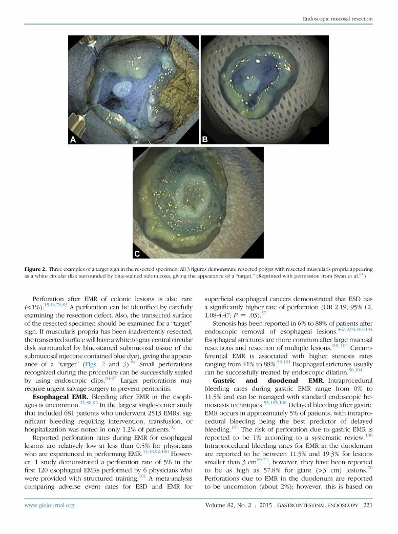
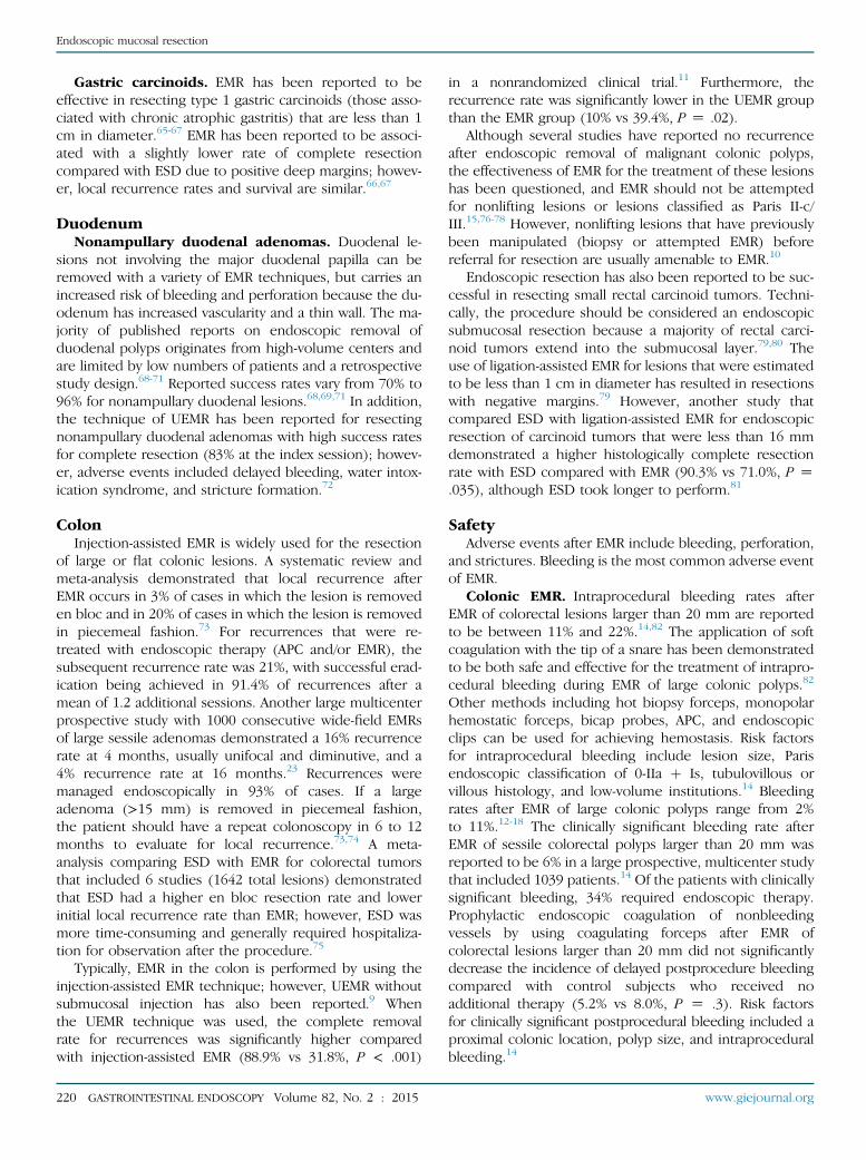
Figure 2. Three examples of a target sign in the resected specimen. All 3 figures demonstrate resected polyps with resected muscularis propria appearingas a white circular disk surrounded by blue-stained submucosa, giving the appearance of a “target.” (Reprinted with permission from Swan et al.84 )
Endoscopic mucosal resection
Perforation after EMR of colonic lesions is also rare(<1%).15,16,76,83 A perforation can be identified by carefullyexamining the resection defect. Also, the transected surfaceof the resected specimen should be examined for a “target”sign. If muscularis propria has been inadvertently resected,the transected surfacewill have awhite to gray central circulardisk surrounded by blue-stained submucosal tissue (if thesubmucosal injectate contained blue dye), giving the appear-ance of a “target” (Figs. 2 and 3).84 Small perforationsrecognized during the procedure can be successfully sealedby using endoscopic clips.84-87 Larger perforations mayrequire urgent salvage surgery to prevent peritonitis.
Esophageal EMR. Bleeding after EMR in the esoph-agus is uncommon.21,88-92 In the largest single-center studythat included 681 patients who underwent 2513 EMRs, sig-nificant bleeding requiring intervention, transfusion, orhospitalization was noted in only 1.2% of patients.92
Reported perforation rates during EMR for esophageallesions are relatively low at less than 0.5% for physicianswho are experienced in performing EMR.53,58,92-100 Howev-er, 1 study demonstrated a perforation rate of 5% in thefirst 120 esophageal EMRs performed by 6 physicians whowere provided with structured training.101 A meta-analysiscomparing adverse event rates for ESD and EMR for
www.giejournal.org
superficial esophageal cancers demonstrated that ESD hasa significantly higher rate of perforation (OR 2.19; 95% CI,1.08-4.47; P Z .03).57
Stenosis has been reported in 6% to 88% of patients afterendoscopic removal of esophageal lesions.46,58,94,101-104
Esophageal strictures are more common after large mucosalresections and resection of multiple lesions.101,104 Circum-ferential EMR is associated with higher stenosis ratesranging from 41% to 88%.58,101 Esophageal strictures usuallycan be successfully treated by endoscopic dilation.58,104
Gastric and duodenal EMR. Intraproceduralbleeding rates during gastric EMR range from 0% to11.5% and can be managed with standard endoscopic he-mostasis techniques.53,105,106 Delayed bleeding after gastricEMR occurs in approximately 5% of patients, with intrapro-cedural bleeding being the best predictor of delayedbleeding.107 The risk of perforation due to gastric EMR isreported to be 1% according to a systematic review.108
Intraprocedural bleeding rates for EMR in the duodenumare reported to be between 11.5% and 19.3% for lesionssmaller than 3 cm69-71; however, they have been reportedto be as high as 57.8% for giant (>3 cm) lesions.70
Perforations due to EMR in the duodenum are reportedto be uncommon (about 2%); however, this is based on
Volume 82, No. 2 : 2015 GASTROINTESTINAL ENDOSCOPY 221
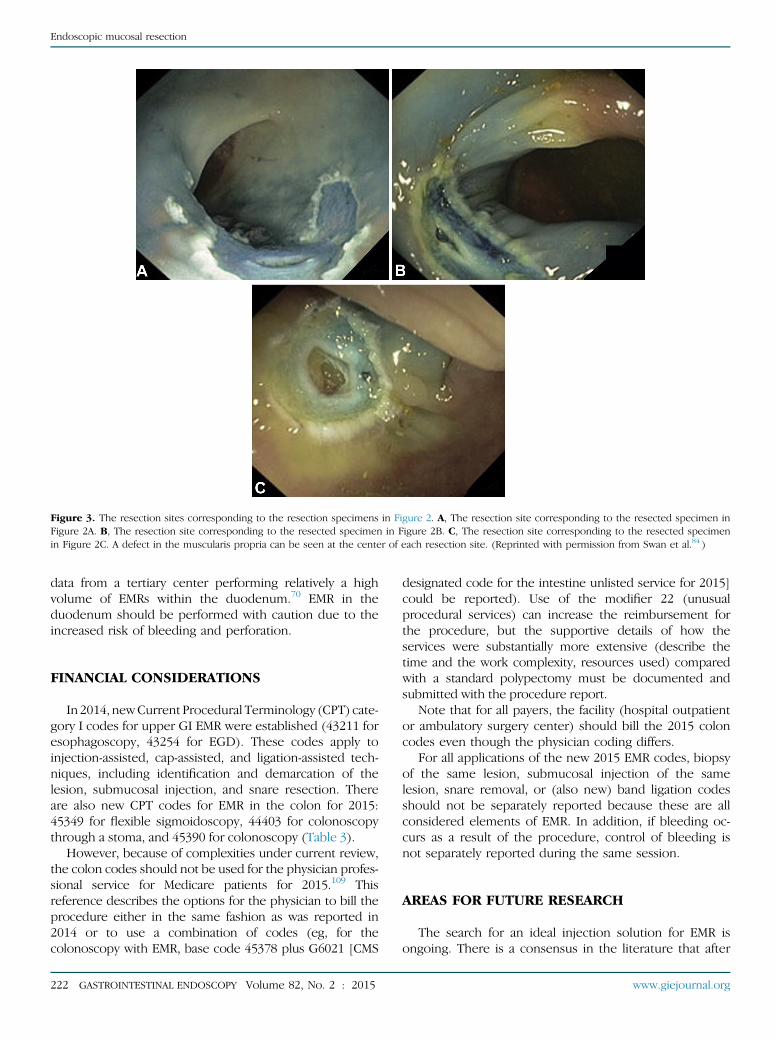
Figure 3. The resection sites corresponding to the resection specimens in Figure 2. A, The resection site corresponding to the resected specimen inFigure 2A. B, The resection site corresponding to the resected specimen in Figure 2B. C, The resection site corresponding to the resected specimenin Figure 2C. A defect in the muscularis propria can be seen at the center of each resection site. (Reprinted with permission from Swan et al.84 )
Endoscopic mucosal resection
data from a tertiary center performing relatively a highvolume of EMRs within the duodenum.70 EMR in theduodenum should be performed with caution due to theincreased risk of bleeding and perforation.
FINANCIAL CONSIDERATIONS
In 2014, newCurrent Procedural Terminology (CPT) cate-gory I codes for upper GI EMR were established (43211 foresophagoscopy, 43254 for EGD). These codes apply toinjection-assisted, cap-assisted, and ligation-assisted tech-niques, including identification and demarcation of thelesion, submucosal injection, and snare resection. Thereare also new CPT codes for EMR in the colon for 2015:45349 for flexible sigmoidoscopy, 44403 for colonoscopythrough a stoma, and 45390 for colonoscopy (Table 3).
However, because of complexities under current review,the colon codes should not be used for the physician profes-sional service for Medicare patients for 2015.109 Thisreference describes the options for the physician to bill theprocedure either in the same fashion as was reported in2014 or to use a combination of codes (eg, for thecolonoscopy with EMR, base code 45378 plus G6021 [CMS
222 GASTROINTESTINAL ENDOSCOPY Volume 82, No. 2 : 2015
designated code for the intestine unlisted service for 2015]could be reported). Use of the modifier 22 (unusualprocedural services) can increase the reimbursement forthe procedure, but the supportive details of how theservices were substantially more extensive (describe thetime and the work complexity, resources used) comparedwith a standard polypectomy must be documented andsubmitted with the procedure report.
Note that for all payers, the facility (hospital outpatientor ambulatory surgery center) should bill the 2015 coloncodes even though the physician coding differs.
For all applications of the new 2015 EMR codes, biopsyof the same lesion, submucosal injection of the samelesion, snare removal, or (also new) band ligation codesshould not be separately reported because these are allconsidered elements of EMR. In addition, if bleeding oc-curs as a result of the procedure, control of bleeding isnot separately reported during the same session.
AREAS FOR FUTURE RESEARCH
The search for an ideal injection solution for EMR isongoing. There is a consensus in the literature that after
www.giejournal.org
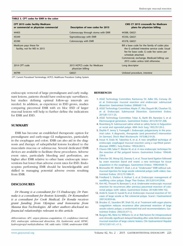
TABLE 3. CPT codes for EMR in the colon
CPT 2015 code: facility Medicareor commercial or physician commercial Description of new codes for 2015
CMS CY 2015 crosswalk for Medicareplans for physician billing
44403 Colonoscopy through stoma with EMR 44388, G6021
45349 Sigmoidoscopy with EMR 45330, G6021
45390 Colonoscopy with EMR 45378, G6021
Medicare pays these forfacility, not for MD in 2015
Bill a base code for the family of codes plusthe G unlisted intestine service code. Usualfee for base code. G code fee varies perschedule attached.
Commercial, exchange, Medicaid billing: use2015 codes unless told otherwise.
2014 CPT code 2015 HCPCS code: for Medicarephysician billing
Long descriptor
44799 G6021 Unlisted procedure, intestine
CPT, Current Procedural Terminology; HCPCS, Healthcare Procedure Coding System.
Endoscopic mucosal resection
endoscopic removal of large premalignant and early malig-nant lesions, patients should have endoscopic surveillance,but studies defining optimal follow-up intervals areneeded. In addition, as experience in ESD grows, studiescomparing piecemeal EMR with en bloc ESD of largermucosal lesions will help to further define the indicationsfor EMR and ESD.
SUMMARY
EMR has become an established therapeutic option forpremalignant and early-stage GI malignancies, particularlyin the esophagus and colon. EMR can also aid in the diag-nosis and therapy of subepithelial lesions localized to themuscularis mucosa or submucosa. Several dedicated EMRdevices are available to facilitate these procedures. Adverseevent rates, particularly bleeding and perforation, arehigher after EMR relative to other basic endoscopic inter-ventions but lower than adverse event rates for ESD. Endo-scopists performing EMR should be knowledgeable andskilled in managing potential adverse events resultingfrom EMR.
DISCLOSURES
Dr Hwang is a consultant for US Endoscopy; Dr Pan-nala is a consultant for Boston Scientific; Dr Komanduriis a consultant for Cook Medical; Dr Konda receivesgrant funding from Olympus and honoraria fromMauna Kea Technologies. All other authors disclosed nofinancial relationships relevant to this article.
Abbreviations: APC, argon plasma coagulation; CI, confidence interval;ESD, endoscopic submucosal dissection; HA, hyaluronic acid; HPMC,hydroxypropyl methylcellulose; OR, odds ratio; UEMR, underwater EMR.
www.giejournal.org
REFERENCES
1. ASGE Technology Committee; Kantsevoy SV, Adler DG, Conway JD,et al. Endoscopic mucosal resection and endoscopic submucosaldissection. Gastrointest Endosc 2008;68:11-8.
2. ASGE Technology Committee; Maple JT, Abu Dayyeh BK, Chauhan SS,et al. Endoscopic Submucosal Dissection. Gastrointest Endosc2015;81:1311-25.
3. ASGE Technology Committee; Tokar JL, Barth BA, Banerjee S, et al.Electrosurgical generators. Gastrointest Endosc 2013;78:197-208.
4. Rosenberg N. Submucosal saline wheal as safety factor in fulgurationor rectal and sigmoidal polypi. AMA Arch Surg 1955;70:120-2.
5. Deyhle P, Jenny S, Fumagalli I. Endoscopic polypectomy in the prox-imal colon. A diagnostic, therapeutic (and preventive?) intervention[in German]. Dtsch Med Wochenschr 1973;98:219-20.
6. Inoue H, Endo M, Takeshita K, et al. A new simplified technique ofendoscopic esophageal mucosal resection using a cap-fitted panen-doscope (EMRC). Surg Endosc 1992;6:264-5.
7. Chaves DM, Sakai P, Mester M, et al. A new endoscopic technique forthe resection of flat polypoid lesions. Gastrointest Endosc 1994;40:224-6.
8. Fleischer DE, Wang GQ, Dawsey S, et al. Tissue band ligation followedby snare resection (band and snare): a new technique for tissueacquisition in the esophagus. Gastrointest Endosc 1996;44:68-72.
9. Binmoeller KF, Weilert F, Shah J, et al. “Underwater” EMR without sub-mucosal injection for large sessile colorectal polyps (with video). Gas-trointest Endosc 2012;75:1086-91.
10. Friedland S, Shelton A, Kothari S, et al. Endoscopic management ofnonlifting colon polyps. Diagn Ther Endosc 2013;2013:412936.
11. Kim HG, Thosani N, Banerjee S, et al. Underwater endoscopic mucosalresection for recurrences after previous piecemeal resection of colo-rectal polyps (with video). Gastrointest Endosc 2014;80:1094-102.
12. Arebi N, Swain D, Suzuki N, et al. Endoscopic mucosal resection of 161cases of large sessile or flat colorectal polyps. Scand J Gastroenterol2007;42:859-66.
13. Brooker JC, Saunders BP, Shah SG, et al. Treatment with argon plasmacoagulation reduces recurrence after piecemeal resection of largesessile colonic polyps: a randomized trial and recommendations. Gas-trointest Endosc 2002;55:371-5.
14. Burgess NG, Metz AJ, Williams SJ, et al. Risk factors for intraproceduraland clinically significant delayed bleeding after wide-field endoscopicmucosal resection of large colonic lesions. Clin Gastroenterol Hepatol2014;12:651-61; e1-3.
Volume 82, No. 2 : 2015 GASTROINTESTINAL ENDOSCOPY 223
Endoscopic mucosal resection
15. Conio M, Repici A, Demarquay JF, et al. EMR of large sessile colorectalpolyps. Gastrointest Endosc 2004;60:234-41.
16. Luigiano C, Consolo P, Scaffidi MG, et al. Endoscopic mucosal resec-tion for large and giant sessile and flat colorectal polyps: a single-center experience with long-term follow-up. Endoscopy 2009;41:829-35.
17. Sato T. A novel method of endoscopic mucosal resection assisted bysubmucosal injection of autologous blood (blood patch EMR). Dis Co-lon Rectum 2006;49:1636-41.
18. Zlatanic J, Waye JD, Kim PS, et al. Large sessile colonic adenomas: useof argon plasma coagulator to supplement piecemeal snare polypec-tomy. Gastrointest Endosc 1999;49:731-5.
19. Veerappan SG, Ormonde D, Yusoff IF, et al. Hot avulsion: a modifica-tion of an existing technique for management of nonlifting areas of apolyp (with video). Gastrointest Endosc 2014;80:884-8.
20. Mannath J, Subramanian V, Singh R, et al. Polyp recurrence afterendoscopic mucosal resection of sessile and flat colonic adenomas.Dig Dis Sci 2011;56:2389-95.
21. Peters FP, Kara MA, Rosmolen WD, et al. Endoscopic treatment ofhigh-grade dysplasia and early stage cancer in Barrett’s esophagus.Gastrointest Endosc 2005;61:506-14.
22. Regula J, Wronska E, Polkowski M, et al. Argon plasma coagulation af-ter piecemeal polypectomy of sessile colorectal adenomas: long-termfollow-up study. Endoscopy 2003;35:212-8.
23. Moss A, Williams SJ, Hourigan LF, et al. Long-term adenoma recur-rence following wide-field endoscopic mucosal resection (WF-EMR)for advanced colonic mucosal neoplasia is infrequent: results andrisk factors in 1000 cases from the Australian Colonic EMR (ACE)study. Gut 2015;64:57-65.
24. Ohkuwa M, Hosokawa K, Boku N, et al. New endoscopic treatment forintramucosal gastric tumors using an insulated-tip diathermic knife.Endoscopy 2001;33:221-6.
25. Ono H, Kondo H, Gotoda T, et al. Endoscopic mucosal resection fortreatment of early gastric cancer. Gut 2001;48:225-9.
26. Yoshida N, Naito Y, Kugai M, et al. Efficacy of hyaluronic acid in endo-scopic mucosal resection of colorectal tumors. J Gastroenterol Hepa-tol 2011;26:286-91.
27. Fujishiro M, Yahagi N, Kashimura K, et al. Comparison of various sub-mucosal injection solutions for maintaining mucosal elevation duringendoscopic mucosal resection. Endoscopy 2004;36:579-83.
28. Yamamoto H, Yahagi N, Oyama T, et al. Usefulness and safety of 0.4%sodium hyaluronate solution as a submucosal fluid “cushion” in endo-scopic resection for gastric neoplasms: a prospective multicenter trial.Gastrointest Endosc 2008;67:830-9.
29. Arantes V, Albuquerque W, Benfica E, et al. Submucosal injection of0.4% hydroxypropyl methylcellulose facilitates endoscopic mucosalresection of early gastrointestinal tumors. J Clin Gastroenterol2010;44:615-9.
30. Bacani CJ, Woodward TA, Raimondo M, et al. The safety and efficacyin humans of endoscopic mucosal resection with hydroxypropylmethylcellulose as compared with normal saline. Surg Endosc2008;22:2401-6.
31. Uraoka T, Fujii T, Saito Y, et al. Effectiveness of glycerol as a submu-cosal injection for EMR. Gastrointest Endosc 2005;61:736-40.
32. Lee SH, Park JH, Park do H, et al. Clinical efficacy of EMR with submu-cosal injection of a fibrinogen mixture: a prospective randomizedtrial. Gastrointest Endosc 2006;64:691-6.
33. Kishihara T, Chino A, Uragami N, et al. Usefulness of sodium hyaluro-nate solution in colorectal endoscopic mucosal resection. Dig Endosc2012;24:348-52.
34. Yoshida N, Naito Y, Inada Y, et al. Endoscopic mucosal resection with0.13% hyaluronic acid solution for colorectal polyps less than 20 mm:a randomized controlled trial. J Gastroenterol Hepatol 2012;27:1377-83.
35. Friedland S, Kothari S, Chen A, et al. Endoscopic mucosal resectionwith an over-the-counter hyaluronate preparation. Gastrointest En-dosc 2012;75:1040-4.
224 GASTROINTESTINAL ENDOSCOPY Volume 82, No. 2 : 2015
36. Arezzo A, Pagano N, Romeo F, et al. Hydroxy-propyl-methyl-celluloseis a safe and effective lifting agent for endoscopic mucosal resectionof large colorectal polyps. Surg Endosc 2009;23:1065-9.
37. Moss A, Bourke MJ, Metz AJ. A randomized, double-blind trial of succi-nylated gelatin submucosal injection for endoscopic resection of largesessile polyps of the colon. Am J Gastroenterol 2010;105:2375-82.
38. Bures J, Kopacova M, Kvetina J, et al. Different solutions used for sub-mucosal injection influenced early healing of gastric endoscopicmucosal resection in a preclinical study in experimental pigs. Surg En-dosc 2009;23:2094-101.
39. FujishiroM, Yahagi N, Kashimura K, et al. Tissue damage of different sub-mucosal injection solutions for EMR.Gastrointest Endosc2005;62:933-42.
40. Tanaka M, Ono H, Hasuike N, et al. Endoscopic submucosal dissectionof early gastric cancer. Digestion 2008;77(Suppl 1):23-8.
41. Kim SY, Han SH, Kim KH, et al. Gastric ischemia after epinephrine in-jection in a patient with liver cirrhosis. World J Gastroenterol 2013;19:411-4.
42. Stevens PD, Lebwohl O. Hypertensive emergency and ventriculartachycardia after endoscopic epinephrine injection of a Mallory-Weiss tear. Gastrointest Endosc 1994;40:77-8.
43. von Delius S, Thies P, Umgelter A, et al. Hemodynamics after endo-scopic submucosal injection of epinephrine in patients with nonvar-iceal upper gastrointestinal bleeding: a matter of concern. Endoscopy2006;38:1284-8.
44. Hirao M, Hatakeyama H, Asanuma T, et al. Endoscopic resection withlocal injection of hypertonic saline epinephrine for the treatment ofearly gastric cancer [in Japanese with an English abstract]. Gan To Ka-gaku Ryoho 1988;15:1466-72.
45. Holt BA, Jayasekeran V, Sonson R, et al. Topical submucosal chro-moendoscopy defines the level of resection in colonic EMR andmay improve procedural safety (with video). Gastrointest Endosc2013;77:949-53.
46. Larghi A, Lightdale CJ, Memeo L, et al. EUS followed by EMR for stag-ing of high-grade dysplasia and early cancer in Barrett’s esophagus.Gastrointest Endosc 2005;62:16-23.
47. Cantor MJ, Davila RE, Faigel DO. Yield of tissue sampling for subepi-thelial lesions evaluated by EUS: a comparison between forceps bi-opsies and endoscopic submucosal resection. Gastrointest Endosc2006;64:29-34.
48. Rodriguez SA, Faigel DO. Endoscopic diagnosis of gastrointestinalstromal cell tumors. Curr Opin Gastroenterol 2007;23:539-43.
49. Kato H, Haga S, Endo S, et al. Lifting of lesions during endoscopicmucosal resection (EMR) of early colorectal cancer: implications forthe assessment of resectability. Endoscopy 2001;33:568-73.
50. Ishiguro A, Uno Y, Ishiguro Y, et al. Correlation of lifting versus non-lifting and microscopic depth of invasion in early colorectal cancer.Gastrointest Endosc 1999;50:329-33.
51. Hayashi N, Tanaka S, Hewett DG, et al. Endoscopic prediction of deepsubmucosal invasive carcinoma: validation of the narrow-band imag-ing international colorectal endoscopic (NICE) classification. Gastro-intest Endosc 2013;78:625-32.
52. Kudo S, Rubio CA, Teixeira CR, et al. Pit pattern in colorectal neoplasia:endoscopic magnifying view. Endoscopy 2001;33:367-73.
53. Oka S, Tanaka S, Kaneko I, et al. Advantage of endoscopic submucosaldissection compared with EMR for early gastric cancer. GastrointestEndosc 2006;64:877-83.
54. Yamasaki M, Kume K, Yoshikawa I, et al. A novel method of endo-scopic submucosal dissection with blunt abrasion by submucosal in-jection of sodium carboxymethylcellulose: an animal preliminarystudy. Gastrointest Endosc 2006;64:958-65.
55. Watanabe K, Ogata S, Kawazoe S, et al. Clinical outcomes of EMR forgastric tumors: historical pilot evaluation between endoscopic sub-mucosal dissection and conventional mucosal resection. GastrointestEndosc 2006;63:776-82.
56. Anders M, Bahr C, El-Masry MA, et al. Long-term recurrence ofneoplasia and Barrett’s epithelium after complete endoscopic resec-tion. Gut 2014;63:1535-43.
www.giejournal.org
Endoscopic mucosal resection
57. Guo HM, Zhang XQ, Chen M, et al. Endoscopic submucosal dissectionvs endoscopic mucosal resection for superficial esophageal cancer.World J Gastroenterol 2014;20:5540-7.
58. Konda VJ, Gonzalez Haba Ruiz M, Koons A, et al. Complete endo-scopic mucosal resection is effective and durable treatment for Bar-rett’s-associated neoplasia. Clin Gastroenterol Hepatol 2014;12:2002-10.
59. Pouw RE, van Vilsteren FG, Peters FP, et al. Randomized trial on endo-scopic resection-cap versus multiband mucosectomy for piecemealendoscopic resection of early Barrett’s neoplasia. Gastrointest Endosc2011;74:35-43.
60. Phoa KN, Pouw RE, van Vilsteren FG, et al. Remission of Barrett’sesophagus with early neoplasia 5 years after radiofrequency ablationwith endoscopic resection: a Netherlands cohort study. Gastroenter-ology 2013;145:96-104.
61. Lian J, Chen S, Zhang Y, et al. A meta-analysis of endoscopic submu-cosal dissection and EMR for early gastric cancer. Gastrointest Endosc2012;76:763-70.
62. Uedo N, Takeuchi Y, Ishihara R. Endoscopic management of earlygastric cancer: endoscopic mucosal resection or endoscopic submu-cosal dissection: data from a Japanese high-volume center and liter-ature review. Ann Gastroenterol 2012;25:281-90.
63. Horiki N, Omata F, Uemura M, et al. Risk for local recurrence of earlygastric cancer treated with piecemeal endoscopic mucosal resectionduring a 10-year follow-up period. Surg Endosc 2012;26:72-8.
64. Yang Z, Wu Q, Liu Z, et al. Proton pump inhibitors versus histamine-2-receptor antagonists for the management of iatrogenic gastric ulcerafter endoscopic mucosal resection or endoscopic submucosaldissection: a meta-analysis of randomized trials. Digestion 2011;84:315-20.
65. Ichikawa J, Tanabe S, Koizumi W, et al. Endoscopic mucosal resectionin the management of gastric carcinoid tumors. Endoscopy 2003;35:203-6.
66. Kim HH, Kim GH, Kim JH, et al. The efficacy of endoscopic submucosaldissection of type I gastric carcinoid tumors compared with conven-tional endoscopic mucosal resection. Gastroenterol Res Pract2014;2014:253860.
67. Sato Y, Takeuchi M, Hashimoto S, et al. Usefulness of endoscopic sub-mucosal dissection for type I gastric carcinoid tumors compared withendoscopic mucosal resection. Hepatogastroenterology 2013;60:1524-9.
68. Abbass R, Rigaux J, Al-Kawas FH. Nonampullary duodenal polyps:characteristics and endoscopic management. Gastrointest Endosc2010;71:754-9.
69. Conio M, De Ceglie A, Filiberti R, et al. Cap-assisted EMR of large, spo-radic, nonampullary duodenal polyps. Gastrointest Endosc 2012;76:1160-9.
70. Fanning SB, Bourke MJ, Williams SJ, et al. Giant laterally spreading tu-mors of the duodenum: endoscopic resection outcomes, limitations,and caveats. Gastrointest Endosc 2012;75:805-12.
71. Kedia P, Brensinger C, Ginsberg G. Endoscopic predictors of success-ful endoluminal eradication in sporadic duodenal adenomas and itsacute complications. Gastrointest Endosc 2010;72:1297-301.
72. Binmoeller KF, Shah JN, Bhat YM, et al. “Underwater” EMR of sporadiclaterally spreading nonampullary duodenal adenomas (with video).Gastrointest Endosc 2013;78:496-502.
73. Belderbos TD, Leenders M, Moons LM, et al. Local recurrence afterendoscopic mucosal resection of nonpedunculated colorectal lesions:systematic review and meta-analysis. Endoscopy 2014;46:388-402.
74. Lieberman DA, Rex DK, Winawer SJ, et al. Guidelines for colonoscopysurveillance after screening and polypectomy: a consensus update bythe US Multi-Society Task Force on Colorectal Cancer. Gastroenter-ology 2012;143:844-57.
75. Wang J, Ge J, Zhang XH, et al. Endoscopic submucosal dissectionversus endoscopic mucosal resection for the treatment of earlyesophageal carcinoma: a meta-analysis. Asian Pac J Cancer Prev2014;15:1803-6.
www.giejournal.org
76. Binmoeller KF, Bohnacker S, Seifert H, et al. Endoscopic snare excisionof “giant” colorectal polyps. Gastrointest Endosc 1996;43:183-8.
77. Kyzer S, Begin LR, Gordon PH, et al. The care of patients with colo-rectal polyps that contain invasive adenocarcinoma. Endoscopic pol-ypectomy or colectomy? Cancer 1992;70:2044-50.
78. Kikuchi R, Takano M, Takagi K, et al. Management of early invasivecolorectal cancer. Risk of recurrence and clinical guidelines. Dis ColonRectum 1995;38:1286-95.
79. Ono A, Fujii T, Saito Y, et al. Endoscopic submucosal resection ofrectal carcinoid tumors with a ligation device. Gastrointest Endosc2003;57:583-7.
80. Soga J. Carcinoids of the rectum: an evaluation of 1271 reportedcases. Surg Today 1997;27:112-9.
81. Park H-W, Byeon J-S, Park YS, et al. Endoscopic submucosal dissectionfor treatment of rectal carcinoid tumors. Gastrointest Endosc 2010;72:143-9.
82. Fahrtash-Bahin F, Holt BA, Jayasekeran V, et al. Snare tip soft coagu-lation achieves effective and safe endoscopic hemostasis duringwide-field endoscopic resection of large colonic lesions (with videos).Gastrointest Endosc 2013;78:158-63.e1.
83. Iishi H, Tatsuta M, Iseki K, et al. Endoscopic piecemeal resection withsubmucosal saline injection of large sessile colorectal polyps. Gastro-intest Endosc 2000;51:697-700.
84. Swan MP, Bourke MJ, Moss A, et al. The target sign: an endoscopicmarker for the resection of the muscularis propria and potentialperforation during colonic endoscopic mucosal resection. Gastroint-est Endosc 2011;73:79-85.
85. Yoshikane H, Hidano H, Sakakibara A, et al. Endoscopic repair by clip-ping of iatrogenic colonic perforation. Gastrointest Endosc 1997;46:464-6.
86. Kim HS, Lee DK, Jeong YS, et al. Successful endoscopic managementof a perforated gastric dysplastic lesion after endoscopic mucosalresection. Gastrointest Endosc 2000;51:613-5.
87. Fujishiro M, Yahagi N, Nakamura M, et al. Successful outcomes of anovel endoscopic treatment for GI tumors: endoscopic submucosaldissection with a mixture of high-molecular-weight hyaluronic acid,glycerin, and sugar. Gastrointest Endosc 2006;63:243-9.
88. Ell C, May A, Gossner L, et al. Endoscopic mucosal resection of earlycancer and high-grade dysplasia in Barrett’s esophagus. Gastroenter-ology 2000;118:670-7.
89. Ell C, May A, Pech O, et al. Curative endoscopic resection of earlyesophageal adenocarcinomas (Barrett’s cancer). Gastrointest Endosc2007;65:3-10.
90. Larghi A, Lightdale CJ, Ross AS, et al. Long-term follow-up of com-plete Barrett’s eradication endoscopic mucosal resection (CBE-EMR)for the treatment of high grade dysplasia and intramucosal carci-noma. Endoscopy 2007;39:1086-91.
91. Manner H, May A, Pech O, et al. Early Barrett’s carcinoma with “low-risk” submucosal invasion: long-term results of endoscopic resectionwith a curative intent. Am J Gastroenterol 2008;103:2589-97.
92. Tomizawa Y, Iyer PG, Wong Kee Song LM, et al. Safety of endoscopicmucosal resection for Barrett’s esophagus. Am J Gastroenterol2013;108:1440-7; quiz 1448.
93. Kato M. Endoscopic submucosal dissection (ESD) is being accepted asa new procedure of endoscopic treatment of early gastric cancer.Intern Med 2005;44:85-6.
94. Fujishiro M, Yahagi N, Kakushima N, et al. Endoscopic submucosaldissection of esophageal squamous cell neoplasms. Clin Gastroenter-ol Hepatol 2006;4:688-94.
95. Gotoda T. A large endoscopic resection by endoscopic submucosaldissection procedure for early gastric cancer. Clin Gastroenterol Hep-atol 2005;3:S71-3.
96. Ono H. Early gastric cancer: diagnosis, pathology, treatment tech-niques and treatment outcomes. Eur J Gastroenterol Hepatol2006;18:863-6.
97. Fujishiro M, Yahagi N, Nakamura M, et al. Endoscopic submucosaldissection for rectal epithelial neoplasia. Endoscopy 2006;38:493-7.
Volume 82, No. 2 : 2015 GASTROINTESTINAL ENDOSCOPY 225
Endoscopic mucosal resection
98. Inoue H. Endoscopic mucosal resection for esophageal and gastricmucosal cancers. Can J Gastroenterol 1998;12:355-9.
99. Yokoi C, Gotoda T, Hamanaka H, et al. Endoscopic submucosal dissec-tion allows curative resection of locally recurrent early gastric cancerafter prior endoscopic mucosal resection. Gastrointest Endosc2006;64:212-8.
100. Tanaka S, Oka S, Kaneko I, et al. Endoscopic submucosal dissection forcolorectal neoplasia: possibility of standardization. Gastrointest En-dosc 2007;66:100-7.
101. van Vilsteren FG, Pouw RE, Seewald S, et al. Stepwise radical endo-scopic resection versus radiofrequency ablation for Barrett’s oesoph-agus with high-grade dysplasia or early cancer: a multicentrerandomised trial. Gut 2011;60:765-73.
102. Seewald S, Akaraviputh T, Seitz U, et al. Circumferential EMR and com-plete removal of Barrett’s epithelium: a new approach to managementof Barrett’s esophagus containing high-grade intraepithelial neoplasiaand intramucosal carcinoma. Gastrointest Endosc 2003;57:854-9.
103. Peters FP, Kara MA, Rosmolen WD, et al. Stepwise radical endoscopicresection is effective for complete removal of Barrett’s esophaguswith early neoplasia: a prospective study. Am J Gastroenterol2006;101:1449-57.
GIE on LinkedIn
Follow GIE on LinkedIn. Followers will receinterviews, podcasts, articles, and tables of Gastrointestinal Endoscopy with Editor Mic
226 GASTROINTESTINAL ENDOSCOPY Volume 82, No. 2 : 2015
104. Qumseya B, Panossian AM, Rizk C, et al. Predictors of esophagealstricture formation post endoscopic mucosal resection. Clin Endosc2014;47:155-61.
105. Catalano F, Trecca A, Rodella L, et al. The modern treatment of earlygastric cancer: our experience in an Italian cohort. Surg Endosc2009;23:1581-6.
106. Nakamoto S, Sakai Y, Kasanuki J, et al. Indications for the use of endo-scopic mucosal resection for early gastric cancer in Japan: a compar-ative study with endoscopic submucosal dissection. Endoscopy2009;41:746-50.
107. Okano A, Hajiro K, Takakuwa H, et al. Predictors of bleeding afterendoscopic mucosal resection of gastric tumors. Gastrointest Endosc2003;57:687-90.
108. Park YM, Cho E, Kang HY, et al. The effectiveness and safety of endo-scopic submucosal dissection compared with endoscopic mucosalresection for early gastric cancer: a systematic review and metaanal-ysis. Surg Endosc 2011;25:2666-77.
109. CMS 2015 G-Codes for Lower GI Endoscopy Procedures FrequentlyAsked Questions. Available at: http://www.asge.org/uploadedFiles/Public_E-Blast_PDFs/G%20code%20FAQs_final.pdf. Accessed January9, 2015.
ive news, updates, and links to authorcontents. Search on LinkedIn for “GIE: hael B. Wallace” and follow us today.
www.giejournal.org