ARTICLE Purification and Anti-pathogenic Prop- erties of ...
9
Korean J. Food Sci. An. 37(5): 743~751 (2017) https://doi.org/10.5851/kosfa.2017.37.5.743 pISSN 1225-8563 · eISSN 2234-246X ARTICLE kosfaj.org 743 Purification and Anti-pathogenic Prop- erties of Immunoglobulin Concentrates from Porcine Blood Tae-Hwan Jung 1 , Jae-Hwan Choi 2 , Kyung-Chul Koh 3 , Woo-Min Jeon 4 , and Kyoung-Sik Han 1,2,4 * Department of Health and Bio-Convergence, Sahmyook University, Seoul 01795, Korea Department of Food Science and Biotechnology, Sahmyook University, Seoul 01795, Korea Korea Meat Research Institute, Gunpo 15871, Korea Department of Animal Biotechnology and Resource, Sahmyook University, Seoul 01795, Korea Abstract During slaughtering, animal blood is typically discarded, resulting in water pollution. How- ever, this discarded blood has valuable components, such as immunoglobulin (Ig). Although several studies have been conducted to develop methods for effective recycling of slaughter- house blood, they have not been commercially utilized in Korea. Here, we extracted an Ig- rich fraction from porcine blood that was then subjected to various in vitro tests, including pathogen growth inhibition, antigenic cross-reactivity, and anti-toxin activity. The porcine immunoglobulin concentrate (PIC) was effectively purified by eliminating other components, such as albumin, and consisted of approximately 63.2±2.9% IgG and 7.2±0.4% IgM on a pro- tein basis. The results showed that it significantly suppressed the growth of pathogenic bacte- ria, and bound to all tested pathogens, including both gram-positive and gram-negative spe- cies, although the degree of activity differed according to strain. The PIC bound to two types of lipopolysaccharide (LPS) obtained from Escherichia coli O111:B4 and Salmonella enter- ica serotype typhimurium in a concentration-dependent manner. In addition, the PIC restored the proliferation activity of the lymphoblast K-562 cells when co-incubated with pathogenic LPS. These results confirm that the PIC prepared in this study is a potentially valuable func- tional food material or diet supplement as an alternative to antibiotics that can protect animals from pathogenic bacteria. Keywords slaughterhouse blood, porcine immunoglobulin concentrate, pathogen growth inhibition, antigenic cross-reactivity, anti-toxin activity Introduction In Korea, most slaughterhouse blood is discarded (Kim et al., 2012; Yun et al., 1998). Not only are the costs of slaughterhouse blood disposal high but also the disposed blood is an environmental problem since it is a major source of water pollution (Jang et al., 2011; Jeon et al., 2013). However, slaughterhouse blood has considerable nutritional value (Quigley et al., 2004), and spray-dried animal pla- sma (SDAP) has been shown to improve the feed intake, weight gain, and morbid- ity of livestock (Pérez-Bosque et al., 2016; Torrallardona, 2010). These effects are greater in animals during challenge with a pathogen or bacterial toxin (Coffey and Cromwell, 1995; Pérez-Bosque et al., 2008). Furthermore, animal plasma contains useful proteins, such as fibrinogen, albumin, and globulin (Hyun and Shin, 1998; Received August 23, 2017 Revised September 10, 2017 Accepted September 11, 2017 *Corresponding author Kyoung-Sik Han Department of Animal Biotechnol- ogy and Resource, Sahmyook Uni- versity, Seoul 01795, Korea Tel: +82-2-3399-1765 Fax: +82-2-3399-1762 E-mail: [email protected] Copyright © Korean Society for Food Science of Animal Resources This is an open access article distri- buted under the terms of the Creat- ive Commons Attribution Non-Com- mercial License (http://creativecom- mons.org/licences/by-nc/3.0) which permits unrestricted non-commercial use, distribution, and reproduction in any medium, provided the original work is properly cited.
Transcript of ARTICLE Purification and Anti-pathogenic Prop- erties of ...

Korean J. Food Sci. An. 37(5): 743~751 (2017)https://doi.org/10.5851/kosfa.2017.37.5.743pISSN 1225-8563 · eISSN 2234-246X
ARTICLE
kosfaj.org 743
Purification and Anti-pathogenic Prop-
erties of Immunoglobulin Concentrates
from Porcine Blood
Tae-Hwan Jung1, Jae-Hwan Choi2, Kyung-Chul Koh3, Woo-Min Jeon4, and
Kyoung-Sik Han1,2,4*
1Department of Health and Bio-Convergence, Sahmyook University, Seoul 01795, Korea2Department of Food Science and Biotechnology, Sahmyook University, Seoul 01795, Korea3Korea Meat Research Institute, Gunpo 15871, Korea4Department of Animal Biotechnology and Resource, Sahmyook University, Seoul 01795, Korea
Abstract
During slaughtering, animal blood is typically discarded, resulting in water pollution. How-
ever, this discarded blood has valuable components, such as immunoglobulin (Ig). Although
several studies have been conducted to develop methods for effective recycling of slaughter-
house blood, they have not been commercially utilized in Korea. Here, we extracted an Ig-
rich fraction from porcine blood that was then subjected to various in vitro tests, including
pathogen growth inhibition, antigenic cross-reactivity, and anti-toxin activity. The porcine
immunoglobulin concentrate (PIC) was effectively purified by eliminating other components,
such as albumin, and consisted of approximately 63.2±2.9% IgG and 7.2±0.4% IgM on a pro-
tein basis. The results showed that it significantly suppressed the growth of pathogenic bacte-
ria, and bound to all tested pathogens, including both gram-positive and gram-negative spe-
cies, although the degree of activity differed according to strain. The PIC bound to two types
of lipopolysaccharide (LPS) obtained from Escherichia coli O111:B4 and Salmonella enter-
ica serotype typhimurium in a concentration-dependent manner. In addition, the PIC restored
the proliferation activity of the lymphoblast K-562 cells when co-incubated with pathogenic
LPS. These results confirm that the PIC prepared in this study is a potentially valuable func-
tional food material or diet supplement as an alternative to antibiotics that can protect animals
from pathogenic bacteria.
Keywords slaughterhouse blood, porcine immunoglobulin concentrate, pathogen growth
inhibition, antigenic cross-reactivity, anti-toxin activity
Introduction
In Korea, most slaughterhouse blood is discarded (Kim et al., 2012; Yun et al.,
1998). Not only are the costs of slaughterhouse blood disposal high but also the
disposed blood is an environmental problem since it is a major source of water
pollution (Jang et al., 2011; Jeon et al., 2013). However, slaughterhouse blood has
considerable nutritional value (Quigley et al., 2004), and spray-dried animal pla-
sma (SDAP) has been shown to improve the feed intake, weight gain, and morbid-
ity of livestock (Pérez-Bosque et al., 2016; Torrallardona, 2010). These effects are
greater in animals during challenge with a pathogen or bacterial toxin (Coffey and
Cromwell, 1995; Pérez-Bosque et al., 2008). Furthermore, animal plasma contains
useful proteins, such as fibrinogen, albumin, and globulin (Hyun and Shin, 1998;
Received August 23, 2017
Revised September 10, 2017
Accepted September 11, 2017
*Corresponding author
Kyoung-Sik Han
Department of Animal Biotechnol-
ogy and Resource, Sahmyook Uni-
versity, Seoul 01795, Korea
Tel: +82-2-3399-1765
Fax: +82-2-3399-1762
E-mail: [email protected]
Copyright © Korean Society for Food
Science of Animal Resources
This is an open access article distri-
buted under the terms of the Creat-
ive Commons Attribution Non-Com-
mercial License (http://creativecom-
mons.org/licences/by-nc/3.0) which
permits unrestricted non-commercial
use, distribution, and reproduction in
any medium, provided the original
work is properly cited.
October 2017 Volume 37 Issue 5
744 https://doi.org/10.5851/kosfa.2017.37.5.743
Pérez-Bosque et al., 2016; Pierce et al., 2005), and the
beneficial effects of the animal plasma are mainly modu-
lated by its immunoglobulin (Ig) components (Lallès et
al., 2009).
Ig derived from animal plasma shows antibacterial acti-
vity against some pathogens by inhibiting their growth
and anti-toxin activity through binding to lipopolysaccha-
ride (LPS) or toxin (Han et al., 2009). It was also reported
that Ig could improve the growth of beneficial bacteria,
such as lactic acid bacteria, and inhibit the growth of harm-
ful pathogenic bacteria in the gut (Balan et al., 2011).
Although several studies that used whole blood or enzyme-
treated plasma, have demonstrated effective utilization of
slaughterhouse blood in Korea (Hyun and Shin, 1999; Ma
et al., 2001; Park, 1997), the blood has not been commer-
cially utilized, and almost all of it is discarded.
In general, slaughterhouse blood accounts for approxi-
mately 3.5% of livestock weight, equaling approximately
7.5 L for a cow, 3.5 L for a pig, 50 mL for a duck, and 35
mL for a chicken (Jeon, 2015). Currently, approximately
523 tons of animal blood is being produced in slaughter-
houses every day, and the amount of slaughterhouse blood
discarded each year is about 126,000 tons (Kim, 2014).
The amount of slaughterhouse blood has been consistently
increasing annually, and more porcine blood is discarded
than any other animal blood (Jeon, 2015). The estimated
cost for the treatment of about 35,000 tons of porcine
blood waste produced in 2014 was 227 million won (Kim
et al., 2016).
Therefore, the aim of this study was to effectively purify
an Ig-rich fraction from porcine blood and evaluate its
commercial potential by assessing its anti-pathogenic pro-
perties using various in vitro assays.
Materials and Methods
Preparation of porcine immunoglobulin concen-
trate (PIC)
Fresh porcine blood was collected from a local slaugh-
terhouse, and immediately mixed with sodium citrate (1%).
Plasma was separated from the porcine blood by centrifu-
gation at 10,000×g for 20 min at 4°C (1736R; LaboGene,
Korea). Calcium chloride (0.5%) was added to the plasma,
which was stirred for 5 min and then incubated for 1 h at
room temperature to promote serum separation. Ammo-
nium sulfate (1.7 M) was slowly added to the serum, stir-
red overnight at 4°C, and then centrifuged at 10,000×g for
20 min. The precipitate was dissolved in distilled water,
and the pH was adjusted to 9. The solution was filtered
through a 10-kDa cutoff membrane using ultrafiltration
(Vivaflow 50 System; Sartorius, Germany), and then the
pH was adjusted to 7.4. The final product (PIC) was spray-
dried using a pilot spray dryer (Yoojin, Korea).
Bacterial strains and cell culture conditions
The bacterial strains used in this study were obtained
from the Korean Collection for Type Cultures (Korea) and
the Korean Culture Center of Microorganisms (Korea). The
pathogenic bacteria were grown in brain heart infusion
medium, columbia broth, marine broth, trypticase soy
broth, or reinforced clostridial medium for 18 h at 37°C
(Table 1). Prior to the experiments, bacteria were subcul-
tured at least three times. For long-term storage, stock cul-
tures were stored in fresh broth containing 20% glycerol
at -80°C. The lymphoblast K-562 cell line was purchased
from the Korean Cell Line Bank (Korea). These cells were
cultured in Roswell Park Memorial Institute 1640 medium
(Gibco, USA) supplemented with 10% fetal bovine serum
(Hyclone), penicillin (100 U/mL), and streptomycin (100
µg/mL) at 37°C in a humidified atmosphere of 5% CO2.
Table 1. Pathogenic strains used in this studyNo. Bacterium Medium
Gram positive
1 Clostridium perfringens ATCC 13124 RCM
2 Clostridium termitidis ATCC 51846 RCM
3 Eubacterium nodatum ATCC 33099 TSB
4 Staphylococcus aureus ATCC 25923 TSB
5 Staphylococcus epidermidis ATCC 12228 TSB
6 Streptococcus mitis KCTC 5650 BHI
7 Streptococcus mutans ATCC 25175 BHI
8 Streptococcus sobrinus ATCC 33478 BHI
Gram negative
9 Actinomyces viscosus ATCC 15988 BHI
10 Eikenella corrodens ATCC 23834 CB
11 Escherichia coli ATCC 9637 TSB
12 Escherichia coli ATCC 43896 TSB
13 Fusobacterium nucleatum ATCC 25586 RCM
14 Prevotella intermedia ATCC 49046 TSB
15 Prevotella nigrescens ATCC 33563 TSB
16 Salmonella enterica KCTC 11862 TSB
17 Salmonella typhimurium KCTC 40253 TSB
18 Shigella flexneri ATCC 12022 TSB
19 Shigella sonnei ATCC 9290 TSB
20 Vibrio vulnificus ATCC 27562 MB
21 Vibrio parahaemolyticus ATCC 33844 MB
ATCC, American Type Culture Collection; KCTC, Korean Collectionfor Type Cultures; RCM, reinforced clostridial medium; TSB, trypti-case soy broth; BHI, brain heart infusion medium; CB, columbia broth;MB, marine broth.

Anti-pathogenic Properties of Porcine Immunoglobulin Concentrate
https://doi.org/10.5851/kosfa.2017.37.5.743 745
Sodium dodecyl sulfate-polyacrylamide gel elec-
trophoresis (SDS-PAGE)
The protein composition of the purified PIC was analy-
zed by SDS-PAGE in a 12.5% acrylamide gel as described
by Laemmli (1970). The samples, which included porcine
IgG (Sigma), porcine plasma, PIC, and bovine serum alb-
umin (BSA, Sigma), were mixed with sample buffer (0.125
M Tris-HCl, 4% SDS, 20% glycerol, and 2% β-mercap-
toethanol, pH 6.8) and heated for 15 min at 98°C. Elec-
trophoresis was conducted at 20 mA (per gel) for 1 h using
a Mini-Protean® Tetra System and PowerPacTM HV (Bio-
Rad, USA), and the gel was stained with Coomassie® brilli-
ant blue G-250 (Bio-Rad) for 2 h. The bands on the gel
were analyzed with a Molecular Imager® GelDocTM XR plus
Imaging system and Image LabTM software (version 5.1;
Bio-Rad).
Measurement of immunoglobulins
The concentrations of IgG and IgM in the purified PIC
were measured by enzyme-linked immunosorbent assay
(ELISA). All reagents were purchased from Bethyl Labo-
ratories, Inc. (USA). The sample and reference were dilu-
ted to an appropriate concentration with sample diluent
(50 mM Tris, 0.14 M NaCl, 1% BSA, 0.05 Tween 20, pH
8.0). Flat-bottom 96-well plates (Nunc) were coated with
affinity-purified antibody (diluted in coating buffer), incu-
bated for 1 h at room temperature, and then washed four
times with washing solution (50 mM Tris, 0.14 M NaCl,
0.05% Tween 20, pH 8.0). The remaining binding sites
were blocked with blocking solution (1% BSA in 50 mM
Tris, 0.14 M NaCl, pH 8.0) for 30 min, and then the plate
was washed four times. The diluted sample and stand- ard
were added to the plate, which was incubated for 1 h, and
then washed four times. An HRP detection antibody was
added to each well and incubated for 1 h. The plate was
washed four times, and then incubated with TMB sub-
strate solution in the dark for 10 min. The reactions were
stopped by the addition of 0.18 M H2SO4, and the absor-
bance was measured at 450 nm using a microplate reader
(Emax; Molecular Devices, USA).
Pathogen growth inhibition
The PIC solution was diluted to 5% (w/v) or 10% (w/v)
in buffered peptone water (BPW; Difco), and a BSA solu-
tion (10%, diluted with BPW) was prepared for use as a
positive control. All of the solutions were filtered through
disposable membrane filter units (0.8 and 0.45 µm; Advan-
tec®) before use in the growth inhibition test. Pathogen
cultures (0.1 mL) were diluted to 1×10⁴ CFU/mL, and then
BSA or PIC solution (0.9 mL) was added. The mixtures
were cultured for 3 h at 37°C, and then incubated on opti-
mal medium agar for 48 h at 37°C for counting. As a neg-
ative control, BPW (0.9 mL) without any protein was used.
All measurements were performed in triplicate, and the
percent growth was calculated as follows: Growth (%) =
(Log10Nm/Log10Nc) × 100; where Nc and Nm are the num-
bers of pathogens in the control and in test mixture, res-
pectively, after 3 h incubation.
Antigenic cross-reactivity
The antigenic cross-reactivity of the PIC was investi-
gated using the method described by Tomita et al. (1995),
with a slight modification. Briefly, the pathogen cultures
were incubated for 18 h at 37°C and then washed three
times with phosphate buffered saline (PBS; Sigma), and
the optical density (OD) at 610 nm of the final pathogen
suspension was adjusted to 1.0. The pathogenic antigens
were bound to a flat-bottom 96-well plate by incubation
overnight at 4°C, and washed four times with washing
buffer (PBS containing 0.05% Tween 20). The remaining
binding sites were blocked by incubation with blocking
buffer (3% BSA in PBS) for 1 h at 37°C and then washed
four times. Then, the PIC solution (100 μL of 10 mg/mL)
was added to each well, and the plate was incubated for 2
h at 37°C. After the plate was washed four times, a HRP-
conjugated goat anti-pig IgG antibody (Bethyl) was added
to each well and incubated for 2 h at 37°C. The absorb-
ance was measured as described above. The positive con-
trol was PIC solution without any pathogen suspension.
All measurements were performed in triplicate, and the
percentage of antigenic cross-reactivity was determined
according to the following formula:
Antigenic cross-reactivity (%) = (mean OD450 of patho-
genic antigen/mean OD450 of positive control) × 100
Anti-toxin activity
The anti-toxin activity of the PIC against LPS was deter-
mined using the method of Yu and Kanost (2002), with a
slight modification. Flat-bottom 96-well plates were coated
with two types of LPS from E. coli O111:B4 or S. enter-
ica serotype Typhimurium (10 μg or 0.1 μg; Sigma) by
overnight incubation at 4°C, and then washed four times
with washing buffer (PBS containing 0.05% Tween 20).
After washing, the plate was blocked by incubation with
blocking buffer (3% BSA in PBS) for 1 h at 37°C, and
October 2017 Volume 37 Issue 5
746 https://doi.org/10.5851/kosfa.2017.37.5.743
then washed again four times. PIC diluted in PBS was
added to each well at various concentrations and incubated
for 2 h at 37°C. After the plate was washed four times, an
HRP-conjugated goat anti-pig IgG antibody (Bethyl) was
added to each well, and the plate was incubated for 2 h at
37°C. The absorbance was measured as described above.
In addition, the binding activity of the PIC to LPS was
investigated using the lymphoblast K-562 cell line and
the 3-(4,5-dimethylthiazol-2-yl)-2,5-diphenyltetrazolium
bromide (MTT) assay according to the method described
by Han et al. (2009), with a slight modification. K-562
cells (1×10⁴ cells/well) were cultured in a 96-well plate
(Nunc) for 24 h at 37°C in the presence of 5% CO2. PIC
solution (50 μL) prepared at various concentrations was
mixed with 50 μL of LPS solution (160 μg/mL) from E.
coli O111:B4 or S. enterica serotype typhimurium, and
the mixture was incubated for 30 min at 37°C. The mix-
tures were transferred to the plate, which was incubated
for 72 h at 37°C. Then, 100 μL of MTT solution (1 mg/
mL) was added to each well, and the plate was incubated
for 4 h at 37°C. The medium was removed, and 150 μL
of dimethyl sulfoxide was added. The plate was shaken
for 10 min at room temperature, and then the absorbance
at 540 nm was measured using a microplate reader.
Statistical analysis
The results were expressed as mean ± SEM, and data
were analyzed using the General Linear Models proce-
dure and the PROC TTEST in the SAS program (SAS
version 9.2; SAS Institute Inc., USA). Statistical signifi-
cance was accepted at p<0.05.
Results
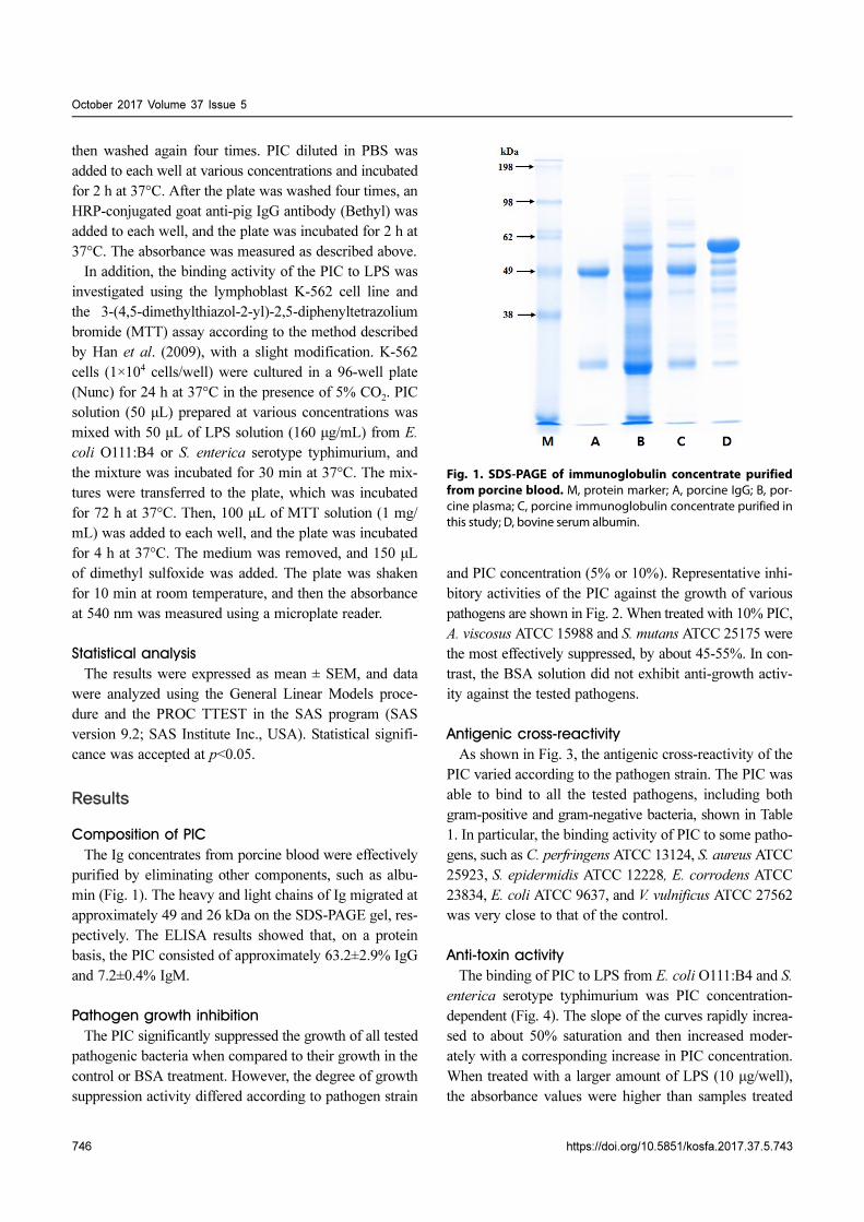
Composition of PIC
The Ig concentrates from porcine blood were effectively
purified by eliminating other components, such as albu-
min (Fig. 1). The heavy and light chains of Ig migrated at
approximately 49 and 26 kDa on the SDS-PAGE gel, res-
pectively. The ELISA results showed that, on a protein
basis, the PIC consisted of approximately 63.2±2.9% IgG
and 7.2±0.4% IgM.
Pathogen growth inhibition
The PIC significantly suppressed the growth of all tested
pathogenic bacteria when compared to their growth in the
control or BSA treatment. However, the degree of growth
suppression activity differed according to pathogen strain
and PIC concentration (5% or 10%). Representative inhi-
bitory activities of the PIC against the growth of various
pathogens are shown in Fig. 2. When treated with 10% PIC,
A. viscosus ATCC 15988 and S. mutans ATCC 25175 were
the most effectively suppressed, by about 45-55%. In con-
trast, the BSA solution did not exhibit anti-growth activ-
ity against the tested pathogens.
Antigenic cross-reactivity
As shown in Fig. 3, the antigenic cross-reactivity of the
PIC varied according to the pathogen strain. The PIC was
able to bind to all the tested pathogens, including both
gram-positive and gram-negative bacteria, shown in Table
1. In particular, the binding activity of PIC to some patho-
gens, such as C. perfringens ATCC 13124, S. aureus ATCC
25923, S. epidermidis ATCC 12228, E. corrodens ATCC
23834, E. coli ATCC 9637, and V. vulnificus ATCC 27562
was very close to that of the control.
Anti-toxin activity
The binding of PIC to LPS from E. coli O111:B4 and S.
enterica serotype typhimurium was PIC concentration-
dependent (Fig. 4). The slope of the curves rapidly increa-
sed to about 50% saturation and then increased moder-
ately with a corresponding increase in PIC concentration.
When treated with a larger amount of LPS (10 μg/well),
the absorbance values were higher than samples treated
Fig. 1. SDS-PAGE of immunoglobulin concentrate purifiedfrom porcine blood. M, protein marker; A, porcine IgG; B, por-cine plasma; C, porcine immunoglobulin concentrate purified inthis study; D, bovine serum albumin.

Anti-pathogenic Properties of Porcine Immunoglobulin Concentrate
https://doi.org/10.5851/kosfa.2017.37.5.743 747
with a smaller amount of LPS concentration (0.1 μg/well).
The anti-toxin activity of the PIC was also examined using
the K-562 cell line. The growth rate of the K-562 cells was
decreased by treatment with LPS (160 μg/mL) but, this
reduction was restored by the addition of PIC (Fig. 5).
Treatment with PIC at 300-500 μg/mL markedly increased
the viability of the K-562 cells (p<0.05 or p<0.01), which
means that the PIC could bind to LPS, reducing the patho-
genicity of LPS.
Fig. 2. Inhibition of pathogen growth by porcine immunoglobulin concentrate. Values are expressed as means ± SEM. Differ-ences were considered to be statistically significant at p<0.01 (**) or p<0.001 (***). BSA, bovine serum albumin; PIC, porcine immuno-globulin concentrate. A, Clostridium perfringens ATCC 13124; B, Streptococcus mutans ATCC 25175; C, Actinomyces viscosus ATCC15988; D, Escherichia coli ATCC 9637.
Fig. 3. Antigenic cross-reactivity of porcine immunoglobulin concentrate against various pathogens. Values are expressed asmeans±SEM. Means without a common letter differ significantly (p<0.001). The pathogens used are listed in Table 1. CON, control.

October 2017 Volume 37 Issue 5
748 https://doi.org/10.5851/kosfa.2017.37.5.743
Discussion
It is known that administration of animal plasma in
livestock improves intestinal barrier function and muco-
sal inflammation as well as growth performance (Bosi et
al., 2004; Peace et al., 2011). Pierce et al. (2005) reported
that the beneficial effects of animal plasma were primar-
ily mediated by the Ig fraction. Administration of Ig inc-
reased the levels of anti-inflammatory cytokines in the
intestinal mucosa (Pérez-Bosque et al., 2008) and redu-
ced the expression of pro-inflammatory cytokines, such
as tumor necrosis factor alpha, interferon gamma, inter-
leukin 2, and interleukin 17, in various in vitro models
(Bosi et al., 2004; Moretó and Pérez-Bosque, 2009). Other
studies have shown that dietary supplements with Ig imp-
roved intestinal health and immunity in clinical trials (As-
muth et al., 2013; Wilson et al., 2013). The Ig concentrate
isolated from porcine blood in this study contained app-
roximately 63% IgG on a protein basis. This suggests its
potential value as a supplement for functional food or ani-
mal diet, because commercial Ig fractions generally con-
tain less than 50% IgG.
In the present study, the PIC not only inhibited the
growth of various pathogens but also directly bound to
their surface. It was reported that IgM had antibacterial
activity and decreased the production of matrix metallo-
proteinase-1 caused by LPS (Zhou et al., 2007). Petschow
et al. (2014) reported that Ig, especially IgG, had antibac-
terial activity against pathogenic bacteria, because Ig inhi-
bited the ability of pathogens or their toxins, which mig-
Fig. 4. The binding of porcine immunoglobulin concentrate to two types of lipopolysaccharide (LPS) from Escherichia coliO111:B4 and Salmonella enterica serotype Typhimurium. Values are expressed as means±SEM. A540, Absorbance at 540 nm.
Fig. 5. Effect of porcine immunoglobulin concentrate on the proliferation of lymphoblast K-562 cells treated with lipopoly-saccharide (LPS). Values are expressed as means ± SEM. Asterisks indicate significant differences, (*) p<0.05 and (**) p<0.01. A, Esche-richia coli O111:B4 LPS (160 μg/mL); B, Salmonella enterica serotype Typhimurium LPS (160 μg/mL). A540, Absorbance at 540 nm.

Anti-pathogenic Properties of Porcine Immunoglobulin Concentrate
https://doi.org/10.5851/kosfa.2017.37.5.743 749
rated through the mucus layer into epithelial cells, and
damaged pathogens through direct binding. Furthermore,
the Fab fragment of IgG imparts diversity, and the Fc frag-
ment can interact with the Fc gamma receptors on some
immune cells, including macrophages and monocytes, thus
stimulating their phagocytic activity (Petschow et al., 2014).
Several studies have reported that the Ig present in SDAP
reduced pathogen adhesion to the gut mucosa as well as
the intestinal inflammation caused by pathogenic bacte-
ria, thus providing immunological benefits (Pérez-Bosque
and Moretó, 2010; Van Dijka et al., 2001).
PIC can bind to LPS on the surface of gram-negative
bacteria and reduce its growth-repressing action on K-562
cells. Hence, PIC may protect the intestine from diseases
caused by gram-negative bacteria and their toxins. Bacte-
rial LPS can cause various tissue pathologies and intesti-
nal permeability disorders, such as Crohn’s disease, nec-
rotizing enterocolitis, and inflammatory bowel disease,
by increasing intestinal epithelial tight junction permea-
bility (Guo et al., 2013; Magata et al., 2015). The entero-
toxigenic E. coli (ETEC) and Salmonella strains used in
the present study induce severe enteropathy. ETEC adheres
to the intestinal epithelium through various routes using
colonization factors, which include more than 20 known
multimeric structures, and it is the most common cause of
diarrhea (O'Ryan et al., 2015). Salmonella strains, as food-
borne enteropathogens, cause serious gastrointestinal inv-
asive diseases (MacLennan et al., 2014). The Ig present
in plasma might provide effective protection from these
bacterial toxin-involved intestinal diseases, and increase
phagocytic activity and lymphocyte proliferation (Balan
and Moughan, 2013; Niewold et al., 2007). Previous stu-
dies have also reported that supplementation with SDAP
reduced the expression of inflammatory cytokines and
prevented the inflammatory diseases caused by LPS (Pérez-
Bosque et al., 2016; Touchette et al., 2002).
Conclusion
In this study, we effectively isolated Ig concentrate from
porcine blood. We demonstrated that the produced PIC
has anti-bacterial and anti-toxin activities, which may pro-
vide protection against pathogenic bacteria. The commer-
cial development of PIC would not only reduce the cost
burden of slaughterhouse blood disposal but may also be
a potentially valuable functional food material or supple-
ment as an alternative to antibiotics in livestock diets.
Acknowledgements
This paper was supported by Korea Institute of Plan-
ning and Evaluation for Technology in Food, Agriculture,
Forestry and Fisheries (IPET) through Agri-Bioindustry
Technology Development Program, funded by Ministry of
Agriculture, Food and Rural Affairs (316029-3).
References
Asmuth, D. M., Ma, Z. M., Albanese, A., Sandler, N. G., Devaraj,
S., Knight, T. H., Flynn, N. M., Yotter, T., Garcia, J. C., Tsu-
chida, E., Wu, T. T., Douek, D. C., and Miller, C. J. (2013) Oral
serum-derived bovine immunoglobulin improves duodenal
immune reconstitution and absorption function in patients
with HIV enteropathy. AIDS 27, 2207-2217.
Balan, P., Han, K. S., Rutherfurd-Markwick, K., Singh, H., and
Moughan, P. J. (2011) Ovine serum immunoglobulin has im-
munomodulatory effects in growing rats gavaged with Salm-
onella enteritidis. J. Nutr. 141, 950-956.
Balan, P. and Moughan, P. J. (2013) Intact but not denatured ovine
serum immunoglobulins positively modulate mucosal imm-
une mediators in the growing rat challenged with Salmonella
enteritidis. Br. J. Nutr. 110, 1031-1039.
Bosi, P., Casini, L., Finamore, A., Cremokolini, C., Merialdi, G.,
Trevisi, P., Nobili, F., and Mengheri, E. (2004) Spray-dried
plasma improves growth performance and reduces inflam-
matory status of weaned pigs challenged with enterotoxigenic
Escherichia coli K88. J. Anim. Sci. 82, 1764-1772.
Coffey, R. D. and Cromwell, G. L. (1995) The impact of environ-
ment and antimicrobial agents on the growth response of early-
weaned pigs to spray-dried porcine plasma. J. Anim. Sci. 73,
2532-2539.
Guo, S., Al-Sadi, R., Said, H. M., and Ma, T. Y. (2013) Lipopoly-
saccharide causes an increase in intestinal tight junction per-
meability in vitro and in vivo by inducing enterocyte memb-
rane expression and localization of TLR-4 and CD14. Am. J.
Pathol. 182, 375-387.
Han, K. S., Boland, M., Singh, H., and Moughan, P. J. (2009) The
in vitro anti-pathogenic activity of immunoglobulin concen-
trates extracted from ovine blood. Appl. Biochem. Biotech-
nol. 157, 442-452.
Hyun, C. K. and Shin, H. G. (1999) Production of angiotensin I
converting enzyme inhibitory peptides from bovine blood
plasma proteins. Korean J. Biotechnol. Bioeng. 14, 600-605.
Hyun, C. K. and Shin, H. K. (1998) Optimization of freeze-drying
conditions for probiotics production with animal blood pro-
teins added medium. Korean J. Appl. Microbiol. Biotechnol.
26, 200-205.
Jang, Y. H., Kim, H. B., Lee, M. H., Baek, H., and Choe, N. H.
(2011) Utilization and hygiene status of animal blood from
slaughterhouse in Korea. J. Prev. Vet. Med. 35, 73-79.
Jeon, Y. W. (2015) Optimization of ultrasonification of slaughter
blood for protein solubilization. Environ. Eng. Res. 20, 163-

October 2017 Volume 37 Issue 5
750 https://doi.org/10.5851/kosfa.2017.37.5.743
169.
Jeon, Y. W., Kim, H. J., Cho, Y. H., and Yoo, H. M. (2013) Biolo-
gical conversion from slaughter blood into amino acid lique-
fied fertilizer. Int. J. Environ. Sci. Dev. 4, 509-513.
Kim, G. E. (2014) Protein degradation in waste blood for liquid
fertilizer production. J. Korean Soc. Urban Environ. 14, 127-
134.
Kim, M. S., Yu, J. E., Min, K. H., Kim, J. H., Choi, I. H., and Nahm,
S. S. (2012) Assessing biological safety of the Hanwoo serum
obtained during slaughtering process. J. Anim. Sci. Technol.
54, 59-63.
Kim, M. Y., Kim, M. A., and Jeong, Y. J. (2016) Characteristics of
iron powder formulation produced from porcine blood by
enzymatic treatment. Korean J. Food Preserv. 23, 753-757.
Laemmli, U. K. (1970) Cleavage of structural proteins during the
assembly of the head of bacteriophage T4. Nature 227, 680-
685.
Lallès, J. P., Bosi, P., Janczyk, P., Koopmans, S. J., and Torrallard-
ona, D. (2009) Impact of bioactive substances on the gastro-
intestinal tract and performance of weaned piglets: A review.
Animal 3, 1625-1643.
Ma, J. S., Shin, K. S., and Park, G. H. (2001) Utilization of porcine
blood and liver in yeast culture for animal diets and effects of
yeast culture on the growth of broiler chicks. J. Anim. Environ.
Sci. 7, 21-28.
MacLennan, C. A., Martin, L. B., and Micoli, F. (2014) Vaccines
against invasive Salmonella disease: Current status and future
directions. Hum. Vaccin Immunother. 10, 1478-1493.
Magata, F., Ishida, Y., Miyamoto, A., Furuoka, H., Inokuma, H.,
and Shimizu, T. (2015) Comparison of bacterial endotoxin
lipopolysaccharide concentrations in the blood, ovarian fol-
licular fluid and uterine fluid: A clinical case of bovine met-
ritis. J. Vet. Med. Sci. 77, 81-84.
Moretó, M. and Pérez-Bosque, A. (2009) Dietary plasma proteins,
the intestinal immune system, and the barrier functions of the
intestinal mucosa. J. Anim. Sci. 87, 92-100.
Niewold, T. A., van Dijk, A. J., Geenen, P. L., Roodink, H., Mar-
gry, R., and van der Meulen, J. (2007) Dietary specific antibo-
dies in spray-dried immune plasma prevent enterotoxigenic
Escherichia coli F4 (ETEC) post weaning diarrhoea in pig-
lets. Vet. Microbiol. 124, 362-369.
O'Ryan, M., Vidal, R., del Canto, F., Carlos Salazar, J., and Mon-
tero, D. (2015) Vaccines for viral and bacterial pathogens cau-
sing acute gastroenteritis: Part II: Vaccines for Shigella, Salm-
onella, enterotoxigenic E. coli (ETEC) enterohemorrhagic E.
coli (EHEC) and Campylobacter jejuni. Hum. Vaccin Immu-
nother. 11, 601-619.
Park, G. H. (1997) Optimum drying condition for slaughter por-
cine blood and its utilization as broiler diets. Korean J. Poult.
Sci. 24, 59-66.
Peace, R. M., Campbell, J., Polo, J., Crenshaw, J., Russell, L., and
Moeser, A. (2011) Spray-dried porcine plasma influences int-
estinal barrier function, inflammation, and diarrhea in weaned
pigs. J. Nutr. 141, 1312-1317.
Pérez-Bosque, A., Miró, L., Amat, C., Polo, J., and Moretó, M.
(2016) The anti-inflammatory effect of spray-dried plasma is
mediated by a reduction in mucosal lymphocyte activation
and infiltration in a mouse model of intestinal inflammation.
Nutrients DOI: 10.3390/nu8100657.
Pérez-Bosque, A., Miró, L., Polo, J., Russell, L., Campbell, J.,
Weaver, E., Crenshaw, J., and Moretó, M. (2008) Dietary pla-
sma proteins modulate the immune response of diffuse gut-
associated lymphoid tissue in rats challenged with Staphylo-
coccus aureus enterotoxin B. J. Nutr. 138, 533-537.
Pérez-Bosque, A. and Moretó, M. (2010) A rat model of mild int-
estinal inflammation induced by Staphylococcus aureus ent-
erotoxin B. Proc. Nutr. Soc. 69, 447-453.
Pérez-Bosque, A., Polo, J., and Torrallardona, D. (2016) Spray
dried plasma as an alternative to antibiotics in piglet feeds,
mode of action and biosafety. Porcine Health Manag. DOI:
10.1186/s40813-016-0034-1.
Petschow, B. W., Blikslager, A. T., Weaver, E. M., Campbell, J.
M., Polo, J., Shaw, A. L., Burnett, B. P., Klein, G. L., and Rho-
ads, J. M. (2014) Bovine immunoglobulin protein isolates for
the nutritional management of enteropathy. World J. Gastro-
enterol. 20, 11713-11726.
Pierce, J. L., Cromwell, G. L., Lindemann, M. D., Russell, L. E.,
and Weaver, E. M. (2005) Effects of spray-dried animal pla-
sma and immunoglobulins on performance of early weaned
pigs. J. Anim. Sci. 83, 2876-2885.
Quigley, J. D., Campbell, J. M., Polo, J., and Russell, L. E. (2004)
Effects of spray-dried animal plasma on intake and apparent
digestibility in dogs. J. Anim. Sci. 82, 1685-1692.
SAS. (2008) SAS/STAT Software for PC. Release 9.2, SAS Insti-
tute Inc., Cary, NC, USA.
Tomita, G. M., Todhunter, D. A., Hogan, J. S., and Smith, K. L.
(1995) Antigenic crossreactivity and lipopolysaccharide neu-
tralization properties of bovine immunoglobulin G. J. Dairy
Sci. 78, 2745-2752.
Torrallardona, D. (2010) Spray dried animal plasma as an alterna-
tive to antibiotics in weanling pigs: A review. Asian-Aust-
ralas. J. Anim. Sci. 23, 131-148.
Touchette, K. J., Carroll, J. A., Allee, G. L., Matteri, R. L., Dyer,
C. J., Beausang, L. A., and Zannelli, M. E. (2002) Effect of
spray-dried plasma and lipopolysaccharide exposure on wea-
ned pigs: I. Effects on the immune axis of weaned pigs. J. Anim.
Sci. 80, 494-501.
Van Dijk, A. J., Everts, H., Nabuurs, M. J. A., Margry, R. J. C. F.,
and Beynen, A. C. (2001) Growth performance of weanling
pigs fed spray-dried animal plasma: A review. Livest. Prod.
Sci. 68, 263-274.
Wilson, D., Evans, M., Weaver, E., Shaw, A. L., and Klein, G. L.
(2013) Evaluation of serum-derived bovine immunoglobulin
protein isolate in subjects with diarrhea-predominant irrita-
ble bowel syndrome. Clin. Med. Insights Gastroenterol. 6, 49-
60.
Yu, X. Q. and Kanost, M. R. (2002) Binding of hemolin to bacter-
ial lipopolysaccharide and lipoteichoic acid. Eur. J. Biochem.
269, 1827-1834.
Yun, S. S., Lee, H. G., Choi, Y. J., Song, E. S., and Juhn, S. L.
Anti-pathogenic Properties of Porcine Immunoglobulin Concentrate
https://doi.org/10.5851/kosfa.2017.37.5.743 751
(1998) Analysis of the plasma proteins from bovine and por-
cine blood and their emulsifying activity. Korean J. Food Sci.
Technol. 30, 988-991.
Zhou, Z. H., Zhang, Y., Hu, Y. F., Wahl, L. M., Cisar, J. O., and
Notkins, A. L. (2007) The broad antibacterial activity of the
natural antibody repertoire is due to polyreactive antibodies.
Cell Host Microbe. 15, 51-61.


![Exploiting Nonlinear Recurrence and Fractal Scaling Prop- erties … · 2018-10-25 · arXiv:0707.0086v1 [nlin.CG] 1 Jul 2007 Exploiting Nonlinear Recurrence and Fractal Scaling Prop-erties](https://static.fdocuments.us/doc/165x107/5e7dfddc4cb867652547420b/exploiting-nonlinear-recurrence-and-fractal-scaling-prop-erties-2018-10-25-arxiv07070086v1.jpg)