Article 42/Number 3/2011 175 due to a myotoxic reaction to the retrobulbar anesthesia, not a palsy...
6
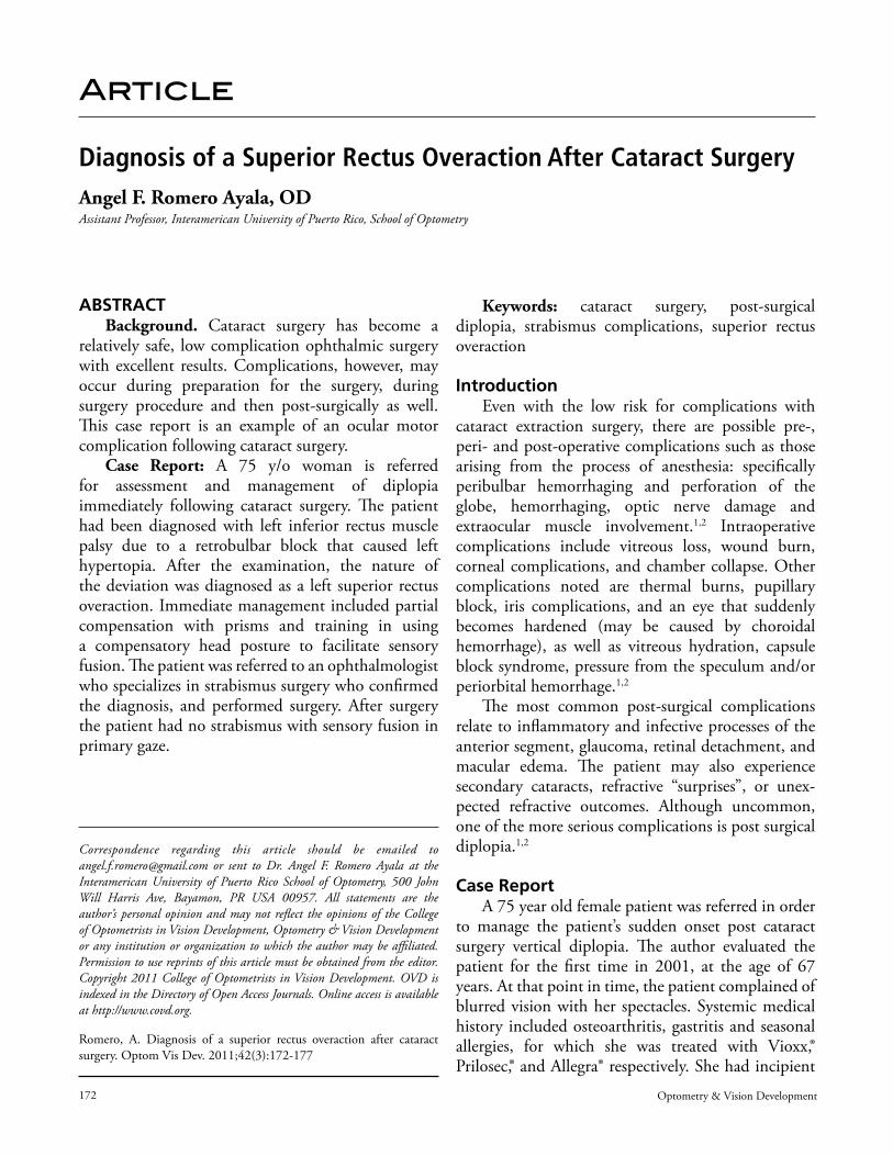
172 Optometry & Vision Development Article Diagnosis of a Superior Rectus Overaction After Cataract Surgery Angel F. Romero Ayala, OD Assistant Professor, Interamerican University of Puerto Rico, School of Optometry ABSTRACT Background. Cataract surgery has become a relatively safe, low complication ophthalmic surgery with excellent results. Complications, however, may occur during preparation for the surgery, during surgery procedure and then post-surgically as well. is case report is an example of an ocular motor complication following cataract surgery. Case Report: A 75 y/o woman is referred for assessment and management of diplopia immediately following cataract surgery. e patient had been diagnosed with left inferior rectus muscle palsy due to a retrobulbar block that caused left hypertopia. After the examination, the nature of the deviation was diagnosed as a left superior rectus overaction. Immediate management included partial compensation with prisms and training in using a compensatory head posture to facilitate sensory fusion. e patient was referred to an ophthalmologist who specializes in strabismus surgery who confirmed the diagnosis, and performed surgery. After surgery the patient had no strabismus with sensory fusion in primary gaze. Keywords: cataract surgery, post-surgical diplopia, strabismus complications, superior rectus overaction Introduction Even with the low risk for complications with cataract extraction surgery, there are possible pre-, peri- and post-operative complications such as those arising from the process of anesthesia: specifically peribulbar hemorrhaging and perforation of the globe, hemorrhaging, optic nerve damage and extraocular muscle involvement. 1,2 Intraoperative complications include vitreous loss, wound burn, corneal complications, and chamber collapse. Other complications noted are thermal burns, pupillary block, iris complications, and an eye that suddenly becomes hardened (may be caused by choroidal hemorrhage), as well as vitreous hydration, capsule block syndrome, pressure from the speculum and/or periorbital hemorrhage. 1,2 e most common post-surgical complications relate to inflammatory and infective processes of the anterior segment, glaucoma, retinal detachment, and macular edema. e patient may also experience secondary cataracts, refractive “surprises”, or unex- pected refractive outcomes. Although uncommon, one of the more serious complications is post surgical diplopia. 1,2 Case Report A 75 year old female patient was referred in order to manage the patient’s sudden onset post cataract surgery vertical diplopia. e author evaluated the patient for the first time in 2001, at the age of 67 years. At that point in time, the patient complained of blurred vision with her spectacles. Systemic medical history included osteoarthritis, gastritis and seasonal allergies, for which she was treated with Vioxx,® Prilosec,® and Allegra® respectively. She had incipient Correspondence regarding this article should be emailed to [email protected] or sent to Dr. Angel F. Romero Ayala at the Interamerican University of Puerto Rico School of Optometry, 500 John Will Harris Ave, Bayamon, PR USA 00957. All statements are the author’s personal opinion and may not reflect the opinions of the College of Optometrists in Vision Development, Optometry & Vision Development or any institution or organization to which the author may be affiliated. Permission to use reprints of this article must be obtained from the editor. Copyright 2011 College of Optometrists in Vision Development. OVD is indexed in the Directory of Open Access Journals. Online access is available at http://www.covd.org. Romero, A. Diagnosis of a superior rectus overaction after cataract surgery. Optom Vis Dev. 2011;42(3):172-177
Transcript of Article 42/Number 3/2011 175 due to a myotoxic reaction to the retrobulbar anesthesia, not a palsy...
172 Optometry & Vision Development
Article
Diagnosis of a Superior Rectus Overaction After Cataract SurgeryAngel F. Romero Ayala, ODAssistant Professor, Interamerican University of Puerto Rico, School of Optometry
ABSTRACTBackground. Cataract surgery has become a
relatively safe, low complication ophthalmic surgery with excellent results. Complications, however, may occur during preparation for the surgery, during surgery procedure and then post-surgically as well. This case report is an example of an ocular motor complication following cataract surgery.
Case Report: A 75 y/o woman is referred for assessment and management of diplopia immediately following cataract surgery. The patient had been diagnosed with left inferior rectus muscle palsy due to a retrobulbar block that caused left hypertopia. After the examination, the nature of the deviation was diagnosed as a left superior rectus overaction. Immediate management included partial compensation with prisms and training in using a compensatory head posture to facilitate sensory fusion. The patient was referred to an ophthalmologist who specializes in strabismus surgery who confirmed the diagnosis, and performed surgery. After surgery the patient had no strabismus with sensory fusion in primary gaze.
Keywords: cataract surgery, post-surgical diplopia, strabismus complications, superior rectus overaction
IntroductionEven with the low risk for complications with
cataract extraction surgery, there are possible pre-, peri- and post-operative complications such as those arising from the process of anesthesia: specifically peribulbar hemorrhaging and perforation of the globe, hemorrhaging, optic nerve damage and extraocular muscle involvement.1,2 Intraoperative complications include vitreous loss, wound burn, corneal complications, and chamber collapse. Other complications noted are thermal burns, pupillary block, iris complications, and an eye that suddenly becomes hardened (may be caused by choroidal hemorrhage), as well as vitreous hydration, capsule block syndrome, pressure from the speculum and/or periorbital hemorrhage.1,2
The most common post-surgical complications relate to inflammatory and infective processes of the anterior segment, glaucoma, retinal detachment, and macular edema. The patient may also experience secondary cataracts, refractive “surprises”, or unex-pected refractive outcomes. Although uncommon, one of the more serious complications is post surgical diplopia.1,2
Case ReportA 75 year old female patient was referred in order
to manage the patient’s sudden onset post cataract surgery vertical diplopia. The author evaluated the patient for the first time in 2001, at the age of 67 years. At that point in time, the patient complained of blurred vision with her spectacles. Systemic medical history included osteoarthritis, gastritis and seasonal allergies, for which she was treated with Vioxx,® Prilosec,® and Allegra® respectively. She had incipient
Correspondence regarding this article should be emailed to [email protected] or sent to Dr. Angel F. Romero Ayala at the Interamerican University of Puerto Rico School of Optometry, 500 John Will Harris Ave, Bayamon, PR USA 00957. All statements are the author’s personal opinion and may not reflect the opinions of the College of Optometrists in Vision Development, Optometry & Vision Development or any institution or organization to which the author may be affiliated. Permission to use reprints of this article must be obtained from the editor. Copyright 2011 College of Optometrists in Vision Development. OVD is indexed in the Directory of Open Access Journals. Online access is available at http://www.covd.org.
Romero, A. Diagnosis of a superior rectus overaction after cataract surgery. Optom Vis Dev. 2011;42(3):172-177
Volume 42/Number 3/2011 173
cataracts, yet her refractive correction allowed for 20/20 monocular visual acuity. She was not evaluated again until the referral in 2009.
In February 2009, the patient was referred from a colleague in order to manage a vertical strabismus with constant diplopia that was present since cataract extraction surgery was performed in the left eye. The history revealed that she underwent cataract extraction in the OD on February 2008, and OS on June 2008. At this time the medical history noted controlled hypertension, hypothyroidism, osteoarthritis, and gastritis. These were being treated with Atenelol, Synthroid,® Indocin,® and Nexium® respectively. She also indicated a history of sinusitis, gallbladder problems, foot surgery and a hysterectomy.
Upon further questioning regarding the chief complaint, the patient noted that the day after the OS cataract extraction, she noticed that the left eye was deviated down and that she had double vision. On the follow-up appointment with the ophthalmic surgeon, he informed the patient about the possibility of an inflammatory response causing the deviation of the eye. The surgeon prescribed prednisone 10 mg two tablets three times a day for 14 days. The patient followed the instructions, but by the third day, she noticed yellow-white grainy plaques in the mouth. At this time the prednisone was discontinued, and the surgeon suggested substitution of the prednisone with Advil. The reaction in the oral mucosa subsided with removal of the prednisone.
At the one-month follow-up appointment, the ophthalmologist instructed her to return for another
follow-up in three months to re-evaluate the strabismus that now presented as a 35Δ left hypertropia. The ophthalmic surgeon indicated that there might be a gradual resolution of the deviation.
Her last visit to the ophthalmologist was on October 2008, where he then diagnosed the patient with a 30Δ left hypertropia due to left inferior rectus muscle palsy, a consequence of a myotoxic reaction to the retrobulbar-blocking agent.
The ophthalmologist provided an unusual referral to a retinologist for a strabismus surgical consult. The retinologist ordered an orbit MRI with fat suppression, after which he also diagnosed with a left inferior rectus muscle palsy. At this visit the retinologist suggested that the surgical procedure to correct the deviation be based on surgical intervention of both eyes. The patient then decided to seek a second opinion.
On the February 2009 evaluation by the author, her uncorrected visual acuities were 20/60 OD, 20/20 OS, while the best-corrected visual acuities were 20/20 OD/OS. Pupils were round, equal and reactive to light with no afferent pupillary defect noted. Confrontation fields were full OD/OS. External ocular health assessment revealed blepharochalasis OU, conjunctival hypertrophy nasal OD/OS, clear corneas, flat healthy irises with a deep, quiet anterior chamber. Intraocular posterior chamber implants were clear as was the posterior subcapsule. Internal ocular health assessment revealed vitreous floaters with signs of early age related macular degeneration and cup to disc ratios of .2 OD/OS. Intraocular pressures using applanation tonometry were 13 mmHg OD/OS.
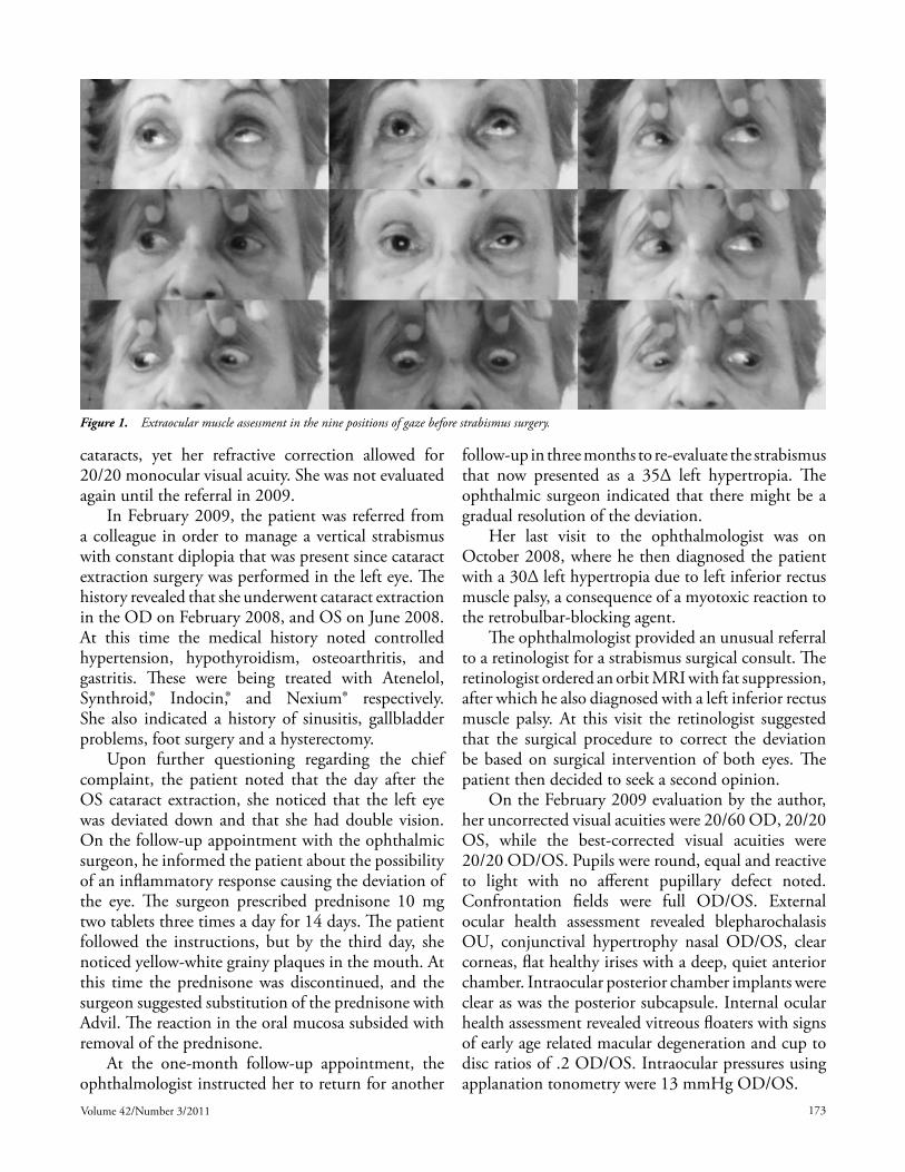
Figure 1. Extraocular muscle assessment in the nine positions of gaze before strabismus surgery.

174 Optometry & Vision Development
Ocular motor assessment revealed a 27Δ left hypertropia with an 8Δ exophoria both at distance and near. Extraocular muscle assessment revealed an increase in the angle of deviation on dextro-supra version, levoversion, levo-supra version, with minimal presentation of the angle of deviation on levo-infra version and almost no deviation on dextro-infra version, and secondary depression. (Figure 1) The Park Three Step test was performed and suggested a left inferior rectus muscle palsy. The patient brought the MRI report, and the radiologist concluded that the eye globes were normal in size and position, with no orbital lesion, and normal extra ocular muscle appearance. Taking into consideration the nature of the strabismus and observation of the pattern of behavior through the nine positions of gaze the diagnosis was determined to be a left superior rectus overaction.
Immediate management for this patient was to use a partial compensating prism. This was a 15 diopter Fresnel prism base down on the spectacle lens of the left eye. She was also taught a compensating head posture with the chin slightly elevated to facilitate sensory fusion. The Worth Four Dot test was performed with the prismatic correction and compensatory head tip resulted in sensory fusion at distance.
Since the patient had the strabismus manifest for eight months, it was obvious that the angle of strabismus was not going to reduce. The patient was then referred to an ophthalmic surgeon who specializes in strabismus surgery for a consult. The strabismus surgeon confirmed the diagnosis of left superior rectus muscle overaction and performed surgery. The
surgical correction consisted of a recession of the left superior rectus and resection of the left inferior rectus with adjustable sutures. The patient has sensory fusion in primary gaze, with a post surgical right hypertropia on secondary gaze elevation that is expected to be asymptomatic and improve over time. Figure 2
Discussion/Literature ReviewThe diagnosis of a left superior rectus over-action
can be reached by careful examination of the ocular motility patterns in the nine positions of gaze. The suspected muscles in this patient’s case, are the two depressors of the left eye, the left inferior rectus and the left superior oblique. Of these two, the most likely suspect would be a paretic inferior rectus, due to the proximity of the needle during retrobulbar anesthesia. Careful observation of the hypertropia of the left eye, reveals that the strabismus does not increase in magnitude in the field of action of either muscle. In fact, the angle of strabismus increases in the field of action of the left superior rectus.
This overaction is precisely the reason why the Park Three Step test results in a paradoxical conclusion. It is the very nature of the superior rectus overaction that limits the ability of the inferior rectus to fully depress the eye globe.
Additionally, the patient’s astute observation that immediately after the surgery the left eye was deviated down and then the left eye was deviated up is also significant. This observation confirms the suspicion that the vertical strabismus in this case is due to an overaction of the superior rectus muscle contraction
Figure 2. Extraocular motor assessment in the nine positions of gaze after strabismus surgery.

Volume 42/Number 3/2011 175
due to a myotoxic reaction to the retrobulbar anesthesia, not a palsy of the left inferior rectus muscle.
Post surgical strabismus is a rare consequence of cataract surgery. A literature review of the causes of vertical deviations as a result of cataract surgery mostly indicates myotoxicity, use of bridle sutures and surgical trauma which includes: perforation of the muscle that cause hemorrhaging or trauma to the nerve.1-19
Retrobulbar anesthesia is used to immobilize the eye globe. The general goal of this injection is to inject a blocking agent within the retrobulbar space to saturate the exposed nerves that innervate the extraocular muscles in order to block their conduction. It has often been seen that the superior oblique may still retain function since the trochlear nerve runs outside the muscle cone. The general approach of the needle is from the inferior temporal orbital rim, and once passing the orbital septum angled up and in toward the muscle cone. Typically lidocaine, bupivacaine or both are used. Complications associated with retrobulbar anesthesia can include optic nerve injury, brainstem anesthesia, globe penetration, globe perforation, retrobulbar hemorrhaging and extraocular muscle damage.1-2
Koide et al, proposes that the mechanisms of post cataract surgery diplopia can be attributed to five possible mechanisms. They are:
1. An improvement in visual acuity may unmask a pre-existing diplopia.
2. An injury to one of the extraocular muscles with use of bridle sutures.
3. Traumatic injury to an extraocular muscle from the needle used for retrobulbar anesthesia.
4. Allergic or toxic reaction to the anesthetic agent.
5. Optical aniseikonia.3
Hamed, made an attempt to categorize the etiology of post cataract surgical onset according to four categories: 1) preexisting or concurrent disorders masked by the cataract, 2) conditions precipitated by the effect of prolonged occlusion by the cataract, 3) surgical trauma, and 4) resulting aphakia or pseudophakia and associated optical aberrations.4
In a retrospective analysis by MacDonald et al, it was found that in a 22-month period the risk of non-transient post surgical diplopia was 2.6 cases per 1000 cataract surgery procedures. They also documented that out of the 21 patients with post
cataract surgery strabismus, 16 had involvement of the inferior rectus muscle and one had the inferior and medial recti muscles involvement, while involvement of the medial rectus, inferior oblique, lateral rectus and the superior rectus had one case each.5 Capó, et al, cite that out of 28 patients with vertical diplopia, 11 presented restriction of the inferior rectus, six had overaction of the inferior rectus, eight overaction of the superior rectus, and three paresis of the superior rectus muscle.6 Johnson reported that persistent vertical diplopia occurred in 32 of 17531 eyes that underwent cataract surgery (0.18%). He also reports that when comparing surgeries that relied on topical anesthesia, as opposed to retrobulbar block, the group that relied on topical anesthesia had no reports of vertical diplopia, and all cases, 32 (0.23%) out of 13714 eyes that had vertical diplopia had retrobulbar anesthesia.7
The issue of the surgeon’s dominant hand as a contributing factor has also been investigated. It has been proposed that the higher incidence of left eye involvement might be due to various techniques used by right-handed surgeons and anesthesists.6,7,8
Esswein and von Noorden reported on nine patients who were referred presenting vertical strabismus after undergoing cataract extraction. These nine cases involved vertically acting muscles: seven had complete or partial paralysis of the inferior rectus muscle, and two had paresis of the superior rectus muscle. It is also notable that six of the patients affected had the right eye affected, and three the left eye.9
Our literature review noted four references that addressed the issue of superior rectus overaction.6,10,11,12 These included Grimmet and Lambert who found four cases of superior rectus overaction out of seven cases in a 25-month period that had strabismus onset after cataract surgery. They based the diagnosis of superior rectus overaction on: 1) hypertropia worse on upgaze, 2) the greatest measured deviation in the field of action of the superior rectus muscle, and 3) normal contralateral ductions. They propose that the superior rectus overaction results from one of three possible injuries to the inferior rectus muscle, muscle fiber damage from the needle, laceration to the anterior ciliary artery causing ischemic damage, or direct trauma to the nerve supply to the inferior rectus. Any of these they propose, will cause inferior rectus muscle palsy, leading to a secondary contraction of the superior rectus muscle.10
176 Optometry & Vision Development
Capó and Guyton studied 19 cases and determined that 16 of these cases had an overaction presenting a larger magnitude deviation in the field of action of the overacting muscle. They propose that myotoxicity from direct injection of local anesthetic causes a transient paresis and is followed by segmental contracture. They also propose that the pattern of segmented contraction affects the overall length of the muscle, resulting in a shorter muscle. Any neural impulse results in a stronger muscle response.12
Capó et al, compared anesthesia methods to the resulting strabismus in 28 vertical strabismus patients manifest after cataract surgery. As part of the investigation, they concluded that the superior rectus may be touched by the retrobulbar anesthetic delivery needle, as opposed to the effect to the inferior rectus which is more traumatic. The inferior rectus is exposed to widespread damage that results in restrictive strabismus. The superior rectus muscle is likely to be touched by the needle causing limited focal damage. This focal, segmental damage leads to focal contraction, reducing the overall length of the muscle, thus causing an overacting effect.6
The theory set forth by Capó et al,6 was confirmed by Kim and Hwang,11 who used thin section MRI imaging of the superior rectus muscle in a patient who presented a vertical deviation after cataract extraction surgery. They confirmed the theory of focal contracture of the superior rectus muscle near the orbital apex.
This theory might also explain Hamed and Mancuso’s findings. They performed computed tomography studies of three patients who developed post cataract surgery hypotropia, due to injury to the inferior rectus muscle. These three patients had segmental retrobulbar enlargement of the inferior rectus and developed hypotropia of the affected eye.13
It is interesting to note that Hunter et al, reported four cases of post surgical diplopia where three had inferior oblique contracture caused by myotoxicity, and another with either mechanical trauma to the inferior oblique or direct nerve anesthetic toxicity. It is also very interesting that three out of the four patients had involvement of the left eye.14
Even though topical anesthesia has been found safer,7 retrobulbar anesthesia is still the procedure of choice to immobilize the eye. Suggestions also include the use of a blunt-tipped canula.12
The management of these patients ranges from compensation with prisms,14 use of botulinum toxin16
to surgical intervention.3,10,16-18 It is not yet clear what role optometric vision therapy may play with these surgically induced disorders.
ConclusionThis case highlights the importance of patient
education, accurate diagnosis and timely, adequate referrals. Proper patient education before cataract surgery should include the discussion of the possibility of post surgical diplopia/strabismus. It is suggested,5,7 that patient education before cataract surgery must include a discussion of the possibility of resultant post cataract extraction surgery strabismus.
Accurate observations are essential for the appropriate diagnosis. In this patient’s case, the presentation of the strabismus and patterns of motility function directed the diagnosis to a muscle overaction. An accurate diagnosis not only ensures the patient receives the appropriate management, but also insures an expected improvement in the patient’s quality of life.
Appropriate patient education will allow for better patient understanding of the causes and possible solutions to the problem. In this case the patient sought a second opinion to better understand her condition. Adequate counseling relieved anxiety due to constant diplopia and cosmetic concerns. Immediate management included prism and compensatory head posture and education that allowed her to carry on her activities of daily living. The patient was satisfied with the immediate resolution of her complaint and could continue with her everyday activities until the surgical consult was completed.
Lastly, an appropriate referral facilitates the best possible clinical outcome. In this case a referral of the patient to an ophthalmic surgeon who specializes in strabismus surgery. The surgery was successful and the patient satisfied.
References1. Benjamin L. Cataract surgery. Philadelphia: Elsevier, 2007:27–32.
2. Hamilton RC. Retrobulbar and peribulbar anesthesia for cataract surgery. In: Steinert RF, Fine IH, Gimbel HV, Koch DD, et al eds. Cataract Surgery: Techniques, Complications, Management. Pennsylvania: Saunders, 2004:79-90.
3. Koide R, Honda M, Kora Y, Ozawa T. Diplopia after cataract surgery. J Cataract Refract Surg 2000;26:1198-1204.
4. Hamed LM. Strabismus presenting after cataract surgery. Ophthalmology 1991;98(2):247-52.
5. MacDonald IM, Reed GF, Wakeman BJ. Strabismus after regional anesthesia for cataract surgery. Can J Ophthalmol 2004;39(3):267-71.

Volume 42/Number 3/2011 177
6. Capó H, Roth E, Johnson T, Muñoz M, et al. Vertical strabismus after cataract surgery. Ophthalmology 1996;103(6):918-21.
7. Johnson DA. Persistent vertical binocular diplopia after cataract surgery. Am J Ophthalmol 2001;132(6):831-35.
8. Hamilton SM, Elsas FJ, Dawson TL. A cluster of patients with inferior rectus restriction following local anesthesia for cataract surgery. J Pediatr Ophthalmol Strabismus 1993;30(5):288-91.
9. Esswein MB, von Noorden GK. Paresis of a vertical rectus muscle after cataract extraction. Am J Ophthalmol 1993;116(4):424-30.
10. Grimmett MR, Lambert SR. Superior rectus overaction after cataract extraction. Am J Ophthalmol 1992;114(1):72-80.
11. Kim JH, Hwang JM. Imaging of the superior rectus in superior rectus overaction after retrobulbar anesthesia. Ophthalmology 2006;113(9):1681-4.
12. Capó H, Guyton DL. Ipsilateral hypertropia after cataract surgery. Ophthalmology 1996;103(5):721–30.
13. Hamed LM, Mancuso A. Inferior rectus muscle contracture syndrome after retrobulbar anesthesia. Ophthalmology 1991;98(10):1506-12.
14. Hunter DG, Lam GC, Guyton DL. Inferior oblique muscle injury from local anesthesia for cataract surgery. Ophthalmology 1995;102(3):501-9.
15. Wylie J, Henderson M, Doyle M, Hickey-Dwyer M. Persistent binocular diplopia following cataract surgery: aetiology and management. Eye 1994;8:543-6.
16. Merino P, Muñoz-Sanz N, Gómez-de-Liaño P, Gutierrez-Partida B, et al. Diplopia after sub-tenon’s anesthesia for cataract surgery. Arch Soc Esp Oftalmol 2006;81:141-6.
17. De Faber JT, von Noorden GK. Inferior rectus muscle palsy after retrobulbar anesthesia for cataract surgery. Am J Ophthalmol 1991;112(2):209-11.
18. Ong-Tone L, Pearce WG. Inferior rectus muscle restriction after retrobulbar anesthesia for cataract extraction. Can J Ophthalmol 1989;24(4):162-65.