
Arryhthmogenic Cardiomyopathy - echo-update.de · Petros Nihoyannopoulos, MD, FRCP Professor of...
36
Petros Nihoyannopoulos, MD, FRCP Professor of Cardiology Imperial College London Hammersmith Hospital, NHLI Arryhthmogenic Cardiomyopathy
Transcript of Arryhthmogenic Cardiomyopathy - echo-update.de · Petros Nihoyannopoulos, MD, FRCP Professor of...

Petros Nihoyannopoulos, MD, FRCP Professor of Cardiology Imperial College London
Hammersmith Hospital, NHLI
Arryhthmogenic Cardiomyopathy
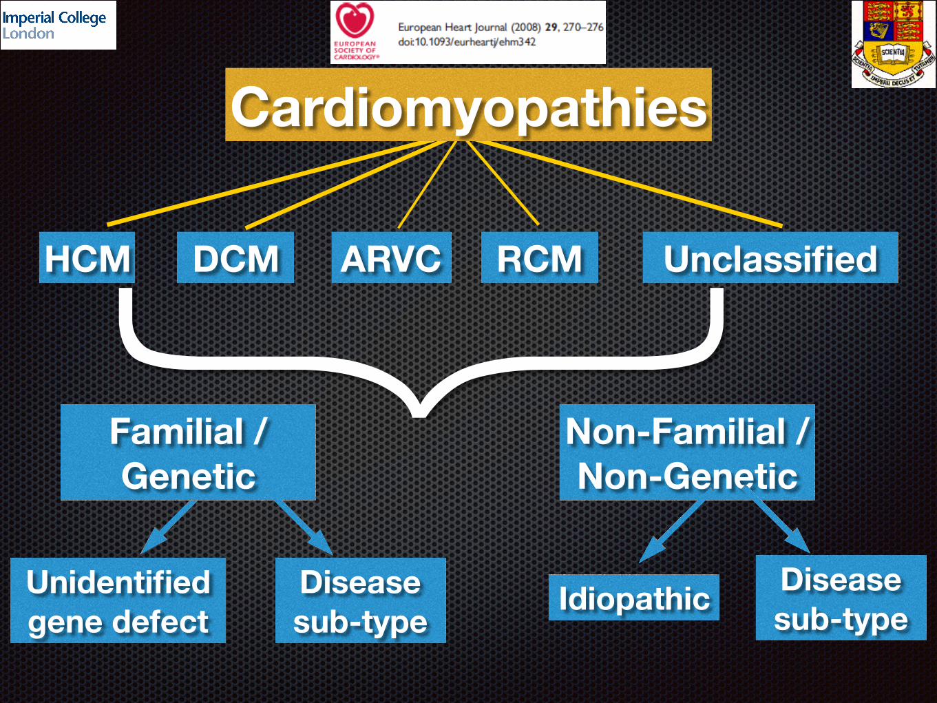
HCM DCM ARVC RCM Unclassified
Familial / Genetic
Non-Familial / Non-Genetic
Unidentified gene defect
Disease sub-type
Idiopathic Disease sub-type
Cardiomyopathies}
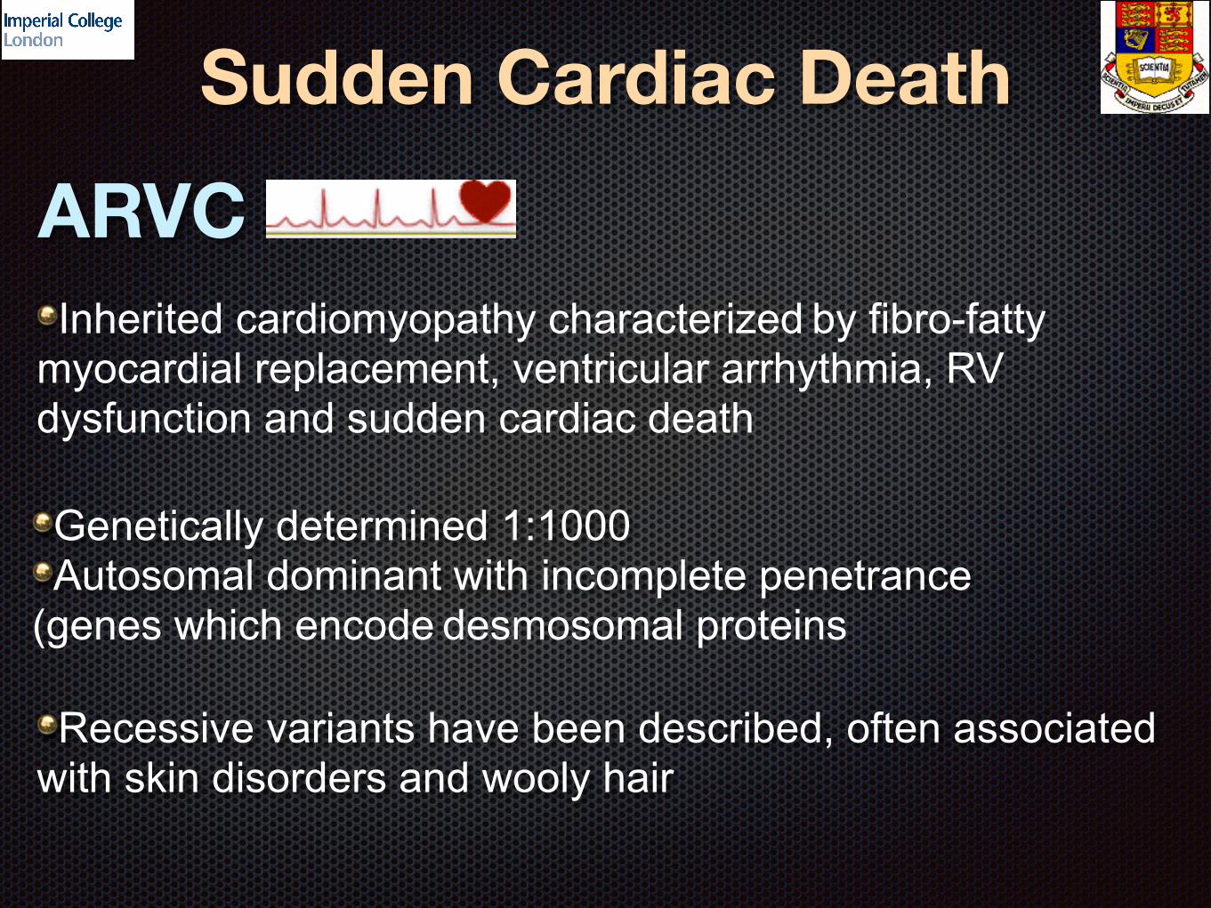
Sudden Cardiac DeathARVC
Genetically determined 1:1000 Autosomal dominant with incomplete penetrance
(genes which encode desmosomal proteins
Recessive variants have been described, often associated with skin disorders and wooly hair
Inherited cardiomyopathy characterized by fibro-fatty myocardial replacement, ventricular arrhythmia, RV dysfunction and sudden cardiac death
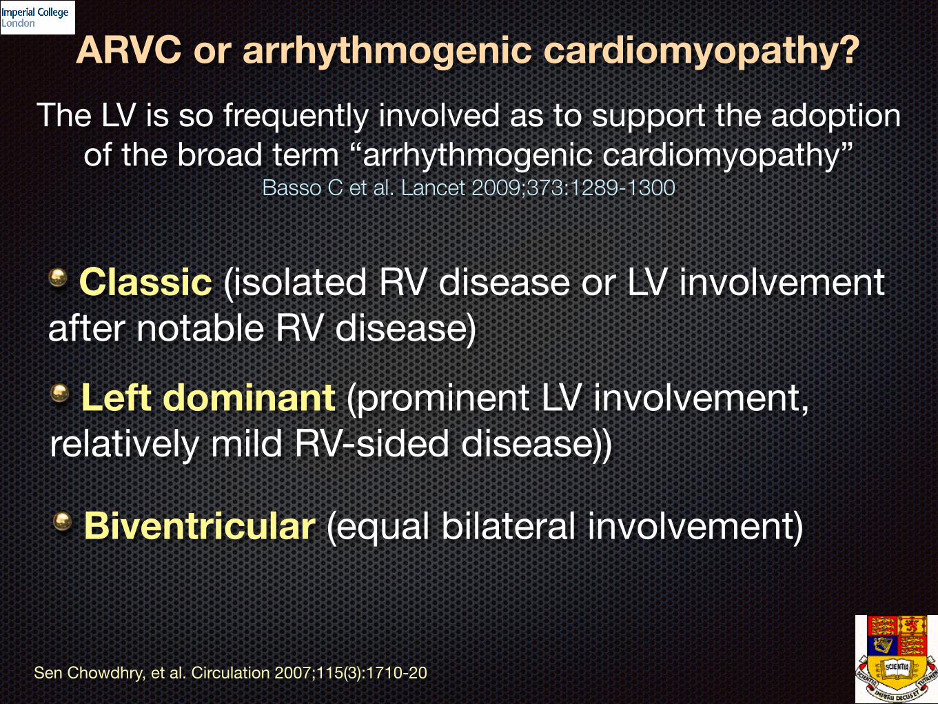
ARVC or arrhythmogenic cardiomyopathy?The LV is so frequently involved as to support the adoption
of the broad term “arrhythmogenic cardiomyopathy”Basso C et al. Lancet 2009;373:1289-1300
Classic (isolated RV disease or LV involvement after notable RV disease)
Left dominant (prominent LV involvement, relatively mild RV-sided disease))
Biventricular (equal bilateral involvement)
Sen Chowdhry, et al. Circulation 2007;115(3):1710-20
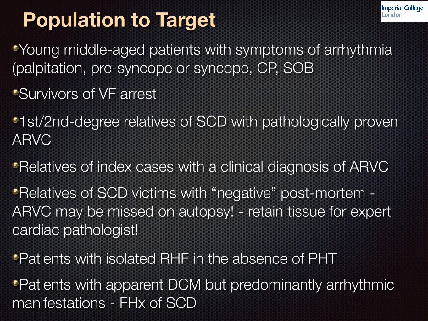
Population to TargetYoung middle-aged patients with symptoms of arrhythmia
(palpitation, pre-syncope or syncope, CP, SOBSurvivors of VF arrest
1st/2nd-degree relatives of SCD with pathologically proven ARVCRelatives of index cases with a clinical diagnosis of ARVCRelatives of SCD victims with “negative” post-mortem -
ARVC may be missed on autopsy! - retain tissue for expert cardiac pathologist!
Patients with isolated RHF in the absence of PHTPatients with apparent DCM but predominantly arrhythmic
manifestations - FHx of SCD

DiagnosisDifficult
No single test to establish or exclude diagnosis
Based on a constellation of clinical, ECG, Echo and other features
Important to make the correct diagnosis
Imaging of questionable use in early disease, few cell can be substrate for fatal arrhythmia
Dilated RV non-specific
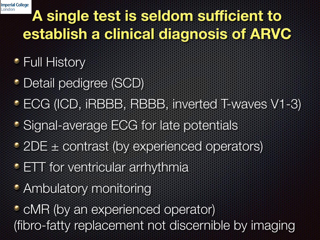
A single test is seldom sufficient to establish a clinical diagnosis of ARVC
Full History Detail pedigree (SCD) ECG (ICD, iRBBB, RBBB, inverted T-waves V1-3) Signal-average ECG for late potentials 2DE ± contrast (by experienced operators) ETT for ventricular arrhythmia Ambulatory monitoring cMR (by an experienced operator)
(fibro-fatty replacement not discernible by imaging
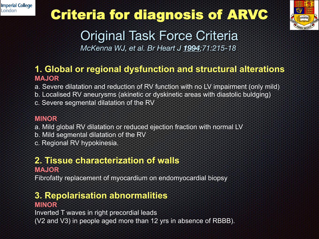
1. Global or regional dysfunction and structural alterations MAJOR
a. Severe dilatation and reduction of RV function with no LV impairment (only mild) b. Localised RV aneurysms (akinetic or dyskinetic areas with diastolic buldging)
c. Severe segmental dilatation of the RV
MINOR
a. Mild global RV dilatation or reduced ejection fraction with normal LV b. Mild segmental dilatation of the RV
c. Regional RV hypokinesia.
2. Tissue characterization of walls MAJOR
Fibrofatty replacement of myocardium on endomyocardial biopsy
3. Repolarisation abnormalities MINOR Inverted T waves in right precordial leads
(V2 and V3) in people aged more than 12 yrs in absence of RBBB).
Criteria for diagnosis of ARVC
Original Task Force CriteriaMcKenna WJ, et al. Br Heart J 1994;71:215-18

Criteria for diagnosis of ARVC
4. Depolarisation/Conduction abnormalities MAJOR Epsilon waves or localized prolongation (>110 msec) of the QRS complex
In right precordial leads (V1 to V3).
MINOR Late potentials (signal averaged ECG)
5. Arrhythmia MINOR
a. LBBB type ventricular tachycardia (sustained and non-sustained) b. Frequent ventricular extrasystoles (more than 1000/24h) on Holter
6. Family history
MAJOR
Familial disease confirmed at necropsy or surgery
MINOR a. Familial history of premature sudden death (<35yrs) due to suspected RV dysplasia.
b. Familial history (clinical diagnosis based on present criteria)
McKenna WJ, et al. Br Heart J 1994;71:215-18

Diffuse Non-epidermolytic Palmoplantar Keratoderma and Woolly Hair (Naxos Disease) Maps to 17q21
Coonar AS, Protonotarios N, Tsatsopoulou A, Needham EWA, Houlston RS, Cliff S, Otter MI, Murday VA, Mattu RK, McKenna WJ.
Circulation, 1998;97:2049 - 2058
- Population: 12,089 - Area: 126.957 km2 (49 sq mi) - Density: 95 /km2 (247 /sq mi)
Palmar plantar keratoderma
Hyperkeratosis without epidermolysis
Locus 17q21
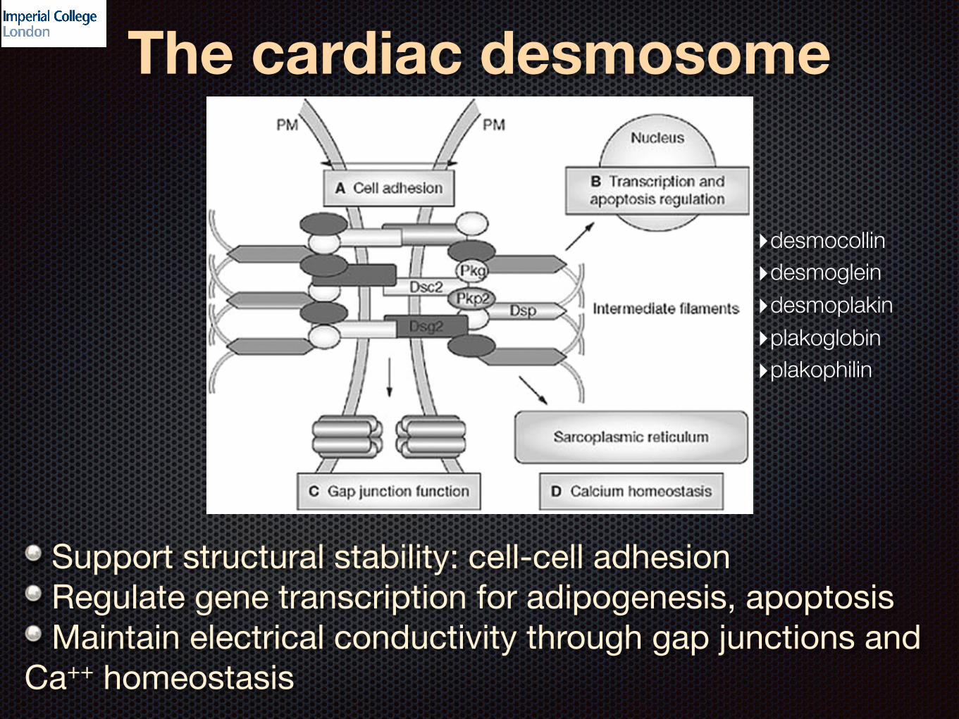
The cardiac desmosome
Support structural stability: cell-cell adhesion Regulate gene transcription for adipogenesis, apoptosis Maintain electrical conductivity through gap junctions and
Ca++ homeostasis
‣desmocollin ‣desmoglein ‣desmoplakin ‣plakoglobin ‣plakophilin

ARVD1 AD (14q23-q24) TGF b3 ARVD2 AD (1q42-q43) RyR2 - idiopathic VT ARVD3 AD (14q12-q22) ? ARVD4 AD (2q32.1-q32.3) ? ARVD5 AD (3q23) TMEM 43 ARVD6 AD (10p12-p14) ? ARVD7 AD (10q22) ? Naxos Disease AR (17q21) JUP (plakoglobin) ARVD8 AD (6p24 DSP (desmoplakin) ARVD9 AD (12p11) PKP-2 (plakophilin) ARVD10 AD (18q12.1) DSG-2(desmplein) ARVD11 AD (18q12.1) DSC-2 (desmocollin) ARVD12 AD (17q21) JUP Carvajal Syndrome AR (6p24) DSP
Diagnosis of ARVC/DGenetic Heterogeneity
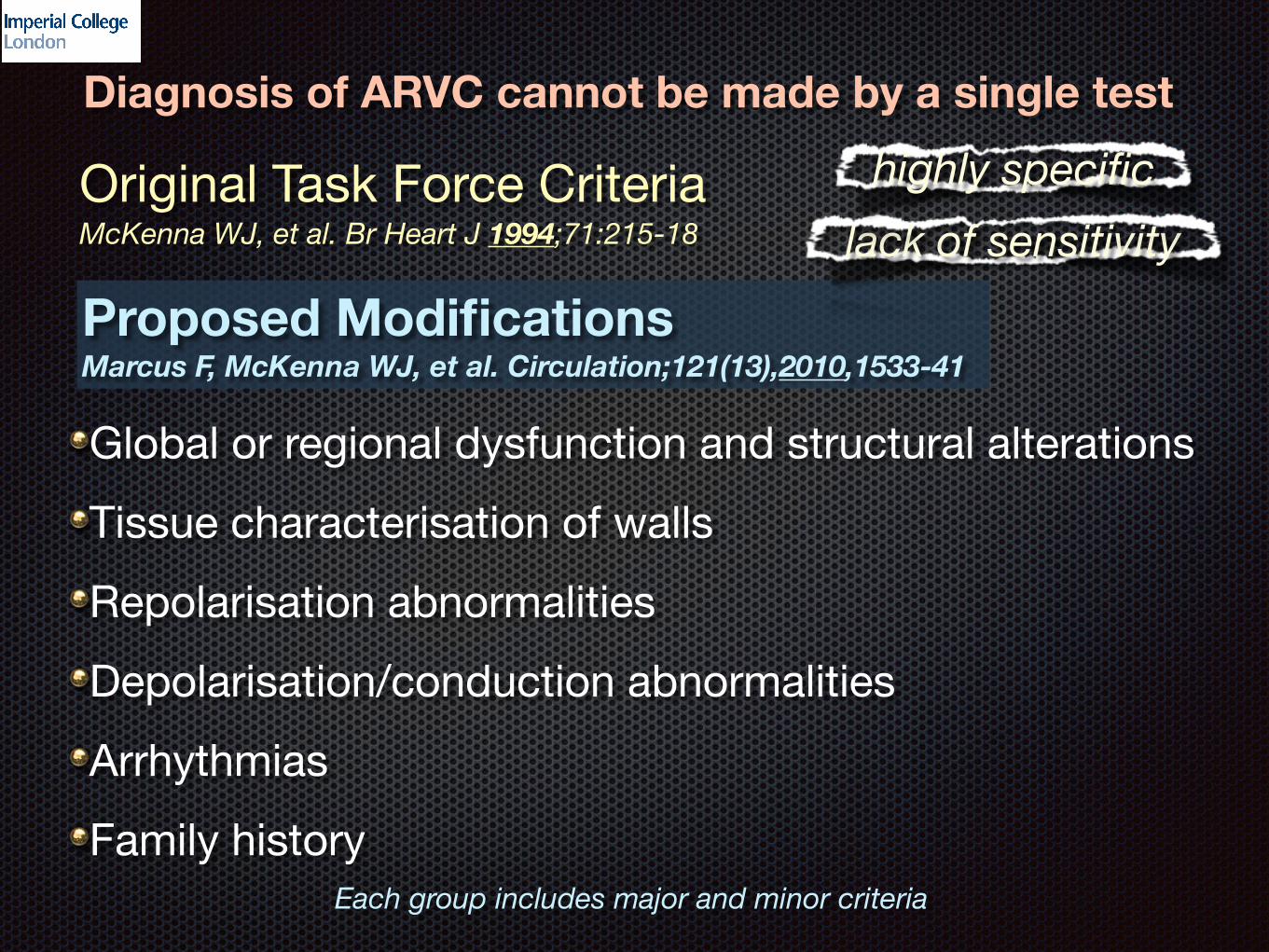
Diagnosis of ARVC cannot be made by a single test
Original Task Force CriteriaMcKenna WJ, et al. Br Heart J 1994;71:215-18
Proposed Modifications Marcus F, McKenna WJ, et al. Circulation;121(13),2010,1533-41
Global or regional dysfunction and structural alterationsTissue characterisation of wallsRepolarisation abnormalitiesDepolarisation/conduction abnormalitiesArrhythmiasFamily history
Each group includes major and minor criteria
lack of sensitivityhighly specific

Global or regional dysfunction or structural alterations
Modified Task Force CriteriaOriginal Task Force Criteria
Major Major (by 2D Echo)
Minor Minor (By 2D Echo)
Severe dilatation& reduction ofRV EF with no or mild LV involvement
Localised RV aneurysms (akinetic ordyskinetic areas with diastolic bulging)
Severe segmental RV dilatation
Mild global RV dilatation and/or reduced EF with normal LV
Mild segmental RV dilatation
Regional RV hypokinesia
Regional RV akinesia, dyskinesia oraneurysm and 1 of the following:-PLAX RVOT≥32mm (≥19mm/m2)-PSAX RVOT≥36mm (≥21mm/m2)-or Fractional Area Change ≤33%
Regional RV akinesia, dyskinesia or aneurysm and 1 of the following:-PLAX RVOT ≥ 29 <32mm(≥16mm/m2 - 19mm/m2)-PSAX RVOT ≥ 32 <36mm(≥18mm/m2 - 21mm/m2)-or FAC >33% ≤40%
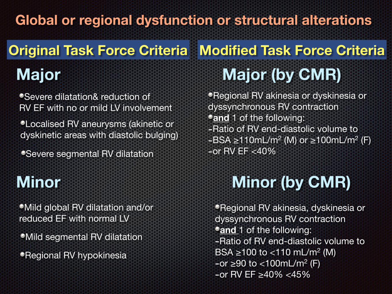
Global or regional dysfunction or structural alterations
Major Major (by CMR)
Minor Minor (by CMR)
Severe dilatation& reduction ofRV EF with no or mild LV involvement
Localised RV aneurysms (akinetic ordyskinetic areas with diastolic bulging)
Severe segmental RV dilatation
Mild global RV dilatation and/or reduced EF with normal LV
Mild segmental RV dilatation
Regional RV hypokinesia
Regional RV akinesia or dyskinesia ordyssynchronous RV contraction and 1 of the following:
-Ratio of RV end-diastolic volume to-BSA ≥110mL/m2 (M) or ≥100mL/m2 (F)-or RV EF <40%
Regional RV akinesia, dyskinesia or dyssynchronous RV contraction and 1 of the following:
-Ratio of RV end-diastolic volume toBSA ≥100 to <110 mL/m2 (M) -or ≥90 to <100mL/m2 (F)-or RV EF ≥40% <45%
Modified Task Force CriteriaOriginal Task Force Criteria
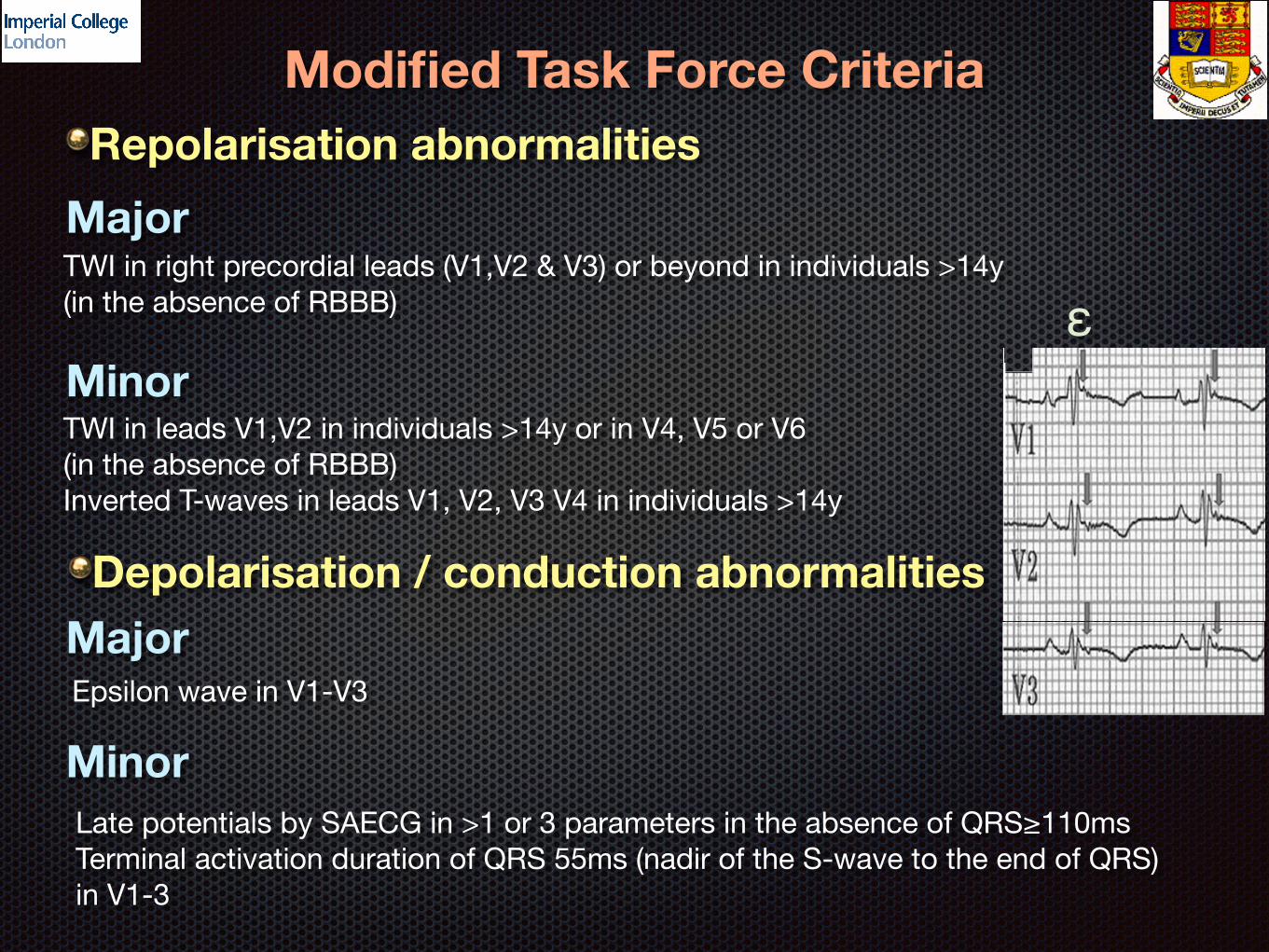
Modified Task Force CriteriaRepolarisation abnormalities
Depolarisation / conduction abnormalities
Major
Minor
Major
Minor
TWI in right precordial leads (V1,V2 & V3) or beyond in individuals >14y (in the absence of RBBB)
TWI in leads V1,V2 in individuals >14y or in V4, V5 or V6 (in the absence of RBBB)Inverted T-waves in leads V1, V2, V3 V4 in individuals >14y
Epsilon wave in V1-V3
Late potentials by SAECG in >1 or 3 parameters in the absence of QRS≥110msTerminal activation duration of QRS 55ms (nadir of the S-wave to the end of QRS) in V1-3
ε
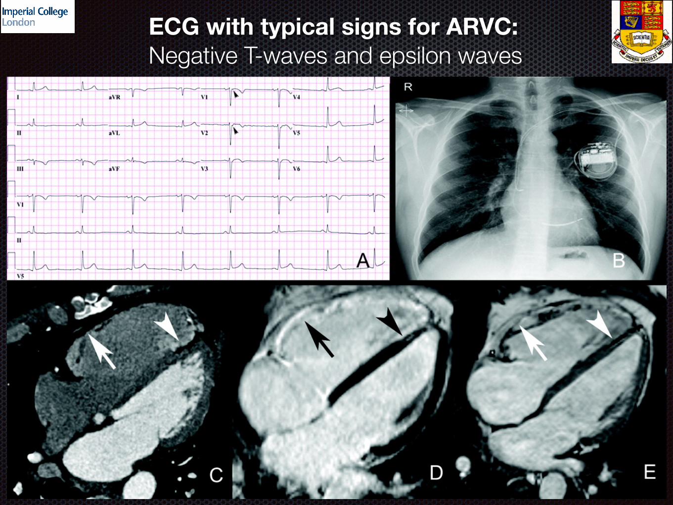
ECG with typical signs for ARVC: Negative T-waves and epsilon waves

Modified Task Force CriteriaTissue characterisation of wall
Arrhythmias
Major
Minor
Major
Minor
Residual myocytes <60% by morphometric analysis (or <50% if estimate), with fibrous replacement of the RV free wall in ≥1 sample, with or without fatty replacement of tissue on endomyocardial biopsy
Residual myocytes 60&-75% by morphometric analysis (or 50%-65% if estimated), with fibrous replacement of the RV free wall in ≥1 sample
NSVT or sustained VT of LBBB morphology with superior axis (negative or indeterminate QRS in II, III, aVF and positive in aVL
NSVT or sustained VT of LBBB morphology with inferior axis (positive in II, III, aVF and negative in aVL500 ventricular extrasystoles / 24h (Holter)
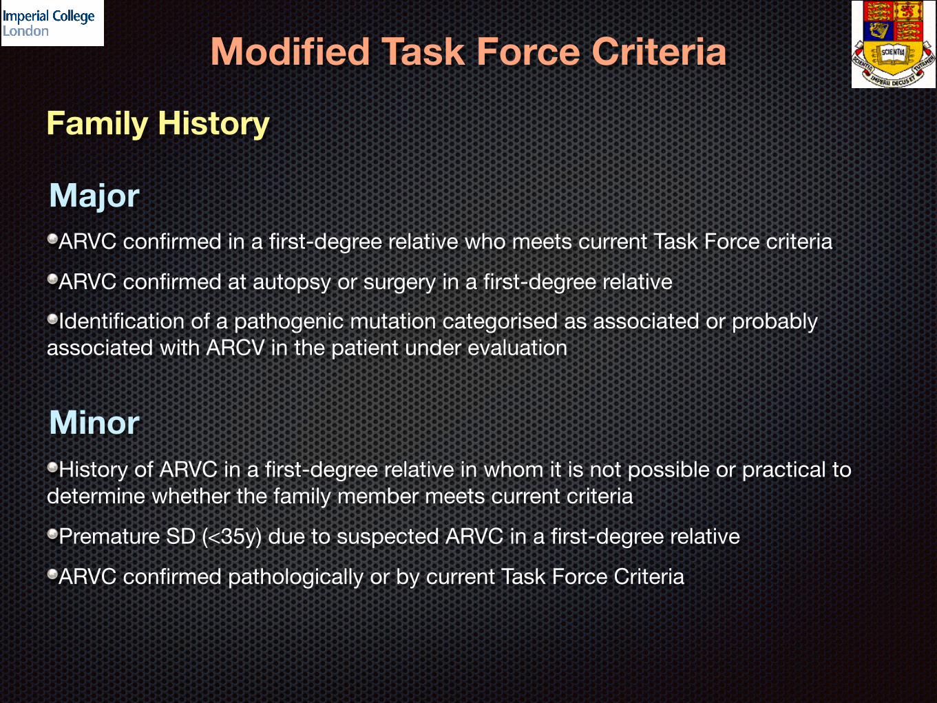
Modified Task Force Criteria
Family History
Major
Minor
ARVC confirmed in a first-degree relative who meets current Task Force criteria
ARVC confirmed at autopsy or surgery in a first-degree relative
Identification of a pathogenic mutation categorised as associated or probably associated with ARCV in the patient under evaluation
History of ARVC in a first-degree relative in whom it is not possible or practical to determine whether the family member meets current criteria
Premature SD (<35y) due to suspected ARVC in a first-degree relative
ARVC confirmed pathologically or by current Task Force Criteria
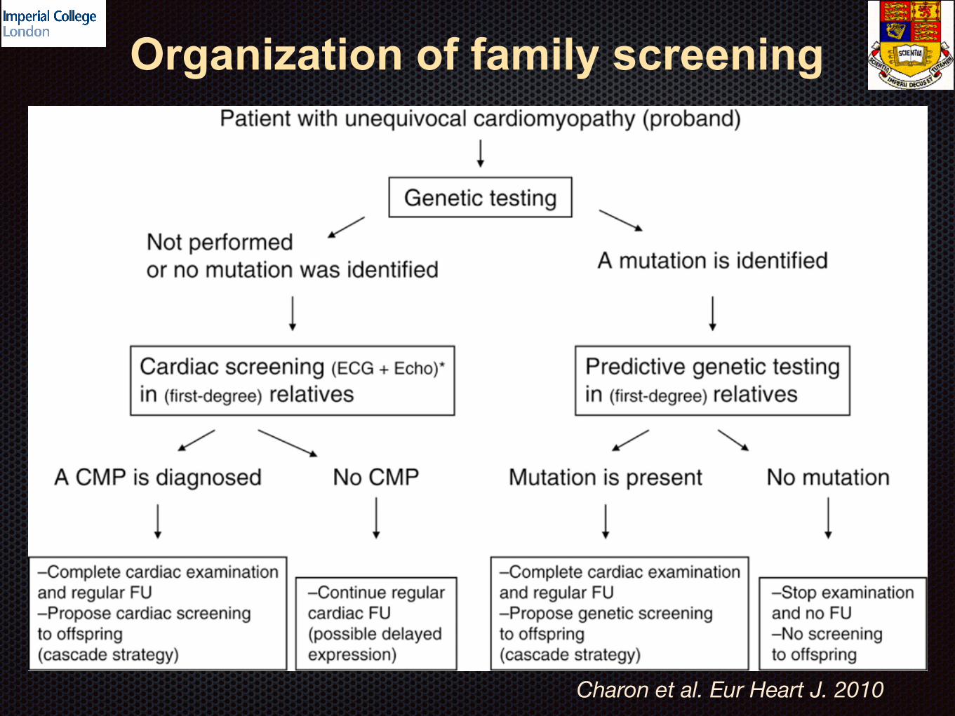
Organization of family screening
Charon et al. Eur Heart J. 2010
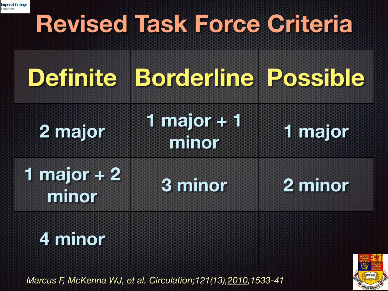
Revised Task Force Criteria
Definite Borderline Possible
2 major 1 major + 1 minor 1 major
1 major + 2 minor 3 minor 2 minor
4 minor
Marcus F, McKenna WJ, et al. Circulation;121(13),2010,1533-41
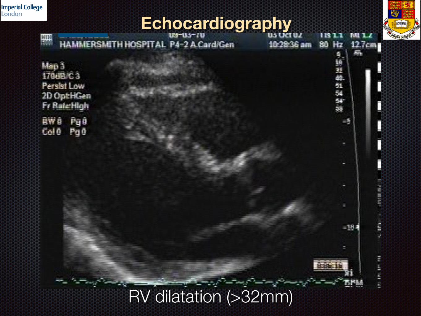
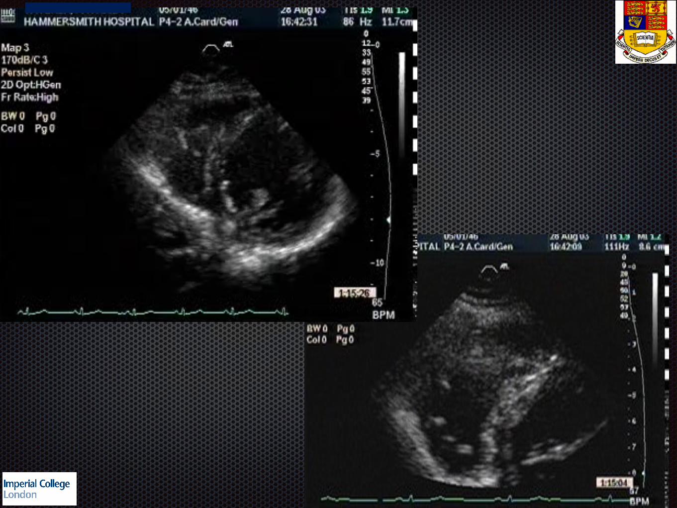
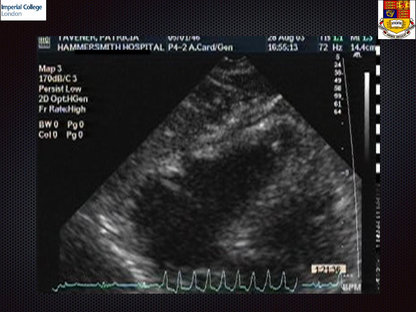
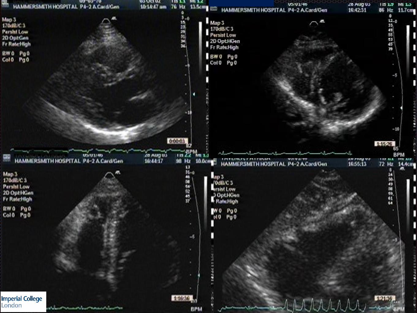
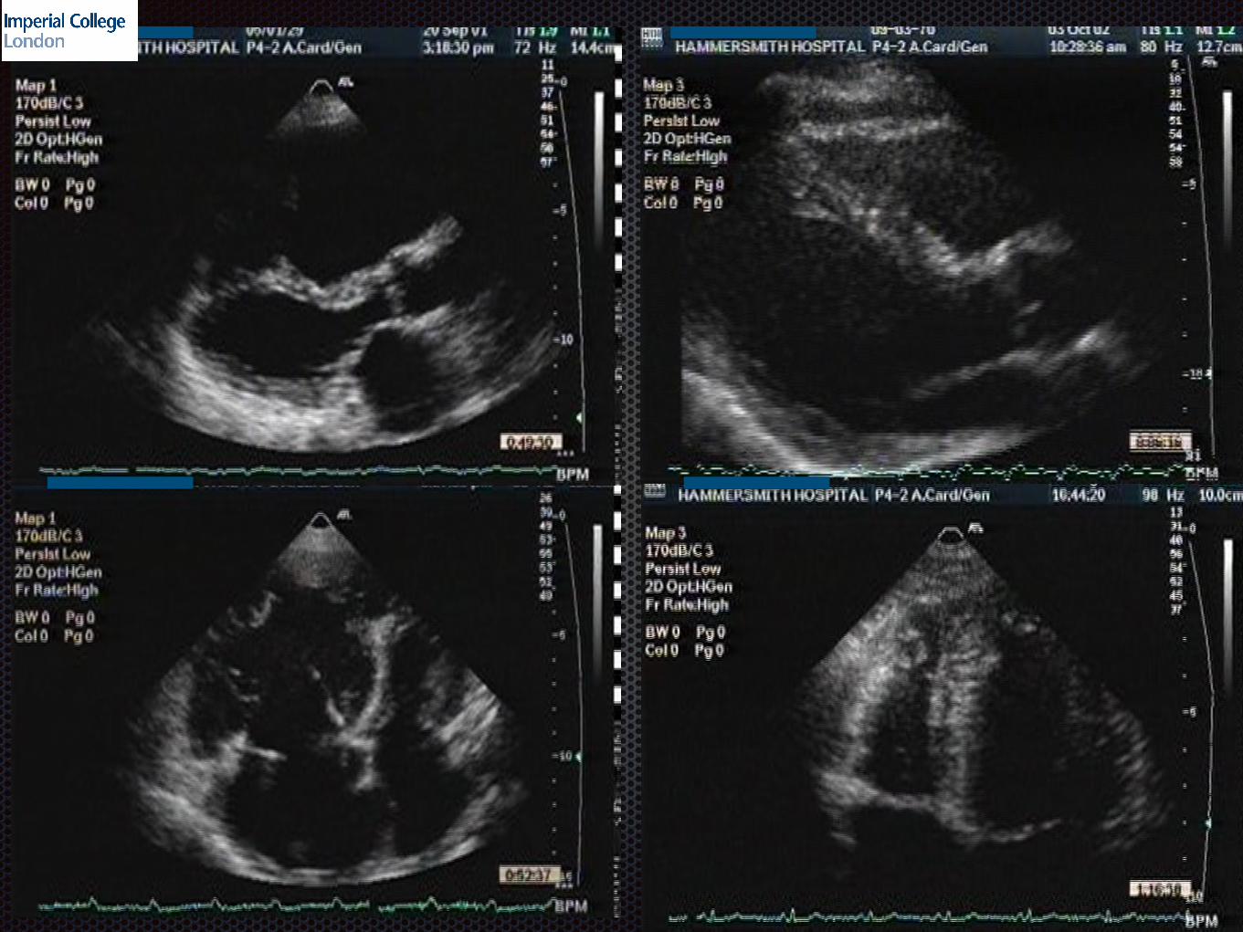
Echocardiography
RV dilatation (>32mm)
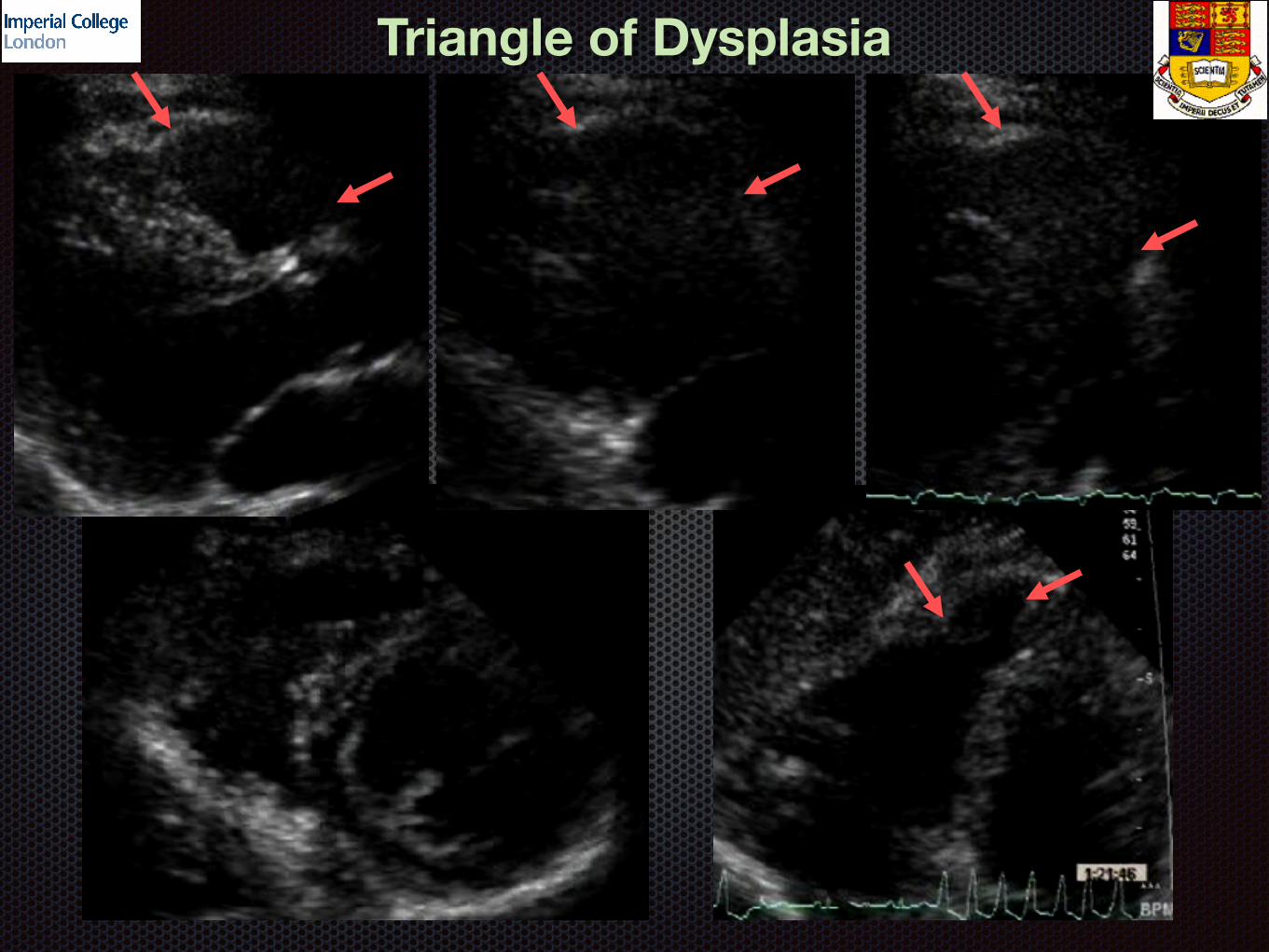
Triangle of Dysplasia


?? RV Dysfunction

RV speckle Tracking
ARVC
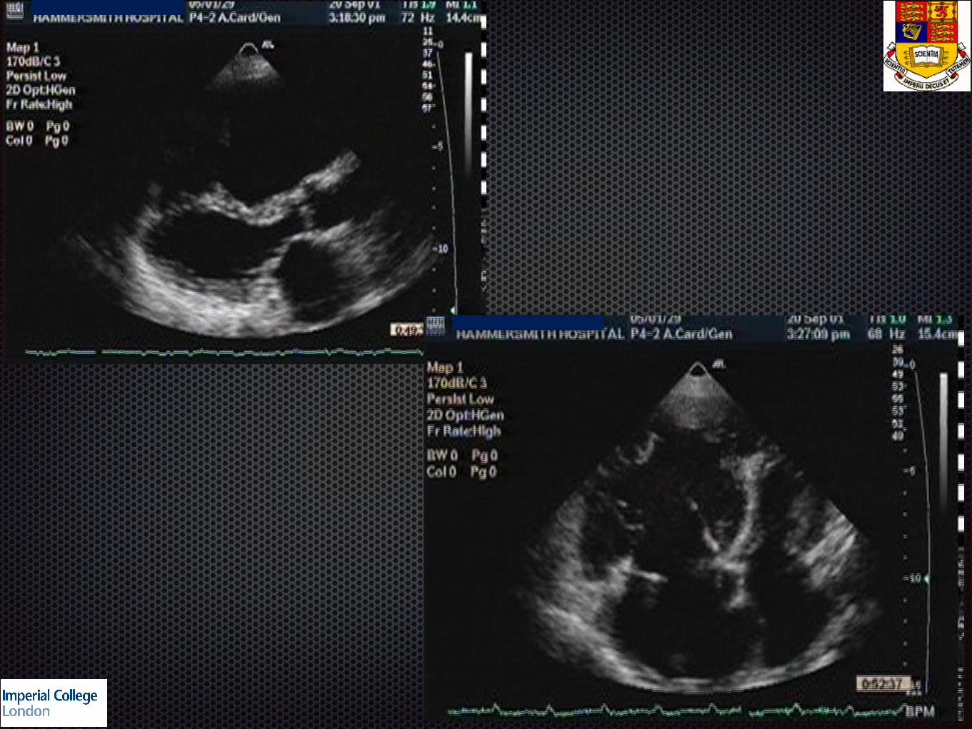
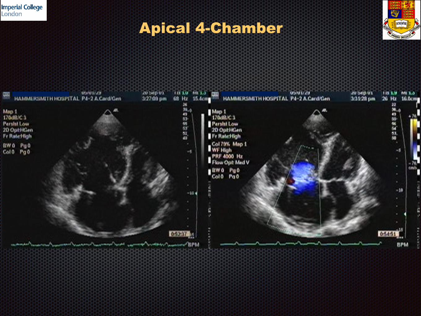
Apical 4-Chamber
Structural alterations
Electrical phenomena
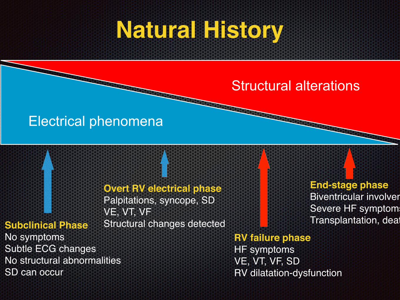
Natural History
Subclinical PhaseNo symptomsSubtle ECG changesNo structural abnormalitiesSD can occur
Overt RV electrical phasePalpitations, syncope, SDVE, VT, VFStructural changes detected
RV failure phaseHF symptomsVE, VT, VF, SDRV dilatation-dysfunction
End-stage phaseBiventricular involvementSevere HF symptomsTransplantation, death

Cardiac Magnetic Resonance Imaging1.5 or 3T scanners Dedicated phased-array coils with multiple elements ECG triggering Expert centres
RV free wall
Morphological: Focal thinning, RVOT enlargement, fatty infiltration with T1 weighted spin echo images (not a task force criterion)
Delayed enhancement: Detection of fibrosis ??prognostic implications not defined LV involvement
Functional: Dilatation, global/regional function, focal aneurysms (subjective), RV volumes, EF
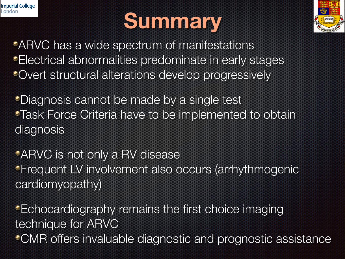
SummaryARVC has a wide spectrum of manifestations Electrical abnormalities predominate in early stages Overt structural alterations develop progressively
Diagnosis cannot be made by a single test Task Force Criteria have to be implemented to obtain
diagnosis
ARVC is not only a RV disease Frequent LV involvement also occurs (arrhythmogenic
cardiomyopathy)
Echocardiography remains the first choice imaging technique for ARVC CMR offers invaluable diagnostic and prognostic assistance