
Apraxia. A Review. -...
53
Apraxia. A Review. Biljana Petreska *,1 , Michela Adriani 2 , Olaf Blanke 2 and Aude G. Billard 1 1 Learning Algorithms and Systems Laboratory (LASA), Ecole Polytechnique Fédérale de Lausanne (EPFL), EPFL-STI-I2S-LASA, Station 9, CH 1015, Lausanne, Switzerland 2 Laboratory of Cognitive Neuroscience (LNCO), Ecole Polytechnique Fédérale de Lau- sanne (EPFL), EPFL-SV-BMI, Station 15, CH 1015, Lausanne, Switzerland [email protected]fl.ch Ph: (+41)-21-693-54-65 Fax: (+41)-21-693-78-50 [email protected] Ph: (+41)-21-693-17-62 Fax: (+41)-21-693-96-25 olaf.blanke@epfl.ch Ph: (+41)-21-693-96-21 Fax: (+41)-21-693-96-25 aude.billard@epfl.ch Ph: (+41)-21-693-54-64 Fax: (+41)-21-693-78-50 1
Transcript of Apraxia. A Review. -...

Apraxia. A Review.
Biljana Petreska∗,1 , Michela Adriani2 , Olaf Blanke2 and Aude G. Billard1
1Learning Algorithms and Systems Laboratory (LASA), Ecole Polytechnique Fédérale
de Lausanne (EPFL), EPFL-STI-I2S-LASA, Station 9, CH 1015, Lausanne, Switzerland
2Laboratory of Cognitive Neuroscience (LNCO), Ecole Polytechnique Fédérale de Lau-
sanne (EPFL), EPFL-SV-BMI, Station 15, CH 1015, Lausanne, Switzerland
[email protected] Ph: (+41)-21-693-54-65 Fax: (+41)-21-693-78-50
[email protected] Ph: (+41)-21-693-17-62 Fax: (+41)-21-693-96-25
[email protected] Ph: (+41)-21-693-96-21 Fax: (+41)-21-693-96-25
[email protected] Ph: (+41)-21-693-54-64 Fax: (+41)-21-693-78-50
1

2
Abstract
Praxic functions are frequently altered following brain lesion, giving rise to
apraxia, a complex pattern of impairments that is difficult to assess or inter-
pret. In this chapter, we review the current taxonomies of apraxia and related
cognitive and neuropsychological models. We also address the questions of
the neuroanatomical correlates of apraxia, the relation between apraxia and
aphasia and the analysis of apraxic errors. We provide a possible explana-
tion for the difficulties encountered in investigating apraxia and also several
approaches to overcome them, such as systematic investigation and model-
ing studies. Finally, we argue for a multidisciplinary approach. For example,
apraxia should be studied in consideration with and could contribute to other
fields such as normal motor control, neuroimaging and neurophysiology.
Introduction
Apraxia is generally defined as “a disorder of skilled movement not caused by weakness,
akinesia, deafferentation, abnormal tone or posture, movement disorders such as tremor
or chorea, intellectual deterioration, poor comprehension, or uncooperativeness" (Heilman
and Rothi, 1993). Apraxia is thus negatively defined, in terms of what it is not, as a higher-
order disorder of movement that is not due to elementary sensory and/or motor deficits.
This definition implies that there are situations where the effector is moved with normal
skill (Hermsdörfer, Mai, Spatt, Marquardt, Veltkamp and Goldenberg, 1996). Puzzling
parts of apraxia are the voluntary-automatic dissociation and context-dependence. On the
one hand, apraxic patients may spontaneously perform gestures that they cannot perform
on command (Schnider, Hanlon, Alexander and Benson, 1997). This voluntary-automatic
dissociation can be illustrated by an apraxic patient that could use his left hand to shave
and comb himself, but could not execute a specific motor action such as opening the

3
hand, so as to let go of an object (Lausberg, Göttert, Münssinger, Boegner and Marx,
1999). In this particular case, focussing on the target of the movement rather than on the
movement itself increased his chances of a successful execution. On the other hand, the
execution of the movement depends heavily on the context of testing (De Renzi, Faglioni
and Sorgato, 1982). It may be well preserved in a natural context, with a deficit that
appears in the clinical setting only, where the patient has to explicitly represent the content
of the action outside of the situational props (Jeannerod and Decety, 1995; Leiguarda and
Marsden, 2000).
Several authors agree that although apraxia is easy to demonstrate, it has proven
difficult to understand. Research on apraxia is filled with confusing terminology, contra-
dictory results and doubts that need to be resolved (Laeng, 2006; Goldenberg, Herms-
dörfer and Spatt, 1996; De Renzi et al., 1982; Graham, Zeman, Young, Patterson and
Hodges, 1999; Koski, Iacoboni and Mazziotta, 2002). Inconsistencies between similar stud-
ies may be explained by differences in methodological and statistical approaches in the
apraxia assessment (i.e., types of gestures used and scoring criteria), chronicity and aetiol-
ogy of damage and brain lesion localization tools (Haaland, Harrington and Knight, 2000).
Therefore, it still stands that our understanding of the neural and cognitive systems un-
derlying human praxis is not well established.
The chapter is structured as follows. We first review existing types of apraxia as well as
important current and historical models of the apraxic deficit. We then consider the inter-
and intra- hemispheric lesion correlates of apraxia. Two other sections are dedicated to the
relationship between praxis and language and to the analysis of apraxic errors. We finally
discuss the current state-of-art in apraxia, and argue for a multidisciplinary approach that
encompasses evidence from various fields such as neuroimaging or neurophysiology.

4
Types of apraxia
This section reviews the current taxonomies of apraxia. Some of the frequently observed
types of apraxia have inspired the apraxia models described in the following section, others
still challenge them.
Ideational apraxia was historically defined as a disturbance in the conceptual organi-
zation of actions. It was first assessed by performing purposive sequences of actions that
require the use of various objects in the correct order (e.g., preparing a cup of coffee)
(Poeck, 1983). It was later accepted that ideational apraxia is not necessarily associated
to complex actions, but is a larger deficit that also concerns the evocation of single ac-
tions. In this view, complex sequences of multiple objects are simply more suitable to
reveal the deficit, possibly because of the heavier load placed on memory and attentional
resources (De Renzi and Lucchelli, 1988). Nonetheless, the term conceptual apraxia was
introduced to designate content errors in single actions, excluding sequence errors in multi-
staged actions with tools1 (Ochipa, Rothi and Heilman, 1992; Heilman, Maher, Greenwald
and Rothi, 1997). In theoretical models, ideational and conceptual apraxia correspond
to a disruption of the conceptual component of the praxis system, i.e., action semantics
memory, described in more detail in the Models of apraxia section (De Renzi and Luc-
chelli, 1988; Graham et al., 1999). Patients with ideational apraxia are not impaired in
the action execution per se, but demonstrate inappropriate use of objects and may fail in
gesture discrimination and matching tasks. For example, a patient was reported to eat
with a toothbrush and brush his teeth with a spoon and a comb. His inability to use tools
could not be explained by a motor production deficit that would characterize ideomotor
apraxia (defined below). Interestingly, although he was able to name the tools and point
to them on command, he could not match the tools with the objects, hence suggesting a
loss of knowledge related to the use of tools.
1Conceptual apraxia is often observed in Alzheimer’s disease.

5
Ideomotor apraxia is considered to be a disorder of the production component of the
praxis system, i.e., sensorimotor action programs that are concerned with the generation
and control of motor activity (Rapcsak, Ochipa, Anderson and Poizner, 1995; Graham
et al., 1999). It is characterized by errors in the timing, sequencing, and spatial organi-
zation of gestural movements (Leiguarda, 2001). Since the conceptual part of the praxis
system is assumed to be intact, patients with ideomotor apraxia should not use objects and
tools in a conceptually inappropriate fashion and should not have difficulty with the serial
organization of an action (De Renzi et al., 1982). Ideational and ideomotor apraxia have
been assessed by testing the execution of various types of gestures: transitive and intran-
sitive (i.e., with or without the use of tools or objects), meaningless non-representational
(e.g., hand postures relative to head) and meaningful representational (e.g., waving good-
bye), complex sequences with multiple objects, repetitive movements, distal and proximal
gestures (e.g., imitation of finger and hand configurations), reaching in peri-personal and
body-centered space (e.g., targets in near space or on the patient’s body), novel movements
(i.e., skill acquisition) or imagined movements. These gestures can also be executed under
different modalities such as: verbal command, imitation, pantomime and tactile or visual
presentation of objects.
The use of various gestures and different modalities to assess apraxia has helped to
uncover many interesting functional dissociations that are listed below. For example,
apraxia was shown to be modality-specific, i.e., the same type of gesture was differentially
impaired according to the modality of testing (De Renzi et al., 1982). One dissociation,
named conduction apraxia, is the syndrome of superior performance on verbal command
than on imitation (Ochipa, Rothi and Heilman, 1994). The opposite pattern has also been
observed: very poor performance on verbal command that improved on imitation or when
seeing the object (Heilman, 1973; Merians, Clark, Poizner, Macauley, Gonzalez Rothi and
Heilman, 1997). The extreme occurrence of conduction apraxia, namely the selective in-
ability to imitate with normal performance on verbal command was termed visuo-imitative

6
apraxia (Merians et al., 1997). In some cases of visuo-imitative apraxia, defective imitation
of meaningless gestures (e.g., fist under chin) contrasts with preserved imitation of mean-
ingful gestures (e.g., hitchhiking) (Goldenberg and Hagmann, 1997; Salter, Roy, Black,
Joshi and Almeida, 2004). A surprising case of double dissociation from this kind of visuo-
imitative apraxia was described in Bartolo, Cubelli, Sala, Drei and Marchetti (2001), where
the patient showed impairment in meaningful gesture production (both on imitation and
verbal command) and normal performance in imitation of meaningless gestures, suggesting
that the patient was able to reproduce only movements he did not identify or recognize as
familiar. Similarly, the apraxic patients in Buxbaum, Sirigu, Schwartz and Klatzky (2003)
responded abnormally to familiar objects (e.g., a key, a hammer or a pen) but normally in
recognizing the hand postures appropriate for novel objects (e.g., parallelepipeds differing
in size and depth). These two studies argue that the reproduction of a gesture may be
constrained by its degree of familiarity, indicating that current models of apraxia would
need some refinement.
Furthermore, the representation of transitive and intransitive actions may be disso-
ciable. In Watson, Fleet, Rothi and Heilman (1986), bilateral apraxia was observed only
for transitive (e.g., hammering) but not intransitive (e.g., hitchhiking, waving goodbye)
movements2. Whereas transitive gestures are constrained by the shape, size and function
of objects, intransitive actions are related to socio-cultural contexts (Cubelli, Marchetti,
Boscolo and Della Sala, 2000; Heath, Roy, Black and Westwood, 2001). The isolated
disturbance of transitive hand movements for use of, recognition and interaction with an
object, in the presence of preserved intransitive movements, was named tactile apraxia and
usually appears in the hand contralateral to the lesion (Binkofski, Kunesch, Classen, Seitz
and Freund, 2001).
As mentioned in the Introduction, contextual cues strongly influence the execution of
actions. Some studies have systematically manipulated the contextual cues in order to
2These patients had lesions in the left supplementary motor area (SMA).

7
assess their relative importance. For example, patients with impaired pantomime of motor
actions showed no deficit in the comprehension of the use of tools or in manipulating the
tools (Halsband, Schmitt, Weyers, Binkofski, Grützner and Freund, 2001). Graham et al.
(1999) also observed dramatic facilitation in the demonstration of tool use when the patient
was given the appropriate or a neutral tool to manipulate3. Interestingly, the patient could
not prevent himself from performing the action appropriate to the tool he was holding,
rather than the action that was requested. In another study however, gesture execution
improved when the object of the action, but not the tool, was given (Clark, Merians,
Kothari, Poizner, Macauley, Rothi and Heilman, 1994). Hence, the addition of visual and
somaesthetic cues may improve certain aspects of apraxic movements, since it provides
mechanical constraints and supplementary information that facilitates the selection of an
adequate motor program (Hermsdörfer, Hentze and Goldenberg, 2006). Nonetheless, there
is the case of a patient that performed much worse when he was actually manipulating the
tool than on verbal command4 (Merians, Clark, Poizner, Jacobs, Adair, Macauley, Rothi
and Heilman, 1999).
Dissociations that concern the nature of the target were also observed. For example,
the left brain damaged patients in Hermsdörfer, Blankenfeld and Goldenberg (2003) had
prolonged movement times and reduced maximum velocities when the movements were
directed toward an allocentric target without visual feedback, but performed normally
when the target was their own nose. Also, a clear dissociation was found in Ietswaart, Crey
and Della Sala (2006) between impaired gesture imitation and intact motor programming
of goal-directed movements, hence arguing against the interpretation of impaired imitation
as a purely executional deficit (see the Models of apraxia section).
A particular type of apraxia is constructional apraxia, originally described by Kleist as
“the inability to do a construction” and defined by Benton as “the impairment in combina-
3The subject had clinically diagnosed corticobasal degeneration.4Ibid.

8
tory or organizing activity in which details must be clearly perceived and in which the re-
lationship among the component parts of the entity must be apprehended” (Laeng, 2006).
Constructional apraxic patients are unable to spontaneously draw objects, copy figures
and build blocks or patterns with sticks, following damage to the dominant but also non-
dominant hemisphere. Hence, constructional apraxia appears to reflect the loss of bilat-
erally distributed components for constructive planning and the perceptual processing of
categorical and coordinate spatial relations (Platz and Mauritz, 1995; Laeng, 2006).
Apraxia can also be observed in mental motor imagery tasks. Motor imagery is con-
sidered as a means of accessing the mechanisms of action preparation and imitation, by
sharing a common neural basis (Jeannerod and Decety, 1995). Apraxic patients were de-
ficient in simulating hand actions mentally and in imagining the temporal properties of
movements5 (Sirigu, Daprati, Pradat-Diehl, Franck and Jeannerod, 1999). Other apraxic
patients showed a deficit in generating and maintaining internal models for planning object-
related actions (Buxbaum, Johnson-Frey and Bartlett-Williams, 2005). These findings
support the notion that the motor impairments observed in apraxic patients result from
a specific alteration in their ability to mentally evoke actions, or to use stored motor
representations for forming mental images of actions.
Apraxia may also be appropriate to reveal the role of feedback during the execution
of a movement. Some apraxic patients were impaired in reaching and aiming move-
ments only in the condition without visual feedback (Ietswaart, Crey, Della Sala and
Dijkhuizen, 2001; Ietswaart et al., 2006) and performed worse during pointing with closed
eyes (Hermsdörfer et al., 2003; Jacobs, Adair, Macauley, Gold, Gonzalez Rothi and Heil-
man, 1999). Interestingly, the patients in Haaland, Harrington and Knight (1999) overshot
the target when feedback of the hand was removed and undershot the target when the feed-
back of the target was unavailable. Importantly, these patients continued to rely on visual
feedback during the secondary adjustment phase of the movement and never achieved nor-
5These patients had posterior parietal lesions.

9
mal end-point accuracy when visual feedback of the hand position or target location was
unavailable. These findings also suggest that ideomotor limb apraxia may be associated
with the disruption of the neural representations for the extrapersonal (spatial location)
and intrapersonal (hand position) features of movement (Haaland et al., 1999).
The importance of feedback signals was demonstrated in one of our own apraxic patients
(unpublished data). We reproduced a seminal study of imitation of meaningless gestures6
by Goldenberg, Laimgruber and Hermsdörfer (2001) on an apraxic patient with left parietal
ischemic lesion. We observed that the patient relied heavily on visual and tactile feedback.
He often needed to bring his hand in the field of vision and corrected the hand posture
by directly comparing it with the displayed stimulus to imitate. He also used tactile
exploration when searching for the correct spatial position on his face. He showed many
hesitations and extensive searching which led to highly disturbed kinematic profiles of the
gesture (shown in Figure 4c, d), but often correct final postures.
Apraxia can also be defined in relation to the selectively affected effector: orofacial
apraxia or buccofacial apraxia, oral apraxia, upper and lower face apraxia (Sala, Maistrello,
Motto and Spinnler, 2006), lid apraxia, limb apraxia, leg apraxia, trunk apraxia, etc. Oral
apraxia for example, is defined as the inability to perform mouth actions such as suck-
ing from a straw or blowing a kiss. It should not be confounded with apraxia of speech
(also called verbal apraxia), which is a selective disturbance of the articulation of words
(Bizzozero, Costato, Sala, Papagno, Spinnler and Venneri, 2000). Motor planning dis-
orders in children are denominated developmental dyspraxia (Cermak, 1985). Apraxia
can also designate a praxic ability impaired in an isolated manner such as: gait apraxia,
apraxic agraphia, dressing apraxia, orienting apraxia and mirror apraxia (i.e., inability
to reach to objects in a mirror (Binkofski, Butler, Buccino, Heide, Fink, Freund and
Seitz, 2003)). When the side of brain lesion and affected hand are considered, the terms
sympathetic and crossed apraxia are used. Apraxia can sometimes be related to the spe-
6Hand postures relative to the head, an example is shown in Figure 4a.

10
cific neural substrate that causes the disorder, for example following subcortical lesions in
corticobasal degeneration (Pramstaller and Marsden, 1996; Jacobs et al., 1999; Merians
et al., 1999; Hanna-Pladdy, Heilman and Foundas, 2001; Leiguarda, 2001) or following le-
sions of the corpus callosum (Watson and Heilman, 1983; Goldenberg et al., 2001; Lausberg
et al., 1999; Lausberg, Davis and Rothenhäusler, 2000; Lausberg and Cruz, 2004). Callosal
apraxia for example is particularly appropriate for disentangling the specific hemispheric
contributions to praxis.
An extensive list of the types of apraxia and their definitions, including types that were
not mentioned above, can be found in the Table in Figure 1. Figure 1
Models of apraxia
Contemporary neuropsychological views of apraxia arise from Liepmann’s influential work
that dates from more than a hundred years ago. Liepmann proposed the existence of an
idea of the movement, “movement formulae”, that contains the “time-space-form picture"
of the action (Rothi, Ochipa and Heilman, 1991). He believed that in right-handers, these
movement formulae are stored in the left parietal lobe, endorsing the view of a left hemi-
spheric dominance for praxis (Faglioni and Basso, 1985; Leiguarda and Marsden, 2000).
To execute a movement, the spatio-temporal image of the movement is transformed into
“innervatory patterns” that yield “positioning of the limbs according to directional ideas”
(Jacobs et al., 1999). Liepmann distinguished between three types of apraxia, that corre-
spond to disruptions of specific components of the model (Goldenberg, 2003; Faglioni and
Basso, 1985). First, a damaged movement formula (i.e., faulty integration of the elements
of an action) would characterize ideational apraxia. Second, failure of the transition from
the movement formula to motor innervation (i.e., inability to translate a correct idea of
the movement into a correct act) is defined as ideomotor apraxia. According to Liepmann,
faulty imitation of movements is a purely executional deficit and proves the separation
11
between the idea and execution of a movement, since in imitation the movement formula is
defined by the demonstration (Goldenberg, 1995; Goldenberg and Hagmann, 1997; Gold-
enberg, 2003). Finally, loss of purely kinematic (kinaesthetic or innervatory) inherent
memories of an extremity is the limb-kinetic variant of apraxia.
Another historically influential model is the disconnection model of apraxia proposed
by Geschwind (1965). According to this model the verbal command for the movement is
comprehended in Wernicke’s area and is transferred to the ipsilateral motor and premotor
areas that control the movement of the right hand (Clark et al., 1994; Leiguarda and
Marsden, 2000). For a left hand movement, the information needs to be further transmitted
to the right association cortex via the corpus callosum. The model postulates that the
apraxic disorder follows from a lesion in the left and right motor association cortices, or a
disruption in their communication pathways. However this model cannot explain impaired
imitation and impaired object use since these tasks do not require a verbal command (Rothi
et al., 1991).
Heilman and Rothi (1993) proposed an alternative representational model of apraxia,
according to which apraxia is a gesture production deficit that may result from the destruc-
tion of the spatiotemporal representations of learned movements stored in the left inferior
parietal lobule. They proposed to distinguish between dysfunction caused by destruction
of the parietal areas (where the spatiotemporal representations of movements would be
encoded), and the deficit which would result from the disconnection of these parietal areas
from the frontal motor areas (Heilman, Rothi and Valenstein, 1982). In the first case,
posterior lesions would cause a degraded memory trace of the movement and patients
would not be able to correctly recognize and discriminate gestures. In the second case,
anterior lesions or disconnections would only provoke a memory egress disorder. Therefore
patients with a gesture production deficit with anterior and posterior lesions should per-
form differently on tasks of gesture discrimination, gesture recognition, and novel gesture
learning.

12
Roy and Square (1985) proposed a cognitive model of limb praxis that involves two
systems, i.e., a conceptual and a production system (illustrated in Figure 2). The concep-
tual system provides an abstract representation of the action and comprises three kinds of
knowledge: (1) knowledge of the functions of tools and objects, (2) knowledge of actions
independent of tools and objects and (3) knowledge about the organization of single actions
into sequences. The production system incorporates a sensorimotor representation of the
action and mechanisms for movement control. Empirical support for the division of the
praxis system into a conceptual and a production component is provided by a patient that
could comprehend and discriminate transitive gestures she was unable to perform (Rapcsak
et al., 1995). This model predicts three patterns of impairment (Heath et al., 2001). First,
a deficit in pantomime but not in imitation would reflect damage to the selection and/or
evocation of actions from long-term memory. Second, a deficit in imitation alone would
indicate a disruption of the visual gestural analysis or translation of visual information
into movement. Finally, concurrent impairment in pantomime and imitation is thought to
reflect a disturbance at the latter, executive stage of gesture production and was the most
frequent pattern observed in Roy, Heath, Westwood, Schweizer, Dixon, Black, Kalbfleisch,
Barbour and Square (2000) and Parakh, Roy, Koo and Black (2004). Figure 2
None of these models predict a number of modality-specific dissociations observed in
neurologically impaired patients, such as preserved gesture execution on verbal command
that is impaired in the visual modality when imitating (Ochipa et al., 1994; Goldenberg
and Hagmann, 1997). To account for these dissociations, Rothi et al. (1991) proposed
a cognitive neuropsychological model of limb praxis which reflects more appropriately the
complexity of human praxis (illustrated in Figure 3a). This multi-modular model has input
that is selective according to the modality, a specific “action semantics system” dissociable
from other semantics systems, an “action reception lexicon” that communicates with an
“action production lexicon” and a separate “nonlexical route” for the imitation of novel and

13
meaningless gestures7 (Rothi, Ochipa and Heilman, 1997). Figure 3
Although this model is widely used to explain data from multiple neurological stud-
ies, it has difficulties concerning several aspects. First, it does not consider the existence
of a selective tactile route to transitive actions (Graham et al., 1999). For example, the
model fails to explain data from a patient profoundly impaired in gesturing in the ver-
bal and visual modalities, but not with the tool in hand (Buxbaum, Giovannetti and
Libon, 2000). Second, imitation of meaningless gestures is assumed to test the integrity
of a direct route from visual perception to motor control. However, Goldenberg et al.
(1996) have shown that this route is far from direct and involves complex intermediate
processing steps. For example, apraxic patients that are impaired in reproducing gestures
on their own bodies are also impaired in replicating the gestures on a life-sized mannikin
(Goldenberg, 1995). Hence, general conceptual knowledge about the human body and
the spatial configuration of body parts seems necessary for performing an imitation task
(Goldenberg, 1995; Goldenberg et al., 1996; Goldenberg and Hagmann, 1997). The belief
that imitation is a rather simple and straightforward visuomotor process is misleading as
one would have to resolve the “body correspondence problem”8 to transpose movements
from bodies with different sizes and different owners which are in addition represented in
different perspectives (Goldenberg, 1995).
To account for the last observation, Cubelli et al. (2000) have revised Rothi et al.’s
cognitive neuropsychological model of limb praxis (illustrated in Figure 3b). They have
added “a visuomotor conversion mechanism” devoted to transcoding the visual input into
appropriate motor programs. They have also suppressed the direct link between the “input”
and “output action lexicon”, leaving only an indirect link through the “action semantics
7The vocabulary was borrowed from the literature of language processing.8Here we give a shortened version of the informal statement of the body correspondence problem.
Given an observed behavior of the model, i.e., a sequence (or hierarchy) of subgoals, find andexecute a sequence of actions using one own’s (possibly dissimilar) embodiment which leads throughthe corresponding subgoals (Nehaniv and Dautenhahn, 2002).

14
system”, as no empirical evidence was found of a patient able to reproduce familiar gestures
with obscure meaning, but not unfamiliar gestures (see Figure 3a, b). Finally, they have
also added a “gestural buffer” aimed at holding a short-term representation of the whole
action. The model predicts five different clinical pictures (for definitions of the different
apraxic disorders please refer to the Table in Figure 1): (1) a deficit of the “action input
lexicon”: pantomime agnosia (i.e., a difficulty in the discrimination and comprehension of
gestures) (2) a deficit of the “action semantics system”: conceptual apraxia without ideo-
motor apraxia, (3) a deficit of the “action output lexicon”: conceptual apraxia with spared
gesture-meaning association, (4) a deficit of the “visuomotor conversion mechanism”: con-
duction apraxia (not observed in their study) and (5) a deficit of the “gestural buffer”: both
ideomotor and ideational apraxia (i.e., impairment in all execution tasks with preserved
ability to perform judgement and categorization tasks).
Buxbaum et al. (2000) further extended Rothi et al.’s cognitive neuropsychological
model of limb praxis, based on their observation of a patient that performed particularly
poorly on tasks that required a spatial transformation of the body. According to their
model (illustrated in Figure 3c), a unitary set of representations named “body schema”
calculates and updates the dynamic positions of the body parts relative to one another.
Importantly, this dynamic body-centered representation of actions is a common processing
stage between the “lexical” and “nonlexical route” and hence subserves both meaningful
and meaningless actions. Note that at the level of the “lexical route”, there is an additional
interaction with the stored representations of learned actions.
Existing models of apraxia still fail to account for additional empirical evidence such
as for example, the differential performance in imitation of hand postures and imitation of
finger configurations shown in Goldenberg and Hagmann (1997) and Goldenberg and Kar-
nath (2006). Furthermore, in a study of ideomotor apraxia, Buxbaum, Kyle and Menon
(2005) provided data which is compatible with the influential “mirror neuron hypothesis”.
Apraxia models cannot easily be reconciled with this hypothesis which, based upon neu-

15
rophysiological observations from the monkey brain, postulates a “mirror neuron system”
underlying both action recognition and action execution (Rizzolatti and Craighero, 2004).
Mirror neurons are a special class of visuomotor neurons, initially discovered in area F5 of
the monkey premotor cortex (see Figure 5), that discharge both when the monkey does a
particular action and when it observes another individual doing a similar action (Gallese,
Fadiga, Fogassi and Rizzolatti, 1996; Rizzolatti and Luppino, 2001; Rizzolatti, Fogassi and
Gallese, 2002). Hence, the “mirror neuron system” is believed to map observed actions
onto the same neural substrate used to execute these actions. As the same representations
appear to subserve both action recognition and action production tasks, it would not be
surprising if the perception of a movement is constrained by its executional knowledge.
Related to apraxia, the “mirror neuron hypothesis” questions the separation of the “input”
and “output lexicon” (Koski et al., 2002).
Contributions of the left and right brain hemispheres
Although most apraxia studies show a left brain hemisphere dominance for praxis, the stud-
ies arguing for a significant involvement of the right hemisphere are numerous. Left brain
damage usually affects both hands, whereas right brain damage affects only the left hand,
suggesting that the left hemisphere is fully competent for processing movement concepts
and also contributes to the generation of movements in the right hemisphere. Apraxic
deficits following left hemisphere lesions are also more frequent (De Renzi, Motti and
Nichelli, 1980; Weiss, Dohle, Binkofski, Schnitzler, Freund and Hefter, 2001), however, in
some rare cases, severe apraxia was observed following right hemisphere lesions (Marchetti
and Sala, 1997; Raymer, Merians, Adair, Schwartz, Williamson, Rothi, Poizner and Heil-
man, 1999). The concept of crossed apraxia was introduced to describe patients with this
opposite pattern of limb apraxia that cannot be explained by handedness. Callosal lesions

16
are most suitable for investigating the issues of hemispheric specialization of praxis. For
example, split-brain patients were apraxic with their left hands, also suggesting a left hemi-
sphere dominance for processing skilled movement (Watson and Heilman, 1983; Lausberg
et al., 1999; Lausberg, Cruz, Kita, Zaidel and Ptito, 2003), but both hemispheres appeared
to contain concepts for skill acquisition (Lausberg et al., 1999) and object use (Lausberg
et al., 2003).
In kinematic studies (described in more detail in The analysis of apraxic errors sec-
tion), only left brain damaged patients were impaired in imitation of meaningless move-
ments (Hermsdörfer et al., 1996; Weiss et al., 2001), as well as in pointing movements
(Hermsdörfer et al., 2003); whereas right brain damaged patients had deficits in slow-
paced tapping and initiation of aiming movements (Haaland and Harrington, 1996). Hence,
the left hemisphere was associated to movement trajectory control (Haaland, Prestopnik,
Knight and Lee, 2004), sequencing and ballistic movements (Hermsdörfer et al., 2003)
and the right hemisphere was related to on-line control of the movement (Hermsdörfer
et al., 2003) and closed-loop processing (Haaland and Harrington, 1996).
A left-right dichotomy was also observed for imitation and matching of hand and fin-
ger configurations (Goldenberg, 1999). Left brain damaged patients had more difficulties
with imitation than matching and vice-versa. In addition, the left hemisphere seemed
fully competent for processing hand postures, but needed the right hemisphere’s contri-
bution for processing finger postures (Goldenberg et al., 2001; Goldenberg, 2001; Sala,
Faglioni, Motto and Spinnler, 2006). It was concluded that the left hemisphere medi-
ates conceptual knowledge about the structure of the human body and that the right
hemisphere is specialized for visually analyzing the gesture (Goldenberg, 1999; Goldenberg
et al., 2001; Goldenberg, 2001).
Finally, several studies observed similar impairment scores following left and right
brain lesions, arguing for a bihemispheric representation of skilled movement (Haaland and
Flaherty, 1984; Kertesz and Ferro, 1984; Roy, Black, Winchester and Barbour, 1992; Roy

17
et al., 2000; Heath et al., 2001). The less frequent, nevertheless well detected incidence of
limb apraxia following right brain lesion, was attributed to the sensitivity and precision
of the assessment methodology. In addition, right hemisphere lesions often led to severe
face apraxia (Bizzozero et al., 2000; Sala, Maistrello, Motto and Spinnler, 2006). Hence, a
model of widespread praxis, distributed across both hemispheres, may be more appropriate
than the unique left lateralised center previously hypothesized. Moreover, it seems that the
degree of left hemisphere dominance varies within subjects and with the type of movement
(Haaland et al., 2004), raising the issue of overlap between the contributions of the right
and left hemisphere to specialized praxic functions.
Intrahemispheric lesion location: a distributed rep-
resentation of praxis?
Several studies have failed to find a consistent association between the locus of the le-
sion within a hemisphere and the severity of apraxia (Basso, Luzzatti and Spinnler, 1980;
Kertesz and Ferro, 1984; Alexander, Baker, Naeser, Kaplan and Palumbo, 1992; Schnider
et al., 1997; Hermsdörfer et al., 2003). Moreover, areas involved in apraxia can also be
damaged in non-apraxic patients (Haaland et al., 1999; Buxbaum et al., 2003). How-
ever, apraxic deficits are most frequent following parietal and frontal lesions, but were
also observed in patients with temporal, occipital and subcortical damage (De Renzi and
Lucchelli, 1988; Goldenberg, 1995; Hermsdörfer et al., 1996; Bizzozero et al., 2000).
More specifically, ideomotor apraxia and motor imagery deficits were observed following
lesions in the left inferior parietal and the left dorsolateral frontal lobes (Haaland et al.,
2000; Buxbaum, Johnson-Frey and Bartlett-Williams, 2005). For example, several studies
suggested that Brodmann areas 39 and 40 (i.e., angular and supramarginal gyri of the
inferior parietal lobule) are critical in visuo-imitative apraxia (Goldenberg and Hagmann,
18
1997; Goldenberg, 2001) and ideomotor limb apraxia (Haaland et al., 1999; Buxbaum
et al., 2003). In addition, the superior parietal lobe appeared crucial in integrating external
visual and intrapersonal somaesthisic information (Heilman, Rothi, Mack, Feinberg and
Watson, 1986; Haaland et al., 1999). Goldenberg and Karnath (2006) subtracted the lesion
overlay of unimpaired from impaired patients and associated disturbed imitation of hand
postures with lesions in the inferior parietal lobe and temporo-parieto-occipital junction,
whereas disturbed imitation of finger postures could be related to lesions in the inferior
frontal gyrus. Interestingly, parts of the middle and inferior frontal gyri, in the vicinity of
Brodmann areas 6, 8 and 46, were involved in all of the ideomotor apraxics in Haaland
et al. (1999). Furthermore, premotor lesions (including lesions to the supplementary motor
area) particularly affected bimanual actions in Halsband et al. (2001) and transitive actions
in (Watson et al., 1986).
It has been difficult to disentangle between the specific contributions of the pari-
etal and the frontal cortices, as lesions in these areas lead to similar deficits (Haaland
et al., 1999; Haaland et al., 2000). For example, target and spatial errors were related to
posterior lesions only (Haaland et al., 2000; Halsband et al., 2001; Weiss et al., 2001; Gold-
enberg and Karnath, 2006), but internal hand configuration errors were present in patients
with anterior and posterior lesions (Haaland et al., 2000; Goldenberg and Karnath, 2006).
Importantly, only patients with posterior lesions, and not anterior lesions, had difficulties
with discriminating between correctly and incorrectly performed actions and with recog-
nizing pantomimes or appropriate hand postures (Halsband et al., 2001; Buxbaum, Kyle
and Menon, 2005).
Apraxia can also develop following subcortical lesions (Pramstaller and Marsden, 1996;
Graham et al., 1999; Jacobs et al., 1999; Merians et al., 1999; Hanna-Pladdy et al., 2001).
In this case, it is not clear whether the apraxia originates from lesions in the basal ganglia,
which are extensively connected to the superior parietal lobe and premotor and supple-
mentary motor areas (Jacobs et al., 1999; Merians et al., 1999), or from the surrounding

19
white matter (i.e., frontoparietal connections) (Pramstaller and Marsden, 1996).
Failure to find clear correlations between specific lesion loci and different apraxic deficits
argues for a wide-spread cortical and subcortical representation of praxis, distributed across
specialized neural systems working in concert (Hermsdörfer et al., 2003; Leiguarda and
Marsden, 2000). However, we believe that a selective damage to one of these systems may
produce a particular pattern of errors tightly related to a subtype of apraxia.
Praxis and Language?
Apraxia is most often seen in association with aphasia (i.e., loss of the ability to speak or
understand speech), which renders the assessment of apraxia very difficult. Indeed, one has
to provide evidence that the patient has understood the commands so that the motor deficit
cannot be attributed to aphasia (De Renzi et al., 1980). Historically, gestural disturbance
in aphasics was considered to be a manifestation of damaged abstract knowledge. This idea
of a common impaired symbolic function underlying aphasia and apraxia was supported
for a long time (Kertesz and Hooper, 1982). However, several large-scale studies failed to
find correlations between subtypes of apraxia and aphasia (Goodglass and Kaplan, 1963;
Lehmkuhl, Poeck and Willmes, 1983; Buxbaum, Kyle and Menon, 2005). Moreover, clear
evidence of a double dissociation between apraxia and aphasia was presented in Papagno,
Della Sala and Basso (1993). For example, some patients were able to verbalize a desired
movement but could not perform it (Goodglass and Kaplan, 1963), whereas other patients
were able to pantomime actions they were unable to name (Rothi et al., 1991). Hence, it
seems that many aspects of language and praxis are subserved by independent, possibly
contiguous neuronal processes, but concomitant deficits may also appear because of shared
neuroanatomical substrates (Kertesz and Hooper, 1982). Nevertheless, the question of how
language is related to praxis is a fascinating one and needs further study, as it can give some
insight into the existence of a supramodal representation of knowledge, or alternatively

20
shed light onto the communication mechanisms between the praxic- and language- specific
representations of knowledge9.
The analysis of apraxic errors
There are extensive quantitative analyses of the severity of apraxic errors in single case
studies and in large samples of brain damaged patients. Qualitative analyses however are
less numerous and unstandardized, but nonetheless essential for precisely understanding
the nature of apraxia. Performances are usually classified in a limited number of response
categories such as10: temporal errors, spatial errors, content errors, substitutive errors,
augmentative errors, fragmentary errors, associative errors (i.e., the correct movement is
replaced by another movement that shares one feature), parapraxic errors (i.e., correct
execution of a wrong movement), wrong body part errors (e.g., patients that execute a
correct movement with the leg instead of the hand), body part as tool errors (i.e., a
body part is used to represent the imagined tool) and perseveration errors (Lehmkuhl
et al., 1983; Platz and Mauritz, 1995; Poeck, 1983; De Renzi and Lucchelli, 1988; Halsband
et al., 2001; Weiss et al., 2001; Lausberg et al., 1999; Lausberg et al., 2003). Perseveration
and body parts as tool errors should be accorded some special interest in future studies,
as they are prominent in apraxia and their occurrence is far from being elucidated (Poeck,
1983; Raymer, Maher, Foundas, Heilman and Rothi, 1997; Lausberg et al., 2003). For
example, even though normal subjects also commit body part as tool errors11, only subjects
with brain lesion cannot correct their error after reinstruction (Raymer et al., 1997).
9Some authors have posited that an action-recognition mechanism might be at the basis oflanguage development (Rizzolatti and Arbib, 1998).
10This list is not extensive. Terminologies can vary a lot across different authors.11There is a hierarchical organization in the performance of actions with increasing difficulty.
Children first acquire the ability to actually use objects, then to demonstrate the action with similarsubstitute objects, then with dissimilar substitute objects, then to use body parts as substitutes,and finally to perform pantomimes with holding imagined objects. This note was taken fromLausberg et al. (2003).

21
A significant step forward in the analysis of apraxic errors was the use of quantitative
3D kinematic motion analysis. These techniques allowed to show many abnormalities in the
kinematic features of apraxic movements such as for example: deficits in spatial accuracy,
irregular velocity profiles, reduced maximum velocities, reduced movement amplitudes, de-
coupling of the relationship between instantaneous wrist velocity and trajectory curvature,
improper linearity of the movement, wrong orientation of the movement in space and/or
deficient joint coordination (Poizner, Mack, Verfaellie, Rothi and Heilman, 1990; Platz and
Mauritz, 1995; Rapcsak et al., 1995; Poizner, Clark, Merians, Macauley, Rothi and Heil-
man, 1995; Poizner, Merians, Clark, Rothi and Heilman, 1997; Merians et al., 1997; Meri-
ans et al., 1999; Clark et al., 1994; Haaland et al., 1999; Binkofski et al., 2001; Herms-
dörfer et al., 2006). An example of an apraxic movement with abnormal kinematics is
shown in Figure 4. Based on kinematic studies it could be concluded that ideomotor
limb apraxia impaired the response implementation but not the preprogramming of the
movement (Haaland et al., 1999) and decoupled the spatial and temporal representations
of the movement (Poizner et al., 1990; Poizner et al., 1995). Importantly, the kinematic
abnormalities observed were often spatial and not temporal, the longer movement times
in the apraxic group could be interpreted as an artefact of the longer distance traveled
(Haaland et al., 1999; Hermsdörfer et al., 2006). However, several authors have advised
against systematically interpreting the irregular kinematics as an indicator for deficient
motor programming or deficient motor implementation (Platz and Mauritz, 1995; Haaland
et al., 1999). For example, no correlation could be found between the kinematic abnormal-
ities and apraxic errors in Hermsdörfer et al. (1996). Indeed, movements with degraded
kinematics frequently reached a correct final position, while, on the contrary, kinematically
normal movements often led to apraxic errors. The abnormal kinematic profile of the ges-
ture probably arose from several corrective and compensatory strategies that the patient
used to cope with the apraxic deficit (Hermsdörfer et al., 1996; Goldenberg et al., 1996).
For example, hesitant and on-line controlled movements generated multi-peaked velocity
22
profiles in our study (see Figure 4d). Hence, according to the authors, the basic deficit
underlying apraxia may concern the mental representation of the target position. Consis-
tently with this hypothesis, it was found that apraxic patients relied more than normal
subjects on online visual information in aiming movements (Ietswaart et al., 2006). Figure 4
Discussion
We have shown in the preceding sections that apraxia has proven very difficult to assess
and understand. Here we will try to provide some hypotheses why these difficulties might
arise and we propose several ways to overcome these.
The complex nature of apraxia. Apraxia designates the impairment of the human
praxis system following brain lesion and has to deal with the high complexity and wide
range of human praxic functions. Therefore studies of apraxia have separately tackled the
faulty execution of many types of gestures (e.g., transitive and intransitive, meaningful and
meaningless, peripersonal and body-centered, etc..) of various end-effectors (e.g., mouth,
face, leg, limb) in different types of modalities (e.g., visual, auditive, tactile presentation
and imitation). The high dimensionality of varying parameters has led to a lack of sys-
tematicity in the apraxia assessment and terminologies used. This has also rendered the
coherent interpretation of the disorder rather arduous.
It follows that there is a great need to discriminate between different types of actions,
as they appear to be differentially impaired in apraxia and hence may involve distinct
underlying mechanisms (see the Types of apraxia section). Indeed, it is very likely that
the mechanisms of imitation and execution of movements vary according to the type of
action that is imitated or executed (Schnider et al., 1997; Goldenberg, 1999; Goldenberg
and Karnath, 2006). This suggests that different categories of actions require the use of
separate systems at some stage of the processing, but the level of separation between the

23
representations underlying actions of different types, or even different actions of the same
type, is not at all clear yet.
We will principally argue that it is important to better understand what a particular
gesture or execution modality implies in terms of brain resources and brain processes when
compared to another gesture/execution modality. For example, a transitive action, i.e., an
action that involves an object, is very different from an intransitive action in the sense
that it provides supplementary tactile input as a result from the interaction with the
object. This tactile sensory input then needs to be integrated to the representation of the
action that relies also on other types of sensory inputs such as visual and proprioceptive.
Moreover, executing a transitive action in a pantomime condition is also different from
executing it with the object in hand, since the action has to be retrieved without the help
of tactile input produced by the object. Indeed the movement is somehow modified, for
example movement amplitudes in normal subjects were larger in the pantomime condition
when compared to actual sawing (Hermsdörfer et al., 2006).
The distinction between meaningful and meaningless gestures would also need some
clarification. The reproduction of a recognized meaningful gesture on the one hand, appears
entirely based on the internal representation of the gesture. Indeed, the knowledge of a
learned skilled act is preferably retrieved from motor memory rather than being constructed
de novo (Halsband et al., 2001). On the other hand, the reproduction of a meaningless
gesture involves a close visual tracking of the imitatee’s body configuration and was mod-
eled by a “visuo-motor conversion mechanism” or a “body schema” (see Figure 3b, c). To
summarize, a meaningful gesture seems to be, to a certain extent, assimilated to a goal that
guides the action from memory, whereas a meaningless gesture is defined as a particular
configuration of the body in space and time, with no external referents (Goldenberg, 2001).
Hence, imitation of meaningless gestures might be used to test the comprehension and repli-
cation of changing relationships between the multiple parts and subdivisions of the refined
and complex mechanical device which is the human body (Goldenberg, 2001). Further-

24
more, a preserved imitation of meaningless gestures is crucial for the apraxic patient as
it might be useful for relearning motor skills. The double dissociation observed between
imitation of meaningless and meaningful gestures argues for completely separate process-
ing systems and is still not accounted for by any of the existing apraxia models previously
described. However, meaningless actions involve novel motor sequences that must be ana-
lyzed and constructed from existing movements (Koski et al., 2002) and both meaningless
and meaningful gestures appear to involve a body schema, i.e., a dynamic model for coding
the body (Buxbaum et al., 2000). Hence, meaningless and meaningful actions may also
share some overlapping conceptual representations.
These examples show that there are some common and some distinct processes involved
in the different types of movements and modalities used for testing apraxia. Identifying the
overlap of these processes would provide a clearer framework for interpreting the patient’s
performance and would simplify the analysis of the lesion correlates. The choice of the test-
ing condition is crucial, as well as identifying the processes inherent to the chosen condition.
However this is a difficult task, since correlations can be found between some very different
and even dissociated types of movements12. For example, kinematic measures of point-
ing movements were correlated to gesture imitation, suggesting that the kinematic deficits
observed during pointing movements are generalized to more global aiming movements,
including movements for imitating hand gestures (Hermsdörfer et al., 2003). Accordingly,
gesture imitation is believed to depend upon some of the same cognitive mechanisms as
reaching and grasping (Haaland et al., 2000), however the level and extent of interplay is
not clear. To make the picture even more complex, the underlying representations may
be componential, for example with separate hand posture representations for transitive
gestures (Buxbaum, Kyle and Menon, 2005). This leads us to two questions that urge to
be answered: (1) what are the basic motor primitives from which all movements are con-
12Surprisingly, single finger tapping was a better predictor of the severity of apraxia than goal-directed grasping and aiming (Ietswaart et al., 2006). Single finger tapping is almost never usedto assess apraxia.

25
structed and (2) which are the motor components that are related to specific movements.
Beyond the complex nature of apraxia. One way to cope with the complex nature
of apraxia is to be even more precise and systematic in assessing the apraxic disorder.
Ideally, the full range of praxic functions, related to different effectors, including mouth,
face and foot should be tested in a complete set of modalities (Koski et al., 2002). Moreover,
we find unfortunate that qualitative measures of the errors, such as kinematic measures of
the movement trajectory (refer to the The analysis of apraxic errors section), are frequently
missing or given in a purely statistical fashion (e.g., 25% of errors in condition A). As such,
these measures do not suffice to understand why the patient succeeds at the execution
of some actions, but not other similar actions. For example, in one study the patient
was able to evoke some actions (using a razor and a comb) fairly consistently, yet others
(hammering and writing) were never produced (Graham et al., 1999). In another study,
the same gestures were not always congruently disturbed across the different modes of
execution, namely on imitation and on verbal command (Jacobs et al., 1999). We believe
that it is this inability to distinguish between different types of errors related to different
types of gestures that has prevented us so far from discovering the precise neuroanatomical
correlates of apraxia, on top of the difficulty to accurately identify the brain lesion. Hence,
the typology and analysis of apraxic errors need to be improved. We encourage extensive
categorization of the errors and their characterization via kinematic methods. In addition,
the errors should be reported in relation to the exact movement and not only specific
condition tested.
We also suggest that studies that assess apraxia should more often integrate tasks
of motor learning, as patients with apraxia may also be deficient in learning new mo-
tor tasks (Heilman, Schwartz and Geschwind, 1975; Rothi and Heilman, 1984; Platz and
Mauritz, 1995; Lausberg et al., 1999). The main motivation in understanding apraxia is
to help the apraxic patients in their everyday lives through the development of efficient

26
rehabilitation methods and training programmes13. Assessing the exact expression of the
apraxic deficit and especially the patient’s motor learning abilities, would help to choose
an appropriate therapy for the patient. Efficiently targeting the movements and praxis
components specifically affected in each patient would accelerate the process of improving
his or her praxic faculties. For the moment, apraxia in relation to motor learning is an
underinvestigated line of research.
Furthermore, we believe that modeling research may prove very helpful to gain some
insight into the details and potential implementation of the processes underlying human
praxis. When a roboticist searches for an algorithm for his robot to manipulate objects,
he or she has to provide with all the different input signals and implement in practice
all the necessary computations and processing resources. For example, the differences
and similarities between reaching to body-centered versus peripersonal cues would become
evident through the development of corresponding algorithms, as they would be explicitly
computed. According to Schaal and Schweighofer (2005), computational models of motor
control in humans and robots often provide solid foundations that can help us to ground
the vast amount of neuroscientific data that is collected today. Thus, biologically inspired
modeling studies such as Sauser and Billard (2006) and Hersch and Billard (2006) seem
to be very promising approaches in the understanding of the nature of gestures and in
emphasizing the differences and similarities of the underlying processes.
Although neuropsychological models are essential for the understanding of apraxia,
they do not address the question of the precise neural representation of the action and
how this representation can be accessed. In a neurocomputational model, one has to take
into account the computational principles of movement that reproduce the behavioral and
kinematic results of the patient, as well as propose a biologically plausible implementation
of the black-box components of apraxia models. In this view, we have a developed a simple
13According to Platz and Mauritz (1995), only patients with ideomotor apraxia and not ideationaland constructional apraxia could benefit from a task-specific sensorimotor training.

27
neurocomputational model described in Petreska and Billard (2006), that accounts for the
callosal apraxic deficit observed in a seminal experimental study of imitation of meaning-
less gestures (Goldenberg et al., 2001). Our model combines two computational methods
for unsupervised learning applied to a series of artificial neural networks. The biologically
inspired and distributed representations of sensory inputs self-organize according to Koho-
nen’s algorithm and associate with antihebbian learning. The appropriate transformations
between sensory inputs needed to reproduce certain gestures are thus learned within a
biologically plausible framework. It is also possible to impair the networks in a way that
accounts for the performance of Goldenberg et al.’s apraxic patient in all of the conditions
of the study. The model also suggests potential neuroanatomical substrates for this task.
We believe that the development of neurocomputational models is a good way to probe
our understanding of apraxia and is compatible with the view of integrating knowledge
from different lines of research, a point which we will defend in the following section.
Toward a multidisciplinary approach. We believe that apraxia can be best dis-
mantled by adopting a multidisciplinary approach. Future models of apraxia will need to
encompass knowledge and data from studies of normal human motor control, human brain
imaging and monkey brain neurophysiology. Fortunately, several authors have already
attempted to combine different sources of evidence: by considering apraxia in the neuro-
physiological framework (e.g., Leiguarda and Marsden (2000)) or by validating a model of
apraxia using neuroimaging methods (e.g., Hermsdörfer, Goldenberg, Wachsmuth, Conrad,
Ceballos-Baumann, Bartenstein, Schwaiger and Boecker (2001), Peigneux, van der Lin-
den, Garraux, Laureys, Degueldre, Aerts, Del Fiore, Moonen, Luxen and Salmon (2004),
Chaminade, Meltzoff and Decety (2005), Mühlau, Hermsdörfer, Goldenberg, Wohlschläger,
Castrop, Stahl, Röttinger, Erhard, Haslinger, Ceballos-Baumann, Conrad and Boecker
(2005)).
Normal human motor control has been extensively studied via behavioral, psychophys-

28
ical, kinematic or computational methods for decades, giving rise to several well estab-
lished principles of movement, such as: spatial control of arm movements (Morasso, 1981),
maps of convergent force fields (Bizzi, Mussa-Ivaldi and Giszter, 1991), uncontrolled man-
ifold concept (Scholz and Schöner, 1999), τ -coupling in the perceptual guidance of move-
ments (Lee, Craig and Grealy, 1999) and inverse and forward internal models (Wolpert and
Ghahramani, 2000). Studies of motor control have also inspired several models for reach-
ing like: minimum jerk trajectory control (Flash and Hogan, 1985), vector-integration-
to-endpoint model (Bullock and Grossberg, 1988), minimum torque change model (Uno,
Kawato and Suzuki, 1989) and stochastic optimal feedback control (Todorov and Jor-
dan, 2002) (for a review refer to Desmurget, Pélisson, Rossetti and Prablanc (1998)).
Proposed models for grasping (e.g., schema design (Oztop and Arbib, 2002)) are reviewed
in Jeannerod, Arbib, Rizzolatti, and Sakata (1995) and models for sensorimotor learn-
ing such as the modular selection and identification for control model (Haruno, Wolpert
and Kawato, 2001) in Wolpert, Ghahramani and Flanagan (2001). In addition, it was
also shown that the amplitude and direction of pointing movements may be independently
processed (Vindras, Desmurget and Viviani, 2005) or that the kinematics and dynamics for
reaching may be separately learned (Krakauer, Ghilardi and Ghez, 1999). Investigation of
apraxia can only benefit from taking into account the rich knowledge of the computational
processes of movement used by the brain and obviously, apraxia models would need to be
compatible with the current general theories of movement control.
Progress in describing the contribution of specific brain regions to human praxis through
the study of brain-damaged patients has been limited by the variability in the size, location
and structures affected by the lesion (Koski et al., 2002). Human brain imaging studies, par-
ticularly positron emission tomography (PET) and functional magnetic resonance (fMRI)
overcome this difficulty to a certain extent and have an essential role in resolving the neu-
roanatomical correlates of human functions. Despite the evident difficulties and limitations
to study movements with neuroimaging, numerous studies have addressed the question of

29
the representation of human praxis, making significant contributions to the understanding
of the neural substrates underlying visuomotor control (see Culham, Cavina-Pratesi and
Singhal (2006) for a review). In order to give an idea of the number of praxis functions
that have been addressed with brain imaging technologies, we will mention some of them:
observation of meaningful and meaningless actions with the intent to recognize or imi-
tate (Decety, Grèzes, Costes, Jeannerod, Procyk, Grassi and Fazio, 1997), hand imitation
(Krams, Rushworth, Deiber, Frackowiak and Passingham, 1998), visually guided reach-
ing (Kertzman, Schwarz, Zeffiro and Hallett, 1997; Desmurget, Epstein, Turner, Prablanc,
Alexander and Grafton, 1999; Grefkes, Ritzl, Zilles and Fink, 2004), object manipulation
and tool-use (Binkofski, Buccino, Stephan, Rizzolati, Seitz and Freund, 1999; Johnson-
Frey, Newman-Norlund and Grafton, 2005), real and/or imagined pantomimes (Moll,
de Oliveira-Souza, J., Cimini Cunha, Souza-Lima and Andreiuolo, 2000; Choi, Na, Kang,
Lee, Lee and Na, 2001; Rumiati, Weiss, Shallice, Ottoboni, Noth and Zilles, 2004) and
sequential organization of actions (Ruby, Sirigu and Decety, 2002). The areas special-
ized for the perception of body parts and postures have been consistently identified14
(Peigneux, Salmon, van der Linden, Garraux, Aerts, Delfiore, Degueldre, Luxen, Orban
and Franck, 2000; Downing, Jiang, Shuman and Kanwisher, 2001). Most importantly,
several brain imaging studies have been conducted in relation to apraxia (Hermsdörfer
et al., 2001; Peigneux et al., 2004; Chaminade et al., 2005; Mühlau et al., 2005) with the
intent to test the neuroanatomical hypothesis of the neuropsychological models previously
described.
Neurophysiological studies allow the investigation of brain processes at the neuronal
level and are essential to the understanding of the principles of neural computation. Cer-
tainly the monkey brain differs from the human brain, however this discrepancy can be
overcome to some extent through the search of homologies (Orban, Van Essen and Van-
14Interestingly, these occipital and visually specialized areas are not only modulated by the visualpresentation of body configurations, but also when the person executes a limb movement (Astafiev,Stanley, Shulman and Corbetta, 2004), indicating a bidirectional flow of the information.

30
duffel, 2004; Sereno and Tootell, 2005; Arbib and Bota, 2003; Rizzolatti et al., 2002).
Sensorimotor processes such as reaching and grasping for example, have been extensively
studied: several parallel parietofrontal circuits were identified, each subserving a particular
sensorimotor transformation (Kalaska, Scott, Cisek and Sergio, 1997; Wise, Boussaoud,
Johnson and Caminiti, 1997; Matelli and Luppino, 2001; Battaglia-Mayer, Caminiti, Lac-
quiniti and Zago, 2003). Without going into the details of the representations used in each
of these functionally distinct parietal and frontal areas (illustrated in Figure 5), we will
mention those which seem relevant for models of apraxia. For example, LIP-FEF neurons
discharge in relation with eye movements and are sensitive to the direction and amplitude
of eye saccades (Platt and Glimcher, 1998), VIP-F4 neurons construct the ’peripersonal’
space confined to the head (Duhamel, Colby and Goldberg, 1998), AIP-F5 neurons me-
diate motor responses selective for hand manipulation and grasping movements (Cohen
and Andersen, 2002), MIP-F2 neurons have a crucial role in the planning, execution and
monitoring of reaching movements (Simon, Mangin, Cohen, Le Bihan and Dehaene, 2002)
where MIP neurons respond to joint rotation (Eskandar and Assad, 1999) and F2 neu-
rons are selective for grip and wrist orientation (Raos, Umiltá, Gallese and Fogassi, 2004).
Furthermore, multiple space representations appear to coexist in the brain that integrate
multisensory inputs (e.g., visual, somatosensory, auditory and vestibular inputs) (Graziano
and Gross, 1998). For example, neurons in area 5 appear to combine visual and somatosen-
sory signals in order to monitor the configuration of the limbs (Graziano, Cooke and Tay-
lor, 2000) and the receptive fields of VIP neurons respond congruently (i.e., with matching
receptive fields) to tactile and visual stimulation (Duhamel et al., 1998). It is very in-
teresting that the modality-specific activities are spatially aligned: the visual receptive
field corresponding to the arm or the face may shift along with that body part when it is
passively moved (Graziano, Hu and Gross, 1997). In addition, neurophysiological data can
give us insight into how the arm posture modulates the activity of somatosensory neurons
(Helms Tilery, Soechting and Ebner, 1996) and how it affects the neurons that compute the

31
trajectory of the hand (Scott, Sergio and Kalaska, 1997). It should be noted that several
sensorimotor transformations are needed in order to grasp an object, the motor command
being in hand coordinates and the object’s location in gaze coordinates. To compute
these transformations, the brain appears to use multiple body-centered frames of references
(Graziano and Gross, 1998): the frames of references underlying VIP area neurons appear
to be organized along a continuum from eye to head coordinates (Duhamel, Bremmer, Ben-
Hamed and Graf, 1997; Avillac, Denève, Olivier, Pouget and Duhamel, 2005) and direct
transformations from head to body-centered representations are possible in the posterior
parietal cortex (Buneo, Jarvis, Batista and Andersen, 2002; Buneo and Andersen, 2006)
with an error estimate of the target position computed in a common eye reference frame
(Batista, Buneo, Snyder and Andersen, 2002; Cohen and Andersen, 2002). Finally, it was
also shown that tools may be integrated into the “body schema” at the neuronal level (Iriki,
Tanaka and Iwamura, 1996; Maravita, Spence and Driver, 2003). Figure 5
To conclude, we strongly believe that this multidisciplinary approach should be bidi-
rectional. Not only apraxia can be interpreted in the neuropsychological and neurophysio-
logical frameworks, but these research domains would also benefit from taking into consid-
eration observations from apraxia. For example, one could learn enormously on how the
normal human praxis system functions by looking at how it is affected by apraxia.
Acknowledgements
This work is supported in part by the Swiss National Science Foundation, through grant
620-066127 of the SFN Professorships Program, by the Sport and Rehabilitation Engineer-
ing Program at EPFL and by the Robotcub Project.

32
References
Alexander, M. P., Baker, E., Naeser, A., Kaplan, E. and Palumbo, C. (1992). Neuropsy-
chological and neuroanatomical dimensions of ideomotor apraxia, Brain 115: 87–107.
Arbib, M. and Bota, M. (2003). Language evolution: neural homologies and neuroinfor-
matics, Neural Networks 16: 1237–1260.
Astafiev, S. V., Stanley, C. M., Shulman, G. L. and Corbetta, M. (2004). Extrastriate
body area in human occipital cortex responds to the performance of motor actions,
Nature Neuroscience 7(5): 542–548.
Avillac, M., Denève, S., Olivier, E., Pouget, A. and Duhamel, J.-R. (2005). Reference
frames for representing visual and tactile locations in parietal cortex, Nature Neuro-
science 8(7): 941–949.
Bartolo, A., Cubelli, R., Sala, S. D., Drei, S. and Marchetti, C. (2001). Double dissociation
between meaningful and meaningless gesture reproduction in apraxia, Cortex 37: 696–
699.
Basso, A., Luzzatti, C. and Spinnler, H. (1980). Is ideomotor apraxia the outcome of
damage to well-defined regions of the left hemisphere? neuropsychological study of
cat correlation, Journal of Neurology, Neurosurgery, and Psychiatry 43: 118–126.
Batista, A. P., Buneo, C. A., Snyder, L. H. and Andersen, R. A. (2002). A common
reference frame for movement plans in the posterior parietal cortex, Nature Reviews
Neuroscience 3: 553–562.
Battaglia-Mayer, A., Caminiti, R., Lacquiniti, F. and Zago, M. (2003). Multiple levels of
representation of reaching in the parieto-frontal network, Cerebral Cortex 13: 1009–
1022.

33
Binkofski, F., Buccino, G., Stephan, K. M., Rizzolati, G., Seitz, R. J. and Freund, H.-J.
(1999). A parieto-premotor network for object manipulation: evidence from neu-
roimaging, Experimental Brain Research 128: 210–213.
Binkofski, F., Butler, A., Buccino, G., Heide, W., Fink, G., Freund, H.-J. and Seitz, R. J.
(2003). Mirror apraxia affects the peripersonal mirror space. a combined lesion and
cerebral activation study, Experimental Brain Research 153: 210–219.
Binkofski, F., Kunesch, E., Classen, J., Seitz, R. J. and Freund, H.-J. (2001). Tactile
apraxia. Unimodal apractic disorder of tactile object exploration associated with pari-
etal lobe lesions, Brain 124: 132–144.
Bizzi, E., Mussa-Ivaldi, F. A. and Giszter, S. (1991). Computations underlying the execu-
tion of movement: A biological perspective, Science 253: 287–291.
Bizzozero, I., Costato, D., Sala, S. D., Papagno, C., Spinnler, H. and Venneri, A. (2000).
Upper and lower face apraxia: role of the right hemisphere, Brain 123: 2213–2230.
Bullock, D. and Grossberg, S. (1988). Neural dynamics of planned arm movements: Emer-
gent invariants and speed-accuracy properties during trajectory formation, Psycho-
logical Review 95(1): 49–90.
Buneo, C. A. and Andersen, R. A. (2006). The posterior parietal cortex: Sensorimotor
interface for the planning and online control of visually guided movements, Neuropsy-
chologia 44(13): 2594–2606.
Buneo, C. A., Jarvis, M. R., Batista, A. P. and Andersen, R. A. (2002). Direct visuomotor
transformations for reaching, Nature Reviews Neuroscience 416: 632–635.
Buxbaum, L. J., Giovannetti, T. and Libon, D. (2000). The role of dynamic body schema in
praxis: Evidence from primary progressive apraxia, Brain and Cognition 44: 166–191.

34
Buxbaum, L. J., Johnson-Frey, S. H. and Bartlett-Williams, M. (2005). Deficient internal
models fro planning hand-object interactions in apraxia, Neuropsychologia 43: 917–
929.
Buxbaum, L. J., Kyle, K. M. and Menon, R. (2005). On beyond mirror neurons: Internal
representations subserving imitation and recognition of skilled object-related actions
in humans, Cognitive Brain Research 25: 226–239.
Buxbaum, L. J., Sirigu, A., Schwartz, M. F. and Klatzky, R. (2003). Cognitive represen-
tations of hand posture in ideomotor apraxia, Neuropsychologia 41: 1091–1113.
Cermak, S. (1985). Developmental dyspraxia, In E. A. Roy (Ed.). Neuropsychological
studies of apraxia and related disorders. Amsterdam: North-Holland pp. 225–248.
Chaminade, T., Meltzoff, A. N. and Decety, J. (2005). An fMRI study of imitation: action
representation and body schema, Neuropsychologia 43(2): 115–127.
Choi, S. G., Na, D. L., Kang, E., Lee, K. M., Lee, S. W. and Na, D. G. (2001). Func-
tional magnetic resonance imaging during pantomiming tool-use gestures, Experimen-
tal Brain Research 139(3): 311–317.
Clark, M., Merians, A. S., Kothari, A., Poizner, H., Macauley, B., Rothi, L. J. G. and
Heilman, K. M. (1994). Spatial planning deficits in limb apraxia, Brain 117: 1093–
1106.
Cohen, Y. E. and Andersen, R. A. (2002). A common reference frame for movement plans
in the posterior parietal cortex, Nature Reviews Neuroscience 3: 553–562.
Cubelli, R., Marchetti, C., Boscolo, G. and Della Sala, S. (2000). Cognition in action:
Testing a model of limb apraxia, Brain and Cognition 44: 144–165.

35
Culham, J. C., Cavina-Pratesi, C. and Singhal, A. (2006). The role of parietal cortex in
visuomotor control: What have we learned from neuroimaging?, Neuropsychologia In
Press.
De Renzi, E., Faglioni, P. and Sorgato, P. (1982). Modality specific and supramodal
mechanisms of apraxia, Brain 105: 301–312.
De Renzi, E. and Lucchelli, F. (1988). Ideational apraxia, Brain 111: 1173–1185.
De Renzi, E., Motti, F. and Nichelli, P. (1980). Imitating gestures. a quantitative approach
to ideomotor apraxia, Arch Neurol 37(1): 6–10.
Decety, J., Grèzes, J., Costes, N., Jeannerod, M., Procyk, E., Grassi, E. and Fazio, F.
(1997). Brain activity during observation of actions, Brain 120: 1763–1777.
Desmurget, M., Epstein, C. M., Turner, R. S., Prablanc, C., Alexander, G. E. and Grafton,
S. T. (1999). Role of the posterior parietal cortex in updating reaching movements to
a visual target, Nature neuroscience 2(6): 563–567.
Desmurget, M., Pélisson, D., Rossetti, Z. and Prablanc, C. (1998). From eze to hand:
Planning goal-directed movements, Neuroscience and Behavioral Reviews 22(6): 761–
788.
Downing, P. E., Jiang, Y., Shuman, M. and Kanwisher, N. (2001). A cortical area selective
for visual processing of the human body, Science 293: 2470–2473.
Duhamel, J.-R., Bremmer, F., BenHamed, S. and Graf, W. (1997). Spatial invariance of
visual receptive fields in parietal cortex neurons, Nature 389: 845–848.
Duhamel, J.-R., Colby, C. L. and Goldberg, M. E. (1998). Ventral parietal area of the
macaque: Congruent visual and somatic response properties, Journal of Neurophysi-
ology 79: 126–136.

36
Eskandar, E. N. and Assad, J. A. (1999). Dissociation of visual, motor and predictive
signals in parietal cortex during visual guidance, Nature Neuroscience 2(1): 88–93.
Faglioni, P. and Basso, A. (1985). Historical perspectives on neuroanatomical correlates
of limb apraxia, Roy EA, editor. Neuropsychological studies of apraxia and related
disorders. Amsterdam: North-Holland pp. 3–44.
Flash, T. and Hogan, N. (1985). The coordination of arm movements: an experimentally
confirmed mathematical model, Journal of Neuroscience 5: 1688–1703.
Gallese, V., Fadiga, L., Fogassi, L. and Rizzolatti, G. (1996). Action recognition in the
premotor cortex, Brain 119: 593Ű609.
Geschwind, N. (1965). Disconnexion syndromes in animals and man. part 1, Brain 88: 237–
94.
Goldenberg, G. (1995). Imitating gestures and manipulating a mannikin – the representa-
tion of the human body in ideomotor apraxia, Neuropsychologia 33(1): 63–72.
Goldenberg, G. (1999). Matching and imitation of hand and finger postures in patients
with damage in the left or right hemispheres, Neuropsychologia 26: 448–455.
Goldenberg, G. (2001). Imitation and matching of hand and finger postures, NeuroImage
14: S132–S136.
Goldenberg, G. (2003). Apraxia and beyond: life and work of Hugo Liepmann, Cortex
39(3): 509–24.
Goldenberg, G. and Hagmann, S. (1997). The meaning of meaningless gestures: A study
of visuo-imitative apraxia, Neuropsychologia 24(3): 222–230.
Goldenberg, G., Hermsdörfer, J. and Spatt, J. (1996). Ideomotor apraxia and cerebral
dominance for motor control, Cognitive Brain Research 3: 95–100.

37
Goldenberg, G. and Karnath, H.-O. (2006). The neural basis of imitation is body part
specific, The Journal of Neuroscience 26(23): 6282–6287.
Goldenberg, G., Laimgruber, K. and Hermsdörfer, J. (2001). Imitation of gestures by
disconnected hemispheres, Neuropsychologia 39: 1432–1443.
Goodglass, H. and Kaplan, E. (1963). Disturbance of gesture and pantomime in aphasia,
Brain 86: 703–720.
Graham, N. L., Zeman, A., Young, A. W., Patterson, K. and Hodges, J. R. (1999). Dys-
praxia in a patient with corticobasal degeneration: the role of visual and tactile inputs
to action, J. Neurol. Neurosurg. Psychiatry 67: 334–344.
Graziano, M. S. A., Cooke, D. F. and Taylor, C. R. S. (2000). Coding the location of the
arm by sight, Science 290: 1782–1785.
Graziano, M. S. A. and Gross, C. G. (1998). Spatial maps for the control of movement,
Current Opinion in Neurobiology 8(2): 195–201.
Graziano, M. S. A., Hu, X. T. and Gross, C. G. (1997). Visuospatial properties of ventral
premotor cortex, Journal of Neurophysiology 77: 2268–2292.
Grefkes, C., Ritzl, A., Zilles, K. and Fink, G. R. (2004). Human intraparietal cortex
subserves visuomotor coordinate transformation, Neuroimage 23: 1494–1506.
Haaland, K. Y. and Flaherty, D. (1984). The different types of limb apraxia errors made
by patients with left vs. right hemisphere damage., Brain Cogn 3(4): 370–84.
Haaland, K. Y. and Harrington, D. L. (1996). Hemispheric asymmetry of movement,
Current Opinion in Neurobiology 6: 796–800.
Haaland, K. Y., Harrington, D. L. and Knight, R. T. (1999). Spatial deficits in ideomotor
limb apraxia. A kinematic analysis of aiming movements, Brain 122: 1169–1182.

38
Haaland, K. Y., Harrington, D. L. and Knight, R. T. (2000). Neural represantations of
skilled movement, Brain 123: 2306–13.
Haaland, K. Y., Prestopnik, J. L., Knight, R. T. and Lee, R. R. (2004). Hemispheric
asymmetries for kinematic and positional aspects of reaching, Brain 127: 1145–1158.
Halsband, U., Schmitt, J., Weyers, M., Binkofski, F., Grützner, G. and Freund, H.-J.
(2001). Recognition and imitation of pantomimed motor acts after unilateral parietal
and premotor lesions: a perspective on apraxia, Neuropsychologia 39: 200–216.
Hanna-Pladdy, B., Heilman, K. M. and Foundas, A. L. (2001). Cortical and subcortical
contributions to ideomotor apraxia: analysis of task demands and error types, Brain
124: 2513–2527.
Haruno, M., Wolpert, D. M. and Kawato, M. (2001). MOSAIC model for sensorimotor
learning and control, Neural Computation 13: 2201–2220.
Heath, M., Roy, E. A., Black, S. E. and Westwood, D. A. (2001). Intransitive limb ges-
tures and apraxia following unilateral stroke, Journal of Clinical and Experimental
Neuropsychology 23(5): 628–642.
Heilman, K. M. (1973). Ideational apraxia - A redefinition, Brain 96: 861–864.
Heilman, K. M., Maher, L. M., Greenwald, M. L. and Rothi, L. J. (1997). Conceptual
apraxia from lateralized lesions, Neurology 49(2): 457–464.
Heilman, K. M., Rothi, L. G., Mack, L., Feinberg, T. and Watson, R. T. (1986). Apraxia
after a superior parietal lesion, Cortex 22(1): 141–150.
Heilman, K. M. and Rothi, L. J. (1993). Apraxia, In K. M. Heilman and E. Valen-
stein (Eds.). Clinical Neuropsychology. Third ed. New York: Oxford University Press
pp. 141–163.
39
Heilman, K. M., Rothi, L. J. and Valenstein, E. (1982). Two forms of ideomotor apraxia,
Neurology 32: 342–346.
Heilman, K. M., Schwartz, H. D. and Geschwind, N. (1975). Defective motor learning in
ideomotor apraxia, Neurology 25(11): 1018–1020.
Helms Tilery, S. I., Soechting, J. F. and Ebner, T. J. (1996). Somatosensory cortical activity
in relation to arm posture: Nonuniform spatial tuning, Journal of Neurophysiology
76(4): 2423–2438.
Hermsdörfer, J., Blankenfeld, H. and Goldenberg, G. (2003). The dependence of ipsilesional
aiming deficits on task demands, lesioned hemisphere, and apraxia, Neuropsychologia
41: 1628–1643.
Hermsdörfer, J., Goldenberg, G., Wachsmuth, C., Conrad, B., Ceballos-Baumann, A. O.,
Bartenstein, P., Schwaiger, M. and Boecker, H. (2001). Cortical correlates of gesture
processing: clues to the cerebral mechanisms underlying apraxia during the imitation
of meaningless gestures, Neuroimage 14: 149–161.
Hermsdörfer, J., Hentze, S. and Goldenberg, G. (2006). Spatial and kinematic features of
apraxic movement depend on the mode of execution, Neuropsychologia 44: 1642–1652.
Hermsdörfer, J., Mai, N., Spatt, J., Marquardt, C., Veltkamp, R. and Goldenberg, G.
(1996). Kinematic analysis of movement imitation in apraxia, Brain 119: 1575–1586.
Hersch, M. and Billard, A. G. (2006). A biologically-inspired model of reaching movements,
Proceedings of the First IEEE/RAS-EMBS International Conference on Biomedical
Robotics and Biomechatronics, pisa, pp. 1067–1072.
Ietswaart, M., Crey, D. P. and Della Sala, S. (2006). Tapping, grasping and aiming in
ideomotor apraxia, Neuropsychologia 44: 1175–1184.

40
Ietswaart, M., Crey, D. P., Della Sala, S. and Dijkhuizen, R. S. (2001). Memory-driven
movements in limb apraxia: is there evidence for impaired communication between
the dorsal and the ventral streams?, Neuropsychologia 39: 950–961.
Iriki, A., Tanaka, M. and Iwamura, Y. (1996). Coding of modified body schema during
tool use by macaque postcentral neurons, Neuroreport 7: 2325–2330.
Jacobs, D. H., Adair, J. C., Macauley, B., Gold, M., Gonzalez Rothi, L. J. and Heilman,
K. M. (1999). Apraxia in corticobasal degeneration, Brain and Cognition 40: 336–354.
Jeannerod, M., Arbib, M., Rizzolatti, G., and Sakata, H. (1995). Grasping objects: the
cortical mechanisms of visuomotor transformation, Trends in Neurosciences 18: 314–
320.
Jeannerod, M. and Decety, J. (1995). Mental motor imagery: a window into the represen-
tational stages of action, Current Opinion in Neurobiology 5: 727–732.
Johnson-Frey, S. H., Newman-Norlund, R. and Grafton, S. T. (2005). A distributed left
hemisphere network active during planning of everyday tool use skills, Cerebral Cortex
15: 681–695.
Kalaska, J. F., Scott, S. H., Cisek, P. and Sergio, L. E. (1997). Cortical control of reaching
movements, Current Opinion in Neurobiology 7: 849–859.
Kertesz, A. and Ferro, J. M. (1984). Lesion size and location in ideomotor apraxia, Brain
107: 921–933.
Kertesz, A. and Hooper, P. (1982). Praxis and language: The extent and variety of apraxia
in aphasia, Neuropsychologia 20(3): 275–286.
Kertzman, C., Schwarz, U., Zeffiro, T. A. and Hallett, M. (1997). The role of posterior
parietal cortex in visually guided reaching movements in humans, Experimental Brain
Research 114: 170–183.

41
Kohonen, T. (2001). Self-Organizing Maps, 3. ed., Springer Series in Information Sciences.
Koski, L., Iacoboni, M. and Mazziotta, J. C. (2002). Deconstructing apraxia: understand-
ing disorders of intentional movement after stroke, Current Opinion in Neurology
15: 71–77.
Krakauer, J. W., Ghilardi, M.-F. and Ghez, C. (1999). Independent learning of in-
ternal models for kinamatic and dynamic control of reaching, Nature neuroscience
2(11): 1026–1031.
Krams, M., Rushworth, M. F. S., Deiber, M.-P., Frackowiak, R. S. J. and Passingham,
R. E. (1998). The preparation, execution and suppression of copied movements in the
human brain, Experimental Brain Research 120(3): 386–398.
Laeng, B. (2006). Constructional apraxia after left or right unilateral stroke, Neuropsy-
chologia 44(9): 1595–1606.
Lausberg, H. and Cruz, R. F. (2004). Hemispheric specialisation for imitation of hand-
head positions and finger configurations: a controlled study in patients with complete
callosotomy, Neuropsychologia 42: 320–334.
Lausberg, H., Cruz, R. F., Kita, S., Zaidel, E. and Ptito, A. (2003). Pantomime to visual
presentation of objects: left hand dyspraxia in patients with complete callosotomy,
Brain 126: 343–360.
Lausberg, H., Davis, M. and Rothenhäusler, A. (2000). Hemispheric specialization in
spontaneous gesticulation in a patient with callosal disconnection, Neuropsychologia
38: 1654–1663.
Lausberg, H., Göttert, R., Münssinger, U., Boegner, F. and Marx, P. (1999). Callosal
disconnection syndrome in a left-handed patient due to infarction of the total length
of the corpus callosum, Neuropsychologia 37: 253–265.

42
Lee, D. N., Craig, C. M. and Grealy, M. A. (1999). Sensory and intrinsic coordination of
movement, Proc. R. Soc. Lond. B 266: 2029–2035.
Lehmkuhl, G., Poeck, K. and Willmes, K. (1983). Ideomotor apraxia and aphasia: An
examination of types and manifestations of apraxic symptoms, Neuropsychologia
21(3): 199–212.
Leiguarda, R. (2001). Limb apraxia: Cortical or subcortical, NeuroImage 14: S137–S141.
Leiguarda, R. C. and Marsden, C. D. (2000). Limb apraxias: higher order disorders of
sensorimotor integration, Brain 123: 860–879.
Maravita, A., Spence, C. and Driver, J. (2003). Multisensory integration and the body
schema: close to hand and within reach, Current Biology 13: R531–R539.
Marchetti, C. and Sala, S. D. (1997). On crossed apraxia. Description of a right-handed
apraxic patient with right supplementary motor area damage, Cortex 33(2): 341–54.
Matelli, M. and Luppino, G. (2001). Parietofrontal circuits for action and space perception
in the macaque monkey, Neuroimage 14: S27–S32.
Merians, A. S., Clark, M., Poizner, H., Jacobs, D. H., Adair, J. C., Macauley, B., Rothi,
L. J. G. and Heilman, K. M. (1999). Apraxia differs in corticobasal degeneration and
left-parietal stroke: A case study, Brain and Cognition 40: 314–335.
Merians, A. S., Clark, M., Poizner, H., Macauley, B., Gonzalez Rothi, L. J. and Heilman,
K. M. (1997). Visual-imitative dissociation apraxia, Neuropsychologia 35(11): 1483–
1490.
Mühlau, M., Hermsdörfer, J., Goldenberg, G., Wohlschläger, A. M., Castrop, F., Stahl, R.,
Röttinger, M., Erhard, P., Haslinger, B., Ceballos-Baumann, A. O., Conrad, B. and
Boecker, H. (2005). Left inferior parietal dominance in gesture imitation: an fMRI
study, Neuropsychologia 43: 1086–1098.

43
Moll, J., de Oliveira-Souza, R., J., P. L., Cimini Cunha, F., Souza-Lima, F. and Andreiuolo,
P. A. (2000). Functional MRI correlates of real and imagined tool-use pantomime,
Neurology 54: 1331–1336.
Morasso, P. (1981). Spatial control of arm movements, Experimental Brain Research
42: 223–227.
Nehaniv, C. L. and Dautenhahn, K. (2002). The correspondence problem, K. Dautenhahn
and C. L. Nehaniv (Eds.), Imitation in Animals and Artifacts, MIT Press pp. 41–61.
Ochipa, C., Rothi, L. J. G. and Heilman, K. M. (1992). Conceptual apraxia in Alzheimer’s
disease, Brain 115: 1061–1071.
Ochipa, C., Rothi, L. J. G. and Heilman, K. M. (1994). Conduction apraxia, Journal of
Neurology, Neurosurgery, and Psychiatry 57: 1241–1244.
Orban, G. A., Van Essen, D. and Vanduffel, W. (2004). Comparative mapping of higher
visual areas in monkeys and humans, Trends in Cognitive Sciences 8(7): 315–324.
Oztop, E. and Arbib, M. A. (2002). Schema design and implementation of the grasp-related
mirror neuron system, Biological Cybernetics 78: 116–140.
Papagno, C., Della Sala, S. and Basso, A. (1993). Ideomotor apraxia without aphasia and
aphasia without apraxia: the anatomical support for a double dissociation, Journal
of Neurology, Neurosurgery, and Psychiatry 56: 286–289.
Parakh, R., Roy, E. A., Koo, E. and Black, S. E. (2004). Pantomime and imitation of
limb gestures in relation to the severity of alzheimer’s disease, Brain and Cognition
55: 272–274.
Peigneux, P., Salmon, E., van der Linden, M., Garraux, G., Aerts, J., Delfiore, G., Deguel-
dre, C., Luxen, A., Orban, G. and Franck, G. (2000). The role of lateral occipi-

44
totemporal junction and area MT/V5 in the visual analysis of upper-limb postures,
Neuroimage 11: 644–655.
Peigneux, P., van der Linden, M., Garraux, G., Laureys, S., Degueldre, C., Aerts, J.,
Del Fiore, G., Moonen, G., Luxen, A. and Salmon, E. (2004). Imaging a cognitive
model of apraxia: The neural substrate of gesture-specific cognitive processes, Human
Brain Mapping 21: 119–142.
Petreska, B. and Billard, A. G. (2006). A neurocomputational model of an imitation deficit
following brain lesion, In Proceedings of 16th International Conference on Artificial
Neural Networks (ICANN 2006). Lecture Notes in Computer Science 4131: 770–779.
Platt, M. L. and Glimcher, P. W. (1998). Response fields of intraparietal neurons with
multiple saccadic targets, Experimental Brain Research 121: 65–75.
Platz, T. and Mauritz, K.-H. (1995). Human motor planning, motor programming, and use
of new task-relevant information with different apraxic syndromes, European Journal
of Neuroscience 7: 1536–1547.
Poeck, K. (1983). Ideational apraxia, Journal of Neurology 230: 1–5.
Poizner, H., Clark, M. A., Merians, A. S., Macauley, B., Rothi, L. J. G. and Heilman,
K. M. (1995). Joint coordination deficits in limb apraxia, Brain 118: 227–242.
Poizner, H., Mack, L., Verfaellie, M., Rothi, L. J. G. and Heilman, K. M. (1990). Three-
dimensional computergraphic analysis of apraxia: Neural representations of learned
movement, Brain 113(1): 85–101.
Poizner, H., Merians, A. S., Clark, M. A., Rothi, L. J. G. and Heilman, K. M. (1997).
Kinematic approaches to the study of apraxic disorders, In Rothi, L. J. G. and Heil-
man, K. M. (Eds). Apraxia: The neuropsychology of Action. Hove, UK: Psychology
Press pp. 93–109.
45
Pramstaller, P. P. and Marsden, C. D. (1996). The basal ganglia and apraxia, Brain
119: 319–340.
Raos, V., Umiltá, M.-A., Gallese, V. and Fogassi, L. (2004). Functional properties of
grasping-related neurons in the dorsal premotor area F2 of the macaque monkey,
Journal of Neurophysiology 92: 1990–2002.
Rapcsak, S. Z., Ochipa, C., Anderson, K. C. and Poizner, H. (1995). Progressive ideomotor
apraxia: Evidence for a selective impairment of the action production system, Brain
and Cognition 27: 213–236.
Raymer, A. M., Maher, L. M., Foundas, A. L., Heilman, K. M. and Rothi, L. J. G. (1997).
The significance of body part as tool errors in limb apraxia, Brain and Cognition
34: 287–292.
Raymer, A. M., Merians, A. S., Adair, J. C., Schwartz, R. L., Williamson, D. J. G., Rothi,
L. J. G., Poizner, H. and Heilman, K. M. (1999). Crossed apraxia: Implications for
handedness, Cortex 35(2): 183–99.
Rizzolatti, G. and Arbib, M. A. (1998). Language within our grasp, Trends in Neuro-
sciences 21(5): 188–194.
Rizzolatti, G. and Craighero, L. (2004). The mirror-neuron system, Annual Review of
Neuroscience 27: 169–192.
Rizzolatti, G., Fogassi, L. and Gallese, V. (2002). Motor and cognitive functions of the
ventral premotor cortex, Current Opinion in Neurobiology 12: 149–154.
Rizzolatti, G. and Luppino, G. (2001). The cortical motor system, Neuron 31: 889Ű901.
Rothi, L. J. G. and Heilman, K. M. (1984). Acquisition and retention of gestures by apraxic
patients, Brain and Cognition pp. 426–437.
46
Rothi, L. J. G., Ochipa, C. and Heilman, K. M. (1991). A cognitive neuropsychological
model of limb praxis, Cognitive Neuropsychology 8(6): 443–458.
Rothi, L. J. G., Ochipa, C. and Heilman, K. M. (1997). A cognitive neuropsychological
model of limb praxis and apraxia, In Rothi, L. J. G. and Heilman, K. M. (Eds).
Apraxia: The neuropsychology of Action. Hove, UK: Psychology Press pp. 29–49.
Roy, E. A., Black, S. E., Winchester, T. R. and Barbour, K. L. (1992). Gestural imitation
following stroke, Brain and Cognition 30(3): 343–346.
Roy, E. A., Heath, M., Westwood, D., Schweizer, T. A., Dixon, M. J., Black, S. E.,
Kalbfleisch, L., Barbour, K. and Square, P. A. (2000). Task demands and limb apraxia
in stroke, Brain and Cognition 44: 253–279.
Roy, E. A. and Square, P. A. (1985). Common considerations in the study of limb, verbal
and oral apraxia, In E. A. Roy (Ed.). Neuropsychological studies of apraxia and related
disorders. Amsterdam: North-Holland pp. 111–161.
Ruby, P., Sirigu, A. and Decety, J. (2002). Distinct areas in parietal cortex involved in
long-term and short-term action planning: A PET investigation, Cortex 38: 321–339.
Rumiati, R. I., Weiss, P. H., Shallice, T., Ottoboni, G., Noth, J. and Zilles, K. (2004).
Neural basis of pantomiming the use of visually presented objects, Neuroimage
21(4): 1224–1231.
Sala, S. D., Faglioni, P., Motto, C. and Spinnler, H. (2006). Hemisphere asymmetry for
imitation of hand and finger movements, goldenberg’s hypothesis reworked, Neuropsy-
chologia 44(8): 1496–1500.
Sala, S. D., Maistrello, B., Motto, C. and Spinnler, H. (2006). A new account of face
apraxia based on a longitudinal study, Neuropsychologia 44(7): 1159–1165.

47
Salter, J. E., Roy, E. A., Black, S. E., Joshi, A. and Almeida, Q. J. (2004). Gestural imita-
tion and limb apraxia in corticobasal degeneration, Brain and Cognition 55(2): 400–
402.
Sauser, E. L. and Billard, A. G. (2006). Parallel and distributed neural models of the
ideomotor principle: An investigation of imitative cortical pathways, Neural Networks
19(3): 285–298.
Schaal, S. and Schweighofer, N. (2005). Computational motor control in humans and
robots, Current Opinion in Neurobiology 6: 675–682.
Schnider, A., Hanlon, R. E., Alexander, D. N. and Benson, D. F. (1997). Ideomotor apraxia:
Behavioral dimensions and neuroanatomical basis, Brain and Language 57: 125–136.
Scholz, J. P. and Schöner, G. (1999). The uncontrolled manifold concept: identifying
control variables for a functional task, Experimental Brain Research 126(3): 289–306.
Scott, S. H., Sergio, L. E. and Kalaska, J. F. (1997). Reaching movements with similar
handpaths but different arm orientations. II. Activity of individual cells in dorsal
premotor cortex and parietal area 5, Journal of Neurophysiology 78: 2413–2416.
Sereno, M. I. and Tootell, R. B. (2005). From monkeys to humans: what do we now know
about brain homologies?, Current Opinion in Neurobiology 15(2): 135–144.
Simon, O., Mangin, J.-F., Cohen, L., Le Bihan, D. and Dehaene, S. (2002). Topographical
layout of hand, eye, calculation, and language-related areas in the human parietal
lobe, Neuron 32: 457–487.
Sirigu, A., Daprati, E., Pradat-Diehl, P., Franck, N. and Jeannerod, M. (1999). Perception
of self-generated movement folowing left parietal lesion, Brain 122: 1867–1874.
Todorov, E. and Jordan, M. I. (2002). Optimal feedback control as a theory of motor
coordination, Nature Neuroscience 5(11): 1226–1235.

48
Uno, Y., Kawato, M. and Suzuki, R. (1989). Formation and control of optimal trajec-
tory in human multijoint arm movement Ů Minimum torque-change model, Biological
Cybernetics 61: 89–101.
Vindras, P., Desmurget, M. and Viviani, P. (2005). Error parsing in visuomotor pointing
reveals independent processing of amplitude nad direction, Journal of Neurophysiology
94: 1212–1224.
Watson, R. T., Fleet, W. S., Rothi, L. J. G. and Heilman, K. M. (1986). Apraxia and the
supplementary motor area, Arch Neurol 43(8): 787–92.
Watson, R. T. and Heilman, K. M. (1983). Callosal apraxia, Brain 106: 391–403.
Weiss, P. H., Dohle, C., Binkofski, F., Schnitzler, A., Freund, H.-J. and Hefter, H. (2001).
Motor impairement in patients with parietal lesions: disturbances of meaningless arm
movement sequences, Neuropsychologia 39: 397–405.
Wise, S. P., Boussaoud, D., Johnson, P. B. and Caminiti, R. (1997). Premotor and pari-
etal cortex: corticocortical connectivity and combinatorial computations, Annu. Rev.
Neurosci. 20: 25–42.
Wolpert, D. M. and Ghahramani, Z. (2000). Computational principles of movement neu-
roscience, Nature Neuroscience 3: 1212–1217.
Wolpert, D. M., Ghahramani, Z. and Flanagan, J. R. (2001). Perspectives and problems
in motor learning, Trends in Cognitive Science 5(11): 487–494.
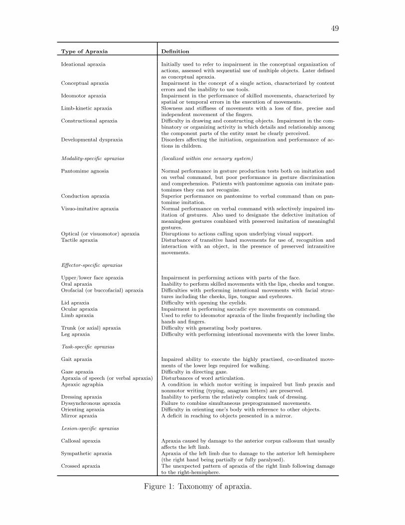
49
Type of Apraxia Definition
Ideational apraxia Initially used to refer to impairment in the conceptual organization ofactions, assessed with sequential use of multiple objects. Later definedas conceptual apraxia.
Conceptual apraxia Impairment in the concept of a single action, characterized by contenterrors and the inability to use tools.
Ideomotor apraxia Impairment in the performance of skilled movements, characterized byspatial or temporal errors in the execution of movements.
Limb-kinetic apraxia Slowness and stiffness of movements with a loss of fine, precise andindependent movement of the fingers.
Constructional apraxia Difficulty in drawing and constructing objects. Impairment in the com-binatory or organizing activity in which details and relationship amongthe component parts of the entity must be clearly perceived.
Developmental dyspraxia Disorders affecting the initiation, organization and performance of ac-tions in children.
Modality-specific apraxias (localized within one sensory system)
Pantomime agnosia Normal performance in gesture production tests both on imitation andon verbal command, but poor performance in gesture discriminationand comprehension. Patients with pantomime agnosia can imitate pan-tomimes they can not recognize.
Conduction apraxia Superior performance on pantomime to verbal command than on pan-tomime imitation.
Visuo-imitative apraxia Normal performance on verbal command with selectively impaired im-itation of gestures. Also used to designate the defective imitation ofmeaningless gestures combined with preserved imitation of meaningfulgestures.
Optical (or visuomotor) apraxia Disruptions to actions calling upon underlying visual support.Tactile apraxia Disturbance of transitive hand movements for use of, recognition and
interaction with an object, in the presence of preserved intransitivemovements.
Effector-specific apraxias
Upper/lower face apraxia Impairment in performing actions with parts of the face.Oral apraxia Inability to perform skilled movements with the lips, cheeks and tongue.Orofacial (or buccofacial) apraxia Difficulties with performing intentional movements with facial struc-
tures including the cheeks, lips, tongue and eyebrows.Lid apraxia Difficulty with opening the eyelids.Ocular apraxia Impairment in performing saccadic eye movements on command.Limb apraxia Used to refer to ideomotor apraxia of the limbs frequently including the
hands and fingers.Trunk (or axial) apraxia Difficulty with generating body postures.Leg apraxia Difficulty with performing intentional movements with the lower limbs.
Task-specific apraxias
Gait apraxia Impaired ability to execute the highly practised, co-ordinated move-ments of the lower legs required for walking.
Gaze apraxia Difficulty in directing gaze.Apraxia of speech (or verbal apraxia) Disturbances of word articulation.Apraxic agraphia A condition in which motor writing is impaired but limb praxis and
nonmotor writing (typing, anagram letters) are preserved.Dressing apraxia Inability to perform the relatively complex task of dressing.Dyssynchronous apraxia Failure to combine simultaneous preprogrammed movements.Orienting apraxia Difficulty in orienting one’s body with reference to other objects.Mirror apraxia A deficit in reaching to objects presented in a mirror.
Lesion-specific apraxias
Callosal apraxia Apraxia caused by damage to the anterior corpus callosum that usuallyaffects the left limb.
Sympathetic apraxia Apraxia of the left limb due to damage to the anterior left hemisphere(the right hand being partially or fully paralysed).
Crossed apraxia The unexpected pattern of apraxia of the right limb following damageto the right-hemisphere.
Figure 1: Taxonomy of apraxia.
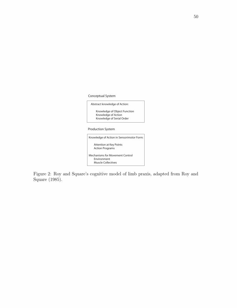
50
Conceptual System
Abstract knowledge of Action:
Knowledge of Object Function
Knowledge of Action
Knowledge of Serial Order
Production System
Knowledge of Action in Sensorimotor Form:
Attention at Key Points
Action Programs
Mechanisms for Movement Control
Environment
Muscle Collectives
Figure 2: Roy and Square’s cognitive model of limb praxis, adapted from Roy andSquare (1985).

51
Auditory Analysis
Auditory/Verbal
Input
Visual Object
Input
Visual Gestural
Input
Phonological Input
Lexicon
Motor Output
Visual Analysis Visual Analysis
Object Recognition
System
Action Input
Lexicon
Action Semantics
Action Output
Lexicon
Innervatory Patterns
Visual Analysis Visual Analysis
Object Recognition
System
Action Input
Lexicon
Action Semantics
Action Output
Lexicon
Gestural Buffer
Visuo-Motor
Conversion
Mechanism
a)
b)
c)
Spatiomotor transformations
from retinotopic to 'intrinsic'
body-centered coding
Dynamic model of body
coded in 'intrinsic' spatial
coordinates (body schema)
Knowledge of tools'
characteristic
movements
Stored portion of
gesture
representation
Knowledge of tools'
characteristic
movements
Figure 3: A cognitive neuropsychological model of limb praxis. The three compo-nents on the right are interchangeable with the empty box in the complete modelon the left. Under a) Rothi et al.’s original model of limb praxis. Under b) the pre-vious model revised by Cubelli et al. and under c) the model extended by Buxbaumet al. For a detailed description see the text. Adapted respectively from Rothi et al.(1997), Cubelli et al. (2000) and Buxbaum et al. (2000).

52
Patient Matched normal subject0
2
4
6
8
10
12
Tim
e (
s)
0 1 2 3 4 5 6 7 830
40
50
60
70
80
90
100
110
120
Should
er
flexi
on e
xtensi
on (
degre
es)
Time (s)
patient
matched normal subject
0 2 4 6 80
0.5
1
1.5
2
2.5
Time (s)
Ha
nd
Ve
loci
ty (
m/s
2)
patientmatched normal subject
a)
b)
c)
d)
Figure 4: An example of the abnormal kinematics of an apraxic movement. A patientwith left ischemic lesions was tested in a study of imitation of meaningless gestures.The stimulus to imitate for this movement is shown under a) and represents a handposture relative to the head. Under b), the movement times of the patient are longerthan those of a matched normal subject (including replacement of the hand in theinitial condition). Under c), the trajectory of the shoulder flexion-extension jointangle of the patient (shown in solid line) contains several irregularities which arethe result from multiple hesitations and changes of directions, whereas the matchednormal subject shoulder flexion-extension trajectory (dashed line) is smooth. Thespeed profile of the patient (solid line) is shown under d) and contains multiple peakswith reduced maximum velocities that contrast with the simple bell-shaped velocityprofile of the matched normal subject (dashed line).

53
Figure 5: Schema of the monkey brain areas and their connectivity. Adapted from(Wise et al., 1997).


