HAVING OUR HEAD EXAMINED. Lesion Lesion INTRODUCTION.
104
HAVING OUR HEAD EXAMINED
-
Upload
julius-cameron -
Category
Documents
-
view
236 -
download
2
Transcript of HAVING OUR HEAD EXAMINED. Lesion Lesion INTRODUCTION.

HAVING OUR HEAD EXAMINED

Lesion
INTRODUCTION

Electroencephalogram (EEG)
RECORDING THE BRAIN’S ELECTRICAL ACTIVITY

CT (Computed Tomography) scan- x-ray photographs + computer composite
PET (Positron Emission Tomography) scan- radioactive glucose. Shows areas of the brain that “light up” during different cognitive tasks
NEUROIMAGING TECHNIQUES

MRI (Magnetic Resonance Imaging)- Brain in a magnetic field. Shows detailed pictures of brain tissue (structure)
fMRI (Functional MRI)- shows function as well as structure. Shows functioning of different areas of the brain by tracking blood flow

OLDER BRAIN STRUCTURES

BrainstemMedullaPonsReticular formation
Introverts and extroverts
THE BRAINSTEM

ThalamusAll the senses EXCEPT smell
THE THALAMUS

Cerebellum“Little brain”Why can’t we
tickle ourselves?
THE CEREBELLUM

Limbic SystemHippocampus
THE LIMBIC SYSTEM

AmygdalaAggression and fear
THE LIMBIC SYSTEM
THE AMYGDALA

HypothalamusInfluence on the pituitary glandReward CentersReward deficiency syndrome
THE LIMBIC SYSTEM
THE HYPOTHALAMUS

THE CEREBRAL CORTEX

CerebrumCerebral cortex
INTRODUCTION


Glial cells (“glue cells”)LobesFrontal lobesParietal lobesOccipital lobesTemporal lobes
STRUCTURE OF THE CORTEX


Motor CortexMapping the Motor Cortex
FUNCTIONS OF THE CORTEX
MOTOR FUNCTIONS

Sensory cortex
FUNCTIONS OF THE CORTEX
SENSORY FUNCTIONS

Functions of the Cortex

Association areasFrontal lobesPhineas Gage
Parietal lobesTemporal lobes
FUNCTIONS OF THE CORTEX
ASSOCIATION AREAS

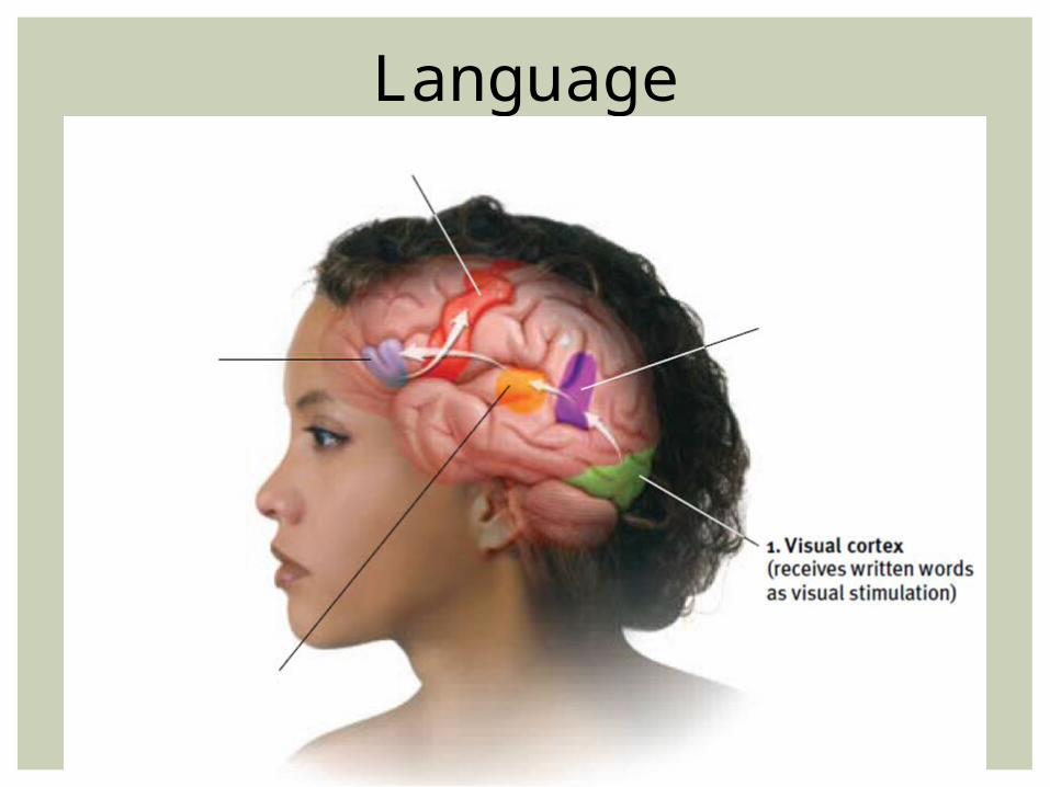
Aphasia- impaired use of language
Broca’s areaWernicke’s area
LANGUAGE

Language
Language

Language

Language

Language

Language

Brain DamagePlasticityNeural prosthetics
THE BRAIN’S PLASTICITY

Reticular Formation: “If someone tickled you, your reticular formation would be activated”
Medulla: think “medals” they hang over your heart and lungs, areas the medulla controls
Pons: Think of a still…calm…pond…you’re getting sleepy…very sleepy…
Thalamus: The “Thalamus” says “Thataway” – directs sensory information
PARTS OF THE BRAIN (WITH MNEMONICS)

Cerebellum: Sarah balances with her cerebellum

Hypothalamus: “Hypo-the llamas”- the llamas need food, water etc. Hypothalamus helps direct these activities

Amygdala- It just sounds scary. “Amygdala” should be the name of a witch in a horror movie- controls aggression and fear
Hippocampus: If you saw a “hippo” on “campus” you would remember- involved in memory

Cerebral cortex: cortex is Latin for “shell” or “husk”- the cerebral cortex is outer layer or “shell” of the brain

Frontal lobe: The “future” is in “front” of us. Involved in making plans and judgments (also speaking and muscle movements)
Parietal lobe: Uh…it’s behind the frontal lobe…?
Temporal lobe: If you have temporary hearing loss, investigate the temporal lobe
Occipital lobe: looks like “optical” – involved in processing visual information

Broca’s area: Broca – Boca
Wernicke’s area: If I asked you to reach in your backpack and take out your “wernicke,” you would not understand

Corpus Callosum- CorPlus CalloSum- adds (connects) the left and right brain together
Kim Peek’s Brain

Reticular Formation: “If someone tickled you, your reticular formation would be activated”
Medulla: think “medals” they hang over your heart and lungs, areas the medulla controls
Pons: Think of a still…calm…pond…you’re getting sleepy…very sleepy…
Thalamus: The “Thalamus” says “Thataway” – directs sensory information
Cerebellum: Sarah balances with her cerebellumHypothalamus: “Hypo-the llamas”- the llamas need food,
water etc. Hypothalamus helps direct these activities
MNEMONICS

Amygdala- It just sounds scary. “Amygdala” should be the name of a witch in a horror movie- controls aggression and fear
Hippocampus: If you saw a “hippo” on “campus” you would remember- involved in memory
Cerebral cortex: cortex is Latin for “shell” or “husk”- the cerebral cortex is outer layer or “shell” of the brain
Frontal lobe: The “future” is in “front” of us. Involved in making plans and judgments (also speaking and muscle movements)
Parietal lobe: Uh…it’s behind the frontal lobe…? Temporal lobe: If you have temporary hearing loss, investigate the temporal lobe
Occipital lobe: looks like “optical” – involved in processing visual information
Broca’s area: Broca – Boca Wernicke’s area: If I asked you to reach in your backpack and take out
your “wernicke,” you would not understand Corpus Callosum- CorPlus CalloSum- adds (connects) the left and right
brain together
MNEMONICS CONT.

OUR DIVIDED BRAIN

Vogel and BogenCorpus-callosumSplit brainMyers and Gazzaniga
SPLITTING THE BRAIN


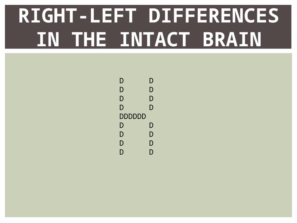
RIGHT-LEFT DIFFERENCES IN THE INTACT BRAIN
D DD DD DD DDDDDDDD DD DD DD D

Hemispheric SpecializationPerceptual tasksLanguageSense of self
RIGHT-LEFT BRAIN DIFFERENCES


THE BRAIN AND CONSCIOUSNESS

Conscious and unconscious choices
INTRODUCTION

Cognitive neuroscience
COGNITIVE NEUROSCIENCE

Dual ProcessingPrimingConscious left brainIntuitive right brain
DUAL PROCESSING

Two-Track MindVisual perception trackVisual action track
THE TWO-TRACK MIND

Left-handednessQ: Is it alright to be left handed?A: No, it is not alright.

Wartime dilemmaTrolley dilemma
NEUROSCIENCE AND MORAL JUDGEMENTS

It’s wartime and you are hiding in the basement with a group of townspeople. Enemy soldiers are outside. Your baby starts to cry loudly; if nothing is done, the soldiers will find you and kill everyone including the baby. The only way to prevent this loss of life is to cover the baby’s mouth; if you do, the baby will smother. What should you do?

A runaway trolley is hurtling down the tracks toward five people who will be killed if it proceeds on its present course. You can save these five people by diverting the trolley onto a different set of tracks, one that has only one person on it, but if you do this that person will be killed. Is it morally permissible to turn the trolley and thus prevent five deaths at the cost of one?

Now consider a slightly different dilemma. Once again, the trolley is headed for five people. You are on a footbridge over the tracks next to a large man. The only way to save the five people is to push this man off the bridge and into the path of the trolley. Is that morally permissible?

When a dilemma is posed, our reasoning processes conflict with our more basic emotional processes and the decision takes longer.

EINSTEIN’S BRAIN

DumberSmarter/efficientNicer
OUR SHRINKING BRAINS

THE END

DEFINITION SLIDES

= tissue destruction; a brain lesion is a naturally or experimentally caused destruction of brain tissue.
LESION

= an amplified recording of the waves of electrical activity that sweep across the brain’s surface. These waves are measured by electrodes placed on the scalp.
ELECTROENCEPHALOGRAM (EEG)

= a series of X-ray photographs taken from different angles and combined by computer into a composite representation of a slice through the body.
Also called CAT scan.
CT (COMPUTED TOMOGRAPHY) SCAN

= a visual display of brain activity that detects where a radioactive form of glucose goes while the brain performs a given task.
PET (POSITRON EMISSION TOMOGRAPHY) SCAN

= a technique that uses magnetic fields and radio waves to produce computer-generated images of soft tissue. MRI scans show brain anatomy.
MRI (MAGNETIC RESONANCE IMAGING)

= a technique for revealing bloodflow and, therefore, brain activity by comparing successive MRI scans. fMRI scans show brain function.
FMRI (FUNCTIONAL MRI)

= the oldest part of the central core of the brain, beginning where the spinal cord swells as it enters the skull; the brainstem is responsible for automatic survival functions.
BRAINSTEM
= the base of the brainstem; controls heartbeat and breathing.
MEDULLA

= a nerve network in the brainstem that plays an important role in controlling arousal.
RETICULAR FORMATION

= the brain’s sensory switchboard, located on top of the brainstem; it directs messages to the sensory receiving areas in the cortex and transmits replies to the cerebellum and medulla.
THALAMUS

= the “little brain” at the rear of the brainstem; functions include processing sensory input and coordinating movement output and balance.
CEREBELLUM

= doughnut-shaped neural system (including the hippocampus, amygdala, and hypothalamus) located below the cerebral hemispheres; associated with emotions and drives.
LIMBIC SYSTEM

= two lima bean-sized neural clusters in the limbic system; linked to emotion.
AMYGDALA

= a neural structure lying below (hypo) the thalamus; it directs several maintenance activities (eating, drinking, body temperature), helps govern the endocrine system via the pituitary gland, and is linked to emotion and reward.
HYPOTHALAMUS

= the intricate fabric of interconnected neural cells covering the cerebral hemispheres; the body’s ultimate control and information-processing center.
CEREBRAL CORTEX

= cells in the nervous system that support, nourish, and protect neurons.
GLIAL CELLS

= portion of the cerebral cortex lying just behind the forehead; involved in speaking and muscle movements and in making plans and judgments.
FRONTAL LOBES

= portion of the cerebral cortex lying at the top of the head and toward the rear; receives sensory input for touch and body position.
PARIETAL LOBES

= portion of the cerebral cortex lying at the back of the head; includes areas that receive information from the visual fields.
OCCIPITAL LOBES

= portion of the cerebral cortex lying roughly above the ears; includes the auditory areas, each receiving information primarily from the opposite ear.
TEMPORAL LOBES

= an area at the rear of the frontal lobes that controls voluntary movements.
MOTOR CORTEX

= area at the front of the parietal lobes that registers and processes body touch and movement sensations.
SENSORY CORTEX

= areas of the cerebral cortex that are not involved in primary motor or sensory functions; rather, they are involved in higher mental functions such as learning, remembering, thinking, and speaking.
ASSOCIATION AREAS

= impairment of language, usually caused by left hemisphere damage either to Broca’s area (impairing speaking) or to Wernicke’s area (impairing understanding).
APHASIA

= controls language expression that directs the muscle movements involved in speech.
BROCA’S AREA

= controls language reception – a brain area involved in language comprehension and expression; usually in the left temporal lobe.
WERNICKE’S AREA

= the brain’s ability to change, especially during childhood, by reorganizing after damage or by building new pathways based on experience.
PLASTICITY

= the formation of new neurons.
NEUROGENESIS

= the large band of neural fibers connecting the two brain hemispheres and carrying messages between them.
CORPUS CALLOSUM

= a condition resulting from surgery that isolates the brain’s two hemispheres by cutting the fibers (mainly those of the corpus callosum) connecting them.
SPLIT BRAIN

= our awareness of ourselves and our environment.
CONSCIOUSNESS

= the interdisciplinary study of the brain activity linked with cognition (including perception, thinking, memory and language).
COGNITIVE NEUROSCIENCE

=the principle that information is often simultaneously processed on separate conscious and unconscious tracks.
DUAL PROCESSING