Apical lesions in Dentistry
25
OD17-7/2 Page 1 CHAPTER 7 APICAL LESIONS Periapical granuloma Radicular cyst Apical abscess Apical scar Surgical defect Periodontal disease Condensing osteitis Osteosclerosis Socket sclerosis Periapical cemental dysplasia Cementifying fibroma (ossifying fibroma) Benign cementoblastoma Florid osseous dysplasia
-
Upload
mahmoud-shaheen -
Category
Education
-
view
54 -
download
3
Transcript of Apical lesions in Dentistry
OD17-7/2 Page 1
CHAPTER 7
APICAL LESIONS
Periapical granuloma
Radicular cyst
Apical abscess
Apical scar
Surgical defect
Periodontal disease
Condensing osteitis
Osteosclerosis
Socket sclerosis
Periapical cemental dysplasia
Cementifying fibroma (ossifying fibroma)
Benign cementoblastoma
Florid osseous dysplasia
OD17-7/2 Page 2
APICAL LESIONS
Whenever a lesion is observed on a radiograph, it must first be described in general
terms before a differential diagnosis is attempted. Is the lesion radiolucent, radiopaque, or
mixed (combination of radiolucency and radiopacity)? Where is the lesion located? The
apices of which teeth are involved? What is the size of the lesion? Is the margin of the
lesion ill-defined, well-defined, or well-defined with a radiopaque border? Is the
appearance of the bone surrounding the lesion: normal, porous, or sclerotic?
The various radiographic appearances of the margins of lesions and the changes in the
surrounding bone have been given clinical interpretation by some diagnosticians based
largely on intuitive analysis rather than on research data. Although the significance of
these signs is sometimes questionable, they are useful in radiographic interpretation. An ill-
defined (diffuse, irregular) periphery is suggestive of a lesion enlarging by invading the
surrounding bone. A well-defined (circumscribed) periphery is suggestive of a self-
contained lesion enlarging by expansion. A well-defined periphery with a hyperostotic
(sclerotic) radiopaque periphery is suggestive of an extremely slow-growing self-contained
lesion enlarging by expansion.
If the bone surrounding a lesion does not show any change, the clinical interpretation is that
of a static lesion. If it shows porosity (destructive breakdown), the interpretation is that of
an invasive process resulting from osteolytic activity. If it shows sclerosis (hyperostosis),
the interpretation is that of resistance to the pathologic process resulting from osteoblastic
activity.

OD17-7/2 Page 3
TYPES OF BORDERS
Fig. 7-1 Well-defined radiolucent lesion at the apex of the maxillary lateral incisor.
The circumscribed radiopaque (sclerotic) border signifies that the lesion is
self-contained, enlarges by expansion, and is slow-growing. There is no
change in the surrounding bone.
Fig. 7-2 Well-defined radiolucent lesion at the apex of the mandibular left second
premolar. The well-defined border signifies that the lesion is self-contained,
and enlarges by expansion. The surrounding bone shows slight sclerosis
(osteoblastic activity) which signifies resistance to the pathologic process.
Fig. 7-3 A radiolucent lesion with diffuse, irregular borders at the apices of the roots of
the maxillary left first molar. The diffuse margins, and the porosity (osteolytic
activity) in surrounding bone signify an invasive process.
OD17-7/2 Page 4
PERIODONTAL SPACE WIDENING (Apical periodontitis)
Radiographically, apparent widening (also called thickening) of the periodontal
ligament space is caused by edema, resulting in accumulation of inflammatory exudate in
the connective tissue of the periodontal ligament. The term apical periodontitis is often
used to describe this pathologic periodontal space widening.
Pathologic periodontal space widening occurs as a result of infection, trauma, orthodontic
treatment, or tooth extrusion. It is imperative that dentists be aware of trauma resulting
from occlusal imbalance, especially that caused by an amalgam, gold, composite resin, or
other restoration that is placed too high in the tooth and results in premature contact with
the opposing tooth. Depending on the extent of the traumatic force and the individual's
physiologic resistance, the widened periodontal space either returns to its normal
appearance after the elimination of the trauma or may form a chronic apical inflammatory
lesion such as an apical granuloma, a radicular cyst, or an apical abscess.
Non-pathologic periodontal space widening occurs as a result of the terminal stage of root
formation (dental papilla), wide marrow space superimposed on a tooth apex, or
superimposition of a tooth apex on a radiolucent anatomy (such as a nasal fossa, maxillary
sinus, mental foramen or submandibular fossa) producing over-exposure or "burnout". An
illusion of loss of lamina dura may also be produced when a tooth apex is superimposed on
a radiolucent anatomy.
Fig. 7-4 Infection (caries) is the cause of the widened periodontal space of the first
molar (apical periodontitis). In this case, the lamina dura is also thickened
(radiopaque line).
Fig. 7-5 Trauma is the cause of the widened periodontal spaces of the central incisors
(apical periodontitis. Clinically, the teeth were retruded.

OD17-7/2 Page 5
Fig. 7-6 Traumatic occlusion, that is, premature contact with the opposing premolars,
is the cause of the widened periodontal spaces of the maxillary premolars
(apical periodontitis). The patient also has alveolar bone loss due to
periodontal disease.
Fig. 7-7 Orthodontic treatment is the cause of the widened periodontal spaces of the
incisors.
Fig. 7-8 Developing root apices of the second molar may be mistaken for widened
periodontal spaces. Root apices complete their formation approximately one
to two years after tooth eruption.
Fig. 7-9 Illusion of widened periodontal space of the root apex of the premolar
because of superimposition on a radiolucent anatomy (maxillary sinus). The
widened periodontal space of the canine may be due to a similar illusion
and/or infection (caries). Also note the illusion of loss of apical lamina dura
on both teeth.
Fig. 7-10 Illusion of widened periodontal spaces of the root apices of the molar
because of superimposition on a radiolucent anatomy (mandibular canal).
Also note the illusion of apical loss of lamina dura.

OD17-7/2 Page 6
APICAL LESION VERSUS ANATOMICAL LANDMARK
To differentiate an apical lesion from an anatomical landmark, make another radiograph
with a different x-ray beam angulation. An anatomical landmark will change its position with
a change in angulation of the x-ray beam. On the other hand, an apical lesion will maintain
the same relationship to the involved tooth.
Another method of differentiation is to follow the continuity of the periodontal space. The
periodontal space will be interrupted in the case of an apical lesion but will be continuous in
the case of an anatomical landmark.
Fig. 7-11 A — Mental foramen. Notice the intact periodontal space.
B — Chronic inflammatory apical lesion. Notice the loss of the apical
periodontal space.
Fig. 7-12 Radiolucent anatomy mistaken for an apical lesion. Mental foramen
superimposed on the root apex of the second premolar may be mistaken for
an apical lesion.

OD17-7/2 Page 7
PERIAPICAL GRANULOMA, RADICULAR CYST AND APICAL ABSCESS
(Inflammatory pulpo-periapical lesions)
The most common pathologic conditions that involve teeth are the inflammatory lesions of
the pulp and periapical areas. Once inflammation (pulpitis) has spread from the dental
pulp, it can produce a variety of apical pathologic changes, the most common of which are
the periapical granuloma, radicular cyst, and apical abscess. Various factors such as the
host's resistance and the virulence of the bacteria affect the local inflammatory response in
the periapical area. Without a microscopic diagnosis, a clinician is frequently unable to
differentiate between a periapical granuloma, a radicular cyst, and an apical abscess.
Radiographic examination is inadequate in making a specific diagnosis.
Inflammatory periapical lesions have certain common clinical characteristics: 1) A history of
painful pulpitis leading to the death of the pulp. 2) A nonvital reaction to electric pulp
testing. In a multirooted tooth where only one root is associated with the pulpo-periapical
pathosis, the tooth will frequently give a vital reaction. 3) Presence of a deep carious lesion
exposing the pulp or a restoration close to the pulp, or a fractured tooth and/or a discolored
crown. 4) Destruction resulting in an interrupted lamina dura of the involved tooth.
A granuloma is formed from the successful attempt of the periapical tissues to neutralize
and confine the irritating toxic products escaping from the root canal. This low grade
inflammation in the tissues continues to induce the proliferation of vascular granulation
tissue. A granuloma may evolve into a radicular cyst or an apical abscess. Clinically, the
lesion is usually asymptomatic but may sometimes exhibit mild pain and sensitivity to
percussion. The affected tooth is nonvital. Radiographically granulomas form small well-
defined radiolucencies. They are the most common periapical lesions and constitute
approximately 50% of all periapical radiolucent lesions.
A radicular cyst (also known as periapical cyst, dental cyst, periodontal cyst) has its origin
from the cell rests of Malassez which are present in periodontal and periapical ligament,
and in periapical granulomas. Most radicular cysts originate from pre-existing granulomas.

OD17-7/2 Page 8
Clinically, the lesion is usually asymptomatic but may sometimes exhibit mild pain and
sensitivity to percussion. The affected tooth is nonvital. A radicular cyst may slowly
enlarge and when large, may cause expansion of the cortical plates. Radiographically, a
radicular cyst forms a large well-defined radiolucency with or without a radiopaque
(hyperostotic) border. The more pronounced the hyperostotic (sclerotic) border, the more
likely is the lesion to be a radicular cyst. It is the second most common periapical lesion
and constitutes approximately 40% of all periapical radiolucent lesions. (See also chapter
on "Cysts of the Jaws").
In approximately 90% of the cases, a well-defined radiolucency at the apex of an untreated
asymptomatic tooth with a nonvital or diseased pulp is either a dental granuloma or a
radicular cyst. Their sizes are the differentiating features: a granuloma is small whereas a
radicular cyst is large. In practice, however, it is not necessary to differentiate a periapical
granuloma from a radicular cyst because both lesions respond quite well to conservative
root canal therapy.
An apical abscess, also called dental or dentoalveolar abscess, usually develops from a
pulpo-periapical inflammatory condition. In the acute stage, the onset of infection is so
sudden that there is no radiographic evidence of an apical lesion. An apical abscess can
develop also from a pre-existing granuloma or cyst. The associated tooth is nonvital, very
painful, extremely sensitive to percussion, and often slightly extruded. The patient will
complain that the tooth feels "high" when it occludes with the opposing tooth. The tooth will
not respond to electrical pulp tests. The application of ice will slightly relieve the pain but
the application of heat will intensify pain. The tooth may demonstrate increased mobility.
Radiographically apical abscesses form large radiolucencies with diffuse irregular borders.
They are the least common of the three pulpo-periapical lesions and constitute
approximately 2% of all periapical radiolucent lesions.
If an apical abscess is permitted to progress without treatment, it may penetrate the cortical
plate at the thinnest and closest part of the tooth apex and form a swelling in the adjacent

OD17-7/2 Page 9
soft tissues. This infection site in the soft tissues is painful. The skin or mucous surface
over the abscess is warm and rubbery to palpation, and demonstrates fluctuance. Once
drainage is established by a sinus tract permitting the pus to drain to the surface, the tooth
and associated swelling are no longer painful since the pain-producing pressure of the
abscess is reduced. The regional lymph nodes may be enlarged and painful. The systemic
temperature may be elevated.
Fig. 7-13 Presence of either a periapical granuloma, a radicular cyst or an abscess at
the root apices of the first molar. The small, well-defined radiolucency is
suggestive (on a speculative basis) of a periapical granuloma. Periapical
granulomas are the most frequently occurring of the three pulpo-periapical
lesions. The involved tooth is nonvital.
Fig. 7-14 Presence of either a periapical granuloma, a radicular cyst or an abscess at
the root apices of the right central and lateral incisors. The large, well-
defined radiolucency with a radiopaque (sclerotic) border is suggestive (on a
speculative basis) of a radicular cyst (periapical cyst). Radicular cysts are
the second most frequently occurring of the three pulpo-periapical lesions.
The involved tooth is nonvital.
Fig. 7-15 Presence of either a periapical granuloma, a radicular cyst or an abscess at
the root apices of the first molar. The large radiolucency with diffuse irregular
border is suggestive (on a speculative basis) of an abscess. Apical
abscesses are the least frequently occurring of the three pulpo-periapical
lesions. The involved tooth is nonvital.

OD17-7/2 Page 10
APICAL SCAR
An inflammatory apical lesion treated by root canal therapy may respond well to
treatment by filling new bone at the site of the lesion. However, the healing process may
sometimes terminate abruptly and leave a small amount of dense scar tissue known as an
apical scar. The scar tissue represents one of the possible end points of healing. It is
composed of dense fibrous tissue and is situated at the apex of a pulpless tooth in which
the root canals have been successfully filled. Microscopic examination reveals fibroblasts
scattered in the collagen fibers. Unlike an apical granuloma, inflammatory cells are not a
feature and vascularity is quite meager. An apical scar is a small, asymptomatic, and well-
circumscribed radiolucency. When observed radiographically over the years, it will either
remain constant in size or diminish slightly.
Fig. 7-16 Apical scar is an area at the apex of a tooth that fails to fill in with osseous
tissue after endodontic treatment. The second premolar was successfully
treated with root canal therapy. The apical radiolucency persisted after
treatment. Patient was recalled after several months. The radiolucency
slightly decreased in size but was still present.
Fig. 7-17 Apical scar. The molar was successfully treated with root canal therapy.
Patient was recalled after several months. The apical radiolucency reduced
in size and persisted.

OD17-7/2 Page 11
SURGICAL DEFECT
A surgical defect is that portion of bone that fails to form osseous tissue. It is frequently
seen periapically after root resection in which the site is filled with dense fibrous (collagen)
tissue instead of bone. It is an asymptomatic persistent radiolucency. An extraction site
can also form a surgical defect. Approximately 75% of all surgically treated periapical
radiolucencies require 1 to 10 years or longer for complete resolution. In the remaining
25%, complete healing does not occur.
Fig. 7-18 Surgical defect in bone is an area that fails to fill in with osseous tissue after
surgery. An apicoectomy was performed at the apex of the left central incisor
and the lesion was curetted. The radiolucency persisted as a surgical defect.
Fig. 7-19 Surgical defect at the extraction site of the mandibular third molar. The
radiolucency of the tooth socket persisted after 1 year.

OD17-7/2 Page 12
PERIODONTAL DISEASE
Periodontal disease is discussed in detail in a previous chapter. Nevertheless, it is
considered here because it produces a relatively common periapical radiolucency in the
advanced stages. The entire bony support of the involved tooth may be completely
destroyed or the tooth may appear to be floating in a radiolucency. Sometimes a narrow
vertical pocket may extend to the apex and appear as a well-defined periapical
radiolucency. This may lead the unwary clinician to a false conclusion of a pulpo-periapical
pathosis. In the advanced stages, these teeth are usually quite mobile and may become
sensitive to percussion; but surprisingly many remain vital, and the demonstration of such
vitality aids the clinician in differentiating a periodontal disease radiolucency from an
inflammatory apical radiolucency (periapical granuloma, radicular cyst and apical abscess).
A clinical examination of supporting tooth structures should be undertaken by probing all
periodontal pockets.
Fig. 7-20 Periodontal disease producing a periapical radiolucency. Vertical bone loss
around the second premolar extends to the apex and appears as a fairly well-
defined radiolucency. The affected tooth is vital.
Fig. 7-21 Periodontal disease producing a periapical radiolucency around the mesial
root of the second molar. Vertical bone loss extends to the apex of the root.
The affected tooth is vital.

OD17-7/2 Page 13
CONDENSING OSTEITIS (Focal sclerosing osteomyelitis)
Condensing osteitis is a reaction of bone induced by inflammation. It occurs mainly at the
apex of a tooth from an infected pulp. The infection from tooth caries reaches the pulp and
progresses to the apical tissues to produce a small periapical radiolucency called rarefying
osteitis. The small rarefying osteitis may be either a periapical granuloma, a radicular cyst
or an abscess. The bone surrounding this rarefying osteitis becomes dense in order to
prevent further spread of the lesion. This dense radiopacity surrounding the rarefying
osteitis is called condensing osteitis. The pulp of the involved tooth is nonvital.
1. Condensing osteitis is a reaction to periapical infection resulting in the
formation of dense bone. The infection usually originates from caries
(sometimes from periodontal disease).
2. Condensing osteitis occurs usually at the apex of a nonvital tooth. On
a radiograph, the tooth may exhibit a large carious lesion or a large
restoration close to the dental pulp.
3. Radiographically, the lesion shows a diffuse radiopacity surrounding a
small central radiolucency at the apex (or apices) of a tooth.
4. Treatment consists of removing the infection either through tooth
extraction or root canal therapy.
5. Prognosis - the high radiopacity of bone tends to disappear partially or
completely in a majority of cases after treatment is given to remove
the infection. Approximately 30% of the cases will not resolve to
normal appearing bone after adequate treatment. The persistence of
the radiopacity should not be of concern to the clinician because of
the innocuous nature of the lesion.
Fig. 7-22 Condensing osteitis at the root apices of the carious first molar. Condensing
osteitis is a reaction to an inflammatory process. Note the diffuse

OD17-7/2 Page 14
radiopacities (condensing osteitis) surrounding the apical radiolucencies
(rarefying osteitis). The involved tooth is nonvital.
Fig. 7-23 Condensing osteitis at the root apices of the carious first molar. Note the
diffuse radiopacities (condensing osteitis) surrounding the apical
radiolucencies (rarefying osteitis). The involved tooth is nonvital.
Fig. 7-24 Condensing osteitis at the root apices of the carious first and second molars.
The involved teeth are nonvital.

OD17-7/2 Page 15
OSTEOSCLEROSIS AND SOCKET SCLEROSIS
Osteosclerosis (enostosis) is one of the common bony lesions found incidentally on a
radiographic examination. It is a well-defined radiopaque mass without any associated
radiolucency. The cause of the lesion is not known (idiopathic). The lesion is
asymptomatic and the teeth associated with it are invariably healthy with vital pulps. There
is neither any pain nor any cortical bone expansion. The lesion may occur either in the
alveolus, in the periapical region, in the intra-radicular or inter-radicular region, below the
crest of the ridge, or in the body of the mandible. It has been suggested that excess
occlusal stress may be one of the factors in osteosclerosis associated with roots of teeth.
When it occurs near the first and second premolars, its formation is speculated to be that of
deposition of sclerotic bone around the root fragment of a deciduous molar which acts as a
nidus. It has been reported that only 10% to 12% of patients with osteosclerosis show root
resorption of an adjoining tooth.
When osteosclerosis occurs in the socket of an extracted tooth as a reparative process, it is
known as socket sclerosis. Radiographically, it is often impossible to distinguish socket
sclerosis from a retained root of a tooth. A dentist may be wrongfully accused of having
failed to remove the tooth in its entirety.
1. Osteosclerosis refers to a localized region of abnormally dense bone.
The cause of the sclerotic bone is either idiopathic or reparative. It is
not associated with any infection.
2. Osteosclerosis can occur in any part of the jaw even at the apices of
vital teeth. Socket sclerosis occurs in the sockets of extracted teeth
and may sometimes be mistaken for retained roots.
3. Radiographically, the lesion shows a well-defined radiopacity. There
is no association with any radiolucency. 10% to 12% of patients show
root resorption.
4. No treatment is necessary.
5. Prognosis - the radiopacity persists without any increase in size.

OD17-7/2 Page 16
Fig. 7-25 Osteosclerosis at the root apices of the first molar. The involved tooth is
vital.
Fig. 7-26 Osteosclerosis between the second premolar and first molar, and at the
mesial root apex of the first molar. The involved tooth is vital. 10% to 12% of
the patients show root resorption.
Fig. 7-27 Osteosclerosis at the distal root of the second molar.
Fig. 7-28 Osteosclerotic bone to the mesial of the molar, preventing the tooth from
further tilting or drifting.
Fig. 7-29 Socket sclerosis. Sclerosing bone deposited in the socket of the extracted
tooth. A dentist may be erroneously accused of having failed to extract the
root. There are no definite radiographic criteria of distinguishing a retained
root from a socket sclerosis.
Fig. 7-30 Socket sclerosis. Although there are no definite radiographic criteria of
distinguishing a retained root from a socket sclerosis, in this case, the diffuse
border distinguishes socket sclerosis from a retained root.
Fig. 7-31 Socket sclerosis between the second premolar and the mesially drifted
second molar.
OD17-7/2 Page 17
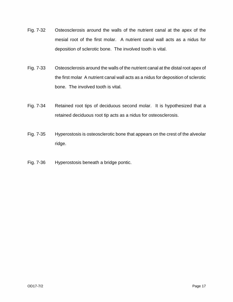
Fig. 7-32 Osteosclerosis around the walls of the nutrient canal at the apex of the
mesial root of the first molar. A nutrient canal wall acts as a nidus for
deposition of sclerotic bone. The involved tooth is vital.
Fig. 7-33 Osteosclerosis around the walls of the nutrient canal at the distal root apex of
the first molar A nutrient canal wall acts as a nidus for deposition of sclerotic
bone. The involved tooth is vital.
Fig. 7-34 Retained root tips of deciduous second molar. It is hypothesized that a
retained deciduous root tip acts as a nidus for osteosclerosis.
Fig. 7-35 Hyperostosis is osteosclerotic bone that appears on the crest of the alveolar
ridge.
Fig. 7-36 Hyperostosis beneath a bridge pontic.

OD17-7/2 Page 18
CEMENTOMAS
a. Periapical cemental dysplasia (periapical cementoma).
b. Cementifying fibroma (ossifying fibroma, cemento-ossifying fibroma)
c. Benign cementoblastoma (true cementoma)
Periapical cemental dysplasia, cementifying fibroma, ossifying fibroma, cemento-ossifying
fibroma, benign cementoblastoma, and florid osseous dysplasia have been grouped by
some under the term "benign fibro-osseous lesions of periodontal ligament origin." These
benign fibro-osseous lesions of periodontal ligament origin are a group of cementum and
bone producing tumors and tumor like proliferations that arise from the cells of the
periodontal ligament. In this text, the florid osseous dysplasia is described as a separate
entity.
All the three types of cementomas have three radiographic appearances:
1) early or osteolytic stage appears radiolucent.
2) mixed or cementoblastic stage appears as a radiolucency containing radiopacities.
3) final or calcified stage appears as a homogeneous radiopacity surrounded by a thin
radiolucent border.
All cementomas are associated with teeth having vital pulps unless, otherwise involved with
caries or trauma.
Periapical Cemental Dysplasia:
Black persons and females of 40 years and older are more commonly affected than white
persons and males to develop periapical cemental dysplasia. Although periapical cemental
dysplasia occurs usually in the mandibular anterior region, it may sometimes be found in
the mandibular posterior region. On a radiograph, the early radiolucent stage of periapical
cemental dysplasia could be misdiagnosed for inflammatory periapical radiolucencies
(periapical granuloma, radicular cyst, abscess). An unalert clinician might needlessly
extract or institute endodontic treatment on a tooth with a normal pulp. The periapical
cemental dysplasia is associated with a vital tooth and is totally asymptomatic. These
features are in contrast to those of an inflammatory lesion which is associated with a
nonvital tooth and may exhibit symptoms. Periapical Cemental Dysplasia (Periapical cementoma)
*Cementifying Fibroma (Ossifying fibroma, cemento-ossifying
*Benign Cementoblastoma (True cementoma)

OD17-7/2 Page 19
fibroma) 1. Believed to be a
reaction of periapical bone. Actual cause unknown. Asymptomatic.
Benign mesenchymal odontogenic neoplasm.
Benign mesenchymal odontogenic neoplasm.
2. Average age is 40
years. Predominantly in black persons and in females.
In young and middle-aged adults.
In teenagers and young adults.
3. Around apices of
teeth. Usually mandibular anteriors.
Around apices of teeth. Usually the mandibular posteriors.
Attached to the apex or apices of mandibular molar or premolar.
4. Vital teeth. Position of
teeth not affected.
Vital teeth. Displacement of teeth or divergence of roots.
Vital teeth. Position of teeth not affected.
5. Usually multilocular.
Sometimes unilocular.
Usually unilocular.
Usually unilocular.
6. Radiographic
appearance has three developmental stages.
Radiographic appearance has three developmental stages.
Radiographic appearance when discovered is usually of the third stage.
7. No expansion of
cortical plates. Self-limiting lesion.
Expansion of cortical plates.
Expansion of cortical plates when large.
8. No treatment
necessary.
Excision of lesion.
Extraction of involved tooth and excision of lesion.
*For details, refer chapter on "Odontogenic Benign Tumors of the Jaws"
OD17-7/2 Page 20
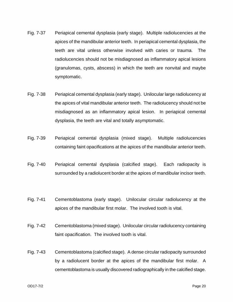
Fig. 7-37 Periapical cemental dysplasia (early stage). Multiple radiolucencies at the
apices of the mandibular anterior teeth. In periapical cemental dysplasia, the
teeth are vital unless otherwise involved with caries or trauma. The
radiolucencies should not be misdiagnosed as inflammatory apical lesions
(granulomas, cysts, abscess) in which the teeth are nonvital and maybe
symptomatic.
Fig. 7-38 Periapical cemental dysplasia (early stage). Unilocular large radiolucency at
the apices of vital mandibular anterior teeth. The radiolucency should not be
misdiagnosed as an inflammatory apical lesion. In periapical cemental
dysplasia, the teeth are vital and totally asymptomatic.
Fig. 7-39 Periapical cemental dysplasia (mixed stage). Multiple radiolucencies
containing faint opacifications at the apices of the mandibular anterior teeth.
Fig. 7-40 Periapical cemental dysplasia (calcified stage). Each radiopacity is
surrounded by a radiolucent border at the apices of mandibular incisor teeth.
Fig. 7-41 Cementoblastoma (early stage). Unilocular circular radiolucency at the
apices of the mandibular first molar. The involved tooth is vital.
Fig. 7-42 Cementoblastoma (mixed stage). Unilocular circular radiolucency containing
faint opacification. The involved tooth is vital.
Fig. 7-43 Cementoblastoma (calcified stage). A dense circular radiopacity surrounded
by a radiolucent border at the apices of the mandibular first molar. A
cementoblastoma is usually discovered radiographically in the calcified stage.

OD17-7/2 Page 21
Fig. 7-44 Cementifying fibroma or ossifying fibroma (osteolytic stage) in the mandibular
first and second molar region. Unilocular radiolucency containing faint
opacification. Well-defined cortical margins.
Fig. 7-45 Cementifying or ossifying fibroma (calcified stage). Occlusal view shows
buccal expansion of the mixed radiopaque-radiolucent lesion.
Fig. 7-46 Cemento-ossifying fibroma (mixed stage). The large unilocular radiolucency
containing calcifications shows expansion of the cortical plate near the
inferior border of the mandible.

OD17-7/2 Page 22
FLORID OSSEOUS DYSPLASIA (Diffuse cementosis, Chronic diffuse sclerosing
osteomyelitis, Gigantiform cementomas, Multiple enostoses, Chronic sclerosing
cementosis)
Florid osseous dysplasia, also known as chronic diffuse sclerosing osteomyelitis, is a
reactive type of fibro-osseous bone disease with a marked predilection for black, middle-
aged females of 40 years and older. Some pathologists consider this bony dysplastic
lesion as an exuberant form of periapical cemental dysplasia. Mandibular involvement is
more common than maxillary involvement. The teeth are vital unless otherwise involved
with caries or trauma. The lesion consists of multiple dense radiopacities surrounded by
narrow peripheral radiolucent rims. These radiopacities are symmetrically distributed in two
or all four jaw quadrants. The coalescing radiopaque masses may sometimes obscure the
peripheral radiolucent rims, resulting in diffuse radiopacities without any associated
radiolucency. Usually there is no jaw expansion and is discovered incidentally during
radiographic examination. Sometimes, advanced cases may show a painless expansion of
the alveolar process; necessitating the need to adjust dentures. The lesion is restricted to
the jawbones and must be differentiated from generalized conditions like Paget's disease
and osteopetrosis.
Tooth extraction in the affected area exposes the avascular cementum to the oral cavity.
This avascular cementum is prone to infection and may lead to chronic suppurative
osteomyelitis with draining fistulous tracts.

OD17-7/2 Page 23
Fig. 7-47 Florid osseous dysplasia. Diffuse opaque masses with radiolucent borders
involving both sides of the mandible.
Fig. 7-48 Florid osseous dysplasia(also known as chronic diffuse sclerosing
osteomyelitis). Pagetoid cotton-wool appearance with diffuse irregularly
shaped radiopaque areas without any associated radiolucency and involving
all four quadrants of the jaws.
Fig. 7-49 Florid osseous dysplasia. Bilaterally located in the mandible,
well-circumscribed, radiopaque masses surrounded by radiolucencies. In
florid osseous dysplasia, the teeth are vital unless otherwise involved with
caries or trauma.
Fig. 7-50 Florid osseous dysplasia. Bilaterally located radiopaque masses surrounded
by peripheral radiolucent borders similar to those of periapical cemental
dysplasia.
OD17-7/2 Page 24
MEDULLARY SPACES, AND TORI
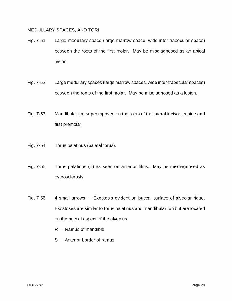
Fig. 7-51 Large medullary space (large marrow space, wide inter-trabecular space)
between the roots of the first molar. May be misdiagnosed as an apical
lesion.
Fig. 7-52 Large medullary spaces (large marrow spaces, wide inter-trabecular spaces)
between the roots of the first molar. May be misdiagnosed as a lesion.
Fig. 7-53 Mandibular tori superimposed on the roots of the lateral incisor, canine and
first premolar.
Fig. 7-54 Torus palatinus (palatal torus).
Fig. 7-55 Torus palatinus (T) as seen on anterior films. May be misdiagnosed as
osteosclerosis.
Fig. 7-56 4 small arrows — Exostosis evident on buccal surface of alveolar ridge.
Exostoses are similar to torus palatinus and mandibular tori but are located
on the buccal aspect of the alveolus.
R — Ramus of mandible
S — Anterior border of ramus

OD17-7/2 Page 25
Periapical Radiolucencies
Granuloma
Radicular Cyst
Abscess
Apical scar
Surgical defect
Periodontal disease
Cementomas (osteolytic stage)
Chronic suppurative osteomyelitis (Described in chapter on "Osteomyelitis")
Periapical Radiopacities
Cementomas (calcified stage)
Condensing osteitis
Osteosclerosis
Socket sclerosis
Florid osseous dysplasia (diffuse cementosis)
Tori
Hypercementosis