AMSA UPM Diabetes Day 2018 NADI · 0!!!!! AMSAUPM$Diabetes$Day$2018$ $ TARIKH’

© JAPI • VOL. 55 • JUNE 2007 www.japi.org 1
Received by mail
Draft-API-JAPI
API-ICP Guidelines on Diabetes 2007
preserve the health of the diabetic.The data from several epidemiological, experimental
- human and animal studies and morerecently the data from several mega trials like the
DCCT, Kumomoto study and the UKPDS haveconvincingly proved the importance of tight metaboliccontrol in arresting the progression and prevention ofmicrovascular disease.4-6
Hyperglycemia contributes to the increased incidenceof macrovascular disease but dyslipidemia,hypertension, central obesity, decreased physical activityand smoking play a major role in acceleratedatherosclerosis seen in diabetics.
Therefore, the complete treatment of diabetic patientsnot only includes meticulous attention to achievementof normoglycemia, but also correction of hypertensionand dyslipidemia, correction of body weight andincrease in physical activity.
It is desirable to have the fasting and post-prandialblood glucose concentration and HbA1c as close tonormal as possible. All the above goals are desirableand can be achieved without significant deteriorationin quality of life. Patient education is also an essentialgoal of any treatment regimen. Patient’s who understandthe importance of achieving these goals and their role inpreserving health will be motivated to do so.
The American Diabetes Association and the EASDhave evolved effective diabetes care programmes, whichhave succeeded in improving the lot of diabetics in thesecountries by bringing down the morbidity and mortality.7
These organizations have brought out consensusguidelines with an aim to achieve the defined goals. Theirexperience has shown that adopting guidelines by theclinicians improves the outcome of treatment. Theseguidelines cannot be adopted or copied, for our country.We must evolve our own programme based on our needs.India is a vast country with a heterogeneous populationwith different religions, cultures, languages, food habits,lifestyles, and traditions. The major portion of thispopulation lives in rural areas with meager facilities interms of health care delivery. Such a programme shouldalso consider the economic realities of our people - hencesuch a programme should not only be available butshould be affordable for the average Indian.
There is an urgent need for creating and adaptingminimal guidelines on diabetes care for our country. Suchguidelines once enunciated should be followed andadhered to by all the health care professionals managing
PREAMBLE
The prevalence of type 2 diabetes is increasing all overthe world particularly in the developing countries.
It has emerged as a major public health problem in ourcountry. The WHO estimated that there were 19.4 millionpersons with diabetes in India in 1995 and that thisnumber is likely to be 57.2 million in 2025. India has thedistinction of having the largest number of diabetics inthe world. Studies in 1980 showed higher prevalencerates of type 2
diabetes among migrant Indians in several countries,compared with their native populations and othermigrant ethnic groups.1 Current prevalence rates are 11-12% in the urban Indian adult population.2 There isevidence that the prevalence of type 2 diabetes isincreasing in rural population also.
Type 2 diabetes amongst Indians occurs at a youngerage, the age at diagnosis being a decade earlier than inthe West. Body mass index is lower by 4 kg/m2 for malesand 6 kg/m2 for females. However abdominal obesitywith increased waist to hip ratio is more common. Strongfamilial aggregation of the disease with high prevalenceamong first degree relatives and vertical transmissionthrough two or more generations is also noted.3 Theearlier age of onset, delayed diagnosis and impropercare lead to an increase in morbidity and mortalityresulting in loss of productivity.
Despite the research and the availability of bettertreatment modalities, the morbidity and mortality isincreasing and is a matter of concern.
Managing patients with diabetes effectively, requiresa great deal of time, effort and patience. The professionaleducation that increases awareness of the importanceof diabetes management is valuable in reducing andpreventing complications of diabetes. Diabetes is aserious, common, costly and controllable disease.Controlling diabetes is easier and cheaper thanmanaging its complications.
The task of rendering quality care to our diabeticpatients is stupendous and challenging. We do not havea national diabetes care programme. A proper programmewill yield rich dividends in terms of prevention of itslong term complications and cutting down the morbidityand mortality in diabetics. Our goal should be to
Figs. not received.
2 www.japi.org © JAPI • VOL. 55 • JUNE 2007
diabetes. In this backdrop, the Indian College ofPhysicians has decided to bring out the guidelines forthe management of type 2 diabetes.The objectives of these guidelines are:1. To arrive at an early and proper diagnosis of
diabetes.2. To provide proper treatment to diabetic patients in
different situations.3. To draw up a monitoring programme which should
be cost-effective in ensuring good control of diabetesand its associated risk factors to protect the patientfrom developing complications.
4. To lay down criteria for what is to be considered asideal, good and acceptable targets to be achieved.
5. To develop a standard education program to beimparted by the family doctor to his patients on eachvisit.
6. To develop guidelines for early detection of itscomplications, appropriate measures to arrest andreverse them.
7. Guidelines regarding timely referral to anappropriate specialist and for coordinating withthem.
8. To develop guidelines for preventive strategy.9. To formulate guidelines for establishment of diabetes
care centre/clinics and measures to accredite them.The family physicians and general practitioners bear
the burden of carrying out the day to day care in mostpatients. Therefore it is essential that they be empoweredwith knowledge and expertise to provide standard care.They should understand goals of therapy and try toachieve the targets of control by using propermanagement policies. By following the consensusguidelines in their day-to-day practice, they can achieveeffective control of diabetes and prevent the developmentof acute and chronic complications of diabetes.
Effective diabetes self-management is an importantconcept in the overall control of the disease. Diabetesself-management is a relatively new approach inimproving control and thereby helping to prevent itscomplications. This approach acknowledges that peoplewith diabetes must ultimately take responsibility for theday-to-day management of their disease. Hence, theyshould receive educational inputs from the diabetic teamto empower them to undertake these responsibilities.
The best model for diabetic care is a team approach,comprising of diabetologist/endocrinologist, familyphysician, diabetes trained nurse, podiatrist, dietician,health educator and various specialists like cardiologist,nephrologist, ophthalmologist specially interested inmanagement of diabetic complications. There should bea close rapport and coordination between the primarycare physician and diabetologist. We do not have enoughnumber of trained para-medical personnel. There is a
need to initiate the development of training facilities forthese paramedical personnel like the diabetic nurseeducator, dietician and the podiatrist.Acknowledgements
I thank the Dean, Dr. Sukumar Mukherjee and theMembers of the Faculty Council of the Indian College ofPhysicians for en trust ing me the task of preparing theIndian Guide lines for Management of Diabetes. I chosea team of eminent diabetologists of the country - Prof. JKAgarwal, Prof. AK Das, Prof. V Seshiah, Prof. SiddharthN Shah and Professor D Maji to fulfill this task.
We took the help of several other diabetologists acrossthe country who gave us their in puts resulting in a ‘draftof the guidelines’. We circulated this to about 200reviewers across the country, who gave further valuablesuggestions, which we have in corporated and finallywe have a consensus document on the “IndianGuidelines for Management of Type 2 Diabetes”.
I sincerely thank the core committee membersProfessor JK Agarwal, Professor AK Das, Professor VSeshiah, Prof. Siddharth N Shan and Professor D Majifor their keen interest and whole hearted cooperation.We thank the members of the working group for theirvaluable in puts and the reviewers for their keen interest,critical suggestions and corrections.
I sincerely thanks USV Limited for wholeheartedlysupporting us; financially for sponsoring the meetingsof the core committee, publication and distribution ofthe guidelines and scientifically for providing therelevant references.
Prof. BK SahayConvenor
Indian Diabetes Guidelines 2002.
REFERENCES
1. King H, Rewers M. On behalf of the WHO Ad Hoc DiabetesReporting Group. Global estimates for the prevalence ofdiabetes mellitus and impaired glucose tolerance in adults.Diabetes Care 1993;16:157-76.
2. Ramachandran A, Snehalatha C, Latha E, Vijay V,Vishwanathan M. Rising prevalence of NIDDM in urbanpopulation in India. Diabetologia 1997;40:232-7.
3. Yajnik CS. The Insulin Resistance Epidemic in India: Fetalorigins, later lifestyle, or both? Nutr Rev 2001;59:1-9.
4. The DCCT Research Group. The effect of intensive treatmentof diabetes on the development and progression of long-term complications in insulin dependent diabetes mellitus.N Engl J Med 1993;329:977-86.
5. Okhubo Y, Hideki K, Araki E, et al. Intensive insulin therapyprevents the progression of diabetic microvascularcomplications in Japanese patients with non-insulindependent diabetes mellitus. A randomized prospective sixyear study. Diab Res Clin Pract 1995;28:103-17.
6. UKPDS Study Group. Intensive blood glucose control withSU and insulin compared with conventional treatment andrisk of complications in patients with type 2 diabetes.(UKPDS 33). Lancet 1998;352:837-53.
7. American Diabetes Association. Clinical Practicerecommendations. Diabetes Care 2001;24 (Suppl 1):S1-145.

© JAPI • VOL. 55 • JUNE 2007 www.japi.org 3
DEFINITION AND EPIDEMIOLOGY
Definition
Diabetes mellitus is a syndrome characterized bychronic hyperglycemia with disturbances ofcarbohydrate, fat and protein metabolism resulting fromeither an absolute or relative deficiency of insulinsecretion and/or action.
**** to incorporate Dr. Munjal’s suggestionThis chronic hyperglycemia of diabetes is associated
with long term damage, dysfunction and failure ofvarious organs especially the eyes, kidneys, nerves, heartand blood vessels.
*** definition on prediabetes to be addedEpidemiology (to get from Dr. A.K. Das)
Prevalence of type 2 diabetes mellitus in India isshowing a progressively upward trend as depicted inthe Table 1.1-4a The study published by Indian Council ofMedical Research (ICMR)5 in 1972 reported a prevalenceof 2.3% which has risen to 12.1% in the year 2000 in theurban population.6
The WHO has also projected this rising trend ofdiabetes.7 The prevalence in India is expected to risefrom 19.4 million in the year 1995 to 57.2 million in theyear 2025 (Table 2).8 As shown in Table 2, India willhave the largest population of diabetics in the worldand will continue to have if preventive measures are notimplemented.
Genetic predisposition, inherent ethnicity, increasedwaist to hip ratio with/without obesity, urbanization,migration and life style changes contribute to this rise inIndians.9 Moreover, type 2 diabetes in Indian populationmay have an on set at a younger age.5,10-12 It is projectedthat equal number of diabetics are undetected for a longtime and hence may present with microvascular andmacrovascular complications at the time of diagnosis.13
CLASSIFICATION
An international Expert Committee, working underthe sponsorship of the American Diabetes Associationwas established in May 1995 to re view the classificationand diagnosis of diabetes mellitus based on etiology.The new classification was published in July, 1997 (Table3).14 The World Health Organization (WHO) hasrecently laid emphasis on oral glucose tolerance test(OGTT) and its importance in the classification anddiagnosis of diabetes.15
Type 1 Diabetes
a. Immune Mediated Diabetes : This form previouslycalled insulin dependent diabetes, type 1 diabetes orjuvenile onset diabetes results from a cellular mediatedautoimmune destruction of the beta cells of the pancreas.Markers of the immune destruction of the beta cell includeislet cell antibodies (ICAs), autoantibodies to insulin(IAAs), autoantibodies to gutamic acid decarboxylase
(GAD 65) and autoantibodies to the tyrosine phosphates,IA-2 and IA-2B. The disease has strong HLAassociations. In this form of diabetes, the rate of beta celldestruction is quite variable, being rapid in someindividuals (mainly infants and children) and slow inothers (mainly adults).
b. Idiopathic Diabetes : Some forms of type 1 diabeteshave no known etiologies. Some of these patients havepermanent insulinopenia and are prone to ketoacidosis,but have no evidence of autoimmunity. Only a minorityof patients with type 1 diabetes fall into this category.However most of those who fall into this category are ofthe African or Asian origin. Individuals with this formof diabetes suffer from episodic ketoacidosis and exhibitvarying degrees of insulin deficiency between episodes.
Table 1: Prevalence of diabetes mellitus in India*** (to beupgraded)
Year Author Place Prevalence (%)Urban Rural
1971 Tripathy et al Cuttack 1.21972 Ahuja et al New Delhi 2.31979 Gupta et al Multicentre 3.0 1.31984 Murthy et al Tenali 4.71986 Patej JC Bhadran 3.81988 Ramachandran et al Kudremukh 5.01989 Kodali et al Gangavathi 2.21989 Rao et al Eluru 1.61991 Ahuja et al New Delhi 6.71992 Ramachandran et al Madras 8.2 2.41997 Ramachandran et al Madras 11.61998 Shekhar shah et al Assam 8.22001 Ramachandran et al Madras 12.1
Table 2 : Top ten countries for number of persons withdiabetes8*** (to be upgraded)
Year 1995 Year 2025No. Country Numbers No. Country Number
in Million in Million
1. India 19.4 1. India 57.22. China 16.0 2. China 37.63. USA 13.9 3. USA 21.94. Russian Fed 8.9 4. Pakistan 14.55. Japan 6.3 5. Indonesia 12.46. Brazil 4.9 6. Russian Fed 12.2
7. Indonesia 4.5 7. Mexico 11.78. Pakistan 4.3 8. Brazil 11.69. Mexico 3.8 9. Egypt 8.810. Ukraine 3.6 10. Japan 8.5
Table 3 : Etiological classification of diabetes mellitus
1. Type 1 diabetes (absolute insulin deficiency)a. Immune mediatedb. Idiopathic
2. Type 2 diabetes (predominantly insulin resistancewith relative insulin deficiency)
3. Other specific types4. Gestational diabetes mellitus (GDM)
4 www.japi.org © JAPI • VOL. 55 • JUNE 2007
This form of diabetes is strongly inherited, lacksimmunological evidence of beta cell autoimmunity andis not HLA associated.Type 2 Diabetes
This form of diabetes was previously referred to asnon-insulin dependent diabetes or adult on set diabetes.Such individuals have relative (rather than absolute)insulin deficiency. This form of diabetes frequently goesundiagnosed for many years because hyperglycemiadevelops gradually and in earlier stages is often notsevere enough for the patient to develop any of thehyperosmolar symptoms of diabetes. Never the less suchpatients are at an increased risk of developingmacrovascular and microvascular complications andthese may be present even at the time of diagnosis. Theseindividuals may be controlled with diet, exercise andoral agents for variable periods of time. How ever, in thecourse of time they are likely to require insulin for betterglycemic control.Other Forms of Diabetes
a. Genetic defects in insulin actionb. Diseases of the exocrine pancreas – Includes
fibrocalculous pancreatopathyc. Endocrinopathiesd. Drug or chemical inducede. In fectionf. Uncommon forms of immune-mediated diabetesg. Other genetic syndromes associated with diabetesGestational Diabetes Mellitus (GDM)
GDM is de fined as any degree of glucose in tolerancewith on set or first recognition during pregnancy. Sixweeks or more after pregnancy ends, the woman shouldbe reclassified into one of the following categories :1. Diabetes2. Impaired fasting glucose3. Impaired glucose tolerance4. Normoglycemia
In majority of GDM cases, glucose regulation willreturn to normal after delivery. Clinical recognition ofGDM is important because therapy, including diet,exercise, insulin and antepartum fetal surveillance canreduce the associated perinatal morbidity and mortality.Although many patients diagnosed with GDM will notdevelop diabetes later in life, others will be diagnosedmany years post-partum as having type 1 diabetes, type2 diabetes, impaired fasting glucose (IFG) or impairedglucose tolerance (IGT).Special Types of Diabetes in India
MODY
MODY is a uncommon monogenic autosomaldominant variety of diabetes seen in the young describedfrom India. The criteria of which is given in Table (criteria
of diagnosis and Types of MODY from Dr. V.Mohan)____*****
The WHO classification (1985)16 had malnutritionrelated diabetes as a separate class (protein deficientdiabetic mellitus (PDDM) and fibrocalculous pancreaticdiabetes mellitus (FCPD)). In the recent ADAclassification,14 FCPD has been included in other specifictypes and PDDM has been deleted. However, this typeof diabetes which is modulated by mal1nutrition isspecially seen in India.9 It is characterized by youngerage of on set, BMI < 18.5 kg/m2, no pancreaticcalcification, relative insulin resistance, non-ketotic butrequiring insulin for control of blood glucose.
Low body weight type 2 diabetes mellitus is also seenin our country and is characterized by BMI less than 19kg/m2 and may not re quire insulin for their glycemiccontrol for a variable period of time. This type of diabetesis not associated with malnutrition. In a multicentricstudy involving nine cities all over the country (1984-1990), the incidence of lean type 2 diabetes mellitus wasob served to be varying from 11-25% at differentcentres.17,18
DIAGNOSIS
The criteria for the diagnosis of diabetes are asfollows:
Symptoms of diabetes associated witha. Random * plasma glucose concentration ≥ 200 mg/
dlb. Fasting ** plasma glucose ≥ 126 mg/dlc. 2 hr plasma glucose ≥ 200 mg/dl during a 75 g OGTT.
For asymptomtic individuals with any one of theabove values, a 75 g OGTT is required to confirm thediagnosis.
(*Random is defined as any time of day with outregard to time since the last meal. The classic symptomsof diabetes include polyuria, polydipsia, Polyphagiaand unexplained weight loss, **Fasting is defined as nocaloric in take for at least eight hours. Plasma glucoseshould be estimated by glucose oxidase method).
Oral Glucose Tolerance Test (OGTT) as Specified byWHO16
Procedure
1. The test should be done after at least three days ofunrestricted diet (more than 150 g carbohydratedaily) and normal physical activity.
2. It should be preceded by 10-16 hours of fasting,during which drinking plain water is permitted.
3. It should be carried out in the resting subject.4. Smoking is not al lowed on the morning of the test
as well as during the test.5. Factors that could influence the test’s interpretation
must be re corded.
© JAPI • VOL. 55 • JUNE 2007 www.japi.org 5
6. Following collection of fasting blood sample, 75 g ofglucose should be dissolved in 250-300 ml of waterand should be drunk over the course of about fiveminutes.
7. The another sample is collected 2 hr after the glucoseload.
8. The test results are interpreted as given in Table 4.Once diagnosed as diabetes, repetition of OGTT is
not required.
The diagnosis should not be based on urine sugaralone.
Pre-Diabetes (IFG/IGT)
An intermediate group of subjects, called impairedfasting glucose (IFG) is recognised as those with FPGlevels > 110 mg/dl but < 126 mg/dl. Impaired glucosetolerance (IGT) is defined as 2-hr post-glucose levels >140 mg/dl but < 200 mg/dl. The IGT and IFG group ofindividuals are important since they have risk ofbecoming diabetic and are prone to developmacrovascular complications.
SCREENING
In view of the rising trend in the prevalence of diabetesand associated morbidities, it is imperative to screen highrisk groups (Table 5) and the population at large. Studiesby ICMR19 and UKPDS20 have shown establishedcomplications even at the time of diagnosis emphasizingthe importance of screening.
Population screening
a. Health check up schemesb. Insurance screeningc. Employment check up schemesd. Diabetic detection campsTools Recommended for Screening
Even though ideally fasting blood glucose should beperformed, an abnormal random plasma glucose valuewould also indicate diabetes. However, such individualsshould be retested at the next visit for establishing thediagnosis of diabetes as per the diagnostic criteria givenin Table 4.
CLINICAL ASSESSMENT
Persistent hyperglycemia is the hall mark of all formsof diabetes mellitus. A detailed evaluation optimizes thecare required to provide better quality of life for thesepatients.
The first visit: The very first visit of the patient is fullyutilized for a detailed medical history and physicalexamination. (See Appendix 1 for patient data card).Medical History
A. Symptoms of hyperglycemia: (polyuria, polydipsia,polyphagia)Weight loss, generalized weaknessPeriarthritisDelayed healing of ulcersVisual disturbancesBalanitis/balano posthitis, orvulvovaginitis/vaginitis
B. Previous history of ketosis, hyperosmolar coma,hypoglycemia; cerebrovascular complications,coronary events, pancreatitis.
C. Symptoms suggesting development and severityof complications: Facial puffiness, pedal edema;frequency, urgency, dysuria, angina, effort intolerance, Claudication (vascular/neurogenic),gangrene, amputation,Sensory impairment - pain, temperature, gaitdisturbance in dark,Foot ulcers - site, size, source, sepsis, associatedcellulitis,Infections (prior or current) - skin, dental,genitourinary, pulmonary tuberculosis,
Bladder and gastrointestinal function; Orthostatichypotension,Erectile dysfunction ** to reword
D. Evaluation for possible causes of secondary diabetesmellitus.
E. Current nutritional status, eating pattern, adequacyof in take; weight history.
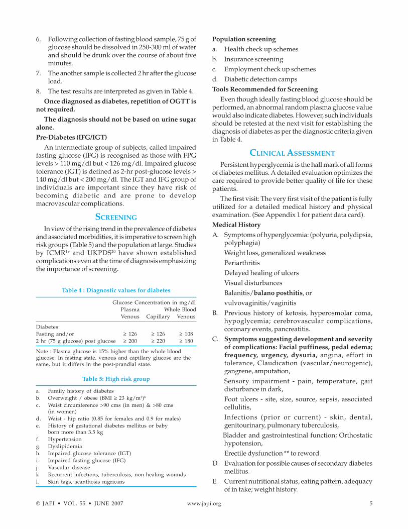
Table 4 : Diagnostic values for diabetes
Glucose Concentration in mg/dlPlasma Whole BloodVenous Capillary Venous
DiabetesFasting and/or ≥ 126 ≥ 126 ≥ 1082 hr (75 g glucose) post glucose ≥ 200 ≥ 220 ≥ 180
Note : Plasma glucose is 15% higher than the whole bloodglucose. In fasting state, venous and capillary glucose are thesame, but it differs in the post-prandial state.
Table 5: High risk group
a. Family history of diabetesb. Overweight / obese (BMI ≥ 23 kg/m2)6
c. Waist circumference >90 cms (in men) & >80 cms(in women)
d. Waist - hip ratio (0.85 for females and 0.9 for males)e. History of gestational diabetes mellitus or baby
born more than 3.5 kgf. Hypertensiong. Dyslipidemiah. Impaired glucose tolerance (IGT)i. Impaired fasting glucose (IFG)j. Vascular diseasek. Recurrent infections, tuberculosis, non-healing woundsl. Skin tags, acanthosis nigricans
6 www.japi.org © JAPI • VOL. 55 • JUNE 2007
F. Risk factors for atherosclerosis:, hypertension,obesity, hyperlipidemia, atherosclerosis in familymembers.
F1. smoking/tobacco and alcohol use
G. Lifestyle, cultural, psychosocial, educational,economic, activity status and exercise history.
H. Gestational history : hyperglycaemia, delivery ofbaby > 3.5 kg, toxemia, stillbirth, polyhydramniosor other complications of pregnancy.
I. Family history of diabetes and its complications andother disorders (Hypertension, hyperlipidemia,coronary artery disease)
J. Previous treatment details and their out comes.K. Current treatment and glycemic status : medications,
compliance, other medications altering glycemicstatus, frequency of monitoring to achieve glycemicgoal.
Physical Examination
� Measures for height, weight, waist circumferenceand hip circumference, (calculation of BMI, waistcircumference and waist - hip ratio)
� Resting pulse, palpation of peripheral pulsesincluding carotid pulses
� Blood pressure, (lying down, sitting up andstanding). Ankle pressure measurement andcalculation of ankle-brachial index**** whereverindicated
� Presence of pallor, pyrexia, dyspnoea, cyanosis,clubbing, dehydration
� Eye - examination including ophthalmoscopicevaluation re viewed by ophthalmologist
� Oral and dental examination� Thyroid palpation� Systemic evaluation
* Cardiac evaluation* Respiratory system examination* Abdominal examination* Neurological examination with special reference
to ankle jerk and peripheral sensation byvibration, pain and touch.
� Foot examination particularly for callosities, inter-digital web deformities and ulcers, cracks of skinand foot pulses
� 10 gm mono filament is a sensitive tool to pick updiabetic foot at risk.
� Skin examination
LABORATORY ASSESSMENT
a. Complete urinalysis: glucose, ketones, protein,sediments
b. CBC (complete blood count): hemoglobin, total
leukocyte count, differential leukocyte count,erythrocyte sedimentation rate (ESR)
c. Fasting and 2 hr post-prandial plasma glucosed. Fasting lipid profile after blood glucose control.e. Electro cardiogramf. X-ray chest PA view to see for cardiac size and
undetected pulmonary tuberculosis.g. Serum creatinine, blood ureah. Test for urinary albumin-creatinine ratio in all
patients, if positive than 24 hour urinary protein tobe done. Microalbuminuria (if possible)
i. HbA1c (if possible)**It is the gold standard though it has issues ofstandardization and cost within our setups.
j. These tests should be done when indicated.� Sputum for AFB� Carotid Doppler - IMT� 2 D-Echocardiography� Urine culture if sediment is abnormal or
symptoms are present� Serum electrolytes� Liver function test**
� SGOT/SGPT (if the patient is on glitazones)� Hs CRF
TMT/Angio (to be given by Dr. Shashank Joshi)
FOLLOW UP EVALUATION
Frequency of follow up visits is determined by thecontrol achieved.
First follow up visit : First follow up visit should bescheduled at 2-4 weeks to evaluate blood glucose controland to reinforce diabetic education, diet, exercise andmanagement plan.
Subsequent visits : Once the blood glucose is undercontrol, the subsequent visits should be at three monthlyinterval to evaluate.� Improvement in symptoms� Compliance for diet, physical activity and drugs� Weight� Blood pressure� HbA1c� Lipid levels if initially abnormal
However plasma glucose (fasting and post-prandial)must be checked every month and if the blood glucosevalues are high, it must be reported to the physician.Yearly investigations
� Physical examination including ophthalmoscopicevaluation and fundoscopy by ophthalmologist.
� Renal evaluation as at first visit and

© JAPI • VOL. 55 • JUNE 2007 www.japi.org 7
microalbuminuria if possible� Chest radiography, if required� Electro cardiogram� Rest of the investigations as in 3-4 monthly visits� Evaluation of patient education and diabetes
awarenessNote: Echocardiography, Doppler studies for foot
pulses, cardiac stress test are to be done in appropriatesituations and these need not be repeated with out properindications. [to come in the box]
GOALS OF THERAPY
The goals of management in a diabetic patient are toprovide� Relief from diabetic symptoms and improvement in
quality of life� Prevention of acute complications like diabetic
ketoacidosis (DKA), hyperosmolar non-ketotic coma(HONK), hypoglycemia and lactic acidosis
� Prevention of infections� Prevention of microvascular complications-
nephropathy, retinopathy and neuropathy.� Prevention of atherosclerotic vascular diseases:
cardiovascular disease, cerebrovascular disease andperipheral vascular disease.
� Prevention of diabetic foot lesions.The published data from several experimental,
epidemiological, human and animal studies and morerecently the data from several megatrials like the DCCT,21
Kumomoto study22 and the UKPDS23 have convincinglyproved the importance of tight metabolic control inarresting and preventing the progression of themicrovascular complications.
Hypertension represents a major risk for bothcardiovascular disease and diabetic nephropathy. Evena modest elevation of blood pressure requires prompttreatment.23 It is suggested that a value of 130/85 mmHg or less is desirable.24,25
Microalbuminuria increase the risk of clinically overtalbuminuria and cardiovascular disease.26 These shouldbe detected early and treated aggressively.
Therefore the complete treatment of diabetic patientsincludes correction of body weight and increasedphysical activity, meticulous attention to achievementof normoglycemia, control of hypertension andcorrection of dyslipidemia.
The goals are mentioned in the Table 6. All these goalsare desirable and can be achieved with out significantdeterioration in quality of life. Patient education is alsoan essential goal of any treatment regimen. Patients whounderstand the importance of achieving these goals andtheir role in preserving health will be motivated to do so.
PRINCIPLES OF MANAGEMENT
The approach to the management of diabetes has notchanged significantly over the years. However, with thedevelopment of newer drugs and better understandingof the pathophysiology of type 2 diabetes, the outlookhas improved considerably.
The initial step after the diagnosis is made, is life-style modification. The patient is advised an appropriatediet and suitable exercise programme, with cessation ofsmoking and alcohol and initiation of metformintherapy. The response to diet and exercise should beassessed for two months in general. The importance ofdiet and exercise as a cost-effective measure in themanagement of diabetes should be understood andimparted to the patients. If adequate glycemic control isachieved, the patient should be motivated to continuethe diet and exercise, while monitoring the blood glucoselevels at regular intervals.
The choice of drug should be individualized basedon the possible underlying pathogenic mechanism ineach case. The anthropometric data - weight, height, bodymass index (BMI = weight in kg/height in mts2), waist-hip ratio (WHR) should be the guiding factors (seeAppendix 2). In obese patients, initiate the therapy withmetformin and in non-obese patients, one can initiatethe therapy with sulfonylureas, glitazones or insulin,depending on the clinical presentation. Whenmonotherapy fails to achieve adequate control,combination therapy is adopted.
A consensus statement from the ADA and theEuropean Association for the Study of Diabetes on theapproach to management of hyperglycemia inindividuals with type 2 diabetes has recently beenpublished. Early intervention with metformin incombination with lifestyle changes (MNT and exercise)with continuing, timely augmentation therapy withadditional agents (including early initiation of insulintherapy) as a means of achieving and maintainingrecommended levels of glycemic control (i.e. A1C <7%for most patients) are highlights of this approach.(Reference: Position Statement: Standards of Medical
Table 6 : Biochemical and clinical endpoints of diabetesmanagement (to be debated -SRJ)***
Parameter Good Fair Poor
Fasting plasma glucose (mg/dl) <110 <126 >126Postprandial (2h) plasma
glucose (mg/dl) <140 <200 >200HbA1c (%) <6.0 <7.0 >7.0Plasma total cholesterol (mg/dl) <180 <200 >200Plasma LDL cholesterol (mg/dl) <100 <130 >130Plasma triglyceride (mg/dl) <150 <180 >180Plasma HDL cholesterol (mg/dl) >45 >40 <40Blood pressure (mm Hg) <120/80 <130/85 >130/85Microalbuminuria (mg/day) <30 <300 >300Ideal body weight (%) >80-<100 <120 >120

8 www.japi.org © JAPI • VOL. 55 • JUNE 2007
Care in Diabetes – 2007. ADA 2007 Guidelines, DiabetesCare 2007; 30:S4-S41.)
However, in certain situations, insulin therapy willhave to be initiated soon after the diagnosis. Thesesituations are:1. When there is an associated infection at the time of
diagnosis2. Myocardial infarction3. Stroke4. Ketoacidosis5. Hyperosmolar coma or pregnancy and6. When the patient has to undergo surgery.
In all these situations the initial therapy will be withinsulin which can be changed after an appropriate timeto oral drugs or non-drug therapy. Yet an other subset ofpatients who require such an approach are patients witha high fasting plasma glucose level at on set i.e. greaterthan 300 mg/dl (>250 mg/dl in lean patients) andpatients with marked loss of weight (> 10 kg).
Monitoring is also an important component ofmanagement of diabetes. This should include not onlythe glycemic status but also for risk factors formacrovascular and microvascular disease such asmicroalbuminuria, hypertension and dyslipidemia.
NON-PHARMACOLOGICAL MANAGEMENT
Diet
Modification of diet is the most important aspect inthe therapeutic plan for patients with diabetes mellitus.Diet therapy consists of the following :1. Maintenance of proper nutrition2. Total number of calories ingested3. Individual food sources that make up these calories4. Distribution of calories through out the day
Selection, moderation and restriction are the keywords in planning. The entire success of dietarymodification in a diabetic subject depends on thejudicious selection of carbohydrates, adequate proteinintake and a determined restriction of total fat in take.
Attainment of optimal body weight results in markedreduction of hyperglycemia and increase in target cellresponse to insulin. Ideal body weight (IBW) of a personcan be calculated by the formula.
IBW (in Kg) = (Height in cm - 100) 0.9The obese and the over weight must be encouraged to
reduce weight. An energy deficit of 500 Kcal daily, willhelp the patient to re duce 500 gm of weight every week.The next step is to calculate the optimal calories Table 7.Calorie in take based on activity is shown in Table 8.
Any person above 50 years may require 10% lesscalories for each decade. Children need base line caloriesof 1000 plus 100 calories for girls and 125 calories for
boys, per year of age up to 12 years.General guidelines for diet planning in diabetes
mellitus is given in Table 9.Region specific diets of 1200, 1800 and 2200 arehighlighted separately in Appendix.
Disclaimer on Diabetes Sweets and Chocolates (ShilpaJoshi)
Exercise
Regular exercises form an important component oftherapy in patients with type 2 diabetes. However acareful assessment of the expected benefits andassociated risks of exercise in individual patients shouldbe made while in corporating an exercise program inthe treatment. Appropriate monitoring should be doneto avoid complications.a. General Principles for Exercise in Diabetics
� Exercise must be done regularly.� An exercise schedule should be one that the
individual enjoys and which suits his/her needs.� Daily exercise is preferable.� The patient must be told that well fitting canvas/
sports shoes should be worn while walking.� The duration of exercise should be 30-60 minutes.� The ideal time is on an empty stomach in the
morning or evening (taking into consideration therisk of hypoglycemia).
� Any exercise should have a warming up and coolingdown period of 5-10 min.
� Most diabetics may need to reduce the dose of insulinand oral drugs when they exercise regularly.
� Before an exercise programme is initiated, a faircontrol of diabetes is to be ensured and a thoroughclinical evaluation of the patient should be madeparticularly with regard to complications of diabetes,hypertension, coronary artery disease, peripheralvascular disease, retinopathy and nephropathy.
� The drugs that a patient is receiving should beascertained and their possible interaction withexercise should be assessed.
Table 7 : Calorie intake based on weight
Category Calorie Requirement
Overweight 20 kcal/kg/dayIdeal weight 30 kcal/kg/dayUnderweight 40 kcal/day
Table 8 : Calorie intake based on activity
Lifestyle Daily calorie requirement
Sedentary 20-25 Kcal/kg of IBWModerately active 26-30 Kcal/kg of IBWStrenuous 31-35 Kcal/kg of IBW

© JAPI • VOL. 55 • JUNE 2007 www.japi.org 9
� It is advisable to individualize the exerciseprescription.
Weight Loss para by Dr. Munjal
b. Evaluation of the Patient Before Exercise
Before beginning an exercise program, the individualwith diabetes mellitus should undergo detailed medicalevaluation with appropriate diagnostic studies.Cardiovascular system
Before embarking on a moderate to high intensityexercise program, an assessment of the cardiovascularrisk status of the individual should be performed.
Any individual with any of the cardiovascular riskfactors will need to undergo a graded exercise test.
Retinopathy
For patients who have proliferative diabeticretinopathy, strenuous activity may precipitate vitreoushemorrhages or traction retinal detachment.
These individuals should avoid exercises that involvestraining and jarring.Nephropathy
Patients with overt nephropathy of ten have a reducedcapacity for exercise. High intensity of strenuousexercises should there fore be avoided.Peripheral neuropathy
Peripheral neuropathy results in loss of protectivesensation in the feet.
Table 9 : Guidelines for diet in diabetes mellitus (to be updated)****
1. Energy (calories)25-30 cal/kg IBW - reduce in obese and increase in underweight
2. Protein0.8 g/kg body weight. Supplement for pregnancy, lactation and growth. Include a small quotaof animal proteins - fish, chicken, milk and yoghurt. Avoid cattle meat and eggs
3. Fats20-25% of total caloriesSaturated: 6-7% of total caloriesPUFA: 6-7% of total caloriesMUFA: 6-7% of total caloriesN6/N3 ratio: 4:1Cooking oil: 0.5 kg/month/person*Total fat intake in the form of cholesterol per day = 300 mg.Note : When prescribing fat in the diet one should take into account the invisible fat in the diet whichnearly contributes to 50% of the required fat.None of the available oils are ideal.26a
The choice of cooking oil should be as follows.a) Use an oil which has a moderate quantity of linoleic acid like ground nut oil, rice bran or sesame.b) Use an oil which has high amounts of linoleic acid (safflower oil, sunflower oil, cotton seed, corn oil)
along with an oil which has relatively low levels of linoleic acid like palm oil.(mix equal quantity or use equal quantity).
or c) Use any of the above oils with alpha linoleic acid certaining oil like mustard and soya bean oil.(* See Appendix 3-5 for content of saturated and unsaturated fatty acids, omega 3:6 content in oils and spices).
4. Carbohydrates55-60% of total calories. Encourage complex carbohydrates i.e. mainly grains, cereals, pulses.*****Beans, vegetables and salads.Avoid simple and refined carbohydrates like sugar, honey and jaggery.Avoid bakery products or deep fried items.
5. FruitsFresh fruits up to 400 g/day. Avoid juices.Ideal fruits are citrus fruits, orange, sweet lime, guava, apple, papaya and watermelon. They provide vitamins,fibre. One portion contains about 40-50 calories. Dry fruits to be avoided.
6. Dietary fibers30-40 g/day preferably from natural sources. Avoid loss from refining and processing. Indian diet is rich in fiber and generallydoes not require addition of fiber supplements. (See Appendix 6).
7a. Common SaltUp to 6 g/day. Reduce intake to 4 g/day in the presence of hypertension, renal failure and heart problems.7b. Condiments and spicesInclude in diet plan. Provide antioxidants, trace elements, minerals and n-3 fatty acids. (See Appendix 5).
7b. Fenugreek8. Artificial sweeteners
Use of aspartame, sucralose, etc in limited quantity is acceptable. The maximum permitted consumption rangefrom 2-4 mg/kg/day. Avoid in pregnancy and lactation.
9. AlcoholAvoid if possible. If not, drastically reduced. It is utilized as carbohydrates. 1 gm of alcohol provides empty calories.Alcohol may exacerbate neuropathy, dyslipidemia, obesity and may worsen the control of diabetes and cause hyperglycemia.
10. TobaccoAvoid smoking and use of tobacco in any form.
10 www.japi.org © JAPI • VOL. 55 • JUNE 2007
Significant peripheral neuropathy is an indication tolimit weight-bearing exercise. Repetitive exercises in anin sensitive feet can lead to ulceration and fractures.
It is necessary to advise proper foot wear to thesepatients. Patients should be taught to monitor for blistersand other potential damage to the feet be fore and afteran exercise session.Autonomic neuropathy
Presence of autonomic neuropathy may limit anindividual’s exercise capacity and increase the risk ofan adverse cardio vascular event during an exercise.
Hypotension and hypertension are more likely todevelop in exercising patients with autonomicneuropathy.
These patients also have difficulties inthermoregulation and should be advised to avoidexercises in extremely hot or cold environments and tobe careful about their hydration.c. Type of Exercises for Diabetic Patients
The best form of exercise recommended to a diabeticis a step wise increase of aerobic exercises. Plain briskwalking is the simplest and safest of all exercises. It canbe started by any one. All the aerobic (isometric) exerciseslike badminton, tennis and basket ball improve thecardio-respiratory functions and utilize a large portionof muscle mass. On the other hand isometric exerciseslike weight lifting, sustained and grip are to be avoidedin diabetics as they increase the arterial pressure and/or precipitable angina.d. Exercise in Special Populations
Elderly : Many of the elderly patients tend to avoidphysical exercise. There is a progressive decline ininsulin sensitivity, muscle mass and strength and lossof mineral from the bones with increasing age. Regularphysical exercise can prevent and reverse these changes.With exercise a better quality of life is attained in thispopulation with reduction in the burden of chronicvascular disease.
Arthritic patients : To recommend upper bodyexercises.
Alternative forms of exercises including Yoga maybe recommended.
Pregnant ladies : To recommend walking and if notfeasible to recommend upper body exercises.e. Benefits of Exercise
Several benefits accrue from a regular exercise schedule.These include :� Improvement in insulin sensitivity.� Reduction of hypertension.� Reduction in weight.� Improvement in lipid profile : reduces serum
triglycerides and increases HDL particularly HDL2
cholesterol.� Improvement in cardiovascular function.� Improvement in the sense of physical and mental
well being.� Minimizing calcium loss.� Improvement in quality of life.
Improving lipid profile and reducing BP is a majorbenefit of exercise on the cardiovascular risk factors.f. Risks of Exercise
There are several potential risks of exercise forpatients with diabetes.
Careful screening for underlying cardiac disease isimportant in all patients with diabetes before startingany exercise.
Exercise may aggravate several complications ofdiabetes and hence all patients should be screenedthoroughly before initiating exercise.
Patients with proliferative retinopathy may developvitreous hemorrhages.
Heavy weight lifting and Valsalva maneuver areparticularly dangerous.g. Special Precautions
� Feet should be inspected daily (before and afterexercise) for cuts, blisters and infections.
� Exercise should be avoided in extreme hot and coldweather conditions.
� Exercise should be avoided during periods of poormetabolic control.
� An exercise program for obese patients with type 2diabetes should start slowly, build up gradually andinclude exercises that are familiar to the patient andleast likely to cause injuries or worsening of longterm diabetic complications.
� Diabetic patients who exercise regularly shouldalways carry quick acting carbohydrate and visiblediabetes identification cards to be used in the eventof hypoglycemia.
11.2.h. Role of Yoga
Several well-planned studies have demonstrated thebeneficial effects of yogic practices in diabetics. Table 11lists the beneficial effects of yogic practices in patientswith diabetes. Some of the asanas that were found toproduce these benefits are Dhanurasana,Ardhamatsayendrasana, Bhujangasana, Naukasana,Halasana, Paschimotasana, and Shavasana pranayam.(See Appendix 7). However, the patient should bethoroughly evaluated by a physician be fore undertakingany yogic practices.
PHARMACOLOGICAL MANAGEMENT
Oral Hypoglycemic Agents
Blood glucose levels are mainly determined by

© JAPI • VOL. 55 • JUNE 2007 www.japi.org 11
absorption of glucose from gut, uptake of glucose byperipheral tissues (muscle, adipose tissue, liver, etc.),hepatic glucose output, and the insulin secretion frompancreas (Fig. 1). In diabetics various oral agents act tomodify these factors, aiding in the control ofhyperglycemia. (Fig. 2, Table 12).a. Sulphonylureas
� The sulphonylureas bind to specific sulphonylureareceptors on b-cell and increase insulin secretion.All sulphonylureas bind to the 140 kDa sub unit ofsulfonylurea receptor whereas glimepiride binds tothe 65 kDa sub unit of the sulphonylurea receptor.
� In lean or normal weight individuals, Su are thepreferred agents of choice.
� Sulfonylureas are preferably given 15-30 min beforemeals.
� For optimal release of duodenal insulin releasing
factor.� Therapy should be initiated with lowest effective
doses and titrated upwards every one to two weeksuntil desired control or maximal dosage is reached.
� If no hypoglycemic effect is observed with half themaximal effective dose, then the drug is not likely tobe effective combination therapy may be explode.
� Sulfonylureas can be combined with metformin,acarbose, thiazolidinediones and insulin to givesynergistic effect. However, they should not becombined with an other sulfonylurea since they actsimilarly and there is no potentiation of action.Infact there is an increase in side effects.
� About 10 to 20% patients fail to respond tosulphonylureas (primary failure).
� Every year about 5% stop responding (secondaryfailure).Secondary failure may be due to insulin deficiency,but various other factors need to be excluded beforeit is labelled as secondary failure.
� Factors to be considered before labeling as secondaryfailure are given in Table 14. In these cases, the failuremay be reversible.
Various clinical trials have failed to conclusivelydemonstrate superiority of one sulphonylurea over theother, when used in optimal doses. In individual cases,switching over from one sulphonylurea to another mayshow some benefit but this may not be long lasting.
Table 10 : Summary of exercise recommendations
Screening: Search for vascular and neurological complicationsincluding silent ischemic heart disease.An electrocardiogram is recommended for all patients.Exercise program and type
AerobicDuration
30-60 minutesFrequency
DailyAvoid complications
Warm up and cool downCarefully select the type of exercise and its intensity
Patient educationMonitoring of plasma glucose by patient and overallprogram by medical personnel
ComplianceMaking exercise enjoyableConvenient locationPositive feedback from involved medical personnel and
family
Table 11 : Beneficial effects of yogic practices
� Reduction of blood pressure� Correction of dyslipidemia� Reduction of insulin resistance and correction of
hyperinsulinemia� Elimination of stress
Fig. 1 : Pathophysiology of type 2 diabetes.
Fig. 2 : Treatment strategies of type 2 diabetes.
Table 12 : Mode of action of various oral antidiabeticagents
Mode of action Class of Drug
Increasing insulin Sulfonylureas,secretion Meglitinides
Reduction of insulin resistance Glitazones, BiguanidesDecreased hepatic glucose Biguanide, Glitazonesoutput
Reduced carbohydrate Alpha glucosidaseabsorption inhibitors
Incretins DPP-IV Inhibitors (Gliptins)
12 www.japi.org © JAPI • VOL. 55 • JUNE 2007
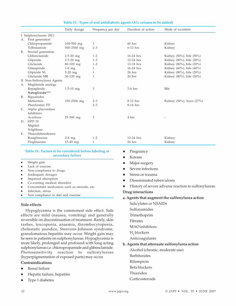
Side effects
Hypoglycemia is the commonest side effect. Sideeffects are mild (nausea, vomiting) and generallyreversible on discontinuation of treatment. Rarely, skinrashes, leucopenia, anaemia, thrombocytopenia,cholestatic jaundice, Stenvens-Johnson syndrome,granulomatous hepatitis may occur. Weight gain maybe seen in patients on sulphonylureas. Hypoglycemia ismore likely, prolonged and profound with long actingsulphonylureas i.e. chloropropamide and glibenclamide.Photosensitivity reaction to sulfonylureas(hyperpigmentation of exposed parts) may occur.Contraindications
� Renal failure� Hepatic failure, hepatitis� Type 1 diabetes
� Pregnancy� Ketosis� Major surgery� Severe infections� Stress or trauma� Disseminated tuberculosis� History of severe adverse reaction to sulfonylureasDrug interactions
a. Agents that augment the sulfonylurea action
Salicylates or NSAIDsSulfonamidesTrimethoprimFibratesMAO inhibitorsH2 blockersAnticoagulants
b. Agents that attenuate sulfonylurea action
Alcohol (chronic, moderate use)BarbituratesRifampicinBeta blockersThiazidesCorticosteroids
Table 13 : Types of oral antidiabetic agents (A1c column to be added)
Daily dosage Frequency per day Duration of action Mode of excretion
I. Sulphonylureas (SU)A. First generation
Chlorpropamide 100-500 mg 1 60 hrs KidneyTolbutamide 500-2500 mg 2-3 6-12 hrs Kidney
B. Second generationGlibenclamide 2.5-20 mg 1-2 16-24 hrs Kidney (50%), bile (50%)Glipizide 2.5-20 mg 1-3 12-24 hrs Kidney (80%), bile (20%)Gliclazide 80-320 mg 1-2 12-18 hrs Kidney (80%), bile (20%)Glimepiride 1-8 mg 1 16-24 hrs Kidney (60%), bile (40%)Glipizide XL 5-20 mg 1 24 hrs Kidney (80%), bile (20%)Gliclazide MR 30-120 mg 1 24 hrs Kidney (80%), bile (20%)
II. Non-Sulfonylurea AgentsA. Meglitinide analogs
Repaglinide 1.5-10 mg 3 2-4 hrs BileNateglinide***
B. BiguanidesMetformin 250-2500 mg 2-3 8-12 hrs Kidney (50%), feces (27%)Phenformin TD 2-3 8-14 hrs
C. Alpha glucosidaseInhibitorsAcarbose 25-300 mg 3 4 hrs -
D. DPP IVMiglitolVolglibose
E. ThiazolidinedionesRosiglitazone 2-8 mg 1-2 12-24 hrs KidneyPioglitazone 15-45 mg 1 24 hrs Kidney
Table 14 : Factors to be considered before labeling assecondary failure
� Weight gain� Lack of exercise� Non-compliance to drugs� Inadequate dosages� Impaired absorption� Co-existing medical disorders� Concomitant medication such as steroids, etc.� Infection, stress� Non compliance to diet and exercise

© JAPI • VOL. 55 • JUNE 2007 www.japi.org 13
EstrogensIsoniazidNicotinic acid
In elderly patients (> 60 years) short actingsulphonylureas are preferred. Glibenclamide shouldbe used with caution in elderly. Chlorpropamideshould not be used in elderly patient and glibenclamideshould be used with caution.b. Non-Sulfonylurea Agents
i. Meglitinide Analogues
� Meglitinide analogues are non-sulphonylureainsulin secretagogues which act on separate non-sulphonylurea receptor binding sites on b-cell andenhance insulin secretion.
� These agents are as efficacious as sulphonylureaswhen used as monotherapy for treatment of type 2diabetes.
Repaglinide/Nateglinide� Repaglinide/Nateglinide is absorbed rapidly (0.5-1
hr) and has a short half life (< 1 hr). Thus it resultsin a rapid but brief release of insulin.
� The starting dose is 0.5/60 mg, with each meal,increased fortnightly to a maximum of 6/180 mg/day.
� It produces fewer and milder hypoglycemic episodesas compared to sulphonylureas.
� 90% of it is excreted in feces. It can be used in patientswith moderate renal insufficiency but is notrecommended for severe renal impairment.
� Weight gain is mild when used in newly diagnosedtype 2 diabetics.
� May be useful in people with erratic food habits.� Useful in religious fasting like Ramadan.
ii. Biguanides
� Metformin is the most commonly used biguanide.� It mediates its effect by enhancing sensitivity of the
hepatic and peripheral tissues to circulating insulinas well as decreasing hepatic glucose output. Itinhibits the intestinal absorption of glucose anddemonstrates anorexic effect.
� The starting dose can be 250 mg twice a day whichis increased by 500 mg every two weeks until desiredtherapeutic goals are achieved or maximum dailydoses (2500 mg) are reached.
� It can be used in an effective combination withsulphonylurea and other oral hypoglycemic agents.
� Metformin, being an antihyperglycemic agent,usually produce hypoglycemia when used asmonotherapy.
� The UKPDS has demonstrated that use of metforminreduces the risk of macrovascular complications.26
� Metformin is now recommended as the first linetreatment in obese type 2 diabetics as monotherapy.
� Currently available sustained release andextended release formulations are also effective.
Other effects
Metformin has a favourable effect on lipids,decreasing triglycerides and LDL cholesterol by 10-15%.Weight loss of 2-3 kg in first six months of treatment hasbeen observed in some studies, whereas in other studies,it has been found to provide a weight stabilizing effect.28
Side effects
Gastrointestinal side effects like abdominaldiscomfort and diarrhoea occur in 20-30% of patients.These can be minimized if metformin is administeredafter meals and with slow titration of doses. Lacticacidosis is an uncommon side effect and is reported inthe frequency of three to nine cases per 100,000 patientyears. It is rare in the absence of other serious hypoxicmedical disorders.Contraindications
� Patients with renal disease (serum creatinine levelmore than 1.4 mg/dl in females and 1.5 mg/dl inmales)*** [to be given by Dr. Munjal]
� Currently the GFR derived from urinary albumincreatinine ratio by using a nomogram [given in theappendix ] is used more to decide the use ofmetformin.
� Hepatic disease� Respiratory insufficiency� Hypoxemic conditions� Acute myocardial infarction� Congestive cardiac failure� Alcohol abuse� Patients undergoing contrast study, metformin
must discontinued for 48 hours and serumcreatinine re-checked within two days to ensurethat metformin can be continued
� Ketosis prone diabetes� Acute complications; severe infections, major
operations and trauma� Bad general condition (e.g. malnutrition,
dehydration)� Metformin should be stopped at least three days
before elective surgery.iii. a - Glucosidase inhibitors
Acarbose, Meglitinide, Voglibose
Acarbose acts by competitively inhibiting a-glucosidase, the enzyme in the small intestine brushborder which breaks down oligosaccharides anddisaccharides into monosaccharides. Thus theconversion of carbohydrates (starch and sugar) to
14 www.japi.org © JAPI • VOL. 55 • JUNE 2007
glucose is delayed.� It is especially useful in decreasing post-prandial
glucose levels (to the extent of 40-50 mg%).� It is beneficial in new on set type 2 diabetics when
fasting hyperglycemia is mild.� It can be combined with sulphonylureas and
biguanides but its glycemic lowering potency ismuch less in comparison (0.5.
� The dosage is 25-50 mg once daily increased to 50-100 mg two to three times in a day. It must be ingestedwith the first bite of food, as the drug must be presentin the small bowel with the food for proper effect.
� Hypoglycemia does not occur if used asmonotherapy. If hypoglycemia results fromcombination therapy with sulphonylurea, treatmentshould be with oral glucose rather than sucrose.
� Meglitinide and voglibose are newer alphaglucosidase inhibitor which have better GItolerance and they are more selective in their alphaglucosidase inhibitory activity. These drugs aremore used in people consuming high carbohydratediets.
Side effects
Bloating, abdominal discomfort, diarrhoea andflatulence. These side effects are more pronounced inpatients on high carbohydrate diet.Contraindications
� Inflammatory bowel disease� Cirrhosis� Serum creatinine more than 2.0 mg/dl� Malabsorption� Intestinal obstructionIncretins***
iv . Thiazolidinediones (Glitazones)
*** 1 paragraph to be added� These agents act by inhibition of gluconeogenesis
in hepatocytes and by improving insulin sensitivityin adipose tissue and skeletal muscles. This effect isbrought about by binding to nuclear peroxisomeproliferator activated receptor-gamma (PPAR-γ)leading to increased glucose transporter expression.The action on adipocytes reduces the plasma freefatty ac ids. This is a major mechanism for restoringinsulin sensitivity.
� The dosage of rosiglitazone is 2-8 mg in one to twodivided doses while that of pioglitazone is 15 to 45mg per day.
� Combination with sulphonylurea is as effective asthe combination of metformin and sulphonylurea.
� Combination with metformin is synergistic due tocomplimentary mode of action.
� The onset of action starts from 2-4 weeks of therapyand the max i mum effect is observed after 11 weeks.
� Combination of glitazones with insulin can be usedwith caution.
Precautions
� As a monotherapy, glitazone does not producehypoglycemia. However in combination, it does soand thus the dose of the other agent must be reduced.
� Treatment with glitazone may lead to resumption ofirregular ovulation. Patients may be at a risk ofpregnancy.
� Glitazones cause a decrease in hemoglobin andhemotocrit. Mean Hb values decline by 2-4%. Thechanges occurred in the first 4-12 weeks of therapy.
� It should be used with caution in patients withedema.
� Body weight must be monitored. Weight gain upto 3kgs is allowed. (*** to be checked)
� In preclinical trials, they have been reported to causeplasma volume expansion and preload inducedcardiac hypertrophy. It should be used with cautionin patients with reduced cardiac reserve.
� Therapy with glitazone should not be initiated ifthe patient exhibits clinical evidence of active liverdisease or the ALT levels exceed 2.5 times the upperlimit of normal.
� If ALT levels remain > 3 times the upper limit ofnormal or if the patient has developed jaundice,glitazone therapy should be discontinued. [to bechecked]888
Contraindications
� Type 1 diabetes� Hypersensitivity to glitazones� Pregnancy� Lactation� Pediatric age group� Dialysis� Hepatic impairment� Severe anaemia� Cardiac failure or history of cardiac failure� History of MYMA Class III, IVSide effects
� Mild to moderate hypoglycemia has been reportedin patients under going therapy with glitazone incombination with a sulphonylurea or insulin inclinical studies.
� Edema was reported in about 4.8% of patients.Edema was re ported most frequently in the study inwhich glitazone was combined with insulin.
� Elevated (≥ 3 times upper limit of normal range)

© JAPI • VOL. 55 • JUNE 2007 www.japi.org 15
serum levels of ALT after glitazone treatment wasreported in 0.26% patients.
� Adverse events of glitazone when used withmetformin are anaemia, weight gain, headache,visual disturbance, arthralgia, hematuria, impotenceand edema.
� Adverse events of glitazone when used withsulfonylurea therapy are weight gain, dizziness,flatulence and edema.
Combination Therapy
The better understanding of the pathophysiology oftype 2 diabetes as well as the development of new drugswith different modes of action has led to theunderstanding of the rationale for combination therapy.
Type 2 diabetes is caused due to insulin resistanceand insulin secretory defects. In any given patient withtype 2 diabetes, while one of these two abnormalitiesmay play a predominant role, the other is also frequentlypresent. For an effective management of this disorderboth these defects have to be corrected. The drugsavailable now act at the various sites and overcomesboth these primary defects. While metformin and theglitazones improve insulin sensitivity and overcomeinsulin resistance, the sulfonylureas and meglitinidederivatives stimulate the b cells to increase the insulinoutput.
Hence there is a role for a combination of both groupof drugs, the insulin sensitizers and the insulinsecretagogues. The common practice is to start withmonotherapy with either of the two and resort to acombination therapy with the addition of a drug fromthe other group when adequate glycemic control is notachieved. The usual combinations are SU + metformin,SU + glitazone, SU + insulin, insulin + metformin,glitazone + metformin. Alpha glucosidase inhibitorscan be combined with SU, metformin or insulin to correctpost-prandial hyperglycemia.
Currently treatment initiation with combinationtherapy is also being practiced which is rational.
When fixed dose combination of SU and metformin isused, it should be preferably given before the meals.
In patients with secondary sulfonylurea failure, whenthere is an in adequate response to the addition ofmetformin, the options would be either to add insulin tothe above combination once at bed time or to stop theoral drugs and add two doses of split mix insulin.
When insulin is added to the SU, a single dose ofintermediate acting insulin (usually in a dose of 10-12units) is given at bed time to provide for basal insulinsupplementation. The small dose required at bed timeprevents weight gain and prevents hyperinsulinemia. Itcorrects the hepatic glucose output and brings downthe fasting glucose and glucotoxicity, thereby makingthe oral preparation effective (Fig. 3).
Role of Indigenous Drugs
Many patients in our country are motivated to useseveral alternative systems of medicine like ayurveda,homeopathy, unani or some indigenous drugs. Severaldrugs have been advocated by alternative medicinepractitioners for the treatment of diabetes such asfenugreek seeds, Pterocarpus marsupium, Momordicachirantri, Eugenic jambolanca, etc.
These drugs by themselves singly or in combinationhave inadequate hypoglycemic effects, their exact modeof action is not clear. However, most of these are rich infibre content and may be effective by interfering anddelaying carbohydrate absorption from the intestines.
Interest in the use of fenugreek seeds has beengenerated by some studies, which have brought out theiruseful role in diabetic patients.28a The additional evidencethat they reduce triglycerides, might prove their role as avery cost-effective strategy in management of thedyslipidemia in diabetes.28b
*** (to be send by Dr. Sahay) There is a need for research and careful evaluation
of these in the management of diabetes. Till then theirrole in the treatment of diabetes will remain inconclusive.Insulin Therapy
a. Indications of Insulin in Type 2 Diabetes
� At on set, if FBG is > 250 mg/dl and/or ketonuria� In stressful situations (acute myocardial infarction,
stroke, fulminant infections, trauma)� During pregnancy� Peri-operative state� Hepatic and renal decompensation� Diabetic ketoacidosis, Diabetic coma, Hyperglycemic
& Hyperosmolar state� Idiosyncrasies to oral anti-diabetic agents� Secondary failure to OHA� Diabetics on steroidsb. Types of Insulin Preparations
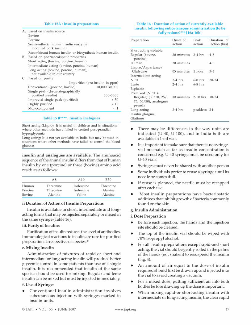
Different types and species of insulins are available.They have different pharmacokinetic properties.Different insulin preparations can be divided based onthe species, duration of action and impurities present(Table 15A & B). Insulin type, species, injection technique,insulin antibodies, site of injection and individual patientresponse differences can affect the onset, degree, andduration of insulin activity. Changing insulin speciesmay affect blood glucose control and should only bedone under the supervision of a health professional withexpertise in diabetes.28
i. Species of Insulin
Insulin of bovine or porcine origin were the onlycommercially available preparations for the first half-century of the insulin era. Currently only bovine, human

16 www.japi.org © JAPI • VOL. 55 • JUNE 2007
Fig. 3 : Algorithm for the management of type 2 diabetes mellitus.
© JAPI • VOL. 55 • JUNE 2007 www.japi.org 17
insulin and analogues are available. The aminoacidsequence of the animal insulin differs from that of humaninsulin by one (porcine) or three (bovine) amino acidresidues as follows:
A 8 A10 B30
Human Threonine Isoleucine ThreoninePorcine Threonine Isoleucine AlanineBovine Alanine Valine Alanine
ii Duration of Action of Insulin Preparations
Insulin is available in short, intermediate and long-acting forms that may be injected separately or mixed inthe same syringe (Table 16).iii. Purity of Insulins
Purification of insulin reduces the level of antibodies.Immunological reactions to insulin are rare for purifiedpreparations irrespective of species.29
e. Mixing Insulin
Administration of mixtures of rapid-or short-andintermediate-or long-acting insulin will produce betterglycemic control in some patients than use of a singleinsulin. It is recommended that insulin of the samespecies should be used for mixing. Regular and lenteinsulin can be mixed but must be injected immediately.f. Use of Syringes
� Conventional insulin administration involvessubcutaneous injection with syringes marked ininsulin units.
� There may be differences in the way units areindicated (U-40, U-100), and in India both areavailable in 1-ml vial.
� It is important to make sure that there is no syringe-vial mismatch as far as insulin concentration isconcerned e.g. U-40 syringe must be used only forU-40 vials.
� Syringes must never be shared with another person� Some individuals prefer to reuse a syringe until its
needle be comes dull.� If reuse is planned, the needle must be recapped
after each use.� Most insulin preparations have bacteriostatic
additives that inhibit growth of bacteria commonlyfound on the skin.
g. Insulin Administration
i. Dose Preparation
� Be fore each injection, the hands and the injectionsite should be cleaned.
� The top of the insulin vial should be wiped with70% isopropyl alcohol.
� For all insulin preparations except rapid-and shortacting, the vial should be gently rolled in the palmsof the hands (not shaken) to resuspend the insulin(Fig. 4).
� An amount of air equal to the dose of insulinrequired should first be drawn up and injected intothe vial to avoid creating a vacuum.
� For a mixed dose, putting sufficient air into bothbottles be fore drawing up the dose is important.
� When mixing rapid-or short-acting insulin withintermediate or long-acting insulin, the clear rapid
Table 15A : Insulin preparations
A. Based on insulin sourceBovinePorcineSemisynthetic human insulin (enzyme
modified pork insulin)Recombinant human insulin or biosynthetic human insulin
B. Based on pharmacokinetic propertiesShort acting (bovine, porcine, human)Intermediate acting (bovine, porcine, human)Long acting (bovine, porcine, human);
not available in our countryC. Based on purity
Impurities (pro-insulin in ppm)Conventional (porcine, bovine) 10,000-30,000Single peak (chromatographically
purified insulin) 300-3000Improved single peak (purified) < 50Highly purified < 10Monocomponent < 1
Table 15 B****. Insulin analogues
Short acting (Lispro): It is useful in children and in situationswhere other methods have failed to control post-prandialhyperglycemiaLong acting: It is not yet available in India but may be used insituations where other methods have failed to control the bloodglucose
Table 16 : Duration of action of currently availableinsulin following subcutaneous administration (to be
fully redone)*** [16a-16b]
Preparation Onset of Peak Duration ofaction action action (hrs)
Short acting/solubleRegular (bovine, 30 minutes 2-4 hrs 4-8
porcine)Human 20 minutes 4-8Lispro/Aspartame/Glulycine 05 minutes 1 hour 3-4
Intermediate actingNPH 2-4 hrs 6-8 hrs 20-24Lente 2-4 hrs 6-8 hrsBiphasicPremixed (NPH +
Regular) (30/70, 25/ 30 minutes 2-10 hrs 18-2475, 50/50), analoguespremix
Long acting 3-4 hrs peakless 24Insulin glargineGlatimer
18 www.japi.org © JAPI • VOL. 55 • JUNE 2007
or short acting insulin should be drawn into thesyringe first.
ii. Injection Procedures
� Injections are given into the subcutaneous tissue.� Most individuals are able to lightly grip a fold of
skin and inject at a 90o angle (Fig. 5).� Thin individuals or children may need to pinch the
skin and inject at a 45o angle to avoid intramuscularinjection, especially in the thigh area.
i. Insulin Pens
Several pen-like devices and insulin-containingcartridges are available that deliver insulinsubcutaneously through a needle. They are easy to use.iii. Injection Site
� Insulin may be injected into the subcutaneous tissueof the upper arm, the anterior and lateral aspects ofthe thigh, the buttocks, and the abdomen (Fig. 6).
� Rotation of the injection site is important to preventlipohypertrophy or lipoatrophy. Rotating within onearea is recommended (e.g. rotating injectionssystematically within the abdomen) rather thanrotating to a different area with each injection. Thispractice may decrease variability in absorption fromday to day.
� Site selection should take into consideration thevariable absorption between sites.
� The abdomen has the fastest rate of absorption,followed by the arms, thighs, and buttocks.
� Exercise increases the rate of absorption from theinjection sites, probably by increasing blood flow tothe skin and perhaps also by local actions.
� The most commonly recommended interval betweeninjection of short-acting (regular) insulin and a mealis 30 min.
c. Storage31
� Vials of insulin not in use should be refrigerated.They should not be kept in the freezer compartment.
� Insulin should not be exposed to direct sunlight� Excess agitation should be avoided to prevent loss
of potency, clumping, frosting, or precipitation.� Insulin in use may be kept at room temperature to
limit local irritation at the injection site, which mayoccur when cold insulin is used. Once the vial isopened, it should be used for a period of 30 days.
� If refrigeration is not available, insulin should bestored in closed cabinets or under the clothes.
� If regular insulin shows haziness, it indicatesbacterial growth and should not be used.
h. Adverse Effects
The main problems associated with insulin use arehypoglycemia and weight gain. Weight gain can besubstantial, and the amount is generally well correlatedwith the total daily dose of insulin.12.2.j. Initiating Insulin Therapy
For initiating insulin it is not necessary to hospitalizethe patient, it can be done at their home. The dosing hasto be individualized depending upon the blood glucoseprofile and clinical setting. It is better to start with smalldoses and modify accordingly every three days.Generally the initial starting dose of insulin should be0.2 units/kg/day.
Fig. 4 : Rolling of insulin vial in the palms of the hands.
Fig. 5 :
Fig. 6 : Sites of insulin injections.

© JAPI • VOL. 55 • JUNE 2007 www.japi.org 19
k. Adding Insulin to Oral Agents
When combinations of oral agents no longer maintainthe level of control desired, insulin is needed. Less doseof insulin is needed and less hyperinsulinemia andweight gain occur when one insulin injection iscombined with oral agents than with multiple insulininjections.l. Multiple Insulin Injections
� For less obese patients (BMI < 30), a bed time injectionof NPH insulin safely controls fastinghyperglycemia.
� When a single insulin injection plus one or moreoral agents no longer maintains good glucosecontrol, two or more injections are needed.
� In contrast to type 1 diabetes, who nearly alwaysrequires three or four injections for good control, longduration type 2 diabetes is usually treated with twoinjections.
� Older persons need careful monitoring to avoidhypoglycemia.
� A regimen of equal amounts of insulin, either NPHand regular insulin mixed by the patients orpremixed, taken before breakfast and dinner is areasonable way to start multiple injections.
� When glitazones are added to insulin regimen,insulin requirements may come down and thus suchsubjects need to be carefully watched forhypoglycemia.
The alternate routes of delivery under evaluationinclude intranasal and intrapulmonary.
MONITORING GLYCEMIC CONTROL
The importance of glycemic control is wellestablished and has been conclusively proved in bothtype 1 and type 2 diabetics by DCCT, Kumomoto andUKPDS studies.21-23 As hyperglycemia is stronglyassociated with the development and progression ofdiabetic complications, the importance of accuratemonitoring of glycemic control cannot be overemphasized.
Table 17 : Parameters of monitoring
UrineGlucoseKetonesAlbumin
BloodGlucoseGlycosylated hemoglobin
1.a. Urine Glucose Testing
Although urine glucose testing is pain less and muchcheaper than blood glu cose test ing, it is mis -lead ingand there fore not rec om mended for rou tine use. It has
lim ited role in the pres ent day man age ment strategyfor diabetes. How ever, some di a betic pa tients con tinueto use urine test ing. It can be con sid ered as an al ter native for mon i tor ing their glycemic sta tus since somemon i tor - ing is better than no mon i tor ing. Test stripsthat quan tify urine glu cose should be used to avoid afalse pos i tive re sult. The urine glu cose
test ing should be done in the post pran dial state in asec ond voided spec i men, so that a neg a tive re sult dur-ing this state in di cates ad e quate glycemic con trol.One should watch for hypoglycemia in such sit uations.1.b. Urine Ketone Testing
Urine ketone test ing is in di cated in the fol low ing :i) At on set of di a be tes in all young pa tients with di
-abetesii) Dur ing pe ri ods of poor met a bolic con trol (blood
glu cose > 250 mg/dl)iii) Dur ing acute ill ness and stress, food de pri va tion
(star va tion)iv) Dur ing preg nancy (early morn ing sam ple)v) When symp toms of ketoacidosis are pres ent (such
as nau sea, vom it ing and ab dom i nal pain)1.c. Urine Albumin Testing
Urine al bu min pro vides a fair in di ca tion of kid neysta tus. If urine al bu min is neg a tive, microalbuminuriashould be tested ev ery year.2.a. Blood Glucose Testing
� Mea sure ment of plasma glu cose is re quired for initial di ag no sis of di a be tes.
� Sub se quently, it is done for mon i tor ing the ad e -quacy of ther apy.
� The fre quency of test ing de pends upon the type ofdi a be tes and the ther apy used.32
� Both FPG and PPG should be mea sured.� Mon i toring must be done with the usual diet and
drug in take. Ex tra diet or heavy diet should not replace the nor mal diet dur ing mon i tor ing.
� Oral glu cose chal lenge is not re quired dur ing moni tor ing.
� The targets for glycemic control are summarized inTable 18.
2.a.i. Recommended Frequency for Plasma
Glucose Measurements
� The mea sure ment of FPG and 2 hr PPG on a weeklyor fort nightly ba sis at start of ther apy. This is to befol lowed by monthly mea sure ments oncesatisfactory control is achieved.
� How ever, those pa tients with type 2 di a be tes onin su lin ther apy as well as stressed sub jects require more fre quent plasma glu cose mea sure mentsas in type 1 di a be tes.

20 www.japi.org © JAPI • VOL. 55 • JUNE 2007
2.a.ii. Self Monitoring of Blood Glucose (SMBG)
� Indications for SMBG in clinical practice are listedin Table 19.
� Type 2 diabetes with altered renal threshold,advanced chronic complications and during periodof acute stress
� Peri-operative state� Labile/brittle diabetes� Neuroglycopenia without warning, nocturnal
hypoglycemia� Pregnancy, acute infection and myocardial infarction� All cases on intensive insulin therapy� Ac cu rate re sults with SMBG are tech nique de -
pend ent, re gard less of whether the strips are readvi su ally or with a glucometer. There fore, the pa -tient’s mea sure ment tech nique should be checkedini tially and at reg u lar in ter vals there af ter.33,34
� A com par i son of the re sults of the pa tients self testing of blood glu cose with si mul ta neous lab o - ratory test ing would con firm the ac cu racy of SMBGin the in di vid ual pa tient.
� The blood glu cose strips should be used within 60days af ter open ing the bot tle.
� Cutting of the strips not recommended.2.b. Glycosylated Hemoglobin Testing
Glycated proteins such as hemoglobin and serumproteins provide measures of glycemia over an extendedperiod of time depending on their half life in thecirculation.
Glycated Hemoglobin
� The de gree of he mo glo bin glycation is pro por -tional to the am bi ent glu cose con cen tra tion andis a mea sure of the av er age glycemia over the pre -ced ing three months.
� HbA1c is the most abundant and correlates best withthe degree of glycemia.
� Var i ous fac tors can al ter the HbA1c lev els. Falselyhigh values are observed in sit u a tions of increasedfetal hemoglo bin and uremia whereas falsely lowval ues are ob served in hemoglobinopathies andhemolytic anae mia.
� A change in HbA1c of 1% would re flect a blood glucose al ter ation of about 30 mg%.
Table to be added by SRJ***� Testing should be per formed rou tinely in all pa -
tients at di ag no sis and then ev ery three monthssub se quently.
� Perform the A1C test at least two times a year inpatients who are meeting treatment goals (and whohave stable glycemic control).
� Perform the A1C test quarterly in patients whosetherapy has changed or who are not meetingglycemic goals. (Ref: ADA 2007 guidelines)**
COMPLICATIONS
1. Acute Metabolic Complications
1.a. Diabetic Ketoacidosis (DKA)35
Di a betic ketoacidosis is an im por tant cause of mor- bid ity and mor tal ity amongst di a bet ics. It is a lifethreat en ing sit u a tion, aware ness, early di ag no sisand efficient management are necessary to reduce mortal- ity. In a good cen tre, mor tal ity should not be morethan 5% which could be mainly due to un der ly ing precip i tat ing fac tors like myo car dial in farc tion or se -vere fulminant in fec tions. DKA de vel ops in sit u ations where there is rel a tive in su lin de fi ciency andthere is coun ter hor mone ex - cess (Fig. 7).Treatment
The treat ment of ketoacidosis aims to cor rect de hy -dra tion, avoid hy per- and hypokalemia and lowerblood glu cose con cen tra tion. The ac i do sis is usu allycor rected by these mea sures and spe cific ther apy forthis is sel dom needed. Ad min is tra tion of in su lin andre place ment of flu - ids, in clud ing elec tro lytes and glucose, are the cor ner stones of the treat ment of DKA. During treat - ment, vi tal signs of the pa tient should be observed closely and timed urine flow and amounts of infu - sions must be re corded. The use of a flow sheet on atimed scale should be main tained.
If a pre cip i tat ing cause of DKA (such as an in fec -tion or myo car dial in farc tion) is de tected, its treatment should be started alongwith the treat ment of DKA.
Table 18 : Targets Glycemic Control for People withdiabetes
Normal Goal Additional ActionSuggested
Whole blood valuesAverage preprandial <100 80-120 <80 >140glucose (mg/dl)Average bedtime glucose <110 100-140 <100 >160(mg/dl)Plasma valuesAverage preprandial <110 90-130 <90 >150glucose (mg/dl)Average bedtime glucose <120 110-150 <110 >180(mg/dl)HbA1c (%) <6 <7 >8
Table 19 : Indications for SMBG
� Type 2 diabets with altered renal threshold, advancedchronic complications and during period of acute stress
� Peri-operative state� Labile/brittle diabetes� Neuroglyceopenia without warning, nocturnal
hypoglycemia� Pregnancy, acute infection and myocardial infarction� All cases on intensive insulin therapy

© JAPI • VOL. 55 • JUNE 2007 www.japi.org 21
In an un con scious pa tient, a nasogastric tube shouldbe in serted to keep the stom ach empty and pre vent aspi ra tion of gas tric con tents.Correction of Dehydration : Fluid and Electrolytes
Normal saline is the fluid of choice for ini tialrehydration. On an av er age - One li ter should be infused in the first hour Next one li ter in the next 2 hrs.Two liters in the next 4 hrs. Two li ters in the next 8 hrs.i.e. 4-6 li ters in 24 hrs. The ef fect of sa line should bemon i tored by blood pres sure, CVP, pulse, neck veins,skin turgor. In a sit - u a tion where serum sodium is 150mEq/L, hypotonic or half nor mal sa line may be used.When the blood glucose concentration reaches 250 mg/dl, the fluid should be changed to 5% dex trose-salinewith concurrent ad min is tra tion of insulin, in or der toavoid hypoglycemia. It takes longer for acidosis to getcorrected than blood glucose.Fluid Overload
Care should be taken not to in fuse a large amount offluid too rapidly in elderly patients, in those with existingheart disease especially congestive cardiac failure andin patients with renal failure.
Potassium Replacement
The plasma potassium may initially be high in thepresence of acidosis. It begins to fall as DKA is treated.Hypokalemia is therefore likely with out potassiumreplacement. There is no definite guideline about whento start potassium replacement. If there is good urinaryout put, potassium replacement should be started earlyduring the treatment of DKA. Potassium chloride (oneampoule, 10 mEq) should be added in the third bottle ofsaline. The potassium level should be evaluated everytwo hours or as necessary. If the response is notadequate, dose must be modified. This should be avoidedin the presence of renal failure. ECG monitoring is helpfulsince tall T waves are indicative of hyperkalemia.Patients not responding to conventional therapy may belooked for other conditions. Ringer lactate and fructoseshould be avoided in DKA.Other Electrolytes
Bicarbonate may be used in severe cases of acidosiswith a pH less than 6.9, bicarbonate levels less than 5mmol/L, patients with DKA complicating acute myocardial infarction and in patients with lactic acidosis. Therisks of bicarbonate therapy are shift of
Withdrawal of insulin Fasting Pancreatic Relative DCounter cell failure Insulin K hormone Stress DeficiencyA excess Insulin resistance DehydrationInsulin
The standard method is low dose infusion of insulinby the intravenous route after an initial bolus of insulin.36-
38 If IV infusion of insulin is difficult to perform, hourlyin tramuscular (IM) injection of insulin is an alternativemethods.38 For an IM administration, the deltoid muscleis preferred. IV insulin therapy is given as an initialdose of 10 units as a bolus, followed by insulin infusionat the rate of 0.1 unit/kg/hour. (50 units of insulin are tobe added to 500 ml of normal saline. The infusion is tobe run at the rate of 1 ml/min). IM regime involves theuse of 0.1 unit/kg/hour of insulin. With these regimens,the plasma glucose level falls (90 mg%) at a predictablerate of around 50 mg/hr. It usually comes down to halfthe initial value in 6-8 hours. If at the end of 2-3 hours,the plasma glucose values do not show a predictablefall, the dose of insulin infusion is doubled i.e. 12 units/hr. Once the plasma glucose levels come down to 250mg%, the fluid replacement should be switched over to
Fig. 7 : Pathogenesis of diabetic ketoacidosis.
Table 20 : Precipitating factors
InfectionOmission of insulinUnknown causeStressful situations
Acute myocardial infarctionCerebrovascular accidentSurgeryTrauma
Table 21 : Signs and symptoms of DKA
Symptoms Signs
Polyuria HyperpneaPolydypsia HypothermiaWeakness Acetone breathLethargy Acidotic breathingMyalgia DyspneaHeadache Acute abdomenAnorexia DehydrationNausea HyporeflexiaVomiting HypotoniaAbdominal pain Stupor/comaBreathlessness
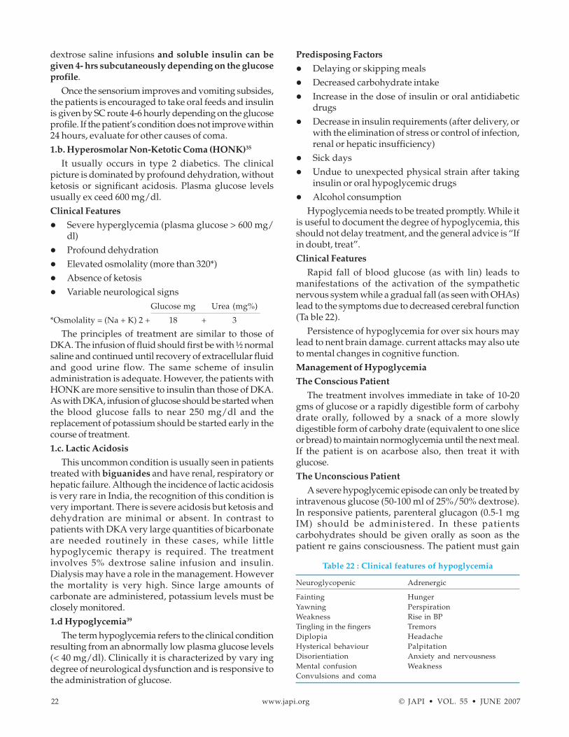
22 www.japi.org © JAPI • VOL. 55 • JUNE 2007
dextrose saline infusions and soluble insulin can begiven 4- hrs subcutaneously depending on the glucoseprofile.
Once the sensorium improves and vomiting subsides,the patients is encouraged to take oral feeds and insulinis given by SC route 4-6 hourly depending on the glucoseprofile. If the patient’s condition does not improve within24 hours, evaluate for other causes of coma.1.b. Hyperosmolar Non-Ketotic Coma (HONK)35
It usually occurs in type 2 diabetics. The clinicalpicture is dominated by profound dehydration, withoutketosis or significant acidosis. Plasma glucose levelsusually ex ceed 600 mg/dl.Clinical Features
� Severe hyperglycemia (plasma glucose > 600 mg/dl)
� Profound dehydration� Elevated osmolality (more than 320*)� Absence of ketosis� Variable neurological signs
Glucose mg Urea (mg%)
*Osmolality = (Na + K) 2 + 18 + 3
The principles of treatment are similar to those ofDKA. The infusion of fluid should first be with ½ normalsaline and continued until recovery of extracellular fluidand good urine flow. The same scheme of insulinadministration is adequate. However, the patients withHONK are more sensitive to insulin than those of DKA.As with DKA, infusion of glucose should be started whenthe blood glucose falls to near 250 mg/dl and thereplacement of potassium should be started early in thecourse of treatment.1.c. Lactic Acidosis
This uncommon condition is usually seen in patientstreated with biguanides and have renal, respiratory orhepatic failure. Although the incidence of lactic acidosisis very rare in India, the recognition of this condition isvery important. There is severe acidosis but ketosis anddehydration are minimal or absent. In contrast topatients with DKA very large quantities of bicarbonateare needed routinely in these cases, while littlehypoglycemic therapy is required. The treatmentinvolves 5% dextrose saline infusion and insulin.Dialysis may have a role in the management. Howeverthe mortality is very high. Since large amounts ofcarbonate are administered, potassium levels must beclosely monitored.1.d Hypoglycemia39
The term hypoglycemia refers to the clinical conditionresulting from an abnormally low plasma glucose levels(< 40 mg/dl). Clinically it is characterized by vary ingdegree of neurological dysfunction and is responsive tothe administration of glucose.
Predisposing Factors
� Delaying or skipping meals� Decreased carbohydrate intake� Increase in the dose of insulin or oral antidiabetic
drugs� Decrease in insulin requirements (after delivery, or
with the elimination of stress or control of infection,renal or hepatic insufficiency)
� Sick days� Undue to unexpected physical strain after taking
insulin or oral hypoglycemic drugs� Alcohol consumption
Hypoglycemia needs to be treated promptly. While itis useful to document the degree of hypoglycemia, thisshould not delay treatment, and the general advice is “Ifin doubt, treat”.Clinical Features
Rapid fall of blood glucose (as with lin) leads tomanifestations of the activation of the sympatheticnervous system while a gradual fall (as seen with OHAs)lead to the symptoms due to decreased cerebral function(Ta ble 22).
Persistence of hypoglycemia for over six hours maylead to nent brain damage. current attacks may also uteto mental changes in cognitive function.Management of Hypoglycemia
The Conscious Patient
The treatment involves immediate in take of 10-20gms of glucose or a rapidly digestible form of carbohydrate orally, followed by a snack of a more slowlydigestible form of carbohy drate (equivalent to one sliceor bread) to maintain normoglycemia until the next meal.If the patient is on acarbose also, then treat it withglucose.The Unconscious Patient
A severe hypoglycemic episode can only be treated byintravenous glucose (50-100 ml of 25%/50% dextrose).In responsive patients, parenteral glucagon (0.5-1 mgIM) should be administered. In these patientscarbohydrates should be given orally as soon as thepatient re gains consciousness. The patient must gain
Table 22 : Clinical features of hypoglycemia
Neuroglycopenic Adrenergic
Fainting HungerYawning PerspirationWeakness Rise in BPTingling in the fingers TremorsDiplopia HeadacheHysterical behaviour PalpitationDisorientiation Anxiety and nervousnessMental confusion WeaknessConvulsions and coma

© JAPI • VOL. 55 • JUNE 2007 www.japi.org 23
consciousness within half an hour. Parenteraldexamethasone is also recommended in unresponsivepatients. It is important to follow glucose level for atleast 24-48 hours in patients where hypoglycemia isinduced by long acting sulphonylureas (glibenclamide)or long acting insulin (Lente/NPH). A long term glucoseinfusion may be necessary and the patient should behospitalized. Occasional patient may take longer timeto cover. Hypoglycemic unawareness is usually seenin type 1 diabetes and may be difficult to recognize.Glucagon although costly and not routinely availableis an alternative to treat insulin induced hypoglycemiaand later needs oral glucose.Prevention of Hypoglycemia
Education of patient and family members is a must.Education of the health careproviders in terms ofdiagnosing diabetes, its management and alsodiagnosing hypoglycemic episodes early is important.
Recognition****2. Chronic Vascular Complications
2.a. Macrovascular Complications
2.a.i. Coronary artery disease
Acute Myocardial Infarction (AMI)
In diabetics as compared to non-diabetic subjects,� The incidence of myocardial in farction is around
three times higher and the outcome of AMI is worse.� Mortality is two times higher in pre-hospital,
hospital and post-hospital periods following theepisode. This is particularly so in patients be low 60years of age and in obese females; more so in thosewith anterior infarctions.40
� Diabetics develop complications of myocar dialinfarction more of ten than nondiabetics. Theseinclude cardiogenic shock, CHF, conductiondisturbances, etc.40-46
� Congestive heartfailure is seen in 44% of diabeticwomen and 25% of diabetic men.
� Infarct sizes are relatively larger and more of tenanterior.* Incidence of silent MI is variable but by all
measures more frequent in patients withdiabetes.
* Painless or silent infarcts occur in 20% ofdiabetics
� Presenting features may be atypical and lead to longlag riod between onset and hospitalization in theintensive coronary care unit (ICCU).47-51
� An estimated 20-40% patients present with atypicalfea tures such as confusion, dyspnoea, cough,fatigue, nausea, vomiting and epigastric distress.
� Ketosis (2-4%) or hyperosmolar state (in elderly) maybe present at admission.
� Sudden cardiac deaths are more common.� CABG or PTCA as an intervention may be decided
on the merit of individual case.
A major concern in the management of AMI in patientsof diabetes is to provide adequate insulin in order torestore glucose utilization and reduce lipolysis andconsequent rise in circu lating free fatty acids.
Diabetic retinopathy is no longer a contraindicationfor thrombolytic therapy.50
Following life saving measures including theadmission to intensive coronary care unit (ICCU),specific anti-diabetic therapy has to be started on apriority basis. The infarction may be massive and post-AMI period more stormy.Diagnosis
On arrival of the patient, random plasma glucose(RPG) is to be estimated immediately. If the patient is nota known diabetic. HbA1c will distinguish between stresshyperglycemia and a diabetic. Urine has to be testedparticularly for ketone bodies. In case the plasma glucoseis more than 200 mg/dl, short acting insulin is to bestarted, preferably by IV infusion.Monitoring for Blood/Plasma Glucose During
Infusion
Monitoring is best done at the bedside by glucosemonitor. Results thus obtained will guide the rate ofinsulin infusion from time to time. Beyond this, regularplasma glucose monitoring along with other parameters(electrolytes and ketone bodies) should be done every 4-6 hours for further management. If the serum potassiumis less than 3.5 mEq/L, potassium solution has to beadded to the insulin infusion device at the rate of 20mEq/L. Special care has to be taken in case of ketosis,hyperosmolar coma. Nutrients (including dextrose,other resuscitation fluids and essential therapy) shouldbe administered through different IV lines. The revivalin the intensified insulin therapy for AMI originated fromDIGAMI study51 which achieved favourable results withadministration of insulin by infusion. Insulin for first24 hours of AMI followed by subcutaneous insulintherapy for three months improved the short and longterm mortality.2.a.ii. Cerebrovascular Disease
Stroke
� Diabetes mellitus (chronic hyperglycemia) is animpor tant risk factor for ischemic but nothemorrhagic stroke.
� The relative risk of ischemic stroke is increased onan average by 2-4 fold in the diabetic population.
� The type and distribution of stroke in diabeticpatients are not significantly dif fer ent from that ofnon-diabetic subjects. Further, to what extenthyperglycemia contributes to promote stroke is not

24 www.japi.org © JAPI • VOL. 55 • JUNE 2007
certain as it is so often associated with hypertension(HTN) and dyslipidemia.
� Framingham study indicates increased cerebralinfarction even with mild glucose intolerance.52-55
Several studies have shown an increase in short andlong term morbidity/mortality in diabetics, who havestroke. Hyperglycemia has been observed to worsen theseverity of the attack.Guidelines for Treatment
� All stroke victims are likely to be dehydrated andshould be started with normal saline at theadmission.
� Random plasma glucose (RPG), glycosylatedhemoglobin (HbAlc), urea, creatinine, electrolytes,urine for ketones should be tested and an initialvalue obtained [to distinguish preexisting diabetesfrom stress hyperglycemia].
� Patient should never be put on dextrose or dextrosesaline to start with.
� The rate and total volume of normal salineadministered in each patient should be determinedafter considering the expected total volume loss,renal/heart status and hematocrit.
� Any excess fluid intake may result in developmentof cerebral edema.
� At times these patients may have complications suchas hyperosmolar non-ketotic state or ketoacidosis.
It is very important to note that plasma glucose shouldnot to be allowed to fall below 160 mg/dl throughoutthe therapy. Hyperglycemia raises lactic acid content ofcerebral ischemic tissues where as cellular damageoccurs in hypoglycemia.
Alongwith maintenance of normoglycaemia, a bloodpressure above 200/100 mm Hg should be controlledvery gradually as acute ing of blood pressure worsensthe ischemia and total outcome of the pa tient. Mannitolinfusion can be given. However efforts to achieve a goodmetabolic control should be accompanied by anaggressive approach towards cerebral reperfusion.Hypoglycemia can manifest as stroke.2.a.iii. Aspirin Therapy in Diabetes
People with diabetes have a two to four fold increasein the risk of dying from the complications ofcardiovascular disease. Both men and women are atincreased risk. Atherosclerosis and vascular thrombosisare major contributors, and it is generally accepted thatplatelets are contributory. Platelets from men and womenwith diabetes are often hypersensitive invitro to plateletaggregating agents. A major mechanism is increasedproduction of thromboxane, a potent vasoconstrictor andplatelet aggregant. Recommendations on the use ofaspirin are given in Table 23.
2.b. Microvascular Complications
2.b.i. Diabetic Nephropathy58
Diabetic nephropathy is one of the common est causeof end-stage renal disease (ESRD). This is due to the inglence of type 2 diabetic, longer life span of diabeticpatients and improved therapeutic options and the factthat patients with diabetic nephropathy are beingaccepted for renal replacement therapy. Prevalence ofall grades of proteinuria was seen in 19.7% of type 2diabetics and amongst them, 5.5% had nephropathy.59The natural history of diabetic nephropathy progressesfrom normoalbuminuria, through a subclinical stage ofurinary albumin excretion called microalbuminuria toovert proteinuria and eventually end-stage renal disease(Ta ble 24).Prevalence at Various Stages
� Acute renal hypertrophy/hyperfunction Present atdiagnosis
� NormoalbuminuriaMost patients in the first 5 yearsA higher dose of aspirin are recommended in patient
of diabetes with stroke (reference to be searched)� Incipient nephropathy Occurs after 5-15 years in
~35% of patients
Table 23 : Recommendations on the use of aspirin
1. Use aspirin therapy as a secondary prevention strategy indiabetic men and women who have evidence of largevessel disease. This includes diabetic men and womenwith a history of myocardial infarction, vascular bypassprocedure, stroke or transient ischemic attack, peripheralvascular disease, claudication, and/or angina.
2. In addition to treating the primary cardiovascular riskfactor(s) identified, consider aspirin therapy as a primaryprevention strategy in high-risk men and women with type1 or type 2 diabetes. This includes diabetic subjects withthe following:* A family history of coronary heart disease* Cigarette smoking* Hypertension* Obesity (> 120% desirable weight)* Albuminuria (micro or macro)* Lipids : Cholesterol > 200 mg/dl
LDL cholesterol > 100 mg/dlHDL cholesterol < 45 mg/dl in men and< 55 mg/dl in womenTriglycerides > 200 mg/dl
* Age > 30 yearsUse of aspirin has not been studied in diabetic individualsunder the age of 30 years.
3. Use enteric-coated aspirin in doses of 75-325 mg/day.4. People with aspirin allergy, bleeding tendency,
anticoagulant therapy, recent gastrointestinal bleeding,and clinically active hepatic disease are not candidates foraspirin therapy.
5. Aspirin therapy should not be recommended for patientsunder the age of 21 years because of the increased risk ofReye’s syndrome associated with aspirin use in thispopulation.

© JAPI • VOL. 55 • JUNE 2007 www.japi.org 25
� Overt diabetic nephropathy Usually develops inabout 35% of patients after 15-25 yrs
� End-stage renal disease Final outcome, usually after25-30 years
Significance of Albuminuria
Microalbuminuria is a marker of generalizedendothelial dysfunction
In type 2 diabetes mellitus, microalbuminuria� May be non-specific� Correlated with systolic hypertension� Associated cardiovascular disease� Associated peripheral vascular disease� Coronary artery disease occurs earlier than ESRD� Associated with premature atherosclerosis� Abnormal lipid pro file� Pronounced increased mortality� Correction of microalbuminuria in solation may not
affect events appreciablyThe type 2 diabetes patients with diabetic kidney
disease usually die from cardiovascular disease suchas coronary artery disease and hence integralmanagement of all risk factors is indicated.Screening for Albuminuria
The methods adopted may be� 24 hour collection along with measurement of
creatinine clearance (mg/24 hours)� Timed collection (µg/min)� Spot collection to look for albumin to creatinine ratio
(mðg/mg creatinine). Sample should not be takenin the immediate post-exercise period60 (Ta ble 25).
In type 1 DM, screening should begin with pubertyand after 5 years duration of diabetes. In tyep 2 DM,however, screening is recommended at the time ofdiagnosis and if negative, every year. Routine urinalysis
should be performed annually. An algorithmicapproach60 is outlined in Fig 8. Treatment OptionsModalities of treatment depend on the stage of diseaseat diagnosis (especially type 1 DM) to prevent onset ofnephropathy. Treat ment approaches are as out lined inTa ble 26.Glycemic Control
� Intensive glycemic control can significantly reducethe risk of developing microalbuminuria and overtnephropathy.
� Metformin should not be used if the serum creatinineis more than 1.4 mg/dl in men and 1.5 mg/dl inwomen.
� Oral antidiabetic drugs recommended for diabeticnephropathy are glipizide, gliclazide, acarbose,repaglinide and glitazones. However they shouldbe used with caution.
Hypertension Control
� In type 1 diabetes mellitus, hypertension is usually
Table 24 : Stages of diabetic nephropathy
Stage Designation Main Characteristics GFR Albumin Excretion BP Treatment
I Hyperfunction Large kidney and 150 May be increased Normal Strict glycemic controlHypertrophy glomerular size reverses the condition
II Normoalbuminuria Normal UAE With/without Normal (often Normal Strict glycemic controlhyperfiltration increased in stress may reduce
situations) hyperfiltrationIII Incipient Persistently 130-160 20-200 µg/min Elevated Microalbuminuria
elevated UAE 3-5% yr stabilized by stricttreatment, GFR alsostable if HbA1C isreduced. Prevention ofprogression is possible
IV Overt Clinical 10-130 >200 µg/min Hypertension Poor control leads toproteinuria greater fall in GFR
V Uremia End stage renal 0-10 Decreasing BP is high Progression can befailure due to retarded by strict
nephron glycemic control,closure BP control
Table 25 : Definitions of abnormal albumin excretion
Category 24 Timed Spot hourcollection collection collection
(mg/24 h) (µg/min) (µg/min creatinine)
Normal <30 <20 <30Microalbuminuria 30-300 20-200 30-300Overt albuminuria >300 >200 >300
Table 26 : Treatment options for diabetic nephropathy
Early Nephropathy� Glycemic control� Treatment of systemic and intraglomerular hypertension� Dietary protein restriction� Modification of risk factors - smoking and dyslipidemiaEnd Stage Renal Disease� To provide symptomatic relief, control of hypertension� Renal replacement therapy

26 www.japi.org © JAPI • VOL. 55 • JUNE 2007
due to the development of nephropathy, while intype 2 diabetes mellitus, hypertension ismultifactorial.
� Both systolic and diastolic hypertension canaccelerate the progression of diabetic nephropathyand aggressive treatment of hypertension maymarkedly retard the rate of fall of GFR.
� The goal of treatment in adults is to maintain asystolic BP < 130 mm Hg and a diastolic BP < 85 mmHg. In patients with isolated systolic hypertensionand an initial systolic BP > 188 mm Hg, the initialgoal is to reduce it to < 160 mm Hg, and in thosewith systolic BP 160-179 mmHg, to lower it by 20mm Hg. If tolerated, further reduction may beattempted.60
� Lifestyle modification should play a major role inthe treatment which includes cessa tion of smoking.
� The drug of choice, as per current dence, is anangiotensin converting enzyme inhibitor (ACEI).This group of drugs has shown benefit in reducingthe progression of microalbuminuria in all patientswith type 1 DM and in normotensive andhypertensive patients with type 2 DM.60
� In normotensive diabetics with normal renalfunction in the absence of albuminuria, there is notenough evidence for the use of ACEIs.60
� Use of betablockers and loop diuretics may beconsidered in the event of the failure of ACEIs andlong acting calcium channel blockers.
� ACEIs should be with held once the serum creatininerises above 2 mg/dl or creatinine clearance falls below 30 ml/min
� The use of AT 2 receptor blockers are cated* If ACEIs are not tolerated* It can be added to ACEIs if the BP control is not
optimum.In the management of hypertension, the specific role
of ARBs have been defined presently. Even combinationof ACEIs and ARBs is advocated. (Reference from Dr.Sahay)Protein Restriction
� Restriction of protein intake can lead to reduction inthe hyperfiltration and intraglomerular pressureand thereby retard the progression of nephropathy.(Use of protein derived from soya bean and itsproducts may be preferable).
� The recommended dietary allowance is 0.8 gm/kg/day. In selected patients with overt nephropathy andfalling GFR, a stricter restriction to 0.6 gm/kg/daymay be useful.
� Diet recommended for diabetes my not require
Fig. 8 : Algorithmic approach to screening for urine albumin excretion.
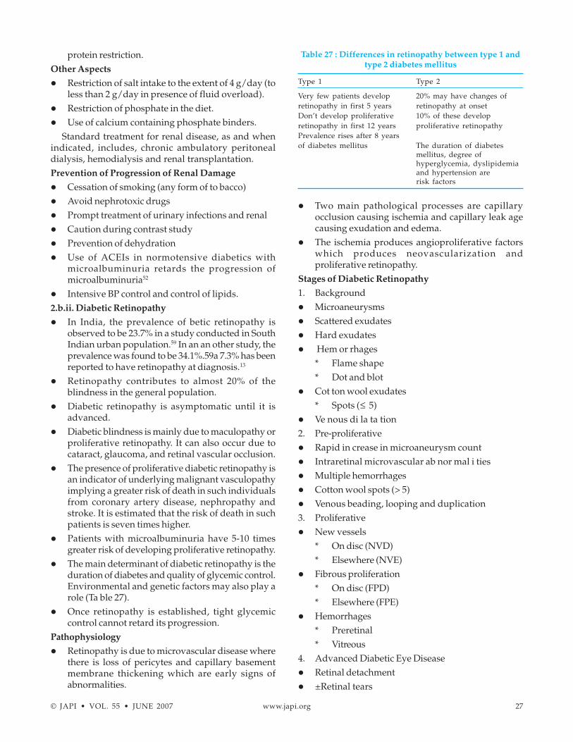
© JAPI • VOL. 55 • JUNE 2007 www.japi.org 27
protein restriction.Other Aspects
� Restriction of salt intake to the extent of 4 g/day (toless than 2 g/day in presence of fluid overload).
� Restriction of phosphate in the diet.� Use of calcium containing phosphate binders.
Standard treatment for renal disease, as and whenindicated, includes, chronic ambulatory peritonealdialysis, hemodialysis and renal transplantation.Prevention of Progression of Renal Damage
� Cessation of smoking (any form of to bacco)� Avoid nephrotoxic drugs� Prompt treatment of urinary infections and renal� Caution during contrast study� Prevention of dehydration� Use of ACEIs in normotensive diabetics with
microalbuminuria retards the progression ofmicroalbuminuria52
� Intensive BP control and control of lipids.2.b.ii. Diabetic Retinopathy
� In India, the prevalence of betic retinopathy isobserved to be 23.7% in a study conducted in SouthIndian urban population.59 In an an other study, theprevalence was found to be 34.1%.59a 7.3% has beenreported to have retinopathy at diagnosis.13
� Retinopathy contributes to almost 20% of theblindness in the general population.
� Diabetic retinopathy is asymptomatic until it isadvanced.
� Diabetic blindness is mainly due to maculopathy orproliferative retinopathy. It can also occur due tocataract, glaucoma, and retinal vascular occlusion.
� The presence of proliferative diabetic retinopathy isan indicator of underlying malignant vasculopathyimplying a greater risk of death in such individualsfrom coronary artery disease, nephropathy andstroke. It is estimated that the risk of death in suchpatients is seven times higher.
� Patients with microalbuminuria have 5-10 timesgreater risk of developing proliferative retinopathy.
� The main determinant of diabetic retinopathy is theduration of diabetes and quality of glycemic control.Environmental and genetic factors may also play arole (Ta ble 27).
� Once retinopathy is established, tight glycemiccontrol cannot retard its progression.
Pathophysiology
� Retinopathy is due to microvascular disease wherethere is loss of pericytes and capillary basementmembrane thickening which are early signs ofabnormalities.
� Two main pathological processes are capillaryocclusion causing ischemia and capillary leak agecausing exudation and edema.
� The ischemia produces angioproliferative factorswhich produces neovascularization andproliferative retinopathy.
Stages of Diabetic Retinopathy
1. Background� Microaneurysms� Scattered exudates� Hard exudates� Hem or rhages
* Flame shape* Dot and blot
� Cot ton wool exudates* Spots (≤ 5)
� Ve nous di la ta tion2. Pre-proliferative� Rapid in crease in microaneurysm count� Intraretinal microvascular ab nor mal i ties� Multiple hemorrhages� Cotton wool spots (> 5)� Venous beading, looping and duplication3. Proliferative� New vessels
* On disc (NVD)* Elsewhere (NVE)
� Fibrous proliferation* On disc (FPD)* Elsewhere (FPE)
� Hemorrhages* Preretinal* Vitreous
4. Advanced Diabetic Eye Disease� Retinal detachment� ±Retinal tears
Table 27 : Differences in retinopathy between type 1 andtype 2 diabetes mellitus
Type 1 Type 2
Very few patients develop 20% may have changes ofretinopathy in first 5 years retinopathy at onsetDon’t develop proliferative 10% of these developretinopathy in first 12 years proliferative retinopathyPrevalence rises after 8 yearsof diabetes mellitus The duration of diabetes
mellitus, degree ofhyperglycemia, dyslipidemiaand hypertension arerisk factors

28 www.japi.org © JAPI • VOL. 55 • JUNE 2007
� Rubeosis iridis� Neovascular glaucoma5. Maculopathy� Macular edema
* Focal* Diffuse
� Ischemic maculopathyTreatment� Periodic ophthalmic checkup� Strict glucose control� Strict control of blood pressure� Lipid control� Cessation of smoking
Condition Recommend Action
NVD Pan-retinal photocoagulation beforevitreous hemorrhage occurs
NVE Pan-retinal photocoagulation before retinalsdetachment occurs
Rubeosis iridis Pan-retinal photocoagulation beforeneovascular glaucoma develops
Pre-proliferative Pan-retinal photocoagulation, ofone eye initially
Vitreo-retinal surgery for the ultimate blindingcomplications of proliferative diabetic retinopathy i.e.severe vitreous hemorrhage, secondary retinaldetachment and neovascular glaucoma (Table 28).Causes of Sudden Blindness in Diabetes
� Retinal detachment� Vitreous hemorrhage and retinal hemorrhage� Central retinal vein thrombosis� Arterial occlusion� Optic neuritis� Acute glaucoma
(See Flow Chart)2.b.iii. Diabetic Neuropathy62
Most common and trouble some complication ofdiabetes mellitus leading to the greatest morbidity andresulting in huge economic burden for diabetic patients
and his family.Diabetic neuropathy is a heterogenous disorder that
encompasses a wide range of abnormalities affectingproximal and distal peripheral sensory and motor nervesas well as the autonomic nervous systems.Clinical Classification
Progressive neuropathiesReversible neuropathiesPressure palsies
Progressive Neuropathies
� Distal symmetrical polyneuropathy� Pre dominantly sensory� Autonomic involvement common (mostly
asymptomatic)� Clinical motor involvement very rare
� Small fibre neuropathy� Autonomic involvement common and usually
symptomatic� Gradual onset� No recovery� Associated with increasing duration of diabetes� Associated with other chronic diabetic
complications� Reversible neuropathy
Flow Chart for Reviewing Patients with Diabetic Retinopathy
Good vision Poor vision
No retinopathy Background retinopathy Retina visible Retina invisible
Exclude Exclude Exclude-Subtle NVD/NVE -Retinal detachment -RD-Macular edema -Ischemic macular edema -Vitreous hemorrhage-Preproliferative signs -Diffuse macular –Neovascular edema glaucoma
Review 12 monthly Review 6 monthly Refer to ophthalmologist if any of theseproblems are present or cannot be excluded.
↓ ↓ ↓
↓ ↓ ↓
↓
↓
↓ ↓ ↓
Table 28 : Examination of the Eyes in Diabetic patients
When to examine What to doAt diagnosis Visual actuity
- distant vision (Snellen chart)- near vision (reading chart)
Annually thereafterBiannually if background Check pupillary light reflexesretinopathy is present for relative after defect
Four monthly if mild pre-proliferative changes occur Fundoscopy, through dilatedpupils
Immedaitley if any changes in Specialist ophthalmologicalvision or visual symptoms investigations
Slit lamp examination of iris,anterior chamber and retina
Measurement of intraocularpressure

© JAPI • VOL. 55 • JUNE 2007 www.japi.org 29
� Mononeuropathies� Femoral (amyotrophy)� Cranial nerve palsies (III and VI)� Truncal radiculopathies
� Acute diffuse painful neuropathy� Stocking disturbution� Sudden onset� Spontaneous recovery� No association with duration of diabetes� No association with other chronic diabetic
complicationsEntrapment Neuropathies
� Median nerve� Ulnar nerve� Lateral popliteal nerve� More common than in the non-diabetic population
Clinical Pattern of Diabetic Peripheral Neuropathy(Figs. 9 - 13).
Clinical Presentation of Large Fiber Neuropathies
� Impaired vibration perception (of ten the firstobjective evidence) and position sense
Fig. 9 : Symmetrical diffuse sensory motor neuropathy.Senosry loss 0 → +++; Pain + → +++;
Tendon Reflexes N → decreased; Motor deficit 0 → +
Fig. 10 : Acute diffuse painful neuropathy.Senosry loss 0 → +; Pain + → +++;
Tendon Reflexes N → decreased; Motor deficit 0
Fig. 11 : Femoral neuropathy (Amyotrophy).Senosry loss 0 → +; Pain + → +++;
Tendon Reflexes; decreased 0 → ; Motor deficit + → +++
Fig. 12 : Other acute Mononeuropathies.Senosry loss 0 → +; Pain + → +++;
Tendon Reflexes N; Motor deficit + → +++
Fig. 13 : Pressure palsies.Senosry loss + → +; Pain + → ++;
Tendon Reflexes N; Motor deficit +-- +++
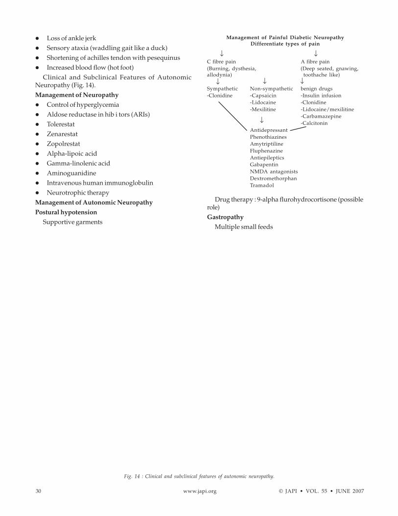
30 www.japi.org © JAPI • VOL. 55 • JUNE 2007
� Loss of ankle jerk� Sensory ataxia (waddling gait like a duck)� Shortening of achilles tendon with pesequinus� Increased blood flow (hot foot)
Clinical and Subclinical Features of AutonomicNeuropathy (Fig. 14).Management of Neuropathy
� Control of hyperglycemia� Aldose reductase in hib i tors (ARIs)� Tolerestat� Zenarestat� Zopolrestat� Alpha-lipoic acid� Gamma-linolenic acid� Aminoguanidine� Intravenous human immunoglobulin� Neurotrophic therapyManagement of Autonomic Neuropathy
Postural hypotension
Supportive garments
Drug therapy : 9-alpha flurohydrocortisone (possiblerole)Gastropathy
Multiple small feeds
Fig. 14 : Clinical and subclinical features of autonomic neuropathy.
Management of Painful Diabetic NeuropathyDifferentiate types of pain
C fibre pain A fibre pain(Burning, dysthesia, (Deep seated, gnawing,allodynia) toothache like)
Sympathetic Non-sympathetic benign drugs-Clonidine -Capsaicin -Insulin infusion
-Lidocaine -Clonidine-Mexilitine -Lidocaine/mexilitine
-Carbamazepine-Calcitonin
AntidepressantPhenothiazinesAmytriptilineFluphenazineAntiepilepticsGabapentinNMDA antagonistsDextromethorphanTramadol
↓↓
↓ ↓↓
↓

© JAPI • VOL. 55 • JUNE 2007 www.japi.org 31
MoseprideDomperidoneErythromycinJejunostomy
Cystopathy
BethanecholSelf catheterization
Gustatory sweat ing
Topical glycopyrrolateSince autonomic neuropathy usually occurs
alongwith peripheral neuropathy and can have seriouscardiorespiratory dysfunction. It is advised that in everydiabetic clinic population the autonomic neuropathymay be looked into by doing a battery of cardiovascularbedside tests. Preoperative workup of a diabetic shouldinclude this autonomic function test.Management of Erectile Dysfunction
One must exclude other causes of erectile dysfunctionsuch as drugs, androgen deficiency and psychologicalfactors. Sildenafil citrate can be administered if it notcontraindicated. Patients on nitrate should not takesildenafil citrate. Intracavernous papavarine injectionshave shown benefit. Intractable cases may be referred tothe urologist.Beside Assessment of Autonomic Neuropathy
� Postural hypotension� Pupillary light re flex� Resting tachy cardia� Post-void distended bladder2.b.iv. Diabetic Foot Syndrome63
Foot problems such as ulcerations, infec tions,gangrene and amputations are quite common in diabeticsubjects. These account for their frequent and prolongedhospitalization, significant morbidity and evenmortality. On a rough estimate nearly 20% of all hospitaladmissions of diabetes are due to foot problems andnearly 5-10% may need foot/leg amputation. Moreover,of all the non-traumatic amputations approximately 50%are related to diabetes.64 The economic and psychologicalstress for the family and the patient are enormous.However with proper footcare these could be preventedor minimized.Etiopathogenesis of Foot Lesions in Diabetics
Peripheral vascular disease (PVD), neuropathy(peripheral and autonomic) and infection are the threemajor etiological factors either singly or in combination.In India, infec tion plays an important part.65
Clinical Evaluation Guidelines
Ischaemic foot
History of claudication, rest painPal pa tion of pe riph eral pulses
An kle-brachial pres sure in dex (ABR be low 0.8indicates ischaemia)
Dopp ler ul tra sound and other testsNeuropathic foot
His tory of noc tur nal painSkin tex ture, fis sur ing, dry nessSemmes Weinstein microfilament testVi bra tion test and an kle jerkMo tor power-atrophyBiothesiometeryNerve con duc tion stud ies
Clinical Spectrum of Diabetic Foot Lesions
� Onychomycosis� Paronychia� In fected foot ul cer� Necrotizing fascitis� Osteomyelitis� Charcot’s neuroarthropathy� Gan greneRisk Factors for Diabetic Foot Lesion
� Insensitive foot-loss of protective sensation leadingto burns or injury
� Altered foot biomechanics - deformed foot� Increased pressure points - corns, callosities,
erythema� Impaired peripheral circulation - ischemic foot� Severe nail pathology - in growing nails� Loss of perspiration - dry skin, fissuring� Limited joint mobility - ‘intrinsic minus’ foot� Bare foot walking� Rat bite� Edema feet - congestive heart failure, nephropathy� Past history of foot ulceration, gangrene, amputation� Epidermophytosis of web space� Cracks� Ill-fitting foot wearWagner’s Classification of Foot Lesions
Grade 0 : Foot at high risk - no ulcerationGrade 1 : Superficial ulcerationGrade 2 : Deep ulcer ationGrade 3 : Deep ulceration with infection and
osteomyelitisGrade 4 : Fore foot or localized gangreneGrade 5 : Extensive gangrene requiring amputation
Measures to Improve Vascular Supply
� Control of diabetes, hypertension, dyslipidemia
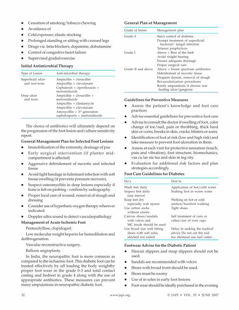
32 www.japi.org © JAPI • VOL. 55 • JUNE 2007
� Cessation of smoking/tobacco chewing� Avoidance of� Cold exposure, elastic stocking� Prolonged standing or sitting with crossed legs� Drugs viz. beta-blockers, dopamine, dobutamine� Control of congestive heart failure� Supervised graded exercise
Initial Antimicrobial Therapy
Type of Lesion Anti-microbial therapy
Superficial ulcer Ampicillin + cloxacillinand non-toxic Ampicillin + clavulanate
Cephalexin + ciprofloxacin +metronidazole
Deep ulcer Ampicillin + cloxacillin +and toxic metronidazole
Ampicillin + clindamycinAmpicillin + clavulanateAmoxycillin + 3rd generationcephalosporin + metronidazole
The choice of antibiotics will ultimately depend onthe progression of the foot lesion and culture sensitivityreport.General Management Plan for Infected Foot Lesions
� Immobilization of the extremity, drainage of pus� Early surgical intervention (if planter mid-
compartment is affected)� Aggressive debridement of necrotic and infected
tissue� Avoid tight bandage in fulminant infection with soft
tissue swelling (it prevents pressure necrosis).� Suspect osteomyelitis in deep lesions especially if
bone is felt on probing - confirm by radiography.� Proper local care of wound, removal of slough and
dressing� Consider use of hyperbaric oxygen therapy wherever
indicated.� Doppler ultra sound to detect vascularpathologyManagement of Acute Ischemic Foot
Pentoxifylline, clopidogrel.Low molecular weight heparin for hemodilution and
defibrogenation.Vascular reconstructive surgery.Balloon angioplasty.In India, the neuropathic foot is more common as
compared to the ischaemic foot. This diabetic foot can betreated effectively by off loading the body weightbyproper foot wear in the grade 0-3 and total contactcasting and bedrest in grade 4 along with the use ofappropriate antibiotics. These measures can preventmany amputations in neuropathic diabetic foot.
Guidelines for Preventive Measures
� Assess the patient’s knowledge and foot carepractices
� Advise essential guidelines for preventive foot care� Advise to consult the doctor if swelling of foot, color
change of toe/nail, pain or throbbing, thick hardskin or corns, breaks in skin, cracks, blisters or sores.
� Identification of foot at risk (low and high risk) andtake measure to prevent foot ulceration in them.
� Assess at each visit for protective sensation (touch,pain and vibration), foot structure, biomechanics,vas cu lar sta tus and skin in teg rity.
� Evaluation for additional risk factors and planstrategies accordingly.
Foot Care Guidelines for Diabetes
Do’s Don’ts
Wash feet daily Application of hot/cold waterInspect feet daily Soaking foot in warm water
(use mirror)Keep feet dry Walking on hot or cold
especially web spaces surface/barefoot walkingUse cotton socks Tight shoes
without elasticCanvas shoes/sandals Self treatment of corn or
with velcro and callus/use of corn capsMC insole should be used
Use broad size well fitting Delay in seeking the medicalshoes with soft soles, advice Do not cut the nailstitched not nailed too shortand use nail cutter
Footwear Advise for the Diabetic Patient
� Hawaii slippers and strap slippers should not beused.
� Sandals are recommended with velcro� Shoes with broad front should be used.� Shoes must be roomy� Use of in soles in early foot lesions� Foot wear should be ideally purchased in the evening
General Plan of Management
Grade of lesion Management plan
Grade 0 Strict control of diabetesPrompt treatment of superficial
bacterial/ fungal infectionTetanus prophylaxis
Grade I Above + Rest of the limbAvoid weight bearingEnsure adequate drainageProper surgical care
Grade II and above Above + broad spectrum antibioticsDebridement of necrotic tissueFrequent dressin, removal of sloughRevascularization proceduresRarely amputations if chronic nonhealing ulcer/gangrene
© JAPI • VOL. 55 • JUNE 2007 www.japi.org 33
� Use custom made shoes for advanced diabeticlesions.
SPECIAL SITUATIONS
1. Infections
The relationship between diabetes mellitus andinfections is synergistic66-68 (Table 29). Infectionsaccount for nearly 10% and 4% of deaths in type 1 andtype 2 diabetics respectively.69 However in developingcountries, these infections are relatively more common(Ta ble 30).
Table 29 : Diabetes and infection : A synergisticcombination
Effects of diabetes on infections*Defective granulocyte, macrophage and lymphocyte function*Infection is more severe, prolonged and resistant to treatmentEffect of infections on diabetes*Worsening hyperglycemia*Precipitation of ketoacidosis
Table 30 : Important infections associated with diabetes
Respiratory SkinTuberculosis DermatophytosisMucormycosis Candidiasis
Staphylococcal infectionsCarbuncles, paronychia
Gastrointestinal Soft tissueEmphysematous Necrotizing fasciitisCholecystitis Infected foot ulcerCandidiasis Malignant otitis externa
Diabetic hand syndromeUrinary tract Bone
Emphysematous Osteomyelitispyelonephritis Uncommon infections
Upper/Lower urinary Fungal meningitistract infection Melioidosis
Salmonella enteritis
Tuberculosis (TB)
Tuberculosis is two to three times more common indiabetics and is an important cause of cachexia inthem.70-73 10% of Diabetics have TB and 20% of TB patientshave associated diabetes. The clinical spectrum ischaracteristically different (Table 31). However,antituberculous therapy can interfere with the glycemiccontrol.74 Most patients have cachexia and would requireinsulin. Occasionally subjects with mild diabetes, earlytuberculosis and normal BMI can be managed on oraldrugs. However, the requirement of oral drugs increases.Antitubercular therapy is however no different from non-diabetics; a four-drug regimen for 2 months and a two-drug regimen for the next 4 months is usually adequate.Reactivation of TB occurs if glycemic control is ineffective.For non-caseous TB, oral drugs can be used whereas forcaseous TB, insulin is to be preferred. Monitoring needsto be done more frequently since the requirements ofantidiabetic drugs may be high. The diabetes should becontrolled intensively and urgently.
Table 31 : Characteristics of diabetes-related tuberculosis
� More advanced� Severe� Lower lobe, bilateral and often nonsegmental� Increased incidence of lung cavitation and sputum
positivity� Higher rates of recrudescence and multidrug resistance.� Predeliction for undernourished and young diabetics.
Mucormycosis
Mucor species cause infection in theimmunocompromised, acidotic and hyperglycemicstates. Rhinocerebral mucormycosis usually presentswith facial (periorbital or perinasal) swelling associatedwith erythema, induration and a bloody discharge. Ablack necrotic eschar on the nasal turbinate may be anearly finding. Later, proptosis, ophthalmoplegia andcranial nerve palsies result. Thrombosis of the cavernoussinus and internal carotid artery may also occur. Tissuebiopsy shows characteristic broad, nonseptate,haphazardly branching hyphae. Mortality is about 50%,despite amphotericin B, surgical debridement andhyperbaric oxygen therapy. Pulmonary mucormycosismimics any chest infection, but the CT scan shows atypical “halo” sign. Amphotericin B is given inincreasing doses with a target dose of 1-1.5 mg/kgintravenously daily.Diabetes and Infection : A Preventable Problem
Infections are a preventable cause of hyperglycemiaand ketoacidosis in di a bet ics.68 The vicious combinationof diabetes and infection can be broken by the judicioususe of antimicrobial therapy and glycemic control. Near-normal glycemia should be the target in such subjects.Insulin therapy should be instituted, and it is oftenpossible to withdraw insulin and start on oral drugsafter the infection has subsided in order to improvepatient compliance.2. Surgery75-76
Surgical intervention in diabetic patients requireadditional care. gery and diabetes both adversely affecteach other. With careful pre-operative assessment andproper preparation, elective surgery can be performed.Choice of appropriate an esthetic agent to avoidmetabolic derangement is recommended. Life-
Table 32 : The spectrum of diabetes-related candidainfection
Infection Therapy
Oral candidiasis Topical ImidazolesParonychia Oral FluconazoleVulvitis Imidazole suppositoriesBalanitis Topical ImidazolesEsophagitis FluconazoleUrinary tract infection Bladder irrigation with(cystourethritis) Amphotericin B/oral
Flucytosin (for 4 days)Invasive/systemic candidiasis Amphotericin B
34 www.japi.org © JAPI • VOL. 55 • JUNE 2007
threatening surgical situations can be treated irrespectiveof blood glucose level with aggressive management ofdiabetic status. No diabetic should be debarred from thebenefit of surgery. Ketoacidosis, osmotic diuresis,electrolyte imbalance and infection alter the surgicaloutcome. General anesthesia induces a stress and resultsin secretion of counter-regulatory hormones raising theblood glucose. Spinal and epidural anaesthesia blockssplanchnic supply leading to fall of plasmacatecholamine requiring less insulin and thus there ismore chance of hypoglycemia. When anesthesia is over,blood glucose starts rising, due to surgical stress.Halothane and enflurane are safer anesthetic agents.Local anesthetic is considered safe in diabetics.
Minor surgery does not require change of medicationif there is good glycemic control.Pre-operative Management (For Major Surgery)
Preoperative Assessment for
� Glycemic status� Cardiac evaluation� Hypertension� Diabetic nephropathy� Diabetic neuropathy� Autonomic neuropathy� Renal parameters
The glycemic control is aimed to achieve a fastingplasma glucose of < 126 mg/dl and a post-prandialglucose of < 200 mg/dl.� In patients with good control on oral antidiabetic
drugs, stop OHAs on the day of surgery and putthem on IV fluids and insulin.
� In patients on long acting antidiabetic agents,despite good control, stop the drugs 3 days beforesurgery and admit the patient for insulin therapy.
� Diabetics who are not under control, are to beadmitted for stabilizing with insulin. The insulintherapy should be individualised depending on theblood glucose profile. Use of multiple subcutaneousshort acting insulin is advisable for effectiveglycemic control.
On the Day of the Surgery
� The most effective and reliable method ofadministration of insulin is the IV route.Subcutaneous route is not recommended.
� It is preferable to take the diabetic patients for surgeryin the morning as the first case.
� Insulin glucose infusion should be used and bloodglucose should be monitored every hourly.
� During surgery, 5% dextrose infusion withappropriate amount of (8-10 units per pint) insulinin the drip or in major surgeries, insulin can beadministered by infusion pumps.
� Continuous blood glucose monitoring isrecommended.
Monitoring of Blood Glucose
Continuous intra- and postoperative ECG is required incase of patient:
� Above the age of 40 years� With DKA� Acid base and/or electrolyte imbalance� Exposed to hypotension during surgery
CVP measure ment is required in:� Cardiac surgery� Dehydration� Renal failure
Monitor urinary output if necessary by catheter in� Renal failure� Hypotension during surgery.
Post-operative Management
In uneventful recovery, the patient can be switched topresurgery antidiabetic therapy after total surgicalhealing (2-4 weeks). Drugs can be started after at least 4weeks if there are no complications of surgery.Use of IV Fluids
1. Dextrose saline/normal saline is used if BP isnormal or low.
2. In situations needing fluid restriction, 10% dextrosecan be infused instead of 5% dextrose with doublethe dose of insulin.
3. Avoid Ringer’s lactate solution.
Table 33 : Treatment of some diabetes-related infections
Infection Organism Drug ofinitial choice
Community S. pneumoniae, Azithromycin oracquired pneumonia H. influenzae, Doxycycline(out-patient) S. aureus, atypical
pathogensCommunity acquired Same as above Ceftriaxone orpneumonia Cefuroxine(hospitalized) (parenteral)
Acute bacterial E. coli, Proteus Trimethoprimcystitis and Gram-neative Sulfamethoxazole
pathogensAcute pyelonephritis Staph. aureus + Ciprofloxacin,/perinephric abscess Same as above for S. aureus
infection;Nafcillin orCefazolin
Necrotizing fasciitis Gram-negative Penicillin G+bacilli, anaerobes, Clindamycin+group A, GentamicinStreptococci
Malignant otitis Pseudomonas Ciprofloxacinexterna aeruginosa (intravenous)
Emphysematous Gram-negative Ampicillin +cholecystitis bacilli anaerobes Sulbactum

© JAPI • VOL. 55 • JUNE 2007 www.japi.org 35
Minor Surgery
The antidiabetic drugs and insulin are stopped onthe day of the surgery. Once the surgery is over, and thepatient is permitted to resume oral feeds, the antidiabeticdrugs are started with half the dose which the patientwas originally taking. On the second post-operative day,full dose of the oral antidiabetic drugs and or insulinare started. Currently, cataract surgery and superficialabscess drainage can be performed if the patient is undergood control with oral antidiabetic agent withoutchanging over to insulin.Conclusion
Diabetic patients who are undergoing plannedsurgery if properly scrutinized and prepared in terms ofall the metabolic factors and organ complications, theextra risk for diabetes during or after surgery is negligible.In uncontrolled severe diabetes, the case should bemanaged as a case of DKA and prepared within a shortpe riod. Diabe tes is not a contraindication for surgery.3. Pregnancy and Diabetes
Diabetes and pregnancy encompasses not only aknown diabetic marching through pregnancy but alsoany form of abnormal glucose tolerance developingduring gestation. The abnormal glucose tolerance of anyetiology, during pregnancy is associated with the highrisk of a poor outcome like miscarriages, still-birth,neonates with heavy birth weight, small for dates,children with lethal or morbid congenital malformations.3.a. Gestational Diabetes Mellitus (GDM)
Gestational diabetes mellitus is defined as any degreeof glucose intolerance with the onset or first recognitionduring pregnancy. The definition applies whetherinsulin or only diet modification is used for treatmentand whether or not the condition persists afterpregnancy. It does not exclude the possibility thatunrecognized glucose intolerance may have antedatedor begun concomitantly with pregnancy. The prevalencemay range from 1 to 14% of all the pregnanciesdepending on the population studied and the diagnostictest employed.
Risk assessment for GDM should be undertaken atthe first prenatal visit. Women with clinicalcharacteristics consistent with a high risk of GDM (Table 34) should undergo glucose testing as soon asfeasible. If they are found not to have GDM at that initialscreening, they should be retested between 24 and 28weeks of gestation. Women of average risk should havetesting undertaken at 24-28 weeks of gestation. Low riskstatus requires no glucose testing, but this category islimited to those women meeting the characteristics asgiven in Ta ble 35.3.b. Screening and Diagnosis
3.b.i. Urine Glucose
Glucosuria is a commonly employed screening test
for the detection of glucose intolerance. Duringpregnancy, the renal threshold for glucose is oftenlowered, due partly to an eight-fold increase inglomerular filtration of glucose, and partly, to anintermittent tubular defect in glucose reabsorption. Lowrenal threshold for glucose during pregnancy rendersglucosuria less specific for detection of GDM and mustnot be used as a diagnostic test.3.b.ii. Blood Glucose
Spot Test : The simplest practical screening procedures,which can be fol lowed is the “Spot Test” based on thestudy in Madras by Seshiah et al. In their study the +2SD figure of the spot test blood glucose (corrected tonearest 5 mg) was 85 mg/dl fasting and 105 mg/dl non-fasting. The approximate plasma glucose values will bearound 90 mg/dl and 120 mg/dl, respec tively. Inpregnant women the fasting and non-fasting glucosenever exceeded the above figures. Hence any pregnantwoman whose plasma glucose value exceeds these cut-off points should be subjected to an oral glucose tolerancetest.
O’Sullivan’s Screening Test : The screening testrecommended by O’Sullivan and Mahan is to do bloodglucose determination one hour after a 50 g oral glucoseload, if plasma glucose is more than 140 mg/dl an oralGTT is ordered. It requires 50 g of glucose and the subjecthas to remain for an additional one hour in the clinic.
American Diabetes Association (ADA) Criteria : TheAmerican Diabetes Association in its position statement2001, recommends Carpenter and Coustan criteria using100 gm OGTT as shown in Ta ble 36. Alternatively, ADAalso recommends that, the diagnosis can be made usinga 75 gm glucose load and the glucose threshold valuesare listed for fasting, 1 hour and 2 hours (Ta ble 36);however, this test is not well validated for the detectionof at risk infants or mothers as the 100 gm OGTT. If any
Table 34 : Indications for screening
� Age > 25 years� Family history of diabetes� Obesity (pre-pregnancy BMI > 25)� Bad obstetric history - previous history of
� Unexplained perinatal loss� IUD• Large for gestational age infant� Congenitally malformed infant� Polyhydroamnios� Pre-eclampsia
� Glucose in second fasting urine sample
Table 35 : Low-risk states where screening is not required
� Age < 25 years� Weight normal before pregnancy� Member of an ethnic group with a low prevalence of GDM� No known diabetes in first degree relatives� No history of abnormal glucose tolerance� No history of poor obstetric outcome

36 www.japi.org © JAPI • VOL. 55 • JUNE 2007
two of the values meet or exceed the cut-off levels the testis positive for GDM. If the test is negative it is repeatedduring the subsequent trimester, particularly in thosewho gain excess weight or shown by clinicalexamination or ultra sound evidence of macrosomia.
WHO Criteria : A standard OGTT should be performedafter overnight fasting by giving 75 gm of glucose.Plasma glucose is measured at fasting and after 2 hours.Pregnant women who meet WHO criteria for IGT anddiabetes are classified as having gestational diabetesmellitus (GDM) (Table 36a).
Table 36a : WHO criteria (plasma glucose)
FPG (mg/dl) 2h PG (mg/dl)
IGT < 126 140-200Diabetes > 126 > 200
Glycosylated Hemoglobin (GHb) : GHb is not suitablefor screening for gestational diabetes, since it yields falsepositive results in 41% and false negative in 26%. GHbestimation is useful in pre-gestational diabetes to knowthe retrospective blood glucose control at the time ofconception if performed in the early first trimester. GHbis also useful in monitoring the control during pregnancybut not for day-to-day management. Serum Fructosamine: Like GHb, serum fructosamine is not a useful screeningtest for gestational diabetes. This test is useful to assessthe short-term state of maternal glucose control duringthe past two weeks.3.b.iii. Recommendations
As per WHO criteria, FPG < 126 mg/dl is normal,whereas by ADA criteria FPG < 95 mg/dl is normal. The2 hour post plasma glucose of > 140 mg/dl by WHOcriteria and > 155 mg/dl by ADA criteria are abnormal.With the effective treatment available, the WHO criteriaof 2 hour PPG > 140 mg/dl and ADA criteria of FPG > 95mgs by identifying a large number of cases have greaterpotential for prevention of fetal morbidity. Hence, wecan adopt FPG > 95 and 2 hour PPG > 140 as diagnosticcriteria for GDM.
GTT with 75 gm glucose
ADA WHO
FPG > 95 < 1262 hour PPG > 155 > 140
If any one of the criteria is fulfilled i.e., fasting or 2 hrmore than the recommended values, the pregnant womanshould be considered as having IABG (isolated abnormalblood glucose) and followed similar to GDM.
If the screene has fasting plasma glucose more than126 mgs and 2 hr post-glucose more than 200 mgs,probably she has been having undetected diabetes priorto conception (pregestational diabetes) which can beconfirmed by glycosylated haemoglobin. The abovesuggested criteria is only for the diagnosis of GDM. Fortreatment of glucose intolerance during pregnancy theeffort should be to maintain fasting 90 ± 5 mgs and 2 hrpost-meal of 120 ± 5 mgs, so that a mean blood glucosearound 105 mgs/dl is maintained as this assures bestfetal outcome.1. To reduce neonatal complications like
hypoglycemia, hypocalcemia, respiratory distresssyndrome, hyperbilirubinemia, polycythemia,intrauterine death etc.
2. To reduce macrosomia3. To reduce the risk to the mother3.c. Management and Monitoring
The meal plan should allow for appropriate maternalweight gain during pregnancy. Better control is achievedby concentrating on the post-prandial values and notallowing them to exceed 120 mg/dl at 2 hour post-meal.It is practical to check the 2 hour post-breakfast and 2hour post-lunch glucose levels. Values exceeding 120mgs/dl require insulin for control. Frequency ofmonitoring should be twice a week during the initialstage, this is because some patients have severe insulinresistance and may require fairly large doses to achievecontrol. Once control is established the monitoring canbe done every 2 weeks during the second trimester andweekly during the third trimester.3.c.i. Diet
In addition to calculated calories as per the IBWduring pregnancy extra caloric requirement is 150, 300and 450 calories in the first, second and third trimesters,respectively. This requirement can be met with by addinga cup of milk (150 cal) which would provide proteinand even calcium.3.c.ii. Insulin
It is mandatory for the mother to learn self injection.Single injection of intermediate acting insulin given inthe morning may be sufficient in some cases. When therequirement goes up, multiple shortacting injections willbe required to control the blood glucose. Split mixeddoses of insulin can also be used. In GDM, in whominsulin is used for the first time it is preferable to choosehuman insulin. Insulin should be stopped on the day ofdelivery or caesarean section due to risk ofhypoglycemia. Insulin analogues are not recommendedduring pregnancy based on the present day information.
Table 36 : Current criteria for diagnosis of GDM usingglucose load (plasma values)
100 g 75 gmg/dl mmol/L mg/dl mmol/L
Fasting 95 5.3 95 5.31 hour 180 10.0 180 10.02 hour 155 8.6 155 8.63 hour 140 7.8
© JAPI • VOL. 55 • JUNE 2007 www.japi.org 37
3.c.iii. Oral Antidiabetic Agents
Agents other than insulin are not advisable. Yet thereare many patients who present during pregnancy withgood control on sulphonylureas. These patients must beswitched to insulin and the risk of congenitalabnormalities should be assessed by ultrasonographyespecially after 14 weeks.3.d. Delivery
GDM alone is not an indication for ean section unlessthere is macrosomia with probable cephalopelvicdisproportion. In Indian settings induction of labor at38th week is reasonable. After delivery all infants shouldbe checked for hypoglycemia and hypocalcemia and ifnecessary given glucose intravenously.3.e. Postpartum
GDM patients should undergo a standard 2 hourOGTT with 75 g of glucose 6 weeks and then 6 monthsafter delivery and thereafter every year. Metaanalysis ofavailable data showed that there is a 40% risk ofdeveloping type 2 diabetes at 15 years. The mostimportant predictors for developing type 2 diabetes area high BMI, the severity of diabetes during pregnancy,early onset during gestations and IGT in the postpartum.There is a small but significant risk of macrovasculardisease in these woman; approximately 8% by the age of60 years.3.f. Pregnancy in Patients with Preexisting
Diabetes Mellitus
Preconception counselling is essential for all diabeticwomen contemplating pregnancy. Diabetics who becomepregnant will have a risk of teratogenecity if the glycemiccontrol is poor during the first 12 weeks of gestation.Hence they should be advised to achieve tight controlbefore conception. During the preconception visit, renalfunction and the eyes should be checked. Those withestablished renal disease i.e. proteinuria > 1 g/24 hrsand creatinine clearance < 70 ml/min and those withproliferative diabetic retinopathy may be discouragedfrom attempting pregnancy because there is a risk ofaccelerated progression of retinopathy andnephropathy. The patients should be converted to orstabilized on animal/human insulin if on OHA. Thefasting and 2 hr post-meal values should be below 95and 120 mg/dl, respectively. In pregnant diabetics, thereis a fall in insulin requirements in the first trimester andthen a gradual rise in the second and the third trimester.Thus frequent modification in the dose of insulin isessential. If a patient gets repeated episodes ofhypoglycemia, it indicates fetal distress. SMBG must bedone atleast daily during pregnancy. Once pregnancyoccurs the eyes should be checked every 3 months. Inthe post-partum period renal status should bereevaluated. There is a risk of pregnancy inducedhypertension and the blood pressure should be regularlymonitored. Control may be achieved with any suitable
combination of short and intermediate acting insulin.The targets are : fasting and pre-meal value of 90 ± 5mgs/dl; a 2 hr post-meal value of 120 ± 5 mg/dl or less.3.g. Management of Diabetics in the Post-partumPeriod
� Insulin is indicated since oral drugs are secreted inthe milk.
� Progester one containing oral contraceptives shouldbe avoided.
� Copper containing IUCD should be avoided due tochances of infection.
� It is preferable for a diabetic mother to complete thefamily and go for sterilization.
4. Travel Days
Diabetics deserve to enjoy their travel as much as non-diabetics. All that it needs is, some advance planningand application of common sense. The need for modifiedrecommendations in Indian circumstances arises onfollowing counts:1. Wide variation in availability of different food items
in different parts of the country.2. Poor hygienic standards of food and water served
in roadside hotels and restaurants.3. Wide variation in temperature in different parts of
the country.4. Scanty medical facility available in many of the
tourist spots in the country.Considering these factors following guidelines can berecommended.81
1. All efforts should be made to achieve a goodmetabolic control before the proposed journey. Thismay not be possible if the journey has to beundertaken at short notice. In such situations theperson must seek advise from his or her physicianand proceed accordingly.
2. Adequate supply of medicines (OHA/insulin),syringes, needles and alcohol swabs should beensured. To obviate the eventuality of running outof supply, in case the travel is unexpectedlyprolonged, a prescription must be obtained from thetreating physician. Generic names of the drugs mustbe mentioned to avoid confusion on account of widevariability in the brand names. Availability of aprescription is also helpful in case the person is everquestioned about the syringes, needle, medicationsand other items carried in relation to treatment andmonitoring of diabetes.
3. In case of international travel, the followingprecautions are desirable.a. One must obtain medical insurance to meet
needs of unexpected emergencies.b. It is quite helpful to obtain a list and address of
the international diabetes federation

38 www.japi.org © JAPI • VOL. 55 • JUNE 2007
organizations. Most countries have suchorganizations. In case of emergency they canco-ordinate with local physicians or hospitals.
c. If the person is traveling across the time zone, itis important to learn about scheduling insulininjections and meals.
d. One must learn reasonably, if not thoroughlyabout meal exchanges because desired type offood stuffs may not be available everywhere.
e. If the patient is traveling by air, it is possible toobtain the required meal on advance requisition.Even if this is not possible, correct food can bepicked from the meal which is served.
All diabetics should carry an identification tag eitherin the form of a card, chain or bracelet while traveling.The travel bag containing medications and prescriptionsshould not be put in the luggage compartment of a car,bus, train or an aeroplane.Management of Hypoglycemia
Glucose powder, sugar cubes, must be kept in the bag.It is always advisable to carry some slow actingcarbohydrate and protein foods, such as biscuits, cheese,milk, sweets, fresh fruits, etc. This is important, asunexpected delays in meal schedules can occur duringtravel. Amongst drinks and beverages, coffee, tea, sugar-free soft drinks, bottled or boiled and chilled water areusually safe. Prolonged driving should be avoided bythe diabetics. At 2-3 hrs intervals some snacks containingslow acting carbohydrates should be consumed. Atslightest suggestion of a hypoglycemic reaction, thevehicle should be pulled over to the side of the road andsome fast acting food mentioned earlier should be taken.Driving should be resumed 10-15 minutes after theperson feels completely comfortable. Diabetics withhistory of hypoglycemia unawareness should not drive.Exercise
During journey one must walk at every possibleopportunity, be it through the aisle of the train oraeroplane or at various stop overs. Daily routine ofexercise must be continued either indoor and outdoor.Monitoring
Monitoring both for blood glucose and urine ketonesshould not be forgotten. Frequent monitoring should helpin minor adjustment of insulin or OHA dosage if theperson has been engaged in fasting or feasting or takingactivities which are strenuous.5. Sick Day Rules
Patients generally tend to omit their medicationsduring intercurrent illnesses. This is extremelydangerous because the stress is almost alwaysassociated with increased section of counter-regulatoryhormones leading to a rise in blood glucose inspite of areduced intake of carbohydrates. Simultaneouslyincreased production of ketoacids might contribute to
the metabolic disturbance converting a trivial illness intoa hyperglycemic emergency. Therefore it is important totake following precaution during sick days.1. Medications : OHA/insulin should not be omitted
just because the person is sick. However doseadjustments must be done depending upon frequentmonitoring of blood sugar and urinary ketones.
2. If the person is unable to consume food, thenadequate hydration must be ensured throughnasogastric tubes or intravenous routes. In all othersituations the patient should be encouraged to takeadequate amount of fluids, particularly containingsalt, glucose and potassium. If the usual meal plansare followed, then sugar-free fluids arerecommended. Fruit juices are good in suchsituations. However if usual meal plans are notfollowed, then the fluid should contain glucose, e.g.,fruit juices, sweetened carbonated beverages likecola, lemonade, coconut water etc.
3. The intercurrent illness, howsoever trivial it may be,should be promptly diagnosed and aggressivelytreated. The importance of bed rest, antipyretics,antibiotics etc. cannot be over emphasized.
6. Fasting Days
� Fasting and feasting is better avoided.82-83� It is preferable to advise not to fast particularly if the
diabetes is uncontrolled.� In case of intermittent fasts, one can omit medication
on the day of the fast.� During fasting, preferred diet should be fruits and
vegetables.� In case the diabetes is controlled, modification in
the dosage and timing of drugs should be advised.� In Ramadan fasting, it is preferable to use the drugs
when the fast is broken� Oral drugs with less chances of hypoglycemia should
be preferred.
TYPE 2 DIABETES IN THE YOUNG(GLIMIPERIDE APPROVED DRUG TO BE ADDED
TO METFORMIN)While type 2 diabetes is usually seen in the adults, it
is also observed in the young.84 There is an increasingrise in the incidence and this rising incidence is alsoobserved in other countries. The contributing factor isthe advent of westernization which has made drasticchanges in the life style of children leading to physicalin activity, unhealthy food hab its and obesity amongstthem.85 In India about 38% of type 2 diabetes is diagnosedbelow the age of 40 years and in about 4.8%, thediagnosis is made below 25 years of age.86 The twocommon non-autoimmune forms of diabetes encounteredin the young are: the early onset type 2 diabetes and thematurity on set diabetes of the young (MODY)
© JAPI • VOL. 55 • JUNE 2007 www.japi.org 39
1. Early Onset Type 2 Diabetes Mellitus87
Characteristics� Family history� Obesity� High plasma glucose values� Absence of ketoacidosis� They respond to lifestyle modification and oral
hypoglycemic agents� It can occur as early as the age of puberty.
C peptide values need to be estimated to differentiateit from type 1 diabetes
Malnutrition modulated diabetes mellitus(MMDM) and fibrocalculus pancreatic diabetes(FCPD) have also been described in this age group.FCPD has a definite marker, whereas MMDM does
not have a specific marker.Patients with MMDM are lean and have a low BMI
and require high doses of insulin to achievenormoglycemia and are resistant to ketosis.88
2. Maturity Onset Diabetes of the Young (MODY)89
There is an an other subset of patients who have astrong family history of diabetes running into threegenerations and onset below 25 yrs of age who aregenerally asymptomatic and do not develop ketosis.Their BMI is generally normal and they respond to oraldrugs.
3. Management of Type 2 Diabetes in the YoungTherapy can be initiated with insulin and thenrevaluated based on the C peptide levels. Those patientswith normal C peptide levels can be treated with OHAs.Special precautions need to be taken because the diseasestarts early and duration may be long.
DIABETES AND HYPERTENSION(FENOFIBRATE WITH LIPIDS LOWERING [UPDATE
ON ANTY DRUGS TO NORMAL LIPIDEMICDIABETES])
Coexistence of hypertension and diabetes is beingincreasingly recognized. 30-35% of hypertensives aredetected to have diabetes. The prevalence of hypertensionis 1.5 to 2 times greater in patients with diabetes mellituscompared with matched non-diabetic individuals.90
More than 40% of diabetics are hypertensive atdiagnosis.91 Coexistence of diabetes and hypertensionincreases the risk of macro- and microvascular disease.Blood pressure should be measured in the supine, sittingand standing position in a diabetic patient to detectevidence of autonomic neuropathy. UKPDS has stressedthe importance of effective blood pressure controlirrespective of the antihypertensive agent used.Lowering blood pressure to a mean of 144/82 mmHgstatistically reduced strokes, diabetes related deaths,heart failure, microvascular complications and visual
loss. The study also shows that polypharmacy i.e. use oftwo or more drugs is required for optimal control.23 TheHOT study in diabetic patients has shown significantlylower risk of cardiovascular disease in those patients assigned to the lowest target blood pressure (< 130/85mmHg).92 In the management of diabetic hypertensives,life-style modifications have to be more aggressive.Pharmacological treatment of hypertension in diabeticpatients differs due to the effects of certain drugs on thelipid profile, insulin sensitivity and glucose metabolism.ACE inhibitors have been shown to slow the rate ofdecline in renal function in diabetic patients. The recentlyreported Heart Outcomes Prevention Evaluation Study(HOPE)93 emphasised its importance to reduce the riskof complications of diabetes. ACE inhibitors arerecommended as first line drugs for management of adiabetic hypertensive.94 Therapy with ACEIs, should notbe initiated in patients with serum creatinine > 3 mg/dland serum K+ > 5 mEq/L. When a patient is on ACEIs,follow up is required and close watch should be doneon the above parameters. If a rising trend is seen, therapyshould be withdrawn. ACEIs are contraindicated inbilateral renal artery stenosis and pregnancy. Longacting calcium channel blockers should be usedwherever ACEIs are contraindicated. Low dose thiazidediuretics are recommended in mild hypertension and incombination with other antihypertensive agents.Frusemide is recommended in patients with fluidoverload and end-stage renal disease. Indapamide ismetabolically neutral and can be used in mildhypertension with diabetes.
Beta-blockers potentially mask hypoglycemicsymptoms, however at present selective beta blockersare not a major contraindication. Further, there is clearevidence of benefits of beta blockers without intrinsicsympathomimetic activity in diabetic patients aftermyocardial infarction.95 Tight metabolic control ofdiabetes, effective blood pressure control and low proteindiet improves overall outcome.
DIABETES AND HYPERLIPIDEMIA
Mean values of lipoproteins and triglycerides (Tg)level, in healthy controls as observed from differentquadrants of India is given in Table 37.95
There is global dysfunction of lipoprotein metabolismin type 2 diabetes. The level and metabolism of plasmalipoproteins (Lp) in patients with diabetes mellitusdepends on several factors as well as the type of diabetesmellitus. The degree of dyslipidemia is more widespread (Table 38).97 Several studies have shown thatserum Tg and serum cholesterol values were raised innewly diagnosed type 2 diabetics and longterm diabeticswith fair to poor glycemic control. These abnormalitiestended to recover with therapy (non-pharmacologicaland pharmacological) and improved glycemiccontrol.98-100 The goals to be achieved in patients with
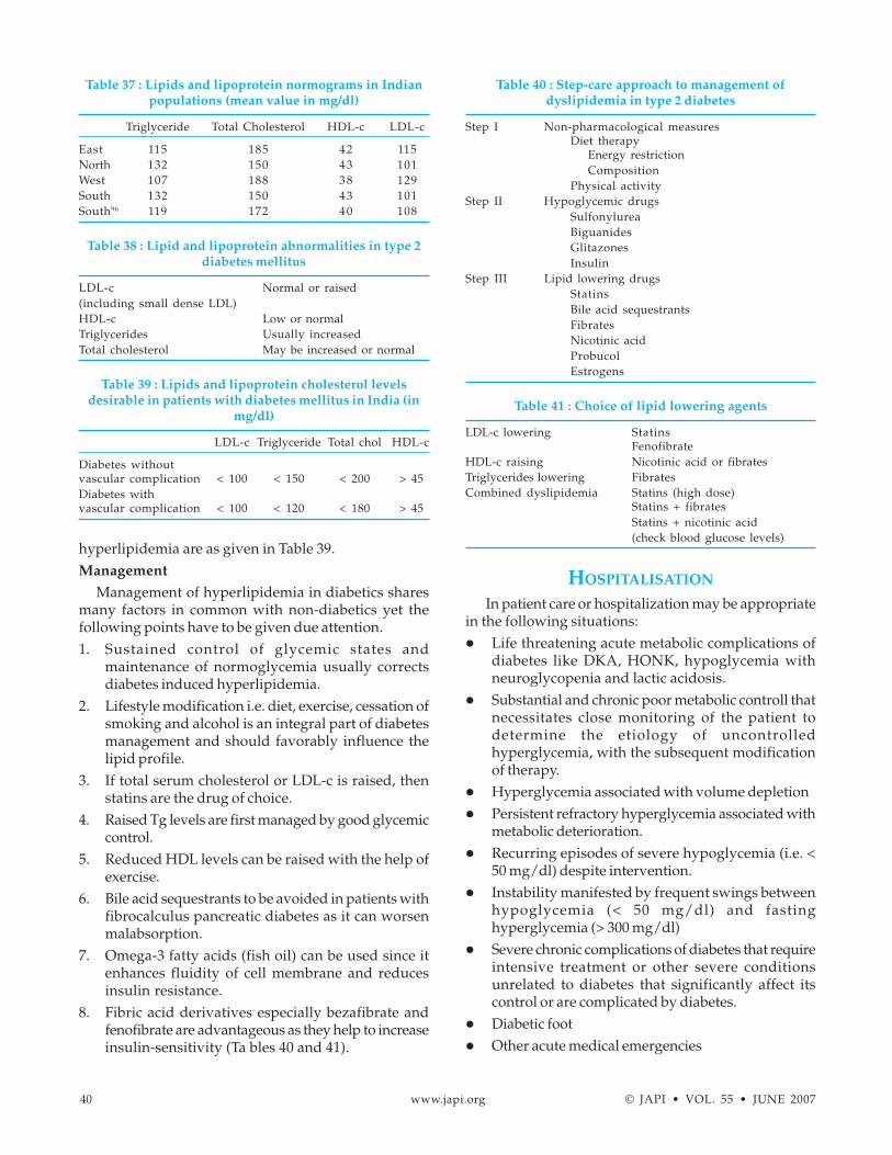
40 www.japi.org © JAPI • VOL. 55 • JUNE 2007
hyperlipidemia are as given in Table 39.Management
Management of hyperlipidemia in diabetics sharesmany factors in common with non-diabetics yet thefollowing points have to be given due attention.1. Sustained control of glycemic states and
maintenance of normoglycemia usually correctsdiabetes induced hyperlipidemia.
2. Lifestyle modification i.e. diet, exercise, cessation ofsmoking and alcohol is an integral part of diabetesmanagement and should favorably influence thelipid profile.
3. If total serum cholesterol or LDL-c is raised, thenstatins are the drug of choice.
4. Raised Tg levels are first managed by good glycemiccontrol.
5. Reduced HDL levels can be raised with the help ofexercise.
6. Bile acid sequestrants to be avoided in patients withfibrocalculus pancreatic diabetes as it can worsenmalabsorption.
7. Omega-3 fatty acids (fish oil) can be used since itenhances fluidity of cell membrane and reducesinsulin resistance.
8. Fibric acid derivatives especially bezafibrate andfenofibrate are advantageous as they help to increaseinsulin-sensitivity (Ta bles 40 and 41).
HOSPITALISATION
In patient care or hospitalization may be appropriatein the following situations:� Life threatening acute metabolic complications of
diabetes like DKA, HONK, hypoglycemia withneuroglycopenia and lactic acidosis.
� Substantial and chronic poor metabolic controll thatnecessitates close monitoring of the patient todetermine the etiology of uncontrolledhyperglycemia, with the subsequent modificationof therapy.
� Hyperglycemia associated with volume depletion� Persistent refractory hyperglycemia associated with
metabolic deterioration.� Recurring episodes of severe hypoglycemia (i.e. <
50 mg/dl) despite intervention.� Instability manifested by frequent swings between
hypoglycemia (< 50 mg/dl) and fastinghyperglycemia (> 300 mg/dl)
� Severe chronic complications of diabetes that requireintensive treatment or other severe conditionsunrelated to diabetes that significantly affect itscontrol or are complicated by diabetes.
� Diabetic foot� Other acute medical emergencies
Table 37 : Lipids and lipoprotein normograms in Indianpopulations (mean value in mg/dl)
Triglyceride Total Cholesterol HDL-c LDL-c
East 115 185 42 115North 132 150 43 101West 107 188 38 129South 132 150 43 101South96 119 172 40 108
Table 38 : Lipid and lipoprotein abnormalities in type 2diabetes mellitus
LDL-c Normal or raised(including small dense LDL)HDL-c Low or normalTriglycerides Usually increasedTotal cholesterol May be increased or normal
Table 39 : Lipids and lipoprotein cholesterol levelsdesirable in patients with diabetes mellitus in India (in
mg/dl)
LDL-c Triglyceride Total chol HDL-c
Diabetes withoutvascular complication < 100 < 150 < 200 > 45Diabetes withvascular complication < 100 < 120 < 180 > 45
Table 40 : Step-care approach to management ofdyslipidemia in type 2 diabetes
Step I Non-pharmacological measuresDiet therapy
Energy restrictionComposition
Physical activityStep II Hypoglycemic drugs
SulfonylureaBiguanidesGlitazonesInsulin
Step III Lipid lowering drugsStatinsBile acid sequestrantsFibratesNicotinic acidProbucolEstrogens
Table 41 : Choice of lipid lowering agents
LDL-c lowering StatinsFenofibrate
HDL-c raising Nicotinic acid or fibratesTriglycerides lowering FibratesCombined dyslipidemia Statins (high dose)
Statins + fibratesStatins + nicotinic acid(check blood glucose levels)
© JAPI • VOL. 55 • JUNE 2007 www.japi.org 41
Admission for Complications of Diabetes or forOther Acute Medical Emergencies
Chronic cardiovascular, neurological, renal and otherdiabetic complications may progress to the stage wherehospital admission is appropriate. In these situations,the need governing admission for the complication perse (e.g. management of end-stage renal disease) are theprimary guidelines for determining whether in patientcare is required. However, in applying such guidelines,the fact that diabetes is present must be considered; thismay result in patients requiring admission whootherwise might be managed on an outpatient basis. Thesame is true for other medical conditions (e.g. infarctions)and treatments (e.g. surgery, chemotherapy) in which1. Diabetes is a confounding factor,2. Rapid initiation of rigorous control of diabetes can
improve outcome (e.g. pregnancy),3. The primary medical problem or the therapeutic
intervention (e.g. large doses of glucocorticoid) cancause a major deterioration in diabetes control, or
4. There is acute onset of retinal, renal, neurological,or cardiovascular complications of diabetes.
PATIENT EDUCATION
Diabetes is a chronic disease that requires lifelongtreatment after the diagnosis is established. Theimportance of tight metabolic control has been broughtout by several recently concluded trials.The aim of the patient education is� Make patient knowledgeable about disease� To build positive attitude towards diabetes� To make him an active partner in the diabetes team� Patient empowerment� Self management� To enable the patient to make informed choice
Patient participation and compliance in the effectivemanagement of diabetes is crucial. A patient who knowsdifferent aspects of the disease will be better off thanthose who are not informed about the disease. In thisbackground patient education is an important andessential component of treatment. This education isimparted in different ways to each patient - this can begiven as information during each visit of the patient byhis physician or family doctor. It can be given in groupsand in diabetic camps where experiences of otherpatients can be exchanged and it will serve as animportant resource. The patient as well as the spouse,siblings and parents of a young patient should get thisinformation. The basic component of this educationshould stress and address the following issues.� What is diabetes and why control diabetes:
explaining briefly about the advantages of goodcontrol in preventing complications, in addition to
relief of symptoms and protection from infections.� How to achieve this control? By diet, exercise and
drugs. The importance and the cost-effectiveness ofdiet and exercise in management of diabetes shouldbe stressed.
� What is the basis of dietary management? Why andhow dietary changes are necessary and useful?
� Similarly importance of regular exercise, yoga andthe precautions to be taken in exercise planning.Integrating exercise in daily activities of theindividual.
� The timing of drug ingestion, insulin administration- timing, injection technique, sites of injection andstorage of insulin.
� Monitoring of plasma glucose levels and other co-morbid conditions.
� Information regarding hypoglycemia - how torecognize and correct and measures to preventrecurrence of hypoglycemia in future.
� Audio visual and other patient education materialcan be kept in the waiting room.
� Emphasis on personal hygiene, foot care.� General information as regards job, marriage and
child bearing.� Self monitoring of blood glucose training.� Role of family members in caring for the patient.� Sick day rules/fasting days.� Harmful effects of alcohol and smoking (tobacco in
any form) and advice regarding cessation.� Targets for control.� The role of mass media such as print media;
newspapers, electronic media, pharmaceuticalindustries, TV, internet and diabetic camps.
The physicians can provide this information at everyvisit to the patient. It may require around 8-10 min. In adiabetes clinic where dietitians, trained nurses, medico-social workers are available, such education can beimparted to the patient. Patient education is a continuousprocess and must be reinforced at every clinic visit of thepatient. (See Appendix 8 for patient information on diet,exercise, self injection, self monitoring, low bloodglucose, eyes and feet).
PREVENTION OF TYPE 2 DIABETES (SMALL 2LINES FROM IDPP RESULTS MAY BE GIVEN)
By the year 2025, diabetes mellitus above 20 years ofage is projected to increase by 35% the world over. Thismeans a diabetic population of 200 million by 2025compared with 135.3 million in 1995.7 The highestincrease will be for India - 59% and China - 68%. Ourcountry will have 57.2 million diabetics compared to19.4 million in 1995. Diabetes will mainly affect themiddle aged population (45-64 years).1,3,101 They will live

42 www.japi.org © JAPI • VOL. 55 • JUNE 2007
long enough to develop chronic diabetic complicationswith a strain on resources in managing the same. Withthis scenario as the background, any effort to preventdiabetes mellitus assumes significance. Type 2 diabetesdevelops as a continum starting with the fullycompensated insulin resistance, normoglycemic andasymptomatic state and progressing to impaired glucosetolerance and later frank diabetes. The basic metabolicdefect is insulin resistance, non-autoimmune mediatedbeta cell dysfunction leading to insulin secretory defectand increased hepatic glucose output.102,103 Identifyingthe individuals at greatest risk is essential in planningpreventive measures. Prevention of diabetes mellitus hasseveral windows of opportunity. They are divided intothree stages:
Primary prevention : To prevent the onset of diabetes.Secondary prevention : To prevent complications of
diabetes in those who are already diagnosed to havediabetes.
Tertiary prevention : To limit the damage caused bydiabetic complications in those who already have somedegree of complications.
Primary prevention is to prevent the onset of diabetesin high risk individuals (Ta ble 42). The aim should be toenhance insulin sensitivity by
� Lifestyle Modification : This involves diet and exercisewhich has been shown to be beneficial in severalstudies.104-106 The chance of developing diabetes isreduced by atleast 50%. The family members shouldreduce the consumption of refined carbohydrates, fat(particularly saturated fat) and alcohol and discontinuesmoking.
� Insulin Sensitizers : Some small studies have shownthe usefulness of sulfonylureas96 and phenformin. Theresults of Diabetes Prevention Program-2 and STOP trialhave recently been discussed at the EuropeanAssociation for the Study of Diabetes (EASD) meetingheld in September 2001. Intensive lifestyle modification,metformin and acarbose have been found to delay theonset of diabetes in impaired glucose tolerantindividuals.108,109 The intervention trial involvingtroglitazone had been discontinued.
� Avoiding diabetogenic drugs such as beta blockers,steroids, phenytoin, immunosupressants, thiazidediuretics, pentamidine and dilantin. SecondaryPrevention : The main objective should be to detect early
undiagnosed cases. The aim of the therapy should be toreverse the disease or to prevent the progression of thedisease. This is done with the help of diet, physicalactivity, drugs and life style modification process.
Tertiary Prevention : The aim is to prevent or limit thedamage caused by the disease in the established caseswhich can be achieved by diet, exercise, drugs, life stylemodification process and demand managementinterventions. Here one must monitor, controll and screenfor complications. The aim is to prevent, arrest or evenregress or reverse the complications of diabetes. Whentreating diabetes, physicians can readily identify theperson at risk of developing diabetes or havingundetected diabetes (the siblings, children of thepatients). mation that lifestyle modification can markedlyreduce the risk of developing diabetes can go a long wayin achieving the goal of prevention.
REF ER ENCES
1. Ramachandran A, Snehalatha C, et al. Rising prevalence ofNIDDM in an urban population in South India. Diabetologia1997;40:232-7.
2. Ramachandran A, Jali MV, et al. High prevalence of diabetesin an urban population in south India. Br Med J 1988;297:587-90.
3. Ramachandran A, Snehalatha C, et al. Prevalence of glucoseintolerance in Asian Indians : Urban-rural difference andsignificance of upper body adioposity. Diabetes Care1992;15:1348-55.
4. Ramachandran A, Snehalatha C, et al. Impact of urbanizationon the lifestyle and on the prevalence of diabetes in nativeAsian Indian population. Diab Res Clin Prac 1999;44:207-13.
4a. Gupta OP, et al. Prevalence of diabetes in India. Adv MetabolicDisorders 1978;9:147-65.
5. Ahuja MMS. Epidemiological studies in diabetes mellitus inIndia. In : Ahuja MMS (ed). Epidemiology of diabetesmellitus in developing countries, New Delhi : Interprint. 1979.
6. Ramachandran A, Snehalatha C, Kapur A, et al . Highprevalence of diabetes and impaired glucose tolerance inIndia : National Urban Diabetes Survey. Diabetologia2001;44:1094-101.
7. King H, Rewers M. WHO Ad Hoc Diabetes Reporting Group: Global estimates for prevalence of diabetes and impairedglucose tolerance in adults. Diabetes 1993;16: 157-77.
8. King H. Diabetes mellitus : a growing international healthcare problem. Int Diab Monitor 1997;9:1-6.
9. Mohan V, Alberti KGMM. Diabetes in the tropics. In :International Textbook of Diabetes Mellitus : Alberti P,Zimmet P, Defronzo RA, Keen H. 2nd edition. Wiley. 1997.
10. Mather HM, Keen H. The Southhall diabetes survey :Prevalence of known diabetes in Asians and Europeans. BrMed J 1985;291:1081-4.
11. Mohan V, Ramachandran A, et al. High prevalence of maturityonset diabetes of the young (MODY) among Indians. DiabetesCare 1985;8:371-4.
12. Asmal AC, Dayal B, et al. Non-insulin dependent diabetesmellitus with young age at onset in Blacks and Indians. S AfrMed J 1981;60:93-6.
13. Mohan Rema, Raj D, Mohan V. Prevalence of retinopathy atdiagnosis among type 2 diabetic patients attending a diabeticcentre in south India. Br J Ophthalmol 2000;84:1058-60.
14. Report of the Expert Committee on the Diagnosis and
Table 42 : High risk individuals
� Inheritence� Age 30 yrs or more� BMI > 24 kg/m2� Impaired glucose tolerance (IGT)� Impaired fasting glucose (IFG)� Women with bad obstetrical history/GDM� sHypertension� Dyslipidemia

© JAPI • VOL. 55 • JUNE 2007 www.japi.org 43
Classification of Diabetes Mellitus. The Expert Committeeon the Diagnosis and Classification on Diabetes Mellitus.Diabetes Care 1997;20:1183-97.
15. World Health Organization : Definition, diagnosis andclassification of diabetes mellitus and its complications :Report of a WHO consultation. Part I. Diagnosis andclassification of diabetes mellitus. Geneva, World HealthOrganization. 1999.
16. World Health Organization : Diabetes mellitus : report of aWHO study group. Geneva, World Health Organisation.Tech Rep Ser 1985;727.
17. Das S. Low body weight type 2 diabetes mellitus. J NutriEnv Med 1999;9:235-45.
18. Low body weight type 2 diabetes mellitus Ed. Das S : InTechnical series, Indian College of Physicians, Associationof Physicians of India. 1999.
19. Vascular complication of diabetes : Indian Data. In Advancesin Endocrinology, Metabolism and Diabetes. Delhi. 1994;2:33-8.
20. UK Prospective Diabetes Study (UKPD) group.Complications in newly diagnosed type 2 diabetic patientsand their association with different clinical and biochemicalrisk factors. Diabetes Research 1990;13:1-11.
21. Diabetes Control and Complications Trial research group.The effect of intensive treatment of diabetes on thedevelopment and progression of long-term complications ininsulin-dependent diabetes mellitus. N Engl J Med1993;329:979-86.
22. Ohkubo Y, Kishikawa H, Araki E, et al. Intensive insulintherapy prevents the progression of diabetic microvascularcomplications in Japanese patients with non-insulindependent diabetes mellitus : a randomized prospective 6year study. Diabetes Res Clin Pract 1995;28:103-17.
23. UK Prospective Diabetes Study Group : Intensive blood -glucose control with sulfonylureas or insulin compared withconventional treatment and risk of complications in patientswith type 2 diabetes (UKPDS 33). Lancet 1998;352:837-53.
24. UK Prospective Diabetes Study Group : Tight blood pressurecontrol and risk of macrovascular and microvascularcomplications in type 2 diabetes (UKPDS 38). BMJ1998;317:703-13.
25. Indian Hypertension Guidelines. 2000.
26. American Diabetes Association. Clinical practicerecommendations 2000. Diabetes Care 2000;23(suppl 1) : s131-s138.
26a. Ghotoorunissa. Requirements of dietary fats to meetnutritional needs to prevent the risk of atherosclerosis. AnIndian perspective. Indian J Med Res 1998;108:191-202.
27. Effect of intensive blood-glucose control with metformin oncomplications in overweight patients with type 2 diabetes(UKPDS 34). UK Prospective Diabetes Study. Lancet1998;352:854-65.
28. Biguanides, Basic aspects and clinical uses, In InternationalText book of Diabetes Mellitus KGMM Alberti, Zimmet PDefronzo RA. Eds. 2nd eds.
28a. Raghuram TC, Sharma RD, Sivakumar B, Sahay BK. Effectof Fenugreek seeds on glucose kinetics in type 2 diabetics.Phytotherapy Research 1994; 8 : 83-6.
28b. Sharma RD, Sarkar DK, Hazra B, et al. Hypolipidemic effectof fenugreek seeds : A chronic study in non-insulin dependentdiabetic patients. Phytother Res 1996; 10 : 332-4.
29. Katzung BG. Basic and Clinical Pharmacology, 7th Ed. 684-705.
30. Home PD. Insulin therapy : In International Text Book ofDiabetes Mellitus. Ed : Alberti, Zimmet and Defronzo. 899-
928.
31. Patient Education by American Diabetes Association. 32.Goldstein DE, Little RR, Lorenz RA, et al. Tests of glycemiain diabetes. Diabetes Care 1995;18:896-909.
33. American Diabetes Association. Standard of medical carefor patients with diabetes mellitus. Diabetes Care2000;23(Suppl 1 ):61-82.
34. Harris MI, Cowie CC, Howie LJ. Self-monitoring of bloodglucose by adults with diabetes in the United Statespopulation. Diabetes Care 1993;16:1116-23.
35. Diabetic ketoacidosis and hyperglycemic non-ketotic coma :In International Textbook of Diabetes Mellitus ed AlbertiZimmet and DeFronzo. 1997; 1215-29.
36. The DCCT Control and Complications Trial Research Group: Epidemiology of severe hypoglycemia in the Diabetes Controland Complications Trial. Am F Med 1991;90:450-9.
37. Davis EA, Keating B, Byrne GC, et al. Hypoglycemia incidenceand clinical predictors in a large population based sampleof children and adolescents with IDDM. Diabetes Care 1997;20 : 22-5.
38. Bolli GB. Counterregulatory mechanism of insulin-inducedhypoglycemia in humans : relevance to the problem ofintensive treatment of IDDM. F Pediatr Endocrinol Metab1998;11(Suppl 1):103-15.
39. Hypoglycemia. In International Text book of diabetesmellitus edited by Alberti, Zimmet and DeFronzo. 1997;1193-214.
40. Jacoby RM, Nesto RW. Acute myocardial infarction in thediabetic patient : pathophysiology, Clinical course andprognosis. J Am Coll Cardiol 1992;20:736-44.
41. Serrano-Rios M, Perez A, Saban Ruiz J. Cardiac complicationsin diabetes. In : World Book of Diabetes in Practice. PrincetonNJ : Elsevier. 1986; 2 : 169-78.
42. Sainani GS, Rajesh S. Diabetes mellitus and cardiovasculardisease. In : Current concepts in diabetes mellitus. SainaniGS Ed. 1993; 73-8.
43. Bhatia SG, Sainani GS, Nayak NJ, et al. Valsalva manoeuvreas a test of autonomic neuropathy in diabetes mellitus. JAssoc Physicians India 1976;24:89-93.
44. Solar N, Bennet M, Pentecost B, et al. Symptomaticmyocardial infarction without chest pain; prevalence andclinical course. Am J Cardiol 1977;40:498-503.
46. Czyzk A, Krolewski A, Szablowskas, et al. Clinical course ofmyocardial infarction among diabetic patients. Diabetic Care1980;4:526-9.
47. Kannel WB, McGee DL. Diabetes and glucose tolerance asrisk factors for cardiovascular disease. The Framinghamstudy. Diabetes Care 1979; 2: 120-31.
48. Garcia MJ, MC Namara PM, Gordon T, Kannel WB. Morbidityand mortality in diabetics in the Framingham population.Diabetes 1974;23:105-16.
49. Raheja BS. Heart disease in diabetes : Aetiopathogenesis. JAssoc Physicians India 1980;28:81-90.
50. Kenneth W, et al. Diabetic retinopathy should not be acontrindication to thrombolytic therapy for acute myocardialinfarction : Review of ocular hemorrhage incidence andlocation in the GUSTO-1 trial. J Am Coll Cardiol 1997;30:1606-10.
51. Malmberg K, Ryden L, Hamsten A, Herlitz J, WaldenstromA, Wedel H. Effects of insulin treatment on cause-specificone-year mortality and morbidity in diabetic patients withacute myocardial infarction. DIGAMI study group. DiabetesInsulin-Glucose in Acute Myocardial Infarction. Eur Heart J1996;17:1337-44.
52. Padma MV, Bajaj JS. Diabetics in stroke. In : Recent concept

44 www.japi.org © JAPI • VOL. 55 • JUNE 2007
in stroke. Pb Ind Coll Phy Ed Bansal BC. Mumbai. 1999; 79.
53. Kennel WB. Current status of epidemiology of brain infarctionassociated with occlusive arterial disease. Stroke 1971;2:295-318K.
54. Palumbo PJ, Elveback IR, Whisnant JP. Neurolgoiccomplications of diabetes mellitus : transient ischaemicattack, stroke and peripheral neuropathy. Adv Neurol1978;19:593-601.
55. Abott RD, Donative RP, MacMohan SW, et al. Diabetics andthe risk of stroke : the Honolulu heart programme. JAMA257:949-52.
56. Colwell JA. Aspirin therapy in diabetes (Technical Review).Diabetes Care 1997;20:1767-71.
57. CAPRIE Steering Committee. A randomised, blinded trial ofclopidogrel versus aspirin in patients at risk of ischaemicevents (CAPRIE). Lancet 1996;348:1329-39.
58. Diabetic nephropathy In : Textbook of Diabetes Ed. JohnPickup and Gareth Williams.
59. Ramachandran A, et al. Prevalence of vascular complicationsand their risk factors in type 2 diabetes. J Assoc PhysiciansIndia 1999;47:1152-6.
59a. Rema M, Ponnaiya M, Mohan V. Prevalence of retinopathy innon-insulin dependent diabetes mellitus at a diabetes centerin southern India. Diab Res Clin Prac 1996;34:29-36.
60. ADA. Clinical Practice Recommendations. Diabetes Care2001;23(suppl 1):s69-s73.
61. Diabetic eye disease In : Textbook of Diabetes. Ed. JohnPickup and Gareth William. 557-612.
62. Diabetic neuropathy In : Textbook of Diabetes. Ed. JohnPickup and Gareth William. 613-50.
63. Diabetic foot. In : Textbook of Diabetes, Ed, John Pickupand Gareth William. 735-44.
64. Most RS, Sinnock P. The Epidemiology of lower limbamputation in diabetic individuals. Diabetes Care 1983;6:87-91.
65. Complications of Diabetes in Indian Scenario In : NIDUS ‘99Das S. Ed. 1999.
66. Johnston CLW. Infection. In : Pickup JC, Williams G Eds.Textbook of Diabetes 2nd ed. London : Blackwell Science.1997; 70.1-70.13.
67. Handwerger BS. The immunology of diabetes mellitus. In :Samter M, Ed. Immunological Disorders 4th ed., Boston :Brown and Co. 1998; 1965-1808.
68. Josh N, Caputo GM, Weitekamp MR, Karchmer AW.Infections in patients with diabetes mellitus. N Engl J Med2000;341:1906-12.
69. Marks HH, Krall LP. Onset, prognsois and mortality indiabetes mellitus. In : Marble A, White P, et al Joslin’s DiabetesMellitus 12th ed. Philadelphia : Lea and Febiger. 1998; 209-54.
70. Sentochnik DE, Eliopoulos GM. Infection and diabetes. InKahn CR, Weir GC, Eds. Joslin’s Diabetes Mellitus 13th ed.New Delhi : BI Waverly. 1998; 867-88.
71. Rieder HC, Cauthen GM, Comstock GW, Snider DE Jr.Epidemiology of tuberculosis in the US. Epidemiol Rev1989;11:79-98.
72. Wilson RM. Infection and diabetes mellitus. In : Pickup JC,Williams G Eds. Textbook of Diabetes. Oxford : BlackwellScientific Publication. 1991; 813-19.
73. Gill GV, Huddle KR, Krige LP. Intensive health screening ofyoung black diabetics. S Afr Med J 1984;65:815-16.
74. Atkin SL, Masson EA, Bodmer CW, Walker BA, White MC.Increased insulin requirement in a patient with type 1diabetes on rifampicin (letter). Diabet Med 1993;10:392.
75. The care of the diabetic patient during surgery In :International textbook of diabetes mellitus. Alberti, Zimmet,DeFronzo, Eds. 1244-53.
76. Surgery and Diabetes Mellitus In : Textbook of diabetes.John Pickup, Gareth Williams Eds. 820-26.
77. Kajos SL, Buchanan TA. Current concepts; GetationalDiabetes Mellitus. N Engl J Med 1999;341:1749-56.
78. American Diabetes Association. Clinical practice guidelines,Gestational Diabetes Mellitus. Diabetes Care 2000;23(suppl1):32-7.
79. Veciana M, Major CA, Morgan MA, Asrat T, Toohey JS, LienJM, Evans AT. Postprandial versus preprandial bloodglucose, monitoring in women with gestational diabetesmellitus, requiring insulin therapy. N Engl J Med1995;333:1237-41.
80. Ryan EA. Long term outlook for women with diabetes inpregnancy, Abstract of 60th Scientific Sessions of theAmerican Diabetes Association, San Antonio, Texas. 2000.
81. Diabetes and Traveling. Dr. Shekhar Shah in Diabetes madeeasy. Swapna Publishing Houe. Guwahati. 2000; 138-39.
82. Kozan GP, Hollerorth HJ, Gordon C, Halford B, Stevnis D.Diabetes Teaching Guide. Joslin Diabetes Centre. 1986.
83. Ahuja MMS, Sharma R. Diabetic Bhoj. New Delhi. BIPublications. 1984; 63.
84. Dabalea D, Pettitt DJ, Jones KL, Arslanian SA. Endocrinol.Metab Clin North Am 1999;28:709.
85. Forgot-Campagna A, Burrows NR, Williamson DF. Thepublic health epidemiology of type 2 diabetes in 371 childrenand adolescents : a case study of American Indianadolescents in the South Western United States. ClinicaChimica Acta 1999;286:81-95.
86. Mohan V, Ramachandran A, Snehalatha C. High prevalenceof maturity onset diabetes of young among Indians. DiabetesCare 1985;8:371-84.
87. Ashida TS, Das AK, Sridhar MG. Investigation on the clinicaland biochemical heterogenecity of early onset type 2 diabetesmellitus. PG dissertation, Pondicherry, University. 1997-2000.
88. Mohan V, Alberti KGMM. Diabetes in the tropics. In :International Text book of Diabetes Mellitus Alberti KGMM,Zimeet P, Defronzo RA, eds. 2nd ed. 1997; 171-88.
89. Sahay BK, Das AK, Mathew T, Varma R. Diabetes in young.In : Postgraduate Medicine, Das AK. Ed. 2001; 15 : 283-7.
90. Paul Anand M. Management of Hypertension with associateddiseases. Das AK, Ed. Medicine update. Association ofPhysicians of India 1988;356-61.
91. Rau NR, Acharya RV, Shah S. Incidence of diabeticcomplications in newly detected cases of NIDDM. NovoNordisk diabetes update. 1999; 35-6.
92. Hansson L, et al. Effects of intensive blood pressure loweringand low dose aspirin on patients with hypertension principalresults of the hypertension optimal; treatment (HOT)randomized trial. Lancet 1998;351:1755-62.
93. Heart Outcomes Prevention Evaluation (HOPE) StudyInvestigators. Effect of ramipril on cardiovascular andmicrovascular outcomes in people with diabetes mellitus :Results of the HOPE Study and MICRO-HOPE sub study.Lancet 2000;355:253-9.
94. The joint National Committee on Prevention, Detection,Evaluation and Treatment of High Blood Pressure. The sixthreport of the Joint National Committee on Prevention,Detection, Evaluation and Treatment of High Blood Pressure.Arch Intern Med 1997;157:2413-36.
95. Das S. Diabetes and Dyslipidemia. Lipid India 2001;7-10.
96. Mohan V, Deepa R, et al. Prevalence of coronary artery disease
© JAPI • VOL. 55 • JUNE 2007 www.japi.org 45
and its relationship to lipids in a selected population inSouth India. The Chennai Urban population study (CUPSNo. 5). J Am Coll Cardiol 2001;38:682-7.
97. Management of diabetic dyslipidemia in adults with diabetes,102-109 in ADA : clinical practice recommendations 2001,Diabetes Care Jan 2001.
98. Shah P, Karmarkar MG. Lipoprotein disorders in non-insulindependent diabetes mellitus. Int Diabetes Dev Countries1991;II:25-9.
99. Zargar AH, Wandrow FA, Wadhwa HB, et al. Serum lipidprofile in non-insulin dependent diabetes mellitus associatedwith obesity. Int J Diabetes Dev Countries 1995;14:9-13.
100. Chandalia HB, Lamba PS. Dyslipidemia and diabetesmellitus. Lipid India 1994;4-16.
101. King H, auibert RE, Herman WH. Global burden of diabetes,1995-2000. Prevalence, numerical estimates and projections.Diabetes Care 1998;21:1414-31.
102. Eriksson J, Franssila-Kaullunki A, Saloranta CM, Widen E,Schalin C, Groop L. Early metabolic defects in persons atincreased risk for non-insulin dependent diabetes mellitus.
N Engl J Med 1989;321:337-43.
103. Leahy JL. Natural History of beta cell dysfunction in NIDDM.Diabetes Care 1990;13:992-1010.
104. Eriksson KF, Lindagarde F. Prevention of type 2 diabetesmellitus by diet and physical exercise, the 6 year Malmofeasibility study. Diabetologia 1991;34:891-8.
105. Huyh, et al. Effects of diet and exercise in preventing NIDDMin people with impaired glucose tolerance. The Du Qing IGTand diabetes study. Diabetes Care 1997;20:537-44.
106. Ramachandran A. Prevention of diabetes - the Indian angle.Int J Diab Dev Countries 1998;18:113-7.
107. Sartox G, Scherten B, Carlstron s, Melanda A, Norden A,Persson G. Ten years follow of subjects with IGT. Preventionof diabetes by tolbutamide and diet regulation. Diabetes1980;29:41-9.
108. Bailey C. The Diabetes Prevention Programme : HeadlineResults. Br J Diab Vas Dis 2001;1:62-4.
109. Trial presented at the EASD, 2001. 37th annual Meeting, 9-13th September.
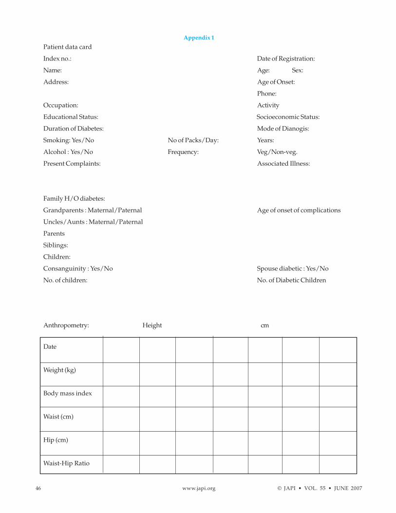
46 www.japi.org © JAPI • VOL. 55 • JUNE 2007
Appendix 1
Patient data card
Index no.: Date of Registration:
Name: Age: Sex:
Address: Age of Onset:
Phone:
Occupation: Activity
Educational Status: Socioeconomic Status:
Duration of Diabetes: Mode of Dianogis:
Smoking: Yes/No No of Packs/Day: Years:
Alcohol : Yes/No Frequency: Veg/Non-veg.
Present Complaints: Associated Illness:
Family H/O diabetes:
Grandparents : Maternal/Paternal Age of onset of complications
Uncles/Aunts : Maternal/Paternal
Parents
Siblings:
Children:
Consanguinity : Yes/No Spouse diabetic : Yes/No
No. of children: No. of Diabetic Children
Anthropometry: Height cm
Date
Weight (kg)
Body mass index
Waist (cm)
Hip (cm)
Waist-Hip Ratio

© JAPI • VOL. 55 • JUNE 2007 www.japi.org 47
Clinical Examination
Date
Pulse
Peripheral pulses
Blood Pressure supine
Blood Pressure Standing
Ankle pressure
Skin lesions
Heart
Lungs
Abdomen
Cranial nerves
Motor system
Sensory system
Autonomic functions
fundus
Investigations
Date
Complete blood count
Fasting blood glucose
Erythrocyte sedimentation rate
Urea
Creatinine
Triglycerides
High density lipoprotein
Low density lipoprotein
Very low density lipoprotein
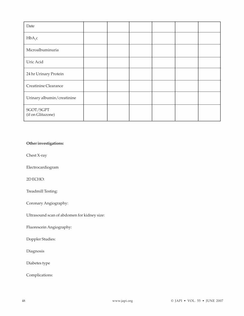
48 www.japi.org © JAPI • VOL. 55 • JUNE 2007
Date
HbA1c
Microalbuminuria
Uric Acid
24 hr Urinary Protein
Creatinine Clearance
Urinary albumin/creatinine
SGOT/SGPT(if on Glitazone)
Other investigations:
Chest X-ray
Electrocardiogram
2D ECHO:
Treadmill Testing:
Coronary Angiography:
Ultrasound scan of abdomen for kidney size:
Fluorescein Angiography:
Doppler Studies:
Diagnosis
Diabetes type
Complications:

© JAPI • VOL. 55 • JUNE 2007 www.japi.org 49
Date/year Onset
Hypertension
Nephropathy
Coronary artery disease
Cardiomyopathy
Neuropathy
Cerebrovascular disease
Retinopathy
Peripheral vascular disease
Ketosis/Hyperosmolarnon-ketotic lactic acidosis.
Treatment advised:
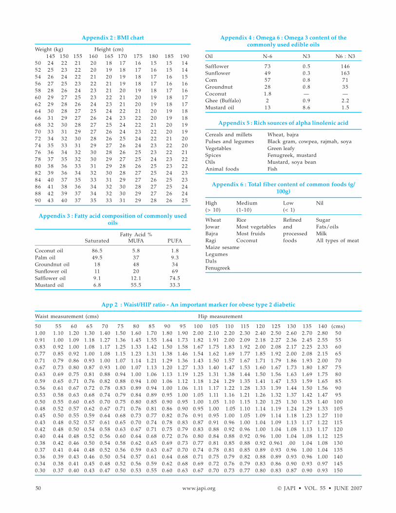
50 www.japi.org © JAPI • VOL. 55 • JUNE 2007
App 2 : Waist/HIP ratio - An important marker for obese type 2 diabetic
Waist measurement (cms) Hip measurement
50 55 60 65 70 75 80 85 90 95 100 105 110 115 120 125 130 135 140 (cms)1.00 1.10 1.20 1.30 1.40 1.50 1.60 1.70 1.80 1.90 2.00 2.10 2.20 2.30 2.40 2.50 2.60 2.70 2.80 500.91 1.00 1.09 1.18 1.27 1.36 1.45 1.55 1.64 1.73 1.82 1.91 2.00 2.09 2.18 2.27 2.36 2.45 2.55 550.83 0.92 1.00 1.08 1.17 1.25 1.33 1.42 1.50 1.58 1.67 1.75 1.83 1.92 2.00 2.08 2.17 2.25 2.33 600.77 0.85 0.92 1.00 1.08 1.15 1.23 1.31 1.38 1.46 1.54 1.62 1.69 1.77 1.85 1.92 2.00 2.08 2.15 650.71 0.79 0.86 0.93 1.00 1.07 1.14 1.21 1.29 1.36 1.43 1.50 1.57 1.67 1.71 1.79 1.86 1.93 2.00 700.67 0.73 0.80 0.87 0.93 1.00 1.07 1.13 1.20 1.27 1.33 1.40 1.47 1.53 1.60 1.67 1.73 1.80 1.87 750.63 0.69 0.75 0.81 0.88 0.94 1.00 1.06 1.13 1.19 1.25 1.31 1.38 1.44 1.50 1.56 1.63 1.69 1.75 800.59 0.65 0.71 0.76 0.82 0.88 0.94 1.00 1.06 1.12 1.18 1.24 1.29 1.35 1.41 1.47 1.53 1.59 1.65 850.56 0.61 0.67 0.72 0.78 0.83 0.89 0.94 1.00 1.06 1.11 1.17 1.22 1.28 1.33 1.39 1.44 1.50 1.56 900.53 0.58 0.63 0.68 0.74 0.79 0.84 0.89 0.95 1.00 1.05 1.11 1.16 1.21 1.26 1.32 1.37 1.42 1.47 950.50 0.55 0.60 0.65 0.70 0.75 0.80 0.85 0.90 0.95 1.00 1.05 1.10 1.15 1.20 1.25 1.30 1.35 1.40 1000.48 0.52 0.57 0.62 0.67 0.71 0.76 0.81 0.86 0.90 0.95 1.00 1.05 1.10 1.14 1.19 1.24 1.29 1.33 1050.45 0.50 0.55 0.59 0.64 0.68 0.73 0.77 0.82 0.76 0.91 0.95 1.00 1.05 1.09 1.14 1.18 1.23 1.27 1100.43 0.48 0.52 0.57 0.61 0.65 0.70 0.74 0.78 0.83 0.87 0.91 0.96 1.00 1.04 1.09 1.13 1.17 1.22 1150.42 0.48 0.50 0.54 0.58 0.63 0.67 0.71 0.75 0.79 0.83 0.88 0.92 0.96 1.00 1.04 1.08 1.13 1.17 1200.40 0.44 0.48 0.52 0.56 0.60 0.64 0.68 0.72 0.76 0.80 0.84 0.88 0.92 0.96 1.00 1.04 1.08 1.12 1250.38 0.42 0.46 0.50 0.54 0.58 0.62 0.65 0.69 0.73 0.77 0.81 0.85 0.88 0.92 0.961 .00 1.04 1.08 1300.37 0.41 0.44 0.48 0.52 0.56 0.59 0.63 0.67 0.70 0.74 0.78 0.81 0.85 0.89 0.93 0.96 1.00 1.04 1350.36 0.39 0.43 0.46 0.50 0.54 0.57 0.61 0.64 0.68 0.71 0.75 0.79 0.82 0.88 0.89 0.93 0.96 1.00 1400.34 0.38 0.41 0.45 0.48 0.52 0.56 0.59 0.62 0.68 0.69 0.72 0.76 0.79 0.83 0.86 0.90 0.93 0.97 1450.30 0.37 0.40 0.43 0.47 0.50 0.53 0.55 0.60 0.63 0.67 0.70 0.73 0.77 0.80 0.83 0.87 0.90 0.93 150
Appendix 2 : BMI chart
Weight (kg) Height (cm)145 150 155 160 165 170 175 180 185 190
50 24 22 21 20 18 17 16 15 15 1452 25 23 22 20 19 18 17 16 15 1454 26 24 22 21 20 19 18 17 16 1556 27 25 23 22 21 19 18 17 16 1658 28 26 24 23 21 20 19 18 17 1660 29 27 25 23 22 21 20 19 18 1762 29 28 26 24 23 21 20 19 18 1764 30 28 27 25 24 22 21 20 19 1866 31 29 27 26 24 23 22 20 19 1868 32 30 28 27 25 24 22 21 20 1970 33 31 29 27 26 24 23 22 20 1972 34 32 30 28 26 25 24 22 21 2074 35 33 31 29 27 26 24 23 22 2076 36 34 32 30 28 26 25 23 22 2178 37 35 32 30 29 27 25 24 23 2280 38 36 33 31 29 28 26 25 23 2282 39 36 34 32 30 28 27 25 24 2384 40 37 35 33 31 29 27 26 25 2386 41 38 36 34 32 30 28 27 25 2488 42 39 37 34 32 30 29 27 26 2490 43 40 37 35 33 31 29 28 26 25
Appendix 3 : Fatty acid composition of commonly usedoils
Fatty Acid %Saturated MUFA PUFA
Coconut oil 86.5 5.8 1.8Palm oil 49.5 37 9.3Groundnut oil 18 48 34Sunflower oil 11 20 69Safflower oil 9.1 12.1 74.5Mustard oil 6.8 55.5 33.3
Appendix 4 : Omega 6 : Omega 3 content of thecommonly used edible oils
Oil N-6 N3 N6 : N3
Safflower 73 0.5 146Sunflower 49 0.3 163Corn 57 0.8 71Groundnut 28 0.8 35Coconut 1.8 — —Ghee (Buffalo) 2 0.9 2.2Mustard oil 13 8.6 1.5
Appendix 5 : Rich sources of alpha linolenic acid
Cereals and millets Wheat, bajraPulses and legumes Black gram, cowpea, rajmah, soyaVegetables Green leafySpices Fenugreek, mustardOils Mustard, soya beanAnimal foods Fish
Appendix 6 : Total fiber content of common foods (g/100g)
High Medium Low Nil(> 10) (1-10) (< 1)
Wheat Rice Refined SugarJowar Most vegetables and Fats/oilsBajra Most fruids processed MilkRagi Coconut foods All types of meatMaize sesameLegumesDalsFenugreek