Antioxidant, anti-inflammatory, and hepatoprotective...
8
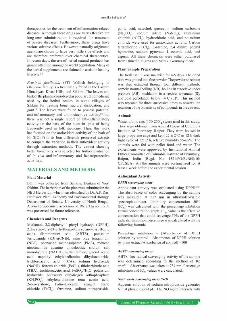
Journal of Pharmacy Research | Vol 11 • Issue 8 • 2017 983 Antioxidant, anti-inflammatory, and hepatoprotective activity of Fraxinus floribunda bark and the influence of extraction process on their bioactivity Arunika Subba 1 , Bhaskar Dutta 2 , Ram Kumar Sahu 3 , Palash Mandal 1 * INTRODUCTION Plants have played a significant role in human health since ancient times. They are known to possess various biological effects including antioxidant, antitumor, antidiabetic, anti-inflammatory, hepatoprotective properties, etc. Recent trend in medicine looks forward for natural sources for the treatment of various ailments. Traditional medicine is supported by various researchers based on their therapeutic potential with less side effects and cost effectiveness. Plants are the source of natural antioxidants which can control oxidative stress resulting from the generation of free radicals. Oxidative stress if not controlled may cause various harmful diseases such as neurodenerative diseases (Alzheimer’s and Parkinson’s disease), arthritis, cancer, rheumatoid. [1,2] Metabolism of certain commonly used drugs such as paracetamol produce free radicals, which induces liver damage. [3,4] Since there are limited treatment and prevention for liver diseases, it is considered to be one of the most serious health problems in the world. [5] Inflammation is another least noticed health issue that is of big concern. It is a defensive response of a body against stress that is characterized by redness, pain, heat, and swelling along with loss of function in the injured area. However, if inflammation is left unchecked, it may lead to onset of several diseases such as rheumatoid arthritis, vasomotor rhinorrhea, and atherosclerosis. It has been observed that several anti-inflammatory, neuroprotective, digestive, antinecrotic, and hepatoprotective drugs have recently been shown to have an antioxidant or free radical scavenging potential as part of their activity. [6] Most of the modern medicines belong to steroidal or non-steroidal anti-inflammatory chemical ABSTRACT Objective: To observe the influence of extraction processes on the bioactivity of Fraxinus floribunda (FF) bark on the basis of antioxidant activity and assess the in vivo anti-inflammatory with hepatoprotective activity in the extracts which has showed better antioxidant potential. Materials and Methods: Antioxidant activity on the basis of phytochemical estimation, free radical scavenging, and antilipid peroxidation activity. Anti-inflammatory activity was observed through carrageenan- induced paw edema, and hepatoprotective effect was studied using carbon tetrachloride induced liver damage in Wister albino rats. Results: Autoclave boiled extract showed highest antioxidant activity in assays of total phenol content (26.98 ± 11.22 mg gallic acid equivalent/g fresh weight tissue), 2,2-diphenyl-1-picryl hydrazyl (0.24 ± 3.65 mg/ml), etc. The same extract inhibited mice paw edema by 55% which was significant after 2 h (P < 0.05) showing anti-inflammatory activity. Lowering of serum glutamic oxaloacetic transaminase, serum glutamic pyruvic transaminase, anti-lipid peroxidation, and bilirubin levels showed hepatoprotective activity against standard formulation Liv-52. Conclusion: The bark of FF is a potential source of antioxidants with anti-inflammatory and hepatoprotective activity. Further research should be done to explore and validate its bioactivity. KEY WORDS: Anti-inflammatory, Antioxidants, Fraxinus floribunda, Hepatoprotection 1 Department of Botany, Plant Physiology and Pharmacognosy Research Laboratory, University of North Bengal, Rajarammohanpur, Siliguri, West Bengal, India, 2 Department of Pharmacology, Dr. B C Roy College of Pharmacy and AHS, Meghnad Saha Sarani, Durgapur, West Bengal, India, 3 Department of Pharmacognosy, Columbia Institute of Pharmacy, Tekari, Raipur, Chattisgarh, India *Corresponding author: Palash Mandal, Department of Botany, Plant Physiology and Pharmacognosy Research Laboratory, University of North Bengal, Rajarammohanpur, Siliguri, West Bengal, India. Phone: +91-9434886123. E-mail: [email protected] Received on: 12-06-2017; Revised on: 26-07-2017; Accepted on: 10-08-2017 Access this article online Website: jprsolutions.info ISSN: 0974-6943 Research Article
Transcript of Antioxidant, anti-inflammatory, and hepatoprotective...
Journal of Pharmacy Research | Vol 11 • Issue 8 • 2017 983
Antioxidant, anti-inflammatory, and hepatoprotective activity of Fraxinus floribunda bark and the influence of extraction process on their bioactivityArunika Subba1, Bhaskar Dutta2, Ram Kumar Sahu3, Palash Mandal1*
INTRODUCTIONPlants have played a significant role in human health since ancient times. They are known to possess various biological effects including antioxidant, antitumor, antidiabetic, anti-inflammatory, hepatoprotective properties, etc. Recent trend in medicine looks forward for natural sources for the treatment of various ailments. Traditional medicine is supported by various researchers based on their therapeutic potential with less side effects and cost effectiveness. Plants are the source of natural antioxidants which can control oxidative stress resulting from the generation of free radicals. Oxidative stress if not controlled may cause various harmful diseases such as neurodenerative diseases (Alzheimer’s and
Parkinson’s disease), arthritis, cancer, rheumatoid.[1,2] Metabolism of certain commonly used drugs such as paracetamol produce free radicals, which induces liver damage.[3,4] Since there are limited treatment and prevention for liver diseases, it is considered to be one of the most serious health problems in the world.[5] Inflammation is another least noticed health issue that is of big concern. It is a defensive response of a body against stress that is characterized by redness, pain, heat, and swelling along with loss of function in the injured area. However, if inflammation is left unchecked, it may lead to onset of several diseases such as rheumatoid arthritis, vasomotor rhinorrhea, and atherosclerosis. It has been observed that several anti-inflammatory, neuroprotective, digestive, antinecrotic, and hepatoprotective drugs have recently been shown to have an antioxidant or free radical scavenging potential as part of their activity.[6] Most of the modern medicines belong to steroidal or non-steroidal anti-inflammatory chemical
ABSTRACT
Objective: To observe the influence of extraction processes on the bioactivity of Fraxinus floribunda (FF) bark on the basis of antioxidant activity and assess the in vivo anti-inflammatory with hepatoprotective activity in the extracts which has showed better antioxidant potential. Materials and Methods: Antioxidant activity on the basis of phytochemical estimation, free radical scavenging, and antilipid peroxidation activity. Anti-inflammatory activity was observed through carrageenan-induced paw edema, and hepatoprotective effect was studied using carbon tetrachloride induced liver damage in Wister albino rats. Results: Autoclave boiled extract showed highest antioxidant activity in assays of total phenol content (26.98 ± 11.22 mg gallic acid equivalent/g fresh weight tissue), 2,2-diphenyl-1-picryl hydrazyl (0.24 ± 3.65 mg/ml), etc. The same extract inhibited mice paw edema by 55% which was significant after 2 h (P < 0.05) showing anti-inflammatory activity. Lowering of serum glutamic oxaloacetic transaminase, serum glutamic pyruvic transaminase, anti-lipid peroxidation, and bilirubin levels showed hepatoprotective activity against standard formulation Liv-52. Conclusion: The bark of FF is a potential source of antioxidants with anti-inflammatory and hepatoprotective activity. Further research should be done to explore and validate its bioactivity.
KEY WORDS: Anti-inflammatory, Antioxidants, Fraxinus floribunda, Hepatoprotection
1Department of Botany, Plant Physiology and Pharmacognosy Research Laboratory, University of North Bengal, Rajarammohanpur, Siliguri, West Bengal, India, 2Department of Pharmacology, Dr. B C Roy College of Pharmacy and AHS, Meghnad Saha Sarani, Durgapur, West Bengal, India, 3Department of Pharmacognosy, Columbia Institute of Pharmacy, Tekari, Raipur, Chattisgarh, India
*Corresponding author: Palash Mandal, Department of Botany, Plant Physiology and Pharmacognosy Research Laboratory, University of North Bengal, Rajarammohanpur, Siliguri, West Bengal, India. Phone: +91-9434886123. E-mail: [email protected]
Received on: 12-06-2017; Revised on: 26-07-2017; Accepted on: 10-08-2017
Access this article online
Website: jprsolutions.info ISSN: 0974-6943
Research Article
Arunika Subba et al.
Journal of Pharmacy Research | Vol 11 • Issue 8 • 2017984
therapeutics for the treatment of inflammation-related diseases. Although these drugs are very effective but long-term administration is required for treatment of severe diseases. Furthermore, these drugs have various adverse effects. However, naturally originated agents are shown to have very little side effects and are therefore preferred over chemical therapeutics. In recent days, the use of herbal natural products has gained attention among the world population. Many of the herbal supplements are claimed to assist in healthy lifestyle.[7]
Fraxinus floribunda (FF) Wallich belonging to Oleaceae family is a tree mainly found in the Eastern Himalayas, Khasi Hills, and Sikkim. The leaves and bark of the plant is considered as popular ethnomedicine used by the herbal healers in some villages of Sikkim for treating bone fracture, dislocation, and gout.[8] The leaves were found to possess potential anti-inflammatory and antinociceptive activity[9] but there was not a single report of anti-inflammatory activity on the bark of the plant in spite of being frequently used in folk medicine. Thus, this work has focused on the antioxidant activity of the bark of FF (BOFF) in its four differently processed extracts to compare the variation in their antioxidant activity through extraction methods. The extract showing better bioactivity was selected for further evaluation of in vivo anti-inflammatory and hepatoprotective activities.
MATERIALS AND METHODSPlant MaterialBOFF was collected from Sankhu, Dentam of West Sikkim. The herbarium of the plant was submitted in the NBU Herbarium which was identified by Dr. A.P. Das, Professor, Plant Taxonomy and Environmental Biology, Department of Botany, University of North Bengal. A voucher specimen, accession no. 9632/Tag no E.S.03 was preserved for future reference.
Chemicals and ReagentsMethanol, 2,2-diphenyl-1-picryl hydrazyl (DPPH), 2,2-azino-bis-(3-ethylbenzothiazoline-6-sulfonic acid) diammonium salt (ABTS), potassium ferricyanide (K3Fe(CN)6), nitro blue tetrazolium (NBT), phenazine methosulphate (PMS), reduced nicotinamide adenine dinucleotide sodium salt monohydrate (NADH), sulfanilamide, glacial acetic acid, naphthyl ethylenediamine dihydrochloride, trichloroacetic acid (TCA), sodium hydroxide (NaOH), ferrous chloride (FeCl2), thiobarbituric acid (TBA), trichloroacetic acid, FeSO4.7H2O, potassium hydroxide, potassium dihydrogen orthophosphate (KH2PO4), ethylene-diamine tetra acetic acid, 2-deoxyribose, Folin–Ciocalteu reagent, ferric chloride (FeCl3), ferrozine, sodium nitroprusside,
gallic acid, catechol, quercetin, sodium carbonate (Na2CO3), sodium nitrite (NaNO2), aluminium chloride (AlCl3), hydrochloric acid, and potassium chloride were used for antioxidant activity. Carbon tetrachloride (CCl4), L-alanine, 2,4 dinitro phenyl hydrazine, sodium pyruvate, L-aspartic acid, and aspirin. All these chemicals were either purchased from Himedia, Sigma and Merck, Germany made.
Plant Sample PreparationThe fresh BOFF was sun dried for 4-5 days. The dried bark was ground into fine powder. The powder specimen was then extracted through four different methods, namely, normal boiling (NB), boiling in autoclave under pressure (AB), soxhlation in a soxhlet apparatus (S), and cold percolation below −4°C (CP). This method was repeated for three successive times to observe the retention of the bioactivity of compounds in the extracts.
AnimalsWister albino rats (150-250 g) were used in this study. They were obtained from Animal House of Columbia Institute of Pharmacy, Raipur. They were housed in large propylene cage and kept 22 ± 2°C in 12 h dark light cycle of 12:12 h, relative humidity 55-65%. The animals were fed with pellet food and water. The experiments were approved by Institutional Animal Ethics Committee of Columbia Institute of Pharmacy, Raipur, India (Regd. No. 1321/PO/ReBi/S/10/CPCSEA). All the animals were acclimatized for at least 1 week before the experimental session.
Antioxidant ActivityDPPH scavenging assayAntioxidant activity was evaluated using DPPH.[10] The absorbance of color scavenging by the sample was measured at 517 nm in ultraviolet-visible spectrophotometer. Inhibitory concentration 50% (IC50) was calculated with the percentage inhibition versus concentration graph. IC50 value is the effective concentration that could scavenge 50% of the DPPH radicals. Inhibition percentage was calculated with the following formula.
Percentage inhibition = [Absorbance of DPPH solution by control – Absorbance of DPPH solution by plant extract/Absorbance of control] ×100
ABTS+ scavenging assayABTS+ free radical scavenging activity of the sample was determined according to the method of Re et al.[11] Absorbance was taken at 734 nm. Percentage inhibition and IC50 values were calculated.
Nitric oxide scavenging assay (NO)Aqueous solution of sodium nitroprusside generates NO at physiological pH. The NO again interacts with

Arunika Subba et al.
Journal of Pharmacy Research | Vol 11 • Issue 8 • 2017 985
oxygen to produce nitrite which can be observed by Greiss reaction at 540 nm.[12] Percentage inhibition was calculated to measure the NO radical scavenging activity of the sample.
Superoxide scavenging activity (SO)SO scavenging activity of the aqueous sample was done by the method of Chou et al.[13] The mixture of NBT chloride, NADH, and PMS along with the sample extract was exposed to fluorescence light for 30 min. Absorbance of color scavenged by the samples was measured at 560 nm and percentage inhibition was calculated.
Reducing power (RP)The aqueous extract (1 ml) was mixed with 2.5 ml buffer (0.3 M), 2.5 ml potassium ferricyanide, and left for incubation at 50°C for 20 min. To the reaction mixture, 2.5 ml TCA was added which was left to cool down and then it was centrifuged. The upper layer (2.5 ml) was collected and 2.5 ml of water was mixed with it followed by addition of the 250 µl ferric chloride. The absorbance was taken at 700 nm.[14]
Metal chelating activityThis method was performed to estimate the chelating activity of the aqueous sample extract for ferrous ions Fe2+.[15] Aqueous extract of 400 µl was diluted in 1600 µl methanol followed by the addition of 40 µl ferrous chloride (2 mM). Ferrozine (80 µl) was added to the mixture and left it for incubation for 10 min. Optical density (OD) value was taken at 562 nm. Percentage inhibition was calculated.
Anti-lipid peroxidation assay (ALP)The thiobarbituric acid reactive species assay was used to measure FeSO4-induced lipid peroxidation in liver homogenates.[16] The oxidation of polyunsaturated fatty acids when reacts with two molecules of thiobarbituric acid (TBA) forms malondialdehyde yielding a pinkish-red which is measured with an absorbance at 532 nm.
Quantitative Estimation of PhytochemicalsTotal phenol content (TPC)Quantitative estimation of TPC was done by the method using Folin–Ciocalteu reagent.[17] Absorbance was taken at 725 nm and the phenol content was calculated as gallic acid equivalent/g fresh weight tissue (FWT).
Total flavonoids content (TFC)TFC was determined as per the method of Sultana et al.[18] The aqueous extract was diluted in distilled water followed by the addition of sodium nitrate, aluminium chloride, and sodium hydroxide. The reaction mixture was vortexed and OD value was
taken at 510 nm. TFC was calculated as quercetin equivalent/g FWT.
Total orthodihydric content (TOPC)TOPC was estimated using Arnow’s reagent.[19] Absorption of pink coloration was measured at 515 nm. The total content was measured as catechol equivalent/g FWT.
In vivo Pharmacological ActivityAcute toxicity studyThe acute toxicity study of aqueous extract of BOFF was determined as per the CPCSEA guideline no 420 (fixed dose method). The aqueous bark extracts were orally given to the rats at the dose level of 5, 50, 300, and 1000 mg/kg, respectively. It was observed that the test showed no mortality even at maximum dose, i.e., 1000 mg/kg body weight (b.w.). Hence, 1/10th (100 mg/kg) of this dose was selected for further study.[20]
Anti-inflammatory activity of carrageenan-induced paw edema (acute model)Anti-inflammatory activity was performed by the method described by Winter and Porter.[21] Acute inflammation was produced by injecting 1% solution of carrageenan into planter surface of rat hind paw at the dose of 0.1 ml/100 g b.w. Wistar albino rats were divided into three groups of six in each. A 0.9% solution of NaCl at a dose of 0.1 ml/100 g p.o. was administered to Group 1 (control). The test drug sample BOFF was administered to animals of Group 2 (FF) at 100 mg/kg b.w. p.o. against the standard drug aspirin at the dose of 100 mg/kg b.w. p.o. to the Group 3 (aspirin). After 30 min, carrageenan solution was injected to the animals of the entire group. The paw edema was measured at the intervals of 30, 60, 120, and 180 min using plethysmometer. The paw edema among different groups was compared, the percentage inhibition of paw edema was determined by the formula, percentage inhibition of paw edema= (Vc−Vt)/Vc) × 100
Vc = Paw edema of control animals,Vt = Paw edema of drug-treated animals.
Hepatoprotective activityThe hepatoprotective activity in the aqueous extract of FF bark was determined by CCl4-induced hepatotoxicity. Acute toxicity studies were conducted using Wistar rat. The animals were fasted overnight before the experiment procedure. The purified water was used as a vehicle to dissolve the extracts. Wistar rats (150-200 g) of either sex were used for hepatoprotective activity. The rats were divided into four groups with six animals in each which were maintained on standard diet and water ad libitum.

Arunika Subba et al.
Journal of Pharmacy Research | Vol 11 • Issue 8 • 2017986
Group 1 (CONT) was given purified water only; Group 2 normal control (NOR CONT) were kept at the dose of 1 ml/kg as a vehicle for 10 days only by oral route. Aqueous extract of BOFF were administered to Group 3 (FF) at a dose of 100 mg/kg by oral route for 10 days, Group 4 (Liv-52) was fed with marketed formulation, Liv-52 syrup only at the dose of 2.5 ml/kg b.w. by oral route for 10 days only.[22] Liv-52 is a popular poly herbal formulation which contains Ayurvedic herbal remedy against viral hepatitis. It is widely prescribed for infective hepatitis.[23] CCl4 at a dose of 0.75 ml/kg b.w. was injected to Group 1, 3, and Group 4 on 3rd, 6th, and 10th day by intraperitoneum route. On 10th day, 1 h after the last dose of CCl4 injection, the blood was collected by retro-orbital route. The blood sample was centrifuged and used for the estimation of various biochemical parameters (serum glutamic oxaloacetic transaminase [SGOT], serum glutamic pyruvic transaminase [SGPT], ALP, and bilirubin levels) which are the markers of liver damage.
Statistical AnalysisThe standard software SPSS was used for different statistical analysis. The values were expressed as mean ± standard error of mean. The data were analyzed using Tukey’s test. The means with P < 0.05 were considered as significant.
RESULTSThe aqueous extracts of BOFF showed potential antioxidant activity but we have focused more on the variation of bioactivity due to the change in extraction processes of the sample.
The box plots (Figures 1-10) clearly illustrate the changes in the bioactivity as well as retention of this activity in various processes after their successive extraction. From the box plots, we have observed that soxhlation process could extract a concentrated amount of bioactive components at the 1st stage of boiling which then rapidly decreases in the 2nd and the 3rd stages due to which the IC50 values in DPPH scavenging activity ranges from low (2.17 mg/ml) in the 1st stage to high (486.61 mg/ml) in the 3rd stage. Similar results were found in ABTS+ (Figure 2), SO (Figure 3), NO scavenging activity (Figure 4) along with metal chelating activity (Figure 5) and RP (Figure 7). In case of normal boiling, autoclave boiling and cold percolation, the bioactivity has gradually decreased from 1st to 3rd stage showing more retention and gradual extraction of bioactive compounds from the plant samples. Among all the processes, autoclave boiling has showed to be an ideal process for extraction of bioactive compounds from the BOFF as it has showed more antioxidant activity on the basis of DPPH, ABTS+, NO, ALP, RP,
Figure 1: 2,2-diphenyl-1-picryl hydrazyl radical scavenging activity. AB: Autoclave boiling, S: Soxhlation, NB: Normal boiling, CP: Cold percolation
Figure 2: 2,2-azino-bis-(3-ethylbenzothiazoline-6-sulfonic acid) diammonium salt radical scavenging activity. AB: Autoclave boiling, S: Soxhlation, NB: Normal boiling, CP: Cold percolation
Figure 3: Superoxide scavenging activity. AB: Autoclave boiling, S: Soxhlation, NB: Normal boiling, CP: Cold percolation
Figure 4: Nitric oxide scavenging activity. AB: Autoclave boiling, S: Soxhlation, NB: Normal boiling, CP: Cold percolation

Arunika Subba et al.
Journal of Pharmacy Research | Vol 11 • Issue 8 • 2017 987
TPC, and TFC. It has also showed higher retention of bioactivity until 3rd stage of extraction. Therefore, extract obtained from autoclave boiling was further
subjected for in vivo assays such as anti-inflammatory and hepatoprotective activity. The process of carrageenan-induced mice paw edema has two phases. During early hyperemia, which occurs 0-2 h after the carrageenan injection, there is an increase in vascular permeability due to the release of histamine, serotonin, and bradykinin. The inflammatory edema reaches its highest level after 2 h and then will start to decline. In our study, paw edema was increased and reached a maximum at 2 h after carrageenan injection. Treatment with FF bark extract at the 100 mg/kg b.w. significantly (P < 0.05) reduced paw edema formation (0.833 ± 0.01 ml) after 2 h as shown in Table 1. Percentage inhibition of paw edema was 55% by the
Figure 5: Metal chelating activity. AB: Autoclave boiling, S: Soxhlation, NB: Normal boiling, CP: Cold percolation
Figure 6: Anti-lipid peroxidation activity. AB: Autoclave boiling, S: Soxhlation, NB: Normal boiling, CP: Cold percolation
Figure 7: Reducing power activity. AB: Autoclave boiling, S: Soxhlation, NB: Normal boiling, CP: Cold percolation
Figure 8: Total phenol content. AB: Autoclave boiling, S: Soxhlation, NB: Normal boiling, CP: Cold percolation
Figure 9: Total flavonoid content. AB: Autoclave boiling, S: Soxhlation, NB: Normal boiling, CP: Cold percolation
Figure 10: Total orthodihydric phenol content. AB: Autoclave boiling, S: Soxhlation, NB: Normal boiling, CP: Cold percolation
Figure 11: Percentage inhibition of paw oedema by bark of Fraxinus floribunda extract compared with aspirin
Arunika Subba et al.
Journal of Pharmacy Research | Vol 11 • Issue 8 • 2017988
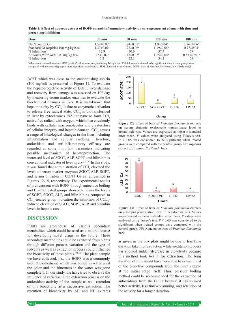
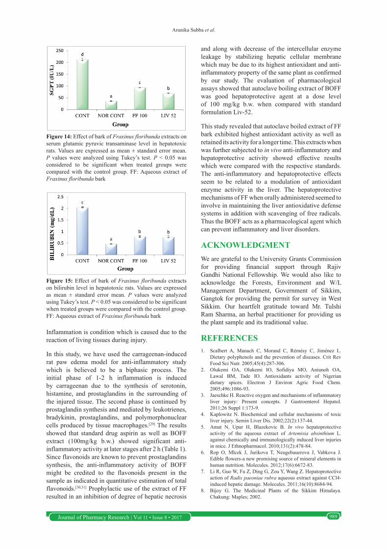
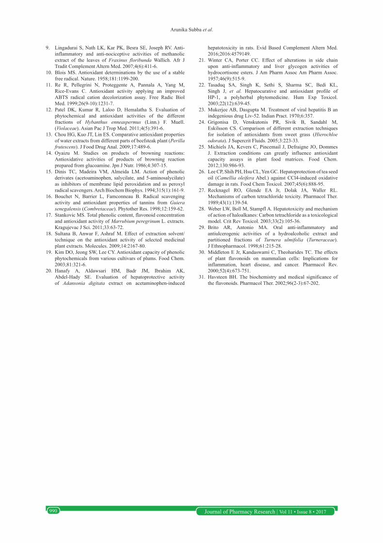
BOFF which was close to the standard drug aspirin (100 mg/ml) as presented in Figure 11. To evaluate the hepatoprotective activity of BOFF, liver damage and recovery from damage was assessed on 10th day by measuring serum marker enzymes to evaluate the biochemical changes in liver. It is well-known that hepatotoxicity by CCl4 is due to enzymatic activation to release free radical state. CCl4 is biotransformed in liver by cytochromes P450 enzyme to form CCl3 active free radical with oxygen, which then covalently binds with cellular macromolecules and creates loss of cellular integrity and hepatic damage. CCl4 causes a range of histological changes to the liver including inflammation and cellular swelling. Therefore, antioxidant and anti-inflammatory efficacy are regarded as some important parameters indicating possible mechanism of hepatoprotection. The increased level of SGOT, ALP, SGPT, and bilirubin is conventional indicator of liver injury.[24-26] In this study, it was found that administration of CCl4 elevated the levels of serum marker enzymes SGOT, ALP, SGPT, and serum bilirubin in CONT Gr as represented in Figures 12-15, respectively. The experimental results of pretreatment with BOFF through autoclave boiling and Liv-52 treated groups showed to lower the levels of SGPT, SGOT, ALP, and bilirubin as compared to CCl4-treated group indication the inhibition of CCL4-induced elevation of SGOT, SGPT, ALP, and bilirubin levels in hepatic rats.
DISCUSSIONPlants are storehouse of various secondary metabolites which could be used as a natural source for developing novel drugs in the future. These secondary metabolites could be extracted from plants through different process variation and the type of solvents as well as extraction process could influence the bioactivity of these plants.[27,28] The plant sample we have collected, i.e., the BOFF was a commonly used ethnomedicine which was boiled in water until the color and the bitterness in the water was gone completely. In our study, we have tried to observe the influence of variation in the extraction process on the antioxidant activity of the sample as well retention of this bioactivity after successive extraction. The retention of bioactivity by AB and NB extracts
Table 1: Effect of aqueous extract of BOFF on anti-inflammatory activity on carrageenan rat edema with time and percentage inhibition
Dose 30 min 60 min 120 min 180 minNaCl control Gr 1.59±0.07b 1.84±0.05c 1.9±0.04b 1.86±0.06b
Standard Gr (aspirin) 100 mg/kg b.w. 1.37±0.02a 1.28±0.06a 1.19±0.05a 0.77±0.04a
% Inhibition 12.9 30.6 37.3 59Fraxinus floribunda 100 mg/kg b.w. 1.5±0.02b 1.43±0.02b 1.25±0.04a 0.833±0.01a
% Inhibition 5.2 22.1 34.1 55Values are expressed as mean±SEM (n=6). P values were analyzed using Tukey’s test. P<0.05 were considered to be significant when treated groups were compared with the control group, a more significant than b and c. SEM: Standard error of mean, BOFF: Bark of Fraxinus floribunda, b.w.: Body weight
Figure 12: Effect of bark of Fraxinus floribunda extracts on serum glutamic oxaloacetic transaminase level in hepatotoxic rats. Values are expressed as mean ± standard error mean. P values were analyzed using Tukey’s test. P < 0.05 was considered to be significant when treated groups were compared with the control group. FF: Aqueous extract of Fraxinus floribunda bark
Figure 13: Effect of bark of Fraxinus floribunda extracts on anti-lipid peroxidation level in hepatotoxic rats. Values are expressed as mean ± standard error mean. P values were analyzed using Tukey’s test. P < 0.05 was considered to be significant when treated groups were compared with the control group. FF: Aqueous extract of Fraxinus floribunda bark
as given in the box plots might be due to less time duration taken for extraction while soxhlation process has showed sudden decrease in bioactivity because this method took 6-8 h for extraction. The long duration of time might have been able to extract most of the bioactive compounds from the plant sample at the initial stage itself. Thus, pressure boiling method could be recommended for the extraction of antioxidants from the BOFF because it has showed better activity, less time-consuming, and retention of the activity for a longer duration.
Arunika Subba et al.
Journal of Pharmacy Research | Vol 11 • Issue 8 • 2017 989
Inflammation is condition which is caused due to the reaction of living tissues during injury.
In this study, we have used the carrageenan-induced rat paw edema model for anti-inflammatory study which is believed to be a biphasic process. The initial phase of 1-2 h inflammation is induced by carrageenan due to the synthesis of serotonin, histamine, and prostaglandins in the surrounding of the injured tissue. The second phase is continued by prostaglandin synthesis and mediated by leukotrienes, bradykinin, prostaglandins, and polymorphonuclear cells produced by tissue macrophages.[29] The results showed that standard drug aspirin as well as BOFF extract (100mg/kg b.w.) showed significant anti-inflammatory activity at later stages after 2 h (Table 1). Since flavonoids are known to prevent prostaglandins synthesis, the anti-inflammatory activity of BOFF might be credited to the flavonoids present in the sample as indicated in quantitative estimation of total flavonoids.[30,31] Prophylactic use of the extract of FF resulted in an inhibition of degree of hepatic necrosis
and along with decrease of the intercellular enzyme leakage by stabilizing hepatic cellular membrane which may be due to its highest antioxidant and anti-inflammatory property of the same plant as confirmed by our study. The evaluation of pharmacological assays showed that autoclave boiling extract of BOFF was good hepatoprotective agent at a dose level of 100 mg/kg b.w. when compared with standard formulation Liv-52.
This study revealed that autoclave boiled extract of FF bark exhibited highest antioxidant activity as well as retained its activity for a longer time. This extracts when was further subjected to in vivo anti-inflammatory and hepatoprotective activity showed effective results which were compared with the respective standards. The anti-inflammatory and hepatoprotective effects seem to be related to a modulation of antioxidant enzyme activity in the liver. The hepatoprotective mechanisms of FF when orally administered seemed to involve in maintaining the liver antioxidative defense systems in addition with scavenging of free radicals. Thus the BOFF acts as a pharmacological agent which can prevent inflammatory and liver disorders.
ACKNOWLEDGMENTWe are grateful to the University Grants Commission for providing financial support through Rajiv Gandhi National Fellowship. We would also like to acknowledge the Forests, Environment and W/L Management Department, Government of Sikkim, Gangtok for providing the permit for survey in West Sikkim. Our heartfelt gratitude toward Mr. Tulshi Ram Sharma, an herbal practitioner for providing us the plant sample and its traditional value.
REFERENCES1. Scalbert A, Manach C, Morand C, Rémésy C, Jiménez L.
Dietary polyphenols and the prevention of diseases. Crit Rev Food Sci Nutr. 2005;45(4):287-306.
2. Olukemi OA, Olukemi IO, Sofidiya MO, Aniunoh OA, Lawal BM, Tade IO. Antioxidants activity of Nigerian dietary spices. Electron J Environ Agric Food Chem. 2005;496:1086-93.
3. Jaeschke H. Reactive oxygen and mechanisms of inflammatory liver injury: Present concepts. J Gastroenterol Hepatol. 2011;26 Suppl 1:173-9.
4. Kaplowitz N. Biochemical and cellular mechanisms of toxic liver injury. Semin Liver Dis. 2002;22(2):137-44.
5. Amat N, Upur H, Blazekovic B. In vivo hepatoprotective activity of the aqueous extract of Artemisia absinthium L. against chemically and immunologically induced liver injuries in mice. J Ethnopharmacol. 2010;131(2):478-84.
6. Rop O, Mlcek J, Jurikova T, Neugebauerova J, Vabkova J. Edible flowers-a new promising source of mineral elements in human nutrition. Molecules. 2012;17(6):6672-83.
7. Li R, Guo W, Fu Z, Ding G, Zou Y, Wang Z. Hepatoprotective action of Radix paeoniae rubra aqueous extract against CCl4-induced hepatic damage. Molecules. 2011;16(10):8684-94.
8. Bijoy G. The Medicinal Plants of the Sikkim Himalaya. Chakung: Maples; 2002.
Figure 14: Effect of bark of Fraxinus floribunda extracts on serum glutamic pyruvic transaminase level in hepatotoxic rats. Values are expressed as mean ± standard error mean. P values were analyzed using Tukey’s test. P < 0.05 was considered to be significant when treated groups were compared with the control group. FF: Aqueous extract of Fraxinus floribunda bark
Figure 15: Effect of bark of Fraxinus floribunda extracts on bilirubin level in hepatotoxic rats. Values are expressed as mean ± standard error mean. P values were analyzed using Tukey’s test. P < 0.05 was considered to be significant when treated groups were compared with the control group. FF: Aqueous extract of Fraxinus floribunda bark
Arunika Subba et al.
Journal of Pharmacy Research | Vol 11 • Issue 8 • 2017990
9. Lingadurai S, Nath LK, Kar PK, Besra SE, Joseph RV. Anti-inflammatory and anti-nociceptive activities of methanolic extract of the leaves of Fraxinus floribunda Wallich. Afr J Tradit Complement Altern Med. 2007;4(6):411-6.
10. Blois MS. Antioxidant determinations by the use of a stable free radical. Nature. 1958;181:1199-200.
11. Re R, Pellegrini N, Proteggente A, Pannala A, Yang M, Rice-Evans C. Antioxidant activity applying an improved ABTS radical cation decolorization assay. Free Radic Biol Med. 1999;26(9-10):1231-7.
12. Patel DK, Kumar R, Laloo D, Hemalatha S. Evaluation of phytochemical and antioxidant activities of the different fractions of Hybanthus enneaspermus (Linn.) F. Muell. (Violaceae). Asian Pac J Trop Med. 2011;4(5):391-6.
13. Chou HG, Kuo JT, Lin ES. Comparative antioxidant properties of water extracts from different parts of beefsteak plant (Perilla frutescens). J Food Drug Anal. 2009;17:489-6.
14. Oyaizu M. Studies on products of browning reactions: Antioxidative activities of products of browning reaction prepared from glucoamine. Jpn J Nutr. 1986;4:307-15.
15. Dinis TC, Madeira VM, Almeida LM. Action of phenolic derivates (acetoaminophen, salycilate, and 5-aminosalycilate) as inhibitors of membrane lipid peroxidation and as peroxyl radical scavengers. Arch Biochem Biophys. 1994;315(1):161-9.
16. Bouchet N, Barrier L, Famconneau B. Radical scavenging activity and antioxidant properties of tannins from Guiera senegalensis (Combretaceae). Phytother Res. 1998;12:159-62.
17. Stankovic MS. Total phenolic content, flavonoid concentration and antioxidant activity of Marrubium peregrinum L. extracts. Kragujevac J Sci. 2011;33:63-72.
18. Sultana B, Anwar F, Ashraf M. Effect of extraction solvent/technique on the antioxidant activity of selected medicinal plant extracts. Molecules. 2009;14:2167-80.
19. Kim DO, Jeong SW, Lee CY. Antioxidant capacity of phenolic phytochemicals from various cultivars of plums. Food Chem. 2003;81:321-6.
20. Hanafy A, Aldawsari HM, Badr JM, Ibrahim AK, Abdel-Hady SE. Evaluation of hepatoprotective activity of Adansonia digitata extract on acetaminophen-induced
hepatotoxicity in rats. Evid Based Complement Altern Med. 2016;2016:4579149.
21. Winter CA, Porter CC. Effect of alterations in side chain upon anti-inflammatory and liver glycogen activities of hydrocortisone esters. J Am Pharm Assoc Am Pharm Assoc. 1957;46(9):515-9.
22. Tasaduq SA, Singh K, Sethi S, Sharma SC, Bedi KL, Singh J, et al. Hepatocurative and antioxidant profile of HP-1, a polyherbal phytomedicine. Hum Exp Toxicol. 2003;22(12):639-45.
23. Mukerjee AB, Dasgupta M. Treatment of viral hepatitis B an indegenious drug Liv-52. Indian Pract. 1970;6:357.
24. Grigonisa D, Venskutonis PR, Sivik B, Sandahl M, Eskilsson CS. Comparison of different extraction techniques for isolation of antioxidants from sweet grass (Hierochloe odorata). J Supercrit Fluids. 2005;3:223-33.
25. Michiels JA, Kevers C, Pincemail J, Defraigne JO, Dommes J. Extraction conditions can greatly influence antioxidant capacity assays in plant food matrices. Food Chem. 2012;130:986-93.
26. Lee CP, Shih PH, Hsu CL, Yen GC. Hepatoprotection of tea seed oil (Camellia oleifera Abel.) against CCl4-induced oxidative damage in rats. Food Chem Toxicol. 2007;45(6):888-95.
27. Recknagel RO, Glende EA Jr, Dolak JA, Waller RL. Mechanisms of carbon tetrachloride toxicity. Pharmacol Ther. 1989;43(1):139-54.
28. Weber LW, Boll M, Stampfl A. Hepatotoxicity and mechanism of action of haloalkanes: Carbon tetrachloride as a toxicological model. Crit Rev Toxicol. 2003;33(2):105-36.
29. Brito AR, Antonio MA. Oral anti-inflammatory and antiulcerogenic activities of a hydroalcoholic extract and partitioned fractions of Turnera ulmifolia (Turneraceae). J Ethnopharmacol. 1998;61:215-28.
30. Middleton E Jr, Kandaswami C, Theoharides TC. The effects of plant flavonoids on mammalian cells: Implications for inflammation, heart disease, and cancer. Pharmacol Rev. 2000;52(4):673-751.
31. Havsteen BH. The biochemistry and medical significance of the flavonoids. Pharmacol Ther. 2002;96(2-3):67-202.