Antioxidant Activity and Hepatoprotective Potential of Agaro-oligosaccharides in Vitro and in Vivo

RESEARCH ARTICLE Open Access
Antioxidant and hepatoprotective potentialof Lawsonia inermis L. leaves against2-acetylaminofluorene induced hepaticdamage in male Wistar ratsManish Kumar1, Paramjeet Kaur1, Madhu Chandel1, Amrit Pal Singh2, Arpana Jain3 and Satwinderjeet Kaur1*
Abstract
Background: Lawsonia inermis (Lythraceae) is an ethnomedicinal plant, traditionally known for curing several ailmentssuch as skin diseases, bacterial infections, jaundice, renal lithiases and inflammation etc. The present work deals withassessment of in vitro antioxidant and in vivo hepatoprotective potential of butanolic fraction (But-LI) of Lawsoniainermis L. leaves.
Methods: Antioxidant activity was evaluated using deoxyribose degradation, lipid peroxidation inhibition and ferricreducing antioxidant power (FRAP) assay. In vivo protective potential of But-LI was assessed at 3 doses [100, 200 &400 mg/kg body weight (bw)] against 2-acetylaminofluorene (2-AAF) induced hepatic damage in male Wistar rats.
Results: But-LI effectively scavenged hydroxyl radicals in deoxyribose degradation assay (IC50 149.12 μg/ml). Fractionalso inhibited lipid peroxidation and demonstrated appreciable reducing potential in FRAP assay. Treatment of animalswith 2-AAF resulted in increased hepatic parameters such as SGOT (2.22 fold), SGPT (1.72 fold), ALP (5.68 fold) and lipidperoxidation (2.94 fold). Different concentration of But-LI demonstrated pronounced protective effects via decreasinglevels of SGOT, SGPT, ALP and lipid peroxidation altered by 2-AAF treatment. But-LI administration also restored thenormal liver architecture as evident from histopathological studies.
Conclusions: The present experimental findings revealed that phytoconstituents of Lawsonia inermis L. possesspotential to effectively protect rats from the 2-AAF induced hepatic damage in vivo possibly by inhibition of reactiveoxygen species and lipid peroxidation.
Keywords: Lawsonia inermis, 2-acetylaminofluorene, Lipid peroxidation, Hepatic damage, Hepatoprotective
BackgroundMutations resulting spontaneously or from environmentalexposure may lead to cancer [1]. Chemical bonds in DNAmolecule abide same laws likewise other chemicals exist-ing at 37 °C in aqueous environment of cell. Likewiseother molecules, existence of DNA also depends upon for-mation and breaking of bonds. So, it is not astonishingthat DNA regularly endures various kinds of chemicaldamages due to spontaneous thermal effects and as resultof attack of other reactive molecules [2]. Various physical
and chemical agents (exogenous agents) causes damageto DNA, many of them are now documented as envir-onmental carcinogens [2, 3]. Studies have shown thatchemicals play an important role in the etiology of sev-eral kinds of human cancers [4, 5]. It is well-known thatexposure to various hazardous chemicals occurs at verylow doses, extends through longer time period of lifeand influence great part of population [6]. Tumorinduction in workers exposed to coal tar in 1775 wasthe earliest known instance of environmental carcino-genesis documented. This very example of environmen-tal carcinogenesis, later led to identification of variouspolycyclic hydrocarbons in coal tar. This also led to thefinding of polycyclic hydrocarbons as skin carcinogens
* Correspondence: [email protected]; [email protected] of Botanical and Environmental Sciences, Guru Nanak DevUniversity, Amritsar 143005, Punjab, IndiaFull list of author information is available at the end of the article
© The Author(s). 2017 Open Access This article is distributed under the terms of the Creative Commons Attribution 4.0International License (http://creativecommons.org/licenses/by/4.0/), which permits unrestricted use, distribution, andreproduction in any medium, provided you give appropriate credit to the original author(s) and the source, provide a link tothe Creative Commons license, and indicate if changes were made. The Creative Commons Public Domain Dedication waiver(http://creativecommons.org/publicdomain/zero/1.0/) applies to the data made available in this article, unless otherwise stated.
Kumar et al. BMC Complementary and Alternative Medicine (2017) 17:56 DOI 10.1186/s12906-017-1567-9
in laboratory animals. Other example was bladdercarcinogenesis incidence among the workers workingin the rubber and chemical industries. This led to therecognition of 2-naphthylamine as bladder carcinogen[2]. With advancement in the science, it is now wellknown that some of cancers are environmental inorigin and can be related directly to different chemical ex-posures [7]. Humans are constantly exposed to plethora ofxenobiotic chemicals and other related environmentalpollutants which are hazardous to the health [8].Liver is the main seat of xenobiotic metabolism and
also carries out various functions in biotransformationincluding amino acid metabolism, lipid metabolism etc.[9–12]. Liver cancer is one of the most common malig-nancies occurring all over the world particularly in Asianand African countries [13]. Various risk factors linkedwith liver cancer are alcohol, food additives, aflatoxins,toxic chemicals from industries, pollutants etc. [14, 15].More than 600 chemicals have been identified whichcan cause liver injury [16, 17]. 2-acetylaminofluorene(2-AAF) is one of the most studied chemical asmodel hepatocarcinogen. It was initially made as aninsecticide, however its use was stopped because ofits carcinogenic nature. It is an aromatic compoundhaving solubility in organic solvents and remainsinsoluble in water [18–21]. 2-AAF induces its carcino-genic effects through metabolic activation via the mixedfunction oxidase system. Activation of 2-AAF leads to theformation of reactive electrophilic forms which react toform DNA adducts [22–24].Nowadays, use of herbal medicines for curing variety
of ailments is gaining popularity including liver diseases[25]. Number of reports are available in the literaturewhich have shown hepatoprotective effects of naturalplant products against various genotoxins, carcinogensand toxic substances including carbon tetrachloride(CCl4), paracetamol, 2-acetylaminofluorene (2-AAF),7, 12-dimethylbenz(a)anthracene (DMBA), thioacetamideetc. [26–34]. Lawsonia inermis L. (L. inermis) commonlyknown as Henna or Mehandi belongs to family Lythra-ceae. Traditionally, the plant is known for its medicinalproperties for the cure of renal lithiases, jaundice, to healwounds, prevent skin inflammation etc. [35–38]. It isalso used by some Nigerian tribes as a therapy againstpoliomyelitis and measles [39]. L. inermis wasreported to contain various phytoconstituents such aschlorogenic acid, ferulic acid, isoferulic acid, gallicacid, o-coumaric acid, m-coumaric acid, myricetin,naringenin-7-o-rutinoside, quercetin, (+)-catechin,(−)-catechin gallate, (−)-epicatechin gallate, vitexin-2′-o-rhamnoside etc. [40]. Hsouna et al. [41] reported phyto-constituents viz. lawsoniaside, lalioside, luteolin-7- O-β-D-glucopyranoside, 2,4,6-trihydroxyacetophenone-2-O-β-D-glucopyranoside, 1,2,4-trihydroxynaphthalene-1-
O-β-D-glucopyranoside from L. inermis leaves. L. inermisshowed numerous medicinal properties viz. antimuta-genic, anticlastogenic, analgesic, anti-inflammatory, anti-pyretic activities etc. [42–44]. Phytoconstituents from L.inermis leaves were reported to possess immunomodula-tory activity [45]. Kaur et al. [46] carried out toxicitystudies on ethanolic extract of L. inermis leaves usingalbino Wistar rats and reported that administration ofrats with 80% ethanolic extract posed no toxicity in thetissues of the organs up to dose of 500 mg/kg bw. An-other study by Alferah [47] reported that administrationof L. inermis leaf solution (200 mg/kg/day) to the rats for42 days did not induce any toxicity in liver, kidney andspleen tissue sections. Selvanayaki and Ananthi [48]reported hepatoprotective effects of aqueous extract of L.inermis against paracetamol induced hepatic damage inmale Albino rats. Hossain et al. [49] studied hepato-protective activity of L. inermis leaves against carbontetrachloride induced liver damage in Wistar albinorats. Dasgupta et al. [50] reported anticarcinogenicactivity of Henna leaves against benzo(a)pyreneinduced forestomach as well as against 7,12 dimethyl-benz(a)anthracene (DMBA)-initiated and croton oil-promoted skin papillomagenesis. In our previous reports[51, 52], we reported extract/fractions of L. inermiswith antioxidant, antiproliferative and apoptosis indu-cing activity. Since But-LI fraction was found toexhibit high antioxidant activity and is rich in variouspolyphenolic phytoconstituents viz. gallic acid, catechin,chlorogenic acid, ellagic acid, kaempferol etc. [51], so weplanned to investigate But-LI fraction from Lawsonia iner-mis L. for modulatory effects against the toxicity inducedby 2-acetylaminofluorene (2-AAF) in male Wistar rats byassessing various serum and liver tissue parameters.
MethodsChemicals2,4,6-tripyridyl-s-triazine (TPTZ), Malondialdehyde (MDA),Deoxyribose, 2-acetylaminofluorene (2-AAF) werepurchased from Sigma Chemical Co. (St Louis, MO,USA). Ascorbic acid and Sodium dodecyl sulfate(SDS) were purchased from Hi-Media, Mumbai, India.All other chemicals used in the present experimentalstudy were of AR grade.
Collection of plant material and preparation of But-LIfractionThe plant material was purchased from local market(Majeeth mandi, Amritsar), identified and authenticatedby Dr. A. S. Soodan, Assoc. Prof., Department of Botanicaland Environmental Sciences, Guru Nanak Dev University,Amritsar. The voucher specimen (no. 6773) has been keptin the Herbarium of the Department. Leaves were washedto ensure that they become free from any kind of dirt as
Kumar et al. BMC Complementary and Alternative Medicine (2017) 17:56 Page 2 of 11
well as other foreign particles and dried in shade. Leaves(3 kg) were grounded to fine powder and extracted atroom temperature to obtain butanolic fraction (But-LI) asdescribed in Kumar et al. [51].
In vitro antioxidant assaysDeoxyribose degradation assayDeoxyribose degradation assay was carried out by themethod of Halliwell et al. [53] and Arouma et al. [54]with slight modifications. In this assay, EDTA (1 mM),FeCl3 (10 mM), hydrogen peroxide (10 mM), 2-deoxyri-bose (10 mM), test sample (1 ml), phosphate buffer andAscorbic acid (1 mM) were mixed in the test tubes andthe contents of reaction mixture were incubated at 37 °Cfor 1 h. After incubation, 1 ml of above mixture was takenand mixed with 1 ml of 2-thiobarbituric acid (TBA) andtricholoacetic acid (TCA) each. Finally reaction mixturewas heated at 80 °C on water bath for 1.5 h. Final absorb-ance of the pink chromogen formed was taken spectro-photometrically at 532 nm using Elisa reader. Rutin wasused as antioxidant standard.Percent hydroxyl radical scavenging potential was
calculated by formula as given below:
Radical scavenging activity %ð Þ ¼ A0 � A1=A0 � 100
where,A0 is the absorbance of reaction mixture + vehicle
solvent,A1 is the absorbance of reaction mixture + test sample.
Lipid peroxidation inhibition assayA modified thiobarbituric acid reactive species (TBARS)assay [55] was performed to determine the lipid peroxidesproduced using egg yolk homogenate as lipid rich media[56]. Various concentrations of test sample were added tothe test tubes containing egg homogenate (0.5 ml of10% v/v). About 50 μl of FeSO4 solution was added to thetest tubes to induce lipid peroxidation and incubated testtubes at 37 °C for 30 min. After ½ hour, 20% acetic acid(pH 3.5), 0.8% of TBA in 1.1% SDS and TCA (20%) wereadded. All the contents of the tubes were mixed properlyand heated at 95 °C for 1 h. After heating, test tubes werecooled, followed by addition of 5 ml of butanol and centri-fuged at 1036 × g for 10 min. The absorbance was taken at532 nm (Systronics 2202 UV–Vis Spectrophotometer).Trolox was used as antioxidant standard.Inhibition of lipid peroxidation (%) was calculated
using the formula as given below:
Radical scavenging activity %ð Þ ¼ 1 ‐ E=Cð Þ � 100
where,C is the absorbance of fully oxidized control,E is the absorbance in the presence of test sample.
Ferric reducing antioxidant power (FRAP) assayFRAP assay was carried out by the method of Benzieand Strain [57]. FRAP reagent was prepared by mixingacetate buffer, TPTZ solution and FeCl3.6H2O solutionin the ratio of 10:1:1. About 3 ml of FRAP reagent wasdispensed into the test tubes followed by addition of300 μl of test sample. Test tubes were shaken to mix thecontent well and incubated at 37 °C for 10 min. Absorb-ance of the reaction mixture was taken at 593 nm(Systronics 2202 UV–Vis Spectrophotometer, India).Trolox was used as standard antioxidant. Increase in theabsorbance of the reaction mixture as compared tocontrol is considered as increase in the reducing poten-tial of test sample.
In vivo hepatoprotective activityExperimental animalsAll the in vivo work was done as per the guidelines ofCommittee for the Purpose of Control and Supervisionof Experiments on Animals (CPCSEA), Ministry ofEnvironment and Forests, Government of India forhousing and experimentation on animals and the studywas approved by the Institutional Animal EthicsCommittee of Guru Nanak Dev University, Amritsar(226/CPCSEA/2014/08). Male Wistar rats weighing240–280 g were used in this study and were procuredfrom the animal house facility of Indian Institute ofIntegrative medicine (IIIM), Jammu (India). After theprocurement, rats were kept in the polypropylene cagesprovided with paddy husk bedding. The temperaturewas maintained at 25 ± 2 °C along with a 12 h light and12 h dark cycle in the animal house of Guru Nanak DevUniversity, Amritsar (Punjab). All the animals were fedon standard pellet diet and water ad libitum and allowedto acclimatize for two weeks before the start ofexperimentation.
Experimental design2-acetylaminofluorene (2-AAF) induced hepatic damagemodel was used to evaluate hepatotoxicity [58]. The ani-mals were randomly divided into seven groups (Fig. 1),each containing four rats (n = 4). The experimentalprotocol was of total 15 days time period. Group Iserved as control group put on standard pellet diet.Group II served as vehicle control and animals receiveddistilled water via oral route (1st to 15th day) and cornoil injection intraperitoneally (i.p.) from 11th to 15thday. Group III served as positive control group wastreated with 2-acetylaminofluorene (2-AAF) (50 mg/kgbw; i.p.) for 5 consecutive days (11th to 15th day). GroupIV (Negative control) was treated with highest dose ofplant extract alone (But-LI; 400 mg/kg bw) from day 1stto 15th, Group V to VII were given 3 doses of plant ex-tract (100, 200 and 400 mg/kg bw from day 1st to 15th)
Kumar et al. BMC Complementary and Alternative Medicine (2017) 17:56 Page 3 of 11
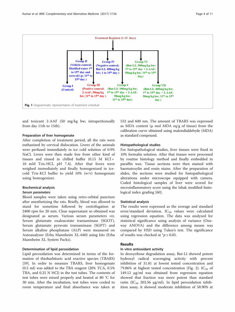
and toxicant 2-AAF (50 mg/kg bw; intraperitoneallyfrom day 11th to 15th).
Preparation of liver homogenateAfter completion of treatment period, all the rats wereeuthanized by cervical dislocation. Livers of the animalswere perfused immediately in ice cold solution of 0.9%NaCl. Livers were then made free from other kind oftissues and rinsed in chilled buffer (0.15 M KCl +10 mM Tris-HCl, pH 7.4). After that livers wereweighed immediately and finally homogenized in ice-cold Tris-KCl buffer to yield 10% (w/v) homogenateusing homogenizer.
Biochemical analysisSerum parametersBlood samples were taken using retro-orbital punctureafter anesthetizing the rats. Briefly, blood was allowed tostand for sometime followed by centrifugation at2400 rpm for 20 min. Clear supernatant so obtained wasdesignated as serum. Various serum parameters viz.Serum glutamate oxaloacetate transaminase (SGOT),Serum glutamate pyruvate transaminase (SGPT) andSerum alkaline phosphatase (ALP) were measured onAutoanalyzer (Erba Mannheim XL-640) using kits (ErbaMannheim XL System Packs).
Determination of lipid peroxidationLipid peroxidation was determined in terms of the for-mation of thiobarbituric acid reactive species (TBARS)[59]. In order to measure TBARS, liver homogenate(0.5 ml) was added to the TBA reagent (20% TCA, 0.5%TBA, and 0.25 N HCl) in the test tubes. The contents oftest tubes were mixed properly and heated at 80 °C for30 min. After the incubation, test tubes were cooled toroom temperature and final absorbance was taken at
532 and 600 nm. The amount of TBARS was expressedas MDA content (μ mol MDA eq/g of tissue) from thecalibration curve obtained using malondialdehyde (MDA)as standard compound.
Histopathological studiesFor histopathological studies, liver tissues were fixed in10% formalin solution. After that tissues were processedby routine histology method and finally embedded inparaffin wax. Tissue sections were then stained withhaematoxylin and eosin stains. After the preparation ofslides, the sections were studied for histopathologicalalterations under microscope equipped with camera.Coded histological samples of liver were scored fornecroinflammatory score using the Ishak modified histo-logical index grading [60].
Statistical analysisThe results were expressed as the average and standarderror/standard deviation. IC50 values were calculatedusing regression equation. The data was analyzed forstatistical significance using analysis of variance (One-way ANOVA) and the difference among means wascompared by HSD using Tukey’s test. The significanceof results was checked at *p ≤ 0.05.
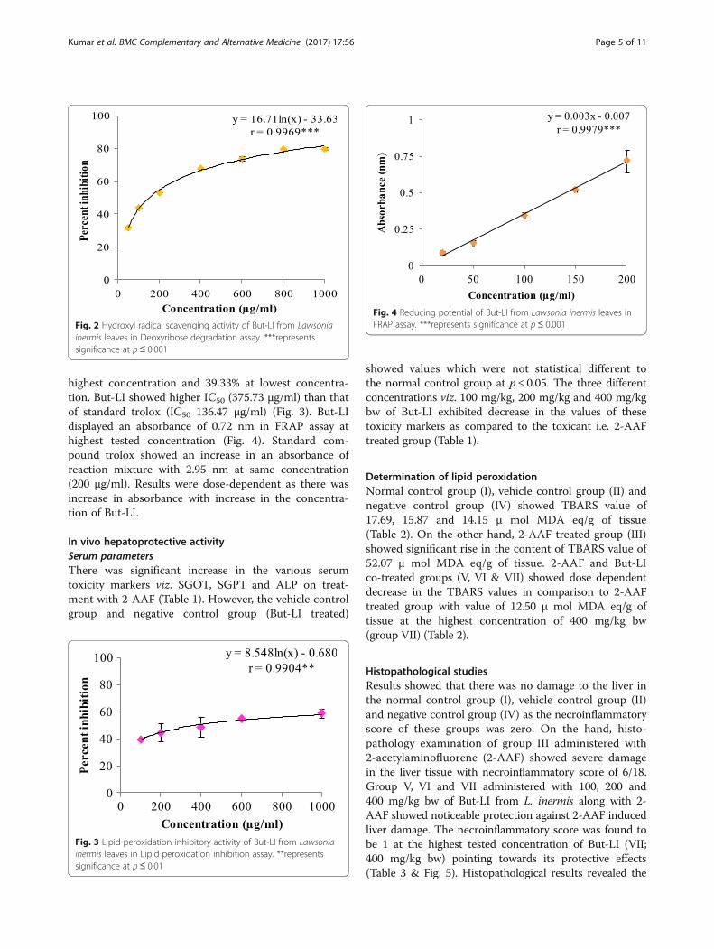
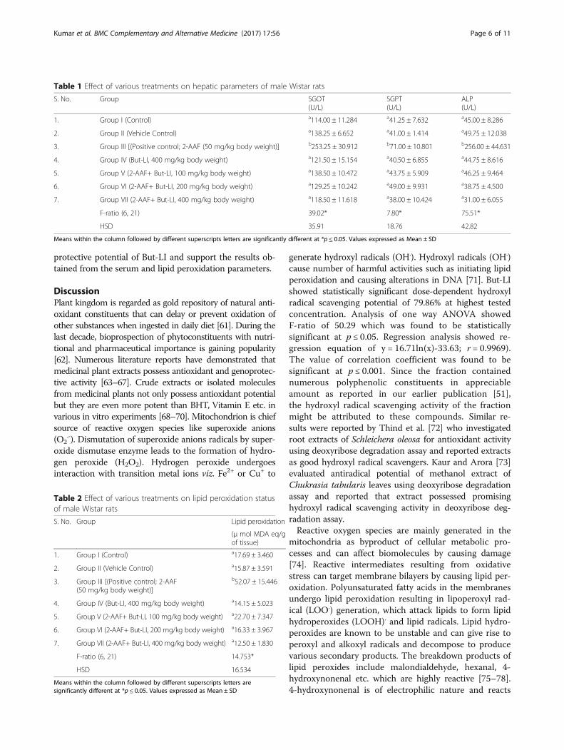
ResultsIn vitro antioxidant activityIn deoxyribose degradation assay, But-LI showed potenthydroxyl radical scavenging activity with percentinhibition of 31.81 at lowest tested concentration and79.86% at highest tested concentration (Fig. 2). IC50 of149.12 μg/ml was obtained from regression equationshowed that fraction was more potent than standardrutin (IC50 203.56 μg/ml). In lipid peroxidation inhib-ition assay, it showed moderate inhibition of 58.90% at
Fig. 1 Diagrammatic representation of treatment schedule
Kumar et al. BMC Complementary and Alternative Medicine (2017) 17:56 Page 4 of 11
highest concentration and 39.33% at lowest concentra-tion. But-LI showed higher IC50 (375.73 μg/ml) than thatof standard trolox (IC50 136.47 μg/ml) (Fig. 3). But-LIdisplayed an absorbance of 0.72 nm in FRAP assay athighest tested concentration (Fig. 4). Standard com-pound trolox showed an increase in an absorbance ofreaction mixture with 2.95 nm at same concentration(200 μg/ml). Results were dose-dependent as there wasincrease in absorbance with increase in the concentra-tion of But-LI.
In vivo hepatoprotective activitySerum parametersThere was significant increase in the various serumtoxicity markers viz. SGOT, SGPT and ALP on treat-ment with 2-AAF (Table 1). However, the vehicle controlgroup and negative control group (But-LI treated)
showed values which were not statistical different tothe normal control group at p ≤ 0.05. The three differentconcentrations viz. 100 mg/kg, 200 mg/kg and 400 mg/kgbw of But-LI exhibited decrease in the values of thesetoxicity markers as compared to the toxicant i.e. 2-AAFtreated group (Table 1).
Determination of lipid peroxidationNormal control group (I), vehicle control group (II) andnegative control group (IV) showed TBARS value of17.69, 15.87 and 14.15 μ mol MDA eq/g of tissue(Table 2). On the other hand, 2-AAF treated group (III)showed significant rise in the content of TBARS value of52.07 μ mol MDA eq/g of tissue. 2-AAF and But-LIco-treated groups (V, VI & VII) showed dose dependentdecrease in the TBARS values in comparison to 2-AAFtreated group with value of 12.50 μ mol MDA eq/g oftissue at the highest concentration of 400 mg/kg bw(group VII) (Table 2).
Histopathological studiesResults showed that there was no damage to the liver inthe normal control group (I), vehicle control group (II)and negative control group (IV) as the necroinflammatoryscore of these groups was zero. On the hand, histo-pathology examination of group III administered with2-acetylaminofluorene (2-AAF) showed severe damagein the liver tissue with necroinflammatory score of 6/18.Group V, VI and VII administered with 100, 200 and400 mg/kg bw of But-LI from L. inermis along with 2-AAF showed noticeable protection against 2-AAF inducedliver damage. The necroinflammatory score was found tobe 1 at the highest tested concentration of But-LI (VII;400 mg/kg bw) pointing towards its protective effects(Table 3 & Fig. 5). Histopathological results revealed the
Fig. 2 Hydroxyl radical scavenging activity of But-LI from Lawsoniainermis leaves in Deoxyribose degradation assay. ***representssignificance at p≤ 0.001
Fig. 3 Lipid peroxidation inhibitory activity of But-LI from Lawsoniainermis leaves in Lipid peroxidation inhibition assay. **representssignificance at p≤ 0.01
Fig. 4 Reducing potential of But-LI from Lawsonia inermis leaves inFRAP assay. ***represents significance at p≤ 0.001
Kumar et al. BMC Complementary and Alternative Medicine (2017) 17:56 Page 5 of 11
protective potential of But-LI and support the results ob-tained from the serum and lipid peroxidation parameters.
DiscussionPlant kingdom is regarded as gold repository of natural anti-oxidant constituents that can delay or prevent oxidation ofother substances when ingested in daily diet [61]. During thelast decade, bioprospection of phytoconstituents with nutri-tional and pharmaceutical importance is gaining popularity[62]. Numerous literature reports have demonstrated thatmedicinal plant extracts possess antioxidant and genoprotec-tive activity [63–67]. Crude extracts or isolated moleculesfrom medicinal plants not only possess antioxidant potentialbut they are even more potent than BHT, Vitamin E etc. invarious in vitro experiments [68–70]. Mitochondrion is chiefsource of reactive oxygen species like superoxide anions(O2
.-). Dismutation of superoxide anions radicals by super-oxide dismutase enzyme leads to the formation of hydro-gen peroxide (H2O2). Hydrogen peroxide undergoesinteraction with transition metal ions viz. Fe2+ or Cu+ to
generate hydroxyl radicals (OH.). Hydroxyl radicals (OH.)cause number of harmful activities such as initiating lipidperoxidation and causing alterations in DNA [71]. But-LIshowed statistically significant dose-dependent hydroxylradical scavenging potential of 79.86% at highest testedconcentration. Analysis of one way ANOVA showedF-ratio of 50.29 which was found to be statisticallysignificant at p ≤ 0.05. Regression analysis showed re-gression equation of y = 16.71ln(x)-33.63; r = 0.9969).The value of correlation coefficient was found to besignificant at p ≤ 0.001. Since the fraction containednumerous polyphenolic constituents in appreciableamount as reported in our earlier publication [51],the hydroxyl radical scavenging activity of the fractionmight be attributed to these compounds. Similar re-sults were reported by Thind et al. [72] who investigatedroot extracts of Schleichera oleosa for antioxidant activityusing deoxyribose degradation assay and reported extractsas good hydroxyl radical scavengers. Kaur and Arora [73]evaluated antiradical potential of methanol extract ofChukrasia tabularis leaves using deoxyribose degradationassay and reported that extract possessed promisinghydroxyl radical scavenging activity in deoxyribose deg-radation assay.Reactive oxygen species are mainly generated in the
mitochondria as byproduct of cellular metabolic pro-cesses and can affect biomolecules by causing damage[74]. Reactive intermediates resulting from oxidativestress can target membrane bilayers by causing lipid per-oxidation. Polyunsaturated fatty acids in the membranesundergo lipid peroxidation resulting in lipoperoxyl rad-ical (LOO.) generation, which attack lipids to form lipidhydroperoxides (LOOH). and lipid radicals. Lipid hydro-peroxides are known to be unstable and can give rise toperoxyl and alkoxyl radicals and decompose to producevarious secondary products. The breakdown products oflipid peroxides include malondialdehyde, hexanal, 4-hydroxynonenal etc. which are highly reactive [75–78].4-hydroxynonenal is of electrophilic nature and reacts
Table 1 Effect of various treatments on hepatic parameters of male Wistar rats
S. No. Group SGOT(U/L)
SGPT(U/L)
ALP(U/L)
1. Group I (Control) a114.00 ± 11.284 a41.25 ± 7.632 a45.00 ± 8.286
2. Group II (Vehicle Control) a138.25 ± 6.652 a41.00 ± 1.414 a49.75 ± 12.038
3. Group III [(Positive control; 2-AAF (50 mg/kg body weight)] b253.25 ± 30.912 b71.00 ± 10.801 b256.00 ± 44.631
4. Group IV (But-LI, 400 mg/kg body weight) a121.50 ± 15.154 a40.50 ± 6.855 a44.75 ± 8.616
5. Group V (2-AAF+ But-LI, 100 mg/kg body weight) a138.50 ± 10.472 a43.75 ± 5.909 a46.25 ± 9.464
6. Group VI (2-AAF+ But-LI, 200 mg/kg body weight) a129.25 ± 10.242 a49.00 ± 9.931 a38.75 ± 4.500
7. Group VII (2-AAF+ But-LI, 400 mg/kg body weight) a118.50 ± 11.618 a38.00 ± 10.424 a31.00 ± 6.055
F-ratio (6, 21) 39.02* 7.80* 75.51*
HSD 35.91 18.76 42.82
Means within the column followed by different superscripts letters are significantly different at *p ≤ 0.05. Values expressed as Mean ± SD
Table 2 Effect of various treatments on lipid peroxidation statusof male Wistar rats
S. No. Group Lipid peroxidation
(μ mol MDA eq/gof tissue)
1. Group I (Control) a17.69 ± 3.460
2. Group II (Vehicle Control) a15.87 ± 3.591
3. Group III [(Positive control; 2-AAF(50 mg/kg body weight)]
b52.07 ± 15.446
4. Group IV (But-LI, 400 mg/kg body weight) a14.15 ± 5.023
5. Group V (2-AAF+ But-LI, 100 mg/kg body weight) a22.70 ± 7.347
6. Group VI (2-AAF+ But-LI, 200 mg/kg body weight) a16.33 ± 3.967
7. Group VII (2-AAF+ But-LI, 400 mg/kg body weight) a12.50 ± 1.830
F-ratio (6, 21) 14.753*
HSD 16.534
Means within the column followed by different superscripts letters aresignificantly different at *p ≤ 0.05. Values expressed as Mean ± SD
Kumar et al. BMC Complementary and Alternative Medicine (2017) 17:56 Page 6 of 11
with glutathione, proteins and also with DNA at higherconcentration [79, 80]. In the present investigation, itwas found that But-LI moderately inhibited the lipidperoxidation dose dependently (y = 8.548ln(x)-0.680;r = 0.9904). The value of correlation coefficient was
found to be significant at p ≤ 0.01. Earlier, we havereported that But-LI fraction harbours high amountof catechin, chlorogenic acid, ellagic acid and kaempferolwhile phytoconstituents such as gallic acid, epicatechinand quercetin were found to be present in moderate
Table 3 Modified hepatic activity index (HAI) grading (necroinflammatory scores)
S. No. Group Periportal orinterface hepatitis(Piecemeal necrosis)
Confluent necrosis Focal (spotty) lyticnecrosis, apoptoticand focal inflammation
Portal inflammat-ion Total score
1. Group I 0 0 0 0 0/18
2. Group II 0 0 0 0 0/18
3. Group III 2 1 1 2 6/18
4. Group IV 0 0 0 0 0/18
5. Group V 1 0 1 1 3/18
6. Group VI 0 0 0 1 1/18
7. Group VII 1 0 0 0 1/18
Fig. 5 Histopathological examination of liver sections of rats belonging to different groups. Group I (Control); Group II (Vehicle control); Group III [(Positivecontrol; 2-AAF (50 mg/kg bw)]; Group IV (But-LI, 400 mg/kg bw); Group V (2-AAF+ But-LI, 100 mg/kg bw); Group VII (2-AAF+ But-LI, 400 mg/kg bw)
Kumar et al. BMC Complementary and Alternative Medicine (2017) 17:56 Page 7 of 11

amount [51]. The lipid peroxidation inhibitory activity ofthe fraction might be due to various polyphenolicconstituents present in it. Nakchat et al. [81] studiedantioxidant activity including anti-lipid peroxidationactivity of boiling water Tamarind seed coat extractand reported that extract effectively inhibited the lipidperoxidation. HPLC analysis of Tamarind seed coatextract showed presence of phenolics constitiuents suchas (+)-catechin, (−)-epicatechin and procyanidin B2 whichmay be resposnsible for its antioxidant activities. Mullaand Swamy [82] studied antioxidant activity of poly-phenolic extract of Portulaca quadrifida and reported thatextract showed antilipid peroxidation activity of 71% withIC50 value of 370.33 ± 2.91 μg/ml. Results of FRAP assayrevealed that But-LI fraction also possessed dose-dependent reducing potential. Analysis of results usingone way ANOVA showed F-ratio of 49.94 which wasfound to be statistically significant at p ≤ 0.05. Regressionanalysis showed regression equation of y = 0.003×-0.007;r = 0.9979. The value of correlation coefficient was foundto be significant at p ≤ 0.001. Reducing ability of the frac-tion may be due to polyphenols present in it. Singh et al.[83] studied leaf, fruit and seed extract of Moringa oleiferafor antioxidant activity and reported that leaf extract pos-sessed good reducing potential in reducing power assay.HPLC analysis of the extract demonstrated the presenceof phenolic constituents such as gallic acid, chlorogenicacid, kaempferol, quercetin, ellagic acid, ferulic acid, andvanillin. Soobrattee et al. [84] studied various polphenolicphytochemicals for reducing potential in FRAP assay.Gallic acid, ellagic acid, chlorogenic acid, quercetin,kaempferol, (−)-epicatechin and (+)-catechin exhibitedFRAP value of 5.25, 4.39, 3.22, 7.39, 1.95, 2.90 and2.47 mmol Fe (II)/L respectively.Several plant extracts and phytochemicals are reported
to modulate the mammalian antioxidant enzymes systemand provide protective effects against cellular damage[85–88]. Liver is the main organ responsible for detoxifi-cation processes occurring in the body. In the liver cells,endoplasmic reticulum is the primary site of metabolism.Hence, this metabolism is termed as hepatic metabolism.Besides liver, there are extrahepatic sites of metabolismwhich include organs such as lungs, kidney, skin, epithe-lial cells of gastrointestinal tract, adrenals and placenta[89–92]. Liver injury in response to various chemicalsresults in the leakage of serum enzymes into the bloodcirculation, thus causing increase in their level in theserum [93]. Sehrawat et al. [94] reported that 2-AAFadministration to rats increased the level of SGOT andSGPT enzymes in serum. In another study, Hasan andSultana [34] reported that 2-AAF treated rats demon-strated high level of serum aspartate aminotransferase(AST) and alanine aminotransferase (ALT). In the presentinvestigation results obtained from serum toxicity markers
such as SGOT, SGPT, ALP demonstrated significantincrease on treatment with 2-AAF as compared to normalcontrol. 2-AAF induced 2.22, 1.72 and 5.68 fold enhance-ments in SGOT, SGPT and ALP levels respectively. Onco-administration of rats with 2-AAF and varying doses ofBut-LI, there was significant decrease in these serumparameters and the serum enzymes levels were restoredtowards normal control levels. The But-LI fraction alonedid not induce any increase in the values of these markersand results were statistically not different to the normalcontrol and vehicle control group at p ≤ 0.05, reflectingnon-toxic nature of But-LI fraction.A study carried out by Selvanayaki and Ananthi [48]
reported aqueous extract of Lawsonia inermis seeds withpotent hepatoprotective effects against paracetamol in-duced hepatic damage in male rats. Extract significantlyreduced the levels of various serum enzymes viz. aspar-tate aminotransferase (AST), acid phosphatase (ACP),alkaline aminotransferase (ALT), alkaline phosphatase(ALP) etc. altered by paracetamol treatment. Recently,Mohamed et al. [95] reported hepatoprotective potentialof methanol extract of L. inermis leaves against carbontetrachloride (CCl4)-induced hepatic damage. It wasfound that extract treatment significantly protected ratsfrom hepatic damage induced by CCl4.Lipid peroxidation is critical marker of oxidative stress
and is coupled with various diseases including cancer[96, 97]. Malondialdehyde (MDA) and lipid hydroperox-ides are produced as the result of lipid peroxidation ofpolyunsaturated fatty acids [86, 92]. Results of thepresent investigation demonstrated that 2-AAF treat-ment resulted in 2.94 fold increase in the MDA level inrats. Further, treatment of rats with But-LI along with 2-AAF reversed the effect of 2-AAF as reflected fromlower level of MDA. Our results are in agreement withprevious studies [34, 88, 94, 95], in which natural plantproducts effectively reduced lipid peroxidation inducedin response to various toxicants. Further, results of histo-pathological examination were also in concordance withresults of other parameters and provided supportive evi-dence regarding protective potential of Lawsonia inermis(But-LI) fraction. It was found that 2-AAF administra-tion to the male Wistar rats caused severe damage tothe liver tissue, since it showed various histopathologicalalterations such as moderate piecemeal necrosis, mildconfluent and spotty necrosis, moderate portal inflam-mation etc. with necroinflammatory score of 6 out of 18.The untreated, vehicle and negative control group rats didnot demonstrate such pathologies in their liver tissueand necroinflammatory score was found to be zero.All the 3 doses (100, 200 and 400 mg/kg bw) pro-vided protection against damage induced by 2-AAFwith necroinflammatory score of 1 out of 18 at high-est tested dose (400 mg/kg bw) and histoarchitecture
Kumar et al. BMC Complementary and Alternative Medicine (2017) 17:56 Page 8 of 11

of the animals in these groups was comparable tountreated control group. Hepatoprotection can beachieved either by reinstating the normal hepaticphysiology or by diminishing the toxic damagingeffect induced by toxicant [98]. The in vivo protectiveactivity of But-LI of Lawsonia inermis against 2-AAFcould be attributed to the various polyphenolic phyto-chemicals present in the fraction.
ConclusionsThe present experimental findings revealed that phyto-constituents of Lawsonia inermis L. possess potential toprotect rats from the 2-AAF induced hepatic damage invivo possibly by inhibition of radicals and lipidperoxidation.
Abbreviations2-AAF: 2-acetylaminofluorene; ALP: Alkaline phosphatase; CPCSEA: Committeefor the purpose of control and supervision of experiments on animals;FRAP: Ferric reducing antioxidant power; MDA: Malondialdehyde; SGOT: Serumglutamic oxaloacetic transaminase; SGPT: Serum glutamic pyruvic transaminase;TBARS: Thiobarbituric acid reactive species; TPTZ: 2,4,6-tripyridyl-s-triazine
AcknowledgementsAll the authors are thankful to the Department of Botanical and EnvironmentalSciences as well as Health Centre of Guru Nanak Dev University, Amritsar forproviding necessary laboratory facilities to carry out this work.
FundingThe authors are thankful to UGC (UPE & CPEPA Programme) and PURSEprogramme of DST, New Delhi for providing financial assistance.
Availability of data and materialsAll the data are included within the paper.
Authors’ contributionsMK carried out major bioactivity part of present work. MK, PK, MC and APScollectively conducted antioxidant and hepatoprotective activities along withstatistically analysis of the data. AJ helped in the histopathological studies.SK designed, supervised and critically checked the manuscript. All authorsread and approved the final version of manuscript.
Competing interestsThe authors declare that they have no competing interests.
Consent for publicationNot applicable.
Ethics approvalAll the in vivo work was done as per the guidelines of Committee for thePurpose of Control and Supervision of Experiments on Animals (CPCSEA),Ministry of Environment and Forests, Government of India for housingand experimentation on animals and the study was approved by theInstitutional Animal Ethics Committee of Guru Nanak Dev University, Amritsar(226/CPCSEA/2014/08).
Author details1Department of Botanical and Environmental Sciences, Guru Nanak DevUniversity, Amritsar 143005, Punjab, India. 2Department of PharmaceuticalSciences, Guru Nanak Dev University, Amritsar 143005, Punjab, India. 3OmDiagnostics, Amritsar, Punjab, India.
Received: 26 March 2016 Accepted: 7 January 2017
References1. Thilly WG. Have environmental mutagens have caused oncomutations in
people? Nat Genet. 2003;34:255–9.2. Bertram JS. The molecular biology of cancer. Mol Aspects Med.
2001;21:167–223.3. Miller JA, Miller EC. Metabolic activation and reactivity of chemical
carcinogens. Mutat Res. 1975;33:25–6.4. Boyland E. A chemist’s view of cancer prevention. Proc R Soc Med.
1967;60:93–9.5. Higginson J. Present trends in cancer epidemiology. Proc Can Cancer Conf.
1969;8:40–75.6. Hemminki K, Sorsa M, Vainio H. Genetic risks caused by occupational
chemicals. Use of experimental methods and occupational risk groupmonitoring in the detection of environmental chemicals causing mutations,cancer and malformations. Scand J Work Environ Health. 1979;5:307–27.
7. Doll R, Peto R. The causes of cancer: quantitative estimates of avoidablerisks of cancer in the United States today. J Natl Cancer Inst. 1981;66:1191–308.
8. Mihailović V, Mihailović M, Uskoković A, Arambašić J, Mišić D, Stanković V,Katanić J, Mladenović M, Solujić S, Matić S. Hepatoprotective effects ofGentiana asclepiadea L. extracts against carbon tetrachloride induced liverinjury in rats. Food Chem Toxicol. 2013;52:83–90.
9. Wolf PL. Biochemical diagnosis of liver diseases. Indian J Clin Biochem.1999;14:59–90.
10. Lee CP, Shih PH, Hsu CL, Yen GC. Hepatoprotection of tea seed oil(Camellia oleifera Abel.) against CCl4-induced oxidative damage in rats.Food Chem Toxicol. 2007;45:888–95.
11. Jia XY, Zhang QA, Zhang ZQ, Wang Y, Yuan YF, Wang HY, Zhao D.Hepatoprotective effects of almond oil against carbon tetrachlorideinduced liver injury in rats. Food Chem. 2011;125:673–8.
12. Liu G, Liu X, Zhang Y, Zhang F, Wei T, Yang M, Wang K, Wang Y, Liu N,Cheng H, Zhao Z. Hepatoprotective effects of polysaccharides extractedfrom Zizyphus jujube cv. Huanghetanzao Int J Biol Macromol. 2015;76:169–75.
13. Qian Y, Ling CQ. Preventive effect of Ganfujian granule on experimentalhepatocarcinoma in rats. World J Gastroenterol. 2004;10:1755–7.
14. Farazi PA, Depinho RA. Hepatocellular carcinoma pathogenesis: from genesto environment. Nat Rev Cancer. 2006;6:674–87.
15. Jemal A, Siegal R, Ward E, Murray T, Xu J, Thun MJ. Cancer statistics. CA CancerJ Clin. 2007;57:43–66.
16. Cengiz N, Kavak S, Guzel A, Ozbek H, Bektas H, Him A, Erdogan E,Balahoroglu R. Investigation of the hepatoprotective effects of Sesame(Sesamum indicum L.) in carbon tetrachloride-induced liver toxicity.J Membr Biol. 2013;246:1–6.
17. Hsu YW, Tsai CF, Chen WK, Lu FJ. Protective effects of seabuckthorn(Hippophae rhamnoides L.) seed oil against carbon tetrachloride-inducedhepatotoxicity in mice. Food Chem Toxicol. 2009;47:2281–8.
18. Wilson RH, De Eds F, Cox Jr AJ. The toxicity and carcinogenic activityof 2-acetaminofluorene. Cancer Res. 1941;1:595–608.
19. Gonzalez FJ, Samore M, McQuiddy P, Kasper CB. Effects of 2-acetylaminofluorene and N-hydroxy-2-acetylaminofluorene on the cellularlevels of epoxide hydratase, cytochrome P-450b, and NADPH-cytochromec (P-450) oxidoreductase messenger ribonucleic acids. J Biol Chem.1982;257:11032–6.
20. Heflich RH, Neft RE. Genetic toxicity of 2-acetylaminofluorene, 2-aminofluoreneand some of their metabolites and model metabolites. Mutat Res.1994;318:73–114.
21. Verna L, Whysner J, Williams GM. 2-Acetylaminofluorene mechanistic dataand risk assessment: DNA reactivity, enhanced cell proliferation and tumorinitiation. Pharmacol Ther. 1996;71:83–105.
22. Lotlikar PD, Enomoto M, Miller JA, Miller EC. Species variations in theN- and ring-hydroxylation of 2-acetylaminofluorene and effects of3-methylcholanthrene pretreatment. Proc Soc Exp Biol Med.1967;125:341–6.
23. Miller EC. Some current perspectives on chemical carcinogenesis inhumans and experimental animals: presidential address. Cancer Res.1978;38:1479–96.
24. Strom SC, Jirtle RL, Michalopoulos G. Genotoxic effects of 2-acetylaminofluoreneon rat and human hepatocytes. Environ Health Perspect. 1983;49:165–70.
25. Gupta M, Majumder UK, Thamilselvan V, Manikandan L, Senthilkumar GP,Suresh R, Kakotti BK. Potential hepatoprotective effect and antioxidant roleof methanol extract of Oldenlandia umbellate in carbon tetrachlorideinduced hepatotoxicity in Wistar rats. Iranian J Pharmacol Ther. 2007;6:5–9.
Kumar et al. BMC Complementary and Alternative Medicine (2017) 17:56 Page 9 of 11

26. Prasad S, Kalra N, Shukla Y. Hepatoprotective effects of lupeol and mangopulp extract of carcinogen induced alteration in Swiss albino mice.Mol Nutr Food Res. 2007;51:352–9.
27. Opoku AR, Ndlovu IM, Terblanche SE, Hutchings AH. In vivo hepatoprotectiveeffects of Rhoicissus tridentata subsp. cuneifolia, a traditional Zulu medicinalplant, against CCl4-induced acute liver injury in rats. S Afr J Bot. 2007;73:372–7.
28. Singh H, Bedi PS, Singh B. Hepatoprotective activity of turmeric and garlicagainst 7–12, dimethylbenzanthracene induced liver damage in Wistaralbino rats. European J Med Plants. 2011;1:162–70.
29. Sharma N, Shukla S. Hepatoprotective potential of aqueous extract ofButea monosperma against CCl4 induced damage in rats. Exp ToxicolPathol. 2011;63:671–6.
30. Akther N, Shawl AS, Sultana S, Chandan BK, Akhter M. Hepatoprotectiveactivity of Marrubium vulgare against paracetamol induced toxicity.J Pharm Res. 2013;7:565–70.
31. Subramanian M, Balakrishnan S, Chinnaiyan SK, Sekar VK, Chandu AN.Hepatoprotective effect of leaves of Morinda tinctoria Roxb. againstparacetamol induced liver damage in rats. Drug Invent Today. 2013;5:223–8.
32. Salama SM, Abdulla MA, Al Rashdi AS, Ismail S, Alkiyumi SS, Golbabapour S.Hepatoprotective effect of ethanolic extract of Curcuma longa onthioacetamide induced liver cirrhosis in rats. BMC Complement Altern Med.2013;13:56.
33. Joshi BC, Prakash A, Kalia AN. Hepatoprotective potential of antioxidantpotent fraction from Urtica dioica Linn. (whole plant) in CCl4 challengedrats. Toxicol Rep. 2015;2:1101–10.
34. Hasan SK, Sultana S. Geraniol attenuates 2- acetylaminofluorene inducedoxidative stress, inflammation and apoptosis in the liver of Wistar rats.Toxicol Mech Methods. 2015;25:559–73.
35. Chopra RN, Nayer SL, Chopra IC. Glossary of India medicinal plants. NewDelhi: CSIR Publications; 1956. p. 151.
36. Bellakhdar J. The traditional Moroccan pharmacopoeia: ancient Arabicmedicine and popular knowledge. Paris: Ibis press; 1997.
37. Kumari P, Joshi GC, Tewari LM. Diversity and status of ethno-medicinal plantsof Almora district in Uttarakhand. India Int J Biodivers Conserv. 2011;3:298–326.
38. Sharma J, Gairola S, Gaur RD, Painuli RM. The treatment of jaundice withmedicinal plants in indigenous communities of the Sub-Himalayan regionof Uttarakhand. India J Ethnopharmacol. 2012;143:262–91.
39. Oladunmoye MK, Kehinde FY. Ethnobotanical survey of medicinal plantsused in treating viral infection among Yoruba tribes of South WesternNigeria. Afr J Microbiol Res. 2011;5:2991–3004.
40. Guha G, Rajkumar V, Ashok Kumar R, Mathew L. Antioxidant activity ofLawsonia inermis extracts inhibits chromium(VI)-induced cellular andDNA toxicity. Evid Based Complement Alternat Med. 2011.doi:10.1093/ecam/nep205.
41. Hsouna AB, Trigui M, Culioli G, Culioli G, Blache Y, Jaoua S. Antioxidantconstituents from Lawsonia inermis leaves: isolation, structure elucidationand antioxidative capacity. Food Chem. 2010;125:193–200.
42. Raja W, Agrawal RC, Ovais M. Evaluation of antimutagenocity effect ofLawsonia inermis (henna) leaf extract in Swiss albino mice. Res J PharmTechnol. 2008;1:278–9.
43. Basirian M, Manjula SN, Mruthunjaya K. In vitro anti-clastogenic activity ofdifferent fractions of roots of Lawsonia inermis on chromosomal aberrationof human lymphocytes against doxocirubicin and cyclophosphamide asclastogens. Int J Med Med Sci. 2013;46:1239–44.
44. Ali BH, Bashir AK, Tanira MO. Anti-inflammatory, antipyretic, and analgesiceffects of Lawsonia inermis L. (henna) in rats. Pharmacology. 1995;51:356–63.
45. Mikhaeil BR, Badria FA, Maatooq GT, Amer M. Antioxidant andimmunomodulatory constituents of henna leaves. Z Naturforsch C.2004;59:468–76.
46. Kaur M, Dangi CB, Singhai A, Singh M, Kosta S, Singh H, Peter J, Jain S.Toxicity profile of ethanolic extract of Lawsonia inermis leaves in albinoWistar rats. WJPPS. 2014;3:835–48.
47. Alferah MAZ. Toxicity induced histological changes in selected organs ofmale (wistar) rats by Lawsonia inermis leaf extract. European J Med Plants.2012;2:151–8.
48. Selvanayaki R, Ananthi T. Hepatoprotective activity of aqueous extract ofLawsonia inermis against paracetamol induced rats. Asian J Pharm Res.2012;2:75–7.
49. Hossain CM, Maji HS, Chakraborty P. Hepatoprotective activity of Lawsoniainermis Linn, warm aqueous extract in carbon tetrachloride induced hepaticinjury in Wister rats. Asian J Pharm Clin Res. 2011;4:106–9.
50. Dasgupta T, Rao AR, Yadava PK. Modulatory effect of Henna leaf(Lawsonia inermis) on drug metabolising phase I and phase IIenzymes, antioxidant enzymes, lipid peroxidation and chemicallyinduced skin and forestomach papillomagenesis in mice. Mol CellBiochem. 2003;245:11–22.
51. Kumar M, Kumar S, Kaur SJ. Identification of polyphenols in leaf extracts ofLawsonia inermis L. with antioxidant, antigenotoxic and antiproliferativepotential. Int J Green Pharm. 2014;8:23–36.
52. Kumar M, Kaur P, Kumar S, Kaur S. Antiproliferative and apoptosis inducingeffects of non-polar fractions from Lawsonia inermis L. in cervical (HeLa)cancer cells. Physiol Mol Biol Plants. 2015;21:249–60.
53. Halliwell B, Gutteridge JMC, Aruoma OI. The deoxyribose method: a simple“test-tube” assay for determination of rate constants for reaction of hydroxylgroups. Anal Biochem. 1987;165:215–9.
54. Aruoma OI, Grootveld M, Halliwell B. The role of iron in ascorbate-dependent deoxyribose degradation. Evidence consistent with a sitespecific hydroxyl radical feneration caused by iron ions bound to thedeoxyribose molecule. J Inorg Biochem. 1987;29:289–99.
55. Ohkowa M, Ohisi N, Yagi K. Assay for lipid peroxides in animal tissue bythiobarbituric acid reaction. Anal Biochem. 1979;95:351–8.
56. Ruberto G, Baratta MT, Deans SG, Dorman HJD. Antioxidant andantimicrobial activity of Foeniculum vulgare and Crithmum maritimumessential oils. Planta Med. 2000;66:687–93.
57. Benzie IFF, Strain JJ. The ferric reducing ability of plasma as a measure of‘antioxidant power’: the FRAP assay. Anal Biochem. 1996;239:70–6.
58. Hasan SK, Khan R, Ali N, Khan AQ, Rehman MU, Tahir M, Lateef A, Nafees S,Mehdi SJ, Rashid S, Shahid A, Sultana S. 18-β Glycyrrhetinic acid alleviates2-acetylaminofluorene-induced hepatotoxicity in Wistar rats: role inhyperproliferation, inflammation and oxidative stress. Hum Exp Toxicol.2015;34:628–41.
59. Devasagayam TPA, Boloor KK, Ramasarma T. Methods for estimating lipidperoxidation: ananalysis of merits and demerits. Indian J Biochem Biophys.2003;40:300–8.
60. Ishak K, Baptista A, Bianchi L, Callea F, De Groote J, Gudat F, Denk H,Desmet V, Korb G, MacSween RNM, Phillips MJ, Portmann BG, Paulsen H,Scheuer PJ, Schmid M, Thaler H. Histological grading and staging of chronichepatitis. J Hepatol. 1995;22:696–9.
61. Girgih AT, He R, Hasan FM, Udenigwe CC, Gill TA, Aluko RE. Evaluation ofthe in vitro antioxidant properties of a cod (Gadus morhua) proteinhydrolysate and peptide fractions. Food Chem. 2015;173:652–9.
62. Wangensteen H, Samuelsen AB, Malterud KE. Antioxidant activity in extractsfrom coriander. Food Chem. 2004;88:293–7.
63. Giao MS, González-Sanjosé ML, Rivero-Pérez MD, Pereira CI, Pintado ME,Malcata FX. Infusions of Portuguese medicinal plants: dependence of finalantioxidant capacity and phenol content on extraction features. J Sci FoodAgric. 2007;87:2638–47.
64. Giao MS, Pereira CI, Fonseca SC, Pintado ME, Malcata FX. Effect of particlesize upon the extent of extraction of antioxidant power from the plantsAgrimonia eupatoria, Salvia sp. and Satureja montana. Food Chem.2009;117:412–6.
65. Kumar M, Kumar S, Kaur SJ. Investigations on DNA protective andantioxidant potential of chloroform and ethyl acetate fractions ofKoelreuteria paniculata Laxm. Afr J Pharm Pharmacol. 2011;5:421–7.
66. Chandel M, Sharma U, Kumar N, Singh B, Kaur S. Antioxidant activity andidentification of bioactive compounds from leaves of Anthocephaluscadamba by ultra-performance liquid chromatography/electrosprayionization quadrupole time of flight mass spectrometry. Asian Pac J TropMed. 2012;5:977–85.
67. Kaur P, Kaur V, Kumar M, Kaur S. Suppression of SOS response in E. coli PQ37, antioxidant potential and antiproliferative action of methanolic extractof Pteris vittata L. on human MCF-7 breast cancer cells. Food Chem Toxicol.2014;74:326–33.
68. Gordon MH, Weng XC. Antioxidant properties of extracts from tanshen(Salvia miltiorrhiza Bunge). Food Chem. 1992;44:119–22.
69. Gu LW, Weng XC. Antioxidant activity and components of Salvia plebeia R.Br. – a Chinese herb. Food Chem. 2001;73:299–305.
70. Pyo YH, Lee TC, Logendrac L, Rosen RT. Antioxidant activity and phenolicscompounds of Swiss chard (Beta vulgaris subspecies cycla) extracts. FoodChem. 2004;85:19–26.
71. Halliwell B, Gutteridge JMC. Free radicals in biology and medicine. Oxford,New York: Oxford University Press; 1999.
Kumar et al. BMC Complementary and Alternative Medicine (2017) 17:56 Page 10 of 11

72. Thind TS, Singh R, Kaur R, Rampal G, Arora S. In vitro antiradical propertiesand total phenolic contents in methanol extract/fractions from bark ofSchleichera oleosa (Lour.) Oken. Med Chem Res. 2011;20:254–60.
73. Kaur R, Arora S. Investigations of antioxidant activity of methanol extract ofChukrasia tabularis. A Juss J Chin Clin Med. 2008;3:200–5.
74. Riess ML, Camara AKS, Kevin LG, An J, Stowe DF. Reduced reactive O2 speciesformation and preserved mitochondrial NADH and [Ca2+] levels duringshort-term 17 °C ischemia in intact hearts. Cardiovas Res. 2004;61:580–90.
75. Halliwell B, Chirico S, Crawford MA, Bjerve KS, Gey KF. Lipid peroxidation: itsmechanism, measurement, and significance. Am J Clin Nutr. 1993;57:715S–24.
76. Gardner HW. Oxygen radical chemistry of polyunsaturated fatty acids.Free Radic Biol Med. 1989;7:65–86.
77. Spiteller P, Kern W, Reiner J, Spiteller G. Aldehydic lipid peroxidationproducts derived from linoleic acid. Biochim Biophys Acta. 2001;1531:188–208.
78. Barrera G, Pizzimenti S, Dianzani MU. Lipid peroxidation: control of cellproliferation, cell differentiation and cell death. Mol Aspects Med. 2008;29:1–8.
79. Esterbauer H, Schaur RJ, Zollner H. Chemistry and biochemistry of 4-hydroxynonenal, malonaldehyde and related aldehydes. Free Radic BiolMed. 1991;11:81–128.
80. Uchida K. 4-Hydroxy-2-nonenal: a product and mediator of oxidative stress.Prog Lipid Res. 2003;42:318–43.
81. Nakchat O, Meksuriyen D, Pongsamart S. Antioxidant and anti-lipidperoxidation activities of Tamarindus indica seed coat in human fibroblastcells. Indian J Exp Biol. 2014;52:125–32.
82. Mulla SK, Swamy P. Antioxidant activity of ethanolic and polyphenolicextracts of Portulaca quadrifida. Int J Biol Pharm Res. 2012;3:392–9.
83. Singh BN, Singh BR, Singh RL, Prakash D, Dhakarey R, Upadhyay G, Singh HB.Oxidative DNA damage protective activity, antioxidant and anti-quorumsensing potentials of Moringa oleifera. Food Chem Toxicol. 2009;47:1109–16.
84. Soobrattee MA, Neergheen VS, Luximon-Ramma A, Aruoma OI, Bahorun T.Phenolics as potential antioxidant therapeutic agents: mechanism andactions. Mutat Res. 2005;579:200–13.
85. Yener Z, Celik I, Ilhan F, Bal R. Effects of Urtica dioica L. seed on lipidperoxidation, antioxidants and liver pathology in aflatoxin-induced tissueinjury in rats. Food Chem Toxicol. 2009;47:418–24.
86. Ansil PN, Nitha A, Prabha SP, Wills PJ, Jazaira V, Latha MS. Protective effectof Amorphophallus campanulatus (Roxb.) Blume. tuber against thioacetamideinduced oxidative stress in rats. Asian Pac J Trop Med. 2011;4:870–7.
87. Xia DZ, Zhang PH, Fu Y, Yu WF, Ju MT. Hepatoprotective activity of puerarinagainst carbon tetrachloride-induced injuries in rats: a randomizedcontrolled trial. Food Chem Toxicol. 2013;59:90–5.
88. Kaur R, Arora S. Interactions of betulinic acid with xenobiotic metabolizingand antioxidative enzymes in DMBA-treated Sprague Dawley female rats.Free Radic Biol Med. 2013;65:131–42.
89. Plant N. Molecular Toxicology. New York: Bios Scientific Publishers; 2003.90. Coleman M. Human drug metabolism: an introduction. 1st ed. UK: John
Wiley & Sons; 2010. p. 13–8.91. Iyanagi T. Molecular mechanism of phase I and phase II drug-metabolizing
enzymes: implications for detoxification. Int Rev Cytol. 2007;260:35–112.92. Taxak N, Bharatam PV. Drug metabolism. A fascinating link between
chemistry and biology. Resonance. 2014;19:259–82.93. Drotman R, Lawhan G. Serum enzymes are indications of chemical induced
liver damage. Drug Chem Toxicol. 1978;1:163–71.94. Sehrawat A, Sharma S, Sultana S. Preventive effect of tannic acid on
2-acetylaminofluorene induced antioxidant level, tumor promotion andhepatotoxicity: a chemopreventive study. Redox Rep. 2006;11:85–95.
95. Mohamed MA, Eldin IM, Mohammed AE, Hassan HM. Effects of Lawsoniainermis L. (Henna) leaves methanolic extract on carbon tetrachloride-induced hepatotoxicity in rats. J Intercult Ethnopharmacol. 2015;5:22–6.
96. Gerber M, Astre C, Segala C, Saintot M, Scali J, Simony-Lafontaine J,Grenier J, Pujol H. Tumor progression and oxidant–antioxidant status.Cancer Lett. 1997;114:211–4.
97. Saintot M, Astre C, Pujol H, Gerber M. Tumor progression and oxidant-antioxidant status. Carcinogenesis. 1996;17:1267–71.
98. Verma VK, Sarwa KK, Kumar A, Zaman MDK. Comparison ofhepatoprotective activity of Swertia chirayita and Andrographis paniculataplant of North East India against CCl4 induced hepatotoxic rats. J PharmRes. 2013;7:647–53.
• We accept pre-submission inquiries
• Our selector tool helps you to find the most relevant journal
• We provide round the clock customer support
• Convenient online submission
• Thorough peer review
• Inclusion in PubMed and all major indexing services
• Maximum visibility for your research
Submit your manuscript atwww.biomedcentral.com/submit
Submit your next manuscript to BioMed Central and we will help you at every step:
Kumar et al. BMC Complementary and Alternative Medicine (2017) 17:56 Page 11 of 11
![Antioxidant Activity and Hepatoprotective Effect of an ...file.scirp.org/pdf/PP_2017112213570116.pdfThis antioxidant activity is manifested by free radical scavenging [9] [10], which](https://static.fdocuments.us/doc/165x107/607b2f15536c6f471e0ff4b1/antioxidant-activity-and-hepatoprotective-effect-of-an-filescirporgpdfpp.jpg)