Antibodies for Immunohistochemistry of Glioma · Antibodies for Immunohistochemistry of Glioma...
4
Antibodies for Immunohistochemistry of Glioma Glioma is a type of brain tumor originated from neuroglia that is a support of neurons. The newly revised WHO2016 includes the classification standards based on the molecular feature in addition to the conventional morphological feature. According to the recent study, the detection of mutations on isocitrate dehydrogenase (IDH) 1/2 gene, ATRX gene and TERT promoter, and co-deletion of 1p/19q are attracting attention in molecular diagnosis of glioma. ATRX (α-Thalassemia/mental retardation syndrome X-linked) is a core component of a chromatin remodeling complex active in telomere biology. ATR-X syndrome is caused by mutation in the ATRX gene on X chromosome. This product is a monoclonal antibody reacts with human ATRX protein. Immunohistochemistry shows ATRX mutation resulted in loss of protein expression. Anti ATRX, Monoclonal Antibody (AMab-6) 【Data】 Immunohistochemical analysis of oligodendroglima [A] and diffuse astrocytoma [B] with anti ATRX, Monoclonal antibody (AMab-6)] Specimen: Paraffin-embedded section from affected area in brain tumor patient A:Oligodendroglioma, IDH-mutant [WHO grade II] B:Diffuse astrocytoma, IDH-mutant [WHO grade II] Thickness of section: 4 μm Antigen retrieval condition: Autoclave for 10 minutes in solution at pH 9.0, then store at room temperature for 40 minutes. Quench: 3% hydrogen peroxide solution Blocking: 5% skim milk solution for 30 minutes Primary antibody: 3 μg/mL Anti ATRX, Monoclonal Antibody (AMab-6) Secondary antibody: HRP-conjugated anti mouse IgG (commercial product) Detection: DAB Counterstain: hematoxylin A: Oligodendroglioma, IDH-mutant: Note the ATRX immunostaining in the nucleus of tumor cells without ATRX mutation. B: Diffuse astrocytoma, IDH-mutant : Note the negative staining of ATRX in the nucleus of tumor cells with ATRX mutation, whereas positive staining was observed in the vascular endothelial cells without ATRX mutation. Data was provided by Yukinari Kato, Department of Regional Innovation, Tohoku University Graduate School of Medicine, Sendai, Japan. Reference: Ogasawara, S., Fujii, Y., Kaneko, M. K., Oki, H., Sabit, H., Nakada, M., Suzuki, H., Ichimura, K., Komori, T. and Kato, Y., “Establishment of anti-Human ATRX monoclonal antibody AMab-6.”, Monoclonal Antibodies in Immunodiagnosis and Immunotherapy, 35(5), 254-258 (2016). Description Clone No.: AMab-6 Host: Mouse Isotype: IgG1・κ Formulation: Liquid / PBS containing sodium azide Concentration: approx. 1 mg/mL (indicated on the label) Application: Western Blot (1 μg/mL~ ) Immunohistochemistry (1 ~ 5 μg/mL) ELISA (1 μg/mL ~) ※Adjust the optimal antibody concentration every experimental system.
Transcript of Antibodies for Immunohistochemistry of Glioma · Antibodies for Immunohistochemistry of Glioma...
Antibodies for Immunohistochemistry of Glioma
Glioma is a type of brain tumor originated from neuroglia that is a support of neurons. The newly revised WHO2016 includes the classification standards based on the molecular feature in addition to the conventional morphological feature. According to the recent study, the detection of mutations on isocitrate dehydrogenase (IDH) 1/2 gene, ATRX gene and TERT promoter, and co-deletion of 1p/19q are attracting attention in molecular diagnosis of glioma.
ATRX (α-Thalassemia/mental retardation syndrome X-linked) is a core component of a chromatin remodeling complex active in telomere biology. ATR-X syndrome is caused by mutation in the ATRX gene on X chromosome. This product is a monoclonal antibody reacts with human ATRX protein. Immunohistochemistry shows ATRX mutation resulted in loss of protein expression.
Anti ATRX, Monoclonal Antibody (AMab-6)
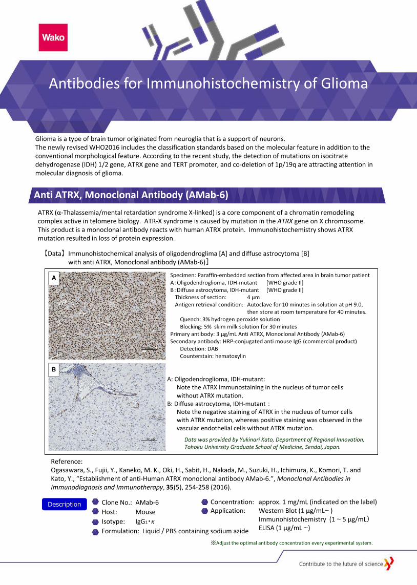
【Data】 Immunohistochemical analysis of oligodendroglima [A] and diffuse astrocytoma [B] with anti ATRX, Monoclonal antibody (AMab-6)]
Specimen: Paraffin-embedded section from affected area in brain tumor patient A:Oligodendroglioma, IDH-mutant [WHO grade II] B:Diffuse astrocytoma, IDH-mutant [WHO grade II]
Thickness of section: 4 μm Antigen retrieval condition: Autoclave for 10 minutes in solution at pH 9.0,
then store at room temperature for 40 minutes. Quench: 3% hydrogen peroxide solution Blocking: 5% skim milk solution for 30 minutes
Primary antibody: 3 μg/mL Anti ATRX, Monoclonal Antibody (AMab-6) Secondary antibody: HRP-conjugated anti mouse IgG (commercial product)
Detection: DAB Counterstain: hematoxylin
A: Oligodendroglioma, IDH-mutant: Note the ATRX immunostaining in the nucleus of tumor cells without ATRX mutation.
B: Diffuse astrocytoma, IDH-mutant : Note the negative staining of ATRX in the nucleus of tumor cells with ATRX mutation, whereas positive staining was observed in the vascular endothelial cells without ATRX mutation.
Data was provided by Yukinari Kato, Department of Regional Innovation, Tohoku University Graduate School of Medicine, Sendai, Japan.
Reference: Ogasawara, S., Fujii, Y., Kaneko, M. K., Oki, H., Sabit, H., Nakada, M., Suzuki, H., Ichimura, K., Komori, T. and Kato, Y., “Establishment of anti-Human ATRX monoclonal antibody AMab-6.”, Monoclonal Antibodies in Immunodiagnosis and Immunotherapy, 35(5), 254-258 (2016).
Description Clone No.: AMab-6 Host: Mouse Isotype: IgG1・κ Formulation: Liquid / PBS containing sodium azide
Concentration: approx. 1 mg/mL (indicated on the label) Application: Western Blot (1 μg/mL~ ) Immunohistochemistry (1 ~ 5 μg/mL) ELISA (1 μg/mL ~)
※Adjust the optimal antibody concentration every experimental system.
Telomerase is a complex of TERT(Telomerase Reverse Transcriptase), template RNA and regulatory subunits, and TERT is a catalytic subunit of telomerase. It is known that telomerase activity is retained in germ cells and tissue stem cells, and also retained high in 80 to 90% of tumor cells. This antibody is a monoclonal antibody against TERT, which is a catalytic subunit of telomerase, and is particularly useful for immunohistochemical staining. It is known that TERT protein expression is suppressed by mutations on the TERT promoter region in some types of glioma. This phenomenon is observed mutually exclusively with the ATRX gene abnormality. TERT promoter mutations are seen in almost all of the gliomas where IDH gene mutation and 1p/19q co-deletion are found, and also seen in some types of gliomas in which IDH gene mutation does not occur.
Anti TERT, Monoclonal Antibody (TMab-6)
Isocitrate dehydrogenase (IDH) is an oxidoreductase that converts isocitric acid and α-ketoglutaric acid to each other. In humans, three forms of IDHs are known: IDH1 (cytoplasmic form, NADP+ dependent), IDH2 (mitochondrial form, NADP+ dependent) and IDH3 (mitochondrial from, NAD+ dependent). It has been reported that mutations on IDH1 and IDH2 genes occur frequently in various cancers, such as cholangiocarcinoma, acute myelogenous leukemia, chondrosarcoma, osteosarcoma and glioma. Wako has antibodies that recognize IDH1, IDH2 and wild type/mutated IDH1/2.
Anti IDH, Monoclonal Antibodies
Description Clone No.: TMab-6 Host: Mouse Isotype: IgM・κ Formulation: liquid / PBS containing sodium azide
Concentration: approx. 1 mg/mL (indicated on the label) Application: Western Blot(1 μg/mL ~) Immunohistochemistry (1 μg/mL ~) ELISA (1 μg/mL ~)
Mutation of IDH1 is almost restricted to Arg at amino acid position 132, and the R132H mutation, where Arg at position 132 is converted to His, has been reported to account for 80 to 90% of the mutations. This is an antibody that specifically recognizes IDH1-R132H in which Arg at position 132 is mutated to His. It does not recognize wild type IDH1 and mutated IDH1 other than R132H.
Anti IDH1-R132H, Monoclonal Antibody (HMab-2) [Wako Cat. No.013-26851]
Anti IDH1-R132H, Monoclonal Antibody (HMab-1) [Wako Cat. No. 018-24081]
This is an antibody (Clone No. HMab-2) that specifically recognizes IDH1-R132H as well as the antibody (Clone No. HMab-2) does. It does not recognize wild type IDH and mutated IDH1 other than R132H. This antibody can be used with high sensitivity even under conditions of low antigen and low antibody concentration.
Description Clone No.: HMab-1 Host: Mouse Isotype: IgG1
Formulation: liquid / PBS containing sodium azide
Concentration: approx. 1mg/mL (indicated on the label) Application: Western Blot (5 μg/mL) Immunohistochemistry (5 ~ 10 μg/mL) ELISA (1 ~ 5 μg/mL)
Description Clone No.: HMab-2 Host: Mouse Isotype: IgG1・κ Formulation: liquid / PBS containing sodium azide
Concentration: approx. 1 mg/mL (indicated on the label) Application: Western Blot (1 μg/mL ~)
Immunohistochemistry (1 μg/mL) ELISA (1 μg/mL ~)
References: 1) Fujii, Y. et al., Biochem. Biophys. Res. Commun., 466(4), 733-739 (2015). 2) Yamamichi, A. et al., Sci. Technol. Adv. Mater., 17, 618-625 (2016).
References: 1) Takano, S. et al., J. Neurooncol., 108(3), 361-373 (2012). 2) Sabit, H. et al., Brain Tumor Pathol., 31(4), 242-246 (2014). 3) Takano, S. et al., Brain Tumor Pathol., 32(3), 169-175 (2015). 4) Kato, Y., Brain Tumor Pathol., 32(1), 3-11 (2015).
※Adjust the optimal antibody concentration every experimental system.
※Adjust the optimal antibody concentration every experimental system.
※Adjust the optimal antibody concentration every experimental system.
IDH1 mutations occur in which Arg at position 132 is converted to Cys, Ser, Gly or Leu, instead of His. This monoclonal antibody recognizes IDH1-R132S in which Arg at position 132 is converted to Ser.
Anti IDH1, Rat Monoclonal Antibody (RcMab-1) [Wako Cat. No. 014-26381]
Anti IDH1-R132S, Monoclonal Antibody (SMab-1) [Wako Cat. No. 015-24091]
This is an antibody that recognizes wild-type/mutated IDH1. It does not recognize IDH2. It can used as a positive and loading control antibody to detect brain tissue-derived proteins at immunohistostaining and western blotting
Anti IDH1, Monoclonal Antibody (RMab-3) [Wako Cat. No. 014-24061]
This is an antibody that recognizes wild-type/mutated IDH1. It can be used for immunohistochemical staining of various types of tissue sections derived from glioma.
Description Clone No.: SMab-1 Host: Mouse Isotype: IgG1
Formulation: liquid / PBS containing sodium azide
Concentration: approx. 1 mg/mL (indicated on the label) Application: Western Blot (5 μg/mL) Immunohistochemistry (5 ~ 10 μg/mL) ELISA (1 ~ 5 μg/mL)
※Adjust the optimal antibody concentration every experimental system.
Description Clone No.: RMab-3 Host: Mouse Isotype: IgG1
Formulation: liquid / PBS containing sodium azide.
Concentration: approx. 1 mg/mL (indicated on the label) Application: Western Blot (5 μg/mL) Immunohistochemistry (5 ~ 10 μg/mL) ELISA (1 ~ 5 μg/mL)
Description Clone No.: RcMab-1 Host: Rat Isotype: IgG2a
Formulation: liquid / PBS containing sodium azide.
Concentration: approx. 1 mg/mL (indicated on the label) Application: Western Blot (0.5 ~ 1 μg/mL) Immunohistochemistry (1 ~ 5 μg/mL) Immunocytochemistry (1 ~ 5 μg/mL)
References: 1) Ikota, H. et al., Brain Tumor Pathol., 32(4), 237-244 (2015). 2) Kaneko, M.K. et al., Tohoku J. Exp. Med., 230, 103-109 (2013). 3) Kato, Y. et al., Biochem. Biophys. Res. Commun., 432, 546-567 (2013). 4) Kaneko, M.K. et al., Monoclon. Antib. Immunodiagn. Immunother., 32(3), 224-228 (2013). 5) Yamamichi, A. et al., Sci. Technol. Adv. Mater., 17(1), 618-625 (2016).
Anti IDH2, Monoclonal Antibody (RMab-22) [Wako Cat. No. 011-24071]
This is an antibody that recognizes wild-type IDH2 and IDH2-R172K/M in mutated IDH2. It does not react to IDH2-R172W. IDH2 mutations occur in which Arg at position 172 is converted to Lys, Met, Gly or Trp.
Description Clone No.: RMab-22 Host: Mouse Isotype: IgG2b
Formulation: liquid / PBS containing sodium azide.
Concentration: approx. 1 mg/mL (indicated on the label) Application: Western Blot (5 μg/mL) Immunohistochemistry (5 ~ 10 μg/mL) ELISA (1 ~ 5 μg/mL)
References: 1) Kaneko, M.K. et al., Biochem. Biophys. Res. Commun., 432, 40-45 (2013).
References: 1) Kaneko, M. K., et al., Tohoku J. Exp. Med., 230, 103-109 (2013). 2) Ogasawara, S. et al., Monoclon. Antib. Immunodiagn. Immunother., 35(5), 254-258 (2016).
References: 1) Kaneko, M. K. et al., Biochem. Biophys. Res. Commun., 406(4), 608-613 (2011). 2) Kato, Y., Brain Tumor Pathol., 32(1), 3-11 (2015).
※Adjust the optimal antibody concentration every experimental system.
※Adjust the optimal antibody concentration every experimental system.
※Adjust the optimal antibody concentration every experimental system.
Wako Pure Chemical Ind., Ltd. www.wako-chem.co.jp 1-2, Doshomachi 3-Chome Chuo-Ku, Osaka 540-8605, Japan Tel: 81-6-6203-3741 Fax: 81-6-6203-1999 Online Catalog: www.e-reagent.com [email protected]
Wako Chemicals USA, Inc. www.wakousa.com 1600 Bellwood Road Richmond, VA 23237, USA Toll-Free (U.S. only): 1-877-714-1920 Tel: 1-804-714-1920 Fax: 1-804-271-7791
Wako Chemicals GmbH www.wako-chemicals.de Europe Office Fuggerstraße 12, D-41468 Neuss, Germany Tel: 49-2131-311-0 Fax: 49-2131-311 100
Listed products are intended for laboratory research use only, and not to be used for drug, food or human use. / Please visit our online catalog to search for other products from Wako; http://www.e-reagent.com / This leaflet may contain products that cannot be exported to your country due to regulations. / Bulk quote requests for some products are welcomed. Please contact us.
1 7 3 G
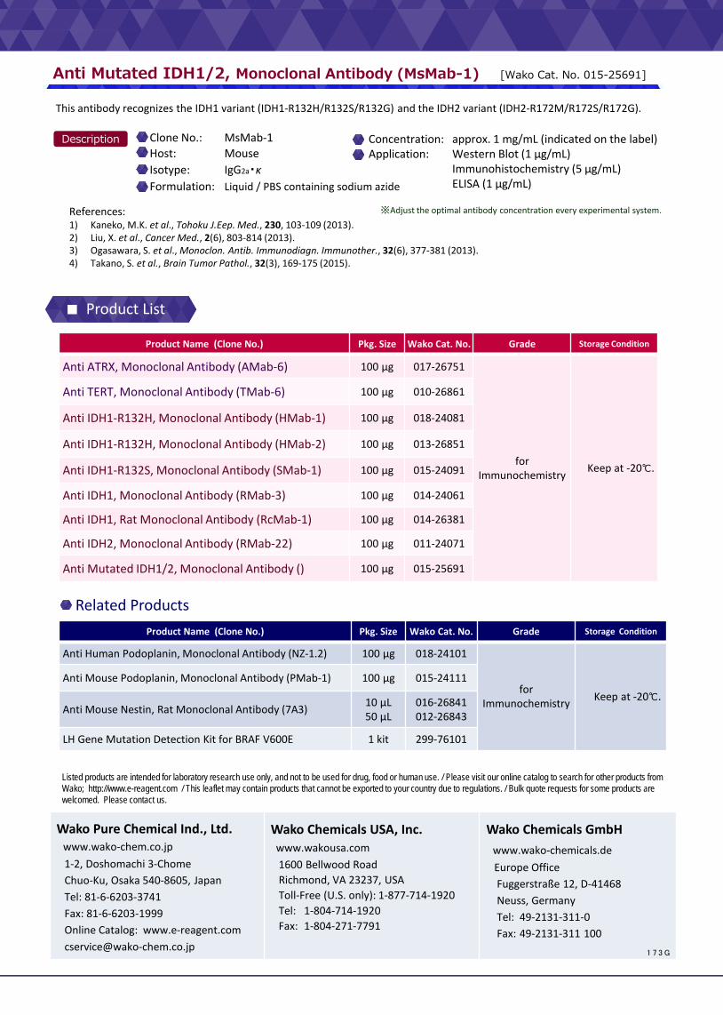
Anti Mutated IDH1/2, Monoclonal Antibody (MsMab-1) [Wako Cat. No. 015-25691]
This antibody recognizes the IDH1 variant (IDH1-R132H/R132S/R132G) and the IDH2 variant (IDH2-R172M/R172S/R172G).
Description Clone No.: MsMab-1 Host: Mouse Isotype: IgG2a・κ Formulation: Liquid / PBS containing sodium azide
Concentration: approx. 1 mg/mL (indicated on the label) Application: Western Blot (1 μg/mL) Immunohistochemistry (5 μg/mL) ELISA (1 μg/mL)
References: 1) Kaneko, M.K. et al., Tohoku J.Eep. Med., 230, 103-109 (2013). 2) Liu, X. et al., Cancer Med., 2(6), 803-814 (2013). 3) Ogasawara, S. et al., Monoclon. Antib. Immunodiagn. Immunother., 32(6), 377-381 (2013). 4) Takano, S. et al., Brain Tumor Pathol., 32(3), 169-175 (2015).
Product Name (Clone No.) Pkg. Size Wako Cat. No. Grade Storage Condition
Anti ATRX, Monoclonal Antibody (AMab-6) 100 μg 017-26751
for Immunochemistry Keep at -20℃.
Anti TERT, Monoclonal Antibody (TMab-6) 100 μg 010-26861
Anti IDH1-R132H, Monoclonal Antibody (HMab-1) 100 μg 018-24081
Anti IDH1-R132H, Monoclonal Antibody (HMab-2) 100 μg 013-26851
Anti IDH1-R132S, Monoclonal Antibody (SMab-1) 100 μg 015-24091
Anti IDH1, Monoclonal Antibody (RMab-3) 100 μg 014-24061
Anti IDH1, Rat Monoclonal Antibody (RcMab-1) 100 μg 014-26381
Anti IDH2, Monoclonal Antibody (RMab-22) 100 μg 011-24071
Anti Mutated IDH1/2, Monoclonal Antibody () 100 μg 015-25691
Product Name (Clone No.) Pkg. Size Wako Cat. No. Grade Storage Condition
Anti Human Podoplanin, Monoclonal Antibody (NZ-1.2) 100 μg 018-24101
for Immunochemistry Keep at -20℃.
Anti Mouse Podoplanin, Monoclonal Antibody (PMab-1) 100 μg 015-24111
Anti Mouse Nestin, Rat Monoclonal Antibody (7A3) 10 μL 50 μL
016-26841 012-26843
LH Gene Mutation Detection Kit for BRAF V600E 1 kit 299-76101
Related Products
※Adjust the optimal antibody concentration every experimental system.
■ Product List