Antibiotic Susceptibility of Probiotic Bacteria...26 Antibiotic Susceptibility of Probiotic Bacteria...
30
26 Antibiotic Susceptibility of Probiotic Bacteria Zorica Radulović 1 , Tanja Petrović 1 and Snežana Bulajić 2 1 University of Belgrade, Faculty of Agriculture 2 University of Belgrade, Faculty of Veterinary Medicine Republic of Serbia 1. Introduction Lactic acid bacteria (LAB) are a heterogeneous group of bacteria widely distributed in nature. These bacteria are found in gastrointestinal (GI) and urogenital tract of humans and animals; they are present on plant material, in milk and meat, and numerous fermented foods. Lactic acid bacteria have been associated with traditional dairy products, cereals, vegetable and meat fermented foods, due to their natural presence leading to spontaneous fermentation. They are also used as starter cultures in industrial food production, as well as in the production of probiotic products due to their potential health benefits to consumer. Milk and dairy products are the most examined food system for the delivery of probiotic bacteria to the human gut. The probiotic concept has progressed and is now in the focus of different research. Significant improvements have been made in selection and characterization of new cultures and their application in food production. The food products, which are produced by traditional methods, exhibit a rich biodiversity with the respect to bacterial contents. From these products, new probiotic strains with the potential functional properties can been isolated and selected. The selected strains have to be further characterized in order to be used in the food industry. Before the probiotics can benefit human health, they must fulfill several criteria including: a) scientifically validated health properties; b) good technological properties meaning that they can be manufactured and incorporated into food products without loosing viability, functionality and technological performance; c) high survival through the upper gastrointestinal tract and high viability at its site of action; d) antagonistic activity to pathogens; e) antibiotic susceptibility; and f) to be able to function in the gut environment. Bearing in mind importance of antibiotic resistance of LAB in food chain, antibiotic susceptibility of potential probiotic strains is a very important criteria for their selection. In the recent decade, releasing of antibiotics in biosphere seriously increased, leading to a strong selective pressure for the emergence and persistence of resistant LAB strains. Since LAB are naturally present in traditionally made fermented food and GI tract and are also added as starter culture or probiotic bacteria in industrial food production, concerns have been raised about the antibiotic resistance of these beneficial bacteria strains. Probiotic bacteria can help maintaining balance in gastrointestinal tract in cases of diarrhea caused by antibiotic treatment. However, there is high risk associated with the ability of these resistant www.intechopen.com
Transcript of Antibiotic Susceptibility of Probiotic Bacteria...26 Antibiotic Susceptibility of Probiotic Bacteria...

26
Antibiotic Susceptibility of Probiotic Bacteria
Zorica Radulović1, Tanja Petrović1 and Snežana Bulajić2 1University of Belgrade, Faculty of Agriculture
2University of Belgrade, Faculty of Veterinary Medicine Republic of Serbia
1. Introduction
Lactic acid bacteria (LAB) are a heterogeneous group of bacteria widely distributed in
nature. These bacteria are found in gastrointestinal (GI) and urogenital tract of humans and
animals; they are present on plant material, in milk and meat, and numerous fermented
foods. Lactic acid bacteria have been associated with traditional dairy products, cereals,
vegetable and meat fermented foods, due to their natural presence leading to spontaneous
fermentation. They are also used as starter cultures in industrial food production, as well as
in the production of probiotic products due to their potential health benefits to consumer.
Milk and dairy products are the most examined food system for the delivery of probiotic
bacteria to the human gut. The probiotic concept has progressed and is now in the focus of
different research. Significant improvements have been made in selection and
characterization of new cultures and their application in food production.
The food products, which are produced by traditional methods, exhibit a rich biodiversity
with the respect to bacterial contents. From these products, new probiotic strains with the
potential functional properties can been isolated and selected. The selected strains have to
be further characterized in order to be used in the food industry. Before the probiotics can
benefit human health, they must fulfill several criteria including: a) scientifically validated
health properties; b) good technological properties meaning that they can be manufactured
and incorporated into food products without loosing viability, functionality and technological
performance; c) high survival through the upper gastrointestinal tract and high viability at
its site of action; d) antagonistic activity to pathogens; e) antibiotic susceptibility; and f) to be
able to function in the gut environment. Bearing in mind importance of antibiotic resistance
of LAB in food chain, antibiotic susceptibility of potential probiotic strains is a very
important criteria for their selection.
In the recent decade, releasing of antibiotics in biosphere seriously increased, leading to a
strong selective pressure for the emergence and persistence of resistant LAB strains. Since
LAB are naturally present in traditionally made fermented food and GI tract and are also
added as starter culture or probiotic bacteria in industrial food production, concerns have
been raised about the antibiotic resistance of these beneficial bacteria strains. Probiotic
bacteria can help maintaining balance in gastrointestinal tract in cases of diarrhea caused by
antibiotic treatment. However, there is high risk associated with the ability of these resistant
www.intechopen.com

Antibiotic Resistant Bacteria – A Continuous Challenge in the New Millennium
550
strains to transmit the resistance gene to pathogenic bacteria in gut microbiota. This can
complicate the treatment of a patient with an antibiotic resistant bacterial infection or
disease. The circulation of genes coding for antibiotic resistance from beneficial LAB in the
food chain via animals to humans is a complex problem. Therefore, there is a need to evaluate
the safety of potential probitic strains regarding their ability to acquire and disseminate
antibiotic resistance determinants in selection of LAB.
In this study, importance of LAB in the food chain will be reviewed. Morphological and
biochemical characteristics of lactobacilli, bifidobactera and enterococci, as well as criteria
for probiotic selection and role of probiotics in health benefit will be discussed. Antibiotic
susceptibility as criteria for potential probiotic bacteria selection and mechanisms of gene
transfers will be considered.
2. Lactic acid bacteria in the GI tract
The human GI tract represents a complex ecosystem in which interactions between food,
microbes and the host cells occur. The bacterial population of normal gut of an adult
comprise of more than 500 different species. The quantity of microbes present in the
intestine (about 1014) exceeds 10-fold the total number of all human cells (Backhed et al.,
2005). The most important function of this intestinal microbiota is to act as a microbial
barrier against pathogens, by so-called competitive exclusion mechanisms, but also
influence the humoral and cellular mucosal immune responses during the neonatal phase of
life, and thereafter to maintain a physiologically-normal steady-state condition throughout
life (Tancrede, 1992). The gut microflora profoundly influences nutritional, physiologic and
protective processes. Both direct and indirect defensive functions are provided by the
normal microbiota. Specifically, gut bacteria directly prevent colonization by pathogenic
organisms by competing for essential nutrients or for epithelial attachment sites. By
producing antimicrobial compounds, volatile fatty acids, and chemically modified bile acids,
indigenous gut bacteria also create a local environment that is generally unfavourable for the
growth of enteric pathogens. This phenomenon is called Colonization Resistance, which can be
defined as the ability of microorganisms belonging to the normal gut microflora to impede the
implantation of pathogens (van der Waaij, 1988). This function of the microflora is also known
as the barrier effect. While probiotic bacteria improve colonization resistance, consensus
thinking is that the importance of LAB as probiotic agents lies more in the indirect
mechanisms such as immunomodulation. When the genetic repertoire of these bacteria is
considered, the GI tract translates into a reservoir of genes encoding numerous
physiological functions from which the human GI tract can benefit. Bacteria represent the
most extensively investigated group of microorganisms. Which species of bacteria will be
long-term colonized in GI tract depends on the biochemical capability of the
microorganisms, the microenvironment determined by the host cells and the available
foodstuffs. Lactobacilli are probably the most well known representative of favourable
microorganisms in GI tract. There is a number of species of lactobacilli reside in the human
intestine in a symbiotic relationship with each other and with other microorganisms
(Claesson et al., 2007). They are generally considered essential for maintaining gut
microfloral health; however, it is the overall balance of the various microorganisms which is
ultimately of most importance.
www.intechopen.com

Antibiotic Susceptibility of Probiotic Bacteria
551
2.1 The composition of the GI tract
The composition of the GI microflora undergoes considerable changes from the day of birth until adulthood. The GI tract of a normal fetus is sterile, but colonization begins immediately after birth and is influenced by the mode of delivery, the infant diet, hygiene levels and medication (Benett et al., 1986; Gronlund et al., 1999). During the first months of life diet has a significant influence on the development of the intestinal flora (Heavey & Rowland, 1999; Stark & Lee, 1982). Within one to two days facultative anaerobes predominate and create a reduced environment that allows the growth of strict anaerobes. Within three to four days, bifidobacteria appear and become predominant. The average number of bifidobacteria in breast-fed infants is 1010–1011 cfu/g. In formula fed infants, bifidobacteria have also been demonstrated to be a numerically important species, but they generally occur in lower numbers than in breastfed infants of the same age (Mountzouris et al., 2002). The predominance of beneficial bacteria in the gut microbiota of breast-fed infants is thought to result from the fermentation of oligosaccharides-non-digestible carbohydrates consisting of several linked monosaccharides (typically 3-10 simple sugars) in breastmilk (Agostoni et al., 2004). Oligosaccharides pass unabsorbed through the small intestine into the colon, where they are fermented by resident bifidobacteria to short-chain fatty acids and lactic acid, reducing the gut pH to approximately 5.7, thus providing the protection against enteric infections (Newburg, 2000, Coppa et al., 2004). In contrast, the gut microflora of formula-fed infants produces a different profile of short-chain fatty acids and a pH in the local microenvironment of approximately 7.0 (Ogawa et al., 1992). Infant faecal flora appears to be stabilized at 4 weeks of age and until weaning when introduction of solid foods takes place (Mountzouris et al., 2002). During weaning, bifidobacteria decrease by 1 log, the microbiota alters from infant-type to adult-type, and a remarkable proliferation of bacteroides, eubacteria, peptostreptococcaceae, and clostridia occur. The faecal flora of children closely resembles that of adults, where the numbers of bacteroidaceae, eubacteria, peptococcaceae, and usually clostridia outnumbered bifidobacteria, which constitute 5-10% of the total flora.
An adult individual’s GI tract is extremely stable and it is very difficult to introduce new species. There are several factors that can alter the composition of the GI flora such as medications (especially antibiotics), diet, climate, aging, illness, stress, pH, infections, geographic location, and even race (Murphy et al., 2009). Thus it is not surprising to find out that the composition of the GI flora not only differs among individuals, but also differs during the life within the same individual. Furthermore, indigenous bacteria are not distributed randomly throughout the gastrointestinal tract but instead are found at population levels and in species distributions that are characteristic of specific regions of the tract.
As shown in Figure 1, there are three main regions that offer very different conditions for the survival of various microorganisms in the GI environment. In the stomach, microbial growth is greatly reduced by the high acidity and presence of oxygen provided by the swallowing. As a result, in the stomach acidotolerant microorganisms and facultative anaerobes such as lactobacilli, streptococci, yeasts, etc. are present. In the second region (small intestine), the microflora consists mainly of facultative anaerobic bacteria such as lactobacilli, streptococci and enterobacteria, and anaerobes such as bifidobacteria, bacteroides and clostridia. In the last region (colon), the number of bacteria is considerably high, due to the low redox potential and relatively high concentration of short-chain fatty acid, and counts 109–1012 cfu/ml (Cummings et al. 1989). The colon has an important role in
www.intechopen.com

Antibiotic Resistant Bacteria – A Continuous Challenge in the New Millennium
552
food digestion, and microflora in this region participates in the transformation of many carbohydrates, proteins and amino acids. The microflora of the colon is very complex and dominated by anaerobic bacteria (Bacteroides spp., Clostridium spp, Bifidobacterium spp., etc), while the facultative anaerobic bacteria are less numerous and represented by lactobacilli, enterococci, streptococci and Enterobacteriaceae. Yeasts (eg., Candida albicans) are relatively poorly represented.
Fig. 1. The average concentration of microorganisms in the GI tract (adapted from Ouwehand & Vesterlund, 2003)
As part of gut microbiota, it is estimated that lactobacilli are present in following
concentrations: 103–106 cfu/ml in the oral cavity; 103 cfu/ml in the stomach; 104 cfu /ml in
the duodenum and jejunum; 108 cfu /ml in the ileum and 109 cfu /ml in the colon (Reuter,
2001; Koll et al., 2008; Ryan et al., 2008). An overview of lactobacilli commonly found in the
GI tract microbiota is shown in Table 1.
Considering that lactobacilli and bifidobacteria constitute a significant population in the GI
tract, this environment represents a good source for the isolation of new strains of LAB.
3. Lactic acid bacteria as probiotic bacteria
By definition probiotics are described as „living microorganisms, which upon ingestion in
certain numbers, exert health benefits beyond inherent basic nutrition“ (Guarner &
Schaafsma, 1998). Regarding the probiotics, the majority of research is focused on bacterial
genera Lactobacillus and Bifidobacterium (Table 2).
Small intestine
104-108 cfu/ml Lactobacillus
Streptococcus Enterobacteria Bacteroides Clostridum
Stomach
101-104 cfu/ml Helicibacter pylori, Lactobacillus Streptococcus
Candida albicans
Colon
109-1012 cfu/ml Bacteroides Bacillus Eubacterium Bifidobacterium Clostridium Peptococcus Peptostreptococcus Ruminococcus Actinomyces Lactobacillus Enterobacteriacae Enterococcus
www.intechopen.com
Antibiotic Susceptibility of Probiotic Bacteria
553
Oral cavity Stomach Small intestine Faeces Colon epithelial biopsies
L. paracasei L. gasseri L. gasseri L. gasseri L. plantarum
L. rhamnosus L. reuteri L. reuteri L. paracasei L. rhamnosus
L. fermentum L. ruminis L. rhamnosus L. ruminis L. paracasei
L. plantarum L. reuteri
L. gasseri L. plantarum
L. salivarius
L. sakei
Table 1. Lactobacillus species distribution in different parts of the GI tract (Lönnermark, 2010)
Lactobacillus sp. Bifidobacteriim sp. Enterococcus sp. Others
L. acidophilus B. bifidum E. faecium Lactococcus lactis ssp. lactis
L.plantarum B. infantis E. faecalis Lactococcus lactis ssp. cremoris
L. casei B. adolescentis Leuconostoc mesenteroides
L. rhamnosus B. longum Pediococcus acidilactici
L. delbrueckii ssp. bulgaricus B. breve Propionibacterium freudenreichii
L. fermentum B. lactis Streptococcus thermophilus
L. johnsonii
L. gasseri
L. salivarius
L. reuteri
Table 2. Lactic acid bacteria used as probiotics (adapted from Gardnier et al., 2002)
They have a reputation of health promoters and they have a significant role as probiotic
bacteria in the production of different foods, particularly in the production of fermented
dairy products. Central position of lactobacilli and bifidobacteria in probiotic formulation is
argumented due to (a) the association of these bacteria with human health, (b) the fact that
they possess the Generally Regarded As Safe (GRAS) status. Enterococci, although not
GRAS organisms, have been used as probiotics too. Some other LAB such as lactococci,
pediococci, Leuconostoc, propionibacteria have also received attention as potential probiotic
cultures (Table 2).
3.1 Lactobacillus
The genus Lactobacillus is large heterogeneous group of microorganisms, which lacks
catalase and cytochromes and is usually microaerophilic, with growth improved under
anaerobic conditions (Kandler & Weiss, 1989). They are Gram-positive nonmotile rods, often
in pairs or chains, coccobacilli to long rods. They have a strictly fermentative metabolism
and convert glucose solely or partly to lactic acid. They are classified as homofermentative
(producing mainly lactic acid) or heterofermentative (producing carbon dioxide, ethanol,
acetic acid and lactic acid). The optimum growth temperature is in the mesophilic range (30-
40ºC), but some strains can grow below 15ºC and some at temperatures up to 55ºC.
Differentiation of Lactobacillus species depends on physiological criteria, carbohydrate
fermentation, biochemical and molecular characterization (Petrovic et al., 2006). Lactobacilli
www.intechopen.com

Antibiotic Resistant Bacteria – A Continuous Challenge in the New Millennium
554
play crucial role in the production of fermented foods: vegetables, meats and dairy
products. Non starter lactic acid bacteria (NSLAB) are lactobacilli which form significant
proportion of the microflora of most cheese varieties during ripening. Many species of
mesophilic Lactobacillus have been isolated from cheese; the ones most frequently
encountered are L. casei, L. paracasei, L. pantarum, L. rhamnosus and L. curvatus (Beresford et
al., 2001). For example, L. plantarum, L. casei ssp. casei and L. brevis have been isolated from
Armada cheese, a Spanish goat milk cheese (Herreros et al., 2003), L. plantarum, L.
paraplantarum, L. paracasei ssp. tolerans, L. sake, L. curvaus and L. pentosus from Batzos, a
traditional Greek cheese made from raw goat′ s milk (Psoni et al., 2003) and L. para.paracasei,
L. plantarum, L. curvatus and L. brevis from Sjenica cheese, traditional Serbian white brined
cheese (Radulović, 2010). Traditional homemade dairy products have great potential for
isolation of new strains, which could be used as starter cultures in food industry, as adjunct
cultures for improving flavour of cheeses, or as probiotic in production of functional foods.
3.2 Bifidobacterium
Species from the genus Bifidobacterium are generally characterized as Gram-positive, non-
spore forming, nonmotile and catalase negative. They are strict anaerobes, although some
species and strains may tolerate oxygen in the presence of carbon dioxide. Within the genus
Bifidobacterium pleomorphism exists and it is described as short regular, thin cells with
pointed ends, long cells with slight bends with a large variety of branching; single or in
chains of many elements; in star-like clusters or disposed in “V” or palisade arrangements
(Scardovi, 1986). In bifidobacteria, glucose catabolism occurs through the fructose 6-
phosphate phodphoketolase pathway, which can be used as distinguishing feature of
bifidobacteria. During fermentation, acetic and lactic acids are produced in molar ratios 3:2.
The optimum temperature for growth is 31-41ºC within the range 25-45ºC. Bifidobacteria are
less acid tolerant than lactobacilli and no growth occurs at pH values less than 4.5 (Scardovi,
1986). Nutritional requirements for growth of bifidobacteria are less complex than those of
lactobacilli, but in some cases bifidobacteria do require specific factors for optimal growth
(Modler et al., 1990). Bifidobacteria are natural habitants of GI tract and strains with
probiotic properties are mainly of human origin. Bifidobacterium species constitute a
significant portion of probiotic cultures used in functional food production.
3.3 Enterococcus
Although lactobacilli and bifidobacteria are most commonly used as probiotics, some
enterococci can also be used as health-promoting bacteria. Species E. faecium and E. faecalis
have been used as probiotic bacteria, although they are not recognized as GRAS organisms.
All species of genus Enterococcus are Gram-positive, non-spore forming, catalasa negative,
facultative anaerobes. They are spherical or ovoid cocci in pairs or short chains. Their
properties, such as the ability to grow at 10ºC and 45ºC in 6.5%NaCl and at pH 9.6 and their
survival heating to 60ºC for 30 min, are used to differentiate enterococci from other Gram-
positive catalase-negative cocci (Franz et al., 1999). Enterococci are homofermentative with
respect to glucose metabolism, although some amounts of formic and lactic acid may be
produced in some media. As the other LAB, enterococci do require B vitamins, amino acids,
purine and pyramidine bases for optimal growth (Garg & Mital, 1991).
www.intechopen.com

Antibiotic Susceptibility of Probiotic Bacteria
555
3.4 LAB associated with therapeutic properties
In recent years an increasing number of probiotic pharmaceutical preparations as well as food supplements are being promoted with health claims. The application of LAB has been more developed for the production of functional foods, where probiotic bacteria have an important role. The commercial probiotic strains, used in functional food production, must be well-substantiated with scientific evidence.
The health benefits of probiotic bacteria can be considered as nutritional or
therapeutic/prophylactic. Nutritional benefits are mainly connected with their role in
enhancing the bioavailability of vitamins and minerals, and an increase of the digestibility of
protein (Tamime et al., 2003). Many researchers (Begley et al., 2006; Ouwehand et al., 2003;
Rodrigeuz et al., 2010) investigated the therapeutic/prophylactic benefits of probiotics and
confirmed their effects:
- Prevention of diarrhoea caused by certain pathogenic bacteria and viruses; - Regulation of intestinal microflora after an antibiotic therapy; - Treating the infection with Helicobacter pylori, responsible for the development of
gastritis, ulcers and gastric cancer; - Improvement of digestion in "lactose-intolerant" individuals, who have reduced
capability for lactose digestion; - Anticancer effect, as a result of the production of certain compounds during its growth; - Reducing cholesterol levels;
- Stimulation of -interferon production, which contributes to increased resistance to some infections;
- Increasing antibody titar (IgG immunoglobulins), which enhances the immune response of an organism;
Health effects are related to microflora modification and strengthening of the gut mucosal
barrier. Some scientific data indicated that probiotic strains have potential in the prevention
and treatment of intestinal and urogenital infections and these cultures may be useful as an
alternatives to antibiotic therapy. The World Health Organization (WHO) has recommended
the reconsideration of microbial interference therapy for infection control (Bengmark, 1998).
Certain probiotic strains have been shown multiple effects including prevention of pathogen
attachment and invasion in cell culture, inhibiting of the growth of enteropatogens in vitro
and enhancement the immune response. Considering thee effects, the usage of probiotics
may decrease antibiotics dependence. Fooks et al., (1999) have suggested that probiotic
bacteria can control the infection in several ways such as the competition for nutrients,
secretion of antimicrobial substances, reduction of pH, blocking of adhesion sites, reduction
of virulence, blocking of toxin receptor sites, immune stimulation and suppression of toxin
production.
Infections by bacterial or viral agents most frequently result in diarrhea. One of the most
investigated probiotic, L.rhamnosus GG, has been very effective in the treatment of viral
diarrhea in children, most cases of which were caused by rotavirus. Other probiotic strains
such as Lactocacillus casei Shirota, B. bifidum and S. thermophillus have also been shown to be
effective in the treatment and prevention of rotavirus diarrhea in children (Korhonen, et al.,
2007; Saavedra et al., 1994).
www.intechopen.com
Antibiotic Resistant Bacteria – A Continuous Challenge in the New Millennium
556
Antibiotic-associated diarrhea (AAD) is a major clinical problem that occurs following antibiotic use. The most serious form of this kind is pseudomembranous colitis. Diarrhea is caused by pathogen overgrowth and in 20% cases the etiological agent is Clostridium difficile, a pathogen that is especially persistent and difficult to treat (Lewis & Freedman, 1998). Antibiotics are often used to treat for pseudomembranous colitis or other AAD, although the relapse may occurs when the probiotic therapy may be especially useful. Oral therapy with L. rhamnosus GG was effective in the prevention of AAD, in treatment of colitis, as well as in traveller's diarrhea (Shah, 2007). A combination of B. bifidum, S. thermophilus, L. delbrueckii spp. bulgaricus and L. acidophilus have been also effective in the prevention of traveller's diarrhea (Black et al., 1989).
On the other hand, many studies have shown no effect of probiotic treatment (Lewis & Freedman, 1998). There is still some doubts regaring the quality and efficacy of probiotic products. Nevertheless, the probiotic food industry is flourishing. In many European counties the market is expanding resulting in sell of probiotic yogurts that account for over 10% of all yogurts sold in Europe (Stanton et al., 2001).
4. Criteria associated with probiotic bacteria
Consortium of LAB constitute a major part of the natural microflora of human intestine and when present in sufficient numbers create a healthy equilibrium between beneficial and potentially harmful microflora in the gut (Dune et al., 2001). For some positive effects on human health, a probiotic strain has to reach the large intestine at a concentration of about 107 viable cells/g (Stanton et al., 2001). Microorganisms ingested with food begin their journey to the lower intestinal tract via the mouth and are exposed during their transit through the GI tract to successive stress factors that influence their survival. The time from entrance to release from the stomach is about 90 min, but further digestive processes have longer residence times (Berrada et al., 1991). Probiotic bacteria must overcome physical and chemical barriers in the GI tract, especially acidic environment of the stomach, and then the activity of hydrolytic enzymes and bile salts in the small intestine. In a typical acid tolerance tests, the viability of potential probiotic organisms is determined by exposing them to low pH in a buffer solution or medium for a certain period of time, during which the number of surviving probiotic bacteria is determined. The generally requirements for probiotics are shown in Table 3.
Acid and bile stability
Adherence to human intestinal cells
Ability to reduce the adhesion of pathogens to surfaces
Colonization of human GI tract
Antagonism against carcinogenic and pathogenic bacteria
Production of anti-microbial substances
Survive the various technological processes of production
Safety evaluation: nonpathogenic, nontoxic, nonallergic, nonmutagenic
Desirable metabolic activity and antibiotic resistance/sensitivity
Clinically validated and documented health effects
Table 3. The desired properties of probiotic strains (adapted from Mattila & Sarraela, 2000)
www.intechopen.com

Antibiotic Susceptibility of Probiotic Bacteria
557
Bile plays an essential role in lipid digestion; it emulsifies and solubilizes lipids and functions as biological detergent. Prior to secretion into the duodenum, bile acids, which are synthesized from cholesterol, are conjugated to either glycine or taurine in liver (Begley et al., 2006). In the colon conjugated bile undergoes to the various chemical changes including deconjugation, dehydroxylation, dehydrogenation, and deglucuronidation, almost solely by microbial activity (Begley et al., 2006). Bile reduces the survival of bacteria by destroying their cell membranes, whose major components are lipids and fatty acids and these modifications may affect not only the cell permeability and viability, but also the interactions between the membranes and the environment. Bile salt hydrolases (BSHs) are generally intracellular, oxygeninsensitive enzymes that catalyze the hydrolysis of bile salts (Liong & Shah, 2005). A number of BSHs have been identified and characterized in probiotic bacteria (Franz et al., 2001a).
The adhesion ability as well as interaction with pathohens is regarded as important selection criteria for potential probiotic strains (Salminen et al. 2010). Adhesive properties of LAB depend on a variety of factors, including non-specifc adhesion determined by electrostatic or hydrophobic forces and specific binding dependent on particular molecules. To examine the adhesive property of LAB, several models have been developed. These include binding to tissue culture cells (Tuomola & Salminen, 1998), radiolabelling (Bernet et al., 1993), intestinal mucus (Ouwehand et al., 2001), extracellular matrix proteins (de Leeuw et al., 2006) and resected colonic tissue (Vesterlund et al., 2005). Although none of these models reflect the complex interactions occurring in the mucosal layer of the digestive tract, they represent a rapid method for the screening of potential probiotic strains.
Other functional property used to characterize probiotics is the production of antimicrobial compounds. Several antimicrobial substances that have considerable advantages in competition with pathogens and other harmful bacteria are produced by LAB (Klare et al., 2007; Radulović et al., 2010a). These substances include fatty acids, organic acids, hydrogen peroxide, and diacetyl, acetoin and the most studied inhibitory peptides called ‘bacteriocins’ (Todorov et al., 2011). The ability of probiotics to establish in the GI tract is enhanced by their ability to eliminate competitors. Some examples of antimicrobial substances produced by probiotic bacteria are presented in Table 4.
Probiotic Compaund
Lactobacillus GG Wide spread antibiotic L. acidophilus Acidolin,Acidophilin, Lactocidin L. delbrueckii ssp. bulgaricus Bulgarican L. plantarum Lactolin L. brevis Lactobacillin,Lactobrevin L. reuteri Reuterin
Table 4. Antimicrobial substances (Fuller, 1992)
Antibiotic susceptibility of potential probiotic strains is also considered as an important selection criterion for potential probiotic status (Hummel et al., 2007). Some LAB may carry potentially transmissible plasmid-encoded antibiotic resistance genes. The transmission of antibiotic resistance genes to unrelated pathogenic or potentially pathogenic bacteria in the gut is a major health concern related with the probiotic application (see detailed information in paragraph 6).
www.intechopen.com
Antibiotic Resistant Bacteria – A Continuous Challenge in the New Millennium
558
Along with above mentioned criteria, selected probiotic strains have to be able to survive well in the food and to have the appropriate technological properties (e.g. acidification during fermentation if required). In addition, it is important that the added probiotic does not adversely affect the taste, smell, and texture of the food or beverage.
In vitro tests based on these selection criteria, although not a definite means of strain selection, may provide an useful initial information. A validation model system, such as a Simulator of the Human Intestinal Microbial Ecosystem (SHIME), which aims to mimic complex physiological and physiochemical in vivo reactions, may also be of value in strain selection. However, the ultimate proof of probiotic effects requires validation in well designed statistically sound clinical trails. Generally, tools that may be employed in such an assessment include in vitro studies, studies of strain properties, pharmacokinetic studies, animal studies, use of intestinal models, human studies and epidemiological surveillance. Each strain needs to be tested separately.
It is evident that the selection of new potential probiotic bacteria is an enormous and time-consuming task with uncertain results.
5. Antibiotic resistance of LAB in the food chain
During the recent years, there has been great concern about the possibility of spreading the
antibiotic resistance in the environment. According to the European Commission (2005), it
has been estimated that one to ten million tons of antibiotics has been released into the
biosphere over the last 60 years. This has lead to very strong selective pressure for the
emergence of resistant bacterial strains. Since LAB are present in the GI tract in large
amounts, LAB resistant to certain antibiotics could benefit the host organism. Nevertheless,
there is a risk associated with the ability of these resistant strains to transmit the resistance
gene to pathogenic bacteria. Literature data pointed out that some LAB, the predominant
microbiota in fermented dairy and meat products, may serve as reservoirs of antibiotic
resistance genes potentially transferable to human pathogens (Mathur & Singh, 2005).
The food chain could be regarded as one of the main pathways for the transmission of antibiotic resistant bacteria from animals to humans (Singer et al., 2003). Molecular analysis of resistance genes localized on transferable genetic elements showed they are identical in humans and animals, which confirm that food of animal origin, particularly sausages and cheeses made from raw milk, serve as a vehicle for the transmission of resistant bacteria and antibiotic resistance determinants. The antibiotics application in sub-therapeutic levels in animal′s drinking water and feed increases the selective pressure and amplify the transfer of antibiotic resistance between bacterial species. Thus there is a direct correlation between the indigenous microflora of the GI tract of animals with the GI tract of humans. Although many species of LAB used as starter and probiotic cultures possess GRAS status, potential risk to human health caused by the genes transfer of antibiotic resistance has not yet been fully defined. To address this aspect, the safety of LAB should be verified with the respect of their ability to acquire and disseminate resistance determinants (Kastner et al., 2006). Particular concern is due to the evidence of a widespread occurrence in this bacterial group of conjugative plasmids and transposons. The presence of transmissible antibiotic resistance markers in the safety evaluation of LAB strains is a very important task since genes conferring resistance to several antimicrobials (e.g. chloramphenicol, erythromycin,
www.intechopen.com

Antibiotic Susceptibility of Probiotic Bacteria
559
streptomycin, tetracycline, and vancomycin) located on transferable genetic elements (plasmids or transposons) have already been characterized in lactococci (Perreten et al., 1997), lactobacilli (Axelsson et al., 1988; Danielsen, 2002) and enterococci (Eaton & Gasson, 2001; Huys et al., 2002) isolated from food. However, the transfer of antibiotic resistance genes from LAB reservoir strains to bacteria in the resident microflora of human GI tract and hence to pathogenic bacteria, has not been fully addressed.
Irrespective of the antibiotic resistance mechanisms and the bacterial taxon involved, the
possibility of spreading an antibiotic resistance determinant through horizontal transfer
relies on its genetic basis. Therefore, a distinction between intrinsic and acquired resistance
has to be made. Antibiotic resistance may be intrinsic for bacterial species or a genus, and it
is characterized by the ability of an organism to survive in the presence of certain
antimicrobial agents, due to its inherent characteristics of resistance. Intrinsic or “natural”
resistance mechanisms involve the absence of the target, low cell permeability, antibiotic
inactivation and the presence of efflux mechanisms. Enterococci are intrinsically resistant to
cephalosporins and low levels of aminoglycoside and clindamycin (Teuber et al., 1999).
Lactobacilli, pediococci and Leuconostoc spp. have been reported to have a high natural
resistance to vancomycin, a property that is useful to separate them from other Gram-
positive bacteria (Hamilton- Miller & Shah, 1998; Simpson et al., 1988). Some lactobacilli
have a high natural resistance to bacitracin, cefoxitin, ciprofloxacin, fusidic acid, kanamycin,
gentamicin, metronidazole, nitrofurantoin, norfloxacin, streptomycin, sulphadiazine,
teicoplanin, trimethoprim/sulphamethoxazole, and vancomycin (Danielsen & Wind, 2003).
For a number of lactobacilli a very high frequency of spontaneous mutation to nitrofurazone
(10-5), kanamycin and streptomycin was found (Curragh & Collins, 1992). From these data it
is clear that inter-genus and inter-species differences exist, and consequently identification
at species level is required in order to interpret phenotypic susceptibility data. Acquired
resistance is a characteristic of some strains within a species usually susceptible to certain
antibiotics and can be horizontally spread among the bacteria. The acquisition of antibiotic
resistance occurs via the mutation of pre-existing genes or by horizontal transmission of
resistance determinants. With some exception, intrinsic resistance and resistance by
mutation are unlikely to be disseminated, although any gene responsible for intrinsic
resistance may spread provided that it is flanked by insertion sequences (European
Commissions); horizontally transferred genes, particularly those carried on mobile genetic
elements, are those most likely to be transmitted (Normark & Normark, 2002). Among the
three well-known mechanisms for horizontal gene exchange between bacteria, namely free
DNA mediated transformation, bacteriophage induced transduction, and conjugation, the
last is acknowledged to be the most relevant for antibiotic resistance gene transfer (Salyers,
1995). Resistance genes are frequently carried by the mobile genetic elements involved in
these mechanisms, such as plasmids and conjugative transposons, which can be freely
exchanged irrespective of genus or species barriers, resulting in resistance transfer or co-
transfer (Levy, 1986). Recently it has been discovered that the so-called “mobilome” also
involves other genetic elements: the transposon can carry integrons, which are not self-
transmissible but carry a gene encoding an integrase, which in turn mobilises resistance
genes borne on the integron as cassettes (Clementi & Aquilanti, 2011). This mechanism
seems to be active only in the context of resistance gene exchange and it surely determines a
substantial increase in the horizontal mobility of these genes.
www.intechopen.com
Antibiotic Resistant Bacteria – A Continuous Challenge in the New Millennium
560
5.1 Procedure for antimicrobial susceptibility/resistance patterns of LAB
As mentioned above, an intrinsic resistance and resistance by mutation are unlikely to be disseminated, so the risk is mainly characterized by horizontally transferred genes. Therefore, distinction between natural and acquired antibiotic resistance among the population of LAB is of a great importance. Analysis of Minimal Inhibitory Concentration (MIC) and their distributions in defined species/antibiotic combinations helps to differentiate between these two resistance mechanisms. When a bacterial strain demonstrates higher resistance to a specific antibiotic than the other strains of the same taxonomical unit, the presence of acquired resistance is indicated and there is a need for further analysis to confirm the genetic basis of resistance.
According to Murray et al., (2003) the MIC distribution of a given antibiotic for a single bacterial species in the absence of resistance mechanisms should approach statistical normality while bimodal distribution of MIC values suggest acquired resistance. For the purpose of identifying bacterial strains with acquired and potentially transferable antibiotic resistance, microbiological breakpoints have been defined. Microbiological breakpoints are set by studying the MIC distribution in the bacterial population and the part of population that clearly deviates from a susceptible majority is considered resistant (Olsson-Liljequist et al., 1997).
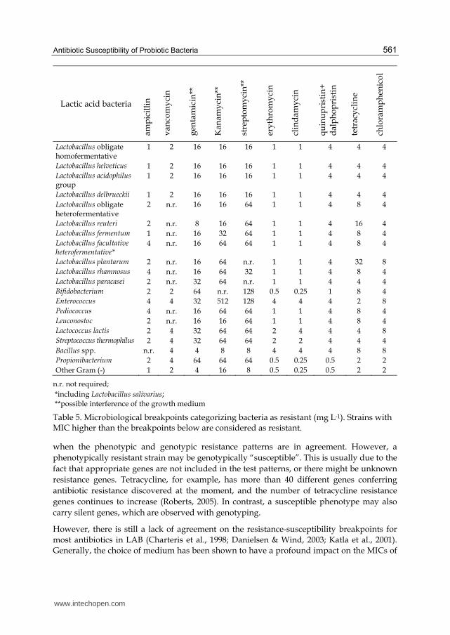
The data used for the definition of microbiological breakpoints, as reported in Table 5, were
derived from the published body of research and from national and European monitoring
procedures. The antibiotics listed: ampicillin, vancomycin, gentamicin, kanamycin,
streptomycin, erythromycin, clindamycin, quinupristin+dalfopristin, tetracycline and
chloramphenicol were chosen to maximise the identification of resistance genotypes by
assessing the resistance phenotypes.
In Gram-positive, bacteria acquired trimethoprim resistance, although occasionally detected is relatively rare. The data available (Korhonen et al., 2007) indicate that within species of lactobacilli the range of apparent trimethoprim resistances can be wide with no clear breakpoint values. Therefore, the MIC testing of trimethoprim for LAB was not considered relevant. Furthermore, testing for linezolid and neomycin is no longer considered necessary. The extremely rare non-mutational resistance to linezolid is due to the acquisition of the cfr gene, which also confers resistance to chloramphenicol (Arias et al., 2008; Toh et al., 2007). Testing for chloramphenicol resistance will efficiently cover for the hazard of acquiring resistance to linezolid. Neomycin is removed from the list since testing for the remaining three aminoglycosides efficiently covers the hazard of acquiring resistance to aminoglycosides.
Antibiotic susceptibility testing may be performed using different phenotypic test methods. In Clinical and Laboratory Standards Institute (CLSI), formerly National Committee on Clinical Laboratory Standards (NCCLS), the approved standards state that the methods of choice are agar dilution and broth microdilution (Anonym, 2007). Other widely used methods include the agar gradient method and commercial methods, such as Etest, which consists of a predefined gradient of antibiotic concentrations on a plastic strip (AbBiomerieux, Sweden). In addition to phenotypic antibiotic resistance determinations, also genotypic detection of particular genes causing resistance may be performed. These genotypic methods include different PCR –based methods, southern hybridization, plasmid profiling and microarray (Ammor et al., 2008; Aquilanti et al., 2007). The situation is clearest
www.intechopen.com
Antibiotic Susceptibility of Probiotic Bacteria
561
Lactic acid bacteria
amp
icil
lin
van
com
yci
n
gen
tam
icin
**
Kan
amy
cin
**
stre
pto
my
cin
**
ery
thro
my
cin
clin
dam
yci
n
qu
inu
pri
stin
+
dal
ph
op
rist
in
tetr
acy
clin
e
chlo
ram
ph
enic
ol
Lactobacillus obligate homofermentative
1 2 16 16 16 1 1 4 4 4
Lactobacillus helveticus 1 2 16 16 16 1 1 4 4 4
Lactobacillus acidophilusgroup
1 2 16 16 16 1 1 4 4 4
Lactobacillus delbrueckii 1 2 16 16 16 1 1 4 4 4
Lactobacillus obligate heterofermentative
2 n.r. 16 16 64 1 1 4 8 4
Lactobacillus reuteri 2 n.r. 8 16 64 1 1 4 16 4
Lactobacillus fermentum 1 n.r. 16 32 64 1 1 4 8 4
Lactobacillus facultative heterofermentative*
4 n.r. 16 64 64 1 1 4 8 4
Lactobacillus plantarum 2 n.r. 16 64 n.r. 1 1 4 32 8
Lactobacillus rhamnosus 4 n.r. 16 64 32 1 1 4 8 4
Lactobacillus paracasei 2 n.r. 32 64 n.r. 1 1 4 4 4
Bifidobacterium 2 2 64 n.r. 128 0.5 0.25 1 8 4
Enterococcus 4 4 32 512 128 4 4 4 2 8
Pediococcus 4 n.r. 16 64 64 1 1 4 8 4
Leuconostoc 2 n.r. 16 16 64 1 1 4 8 4
Lactococcus lactis 2 4 32 64 64 2 4 4 4 8
Streptococcus thermophilus 2 4 32 64 64 2 2 4 4 4
Bacillus spp. n.r. 4 4 8 8 4 4 4 8 8
Propionibacterium 2 4 64 64 64 0.5 0.25 0.5 2 2
Other Gram (-) 1 2 4 16 8 0.5 0.25 0.5 2 2
n.r. not required;
*including Lactobacillus salivarius; **possible interference of the growth medium
Table 5. Microbiological breakpoints categorizing bacteria as resistant (mg L-1). Strains with MIC higher than the breakpoints below are considered as resistant.
when the phenotypic and genotypic resistance patterns are in agreement. However, a
phenotypically resistant strain may be genotypically “susceptible”. This is usually due to the
fact that appropriate genes are not included in the test patterns, or there might be unknown
resistance genes. Tetracycline, for example, has more than 40 different genes conferring
antibiotic resistance discovered at the moment, and the number of tetracycline resistance
genes continues to increase (Roberts, 2005). In contrast, a susceptible phenotype may also
carry silent genes, which are observed with genotyping.
However, there is still a lack of agreement on the resistance-susceptibility breakpoints for
most antibiotics in LAB (Charteris et al., 1998; Danielsen & Wind, 2003; Katla et al., 2001).
Generally, the choice of medium has been shown to have a profound impact on the MICs of
www.intechopen.com
Antibiotic Resistant Bacteria – A Continuous Challenge in the New Millennium
562
LAB. The recommended growth media by the National Committee for Clinical Laboratory
Standards (Mueller-Hinton agar) (NCCLS, 2002) and by the British Society for Antimicrobial
Chemotherapy (Iso-Sensitest agar) (Andrews, 2001) do not support growth of all LAB. MRS
medium, that generally supports the growth of LAB much better, is not always compatible
to the Iso-Sensitest medium for the use in susceptibility testing, as was reported for various
classes of antibiotics (Huys et al., 2002). Furthermore, there are still no guidelines available
for the interpretation of susceptibility test results of commensal or food-associated bacteria.
Additionally, MIC breakpoints values have been shown to be species specific and thus vary
between species of the same genera (Danielsen & Wind, 2003). Also, distinguishing between
intrinsic, non-specific and acquired resistance is difficult and requires, besides the
evaluation of genetic base of resistance, that the antimicrobial-resistance patterns of many
LAB species from different sources may be compared (Teuber et al., 1999).
5.2 Mobile genetic elements in LAB
A prerequisite for LAB to acquire antibiotic resistance genes from other bacteria is their
ability to communicate actively and passively with these bacteria with the aid of conjugative
plasmids and transposons. Conjugative plasmids and transposons are common in LAB, and
due to their wide environmental distribution, it is possible that these commensal bacteria act
as vectors for the dissemination of antibiotic resistance determinants to the consumer via the
food chain. Plasmids are found in many genera of LAB, characterized by different size,
function and distribution (Davidson et al., 1996; Wang & Lee, 1997). The functions related to
the plasmids include hydrolysis of proteins, metabolism of carbohydrates, amino acids and
citrate, production of bacteriocins and exopolysaccharides, and resistance to antibiotics,
heavy metals and phages. At least 25 species of lactobacilli contain native plasmids (Wang &
Lee, 1997), and often appear to contain multiple (from 1 to 16) different plasmids in a single
strain. R-plasmids encoding tetracycline, erythromycin, chloramphenicol, or macrolide-
lincomycin-streptogramin resistance have been reported in L. reuteri (Lin et al., 1996;
Tannock et al., 1994), L. fermentum (Fons et al., 1997; Ishiwa & Iwata, 1980), L. acidophilus
(Vescovo et al., 1982), and L. plantarum (Danielsen, 2002) isolated from raw meat, silage and
faeces. The reported prevalence of antibiotic resistance genes such as erythromycin,
vancomycin, tetracycline, chloramphenicol, and gentamicin resistance genes, on transferable
genetic elements in enterococci is more extensive, both on plasmids (Murray et al., 1988) and
transposons (Clewell et al., 1995; Perreten et al., 1997a; Rice & Marshall, 1994;). A multiple
antibiotic resistance plasmid was reported in a L. lactis strain isolated from cheese (Perreten
et al., 1997b), encoding streptomycin, tetracycline and chloramphenicol resistance.
Conjugative transposons are the major vehicle regarding antibiotic resistance transport in
LAB. They have been discovered in E.faecalis (Tn916, Tn918, Tn920, Tn925, Tn2702), E.
faecium (Tn5233) and L. lactis (Tn5276, Tn5301). In enterococci and streptococci, resistances to
tetracycline (tet (M)), erythromycin (ermAM, erm), chloramphenicol (cat) and kanamycin
(aphA-3) have been determined. In lactococci, code for nisin (nis) production and sucrose
fermentation (sac) has been observed. These transposons vary in size between 16 and 70 kb
and may be inserted into plasmids or the chromosome in one or multiple copies. They may
mobilize plasmids or chromosomal genes. The most remarkable observation is the extreme
host range, which is the property of the Tn916/Tn1545 family.
www.intechopen.com

Antibiotic Susceptibility of Probiotic Bacteria
563
5.3 Horizontal transferability of antibiotic resistance from LAB in food chain
The possible transfer of antibiotic resistance genes between bacterial species have been studied mostly in harmful or pathogenic species, but also recently in LAB. The vast majority of the experiments have been made in vitro, using methods such as filter-mating (Klare et al., 2007, Ouoba et al.,2008), although these in vitro methods do not mimic the circumstances in nature, and results obtained cannot be compared with the results achieved or expected using in vivo methods. The transferability of antibiotic resistance genes in the GI tract from LAB is not straightforward, since the GI tract is a hostile environment to many allochthonous bacteria. Moreover, studies made in vivo usually are based on “worst-case scenario”, simulating very high daily intake of food products containing the resistant bacteria (Jacobsen et al., 2007). The potentially transferable genes in LAB have been described in multiple studies and have been reviewed in Ammor et al. (2007). Two of the most commonly observed resistance genes in LAB found so far are tet(M) for tetracycline resistance and erm(B) for erythromycin, followed with cat genes coding for chloramphenicol resistance (Cataloluk & Gogebakan 2004; Danielsen, 2002).
Enterococci are known to be very well receptive for conjugation (Clewell & Weaver, 1989), but are also successful donor organisms for the transfer of antibiotic resistance genes to unrelated enterococci (Rice et al., 1998), lactobacilli (Shrago & Dobrogosz, 1988), other Gram-positives including Bacillus subtilis (Christie et al., 1987), Staphylococcus (Young et al., 1987) and Listeria spp. (Charpentier et al., 1997; Perreten et al., 1997b), and even Gram-negative bacteria (Courvalin, 1994). Moreover, the transfer of conjugative elements, including a plasmid-encoded kanamycin resistance and a transposon-encoded tetracycline and erythromycin resistance (Doucet-Populaire et al., 1991), were shown to be transferable from E. faecalis to Escherichia coli and Listeria monocytogenes, respectively, in the digestive tract of mice. In contrast, reports of conjugative transfer of antibiotic resistance genes in other LAB are rare. Two in vivo studies were performed, to examine the possibility of conjugative transfer between native Gram-positive members of the gut. Therefore, the broad host range conjugative plasmid pAMβ1 was transferred in vitro to L. reuteri (Morelli et al., 1988) and L. lactis (Igimi et al., 1996) and administered orally or using gastric intubation to mice. By analysis of faecal content, plasmid transfer to E. faecalis was observed in both studies.
In order to fully understand the extent to which LAB strains transfer resistance genes in the natural environment, it is essential to study genetic exchange in this context. Toomey et al., (2009) reported on the ability of wild-type antibiotic resistance determinants [erm(B) and tet(M)], present in LAB strains isolated from food sources, to be transferred to recipient strains. In vitro mating, using a traditional filter mating technique, showed that all four LAB mating pairs transferred their resistance determinants at high frequencies. By employing two in vivo models, an alfalfa sprout plant and an animal rumen model Toomey et al.., (2009) demonstrated the transfer of resistance determinants between all four LAB mating pairs in these models. Previously, in vivo transfer between LAB has only been shown in the gastrointestinal tracts of gnotobiotic rats (Jacobsen et al., 2007) and mice (McConnell et al., 1991; Morelli et al., 1988). The transfer frequencies have been observed to increase when the animals have received the antibiotic in question at subtherapeutic levels (Igimi et al., 1996; Licht et al., 2003; Salyers & Shoemaker 1996) in their drinking water or feed, suggesting that increasing the antibiotic pressure can amplify the transfer of antibiotic resistance between
www.intechopen.com
Antibiotic Resistant Bacteria – A Continuous Challenge in the New Millennium
564
bacterial species. All of these above studies indicate that antibiotic resistant factors may be transferred from food related bacterium species (LAB) to other, potentially pathogenic species. The risks associated need to be considered, in light of the increasing concerns related to food as a potential reservoir for antibiotic resistance determinants.
6. Antibiotic resistance/susceptibility patterns of specific LAB genera applicable as probiotics
Some features appeared to be shared by the majority of LAB; in particular, it was reported that most LAB species are resistant to metronidazole and that they are all intrinsically resistant to sulphonamides and trimethoprim, while they are usually susceptible to piperacillin and piperacillin plus tazobactam. On the other hand, clear differences were highlighted among different LAB genera, although well-defined species-specific profiles were not always identifiable. A high resistance to cefoxitin was acknowledged for Lactococcus, Leuconostoc, and Lactobacillus, whereas, as regards vancomycin, Leuconostoc, Pediococcus and most lactobacilli species were recognised as intrinsically resistant and most Lactococcus isolates as highly susceptible.
Lactobacilli widely used in starter cultures or as probiotics in dairy products enter human intestines in large numbers and there interact with the intestinal microbiota (Teuber et al., 1999). Therefore they have the potential to serve as hosts for antibiotic-resistance genes, with the risk of transferring the genes to opportunistic or pathogenic bacteria. Routine antibiotic susceptibility testing has been advocated as an essential selection criterion for potentially starter or probiotic Lactobacillus cultures (Charteris & Kelly, 1993). The Lactobacillus species have been found susceptible to many cell wall synthesis inhibitors, like penicillins and ampicillin (Danielsen & Wind 2003, Coppola et al., 2005), in contrast to glycopeptides such as vancomycin, most Lactobacillus species, excluding obligate heterofermentative species, have been found to be resistant to these types of antibiotics. However, the resistance towards vancomycin has been demonstrated being as intrinsic (Tynkkynen et al., 1998) due the presence of D-alanine: D-alanine ligase-related enzymes (Elisha & Courvalin, 1995) and should not be compared with transmissible, plasmid–mediated resistance found in enterococci (Leclercq et al., 1992). As a general rule, lactobacilli have a high natural resistance to bacitracin, cefoxitin, ciprofloxacin, fusidic acid, kanamycin, gentamicin, metronidazole, nitrofurantoin, norfloxacin, streptomycin, sulphadiazine, teicoplanin (Danielsen & Wind, 2003). In addition, resistance against inhibitors of nucleic acid synthesis, such as trimethoprim, seems to be intrinsic, although further characterizations are required on this topic (Ammor et al., 2007). Resistance to tetracycline has been observed more often among Lactobacillus species, and it has been shown to have a wide range of MICs (Korhonen et al., 2008), also with a multimodal distribution of MICs, probably due to the extensive variability of tetracycline resistance mechanisms conferring diverse levels of susceptibility (Roberts, 2005). Especially with tetracycline, molecular methods should be applied in order to reveal the nature of resistance, i.e. is it due to intrinsic mechanisms, mutation or added, mobile genes.
Screening of antibiotic-resistance profile among Lactobacillus strains used in dairy products such as probiotics or as starters is now tending to become systematic. Coppola et al., (2005) pointed out that all of 63 L. rhamnosus strains isolated from Parmigiano Reggiano cheese showed resistance to six antibiotics (cefixime, vancomycin, neomycin, enoxacin, peflxacin,
www.intechopen.com

Antibiotic Susceptibility of Probiotic Bacteria
565
and sulphamethoxazole plus trimetoprim). Investigating the current antibiotic-resistance situation in microbial food additives in Switzerland, Kastner et al., (2006) determined that among 74 Lactobacillus isolates applicable as starter or probiotic cultures, two antibiotic resistances were detected in probiotic cultures. The genetic base of those resistances was confirmed; the tetracycline resistance gene tet(W) in L. reuteri SD 2112 (residing on a plasmid) and the lincosamide resistance gene lnu(A) in L. reuteri SD 2112. The similar trend was noticed in study of Katla et al., (2001). Only one of the 189 Lactobacillus strains isolated from Norwegian dairy products such as yoghurt, sour cream, fermented milk and cheese was classified as high level resistant to streptomycin. In contrast, a study conducted on “home-made“ spanish cheese (Serena, Gamonedo, Cabrales) revealed the presence of lactobacilli resistant to penicillin G, cloxacillin, streptomycin, gentamycin, tetracycline, erythromycin and chloramphenicol (Herrero et al., 1996).
L. lactis strains were sensitive to amikacin, ampicillin, 1st generation cephalosporin, chloramphenicol, erythromycin, gentamicin, imipenem, oxacillin, penicillin, pipericillin, sulphonamide, tetracycline, trimethoprim/sulfomethoxazole, and vancomycin (de Fabrizio et al., 1994). A slightly lowered susceptibility was observed towards carbenicillin, ciprofloxacin, dicloxacillin and norfloxacin. Intrinsic resistances were recorded towards colistin, fosfomycin, pipemidic acid and rifamycin. Orberg & Sandine (1985) demonstrated that investigated strains of L. lactis subsp. cremoris and subsp. lactis were all resistant to thrimethoprim and almost all to sulphathiazole. Resistance to gentamicin, kanamycin, lincomycin, neomycin, rifampin and streptomycin varied.
The enterococcal strains are naturally tolerant to β-lactams, cephalosporins, lincosamides and polymyxins. A specific cause for concern and a factor contributing to the pathogenesis of enterococci is the resistance they acquire to aminoglycosides, tetracyclines, macrolides, chloramphenicol, penicillin, and ampicillin (Gray et al., 1991) and their capacity to exchange genetic information by conjugation. Enterococcal food isolates (mainly E. faecalis and E. faecium) were analysed for resistances to a broader range of different antibiotics using phenotypic susceptibility testing, both in raw meat (Knudtson & Hartman, 1993; Quednau et al., 1998) and fermented milk and meat products (Franz et al., 2001; Teuber & Perreten, 2000). Their data suggest a high prevalence of (multiple) antibiotic resistant enterococci in foods, which nevertheless were mostly susceptible to the clinically relevant antibiotics ampicillin and vancomycin. Enterococci from European cheeses, mainly belonging to E. feacalis and E. faecium, are susceptible to different antibiotics in different proportions (Teuber et al., 1999; Franz et al., 2001). From the study of European cheeses Teuber et al. (1999) ascertained that the incidence for vancomycin resistance among enterococcal isolates was as low as 4%. When Franz et al. (2001) tested 47 E. faecalis strains, isolated mostly from cheeses, they were all susceptible to vancomycin. Bulajić & Mijačević (2011) pointed out that among enterococcal strains isolated from autochthonous Sombor cheese, only one strain showed vancomycin resistance. In contrast, Citak et al. (2004) have shown resistance to vancomycin among the population of enterococci isolated from Turkish white cheeses and was found in 96.8% of E. faecalis isolates, and 76% of E. faecium strains. The susceptibility to vancomycin is of great importance as this glycopeptide antibiotic is one of the last therapeutic options in clinical therapy.
Bifidobacteria are generally considered to be food-grade organisms that do not impose health risks on the consumer or the environment. Nevertheless, it should be noted that rare
www.intechopen.com
Antibiotic Resistant Bacteria – A Continuous Challenge in the New Millennium
566
cases of Bifidobacterium-associated gastrointestinal and extra-intestinal infections have been described. In contrast to susceptibility testing of clinically important bacteria, no standard procedures are specifically dedicated to the determination of resistance phenotypes in Bifidobacterium strains. To date, a large variety of methods and protocols have been described for antimicrobial susceptibility testing of bifidobacteria, including agar (overlay) disc diffusion, broth dilution and agar dilution. In addition, various growth media have been used primarily on the basis that they meet the complex growth requirements of bifidobacteria. As opposed to conventional susceptibility test media such as Mueller–Hinton and Iso-Sensitest medium none of these Bifidobacterium-specific media are well defined in terms of minimal interaction between specific antimicrobial agents and growth medium components. Recently, a newly defined medium formulation referred to as the Lactic acid bacteria Susceptibility test Medium supplemented with cysteine (LSM + cysteine) was proposed for susceptibility testing of bifidobacteria.
Moubareck et al., (2005) were tested the fifty bifidobacterial strains, isolated from humans,
animals or probiotic products for susceptibility to 30 antibiotics by disc diffusion test on Brucella agar supplemented with 5% laked sheep blood and vitamin K (1mg/L). All strains
were sensitive to penicilins: penicillin G, amoxicillin, piperacillin, ticarcillin, imipenem, and usually anti-Gram-positive antibiotics (macrolides, clindamycin, vancomycin and
teicoplanin). Most isolates (70%) were resistant to fusidic acid and, as expected, high resistance profile were observed for aminoglycosides. Potentially acquired resistance was
only observed against tetracycline and minocycline, in 14% of the tested strains. For the first time, Moubareck et al., (2005) identified tet(W) as the gene responsible for tetracycline
resistance in Bifidobacterium pseudocatenulatum and B. bifidum. Interestingly, the tet(W) gene was previously found in human B. longum and three genera of rumen obligate anaerobes,
suggesting intergenic transfer of this resistance gene between anaerobic bacteria (Scott et al., 2000). In the study of Masco et al., (2006), the LSM + cysteine medium was used to
determine the susceptibility profile of 100 bifidobacterial isolates (strains of animal and human origin, isolates from probiotic products and strains from clinical sources) to 15
common antimicrobial agents. All strains tested were susceptible to amoxicillin, chloramphenicol, erythromycin, quinupristin/dalfopristin, rifampicin and vancomycin. The
date from this study (Masco et al., 2006) also reinforce earlier observations indicating that bifidobacteria are intrinsically resistant to gentamicin, sulfamethoxazole and polymyxin B.
Susceptibility to trimethoprim, trimethoprim/sulfamethoxazole, ciprofloxacin, clindamycin, tetracycline and minocycline was variable. The tet(W) gene was responsible for tetracycline
resistance in 15 strains including 7 probiotic isolates belonging to the taxa Bifidobacterium
animalis subsp. lactis and B. bifidum. This gene was present in a single copy on the
chromosome and did not appear to be associated with the conjugative transposon TnB1230 previously found in tet(W)-containing Butyrivibrio fibrisolvens.
7. Conclusion
The selective pressure imposed by the use of antimicrobial agents plays a key role in the emergence of resistant bacteria. Under selective pressure, the numbers of these bacteria increase and some may transmit their resistance genes to other members of the population.. The food chain was considered as the main route of transmission of antibiotic resistant lactic acid bacteria between the animals and human population. Fermented dairy products and
www.intechopen.com

Antibiotic Susceptibility of Probiotic Bacteria
567
fermented meats, which are not heat-treated before consumption, provide a vehicle for antibiotic resistant LAB with a direct link between the animal indigenous microflora and the human gastrointestinal tract. There is the potential health risk, due to the transfer of antibiotic resistance genes from LAB to bacteria in the human gastrointestinal tract, especially to pathogenic bacteria.
Lactic acid bacteria used as starter cultures or probiotic bacteria, enter into human intestines in large number where they interact with the intestinal microflora. Since there has been a significant rise in the consumption of probiotic products, it is important that probiotics are well documented regarding antibiotic resistance profile. The ability to transfer antibiotic resistance genes must be considered as an important parameter for the selection of the probiotic strains. Continuous attention should be paid to the selection of probiotic strains free of transferable antibiotic-resistance determinants. Without doubt, the uncontrolled use of antimicrobial agents in farming practice has assisted the spread of resistant organisms. Therefore a much stricter control over the use of these drugs is essential.
8. Acknowledgment
This work was supported by Ministry of Education and Science of Republic of Serbia (Project No. 46010).
9. References
Agostoni, C., Axelsson, I., Goulet, O., Koletzko, B., Michaelsen, K.F. & Puntis, J. W., (2004). Prebiotic Oligosaccharides in Dietetic Products for Infants: A Commentary by the ESPGHAN Committee on Nutrition. Journal of Pediatric Gastroenterology Nutrition, Vol. 39, pp. 465-473.
Ammor, M. S., Florez, A. B. & Mayo, B. (2007). Antibiotic Resistance in Nonenterococcal Lactic Acid Bacteria and Bifidobacteria. Food Microbiology, Vol. 24, pp. 559-570.
Ammor, M. S., Florez, A. B., van Hoek, A. H., de Los Reyes-Gavilan, C. G., Aarts, H. J., Margolles, A. & Mayo, B. (2008). Molecular Characterization of Intrinsic and Acquired Antibiotic Resistance in Lactic Acid Bacteria and Bifidobacteria. Journal of Molecular Microbiology and Biotechnology, Vol. 14, pp. 6-15.
Andrews, J. M. (2001). BSAC Standardized Disc Susceptibility Testing Method. Journal of Antimicrobial Chemotherapy, Vol. 48 Suppl .1, pp. 43-57.
Anonym (2007). Methods for Antimicrobial Susceptibility Testing of Anaerobic Bacteria. Approved Standard. CLSI document M11-A7. Applied and Environmental Microbiology, 72, pp. 1729-1738.
Aquilanti, L., Silvestri, G., Zannini, E., Osimani, A., Santarelli, S. & Clementi, F. (2007). Phenotypic, Genotypic and Technological Characterization of Predominant Lactic Acid Bacteria in Pecorino Cheese from Central Italy. Journal of Applied Microbiology, Vol. 103, pp. 948-960.
Arias, C. A., Vallejo, M., Reyes, J., Panesso, D., Moreno, J., Castaneda, E., Villegas, M. V., Murray, B. E. & Quinn, J. P. (2008). Clinical and Microbiological Aspects of Linezolid Resistance Mediated by the cfr Gene Encoding a 23S rRNA Methyltransferaze. Journal of Clinical Microbiology, Vol. 46, pp. 892-896.
www.intechopen.com

Antibiotic Resistant Bacteria – A Continuous Challenge in the New Millennium
568
Axelsson, L.T., Ahrne, S., Andersson, M.C. & Stahl, S.R. (1988). Identification and Cloning of a Plasmid-Encoded Erythromycin Resistance Determinant from Lactobacillus reuteri G4. Plasmid, Vol. 20, pp. 171-174.
Backhed, F.; Ley, R.E.; Sonnenburg, J.L.; Peterson, D.A. & Gordon, J.I. (2005). Host-Bacterial Mutualism in the Hhuman Intestine. Science, Vol. 307, pp. 1915–1920.
Begley, M, Hill, C., & Gahan, C. G. M., (2006). Bile Salt Hydrolase Activity in Probiotics. Applied and Environmental Microbiology, Vol. 72, No. 3, pp. 1729-1738.
Bengmark, S. (1998). Ecological Control of the Gastrointestinal Tract. Rhe Role of Probiotic Flora. Gut, Vol. 27, pp. 2-7.
Bennet, R., Eriksson, M., Nord, C.E. & Zetterström, R. (1986). Fecal Bacterial Microflora of Newborn Infants During Intensive Care Management and Treatment with Five Antibiotic Regimens. Pediatric Infectious Disease, Vol. 5, pp. 533-539.
Beresford, T. P., Fitzimons, N. A., Brenan, N. L. & Cogan, T. M. (2001). Recent Advances in Cheese Microbiology. International Dairy Journal, Vol. 11, pp. 256-274.
Bernet, M. F., Brassart, D., Neeser, J. R. & Servin, A. L. (1994). Lactobacillus acidophillus LA 1 Binds to Cultured Human Intestinal Cell Lines and Inhibits Cell Attachment and Cell Invasion by Enterovirulent Bacteria. Gut, Vol. 35, pp. 483-489.
Bernet, M. F., Brassart, D., Neeser, J.R. & Servin, A.L. (1993). Adhesion of Human Bifidobacterial Strains to Cultured Human Intestinal Epithelial Cells and Inhibition of Enteropathogen–Cell Interactions. Applied and Enviromental Microbiology., Vol. 59, pp. 4121–4128.
Berrada, N., Lemeland, J.F., Laroche, G., Thovenot P. & Piaia,M. (1991). Bifidobactrium from Fermented Milk: Survival During Gastic Transit. Journal of Dairy Science, Vol. 74, pp. 409-413.
Black, F. T. Anderson, P.L., Oeskov, J., Gaarskev, K. & Laulund, S. (1989). Prophylactic Efficacy of Lactobacilli on Traveler′s Diarrhea. Travel Medicine, Vol. 7, pp. 333-335.
Bulajić, S. & Mijačević, Z. (2011). Antimicrobial Susceptibility of Lactic Acid Bacteria Isolated from Sombor Cheese. Acta Veterinaria, Vol. 61, No. 2-3, pp. 247-258.
Cataloluk, O. & Gogebakan, B. (2004). Presence of Drug Resistance in Intestinal Lactobacilli of Dairy and Human Origin in Turkey. FEMS Microbiology Letters, Vol. 236, pp. 7-12.
Charpentier, E. & Courvalin, P. (1997). Emergence of the Trimethoprim Resistance gene dfrD in Listeria monocytogenes BM4293. Antimicrobial Agents and Chemoterapy, Vol. 41, pp. 1134-1136.
Charteris, W. P., Kelly, P. M., Morelli, L. & Collins, J. K. (1998). Antibiotic Susceptibility of Potentially Probiotic Lactobacillus Species. Journal of Food Protection, Vol. 61, pp. 1636-1643.
Charteris, W. P. & Kelly, P. M. (1993). In Vitro Antibiotic Susceptibility of Potentially Probiotic Lactobacilli and Bifidobacteria. In Second Annual Report, EU FLAIR Project No. AGRF-CT91-0053 ed. Morelli, L. Brussels: Commission of the European Communities.
Christie, P. J., Korman, R. Z., Zahler, S. A., Adsit, J. C. & Dunny, G. M. (1987). Two Conjugation Systems Associated with Streptococcus faecalis plasmid pCF10: Identification of a Conjugative Transposon that Transfers Between S. facealis and Bacillus subtilis. Journal of Bacteriology, Vol. 169, pp. 2529-2536.
www.intechopen.com
Antibiotic Susceptibility of Probiotic Bacteria
569
Citak, S., Yucel, N. & Orhan, S. (2004). Antibiotic Resistance and Incidence of Enterococcus Species in Turkish White Cheese. International Journal of Dairy Technology, Vol. 57, pp. 27-31.
Claesson, M. J., van Sinderen, D. & O'Toole, P. W. (2007). The genus Lactobacillus – A Genomic Basis of Understanding its Diversity. FEMS Microbiology Letters, Vol. 269, No.1, pp. 22-28.
Clementi, F. & Aquilanti, F. A. (2011). Recent Investigations and Updated Criteria for the Assessment of Antibiotic Resistance in Food Lactic Acid Bacteria. Anaerobe (2011) 1-5, Article in Press.
Clewell, D. B. Weaver, K. E. (1989). Sex Pheromones and Plasmid Transfer in Enterococcus faecalis. Plasmid, Vol. 21, pp. 175-184.
Coppa, G., Bruni, S., Morelli, L., Soldi, S. & Gabrielli, O. (2004). The First Prebiotics in Humans: Human Milk Oligosaccharides. Journal of Clinical Gastroenterology, Vol. 38, pp. 80-83.
Coppola, R., Succi, M., Tremonte, P., Reale, A., Salzano, G. & Sorrentino, E. (2005). Antibiotic Susceptibility of Lactobacillus rhamnosus Strains Isolated from Parmigiano Reggiano Cheese. Lait, Vol. 85, pp. 193-204.
Courvalin, P. (1994). Transfer of Antibiotic Resistance Genes Between Gram-positive and Gram-negative Bacteria. Antimicrobial Agents and Chemotherapy, Vol. 38, pp. 1447-1451.
Cummings, J. H., Gibson, G. R. & Macfarlane, G. T. (1989). Qualitative Estimates of Fermentation in the Hind Gut of Man. Acta Veterinaria Scandinavica Suppl. Vol. 86, pp. 76-82.
Curragh, H. J. & Collins, M. A. (1992). High-levels of Spontaneous Drug-Resistance in Lactobacillus. Journal of Applied Bacteriology, Vol. 73, pp. 31-36.
Danielsen, M. & Wind, A. A. (2003). Susceptibility of Lactobacillus spp. to Antimicrobial Agents. International Journal of Food Microbiology, Vol. 82, pp. 1-11.
Danielsen, M. (2002). Characterization of the Tetracycline Resistance Plasmid pMD5057 from Lactobacillus plantarum 5057 Reveals a Composite Structure. Plasmid, Vol. 48, pp. 98-103.
Davidson, B. E., Kordias, N., Dobos, M. & Hillier, A. J. (1996). Genomic Organization of Lactic Acid Bacteria. Antonie Van Leeuwenhoek International Journal of General and Molecular Microbiology, Vol. 70, pp. 161-183.
de Leeuw, E., Li, X. & Lu, W. (2006). Binding Characteristics of the Lactobacillus brevis ATCC 8287 Surface Layer to Extracellular Matrix Proteins. FEMS Microbiology Letters, Vol. 260, pp. 210-215.
deFabrizio, S. V., Parada, J. L. & Torriani, S. (1994). Antibiotic Resistance of Lactococcus lactis – an Approach of Genetic Determinants Location through the Model System. Microbiologie-Aliments-Nutritions, Vol. 12, pp. 307-315.
Doucet-Populaire, F., Trieu-Cuot, P., Dosbaa, I., Andremont, A. & Courvalin, P. (1991). Inducible transfer of conjugative transposon Tn1545 from Enterococcus faecalis to Listeria monocytogenes in the digestive tracts of gnotobiotic mice. Antimicrobial Agents and Chemotherapy, Vol. 35, pp. 185-187.
Dunne, C., O'Mahony, L., Murphy, L., Thornton, G., Morrissey, D., O'Halloran, S., Feeney, M., Flynn, S., Fitzgerald G., Daly, C., Kiely, B., O 'Sullivan, G.C., Shanahan, F. & Collins, J.K. (2001). In vitro Selection Criteria for Probiotic Bacteria of Human
www.intechopen.com

Antibiotic Resistant Bacteria – A Continuous Challenge in the New Millennium
570
Origin: Correlation with In vivo Findings. American Journal of Clinical Nutrition, Vol. 73, pp. 386-392.
Eaton, T. J. & Gasson, M. J. (2001). Molecular Screening of Enterococcus Virulence Determinants and Potential for Genetic Exchange between Food and Medical Isolates. Applied and Environmental Microbiology, Vol. 67, pp. 1628-1635.
EFSA. (2008). Technical Guidance Prepared by the Panel on Additives and Products or Substances used in Animal Feed (FEEDAP) on the Update of the Criteria Used in the Assessment of Bacterial Resistance to Antibiotics of Human and Veterinary Importance. The EFSA Journal, pp. 1-15.
Elisha, B. G. & Courvalin, P. (1995). Analysis of Genes Encoding Dalanine: d-Alanine Ligase-Related Enzymes in Leuconostoc mesenteroides and Lactobacillus spp. Gene, Vol. 152, pp. 79-83.
European Commissions (2005). Opinion of the Scientific Committee on Animal Nutrition on the Criteria for Assessing the Safety of Micro-organisms Resistant to Antibiotics of Human Clinical and Veterinary Importance. European Commissions, Health and Consmer protection Directorate General, Directorate C, Scientific Opinions, Brussels, Belgium.
Fons, M., Hege, T., Lafire, M., Raibaud, P., Ducluzeau, R. & Maguin, E. (1997). Isolation and Characterization pf a Plasmid from Lactobacillus fermentum Connferring Erythromycin Resistance. Plasmid, Vol. 37, pp. 199-203.
Fooks, L. J., Fuller, R. & Gibson, G. R. (1999). Prebiotics, Probiotic and Human Gut Microbiology. International Dairy Journal, Vol. 9, pp. 53-61.
Franz, C. M. A. P., Holzapfel, W. H. & Stiles, M. E. (1999). Enterococci at the Crossroads of Food Safety. International Journal of Food Microbiology, Vol. 47, pp. 193-197.
Franz, C. M., Muscholl-Silberhorn, A. B., Yousif, N.M.K., Vancanneyt, M., Swings, J. & Holzapfel, W. H. (2001). Incidence of Virulence Factors and Antibiotic Resistance among Enterococci Isolated from Food. Applied and Environmental Microbiology, Vol. 67, pp. 4385-4389.
Franz, C.M.A.P., Specht I., Haberer P. & Holzapfel W. H. (2001a). Bile Salt Hydrolase Activity of Enterococci Isolated from Food: Screening and Quantitative Determination. Journal of Food Protection, Vol. 64, No 5, pp. 725-729.
Fuller, R. (1992). Probiotics: The scientific basis, ISBN 0-412-40850-3, Chapman & Hall, London, Gardnier, E. G., Ross, P. R., Kellz, M. P., Stanton, K., Collins, K. K. & Fitzgerald, G. (2002).
Microbiology of Therapeutic Milks, In: Dairy Microbiology Handbook, Third Edition, R. K. Robinson (Ed.), Wiley-Interscience, Inc., 431-478, ISBN 0-471-38596-4, New York, USA.
Garg, S. K. & Mital, B. K. (1991). Enterococci i n Milk and Milk Products. Critical Reviews in Microbiology, Vol. 18, pp. 15-45.
Gevers, D., Huys, G. & Swings, J. (2003). In Vitro Conjugal Transfer of Tetracycline Resistance from Lactobacillus Isolates to Other Gram-positive Bacteria. FEMS Microbiology Letters, Vol. 225, pp. 125-130.
Gray, C. M., Stewart, D. & Pedler, S. J. (1991). Species Identification and Antibiotic Susceptibility Testing of Enterococci Isolated from Hospitalized Patients. Antimicrobial Agents and Chemotherapy, Vol 35, pp. 1943-1945.
Gronlund, M. M., Lehtonen, O. P., Erola, E.& Kero, P. (1999). Fecal Microflora in Healthy Infants Born by Different Methods of Delivery: Permenent Changes in Intestinal
www.intechopen.com

Antibiotic Susceptibility of Probiotic Bacteria
571
Flora after Cesarean Delivery. Journal of Pediatric Gastroeneterological Nutrition, Vol. 28, pp. 19-25.
Guarner, F. & Schaafsma, G. J. (1998). Probiotics. International Journal of Food Microbiology, Vol. 39, pp. 237-238.
Hamilton-Miller, J. M. T & Shah, S. (1998). Vancomycin Susceptibility as an Aid to the Identification of Lactobacilli. Letters in Applied Microbiology, Vol. 26, pp. 153-154.
Herrero, M., Mayo, B. Ganzales, B. & Suarez, J.E. (1996). Evaluation of Technologically Important Traits in Lactic Acid Bacteria Isolated from Spontaneous Fermentations. Journal of Applied Bacteriology, Vol. 81, pp. 567-570.
Herreros, M. A., Fresno, J.M., Gonzalez Prieto, M. J. & Tornadijo, M. E. (2003). Technological Characterization of Lactic Acid Bacteria Isolated from Armada Cheese (a Spanish Goat ′s milk Cheese). International Dairy Journal, Vol.13, pp. 469-479.
Hevey, P. & Rowland, I. (1999). The Gut Microflora of The Developing Infant: Microbiology and Metabolism. Microbial Ecology in Health and Disease. Vol. 11, pp. 75-83.
Hummel, A.S.; Hertel, C.; Holzapfel, W.H. & Franz, C. (2007). Antibiotic Resistances of Starter and Probiotic Strains of Lactic Acid Bacteria. Applied and Enviromental Microbiology, Vol.73, pp. 730-739.
Huys, G., D’Haene, K. & Swings, J. (2002). Influence of the Culture Medium on Antibiotic Susceptibility Testing of Food-Associated Lactic Acid Bacteria with the Agar Overlay Disc Diffusion Method. Letters in Applied Microbiology, Vol. 34, pp. 402-406.
Igimi, S., Ryu, C.H., Park, S.H., Sasaki, Y., Sasaki, T. & Kumagai, S.. (1996). Transfer of Conjugative Plasmid pAM beta 1 from Lactococcus lactis to Mouse Intestinal Bacteria. Letters of Applied Microbiology, Vol. 23, pp. 31-35.
Ishiwa, H & Iwata, S. (1980). Drug Resistance in Lactobacillus fermentum. Journal of General and Applied Microbiology, Vol. 26, pp. 71-74.
Jacobsen, I., Wicks, A., Hammer, K., Huys, G., Gevers, D. & Andersen, S.R. (2007). Horizontal Transfer of tet(M) and erm(B) Resistance Plasmids from Food Strains of Lactobacillus plantarum to Enterococcus faecalis JH2-2 in The Gastrointestinal Tract of Gnotobiotic Rats. FEMS Microbiology Ecology, Vol. 59, pp. 158-166.
Kandler, O. & Weiss, N. (1989). Genus Lactobacillus, In: Bergeys Manual of Systematic Bacteriology, P. H. Snearh, N. S. Mair, M. E. Sharpe, J. G. Holt (Ed.), Vol. 2, 1418-1434, ISBN 0-683-07893-3, Williams & Wilkins Baltimore, USA.
Kastner, S., Parreten, V., Bleuler, H., Hugenschmidt, G., Lacroix, C & Meile, L. (2006) Antibiotic Susceptibility Patterns and Resistance Genes of Starter Culture and Probiotic bacteria Used in Food. Systematic and Applied Microbiology, Vol. 29, pp. 145-55.
Katla, A. K., Kruse, H., Johnsen, G. & Herikstad, H. (2001). Antimicrobial Susceptibility of Starter Culture Bacteria Used in Norwegian Dairy Products. International Journal of Food Microbiology, Vol. 67, pp. 147-152.
Klare, I., Konstabel, C., Werner, G., Huys, G., Vankerckhoven, V., Kahlmetrr, G., Hildebrandt, B., Muller Bertling , S., Witte, W & Goossens, H. (2007). Antimicrobial Susceptibilities of Lactobacillus, Pediococcus and Lactococcus Human Isolates and Culture Intended for Probiotic or Nutritional Use. Journal of Antimicrobial Chemotherapy, Vol. 59, pp. 900-912.
www.intechopen.com
Antibiotic Resistant Bacteria – A Continuous Challenge in the New Millennium
572
Knudtson, L. M. & Hartman, P.A. (1993). Antibiotic Resistance among Enterococcal Isolates from Environmental and Clinical Sources. Journal of Food Protection, Vol. 56, pp. 489-492.
Koll, P., Mandar, R.., Marcotte, H., Leibur, E., Mikelsaar, M. & Hammarstrom, L. (2008). Characterization of Oral Lactobacilli as Potential Probiotics for Oral Health. Oral Microbiol Immunol., Vol. 23, No 2, pp. 139-147.
Korhonen, J. M. Sclivagnotis, Y. & von Wright, A. (2007). Characterization of Dominant Cultivable Lactobacilli and their Antibiotic Resistance Profiles from Fecal Samples of Weaning Piglets. Journal of Applied Microbiology, Vol. 103, pp. 2496-2503.
Leclercq, R., Dutka-Malen, S., Brisson-Noel, A., Molinas, C., Derlot, E., Arthur, M., Duval, J. & Courvalin, P. (1992). Resistance of Enterococci to Aminoglycosides and Glycopeptides. Clinical Infectious Diseases, Vol. 15, pp. 495-501.
Levy, S. B. (1986). Ecology of Antibiotic Resistance Determinants. In:Levy, SB, Novick, RP (eds) Antibiotic resistance genes: ecology, transfer and expression. Cold Spring Harbor Press, New York, pp. 17-29.
Lewis, S. J. & Freedman, A. R. (1998). The Use of Biotherapeutic Agents in the Prevention and Treatment of Gastrointestinal Disease. Alimentary Pharmacology and Therapeutics, Vol. 12, pp. 807-822.
Licht, T. R., Struve, C., Christensen, B. B., Poulsen, R. L., Molin, S. & Krogfelt, K. A. (2003). Evidence of Increased Spread and Establishment of Plasmid RP4 In The Intestine Under Sub-inhibitory Tetracycline concentrations. FEMS Microbiology Ecology, Vol. 44, pp. 217-233.
Lin, C. F., Fung, Z .F, Wu, C. L. & Chung, T. C. (1996). Molecular Characterization of a Plasmidborne (pTC82) Chloramphenicol Resistance Determinant (cat-Tc) from Lactobacillus reuteri. Plasmid, Vol. 36, pp. 116-124.
Liong, M.T. & Shah, N.P. (2005). Bile Salt Deconjugation Ability, Bile Salt Hhydrolase Activity and Cholesterol Co-precipitation Ability of Lactobacilli Strains. International Dairy Jounal, Vol. 15, No 4, pp. 391-398.
Lönnermark, E. (2010). Lactobacilli in the Normal Microbiota and Probiotic Effects of Lactobacillus plantarum. PhD Thesis, Department of Infectious medicine, Sahlgrenska Academy, University of Gothenburg, Sweden.
Masco, I., Van Hoorde, De Brandt, E., Swings, J. & Huys, G. (2006). Antimicrobial Susceptibility of Bifidobacterium Strains from Humans, Animals and Probiotic Products. Journal of Antimicrobial Chemotherapy, Vol. 58, pp. 85-94
Mathur, S. & Singh, R. (2005). Antibiotic Resistance in Lactic Acid Bacteria-a review. International Journal of Food Microbiology, Vol. 105, pp. 281-95.
Mattila, T. & Saarela, M. (2000). Probiotic Functional Foods. In: Functional foods, Williams and Gibson R.G. (Ed), 287-313. CRC Press LLC. Boca Raton, Boston, New York, Washington, DC.
McConnell, M. A., Mercer, A. A. & Tannock, G. W. (1991). Transfer of Plasmid pAM-1 between Members of Microflora Inhabiting the Murine Digestive Tract and Modification of the Plasmid in a Lactobacillus reuteri Host. Microbial Ecology in Health and Disease, Vol. 4, pp. 343-355.
Modler, H. V., McKellar, R. C. & Yaguchi, M. (1990). Bifidobacteria and Bifidogenic Factors. Canadian Institute of Food Science and Technology Journal, Vol. 23, pp. 29-41.
www.intechopen.com

Antibiotic Susceptibility of Probiotic Bacteria
573
Morelli, L., Sarra, P.G. & Bottazzi, V. (1988). In Vivo Transfer of pAM-beta-1 from Lactobacillus reuteri to Enterococcus faecalis. Journal of Applied Bacteriology, Vol. 65, pp. 371-375.
Moubareck, C., Gavini, F., Vaugien, L., Butel, M. J. & Doucet-Populaire, F. (2005). Antimicrobial Susceptibility of Bifidobacteria. Journal of Antimicrobial Chemotherapy, Vol. 55, pp. 38-44.
Mountzouris, K.C; McCartney, A.L; & Gibson, G.R. (2002). Intestinal Microflora of Human Infants and Current Trends for its Nutritional Modulation. British Journal of Nutrition, Vol. 87, pp. 405–420.
Murphy, E., Murphy, C. & O′Mahony, L. (2009). Influence of the Gut Microbiota with Aging. In: Microbiology and Aging: Clinical Manifestations, S.L. Percival, (Ed.), Springer-Verlag, ISBN 1588296407, New York, USA.
Murray, B. E., An, F. Y. & Clewell, D. B. (1988). Plasmid and Pheromone Response of the Betalactamase Producer Streptococcus (Enterococcus) faecalis HH22. Antimicrobial Agents and Chemotherapy, Vol. 32, pp. 547-551.
Murray, P. R., Baron, E. J., Jorgensen, J. H., Pfaller, M. A. & Yolken R. H. (2003). Manual of Clinical Microbiology, American Society for Microbiology, Washington.
National Committee for Clinical Laboratory Standards (NCCLS). (2002). Performance Standards for Antimicrobial Susceptibility Testing, Twelfth informational supplement (M100-S12)
Newburg, D.S. (2000). Oligosaccharides in Human Milk and Bacterial Colonization. Journal of Pediatric Gastroenterological Nutrition, Vol. 30, 2, pp. 8-17.
Normark, B. H. & Normark, S. (2002). Evolution and Spread of Antibiotic Resistance. Journal of International Medicine, Vol. 252, pp. 91-106
Ogawa, K., Ben, R.A., Pons, S., de Paolo, M. I. & Bustos Fernández, L. (1992). Volatile Fatty Acids, Lactic Acid, and pH in the Stools of Breast-fed and Bottlefed infants. Journal of Pediatric Gastroenterological Nutrition, Vol. 15, pp. 248-252.
Olsson-Liljequist, B., Larsson, P., Walder, M. & Miorner, H. (1997). Antimicrobial Susceptibility Testing in Sweden, III. Methodology for Susceptibility Testing in Sweden. Scandinavian Journal of Infectious Disease, 105S, pp. 13-23
Orberg, P. K. & Sandine, W.E. (1985). Survey of Antimicrobial Resistance in Lactic Streptococci. Applied and Environmental Microbiology, Vol. 49, pp. 538-542
Ouoba, L. I., Lei, V. & Jensen, L. B. (2008). Resistance of Potential Probiotic Lactic Acid Bacteria and Bifidobacteria of African and European Origin to Antimicrobials: Determination and Transferability of the Resistance Genes to Other Bacteria. International Journal of Food Microbiology, Vol. 121, pp. 217-224
Ouwehand, A. C. & Vesterlund, S. (2003). Health Aspects of Probiotics. Drugs, Vol. 6, pp. 573-580.
Ouwehand, A. C., Bianchi, Salvadori, B., Fonden, R., Mogensen, G., Salminen, S. & Sellara, R. (2003). Health Effects of Probiotics and Culture-Containing Dairy Products in Humans. Bulliten of the International Dairy Federation, Vol. 380, pp. 4-19.
Ouwehand, A. C., Tuomola, E. M., Tolkko, S. & Salminen, S. (2001). Assessment of Adhesion Properties of Novel Probiotic Strains to Human Intestinal Mucus. International Journal of Food Microbiology, Vol. 64, pp. 119-126.
Perreten, V., Schwarz, F., Cresta, L., Boeglin, M., Dasen, G. & Teuber, M. (1997a). Antibiotic Resistance Spread in Food. Nature, Vol. 389, pp. 801-802.
www.intechopen.com
Antibiotic Resistant Bacteria – A Continuous Challenge in the New Millennium
574
Perreten, V., Kolloffel, B. & Teuber, M. (1997b). Conjugal Transfer of the Tn916-like Transposon TnFO1 from Enterococcus faecalis Isolated from Cheese to Other Gram-positive Bacteria. Systematic and Applied Microbiology, Vol. 20, pp. 27-38.
Petrovic, T., Niksic, M. & Bringel, F. (2006). Strain Typing with ISLpl1 in Lactobacilli. FEMS Microbiology Letters, Vol.255, pp. 1-10.
Psoni, L., Tzanetakis, N. & Litopoulou-Tzanetaki, E. (2005). Microbiological Characteristics of Batzos, a Traditional Greek Cheese from Raw Goat′s Milk. Food Microbiology Vol. 20, No. 5, pp. 575-582.
Quednau, M., Ahrne, S., Petersson, A.C. & Molin, G. (1998). Antibiotic-Resistant Strains of Enterococcus Isolated from Swedish and Danish Retailed Chicken and Pork. Journal of Applied Microbiology, Vol. 84, pp. 1163-1170
Radulović, Z. (2010). Autochthonous Lactic Acid Bacteria as Starter Cultures. Faculty of Agriculture University of Belgrade, ISBN 978-86-7843-081-9, Belgrade, Serbia.
Radulović, Z., Petrović, T., Nedović, V., Dimitrijević, S., Mirković, N., Petrušić, M. & Paunović, D. (2010a). Characterization of autochthonous Lactobacillus paracasei strains on potential probiotic ability. Mljekarstvo, Vol. 60, No.2, pp. 86-93.
Reuter, G. (2001). The Lactobacillus and Bifidobacterium Microflora of The Human Intestine: Composition and Succession. Current Issues Intestinal Microbiology, Vol. 2, pp. 43-53.
Rice, L. & Marshall, S. H. (1994). Insertions of IS256-like Element Flanking the Chromosomal Beta-lactamase Gene of Enterococcus faecalis CX19. Antimicrobial Agents and Chemotherapy, Vol. 38, pp. 693-701
Rice, L.B., Carias, L.L., Donskey, C.L. & Rudin, S.D. (1998). Transferable, Plasmid-Mediated VanB-type Glycopeptide Resistance in Enterococcus faecium. Antimicrobial Agents and Chemotherapy, Vol. 42, pp. 963-964
Roberts, M.C. (2005). Update on Acquired Tetracycline Resistance Genes. FEMS Microbiology Letters, Vol. 245, pp. 195-203
Rodriguez, C., Medici, M., Mozzi, F. & de Valdez, G. F. (2010). Therapeutic Effect of Streptpcoccus thermophilus CRL 1190 – Fermented Milk on Chronic Gastritis. World Journal of Gastroenerology, Vol. 16, pp. 1622-1630.
Ryan, K.A.; Jayaraman, T.; Daly, P.; Canchaya, C.; Curran, S.; Fang, F.; Quigley, E.M. & O'Toole, P.W. (2008) Isolation of Lactobacilli with Probiotic Properties from the Human Stomach. Letters of Applied Microbiology, Vol.47, pp. 269-274.
Saavedra, J. M., Bauman, N., Oung, I., Perman, J. & Yolken, R. (1994). Feeding of Bifidobacterium bifidum and Streptococcus thermophilus to Infants in Hospital for Prevention of Diarrhoea and Shedding of Rotavirus, The Lancet, Vol. 344, pp. 1046-1049.
Salminen, S., Nybom, S., Meriluoto, J., Carmen Collado, M., Vesterlund, S. & El-Nezami, H. 2010. Interaction of Probiotics and Pathogens—Benefits to Human Health? Current Opinion in Biotehnology, Vol. 21, pp. 157-167.
Salyers, A. A. & Shoemaker, N.B. (1996). Resistance Gene transfer in Anaerobes: New Insights, New problems. Clinical Infectious Diseases, Vol. 23 Suppl. 1, S36-43
Salyers, A. A. (1995). Antibiotic Resistance Transfer in the Mammalian Intestinal Tract: Implications for Human Health, Food Safety and Biotechnology, Heidelberg:Springer-Verlag, New York, USA.
www.intechopen.com

Antibiotic Susceptibility of Probiotic Bacteria
575
Scardovi, V. (1986). Genus Bifidobacterium, In: Bergeys Manual of Systematic Bacteriology, P. H. Snearh, N. S. Mair, M. E. Sharpe, J. G. Holt (Ed.), Vol. 2, 1418-1434, ISBN 0-683-07893-3, Williams & Wilkins Baltimore, USA.
Scott, K. P., Melville, C.M., Barbosa, T.M. et al. (2000). Occurrence of the New Tetracycline Resistance Gene tet(W) in Bacteria from the Human Gut. Antimicrobial Agents and Chemotherapy, Vol. 44, pp. 775-777.
Shah, P. N. (2007). Functional Cultures and Health Benefits. International Dairy Journal, Vol. 17, pp. 1262-1277.
Shrago, A.W. & Dobrogosz, W.J. (1988). Conjugal Transfer of Group-B Streptococcal Plasmids and Comobilization of Escherichia coli-Streptococcus Shuttle Plasmids to Lactobacillus plantarum. Applied and Environmental Microbiology, Vol. 54, pp. 824-826.
Simpson, W. J., Hammond, J.R.M. & Miller, R.B. (1988). Avoparcin and Vancomycin-Useful Antibiotics for the Isolation of Brewery Lactic Acid Bacteria. Journal of Applied Bacteriology, Vol. 64, pp. 299-309.
Singer, R. S., Finch, R., Wegener, H.C., Bywater, R., Walters, J. & Lipstich, M. (2003). Antibiotic resistance - The Interplay Between antibiotic use in Animals and Human Beings. Lancet Infectious Diseases, Vol. 3, pp. 47-51.
Stanton, C., Gardiner, G., Meehan, H., Collins, J. K., Fitzgerald, G., Lynch, P. B. & Ross, R. P. (2001). Market Potential for Probiotics. The American Journal of Clinical Nutrition, Vol. 73 (suppl.), pp. 4765-4835.
Stark, P.L., & Lee, A. (1982). The Microbial Ecology of the Large Bowel of Breast-fed and Formula-fed Infants During the First Year of Life. Journal of Medical Microbiology, Vol. 15, pp. 189-203.
Tamime, A., Božanić, R., Rogelj, I. (2003). Probiotički fermentirani mliječni proizvodi. Mljekarstvo, Vol. 53, pp. 111-134.
Tancrede, C. (1992). Role of Human Microflora in Health and Disease. European Journal of Clinical Microbiology Infection Disease, 11, pp. 1012–1015.
Tannock, G.W., Luchansky, J.B., Miller, L., Connell, H., Thodeandersen, S., Mercer, A.A. & Kalenhammer, T.R. (1994). Molecular Characterization of a Plasmid Borne (pGT633) Erythromycin Resistance Determinant (ermGT) from Lactobacillus reuteri 100-63. Plasmid, Vol. 31, pp. 60-71
Teuber, M & Perreten, V. (2000). Role of Milk and Meat Products as Vehicles for Antibiotic Resistant Bacteria. Acta Veterinaria Scandinavica, pp. 75-87.
Teuber, M., Meile, L. & Schwarz, F. (1999). Acquired Antibiotic Resistance in Lactic Acid Bacteria from Food. Antonie van Leeuvenhoek, Vol. 76, pp. 115-137.
Todorov, S.D., Furtado, D.N., Saad, S. M. I. & Tome, E. (2011). Potential Beneficial Properties of Bacteriocin-Producing Lactic Acid Bacteria Isolated from Smoked Salmon. Journal of Applied Microbiology, 110, No 4, pp. 971-986.
Toh, S. M., Xiong, L., Arias, C. A., Villegas, M. V., Lolans, K., Quinn, J. & Mankin, A. S. (2007). Acquisition of a Natural Resistance Gene Renders a Clinical Strains of Methicilllin Resistant Staphylococcus aureus Resistant to the Synthetic Antibiotic Linezolid. Molecular Microbiology, Vol. 64, pp. 1506-1514.
Toomey, N., Monaghan, A., Fanning, S. & Bolton, D. (2009). Transfer of Antibiotic Resistance Marker Genes Between Lactic Acid Bacteria in Model Rumen and Plant Environments. Applied and Environmental Microbiology, Vol. 75, No. 10, pp. 346-3152.
www.intechopen.com
Antibiotic Resistant Bacteria – A Continuous Challenge in the New Millennium
576
Tuomola, E.M. & Salminen, S.J. (1998). Adhesion of Some Probiotic and Dairy Lactobacillus Strains to Caco-2 Cell Cultures. International Journal of Food Microbiology, Vol.41, pp. 45–51.
Tynkkynen, S., Singh, K. V. & Varmanen, P. (1998). Vancomycin Resistance Factor of Lactobacillus rhamnosus GG in Relation to Enterococcal Vancomycin Resistance (van) Genes. International Journal of Food Microbiology, Vol. 41, pp.195-204. UK.
van der Waaij, D. (1988). Evidence of Imunoregulation of the Composition of Intestinal Microflora and Its Practical Consequences. European Journal Clinical Microbiology Infectious Diseases, Vol. 7, pp. 103-106.
Vescovo, M., Morelli, L. & Botazzi, V. (1982). Drug resistance plasmids in Lactobacillus acidophilus and Lactobacillus reuteri. Applied and Environmental Microbiology, Vol. 43, pp. 50-56.
Vesterlund, S., Paltta, J., Karp, M. & Ouwehand, A. C. (2005). Adhesion of Bacteria to Resected Human Colonic Tissue: Quantitative Analysis of Bacterial Adhesion and Viability. Research in Microbiology, Vol. 156, pp. 238–244.
Wang, T.T. & Lee, B.H. (1997). Plasmids in Lactobacillus. Critical Reviews in Biotechnology, Vol. 17, pp. 227-272.
Young, H.K., Skurray, K.A. & Amyes, S.K. (1987). Plasmid-Mediated Trimethoprim-Resistance in Staphylococcus aureus. Characterization of the First Gram-Positive Plasmid Dihydrofolate Reductase (type S1). Biochemistry Journal, Vol. 243, No. 1, pp. 309-312.
www.intechopen.com
Antibiotic Resistant Bacteria - A Continuous Challenge in the NewMillenniumEdited by Dr. Marina Pana
ISBN 978-953-51-0472-8Hard cover, 576 pagesPublisher InTechPublished online 04, April, 2012Published in print edition April, 2012
InTech EuropeUniversity Campus STeP Ri Slavka Krautzeka 83/A 51000 Rijeka, Croatia Phone: +385 (51) 770 447 Fax: +385 (51) 686 166www.intechopen.com
InTech ChinaUnit 405, Office Block, Hotel Equatorial Shanghai No.65, Yan An Road (West), Shanghai, 200040, China
Phone: +86-21-62489820 Fax: +86-21-62489821
Antibiotic-resistant bacterial strains remain a major global threat, despite the prevention, diagnosis andantibiotherapy, which have improved considerably. In this thematic issue, the scientists present their results ofaccomplished studies, in order to provide an updated overview of scientific information and also, to exchangeviews on new strategies for interventions in antibiotic-resistant bacterial strains cases and outbreaks. As aconsequence, the recently developed techniques in this field will contribute to a considerable progress inmedical research.
How to referenceIn order to correctly reference this scholarly work, feel free to copy and paste the following:
Zorica Radulović, Tanja Petrović and Snežana Bulajić (2012). Antibiotic Susceptibility of Probiotic Bacteria,Antibiotic Resistant Bacteria - A Continuous Challenge in the New Millennium, Dr. Marina Pana (Ed.), ISBN:978-953-51-0472-8, InTech, Available from: http://www.intechopen.com/books/antibiotic-resistant-bacteria-a-continuous-challenge-in-the-new-millennium/antibiotic-susceptibility-of-probiotic-bacteria

© 2012 The Author(s). Licensee IntechOpen. This is an open access articledistributed under the terms of the Creative Commons Attribution 3.0License, which permits unrestricted use, distribution, and reproduction inany medium, provided the original work is properly cited.


![l^r ø Þ1‰ …^Þæ Ñ^ËôÞ · l^r ø Þ1‰ …^Þæ Ñ^ËôÞ ë!*Ô E wš1Z*Ññ)ÕäO‘]|ÔòsZ‹ úŠ0L \RçLG.(ZÔg§---ddddEEEGGGGIIII LLLL 5‡~k,’ÆyÒÆ »æME%MïG!MZš](https://static.fdocuments.us/doc/165x107/5e85f10cb0cf6221b3008f3b/lr-1a-lr-1a-e-w1zoasza.jpg)