Antepartum and intrapartum foetal monitoring
132
ASSESSMENT OF FOETAL WELL BEING ANTEPARTUM AND INTRAPARTUM DR RAJEEV SOOD ASSOC. PROF. OBG KAMLA NEHRU HOSPITAL INDIRA GANDHI MEDICAL COLLEGE SHIMLA
-
Upload
rajeevsood2009 -
Category
Health & Medicine
-
view
273 -
download
2
Transcript of Antepartum and intrapartum foetal monitoring

ASSESSMENT OF FOETAL WELL BEING ANTEPARTUM AND INTRAPARTUM
DR RAJEEV SOOD ASSOC. PROF. OBG
KAMLA NEHRU HOSPITAL
INDIRA GANDHI MEDICAL COLLEGE SHIMLA

INTRODUCTION ANTENATAL FETAL SURVEILLANCE IS
ASSESSMENT OF FETAL WELL BEING INANTEPARTUM PERIOD TO ENSUREDELIVERY OF HEALTHY NEONATE.
Two main objectives are:-
Early detection of fetuses at risk toprevent perinatal mortality andmorbidity.
Find out normal fetuses and avoidunwarranted interventions.
2

ANTEPARTUM FETAL ASSESSMENT METHODS:- 1a CLINICAL ASSESSMENT Weight gain Fundal height Abdominal girth Auscultation of fetal heart 1b fetal movement count by mother 2.ultrasound for fetal parameters 3.biochemical tests 4.NST 5.VAST 6.CST
3
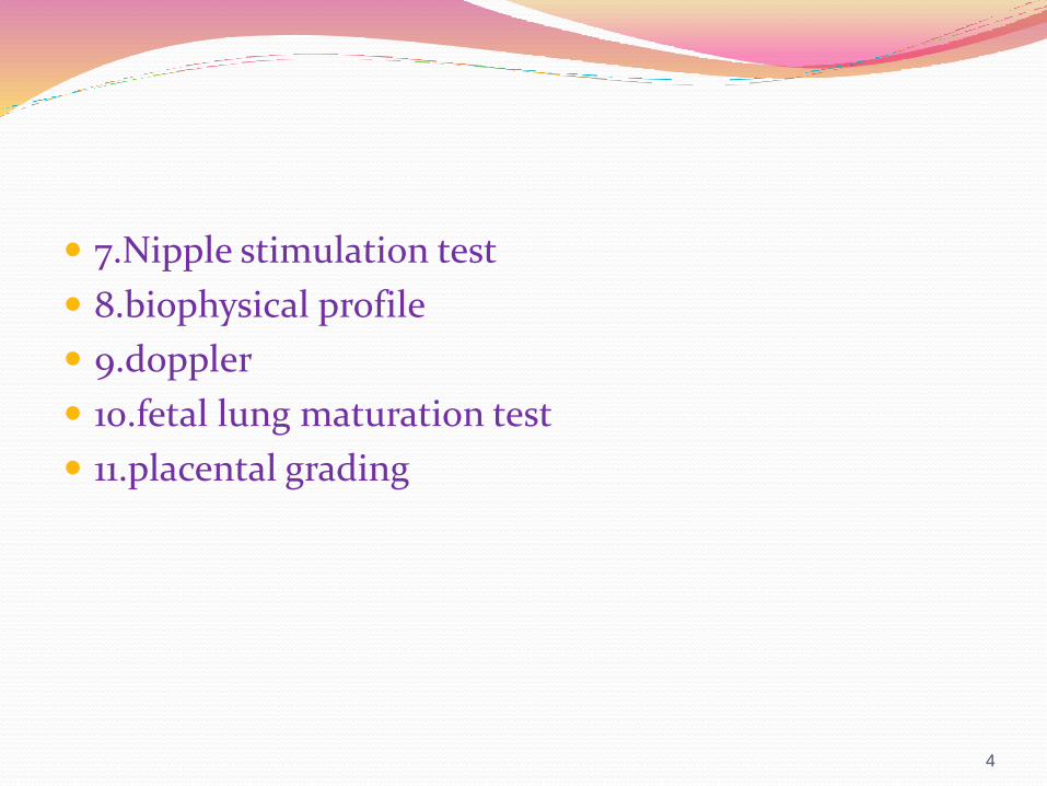
7.Nipple stimulation test
8.biophysical profile
9.doppler
10.fetal lung maturation test
11.placental grading
4
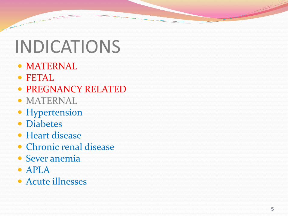
INDICATIONS MATERNAL FETAL PREGNANCY RELATED MATERNAL Hypertension Diabetes Heart disease Chronic renal disease Sever anemia APLA Acute illnesses
5
Fetal
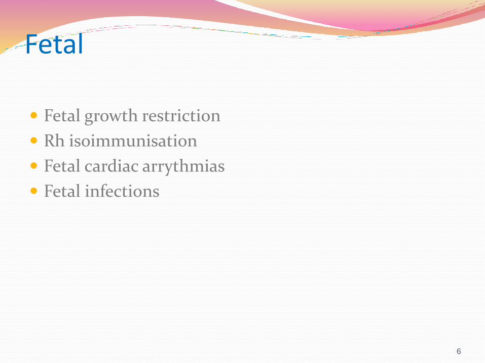
Fetal growth restriction
Rh isoimmunisation
Fetal cardiac arrythmias
Fetal infections
6
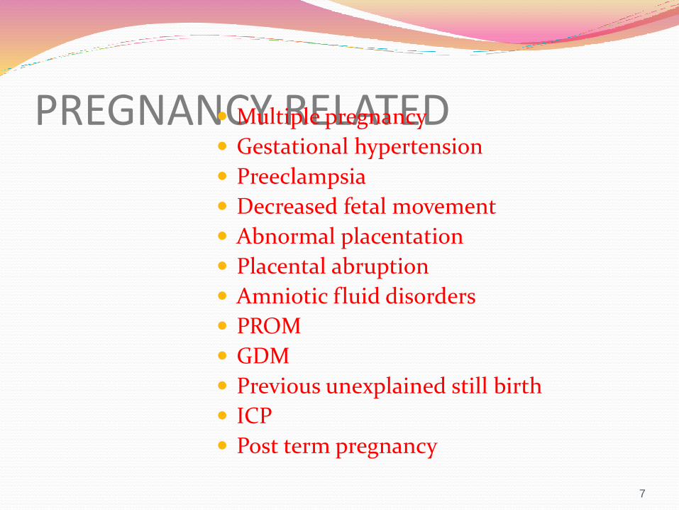
PREGNANCY RELATED Multiple pregnancy
Gestational hypertension
Preeclampsia
Decreased fetal movement
Abnormal placentation
Placental abruption
Amniotic fluid disorders
PROM
GDM
Previous unexplained still birth
ICP
Post term pregnancy
7
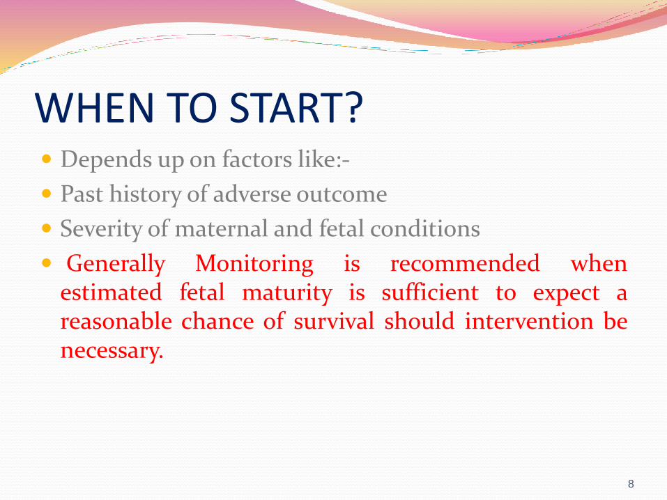
WHEN TO START? Depends up on factors like:-
Past history of adverse outcome
Severity of maternal and fetal conditions
Generally Monitoring is recommended whenestimated fetal maturity is sufficient to expect areasonable chance of survival should intervention benecessary.
8
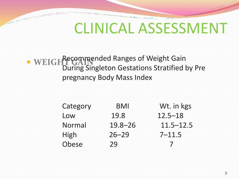
CLINICAL ASSESSMENT
WEIGHT GAIN
9
Recommended Ranges of Weight GainDuring Singleton Gestations Stratified by Pre pregnancy Body Mass Index
Category BMI Wt. in kgsLow 19.8 12.5–18Normal 19.8–26 11.5–12.5High 26–29 7–11.5Obese 29 7
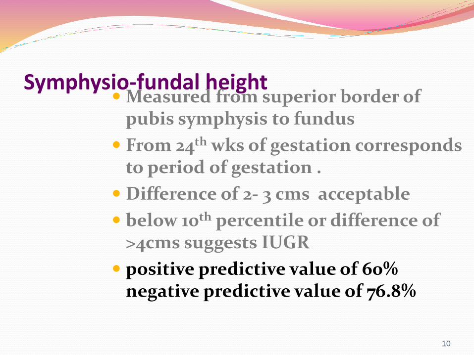
Symphysio-fundal height Measured from superior border of
pubis symphysis to fundus
From 24th wks of gestation corresponds to period of gestation .
Difference of 2- 3 cms acceptable
below 10th percentile or difference of >4cms suggests IUGR
positive predictive value of 60% negative predictive value of 76.8%
10
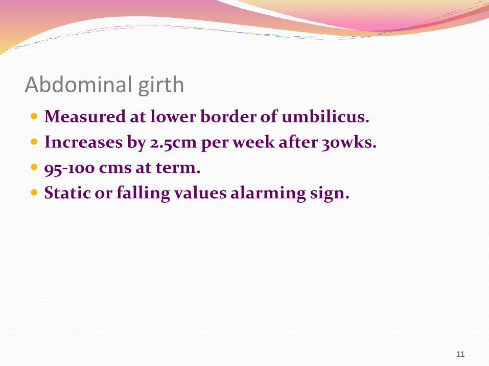
Abdominal girth Measured at lower border of umbilicus.
Increases by 2.5cm per week after 30wks.
95-100 cms at term.
Static or falling values alarming sign.
11
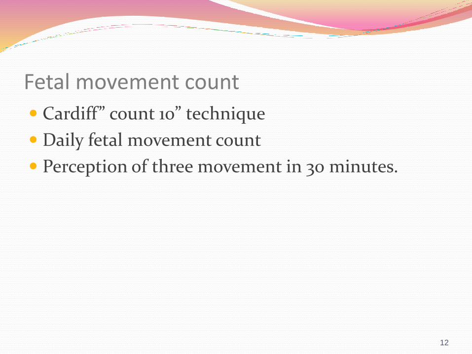
Fetal movement count
Cardiff” count 10” technique
Daily fetal movement count
Perception of three movement in 30 minutes.
12
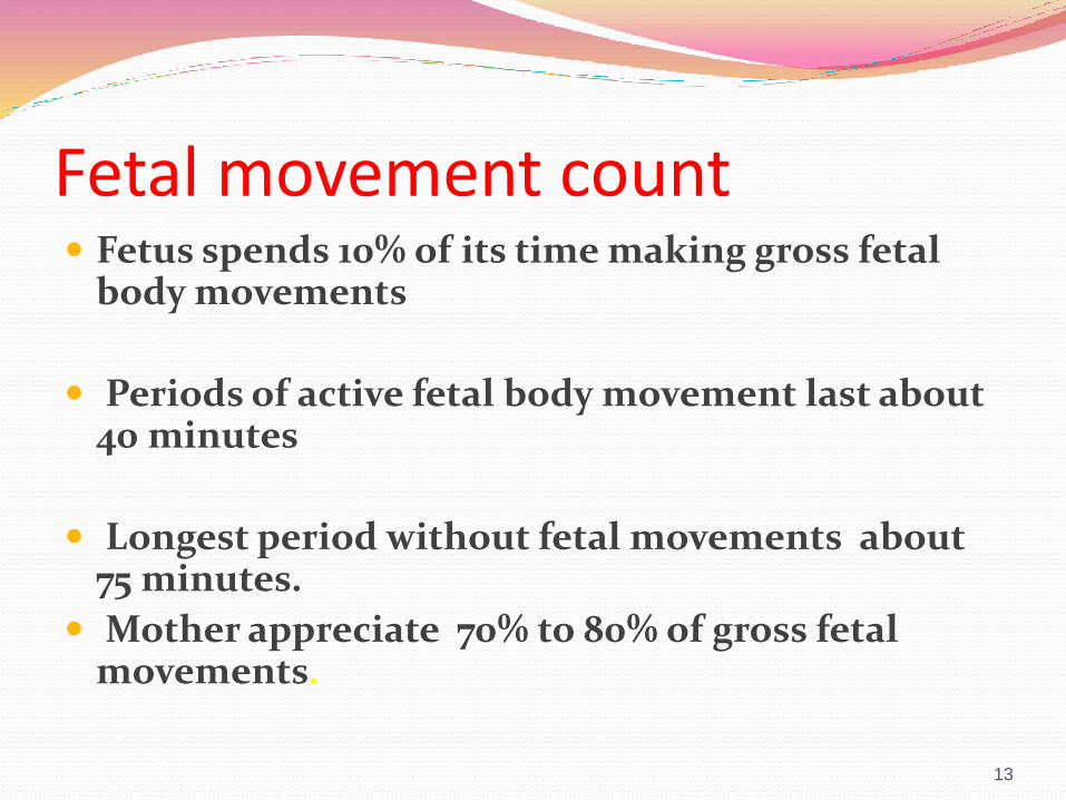
Fetal movement count Fetus spends 10% of its time making gross fetal
body movements
Periods of active fetal body movement last about 40 minutes
Longest period without fetal movements about 75 minutes.
Mother appreciate 70% to 80% of gross fetal movements.
13
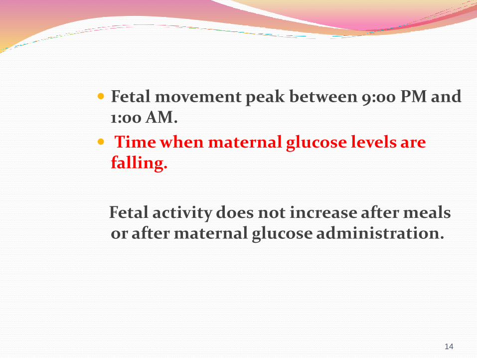
Fetal movement peak between 9:00 PM and 1:00 AM.
Time when maternal glucose levels are falling.
Fetal activity does not increase after meals or after maternal glucose administration.
14

Factors affecting maternal perception of fetal movement Fetal and placental factors :-
Placental location
The length and type of fetal movements
Amniotic fluid volume (AFV)
Maternal factors :-
Parity, obesity.
Psychological factors anxiety.
15
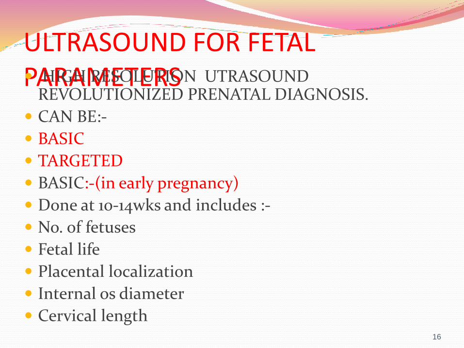
ULTRASOUND FOR FETAL PARAMETERS HIGH RESOLUTION UTRASOUND
REVOLUTIONIZED PRENATAL DIAGNOSIS.
CAN BE:-
BASIC
TARGETED
BASIC:-(in early pregnancy)
Done at 10-14wks and includes :-
No. of fetuses
Fetal life
Placental localization
Internal os diameter
Cervical length16
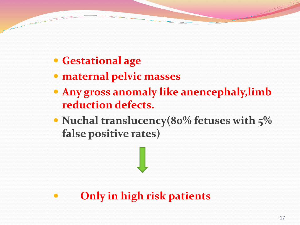
Gestational age
maternal pelvic masses
Any gross anomaly like anencephaly,limbreduction defects.
Nuchal translucency(80% fetuses with 5% false positive rates)
Only in high risk patients
17
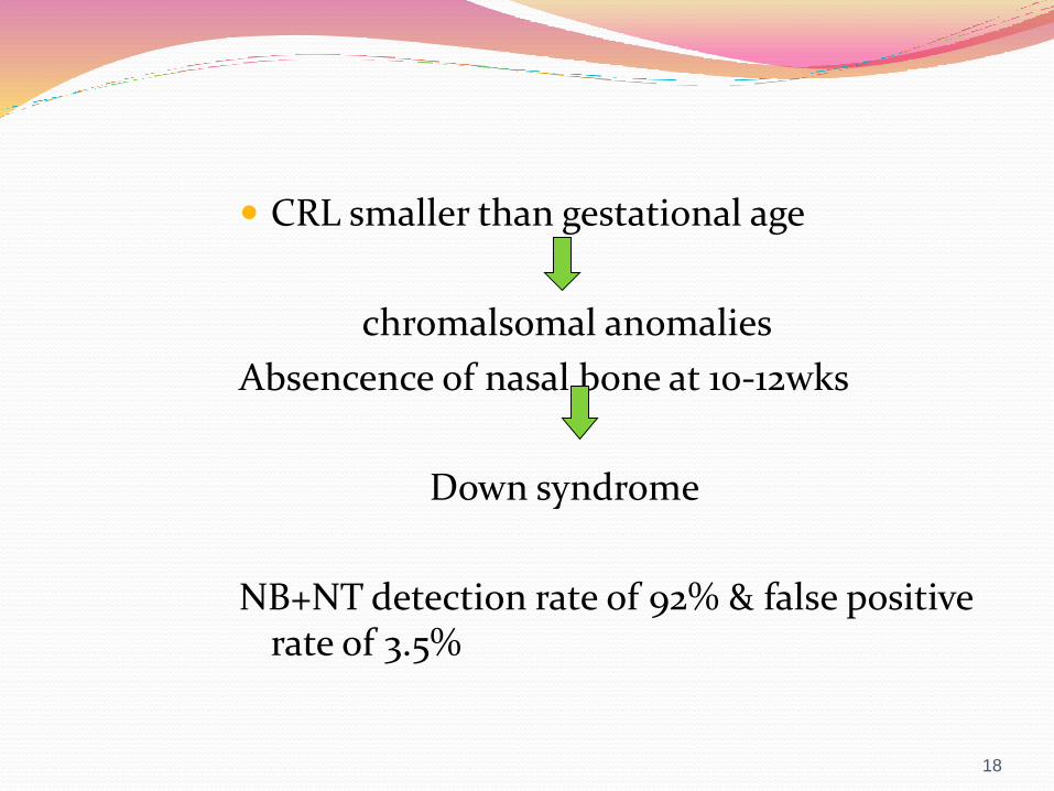
CRL smaller than gestational age
chromalsomal anomalies
Absencence of nasal bone at 10-12wks
Down syndrome
NB+NT detection rate of 92% & false positive rate of 3.5%
18
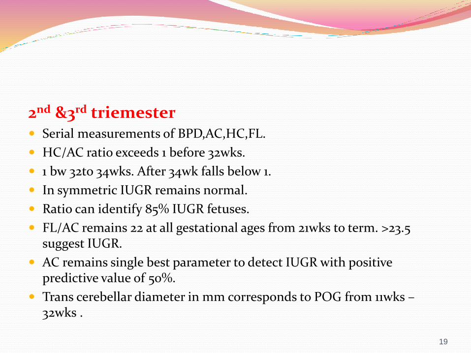
2nd &3rd triemester Serial measurements of BPD,AC,HC,FL.
HC/AC ratio exceeds 1 before 32wks.
1 bw 32to 34wks. After 34wk falls below 1.
In symmetric IUGR remains normal.
Ratio can identify 85% IUGR fetuses.
FL/AC remains 22 at all gestational ages from 21wks to term. >23.5 suggest IUGR.
AC remains single best parameter to detect IUGR with positive predictive value of 50%.
Trans cerebellar diameter in mm corresponds to POG from 11wks –32wks .
19

AMNIOTIC FLUID VOLUME Single deepest pocket >2cm normal
Or
Amniotic fluid index 5-25cm.
20
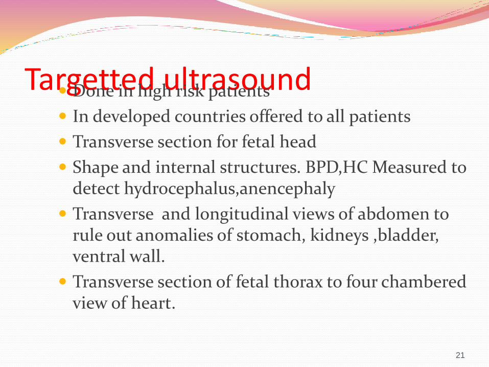
Targetted ultrasound Done in high risk patients
In developed countries offered to all patients
Transverse section for fetal head
Shape and internal structures. BPD,HC Measured to detect hydrocephalus,anencephaly
Transverse and longitudinal views of abdomen to rule out anomalies of stomach, kidneys ,bladder, ventral wall.
Transverse section of fetal thorax to four chambered view of heart.
21
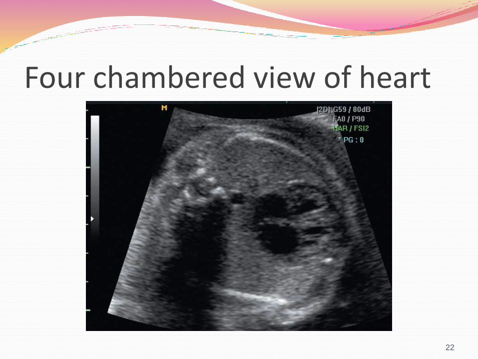
Four chambered view of heart
22

Identify three long bones in each limb
and any achondroplasia is looked for.
Saggittal ,coronal and transverse views taken to rule out spinabifida.
cephalic index
Biparietal diameter/occipitofrontal diameter
23
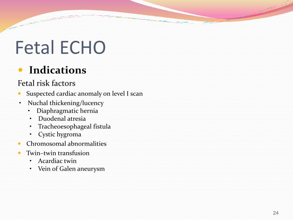
Fetal ECHO IndicationsFetal risk factors Suspected cardiac anomaly on level I scan
• Nuchal thickening/lucency• Diaphragmatic hernia• Duodenal atresia• Tracheoesophageal fistula• Cystic hygroma
Chromosomal abnormalities
Twin–twin transfusion• Acardiac twin• Vein of Galen aneurysm
24

Maternal risk factors •
Congenital heart disease• Exposure to teratogen
Diabetes
Maternal infectionsFamilial risk factors• Trisomy 21 (Down)
25
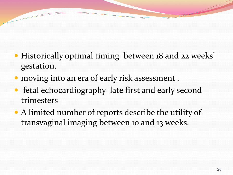
Historically optimal timing between 18 and 22 weeks’ gestation.
moving into an era of early risk assessment .
fetal echocardiography late first and early second trimesters
A limited number of reports describe the utility of transvaginal imaging between 10 and 13 weeks.
26
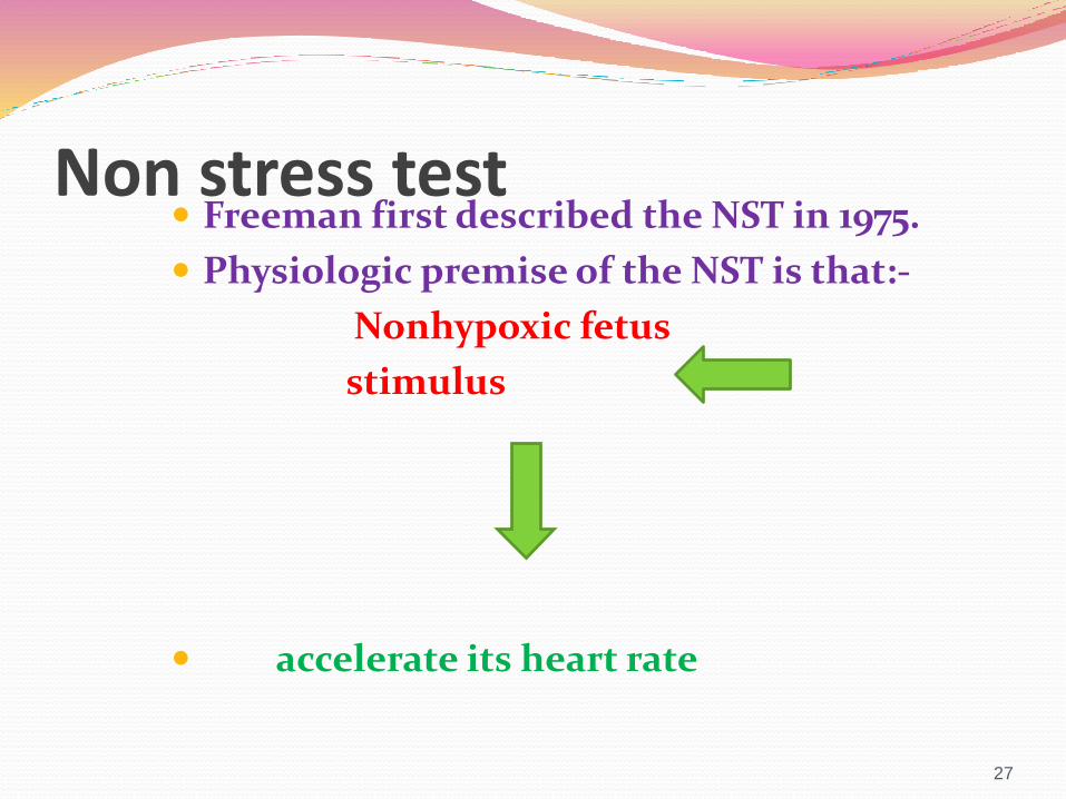
Non stress test Freeman first described the NST in 1975.
Physiologic premise of the NST is that:-
Nonhypoxic fetus
stimulus
accelerate its heart rate
27
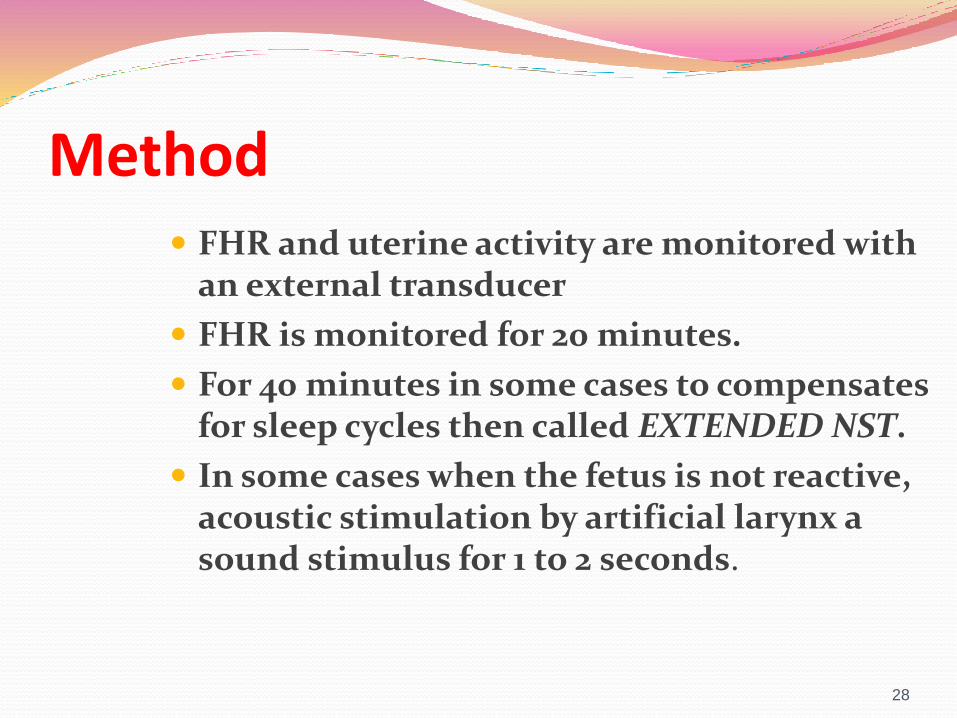
Method FHR and uterine activity are monitored with
an external transducer
FHR is monitored for 20 minutes.
For 40 minutes in some cases to compensates for sleep cycles then called EXTENDED NST.
In some cases when the fetus is not reactive, acoustic stimulation by artificial larynx a sound stimulus for 1 to 2 seconds.
28

INTERPRETATION Reactive NST presence of two accelerations of
15bpm over base line for 15sec in a 20-minute time period with or without fetal movements.
Nonreactive NST absence of two accelerations in a 40-minute period with or without acoustic stimulation over a 40-minute period.
29
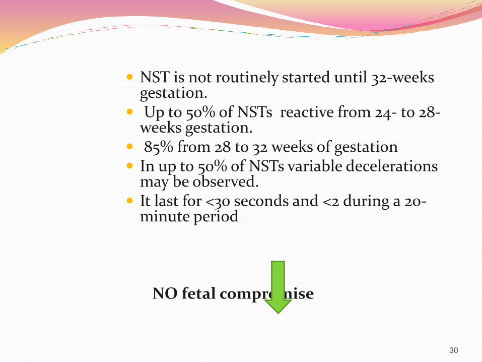
NST is not routinely started until 32-weeks gestation.
Up to 50% of NSTs reactive from 24- to 28-weeks gestation.
85% from 28 to 32 weeks of gestation In up to 50% of NSTs variable decelerations
may be observed. It last for <30 seconds and <2 during a 20-
minute period
NO fetal compromise
30
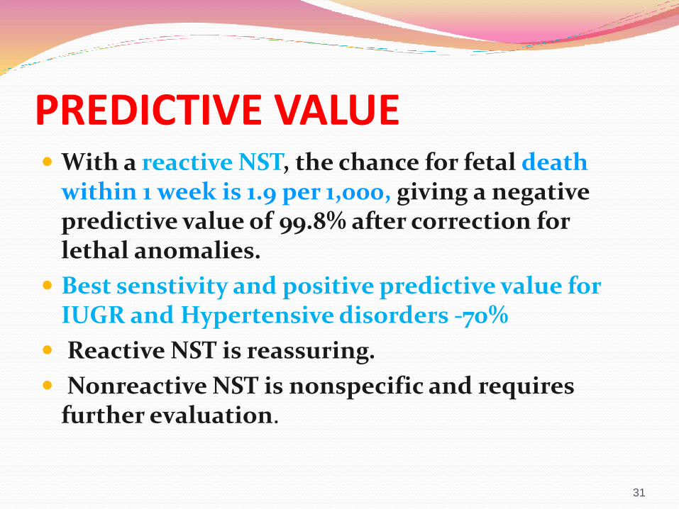
PREDICTIVE VALUE With a reactive NST, the chance for fetal death
within 1 week is 1.9 per 1,000, giving a negative predictive value of 99.8% after correction for lethal anomalies.
Best senstivity and positive predictive value for IUGR and Hypertensive disorders -70%
Reactive NST is reassuring.
Nonreactive NST is nonspecific and requires further evaluation.
31
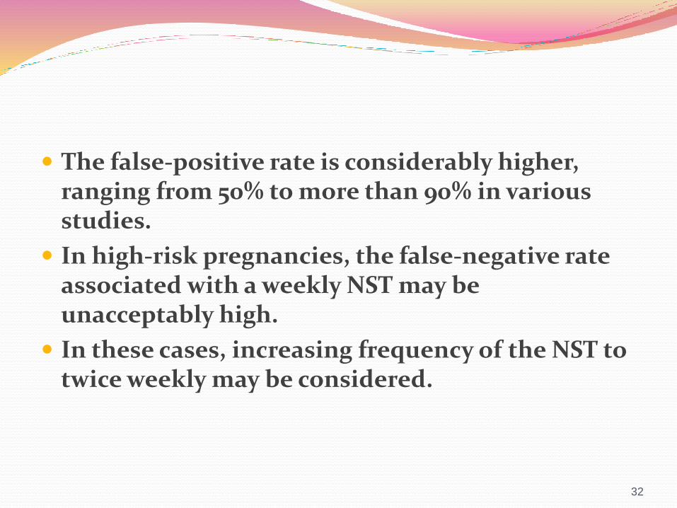
The false-positive rate is considerably higher, ranging from 50% to more than 90% in various studies.
In high-risk pregnancies, the false-negative rate associated with a weekly NST may be unacceptably high.
In these cases, increasing frequency of the NST to twice weekly may be considered.
32
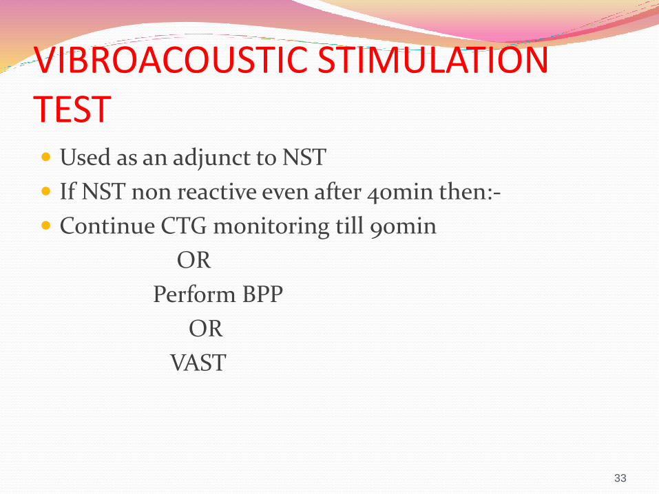
VIBROACOUSTIC STIMULATION TEST Used as an adjunct to NST
If NST non reactive even after 40min then:-
Continue CTG monitoring till 90min
OR
Perform BPP
OR
VAST
33

Auditory brainstem response functional at 26 to 28 weeks’ gestation.
VAST increase the incidence of reactive NSTs after 26 weeks’ gestation .
Reduce the testing time.
artificial larynx that generates sound 82 Db -100db with a frequency of 80 Hz.
34

A stimulus for 3 seconds or less is applied near the fetal head.
If the NST remains nonreactive
Stimulus is repeated at 1-minute intervals up to three times.
35
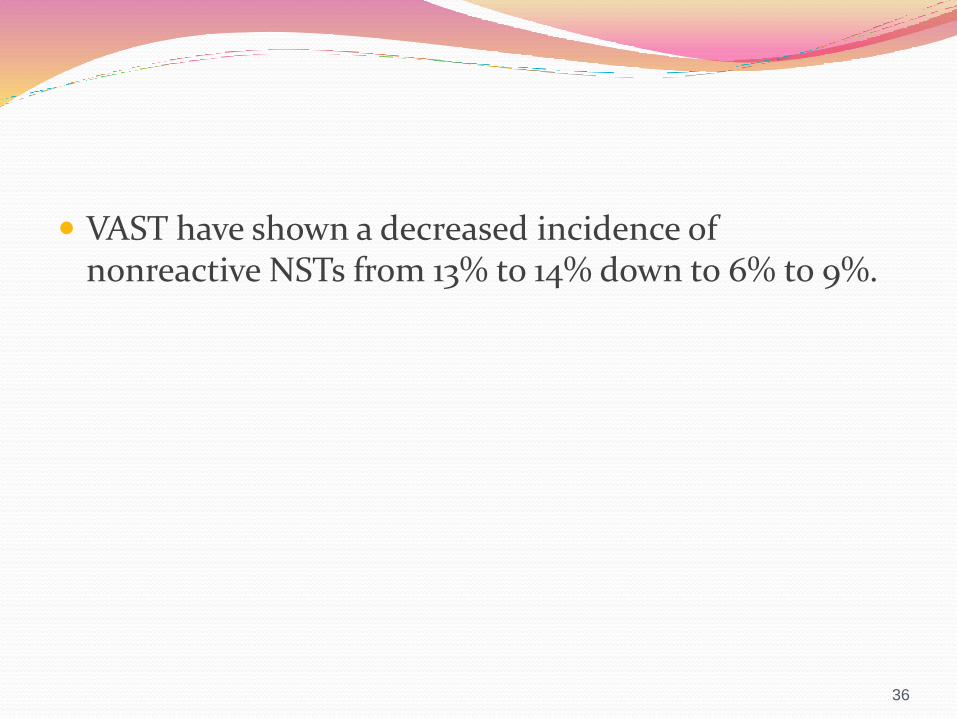
VAST have shown a decreased incidence of nonreactive NSTs from 13% to 14% down to 6% to 9%.
36
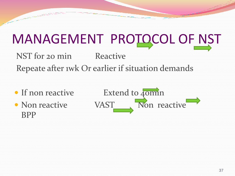
MANAGEMENT PROTOCOL OF NSTNST for 20 min Reactive
Repeate after 1wk Or earlier if situation demands
If non reactive Extend to 40min
Non reactive VAST Non reactive BPP
37

BIOPHYSICAL PROFILE Thorough evaluation of fetal well-being .
Potential to significantly reduce the false-positive rate of the NST/CST.
The BPP was initially described by Manning and colleagues .
Rationale:-Fetal biophysical activities controlled by centers in the fetal brain sensitive to varying degrees of hypoxia.
38
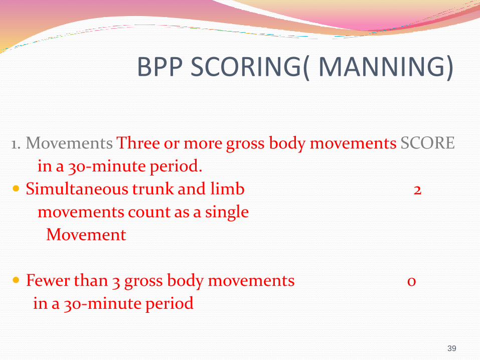
BPP SCORING( MANNING)
1. Movements Three or more gross body movements SCORE
in a 30-minute period.
Simultaneous trunk and limb 2
movements count as a single
Movement
Fewer than 3 gross body movements 0
in a 30-minute period
39
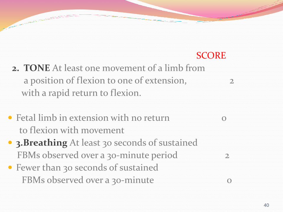
SCORE
2. TONE At least one movement of a limb from
a position of flexion to one of extension, 2
with a rapid return to flexion.
Fetal limb in extension with no return 0
to flexion with movement
3.Breathing At least 30 seconds of sustained
FBMs observed over a 30-minute period 2
Fewer than 30 seconds of sustained
FBMs observed over a 30-minute 0
40
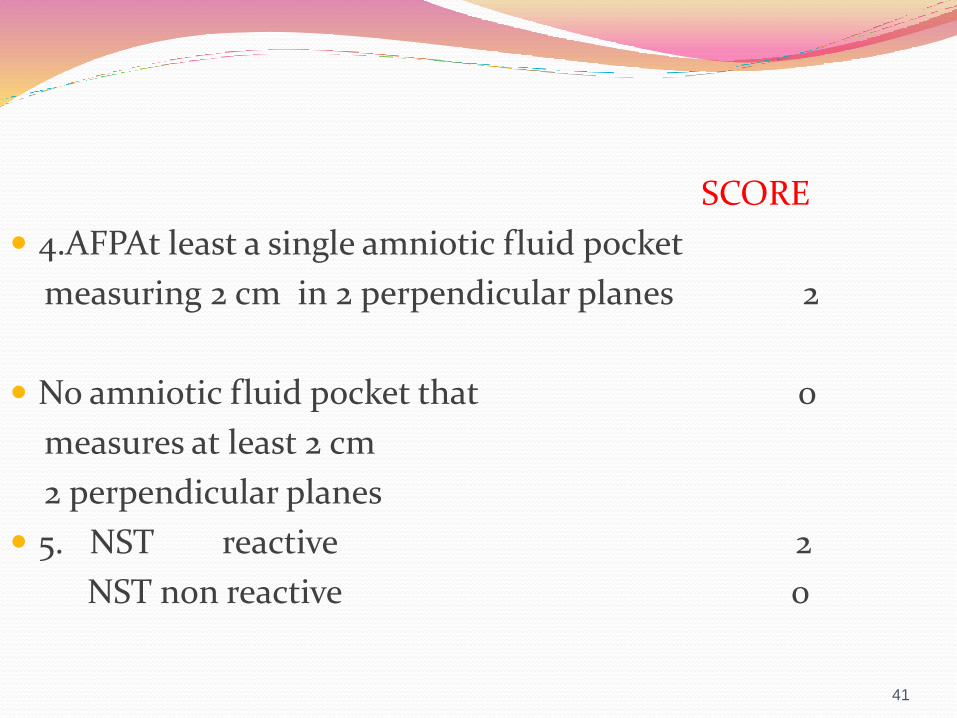
SCORE
4.AFPAt least a single amniotic fluid pocket
measuring 2 cm in 2 perpendicular planes 2
No amniotic fluid pocket that 0
measures at least 2 cm
2 perpendicular planes
5. NST reactive 2
NST non reactive 0
41
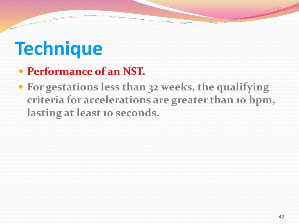
Technique Performance of an NST.
For gestations less than 32 weeks, the qualifying criteria for accelerations are greater than 10 bpm, lasting at least 10 seconds.
42
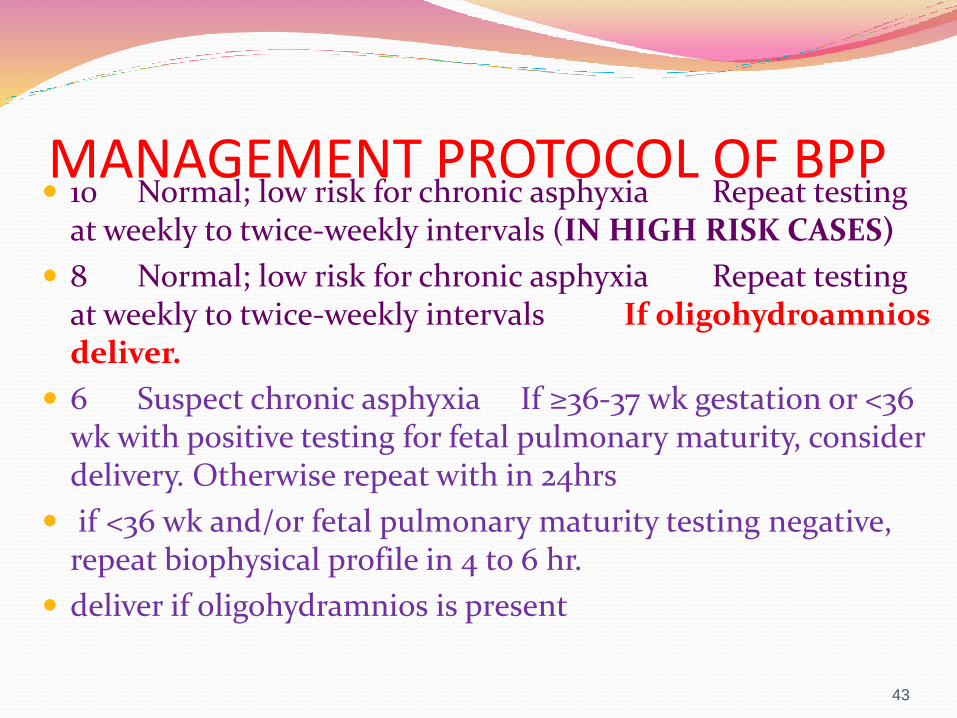
MANAGEMENT PROTOCOL OF BPP 10 Normal; low risk for chronic asphyxia Repeat testing
at weekly to twice-weekly intervals (IN HIGH RISK CASES)
8 Normal; low risk for chronic asphyxia Repeat testing at weekly to twice-weekly intervals If oligohydroamniosdeliver.
6 Suspect chronic asphyxia If ≥36-37 wk gestation or <36 wk with positive testing for fetal pulmonary maturity, consider delivery. Otherwise repeat with in 24hrs
if <36 wk and/or fetal pulmonary maturity testing negative, repeat biophysical profile in 4 to 6 hr.
deliver if oligohydramnios is present
43
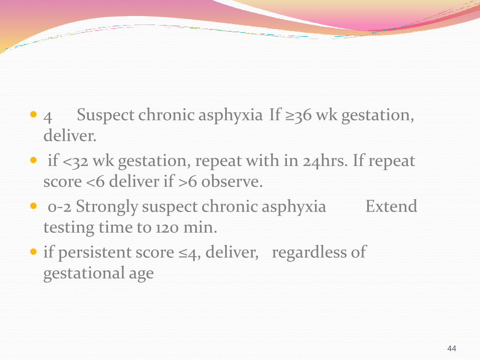
4 Suspect chronic asphyxia If ≥36 wk gestation, deliver.
if <32 wk gestation, repeat with in 24hrs. If repeat score <6 deliver if >6 observe.
0-2 Strongly suspect chronic asphyxia Extend testing time to 120 min.
if persistent score ≤4, deliver, regardless of gestational age
44
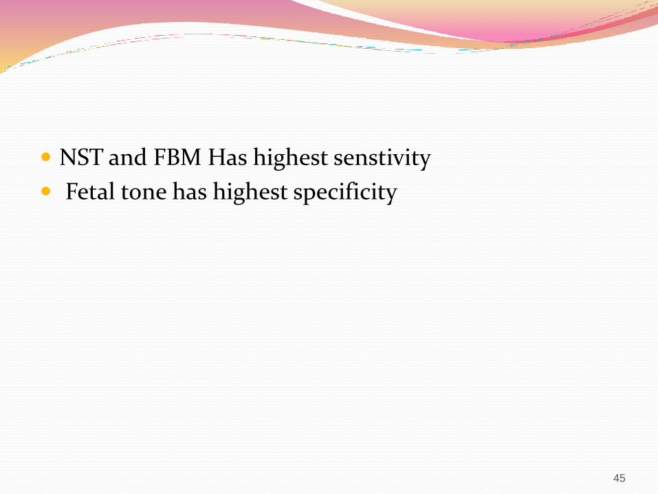
NST and FBM Has highest senstivity
Fetal tone has highest specificity
45
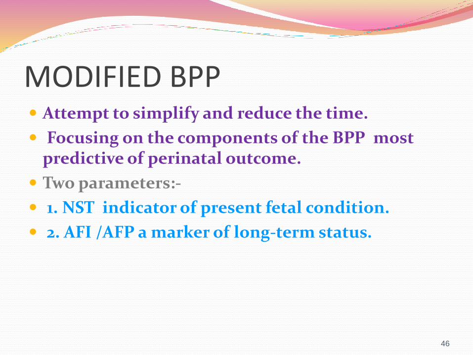
MODIFIED BPP Attempt to simplify and reduce the time.
Focusing on the components of the BPP most predictive of perinatal outcome.
Two parameters:-
1. NST indicator of present fetal condition.
2. AFI /AFP a marker of long-term status.
46
Contraction Stress Test
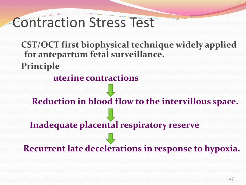
CST/OCT first biophysical technique widely applied for antepartum fetal surveillance.
Principle
uterine contractions
Reduction in blood flow to the intervillous space.
Inadequate placental respiratory reserve
Recurrent late decelerations in response to hypoxia.
47
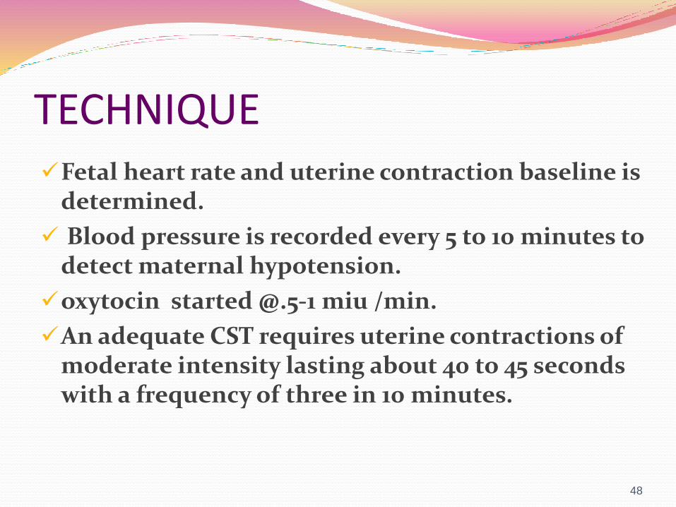
TECHNIQUEFetal heart rate and uterine contraction baseline is
determined.
Blood pressure is recorded every 5 to 10 minutes to detect maternal hypotension.
oxytocin started @.5-1 miu /min.
An adequate CST requires uterine contractions of moderate intensity lasting about 40 to 45 seconds with a frequency of three in 10 minutes.
48

INTERPRETATIONNegative: No late or significant variable
decelerations
Positive: Late decelerations with at least 50% of contractions
Suspicious: Intermittent late or variable decelerations
Hyperstimulation: Decelerations with contractions longer than 90 seconds’ duration or 2-minute frequency
Unsatisfactory: Fewer than three contractions per 10 minutes or an uninterpretable tracing
49

PREDICTIVE VALUE OF CST A negative CST good fetal outcome.
incidence of perinatal death within 1 week of a negative CST (i.e., the false-negative rate) to be less than 1 per 1000.
50

MANAGEMENT PROTOCOL OF CST Positive CST is usually repeated in 24 hours .
This is of historical importance .
Not used now.

CONTRAINDICATIONS Placenta previa
Previous CS
Multiple gestation
Polyhydroamnios
History of preterm
Incompetent cervix
52
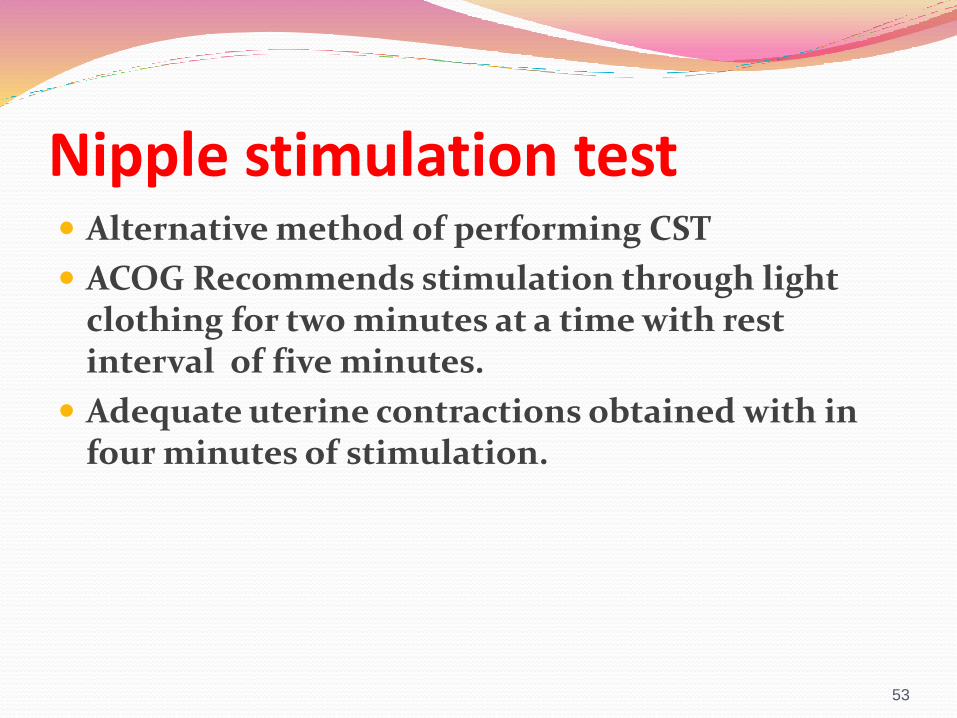
Nipple stimulation test Alternative method of performing CST
ACOG Recommends stimulation through light clothing for two minutes at a time with rest interval of five minutes.
Adequate uterine contractions obtained with in four minutes of stimulation.
53
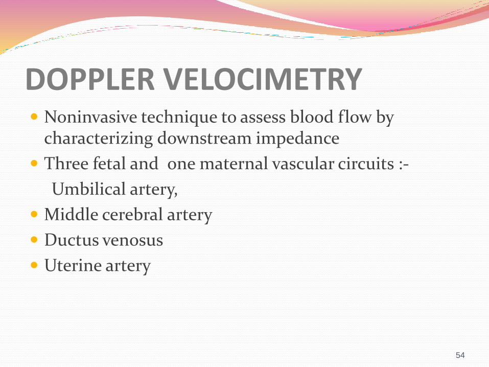
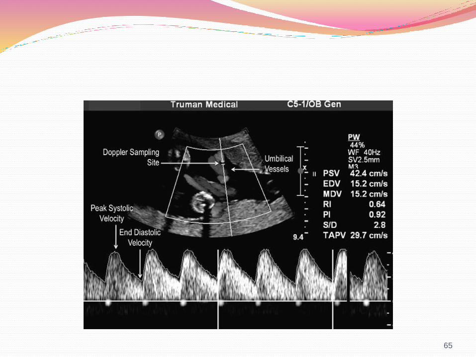
DOPPLER VELOCIMETRY Noninvasive technique to assess blood flow by
characterizing downstream impedance
Three fetal and one maternal vascular circuits :-
Umbilical artery,
Middle cerebral artery
Ductus venosus
Uterine artery
54

INDICATIONS IUGR
PIH
GDM
RH ISOIMMUNISATION
INTRAHEPATIC CHOLISTASIS OF PREGNANCY
55

UTERINE ARTERY
INDICATIONS
(1) history of Preeclampsia
(2) previous child with IUGR
(3) unexplained high maternal
serum alpha-fetoprotein level
(4) high human chorionic gonadotropin
level.
(5) thrombophilias
56
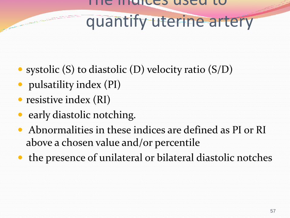
The indices used to quantify uterine artery
systolic (S) to diastolic (D) velocity ratio (S/D)
pulsatility index (PI)
resistive index (RI)
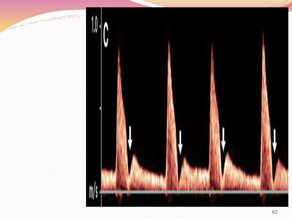
early diastolic notching.
Abnormalities in these indices are defined as PI or RI above a chosen value and/or percentile
the presence of unilateral or bilateral diastolic notches
57

How to calculate indices S/D ratio
systolic peak velocity/diastolic peak velocity
Resistance index (RI)
systolic- end diastolic peak velocity/systolic peak velocity
Pulsatility index (PI)
systolic-end diastolic peak velocity/time averaged maximum −velocity
58
Uterine Doppler screening is commonly performed around 20 weeks.
increased uterine artery impedance to flow at 20–24 weeks follow-up at 26–28 weeks.
Cut-off values at 23 weeks' gestation are:-
a mean PI above 1.5–1.61.
mean RI above 0.57–0.58.
Bilateral notches are found in about 25–30% of pregnancies at 12 weeks.
10–15% at 20 weeks .
5% at 24 weeks.
sensitivity is up to 85% when performed between 22 and 23 weeks’ gestation.
high risk patients given low-dose aspirin because of bilateral uterine artery notching at 12 to 14 weeks have an 80% reduction of placental disease
60
An early diastolic notch in the uterine arteries at 12 to 14 weeks suggest delayed trophoblastinvasion.
Persistence “notching” beyond 24 weeks
confirmatory evidence.
61
62
sensitivities and specificities of uterine artery Doppler in low-risk populations varied from 34% to 76% and 83% to 93%, respectively.
63
For both preeclampsia and IUGR uterine artery Doppler more accurate in the second than the first trimester.
Increased PI with notching in the second trimester best predictor of preeclampsia.
64
65
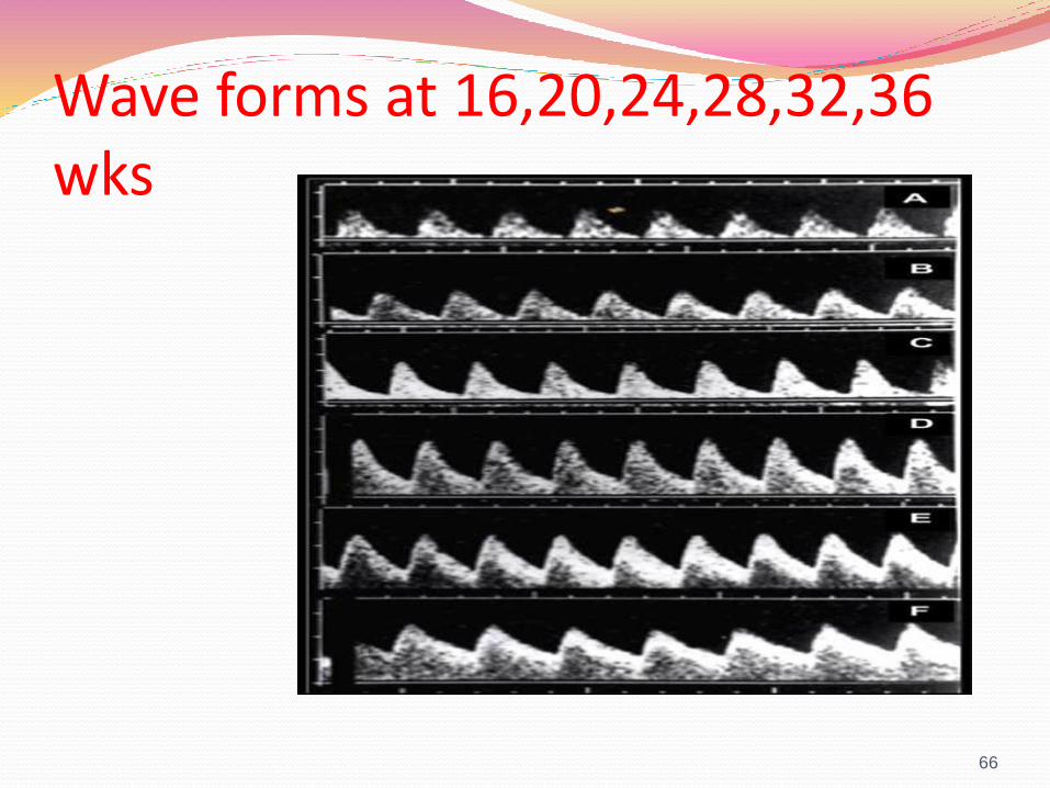
Wave forms at 16,20,24,28,32,36 wks
66
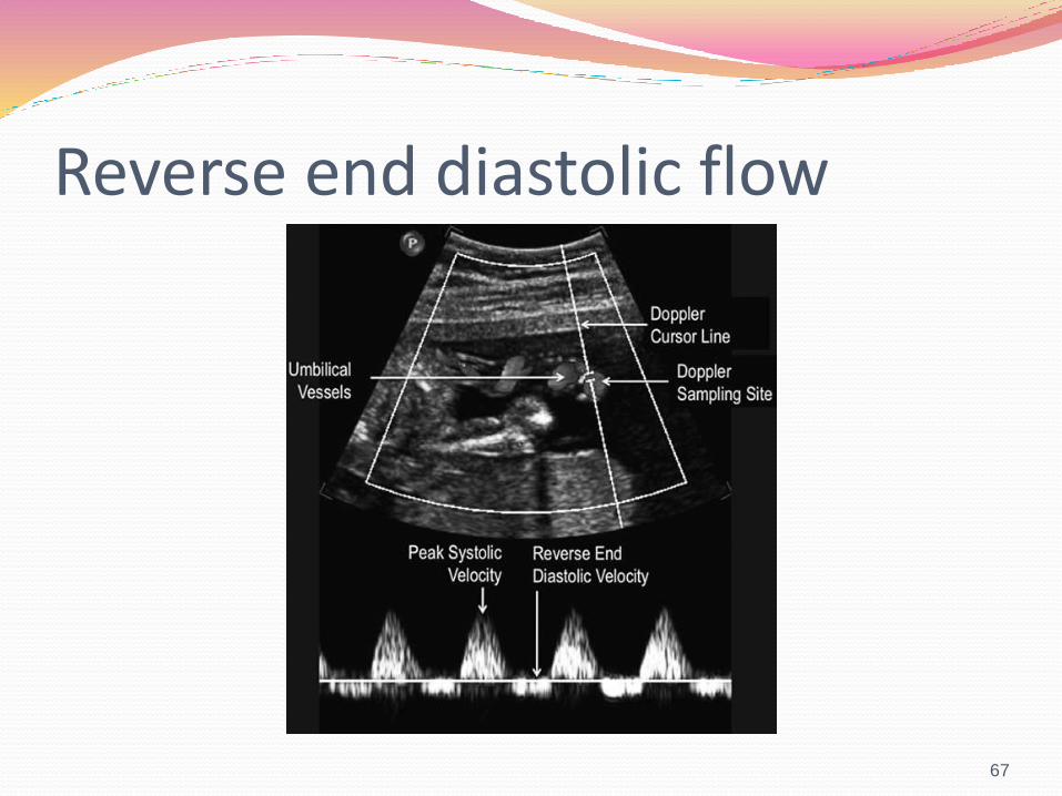
Reverse end diastolic flow
67
FACTS SHEET RI had the best discriminatory
ability when compared with the S/D ratio (P<.05),the PI (P<.001).S/D ratio, however, remains the most popularindex.
S/D ratio less than or equal to 3.0. Resistance index less than or equal
to 0.6 is considered normal after 27 completedweeks of pregnancy.
Benefits of this technique before 28 weeks ofgestation are uncertain.
68

Diagnostic feature of umbilical artery Doppler waveform is the end diastolic flow.
Absent or reverse end diastolic flow ominous finding.
frequency of absent end diastolic flow is approximately 2% in high-risk pregnancies .
0.3% in a general obstetric population.
In pregnancies complicated with FGR, fetal surveillance should consist of weekly umbilical Doppler.
.
69
BPP or NST should be used either as a backup test or simultaneously with the umbilical artery Doppler.
Umbilical Doppler index is high or increasing
weekly umbilical Doppler ultrasound
+
Twice wkly NST/ BPP
70
MANAGEMENT WITH ABSENT END DIASTOLIC FLOW
Guided by the gestational age. >34 completed weeks Delivery
Bw 28 to 34 completed conservative
Daily umbilical artery Doppler, NST, and BPP (or modified BPP)+ Ductus venosus
Reverse flow at any gestational age beyond 28 weeks Delivery
71
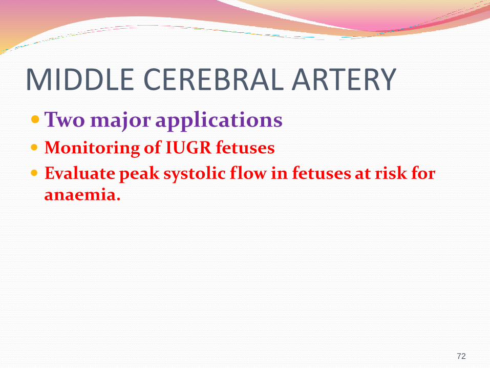
MIDDLE CEREBRAL ARTERYTwo major applications Monitoring of IUGR fetuses
Evaluate peak systolic flow in fetuses at risk for anaemia.
72
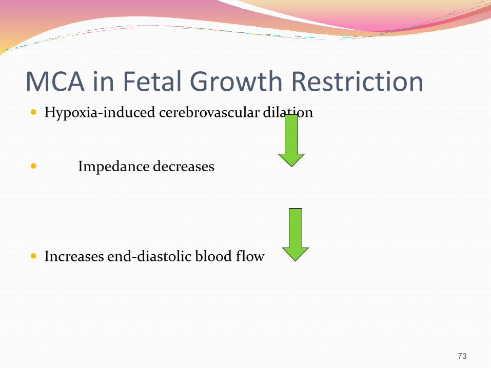
MCA in Fetal Growth Restriction Hypoxia-induced cerebrovascular dilation
Impedance decreases
Increases end-diastolic blood flow
73
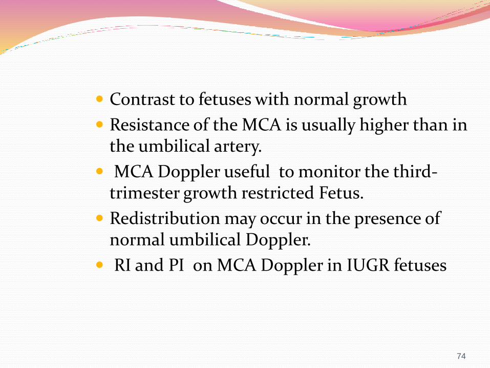
Contrast to fetuses with normal growth
Resistance of the MCA is usually higher than in the umbilical artery.
MCA Doppler useful to monitor the third-trimester growth restricted Fetus.
Redistribution may occur in the presence of normal umbilical Doppler.
RI and PI on MCA Doppler in IUGR fetuses
74
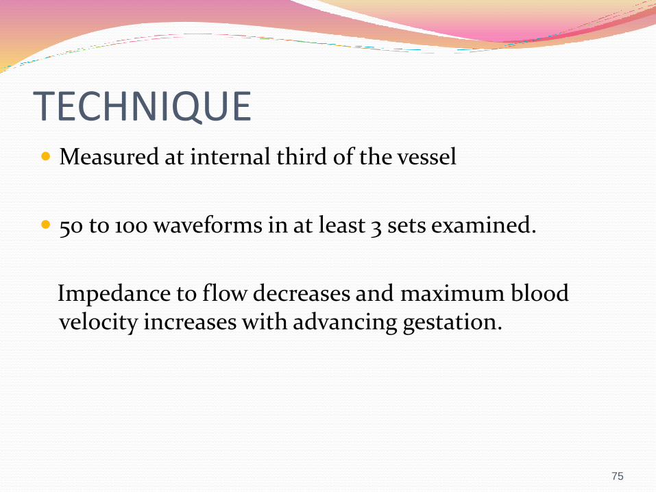
TECHNIQUE Measured at internal third of the vessel
50 to 100 waveforms in at least 3 sets examined.
Impedance to flow decreases and maximum blood velocity increases with advancing gestation.
75

MCA-PSV new development.
better parameter in the prediction of perinatalmortality than PI/RI.
MCA PI in IUGR fetuses can normalize in later stages.
MCA-PSV becomes abnormal, remains as such.
Ratio of MCA PI to Umbilical PI >1.5
in normal fetal circulatory condition.
76
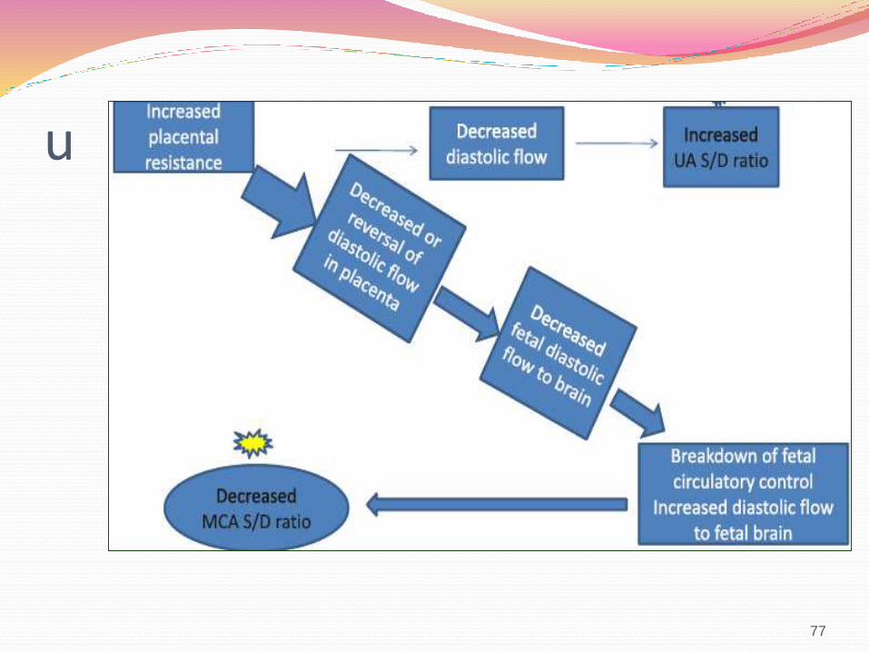
u
77
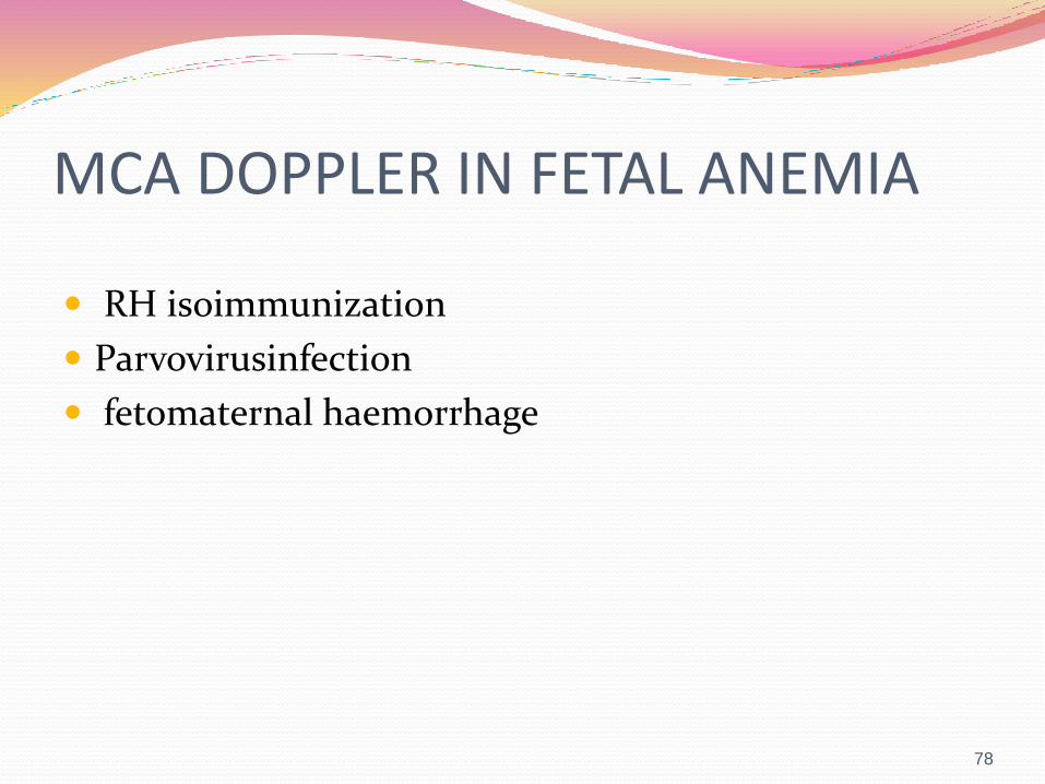
MCA DOPPLER IN FETAL ANEMIA
RH isoimmunization
Parvovirusinfection
fetomaternal haemorrhage
78
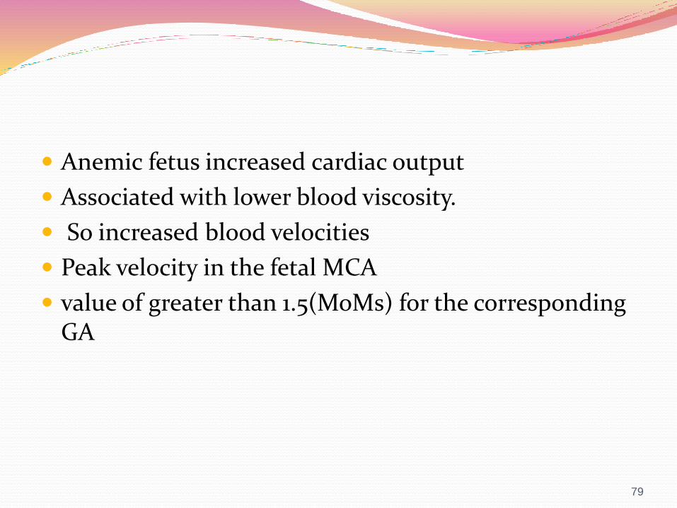
Anemic fetus increased cardiac output
Associated with lower blood viscosity.
So increased blood velocities
Peak velocity in the fetal MCA
value of greater than 1.5(MoMs) for the corresponding GA
79
80
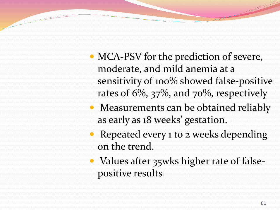
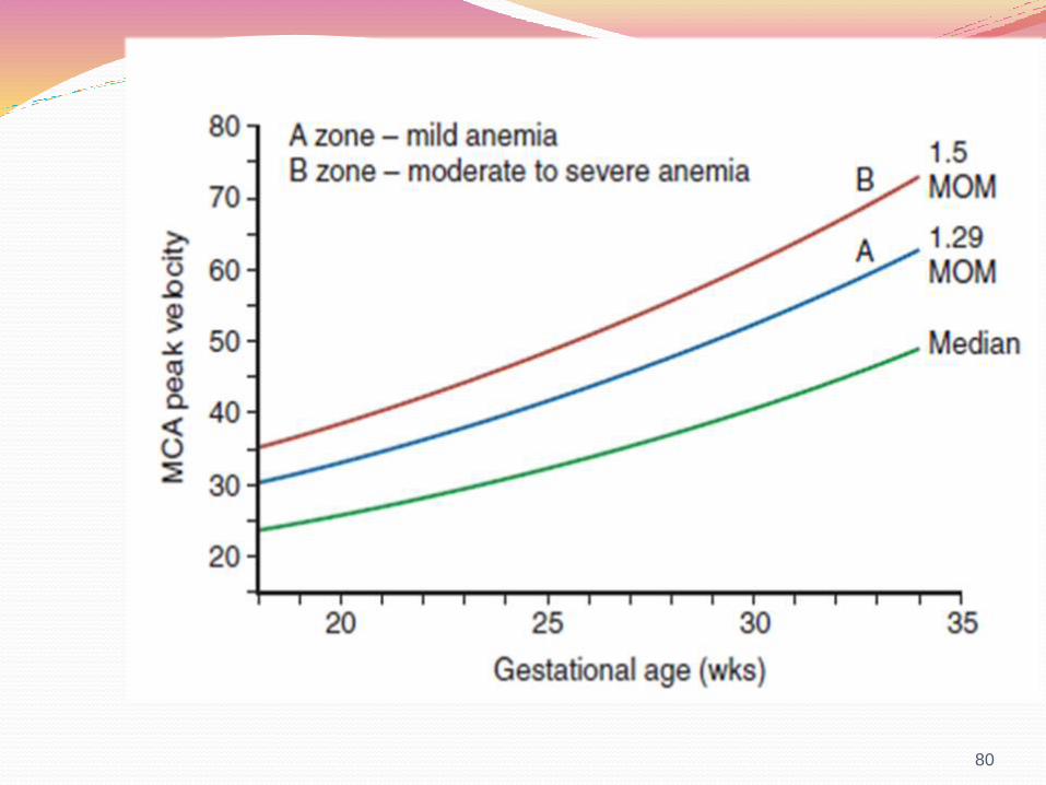
MCA-PSV for the prediction of severe, moderate, and mild anemia at a sensitivity of 100% showed false-positive rates of 6%, 37%, and 70%, respectively
Measurements can be obtained reliably as early as 18 weeks’ gestation.
Repeated every 1 to 2 weeks depending on the trend.
Values after 35wks higher rate of false-positive results
81

Ductus Venosus
Connects the intra-abdominal portion of the umbilical vein with the inferior vena cava at its inlet to the right atrium.
Shunt plays a critical role in the delivery of well-oxygenated blood to the left side of fetal heart.
Sample siteInlet, where the highest velocities are recorded
waveform of the ductus venosus triphasic.
82

Ductus venosus waveforms
83
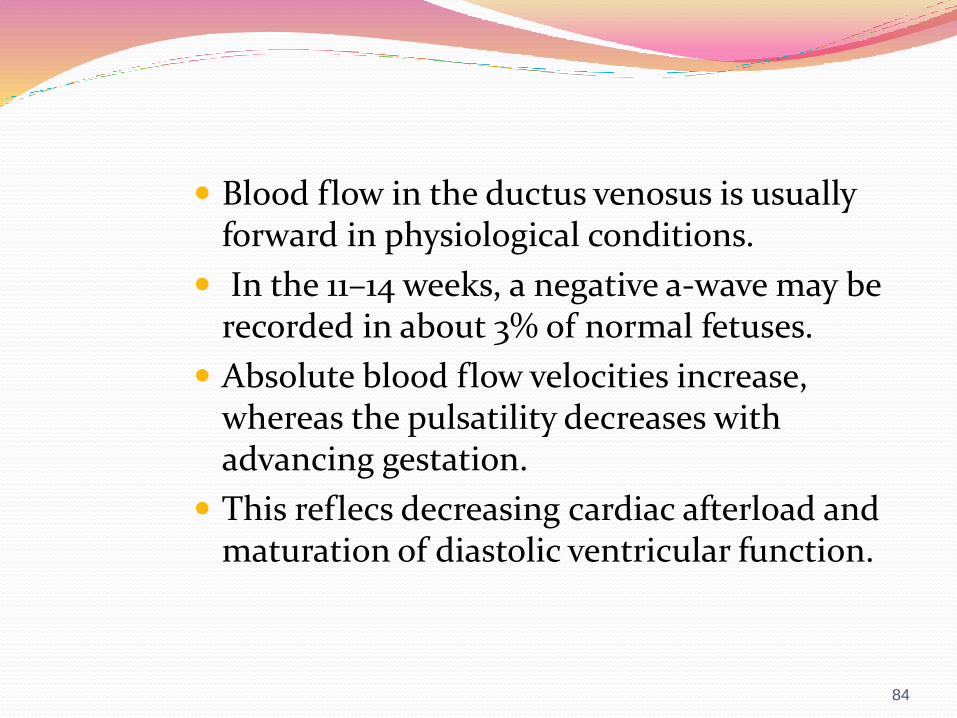
Blood flow in the ductus venosus is usually forward in physiological conditions.
In the 11–14 weeks, a negative a-wave may be recorded in about 3% of normal fetuses.
Absolute blood flow velocities increase, whereas the pulsatility decreases with advancing gestation.
This reflecs decreasing cardiac afterload and maturation of diastolic ventricular function.
84
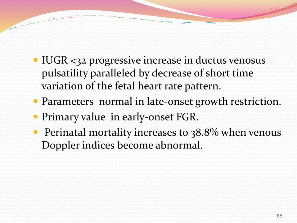
IUGR <32 progressive increase in ductus venosuspulsatility paralleled by decrease of short time variation of the fetal heart rate pattern.
Parameters normal in late-onset growth restriction.
Primary value in early-onset FGR.
Perinatal mortality increases to 38.8% when venous Doppler indices become abnormal.
85

Management goals & protocol Prevention of stillbirth
Delivery based on an accurate assessment of fetal versus neonatal risks.
Abnormal venous Doppler indices, mandate higher testing frequency, up to daily testing.
Reversal of DV a wave increases the risk for an abnormal biophysical profile score within 1 to 8 days.
Reversal of DV a wave only becomes an independent risk factor for neonatal morbidity and mortality after 27 weeks’ gestation.
86
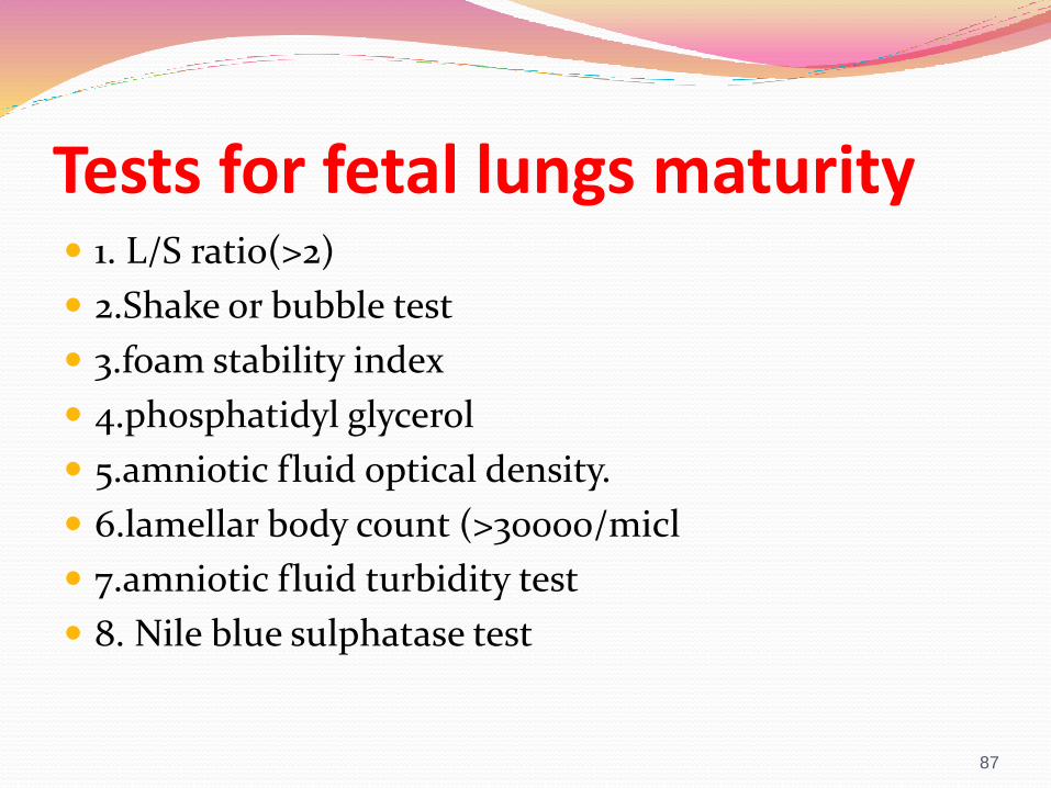
Tests for fetal lungs maturity 1. L/S ratio(>2)
2.Shake or bubble test
3.foam stability index
4.phosphatidyl glycerol
5.amniotic fluid optical density.
6.lamellar body count (>30000/micl
7.amniotic fluid turbidity test
8. Nile blue sulphatase test
87

GOAL The timely identification and rescue of the fetus at
risk of neonatal and long term morbidity from
intrapartum hypoxic insult
Intrapartum monitoring FHR monitoring –
Intermittent auscultation(IA)
Electronic fetal monitoring(EFM)
Fetal Scalp pH
Fetal Pulse oximetry
Fetal scalp lactate testing
ST waveform analysis
FETAL HEART RATE MONITORING External FHR monitoring-
Hand-held Doppler ultrasound probe
External transducer

TECHNICAL CONSIDERATIONS Basis for FHR monitoring is beat to beat recording
For practical purposes ,this is possible only when direct fetal electrocardiograms are recorded with a scalp electrode.
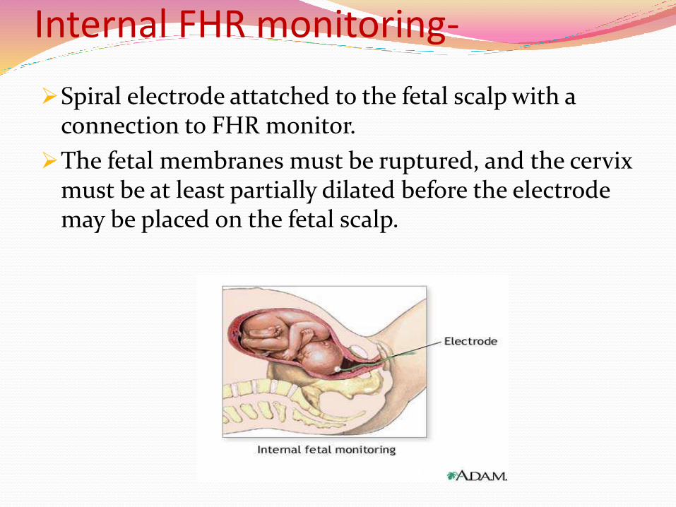
Internal FHR monitoring-
Spiral electrode attatched to the fetal scalp with a connection to FHR monitor.
The fetal membranes must be ruptured, and the cervix must be at least partially dilated before the electrode may be placed on the fetal scalp.
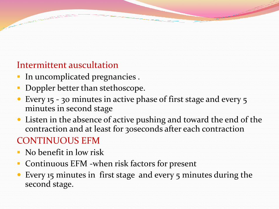
Intermittent auscultation In uncomplicated pregnancies .
Doppler better than stethoscope.
Every 15 - 30 minutes in active phase of first stage and every 5 minutes in second stage
Listen in the absence of active pushing and toward the end of the contraction and at least for 30seconds after each contraction
CONTINUOUS EFM No benefit in low risk
Continuous EFM -when risk factors for present
Every 15 minutes in first stage and every 5 minutes during the second stage.

Fetal Assessment : IA & EFM
Surveillence
Acceptable methods
Low-Risk
Pregnancies
High-Risk
Pregnancies
Intermittent Auscultation* Yes Yes (a)
Continuous Electronic Fetal
Monitoring (EFM)
Yes Yes (b)
Evaluation Intervals
First-stage Labour 30 min 15 min (a,b)
Second –stage labour 15 min 5 min (a,c)
•a- before, during and especially after a contraction for 60 sec
•b- includes evaluation in every 15 min
• c- evaluation in every 5 min
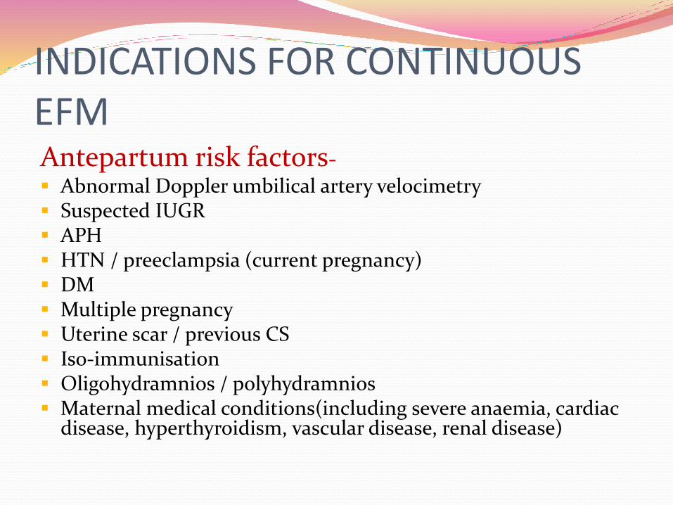
INDICATIONS FOR CONTINUOUS EFMAntepartum risk factors- Abnormal Doppler umbilical artery velocimetry Suspected IUGR APH HTN / preeclampsia (current pregnancy) DM Multiple pregnancy Uterine scar / previous CS Iso-immunisation Oligohydramnios / polyhydramnios Maternal medical conditions(including severe anaemia, cardiac
disease, hyperthyroidism, vascular disease, renal disease)
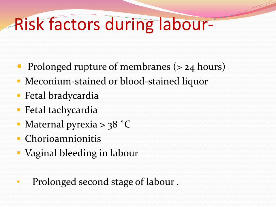
Risk factors during labour-
Prolonged rupture of membranes (> 24 hours)
Meconium-stained or blood-stained liquor
Fetal bradycardia
Fetal tachycardia
Maternal pyrexia > 38 ˚C
Chorioamnionitis
Vaginal bleeding in labour
• Prolonged second stage of labour .
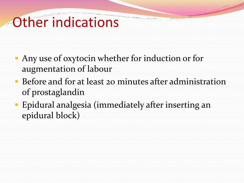
Other indications
Any use of oxytocin whether for induction or for augmentation of labour
Before and for at least 20 minutes after administration of prostaglandin
Epidural analgesia (immediately after inserting an epidural block)
Electronic fetal monitoring Various components include
-Baseline
-Variability
-Accelerations
-Decelerations

External fetal monitoringBASELINE
The mean FHR rounded to increments of 5 bpm during a 10-minute segment, excluding:
—Periodic or episodic changes
—Periods of marked FHR variability
—Segments of baseline that differ by more than 25 bpm
The baseline must be for a minimum of 2 minutes in any 10-minute segment
Normal : 110–160 bpm
Tachycardia: > 160 bpm
Bradycardia: <110 bpm
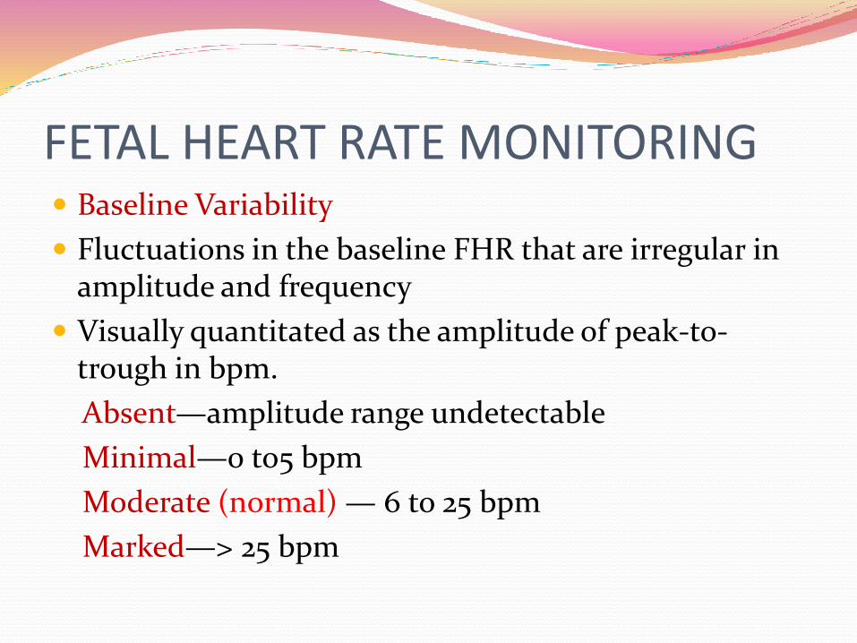
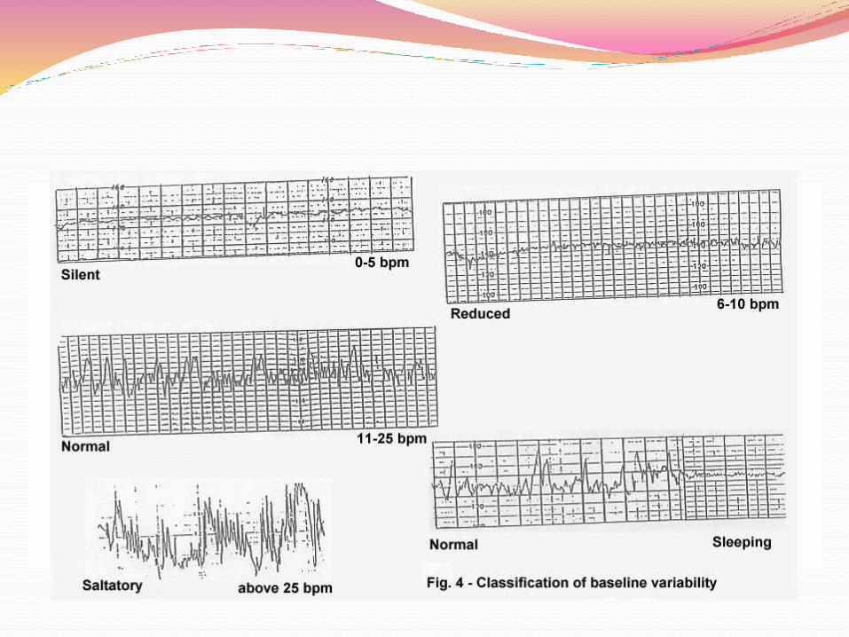
FETAL HEART RATE MONITORING Baseline Variability
Fluctuations in the baseline FHR that are irregular in amplitude and frequency
Visually quantitated as the amplitude of peak-to-trough in bpm.
Absent—amplitude range undetectable
Minimal—0 to5 bpm
Moderate (normal) — 6 to 25 bpm
Marked—> 25 bpm
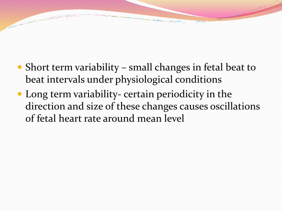
Short term variability – small changes in fetal beat to beat intervals under physiological conditions
Long term variability- certain periodicity in the direction and size of these changes causes oscillations of fetal heart rate around mean level
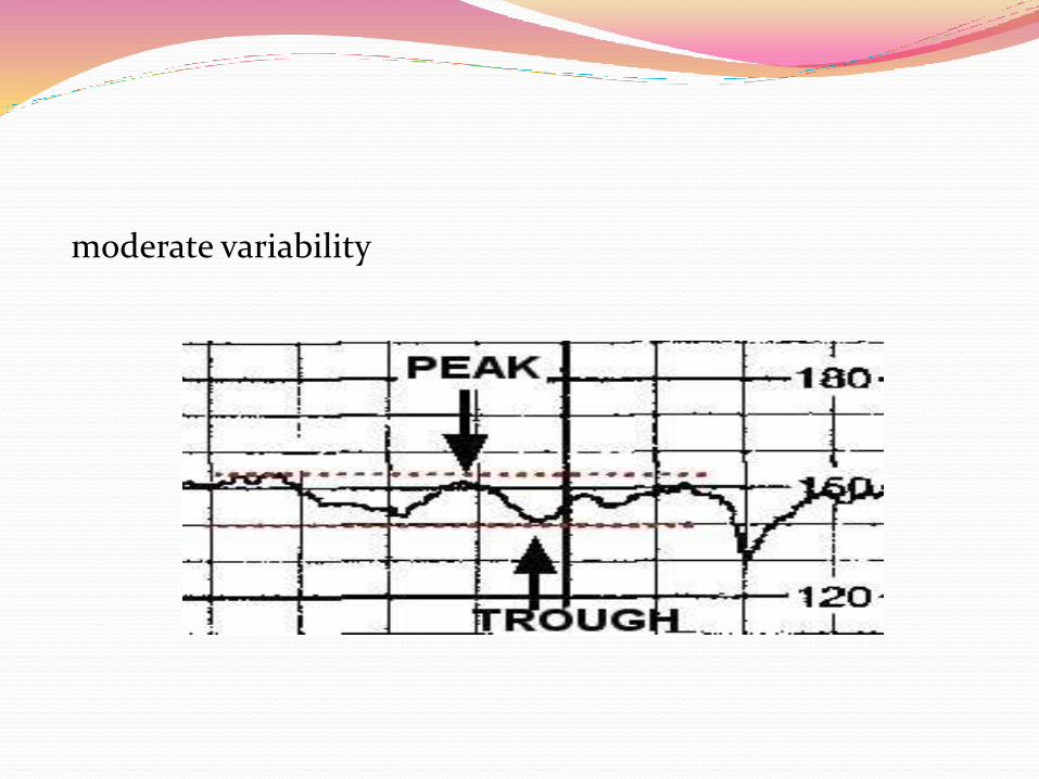
moderate variability
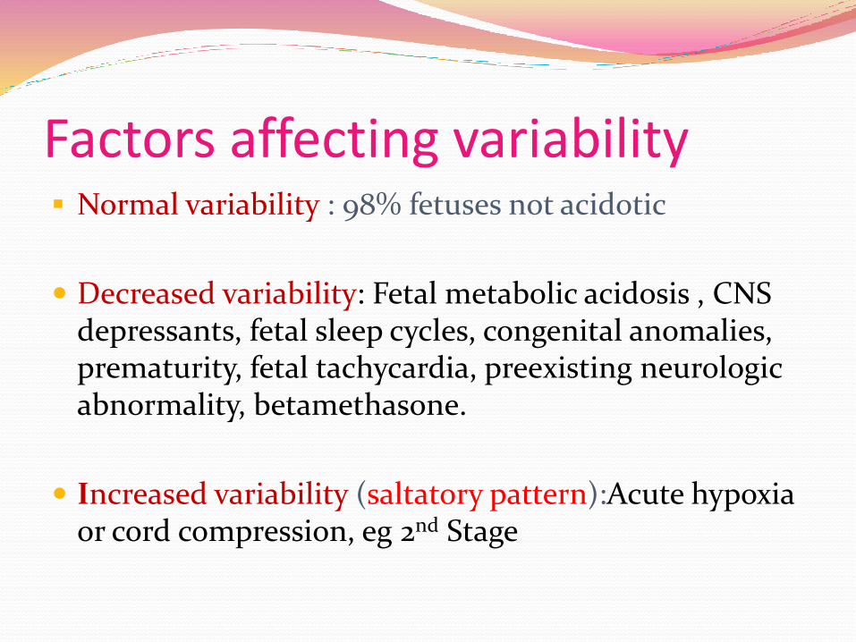
Factors affecting variability Normal variability : 98% fetuses not acidotic
Decreased variability: Fetal metabolic acidosis , CNS depressants, fetal sleep cycles, congenital anomalies, prematurity, fetal tachycardia, preexisting neurologic abnormality, betamethasone.
Increased variability (saltatory pattern):Acute hypoxia or cord compression, eg 2nd Stage
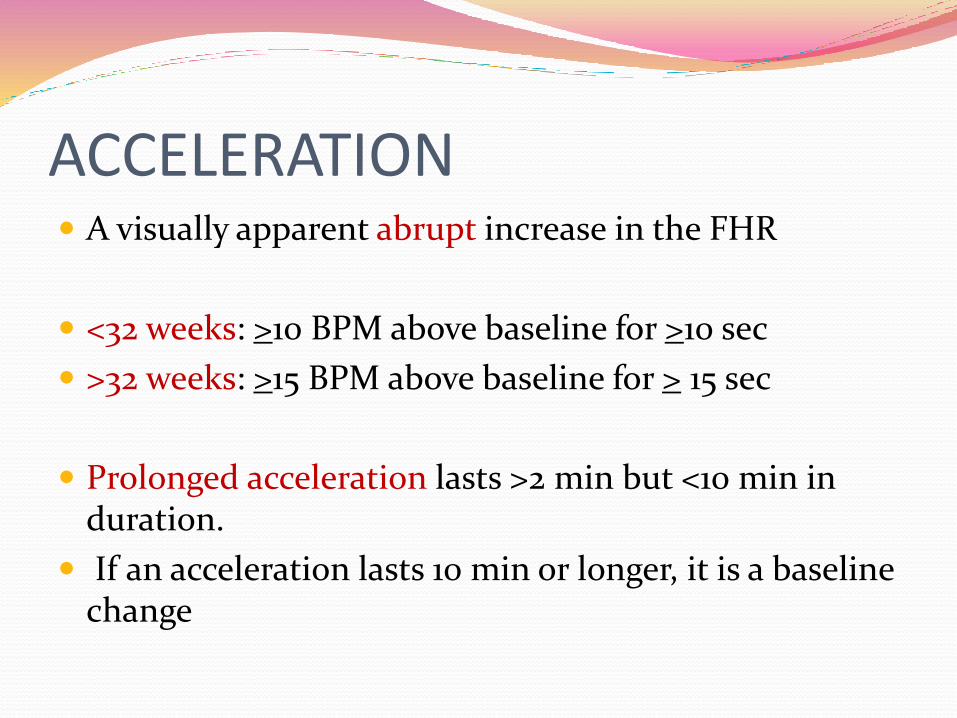
ACCELERATION A visually apparent abrupt increase in the FHR
<32 weeks: >10 BPM above baseline for >10 sec
>32 weeks: >15 BPM above baseline for > 15 sec
Prolonged acceleration lasts >2 min but <10 min in duration.
If an acceleration lasts 10 min or longer, it is a baseline change
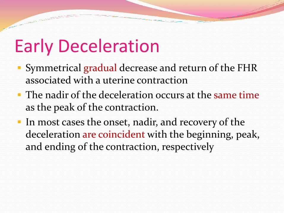
Early Deceleration Symmetrical gradual decrease and return of the FHR
associated with a uterine contraction
The nadir of the deceleration occurs at the same time as the peak of the contraction.
In most cases the onset, nadir, and recovery of the deceleration are coincident with the beginning, peak, and ending of the contraction, respectively
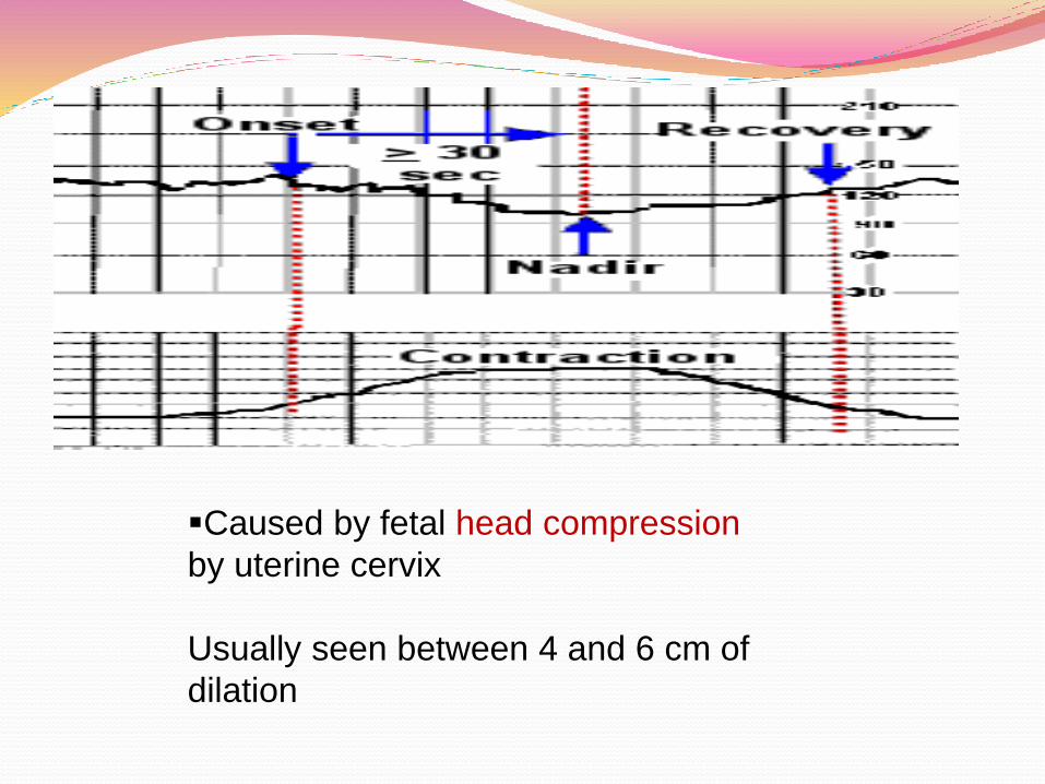
Caused by fetal head compression
by uterine cervix
Usually seen between 4 and 6 cm of
dilation
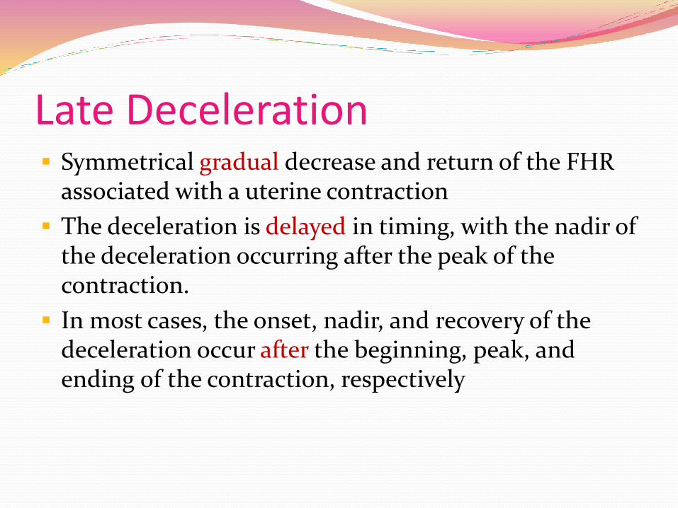
Late Deceleration Symmetrical gradual decrease and return of the FHR
associated with a uterine contraction
The deceleration is delayed in timing, with the nadir of the deceleration occurring after the peak of the contraction.
In most cases, the onset, nadir, and recovery of the deceleration occur after the beginning, peak, and ending of the contraction, respectively
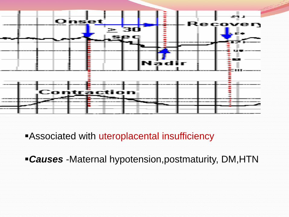
Associated with uteroplacental insufficiency
Causes -Maternal hypotension,postmaturity, DM,HTN
Variable Deceleration Visually apparent abrupt decrease in FHR
The decrease in FHR is ≥ 15 bpm , lasting ≥ 15 sec, and <2 minutes in duration.
When variable decelerations are associated with uterine contractions, their onset, depth, and duration commonly vary with successive uterine contractions.
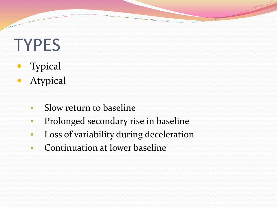
TYPES Typical
Atypical
Slow return to baseline
Prolonged secondary rise in baseline
Loss of variability during deceleration
Continuation at lower baseline
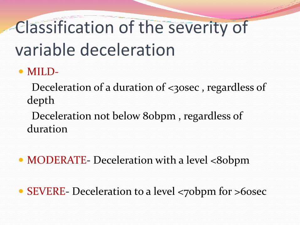
Classification of the severity of variable deceleration MILD-
Deceleration of a duration of <30sec , regardless of depth
Deceleration not below 80bpm , regardless of duration
MODERATE- Deceleration with a level <80bpm
SEVERE- Deceleration to a level <70bpm for >60sec
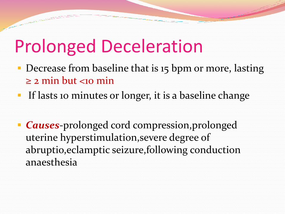
Prolonged Deceleration Decrease from baseline that is 15 bpm or more, lasting
≥ 2 min but <10 min
If lasts 10 minutes or longer, it is a baseline change
Causes-prolonged cord compression,prolongeduterine hyperstimulation,severe degree of abruptio,eclamptic seizure,following conduction anaesthesia
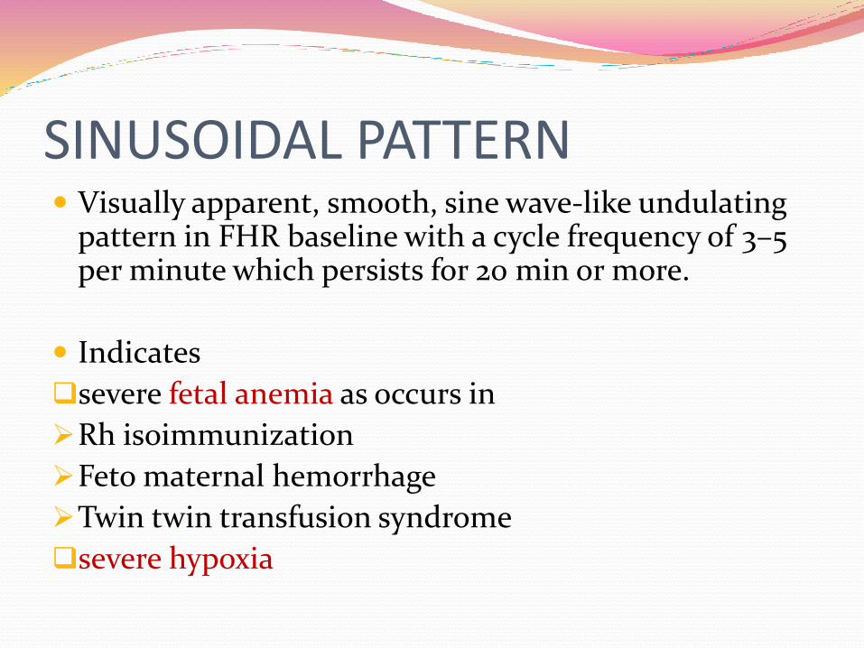
SINUSOIDAL PATTERN Visually apparent, smooth, sine wave-like undulating
pattern in FHR baseline with a cycle frequency of 3–5 per minute which persists for 20 min or more.
Indicates
severe fetal anemia as occurs in
Rh isoimmunization
Feto maternal hemorrhage
Twin twin transfusion syndrome
severe hypoxia

A
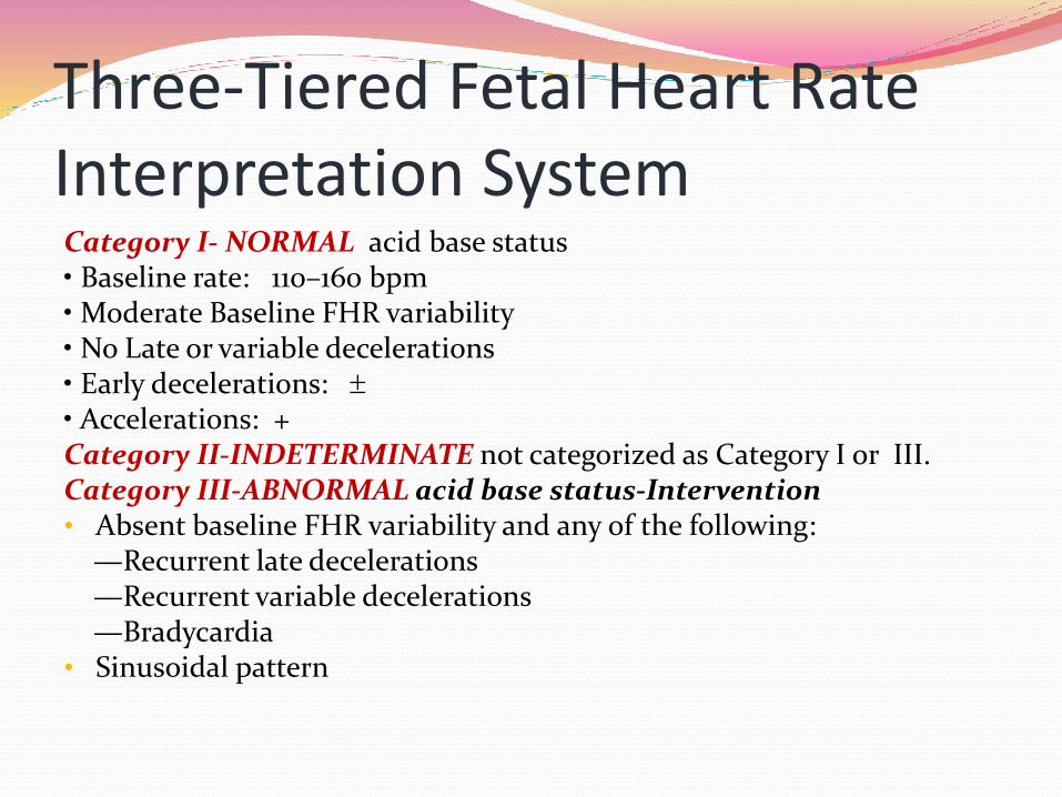
Three-Tiered Fetal Heart Rate Interpretation SystemCategory I- NORMAL acid base status• Baseline rate: 110–160 bpm• Moderate Baseline FHR variability• No Late or variable decelerations• Early decelerations: • Accelerations: +Category II-INDETERMINATE not categorized as Category I or III.Category III-ABNORMAL acid base status-Intervention• Absent baseline FHR variability and any of the following:
—Recurrent late decelerations—Recurrent variable decelerations—Bradycardia
• Sinusoidal pattern
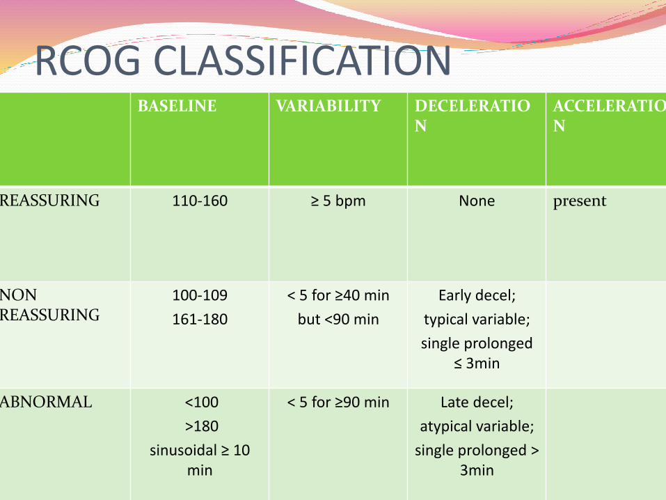
RCOG CLASSIFICATIONBASELINE VARIABILITY DECELERATIO
NACCELERATION
REASSURING 110-160 ≥ 5 bpm None present
NONREASSURING
100-109
161-180
< 5 for ≥40 min
but <90 min
Early decel;
typical variable;
single prolonged ≤ 3min
ABNORMAL <100
>180
sinusoidal ≥ 10 min
< 5 for ≥90 min Late decel;
atypical variable;
single prolonged > 3min

Ancillary tests that can aid in the management of Category II or Category III FHR tracings-
Four techniques are available to stimulate the fetus:
1)fetal scalp sampling,
2) Allis clamp scalp stimulation,
3) vibroacoustic stimulation, and
4) digital scalp stimulation

Standard interventions for NRFS -
Supplemental oxygen
Discontinuation of any labor stimulating agent
Changing maternal position
Resolution of maternal hypotension-hydration.
P/V to determine umbilical cord prolapse, rapid cervical dilation, or descent of the fetal head,ARM
Assessment of uterine contraction .
Tocolytics-in tachysystole with associated FHR changes.
When the FHR tracing includes recurrent variable decelerations -Amnioinfusion
MANAGMENTSuspicious CTG-
If inadequate quality-check contact and connections
If hypercontractility-discontinue oxytocin, consider tocolytics
Maternal tachycardia,pyrexia,dehydration, hypotension
Supine? Epidural? sedation? drugs?
i/v crystalloid bolus; 10 L/min O2
If persistent→ do ancillary tests
Pathological CTG
FBS if feasible
If not feasible-expedite delivery (within 30 min)

Effects of Medications on FHR PatternsNarcotics- decreased variability and accelerationsCorticosteroids- Decreased variability (with beta-methasone but not dexamethasone)Magnesium sulfate- A significant decrease in short-term variability, clinically insignificant
decrease in FHR inhibits the increase in accelerations with advancing gestational age
Epidural analgesia- decreased variability and accelerationsTerbutaline- Increase in baseline FHR
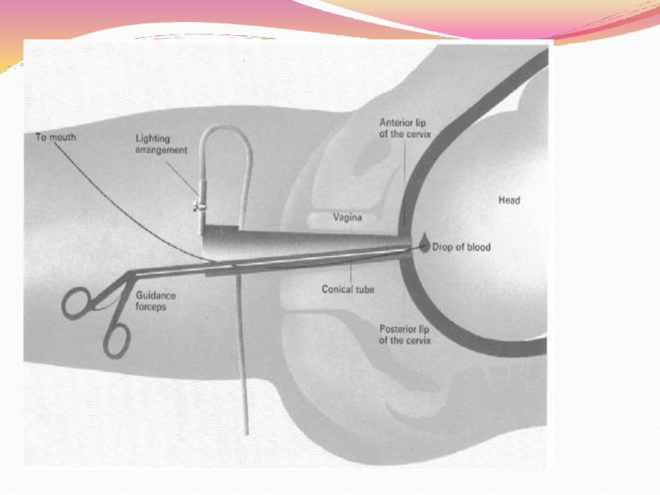
FETAL SCALP PH In women with "abnormal“ fetal heart rate tracings .
Cervix needs to be 4-5cm dilated and Vx at -1 st or below
pH <7.20 –fetal acidosis: deliver
pH 7.20-7.25 – borderline, repeat in 30 min or deliver if rapid fall
pH > 7.25 – reassuring, repeat if FH abnormality persists
Greater utility of scalp pH is in its high negative predictive value (97–99%).
Contraindications Maternal infection (HIV, hepatitis, HSV) Fetal bleeding disorders (e.g. haemophilia) Prematurity < 34 weeks Face presentation

FETAL PULSE OXIMETRY
Acidosis: O2 sat. <30% for >2min
Approved by FDA for use in fetuses with NRFS in May 2000
The ACOG currently recommends against its use until further studies are available to confirm its efficacy and safety
Insufficient evidence for its use as an adjunct or independent of electronic fetal surveillance.
FETAL SCALP LACTATE TESTING Higher sensitivity and specificity than scalp pH
> 4.8 mmol/L : acidosis
Clinical trial that compared the use of scalp pH to scalp lactate level did not demonstrate a difference in the rate of acidemia at birth, Apgar scores, or neonatal intensive care unit admissions
Not recommended for routine use

ST WAVEFORM ANALYSIS Method: STAN S31 fetal heart monitor(USFDA)
Scalp electrodes
The electrical fetal cardiac signal – P wave, QRS complex, and T wave – is amplified and fed into a cardiotachometer for heart rate calculation
Restrict fetal ST waveform analysis to those with non reassuring fetal status on EFM
The use of ST waveform analysis for the intrapartum
assessment of the compromised fetus is not recommended
for routine use at this time.

Thankyou
132