
Computerised analysis of antepartum foetal heart ...
10
Full Terms & Conditions of access and use can be found at http://www.tandfonline.com/action/journalInformation?journalCode=ijog20 Download by: [Universita Studi di Pavia] Date: 10 February 2017, At: 08:11 Journal of Obstetrics and Gynaecology ISSN: 0144-3615 (Print) 1364-6893 (Online) Journal homepage: http://www.tandfonline.com/loi/ijog20 Computerised analysis of antepartum foetal heart parameters: New reference ranges Natascia Giuliano, Maria Laura Annunziata, Francesca Giovanna Esposito, Salvatore Tagliaferri, Andrea Di Lieto, Giovanni Magenes, Maria Gabriella Signorini, Marta Campanile & Domenico Arduini To cite this article: Natascia Giuliano, Maria Laura Annunziata, Francesca Giovanna Esposito, Salvatore Tagliaferri, Andrea Di Lieto, Giovanni Magenes, Maria Gabriella Signorini, Marta Campanile & Domenico Arduini (2016): Computerised analysis of antepartum foetal heart parameters: New reference ranges, Journal of Obstetrics and Gynaecology To link to this article: http://dx.doi.org/10.1080/01443615.2016.1239069 Published online: 06 Dec 2016. Submit your article to this journal Article views: 11 View related articles View Crossmark data brought to you by CORE View metadata, citation and similar papers at core.ac.uk provided by Archivio istituzionale della ricerca - Politecnico di Milano
Transcript of Computerised analysis of antepartum foetal heart ...
Full Terms & Conditions of access and use can be found athttp://www.tandfonline.com/action/journalInformation?journalCode=ijog20
Download by: [Universita Studi di Pavia] Date: 10 February 2017, At: 08:11
Journal of Obstetrics and Gynaecology
ISSN: 0144-3615 (Print) 1364-6893 (Online) Journal homepage: http://www.tandfonline.com/loi/ijog20
Computerised analysis of antepartum foetal heartparameters: New reference ranges
Natascia Giuliano, Maria Laura Annunziata, Francesca Giovanna Esposito,Salvatore Tagliaferri, Andrea Di Lieto, Giovanni Magenes, Maria GabriellaSignorini, Marta Campanile & Domenico Arduini
To cite this article: Natascia Giuliano, Maria Laura Annunziata, Francesca Giovanna Esposito,Salvatore Tagliaferri, Andrea Di Lieto, Giovanni Magenes, Maria Gabriella Signorini, MartaCampanile & Domenico Arduini (2016): Computerised analysis of antepartum foetal heartparameters: New reference ranges, Journal of Obstetrics and Gynaecology
To link to this article: http://dx.doi.org/10.1080/01443615.2016.1239069
Published online: 06 Dec 2016.
Submit your article to this journal
Article views: 11
View related articles
View Crossmark data
brought to you by COREView metadata, citation and similar papers at core.ac.uk
provided by Archivio istituzionale della ricerca - Politecnico di Milano
ORIGINAL ARTICLE
Computerised analysis of antepartum foetal heart parameters:New reference ranges
Natascia Giulianoa, Maria Laura Annunziataa, Francesca Giovanna Espositoa, Salvatore Tagliaferria ,Andrea Di Lietoa, Giovanni Magenesb, Maria Gabriella Signorinic, Marta Campanilea and Domenico Arduinid
aDepartment of Obstetrical-Gynaecological, Urological Science and Reproductive Medicine, Federico II University, Naples, Italy; bDepartmentof Electrical, Computer and Biomedical Engineering, University of Pavia, Pavia, Italy; cDepartment of Electronic, Information andBioengineering, Politecnico of Milano, Milan, Italy; dDepartment of Obstetrics and Gynaecology, Foetal Medicine Centre, University of Rome“Tor Vergata”, Rome, Italy
ABSTRACTWe selected 4012 cCTG records (one trace for each patient) performed in healthy pregnancies from30th to 42nd gestational week using foetal heart rate (FHR), short-term variability (STV), long-termirregularity (LTI), Delta, approximate entropy (ApEn), spectral components as low frequency (LF), medianfrequency (MF), high frequency (HF) and LF/(HFþMF) ratio were analysed. Reference nomograms werecreated and sensitivity and specificity for the prediction of foetal compromise were calculated whichwere 90% and 89%, respectively. Changes of cCTG parameters according to gestational week were eval-uated: FHR (r¼�.65) and LF (r¼�.87) showed a statistically significant reduction (p< .05) with gesta-tional age. STV (r¼ .59), LTI (r¼ .69), Delta (r¼ .67), and MF (r¼ .88) showed a statistically significantincrease (p< .05) with gestational age. In contrast, for ApEn (r¼�.098), HF (r¼ .14) and LF/(HFþMF)ratio (r¼�.47) a non-statistically significant change was found (p> .05). The identification of referenceranges for cCTG indexes in according to gestational age could provide a more objective examination ofcCTG trace.
KEYWORDSComputerised cardiotocog-raphy; antepartum foetalmonitoring; foetal heartrate; gestational age;reference ranges
Introduction
Cardiotocography is the most widespread method of foetalsurveillance worldwide. During the last decades, traditionalmonitoring systems received a fundamental improvement bynew technological devices allowing the overcoming of sub-jective interpretation while providing objective informationabout foetal wellbeing (Bernardes et al. 1997; Figueras et al.2005).
Computerised cardiotocography (cCTG) improves foetalmonitoring reliability as it standardises CTG evaluation withautomatic estimation of foetal heart rate (FHR) parameters(Signorini et al. 2003). In fact, cCTG is characterised by object-ivity and consistency (Dawes et al. 1996) as it performs anautomatic trace analysis, implementing diagnostic criteriaaccepted in clinical obstetric practise (Gagnon et al. 1993;Pardey et al. 2002).
Nowadays, the available foetal monitoring guidelines,published by the American College of Obstetricians andGynaecologists (ACOG) (ACOG 1989, 1995, 2005, 2009), theNational Institute of Child Health and Human Development(NICHHD) (Zuspan et al. 1979; NICHHD 1997; Macones et al.2008), the Royal College of Obstetricians and Gynaecologists(RCOG) (RCOG 2001), and the National Institute of ClinicalExcellence (NICE) (NICE 2007) focus specifically on
intrapartum monitoring, although some try to apply thesame principles to antepartum tracings (Macones et al.2008; ACOG 2009). The use of computerised analysis duringthe antepartum period has some problems. The first one isthe difficulty of transforming the linguistic description ofFHR parameters into numerical algorithms (Wr�obel et al.2013). Second, even when various algorithms have beendeveloped, only some cCTG parameters have been investi-gated. In fact, one of the most widespread system of ana-lysis, the Oxford Foetal Care System (Serra et al. 2009)analyses only linear parameters such as baseline foetal heartrate (Basal FHR), long-term FHR variation (LTV), short-termFHR variation (STV), episodes of high/low FHR variation,accelerations and decelerations. While many studies werebased on the Oxford analyses, few studies focussed theirattention on cCTG trace analysis using the 2CTG2 systemthat measures additional cCTG parameters such as non-lin-ear features. Moreover, the major limitations are related tothe few attempts to evaluate clinical implication of cCTGparameters as the absence of their analysis in relation withneonatal outcome. Most of the studies focussed on the pre-dictive value of cCTG parameters considered only their rela-tion with the Apgar score, not including other importantparameters such as umbilical cord pH at birth (Bernardeset al. 1997; Park et al. 2001).
CONTACT Natascia Giuliano [email protected] Department of Obstetrical-Gynaecological, Urological Science and Reproductive Medicine of theFederico II University, Naples, Italy� 2016 Informa UK Limited, trading as Taylor & Francis Group
JOURNAL OF OBSTETRICS AND GYNAECOLOGY, 2016http://dx.doi.org/10.1080/01443615.2016.1239069
In the present study we investigated several aspects ofcCTG analysis trying to simplify its clinical application.
The aims of our study were:
1. Evaluating how the cCTG (linear and non-linear) parame-ters analysed with the 2CTG2 system change throughgestation. This could provide new information about theclinical meaning of numerical parameters, and they, inturn, may explain physiological or paraphysiological phe-nomena that occur in foetus.
2. Creating reference normality values for all cCTG parame-ters investigated, as a function of gestational age, in apopulation of healthy pregnancies. Supplementing thefirst tables developed by Arduini et al. (1993), we wantedto provide new reference values for each week, from the30th to the 42th week, for uncomplicated pregnancies,based on our clinical experience and using our system.
3. Providing a clinical validation of the 2CTG2 system toevaluate the usefulness of our system and the newtables, in relationship with foetal outcome.
Materials and methods
Data collection
This is a prospective study performed from October 2012 toOctober 2014. The population study consists of pregnantwomen who underwent to antenatal computerised cardioto-cographic exam at the Department of Obstetrical-Gynaecological and Urological Science and ReproductiveMedicine of the Federico II University, Naples, Italy. The prin-ciples of the Helsinki Declaration were followed, and a writtenconsent authorised the use of clinical information from cCTGrecords of each patient for scientific aims. We used PhilipsAvalon Foetal Monitor FM30 cardiotocographs, with an ultra-sound transducer and a transabdominal tocodynamometer.The Cardiotocograph was interfaced to 2CTG2 system (SEA,Italy) for computerised analysis (Arduini et al. 1993). We usedMantel’s algorithm, based on a low-pass digital system thatcrosses the tracing five times, starting from a value deter-mined by histogram analysis of the FHR distribution (Mantelet al. 1990). In fact, Mantel’s algorithm is generally satisfactorywhen the FHR tracings are regular with long and stable FHRsegments, which are found most commonly during the ante-partum period (Nidhal et al. 2011).
From 9732 cCTG records archived during these years, weselected 4012 antenatal nonstress cCTG traces from 30th to42nd gestational week, associated with documented maternaland foetal normal outcome. cCTG traces were carried out forroutine antenatal care (“term foetuses”) or as control (“pre-term feuses”, for the study aim). Only one trace per foetuswas analysed and the selected cCTG trace was the first per-formed at our Department.
Gestational age was obtained from foetal crown-rumplength ultrasonographic measurement performed at 8–14weeks. The last menstrual period-based date was only cor-rected if the interval between the ultrasound and the men-strual dates was greater than 1 week (Butt et al. 2014).
Inclusion criteria were: Caucasian ethnic, singleton healthypregnancies, neonatal weight above the 10th percentile for
gestational age and sex (Hadlock et al. 1985), absence ofstructural or chromosomal abnormalities, normal umbilicalartery Doppler (Yeh et al. 2012), absence of maternal dis-eases. Traces from foetuses below the 30th gestational weekwere excluded because the most part of them did not meetthe inclusion criteria.
FHR signals were obtained in a controlled clinical environ-ment, with the mothers lying in a semi-Fowler’s position in arelaxed condition. Each nonstress test was at least 20minuteslong. The FHR analysis was carried out in each signal using a10-min segment of trace in order to obtain the same lengthof analysis segment. We selected segment in absence of largeFHR alterations (e.g. accelerations and/or decelerations) andother phenomena such us uterine contraction and/or foetalmovements. Only records of acceptable quality (signal loss<50%) were included in the study.
The parameters assessed are divided in: time domainparameters (baseline FHR, STV, LTI, Delta), frequency domainparameters, computed by adopting a power spectral estima-tion (LF, MF, HF and LF/(HFþMF) ratio) and nonlinear param-eter (ApEn).The resulting cCTG indexes were computed foreach gestational week. The cCTG records were divided bygestational age: 30th week (nrec¼157), 31st week (nrec¼169),32nd week (nrec¼210), 33rd week (nrec¼217), 34th week(nrec¼249), 35th week (nrec¼237), 36th week (nrec¼378),37th week (nrec¼456), 38th week (nrec¼425), 39th week(nrec¼516), 40th week (nrec¼432), 41st week (nrec¼369),42nd week (nrec¼197).
Acquisition and elaboration of CTG signals
FHR signals were acquired at a sampling frequency of 2 Hz. Ifthe signals are measured in beats per minute (bpm), wecalled them S120(i), where 120 is the number of samples in aminute (i); if they are expressed in milliseconds, we calledthem T120(i). For the computation of FHR parameters we useda signal obtained by opportunely under sampling S120(i). Thisis accomplished by replacing each group of five consecutivepoints in the original series with their average value; in thisway the resulting signal is composed of 24 points per minute(0.4 Hz). We referred to this signal as S24(i) if the signal isexpressed in bpm, or as T24(i) if it is expressed in millisec-onds. The 2CTG2 programme always computes the parame-ters on the entire signal. During the acquisition, all theparameters are updated every minute.
Baseline, accelerations and decelerationsThe baseline is a running average of the FHR where accelera-tions and decelerations are defined as deviation of the FHRfrom the baseline lasting a sufficient amount of time. In the2CTG2 software, the baseline is computed by using the algo-rithm developed by Mantel (Mantel et al. 1990; Preboth2000). Accelerations and decelerations are defined as follows(Arduini et al. 1993):
� Small acceleration: FHR elevation over the greater baselinebetween 10 and 15 bpm for at least 15 s.
� Big acceleration: FHR elevation over the greater baselineof 15 bpm for at least 15 s.
2 N. GIULIANO ET AL.
� Decelerations:� FHR lower than the baseline for an amplitude of at
least 20 bpm and for a duration of at least 30 s.� FHR lower than the baseline for an amplitude of at
least 10 bpm and for a duration of at least 60 s.
Short-term variabilitySTV quantifies FHR variability over a very short time scale,usually on a beat to beat basis. In the automated system2CTG2, this index is implemented by adopting the definitionof Arduini et al. (1993). Given one minute of RR signal, T24(i)in ms and i[[1;24], STV is defined as
STV ¼ mean T24 iþ 1ð Þ � T24 ið Þj j½ �i¼P23
i¼1T24ðiþ 1Þ � T24ðiÞj j
23
Arduini excludes from the calculation big accelerationsand decelerations.
Long-term irregularityGiven a three minutes T24(i) signal with i[[1; 72], LTI isdefined as the interquartile range (1/4; 3/4) of the distributionof the modula m24 (j) with i[[1; 71]
m24 jð Þ ¼ T224 jþ 1ð Þ þ T224 jð Þ� �1=2
The definition is the same provided by De Haan (ACOG1989), with the exception of a window of 72 (and not 512)samples long.
Arduini excludes from the calculation big accelerationsand decelerations.
DeltaGiven a minute of signal in millisecond T24(i) with i[[1; 24],Delta is defined as the difference between the maximum andminimum FHR value,
Delta ¼ maxT24 ið Þ �minT24 ið ÞArduini et al. (1993) excludes from the calculation big
accelerations and decelerations.
Approximate entropyApEn is a collection of statistical indexes. It measures theregularity and, indirectly, the correlation and the persistenceof a signal: small values indicate reduced signal irregularity.We use the original definition by Pincus (1995):
ApEn m; rð Þ ¼PN�mþ1
i¼1logCiðm; rÞ
N�mþ 1�
PN�m
i¼1log Ciðmþ 1; rÞ
N�m
where m is a natural number, r a positive real and N¼ 360.The approximate entropy is computed over windows of FHRsignal 3min long.
Spectral analysisParameters in the frequency domain were obtained fromautoregressive power spectrum estimation. In the FHR
spectrum we identify four contributions: the very low fre-quency (VLF 0–0.03Hz), which is related to long period andnon-linear contributions, the low frequency (LF 0.03–0.15Hz),which is mainly correlated with neural sympathetic activityand the high frequency (HF 0.5–1Hz), which marks the pres-ence of foetal breathing. The middle frequency (MF0.15–0.5 Hz) depends on foetal movements and maternalbreathing activity and it is typical of FHR spectrum. LF/(HFþMF) ratio quantifies the autonomic balance betweenneural control mechanisms from different origin (Signoriniet al. 2003).
Statistical analysis
The statistical software package IBM SPSS 20.0 forWindows was used for data analysis. FHR, STV, LTI, Delta,ApEn and spectral components, as LF, MF, HF, LF/(HFþMF)ratio and their distribution throughout third trimester ofgestation were analysed. The Kolgomorov–Smirnov testfor normality showed a non-normal distribution. TheKruskal–Wallis test was computed to evaluate the statisticalsignificant variation of cCTG parameters with gestationalage. The relationship between cCTG parameters and gesta-tional age was analysed through the Spearman’s rank cor-relation coefficient. Threshold for statistical significance wasp< .05.
The following percentiles for each parameter were calcu-lated: 5th; 50th; 95th.
In order to verify the clinical validity of our referenceranges, we realised a second study that included
A new sample of newborns made up 328 babies: 118with a favourable outcome (pH >7.05 and BD <12mmol/L,normal Apgar scores >7 both at 1 and 5min, delivery at/orbeyond 37th gestational week, intact survival beyond 28days of postnatal life), 210 with a unfavourable outcome (pH<7.05 and BD >12mmol/L, normal Apgar scores <7 both at1 and 5min, delivery before 37th gestational week, necessityof “neonatal intensive care unit for 24 h”). Antepartum cCTGtraces performed by their mothers were compared with ourreference ranges. Therefore, ROC curves, sensitivity and speci-ficity were obtained in consideration of the number ofbabies with favourable outcome whose cCTG parameterswere falling between the 5th and the 95th percentile. Thesame evaluation was performed for unfavourable outcomebabies.
Results
For all the traces, the median duration of the trace until crite-ria of reassurance were met (maximum 60min) was 32min(Preboth 2000).
The median value for each parameter was calculated andtheir distribution through gestational age was elaborated. InTable 1 was reported the median value of cCTG paremetersfirst and before the 37th gestational week. FHR and ApEnshowed a progressive decrease throughout pregnancy, whilethere is an increase of the values of STV, Delta and LTI.LF component and MF decreased, while HF increased.LF/(HFþMF) ratio reduced.
JOURNAL OF OBSTETRICS AND GYNAECOLOGY 3
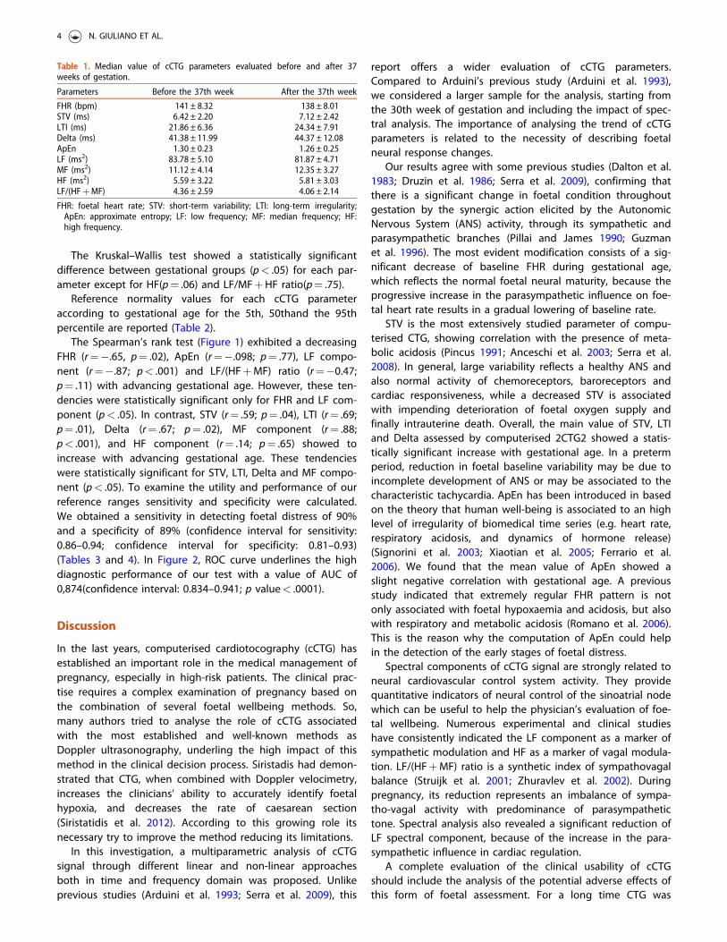
The Kruskal–Wallis test showed a statistically significantdifference between gestational groups (p< .05) for each par-ameter except for HF(p¼ .06) and LF/MFþHF ratio(p¼ .75).
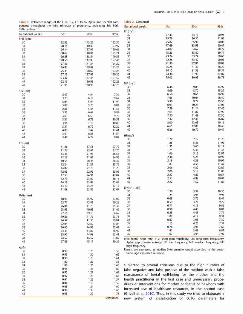
Reference normality values for each cCTG parameteraccording to gestational age for the 5th, 50thand the 95thpercentile are reported (Table 2).
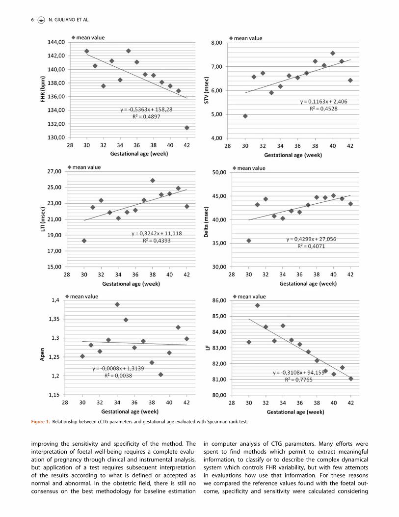
The Spearman’s rank test (Figure 1) exhibited a decreasingFHR (r¼�.65, p¼ .02), ApEn (r¼�.098; p¼ .77), LF compo-nent (r¼�.87; p< .001) and LF/(HFþMF) ratio (r¼�0.47;p¼ .11) with advancing gestational age. However, these ten-dencies were statistically significant only for FHR and LF com-ponent (p< .05). In contrast, STV (r¼ .59; p¼ .04), LTI (r¼ .69;p¼ .01), Delta (r¼ .67; p¼ .02), MF component (r¼ .88;p< .001), and HF component (r¼ .14; p¼ .65) showed toincrease with advancing gestational age. These tendencieswere statistically significant for STV, LTI, Delta and MF compo-nent (p< .05). To examine the utility and performance of ourreference ranges sensitivity and specificity were calculated.We obtained a sensitivity in detecting foetal distress of 90%and a specificity of 89% (confidence interval for sensitivity:0.86–0.94; confidence interval for specificity: 0.81–0.93)(Tables 3 and 4). In Figure 2, ROC curve underlines the highdiagnostic performance of our test with a value of AUC of0,874(confidence interval: 0.834–0.941; p value< .0001).
Discussion
In the last years, computerised cardiotocography (cCTG) hasestablished an important role in the medical management ofpregnancy, especially in high-risk patients. The clinical prac-tise requires a complex examination of pregnancy based onthe combination of several foetal wellbeing methods. So,many authors tried to analyse the role of cCTG associatedwith the most established and well-known methods asDoppler ultrasonography, underling the high impact of thismethod in the clinical decision process. Siristadis had demon-strated that CTG, when combined with Doppler velocimetry,increases the clinicians’ ability to accurately identify foetalhypoxia, and decreases the rate of caesarean section(Siristatidis et al. 2012). According to this growing role itsnecessary try to improve the method reducing its limitations.
In this investigation, a multiparametric analysis of cCTGsignal through different linear and non-linear approachesboth in time and frequency domain was proposed. Unlikeprevious studies (Arduini et al. 1993; Serra et al. 2009), this
report offers a wider evaluation of cCTG parameters.Compared to Arduini’s previous study (Arduini et al. 1993),we considered a larger sample for the analysis, starting fromthe 30th week of gestation and including the impact of spec-tral analysis. The importance of analysing the trend of cCTGparameters is related to the necessity of describing foetalneural response changes.
Our results agree with some previous studies (Dalton et al.1983; Druzin et al. 1986; Serra et al. 2009), confirming thatthere is a significant change in foetal condition throughoutgestation by the synergic action elicited by the AutonomicNervous System (ANS) activity, through its sympathetic andparasympathetic branches (Pillai and James 1990; Guzmanet al. 1996). The most evident modification consists of a sig-nificant decrease of baseline FHR during gestational age,which reflects the normal foetal neural maturity, because theprogressive increase in the parasympathetic influence on foe-tal heart rate results in a gradual lowering of baseline rate.
STV is the most extensively studied parameter of compu-terised CTG, showing correlation with the presence of meta-bolic acidosis (Pincus 1991; Anceschi et al. 2003; Serra et al.2008). In general, large variability reflects a healthy ANS andalso normal activity of chemoreceptors, baroreceptors andcardiac responsiveness, while a decreased STV is associatedwith impending deterioration of foetal oxygen supply andfinally intrauterine death. Overall, the main value of STV, LTIand Delta assessed by computerised 2CTG2 showed a statis-tically significant increase with gestational age. In a pretermperiod, reduction in foetal baseline variability may be due toincomplete development of ANS or may be associated to thecharacteristic tachycardia. ApEn has been introduced in basedon the theory that human well-being is associated to an highlevel of irregularity of biomedical time series (e.g. heart rate,respiratory acidosis, and dynamics of hormone release)(Signorini et al. 2003; Xiaotian et al. 2005; Ferrario et al.2006). We found that the mean value of ApEn showed aslight negative correlation with gestational age. A previousstudy indicated that extremely regular FHR pattern is notonly associated with foetal hypoxaemia and acidosis, but alsowith respiratory and metabolic acidosis (Romano et al. 2006).This is the reason why the computation of ApEn could helpin the detection of the early stages of foetal distress.
Spectral components of cCTG signal are strongly related toneural cardiovascular control system activity. They providequantitative indicators of neural control of the sinoatrial nodewhich can be useful to help the physician’s evaluation of foe-tal wellbeing. Numerous experimental and clinical studieshave consistently indicated the LF component as a marker ofsympathetic modulation and HF as a marker of vagal modula-tion. LF/(HFþMF) ratio is a synthetic index of sympathovagalbalance (Struijk et al. 2001; Zhuravlev et al. 2002). Duringpregnancy, its reduction represents an imbalance of sympa-tho-vagal activity with predominance of parasympathetictone. Spectral analysis also revealed a significant reduction ofLF spectral component, because of the increase in the para-sympathetic influence in cardiac regulation.
A complete evaluation of the clinical usability of cCTGshould include the analysis of the potential adverse effects ofthis form of foetal assessment. For a long time CTG was
Table 1. Median value of cCTG parameters evaluated before and after 37weeks of gestation.
Parameters Before the 37th week After the 37th week
FHR (bpm) 141 ± 8.32 138 ± 8.01STV (ms) 6.42 ± 2.20 7.12 ± 2.42LTI (ms) 21.86 ± 6.36 24.34 ± 7.91Delta (ms) 41.38 ± 11.99 44.37 ± 12.08ApEn 1.30 ± 0.23 1.26 ± 0.25LF (ms2) 83.78 ± 5.10 81.87 ± 4.71MF (ms2) 11.12 ± 4.14 12.35 ± 3.27HF (ms2) 5.59 ± 3.22 5.81 ± 3.03LF/(HFþMF) 4.36 ± 2.59 4.06 ± 2.14
FHR: foetal heart rate; STV: short-term variability; LTI: long-term irregularity;ApEn: approximate entropy; LF: low frequency; MF: median frequency; HF:high frequency.
4 N. GIULIANO ET AL.
subjected to several criticisms due to the high number offalse negative and false positive of the method with a falsereassurance of foetal well-being for the mother and thehealth practitioner in the first case and unnecessary proce-dures or interventions for mother or foetus or newborn withincreased use of healthcare resources, in the second case(Grivell et al. 2010). Thus, in this study we tried to elaborate anew system of classification of cCTG parameters for
Table 2. Reference ranges of the FHR, STV, LTI, Delta, ApEn, and spectral com-ponents throughout the third trimester of pregnancy, indicating 5th, 50th,95th centiles.
Gestational weeks 5th 50th 95th
FHR (bpm)30 132.52 143.20 152.3031 128.15 140.48 153.9232 126.14 137.91 150.6633 130.62 140.01 155.2134 126.83 138.54 149.5535 128.58 142.93 157.4036 127.46 141.34 154.2237 126.82 139.07 152.2338 125.41 138.69 153.2439 127.13 137.93 149.2640 123.47 137.46 151.5241 122.13 136.93 152.2842 121.95 130.95 142.70
STV (ms)30 2.47 4.84 7.3931 3.23 6.19 9.6232 3.64 5.94 11.9433 3.08 5.75 9.0934 2.85 5.44 11.5335 3.32 6.64 9.8936 3.61 6.53 9.7037 3.31 6.79 10.5838 3.96 7.10 10.8539 3.31 6.72 12.2040 4.00 7.42 12.4141 3.51 6.83 11.5442 3.49 6.22 9.82
LTI (ms)30 11.46 17.55 27.7931 11.78 22.41 33.7632 13.38 21.96 36.5633 12.17 21.01 34.9534 10.56 20.34 36.4235 12.24 21.31 33.7936 14.02 21.78 31.2837 12.03 22.99 35.9538 12.53 24.91 42.8739 12.79 23.07 38.1540 14.19 23.82 35.4941 13.19 24.26 37.1942 11.09 23.82 37.07
Delta (ms)30 18.99 35.92 53.0931 22.77 42.89 69.2232 26.04 41.75 75.3333 23.93 40.05 61.4934 22.16 39.15 64.6235 19.86 41.76 63.7836 24.57 41.30 59.8437 22.04 42.67 63.8438 26.66 44.92 62.4239 24.31 43.87 68.9940 25.90 44.99 63.4141 24.52 44.57 63.8242 27.85 45.17 59.29
ApEn30 0.99 1.25 1.5331 0.99 1.30 1.6232 0.98 1.25 1.6133 1.00 1.29 1.5834 1.06 1.35 1.6835 0.94 1.36 1.8036 0.92 1.27 1.6937 0.97 1.28 1.6538 0.91 1.23 1.5739 0.84 1.19 1.5940 0.65 1.24 1.9041 0.98 1.33 1.6642 0.95 1.29 1.73
(continued)
Table 2. Continued
Gestational weeks 5th 50th 95th
LF (ms2)30 77.43 84.13 90.9431 75.78 86.30 91.0132 75.82 85.90 90.0633 77.69 83.95 90.4734 74.82 84.62 90.2735 75.23 83.80 89.7736 73.74 83.74 88.6637 72.26 83.42 90.0338 71.96 82.81 89.0339 75.26 81.40 88.2040 74.15 81.53 88.3741 74.38 81.90 87.8242 75.02 80.95 88.78
MF (ms2)30 6.46 9.80 14.9231 4.09 8.70 15.2732 6.58 9.66 16.9233 7.63 10.04 16.4034 5.90 9.77 15.5635 6.03 10.23 17.0336 7.39 11.14 17.4537 7.63 11.82 17.9938 7.30 11.49 17.5039 7.32 12.40 16.8240 8.00 13.03 19.1641 6.65 12.80 18.4542 6.36 10.72 19.47
HF(ms2)30 1.79 7.12 11.2031 1.85 5.46 11.9532 1.35 3.60 12.1733 1.74 5.31 11.3434 1.66 5.12 12.8135 2.38 5.26 10.9236 2.18 4.38 10.9137 1.83 4.50 11.4238 2.06 4.82 12.8739 2.06 5.19 11.0340 2.27 4.87 10.5441 2.53 4.92 10.0142 2.22 7.52 11.85
LF/(HFþMF)30 1.20 3.34 10.3031 1.24 3.96 9.9132 0.68 3.72 8.4133 0.59 3.21 9.2234 0.71 4.13 9.8935 0.90 4.40 8.8136 0.85 4.43 7.7737 1.02 4.12 9.3438 0.67 3.95 7.3939 1.15 4.25 7.3440 0.78 3.95 7.6541 1.41 3.48 6.8742 1.07 3.98 7.97
FHR: foetal heart rate; STV: short-term variability; LTI: long-term irregularity;ApEn: approximate entropy; LF: low frequency; MF: median frequency; HF:high frequency.
Results are expressed as median (interquantile range) according to the gesta-tional age expressed in weeks.
JOURNAL OF OBSTETRICS AND GYNAECOLOGY 5
improving the sensitivity and specificity of the method. Theinterpretation of foetal well-being requires a complete evalu-ation of pregnancy through clinical and instrumental analysis,but application of a test requires subsequent interpretationof the results according to what is defined or accepted asnormal and abnormal. In the obstetric field, there is still noconsensus on the best methodology for baseline estimation
in computer analysis of CTG parameters. Many efforts werespent to find methods which permit to extract meaningfulinformation, to classify or to describe the complex dynamicalsystem which controls FHR variability, but with few attemptsin evaluations how use that information. For these reasonswe compared the reference values found with the foetal out-come, specificity and sensitivity were calculated considering
Figure 1. Relationship between cCTG parameters and gestational age evaluated with Spearman rank test.
6 N. GIULIANO ET AL.
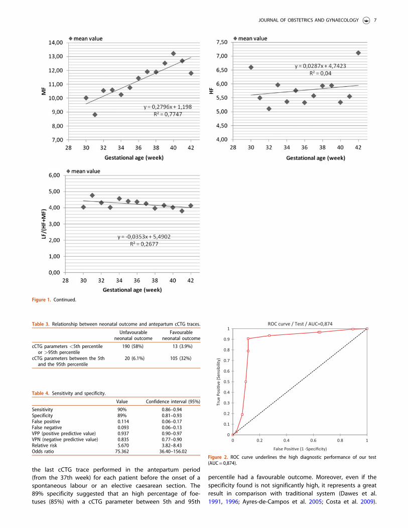
the last cCTG trace performed in the antepartum period(from the 37th week) for each patient before the onset of aspontaneous labour or an elective caesarean section. The89% specificity suggested that an high percentage of foe-tuses (85%) with a cCTG parameter between 5th and 95th
percentile had a favourable outcome. Moreover, even if thespecificity found is not significantly high, it represents a greatresult in comparison with traditional system (Dawes et al.1991, 1996; Ayres-de-Campos et al. 2005; Costa et al. 2009).
Figure 1. Continued.
Table 3. Relationship between neonatal outcome and antepartum cCTG traces.
Unfavourableneonatal outcome
Favourableneonatal outcome
cCTG parameters <5th percentileor >95th percentile
190 (58%) 13 (3.9%)
cCTG parameters between the 5thand the 95th percentile
20 (6.1%) 105 (32%)
Table 4. Sensitivity and specificity.
Value Confidence interval (95%)
Sensitivity 90% 0.86–0.94Specificity 89% 0.81–0.93False positive 0.114 0.06–0.17False negative 0.093 0.06–0.13VPP (positive predictive value) 0.937 0.90–0.97VPN (negative predictive value) 0.835 0.77–0.90Relative risk 5.670 3.82–8.43Odds ratio 75.362 36.40–156.02
0
0.1
0.2
0.3
0.4
0.5
0.6
0.7
0.8
0.9
1
0 0.2 0.4 0.6 0.8 1
True
Pos
i�ve
(Sen
sibi
lity)
False Posi�ve (1 -Specificity)
ROC curve / Test / AUC=0,874
Figure 2. ROC curve underlines the high diagnostic performance of our test(AUC= 0,874).
JOURNAL OF OBSTETRICS AND GYNAECOLOGY 7
In fact, in our study, only 6,1% of the analysed babies hadunfavourable outcome and antepartum trace between the5th and the 95th percentile, while 3.9% of the babies withfavourable outcome and cCTG parameters<5th or>95th per-centile. These results suggest a possible improvement inpregnancy clinical management through the cCTG antepar-tum monitoring. A recent Cochrane del 2010 (Grivell et al.2010) showed a significant reduction in perinatal mortalityusing cCTG versus traditional CTG. This study represents a fur-ther step in this direction and towards the prediction of foe-tal outcome based on non-invasive and standard technology.
Strength and limitations of the study
Even if our study was based on a large sample of patients, itwas not a multicenter study; so our results could be per-ceived as of low clinical impact. However, our single-centretrial was based on a direct evaluation of all aspects of trialconduct, including data acquisition, quality control, data man-agement, and data analysis. Moreover, the strength of thestudy is related to our evaluation of clinical application of thereference range calculated throughout the estimation of sen-sitivity and specificity. Finally, in our previous study(Annunziata et al. 2016), we showed that cCTG modificationsduring the different stages of labour reflected the physiologicincreased activation of the autonomous nervous system. Webelieve that the use of computerised FHR analysis performedduring labour it is possible to get more information from foe-tal cardiac signal, in comparison with the traditional tracing.
Acknowledgements
All the authors have fulfilled all the conditions required for authorship.
Disclosure statement
There are not conflicts of interest with the topic matter of this manu-script, as described in the instructions to authors.
ORCID
Salvatore Tagliaferri http://orcid.org/0000-0002-8699-6544
References
American College of Obstetricians and Gynaecologists (ACOG). 1989.Intrapartum foetal heart rate monitoring. ACOG Technical Bulletin132:1–6.
American College of Obstetricians and Gynaecologists (ACOG). 1995.Technical bulletin 207: foetal heart rate patterns: monitoring, interpret-ation, and management. International Journal of Gynecology andObstetrics 51:65–74.
American College of Obstetricians and Gynaecologists (ACOG). 2005.Practise bulletin 70: intrapartum foetal heart rate monitoring.Obstetrics and Gynecology 106:1453–1460.
American College of Obstetricians and Gynaecologists (ACOG). 2009.Practise bulletin 106: intrapartum foetal heart rate monitoring: nomen-clature, interpretation, and general management principles. Obstetricsand Gynecology 114:192–202.
Anceschi MM, Piazze JJ, Ruozi-Berretta A, Cosmi E, Cerekja A, Maranghi L,Cosmi EV. 2003. Validity of short term variation (STV) in detection offetal acidemia. Journal of Perinatal Medicine 31:231–236.
Annunziata ML, Tagliaferri S, Esposito FG, Giuliano N, Mereghini F, DiLieto A, et al. 2016. Computerized analysis of fetal heart rate variabilitysignal during the stages of labor. Journal of Obstetrics andGynaecology Research 42:258–265.
Arduini D, Rizzo G, Piana G, Bonalumi A, Brambilla P, Romanini C. 1993.Computerised analysis of foetal heart rate: I. description of the system(2CTG). Maternal Foetal Investment 3:159.
Ayres-de-Campos D, Costa-Santos C, Bernardes J, SisPorto MulticentreValidation Study Group. 2005. Prediction of neonatal state by com-puter analysis of foetal heart rate tracings: the antepartum arm of theSisPorto multicentre validation study. European Journal of Obstetricsand Gynecology and Reproductive Biology 118:52–60.
Bernardes J, Costa-Pereira A, Ayres-de-Campos D, Van Geijn HP, Pereira-Leite L. 1997. Evaluation of interobserver agreement of cardiotoco-grams. International Journal of Gynaecology and Obstetrics 57:33–37.
Butt K, Lim K, Society of Obstetricians and Gynaecologists of Canada.2014. Determination of gestational age by ultrasound. Journal ofObstetrics and Gynaecology Canada 36:171–183.
Costa A, Ayres-de-Campos D, Costa F, Santos C, Bernardes J. 2009.Prediction of neonatal acidemia by computer analysis of foetal heartrate and ST event signals. American Journal of Obstetrics andGynecology 201:e1–e6.
Dalton KJ, Phill D, Dawes GS, Patrick JE. 1983. The autonomic nervoussystem and fetal heart rate variability. American Journal of Obstetricsand Gynecology 146:456–462.
Dawes GS, Moulden M, Redman CW. 1991. System 8000: computerizedantenatal FHR analysis. Journal of Perinatal Medicine 19:47–51.
Dawes GS, Moulden M, Redman CWG. 1996. Improvements in computer-ized fetal heart rate analysis antepartum. Journal of Perinatal Medicine24:25–36.
Druzin ML, Hutson JM, Edersheim TG. 1986. Relationship of baseline fetalheart rate to gestational age and fetal sex. American Journal ofObstetrics and Gynecology 154:1102–1103.
Ferrario M, Signorini MG, Magenes G, Cerutti S. 2006. Comparison ofentropy-based regularity estimator: application to the foetal heart ratesignal for the identification of foetal distress. IEEE Transactions on Bio-Medical Engineering 53:119–125.
Figueras F, Albela S, Bonino S, Palacio M, Barrau E, Hernandez S, et al.2005. Visual analysis of antepartum foetal heart rate tracings: inter-and intra-observer agreement and impact of knowledge of neonataloutcome. Journal of Perinatal Medicine 33:241–245.
Gagnon R, Campbell MK, Humse C. 1993. A comparison between visualand computer analysis of antepartum foetal heart rate tracings.American Journal of Obstetrics and Gynecology 168:842–847.
Grivell RM, Alfirevic Z, Gyte GM, Devane D. 2010. Antenatal cardiotocog-raphy for foetal assessment. Cochrane Database of Systematic ReviewsCD007863.
Guzman ER, Vintzileos AM, Martins M, Benito C, Houlihan C, Hanley M.1996. The efficacy of individual computer heart rate indices indetencting acidemia at birth in growth-restricted foetuses. Obstetricsand Gynecology 87:969–974.
Hadlock FP, Harrist RB, Sharman RS, Deter RL, Park SK. 1985. Estimationof foetal weight with the use of head, body, and femur measure-ments–a prospective study. American Journal of Obstetrics andGynecology 151:333–337.
Macones GA, Hankins GDV, Spong CY, Hauth J, Moore T. 2008. The 2008national institute of child health and human development workshopreport on electronic foetal monitoring: update on definitions, interpret-ation, and research guidelines. Obstetrics and Gynecology 112:661–666
Mantel R, van Geijn HP, Caron FJ, Swartjes JM, van Woerden EE, JongsmaHW. 1990. Computer analysis of antepartum fetal heart rate: 1. Baselinedetermination. International Journal of Biomedical Computing 25:261–272.
National Collaborating Centre for Women's and Children’s Health, com-missioned by the National Institute for Health and Clinical Excellence(NICE). 2007. Intrapartum care. London: RCOG Press.
National Institute of Child Health and Human Development researchplanning workshop (NICHHD). 1997. Electronic foetal heart rate
8 N. GIULIANO ET AL.
monitoring: research guidelines for interpretation. American Journal ofObstetrics and Gynecology 177:1385–1390.
Nidhal S, Ali MMA, Zaidan AA, Zaidan BB, Najah H. 2011. Computerisedalgorithm for foetal heart rate baseline and baseline variability estima-tion based on distance between signal average and a value.International Journal of Pharmacology 7:228–237.
Pardey J, Moulden M, Redman CWG. 2002. A computer system for thenumerical analysis of nonstress tests. American Journal of Obstetricsand Gynecology 186:1095–1103.
Park MI, Hwang JH, Cha KJ, Park YS, Koh SK. 2001. Computerized analysisof fetal heart rate parameters by gestational age. International Journalof Gynaecology and Obstetrics 74:157–164.
Pillai M, James D. 1990. The development of fetal heart rate patterns dur-ing normal pregnancy. Obstetrics and Gynecology 76:812–816.
Pincus SM. 1991. Approximate entropy as a measure of system complex-ity. Proceedings of National Academy of Sciences of the United Statesof America 88:2297–2301.
Pincus SM. 1995. Approximate entropy (ApEn) as a complexity measure.Chaos 5:110–117.
Preboth M. 2000 ACOG guidelines on antepartum foetal surveillance.American college of obstetricians and gynaecologists. American FamilyPhysician 62:1184,1187–1188.
Romano M, Bracale M, Cesarelli M, Campanile M, Bifulco P, De Falco M,et al. 2006. Antepartum cardiotocography: a study of fetal reactivity infrequency domain. Computers in Biology and Medicine 36:619–633.
Royal College of Obstetricians and Gynaecologists (RCOG). 2001. The useof electronic foetal monitoring. Evidence-based clinical guideline,number 8. London: RCOG Press.
Serra V, Bellver J, Moulden M, Redman CW. 2009. Computerised analysisof normal foetal heart rate pattern throughout gestation. Ultrasoundin Obstetrics and Gynecology 34:74–79.
Serra V, Moulden M, Bellver J, Redman CW. 2008. The value of the short-term foetal heart rate variation for timing the delivery of growth-retarded foetuses. BJOG 115:1101–1107.
Signorini MG, Magenes G, Cerutti S, Arduini D. 2003. Linear and nonlinearparameters for the analysis of fetal heart rate signal from cardiotoco-graphic recordings. IEEE Transactions on Biomedical Engineering50:365–374.
Siristatidis C, Kassanos D, Salamalekis G, Creatsa M, Chrelias C, CreatsasG. 2012. Cardiotocography alone versus cardiotocography plusDoppler evaluation of the foetal middle cerebral and umbilicalartery for intrapartum foetal monitoring: a Greek prospective con-trolled trial. Journal of Maternal Foetal Neonatal Medicine25:1183–1187.
Struijk PC, Ursem NT, Mathews J, Clark EB, Keller BB, Wladimiroff LW.2001. Power spectrum analysis of heart rate and blood flow velocityvariability measured in the umbilical and uterine arteries in early preg-nancy: a comparative study. Ultrasound in Obstetrics and Gynecology17:316–321.
Wr�obel J, Horoba K, Pander T, Je _zewski J, Czaba�nski R. 2013. Improvingfoetal heart rate signal interpretation by application of myriad filtering.Biocybernetics and Biomedical Engineering 33:211–221.
Xiaotian L, Daan Z, Shufeng Z. 2005. Approximate entropy of fetal heartrate variability as a predictor of fetal distress in women at termpregnancy. Acta Obstetricia et Gynecologica Scandinavica84:837–843.
Yeh P, Emary K, Impey L. 2012. The relationship between umbilical cordarterial pH and serious adverse neonatal outcome: analysis of 51,519consecutive validated samples. BJOG 119: 824–831.
Zhuravlev YE, Rassi D, Mishin AA, Emery SJ. 2002. Dynamic analysis ofbeat-to-beat fetal heart rate variability recorded by SQUID magnetom-eter: quantification of sympatho-vagal balance. Early HumanDevelopment 66:1–10.
Zuspan FP, Quilligan EJ, Iams JD, van Geijn HP. 1979. Predictors of intra-partum fetal distress: the role of electronic fetal monitoring. Report ofthe national institute of child health and human development consen-sus development task force. American Journal of Obstetrics andGynecology 135:287–291.
JOURNAL OF OBSTETRICS AND GYNAECOLOGY 9