


ANATOMY & PHYSIOLOGY INTEGUMENTARY SYSTEM PART 2 Appendages of the Skin.
38
ANATOMY & PHYSIOLOGY INTEGUMENTARY SYSTEM PART 2 Appendages of the Skin
-
Upload
shonda-wilkinson -
Category
Documents
-
view
235 -
download
1
Transcript of ANATOMY & PHYSIOLOGY INTEGUMENTARY SYSTEM PART 2 Appendages of the Skin.
ANATOMY & PHYSIOLOGYINTEGUMENTARY SYSTEM
PART 2
Appendages of the Skin
Skin Appendages
Glandsall arise from stratum basale then
extend into dermis & subcutaneous layers
all exocrine glands Release product thru a duct onto surface of skin
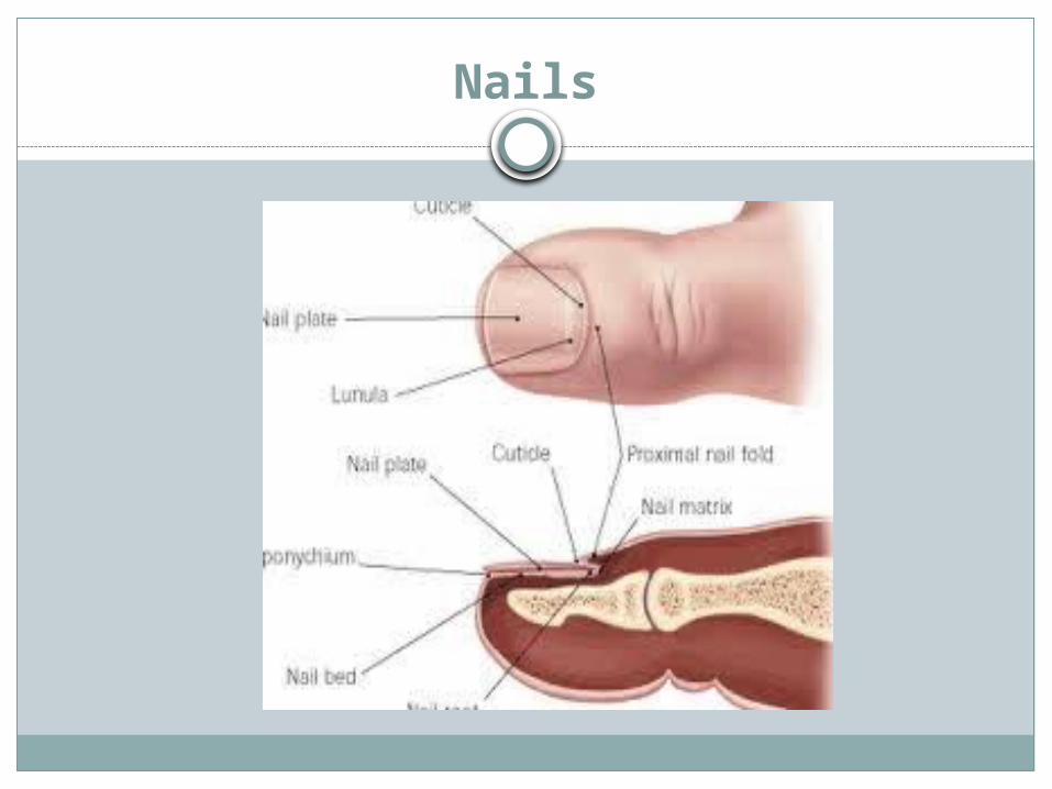
Hair & Hair FolliclesNails
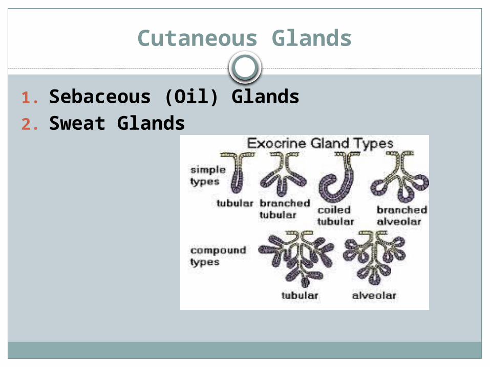
Cutaneous Glands
1. Sebaceous (Oil) Glands2. Sweat Glands

Sebaceous Glands
All over body except soles & palmsmost ducts empty onto hair follicle,
rest empty directly onto skin
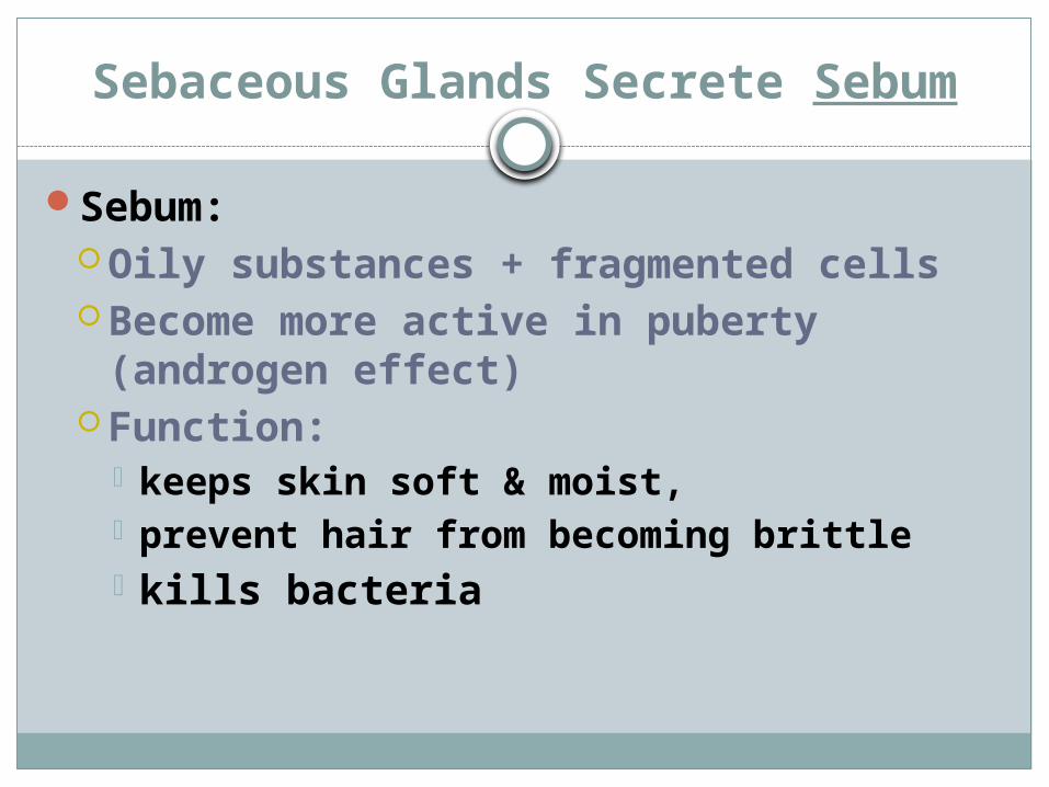
Sebaceous Glands Secrete Sebum
Sebum:Oily substances + fragmented cellsBecome more active in puberty
(androgen effect)Function:
keeps skin soft & moist, prevent hair from becoming brittle kills bacteria
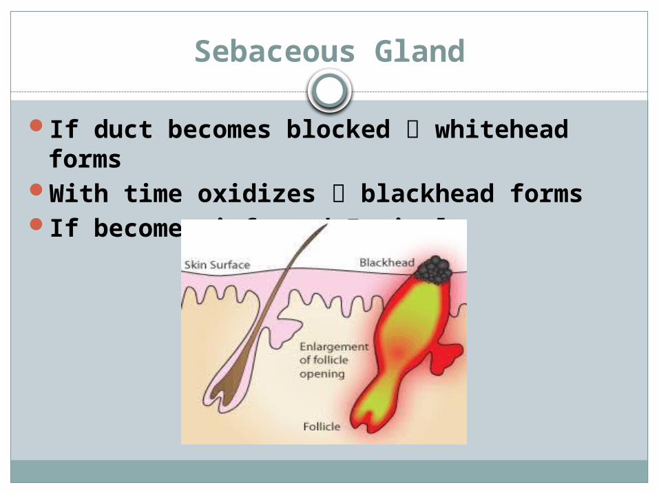
Sebaceous Gland
If duct becomes blocked whitehead forms
With time oxidizes blackhead formsIf becomes infected pimple
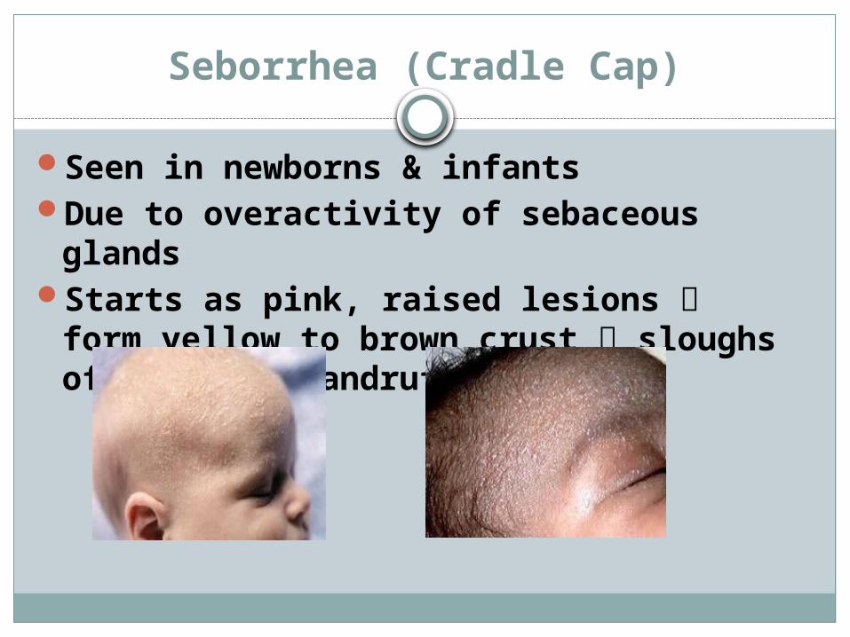
Seborrhea (Cradle Cap)
Seen in newborns & infantsDue to overactivity of sebaceous
glandsStarts as pink, raised lesions form
yellow to brown crust sloughs off as oily dandruff
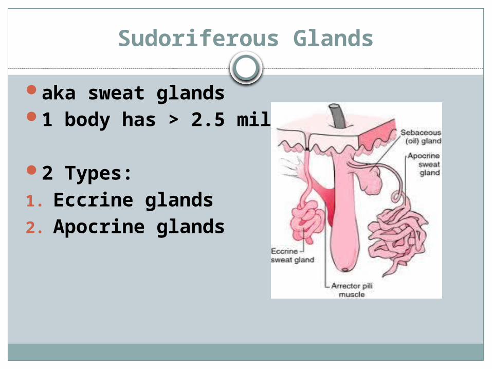
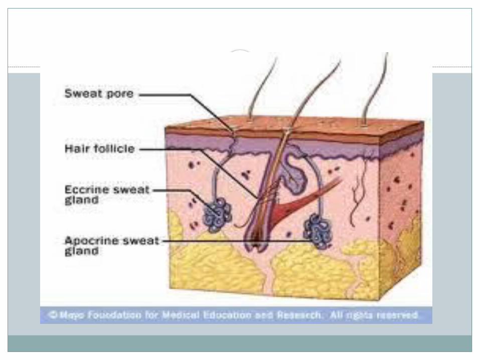
Sudoriferous Glands
aka sweat glands1 body has > 2.5 million
2 Types:1. Eccrine glands2. Apocrine glands

Eccrine Sweat Glands
more of these than apocrine sweat glands
Product: Sweat
Sweat is made up of:WaterNaClVitamin CUrea & uric acidLactic acid
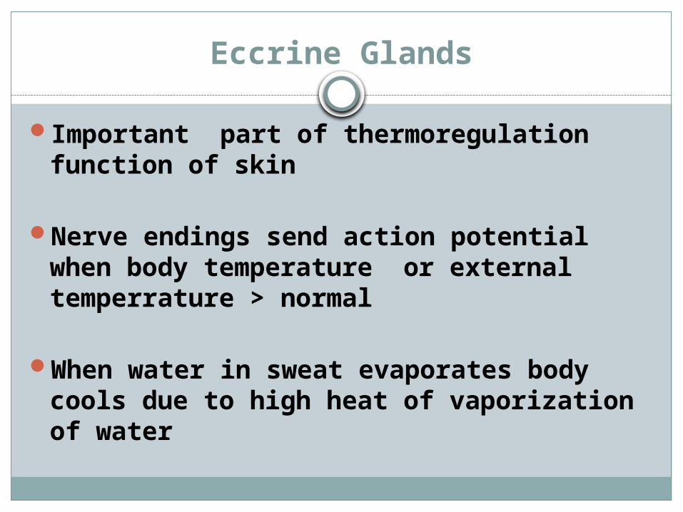
Eccrine Glands
Important part of thermoregulation function of skin
Nerve endings send action potential when body temperature or external temperrature > normal
When water in sweat evaporates body cools due to high heat of vaporization of water
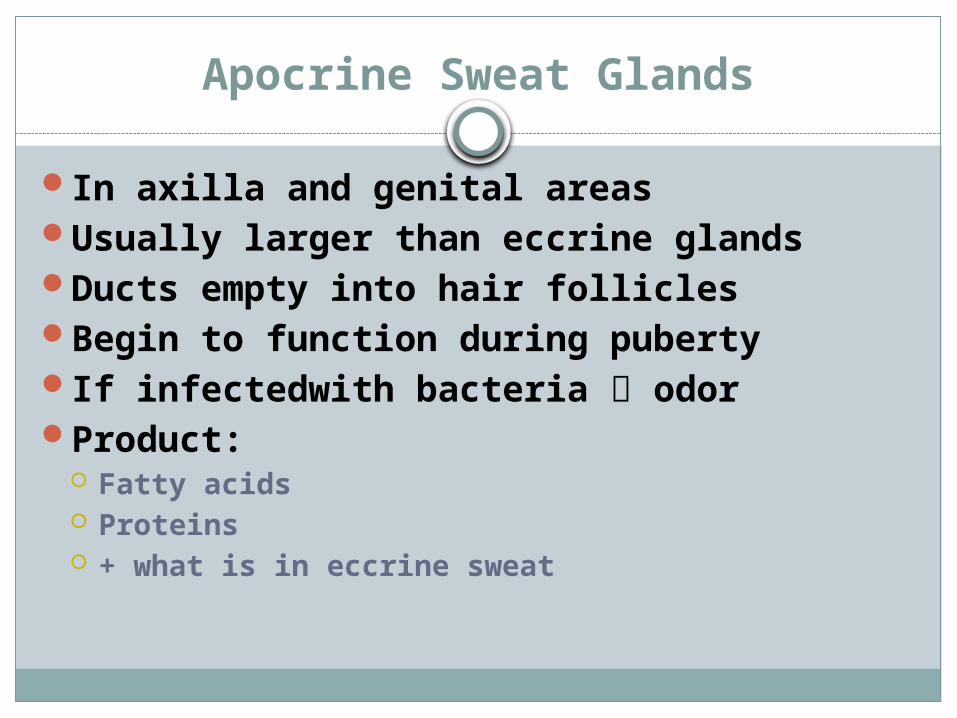
Apocrine Sweat Glands
In axilla and genital areasUsually larger than eccrine glandsDucts empty into hair folliclesBegin to function during pubertyIf infectedwith bacteria odorProduct:
Fatty acids Proteins + what is in eccrine sweat

Hair Follicles
Scattered all over body except palms & soles
Functions:Protection Insulation

Homeostatic Imbalances
Skin is largest organ so see many skin conditions
Most common ailments fall into categories1. Infections2. Allergies3. Skin cancer4. Burns
INTEGUMENTARY SYSTEMPART 3
Homeostatic Imbalances of the Skin

Bacterial Infections
Boils & Carbuncles Infected sebaceous glands (especially
neck)Staphylococcus areus

Impetigo
See blister-like pink lesions around nose, mouth yellow crusty
Elementary school ageExtremely contagious staph
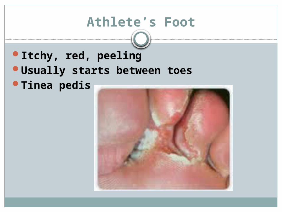
Athlete’s Foot
Itchy, red, peelingUsually starts between toesTinea pedis
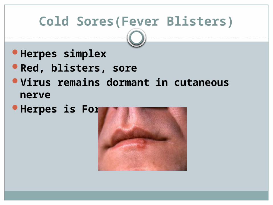
Cold Sores(Fever Blisters)
Herpes simplexRed, blisters, soreVirus remains dormant in cutaneous
nerveHerpes is Forever

Contact Dermatitis
Itching, redness, swellingSkin reacting to metal, chemical (in food,
poison ivy)
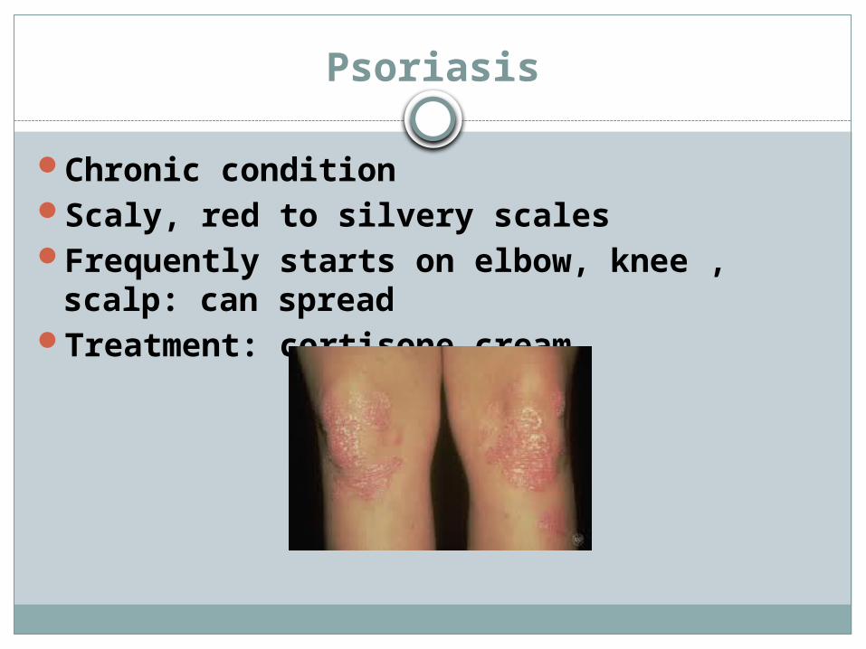
Psoriasis
Chronic conditionScaly, red to silvery scalesFrequently starts on elbow, knee , scalp:
can spreadTreatment: cortisone cream
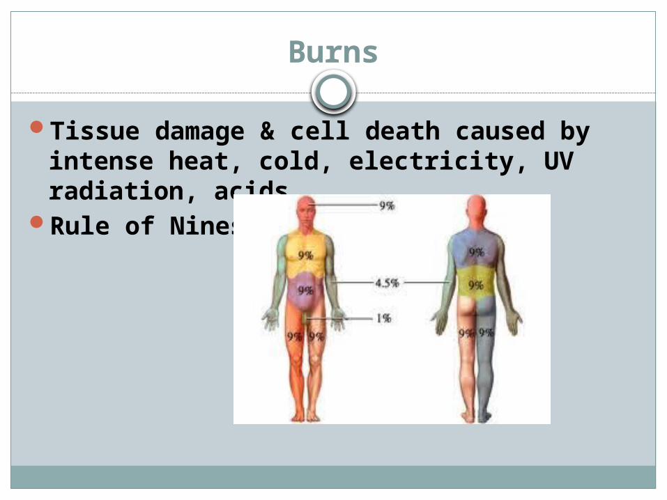
Burns
Tissue damage & cell death caused by intense heat, cold, electricity, UV radiation, acids
Rule of Nines:
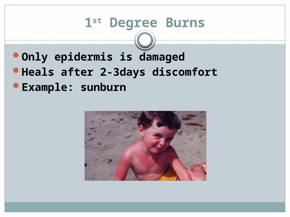
1st Degree Burns
Only epidermis is damagedHeals after 2-3days discomfortExample: sunburn
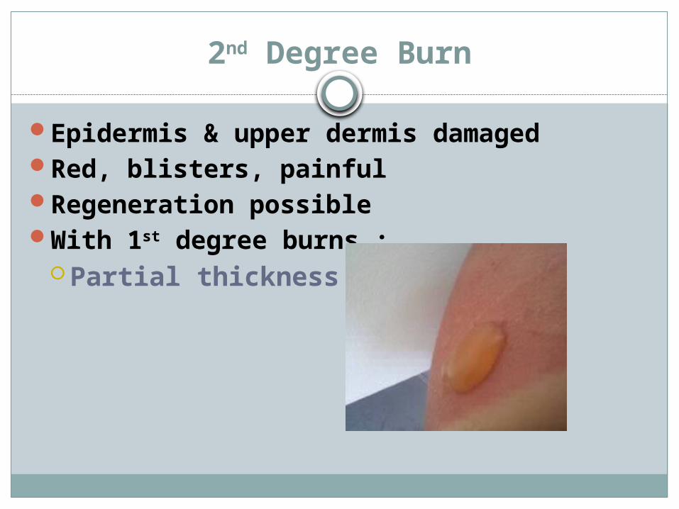
2nd Degree Burn
Epidermis & upper dermis damagedRed, blisters, painfulRegeneration possible With 1st degree burns :
Partial thickness burns
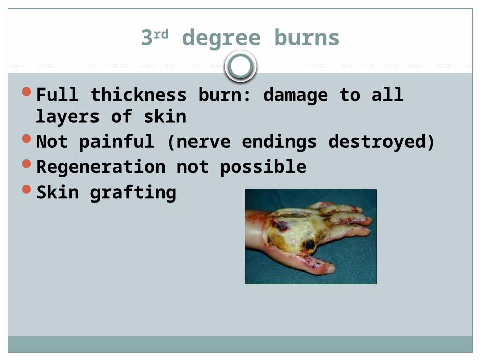
3rd degree burns
Full thickness burn: damage to all layers of skin
Not painful (nerve endings destroyed)Regeneration not possibleSkin grafting
Skin Cancer
Fastest rising cancer in young adults3 kinds:1. Basal Cell Carcinoma2. Squamous Cell Carcinoma3. Malignant Melanoma
4. Metastasis to Skin
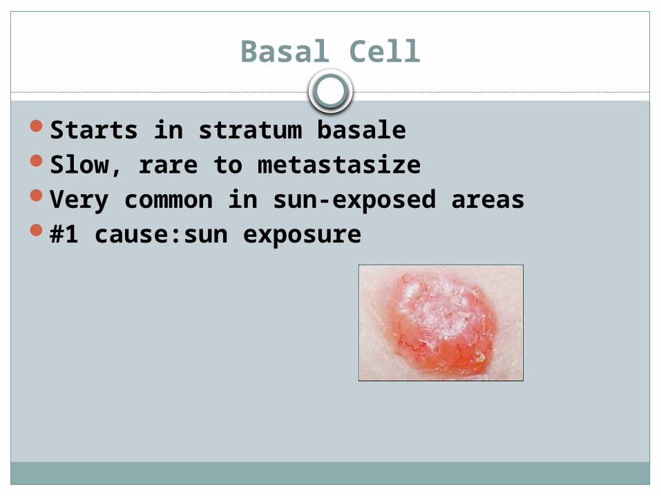
Basal Cell
Starts in stratum basaleSlow, rare to metastasizeVery common in sun-exposed areas#1 cause:sun exposure
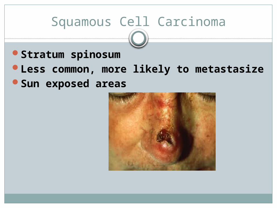
Squamous Cell Carcinoma
Stratum spinosumLess common, more likely to metastasizeSun exposed areas
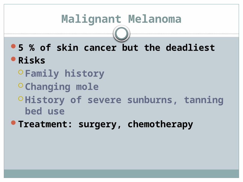
Malignant Melanoma
5 % of skin cancer but the deadliestRisks
Family historyChanging moleHistory of severe sunburns, tanning
bed useTreatment: surgery, chemotherapy

ABCD Rule
A: asymmetry, pigmentation not uniformB: border irregularityC: colors vary in same spotD: diameter > end of pencil eraser
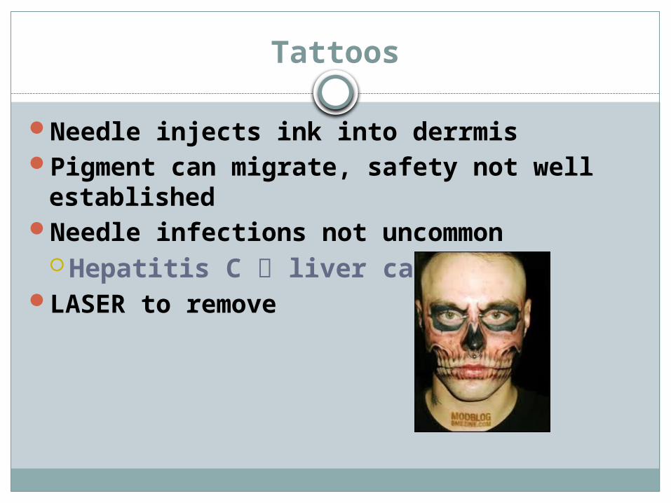
Tattoos
Needle injects ink into derrmisPigment can migrate, safety not well
establishedNeedle infections not uncommon
Hepatitis C liver cancerLASER to remove

Development of the Skin
Lanuga: soft , fine hairs that develop in 5th -6th month of pregnancy
Vernix caseosa: creamy, thick, white substance produced by sebaceous glands in 2nd half pregnancyKeeps skin soft, moist

Development of the Skin
Milia:small white spots frequently seen on newborn – 3rd week after birth
Accumulations in sebaceous glands
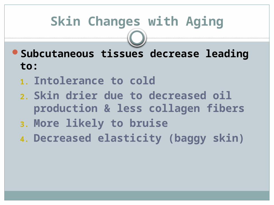
Skin Changes with Aging
Subcutaneous tissues decrease leading to:1. Intolerance to cold2. Skin drier due to decreased oil
production & less collagen fibers3. More likely to bruise 4. Decreased elasticity (baggy skin)