Anatomy - CameroonHealth Anatomy.pdf... · Palmar or Volar = the front (anterior) ... Anatomy...
60
www.internationalhealthinitiatives.com Cameroon Upper Extremity Rehabilitation Project section Anatomy 1
Transcript of Anatomy - CameroonHealth Anatomy.pdf... · Palmar or Volar = the front (anterior) ... Anatomy...

www.internationalhealthinitiatives.com
Cameroon Upper Extremity Rehabilitation Project
sect ion
Anatomy
1

Anterior
=
the front of
the body
Posterior
=
the back of
the body
Superior
=
towards the
head
Inferior
=
away from the
head
Anatomy Terms

Medial
=
toward the
middle of the
body
Lateral
= away from the
middle of
the body
Proximal
=
towards the
center of the body
Distal
=
away from the
center of the body
Anatomy Terms
Dorsal
=
the back (posterior)
of the hand
Palmar or
Volar
=
the front (anterior)
of the hand, the palm
Anatomy Terms

Flexion
=
bending motion that decreases the angle between bones of
a joint
Extension
= a straightening motion that increases the angle between
bones of a joint
Wrist Flexion
Wrist
Extension
Anatomy Movements
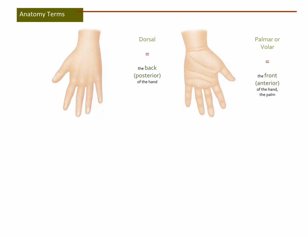
Anatomy Movements
Pronation
=
Turning to palm of the hand down to face the floor
Supination
=
Turning to palm of the hand to up to face the ceiling
Radial Deviation
=
Turning to hand toward the thumb
Ulnar Deviation
= Turning to hand
toward the
little finger
Anatomy Movements

Forward Elevation
=
Lifting the arm from the shoulder joint forward above the
head
Anatomy Movements
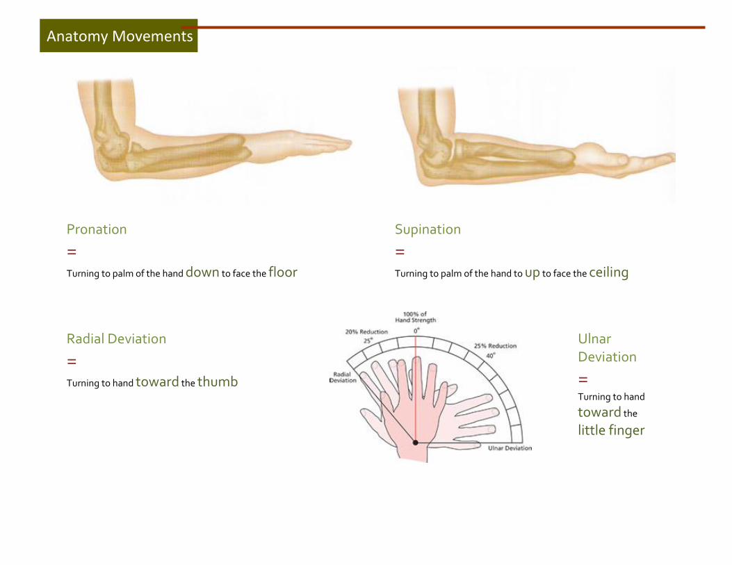
Thumb Movements
Flexion=
Bending the thumb down towards the middle of the palm of the hand
Extension=
Moving the thumb away from the fingers, in the same plane as the fingers
(also called radial abduction)
Abduction=
Moving thethumbaway from the fingers, perpendicular to the fingers
Adduction=
Moving the thumb back towards the fingers, in the same plane as the
fingers
Opposition =
Bringing the thumb out and across the fingers
(i.e. the thumb is now opposite the fingers)
Anatomy Movements
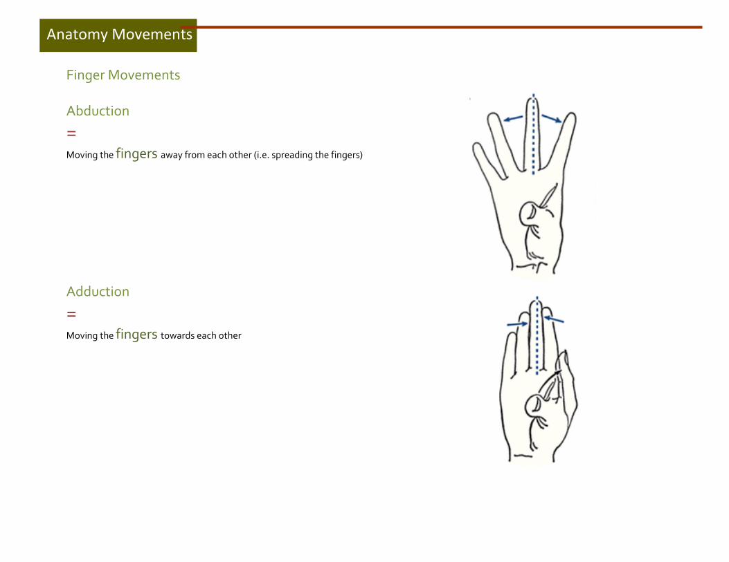
Finger Movements
Abduction
=
Moving the fingers away from each other (i.e. spreading the fingers)
Adduction
=
Moving the fingers towards each other
Anatomy Movements

Anterior (front) View
Skeletal System (Bones)

Posterior (back) View
Skeletal System (Bones)
Skeletal System (Bones)

Anterior (front or volar or palmar) View
MCP =Metacarpal-Phalangeal Joint
PIP =Proximal Inter-Phalangeal Joint
DIP =Distal Inter-Phalangeal Joint
PhysioTherapistsLikeShoulders (pisiform) (Triquetrum) (Lunate) (Scaphoid)
ThatTheyC anH eal! (Trapezium) (Trapezoid) (Capitate) (Hamate)
Skeletal System (Bones)
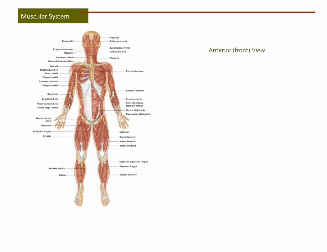
Anterior (front) View
Muscular System

Posterior (back) View
Muscular System
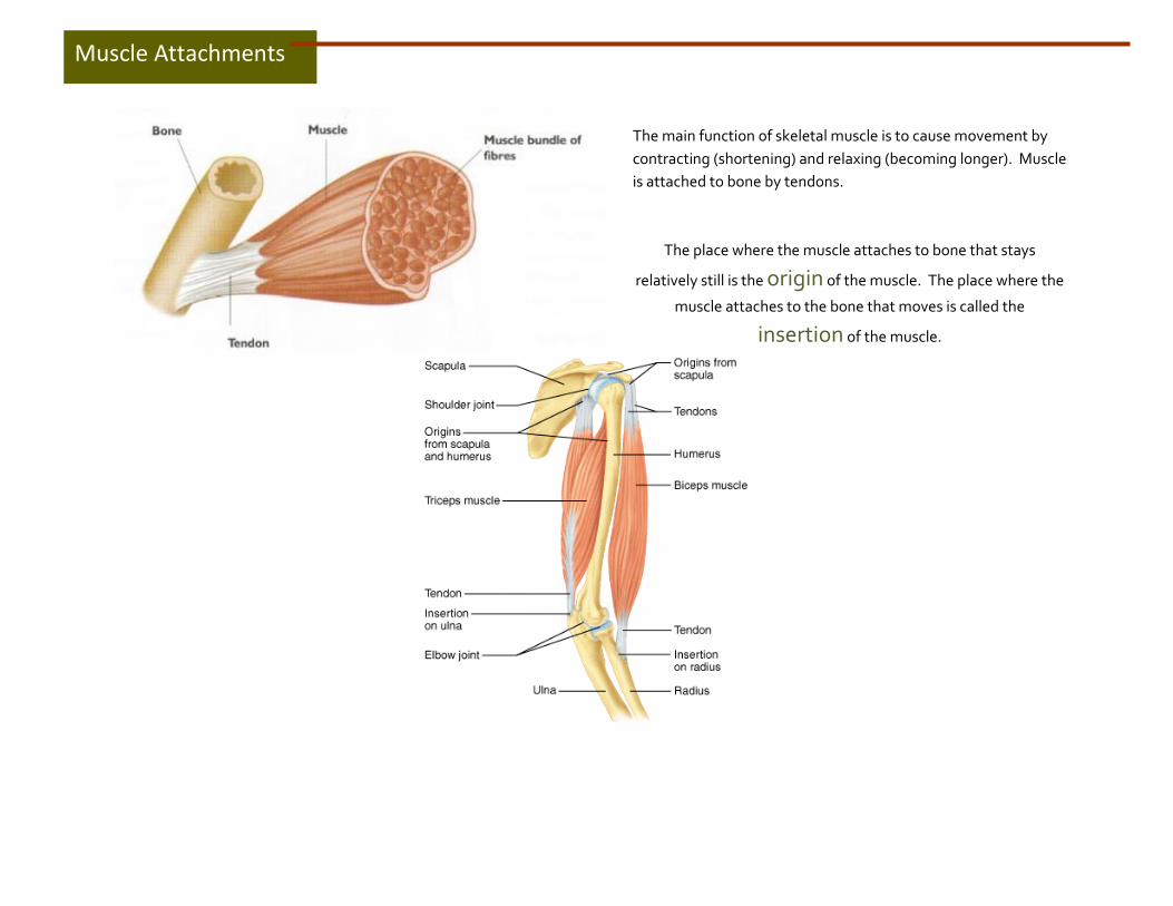
The main function of skeletal muscle is to cause movement by
contracting (shortening) and relaxing (becoming longer). Muscle
is attached to bone by tendons.
The place where the muscle attaches to bone that stays
relatively still is the origin of the muscle. The place where the
muscle attaches to the bone that moves is called the
insertion of the muscle.
Muscle Attachments

Isometric Muscle Contractions
Isometric muscle contraction is when a muscle increases tension but the actual length of the muscle does not change. This means that the joint that the muscle works on does not move. Imagine holding your elbow at 90° and then somebody puts a heavy weight in your hand. This is an isometric muscle contraction. Another example is the muscles of the neck that contract isometrically to keep the head up.
Isotonic Muscle Contractions
Isotonic muscle contractions enable us to move around. There are two types of isotonic muscle contractions:
1. Concentric 2. Eccentric
Muscle Contractions
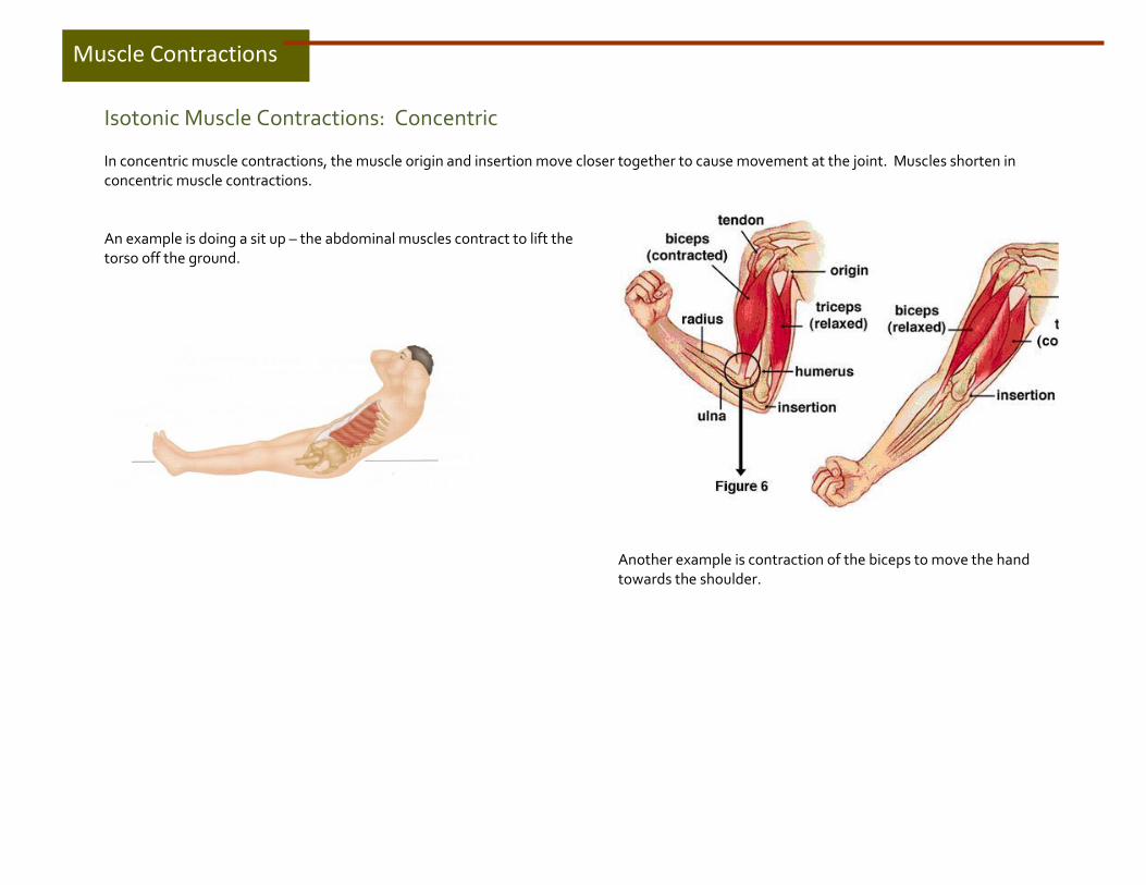
Isotonic Muscle Contractions: Concentric
In concentric muscle contractions, the muscle origin and insertion move closer together to cause movement at the joint. Muscles shorten in concentric muscle contractions.
An example is doing a sit up – the abdominal muscles contract to lift the torso off the ground.
Another example is contraction of the biceps to move the hand towards the shoulder.
Muscle Contractions
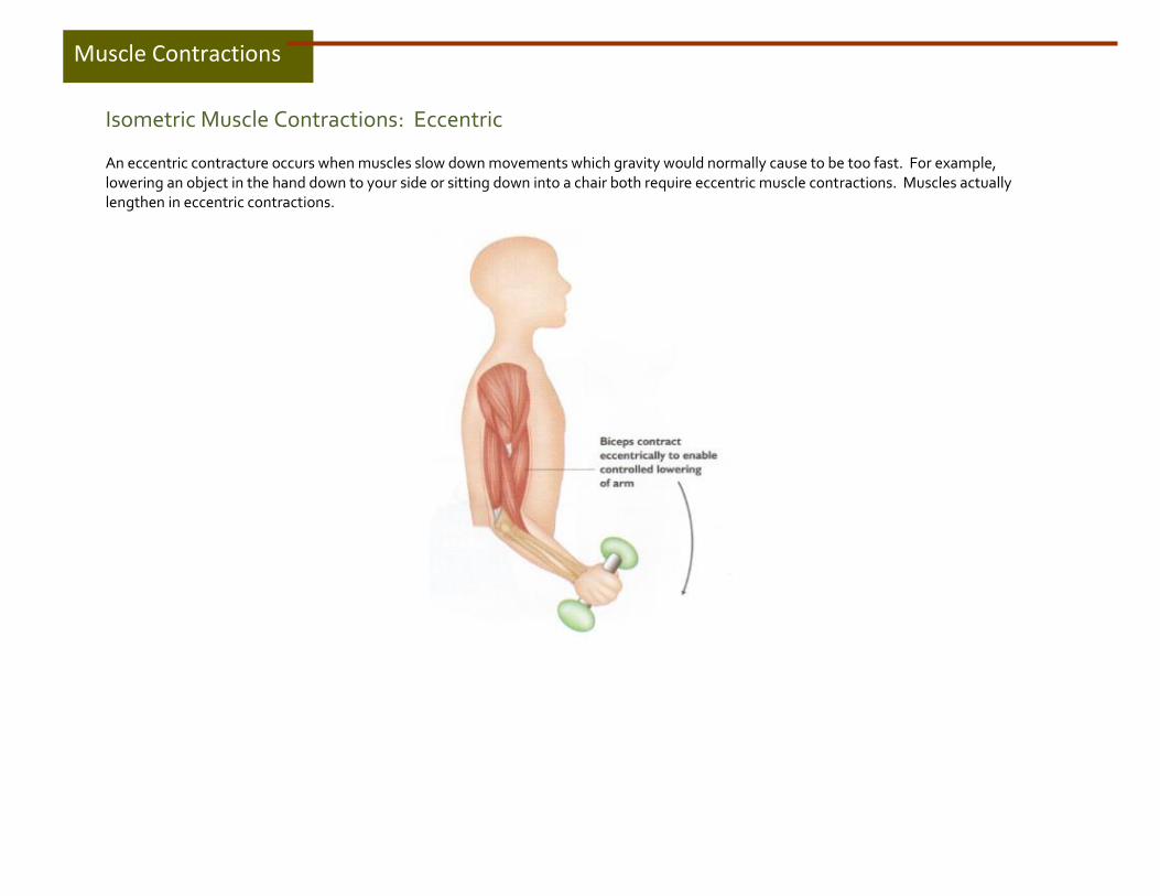
Isometric Muscle Contractions: Eccentric
An eccentric contracture occurs when muscles slow down movements which gravity would normally cause to be too fast. For example, lowering an object in the hand down to your side or sitting down into a chair both require eccentric muscle contractions. Muscles actually lengthen in eccentric contractions.
Muscle Contractions

Group Actions of Muscles
Muscles work together or against each other to cause movement. What one muscle can do, another muscle can undo. Muscles can also provide added support or stability to allow movements to occur elsewhere.
There are 4 functional groups of muscles:
1. Prime Mover or Agonist 2. Antagonist 3. Synergist 4. Fixator
1. Prime Mover or Agonist This is a muscle that contracts to cause movement. For example, biceps is the prime mover for elbow flexion. There are other muscles that assist the prime mover but with less effect or strength; these are called assistant or secondary movers.
2. Antagonist
The muscle on the opposite side of a joint to the prime mover is called the antagonist. The antagonist must relax to allow the prime mover to contract. For example, in order for the biceps to flex the arm at the elbow, the triceps on the back of the arm must relax to allow this movement to occur.
Muscle Contractions
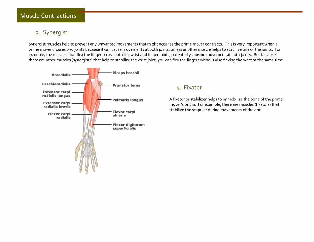
3. Synergist
Synergist muscles help to prevent any unwanted movements that might occur as the prime mover contracts. This is very important when a prime mover crosses two joints because it can cause movements at both joints, unless another muscle helps to stabilize one of the joints. For example, the muscles that flex the fingers cross both the wrist and finger joints, potentially causing movement at both joints. But because there are other muscles (synergists) that help to stabilize the wrist joint, you can flex the fingers without also flexing the wrist at the same time.
4. Fixator
A fixator or stabilizer helps to immobilize the bone of the prime mover’s origin. For example, there are muscles (fixators) that stabilize the scapular during movements of the arm.
Muscle Contractions

The nervous system contains a network of cells called neurons that coordinate actions and transmit signals. The nervous system consists of two parts: the central nervous system (CNS) and the peripheral nervous system (PNS). The CNS consists of the brain and spinal cord. The PNS consists of all other nerves outside of the brain and spinal cord. The function of the PNS is to connect the CNS to the limbs and organs and to send sensory information from the body back to the spinal cord. It is the PNS that we will focus on. The nerve cell and the muscle cells it makes contact with are called a motor unit. The nerve transmits a signal to cause the motor unit to contract.
Nervous System
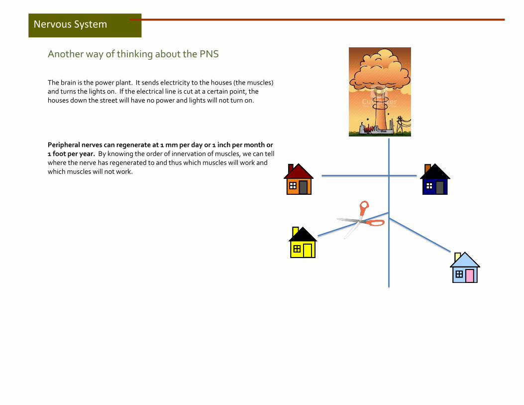
Another way of thinking about the PNS The brain is the power plant. It sends electricity to the houses (the muscles) and turns the lights on. If the electrical line is cut at a certain point, the houses down the street will have no power and lights will not turn on. Peripheral nerves can regenerate at 1 mm per day or 1 inch per month or 1 foot per year. By knowing the order of innervation of muscles, we can tell where the nerve has regenerated to and thus which muscles will work and which muscles will not work.
Nervous System

The Brachial Plexus The brachial plexus is a network of nerve fibers that proceeds through the neck, the axilla (armpit) and into the arm and hand. The brachial plexus is responsible for all the cutaneous (skin) and muscular innervation of the upper limb (except the trapezius muscle).
Nervous System

The Median Nerve
The median nerve innervates the following muscles, in this order:
1. Pronator Teres 2. Flexor Carpi Radialis 3. Palmaris Longus 4. Flexor Digitorum Superficialis Anterior Interosseous Nerve (median nerve) 5. Flexor Digitorum Profundus (index and middle) 6. Flexor Pollicis Longus 7. Pronator Quadratus Palmar Recurrent Motor Branch (median) 8. Abductor Pollicis Brevis 9. Opponens Pollicis 10. Flexor Pollicis Brevis Common Palmar Digital Nerve (median) 11. Lumbricals 1 & 2
Nervous System

The Radial Nerve
The radial nerve innervates the following muscles, in this order:
1. Triceps 2. Anconeus 3. Brachioradialis 4. Extensor Carpi Radialis Longus 5. Extensor Carpi Radias Brevis 6. Supinator
Posterior Interosseous Nerve (radial nerve) 7. Extensor Digitorum (Communis) 8. Extensor Digiti Minimi 9. Extensor Carpi Ulnaris 10. Abductor Pollicis Longus 11. Extensor Pollicis Longus 12. Extensor Pollicis Brevis 13. Extensor Indicis
Nervous System
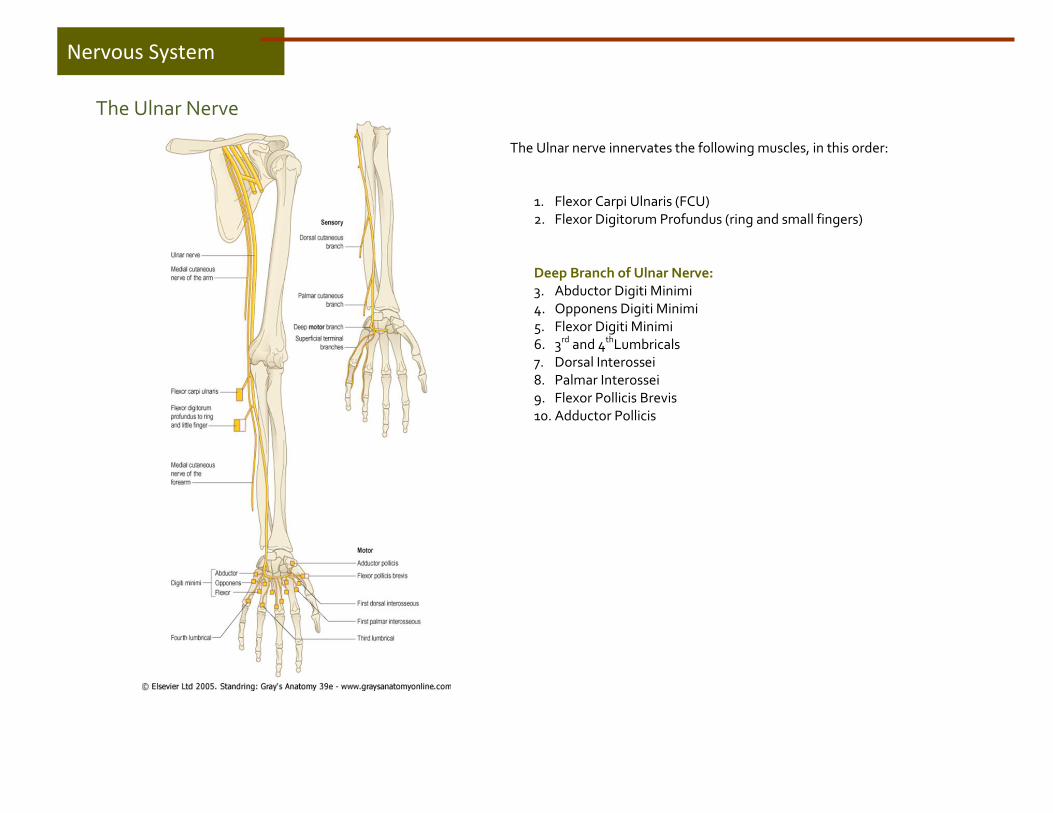
The Ulnar Nerve
The Ulnar nerve innervates the following muscles, in this order:
1. Flexor Carpi Ulnaris (FCU) 2. Flexor Digitorum Profundus (ring and small fingers) Deep Branch of Ulnar Nerve: 3. Abductor Digiti Minimi 4. Opponens Digiti Minimi 5. Flexor Digiti Minimi 6. 3rd and 4thLumbricals 7. Dorsal Interossei 8. Palmar Interossei 9. Flexor Pollicis Brevis 10. Adductor Pollicis
Nervous System

Pronator Teres
Origin:
Insertion:
Action:
Nerve:
Medial epicondyle of humerus and ulna Middle of lateral surface of radius
Pronates forearm. Helps flex elbow also. Median nerve
Muscles innervated by the
Median Nerve

Flexor Carpi Radialis
Origin:
Insertion:
Action:
Nerve:
Medial epicondyle of humerus Base of 2nd and 3rd metacarpal bones
Flexes hand at wrist. Helps with radial deviation.
Median nerve
Muscles innervated by the
Median Nerve

Palmaris Longus
Origin:
Insertion:
Action:
Nerve:
Medial epicondyle of humerus Flexor retinaculum and palmar aponeurosis Flexes hand at wrist. Tightens palmar aponeurosis Median nerve
Muscles innervated by the
Median Nerve

Flexor Digitorum Superficialis
Origin:
Insertion:
Action:
Nerve:
Medial epicondyle of humerus and ulna 4 tendons attach to the middle phalanges of index, middle, ring and little fingers
Flexes proximal interphalangeal joints Median nerve
Muscles innervated by the
Median Nerve

Flexor Digitorum Profundus
Origin:
Insertion:
Action:
Nerve:
Anterior and medial aspect of ulna 4 tendons attach to base of distal phalanges of index to little fingers
Flexes distal interphalangeal joints Median nerve innervates the index and middle fingers (anterior interosseous branch)
Muscles innervated by the
Median Nerve

Flexor Pollicis Longus
Origin:
Insertion:
Action:
Nerve:
Anterior aspect of radius and interosseous membrane Base of distal phalanx of thumb
Flexes distal phalanx of thumb Median nerve (anterior interosseous branch)
Muscles innervated by the
Median Nerve
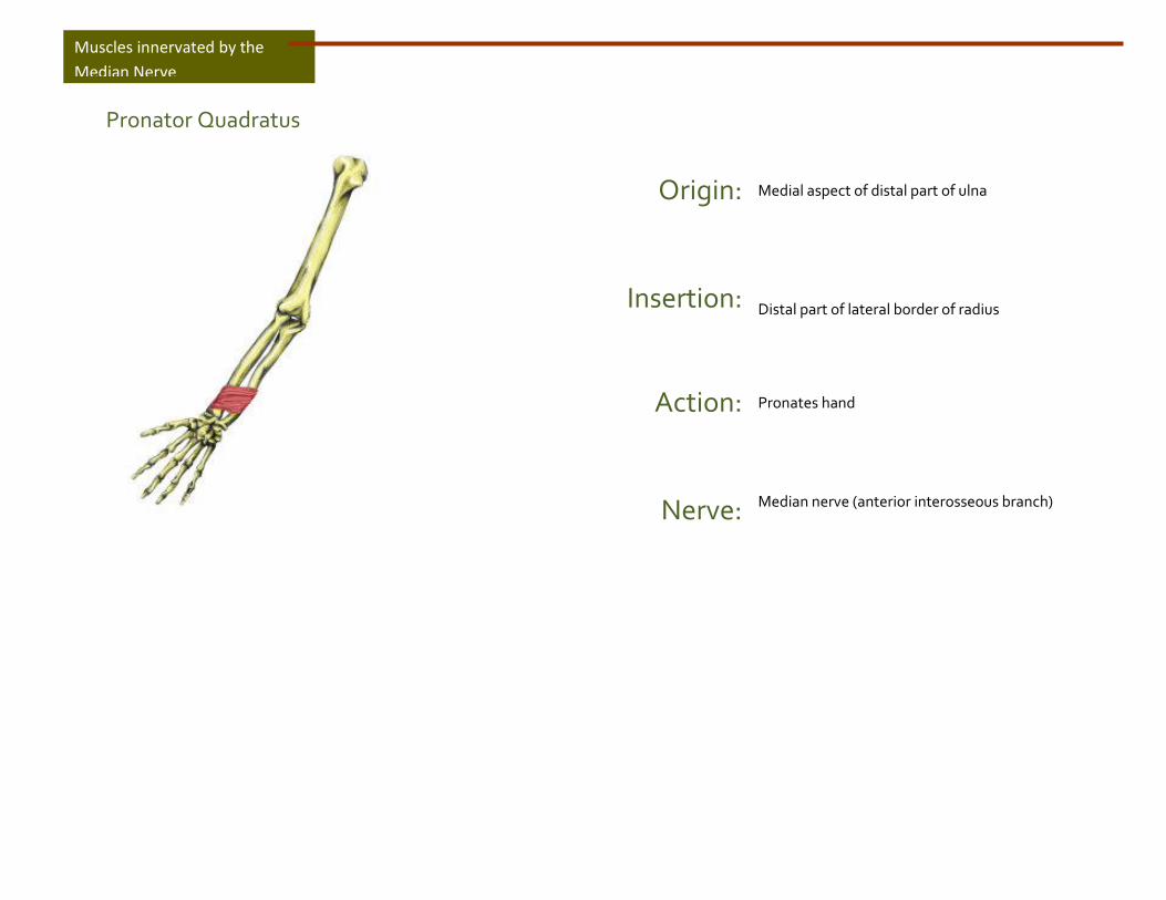
Pronator Quadratus
Origin:
Insertion:
Action:
Nerve:
Medial aspect of distal part of ulna Distal part of lateral border of radius
Pronates hand Median nerve (anterior interosseous branch)
Muscles innervated by the
Median Nerve

Abductor Pollicis Brevis
Origin:
Insertion:
Action:
Nerve:
Flexor retinaculum and scaphoid and trapezium Base of proximal phalanx of thumb
Abducts thumb at carpo-metacarpal and metacarpo-phalangeal joints Median nerve (palmar recurrent motor branch)
Muscles innervated by the
Median Nerve

Opponens Pollicis
Origin:
Insertion:
Action:
Nerve:
Flexor retinaculum and trapezium Lateral side of 1st metacarpal (thumb)
Opposes thumb to fingers Median nerve (palmar recurrent motor branch) )
Muscles innervated by the
Median Nerve

Flexor Pollicis Brevis
Origin:
Insertion:
Action:
Nerve:
2 heads: 1) Superficial: flexor retinaculum and trapezium and 2) Deep: floor of carpal bones Lateral side of 1st metacarpal and base of proximal phalanx of thumb
Flexes proximal phalanx of thumb at metacarpo-phalangeal joint Superficial head is innervated by the median nerve (palmar recurrent motor branch)
Muscles innervated by the
Median Nerve

Lumbricals 1 and 2
Origin:
Insertion:
Action:
Nerve:
Tendons of flexor digitorum profundus of index and middle fingers Lateral side of extensor expansions of index and middle finger
Flex the metacarpo-phalangeal joints and extend the inter-phalangeal joints of index and middle fingers Median nerve (common palmar digital nerve)
Muscles innervated by the
Median Nerve

Triceps
Origin:
Insertion:
Action:
Nerve:
Scapula and humerus Olecranon process of the ulna
Extends the elbow joint
Radial nerve
Muscles innervated by the
Radial Nerve
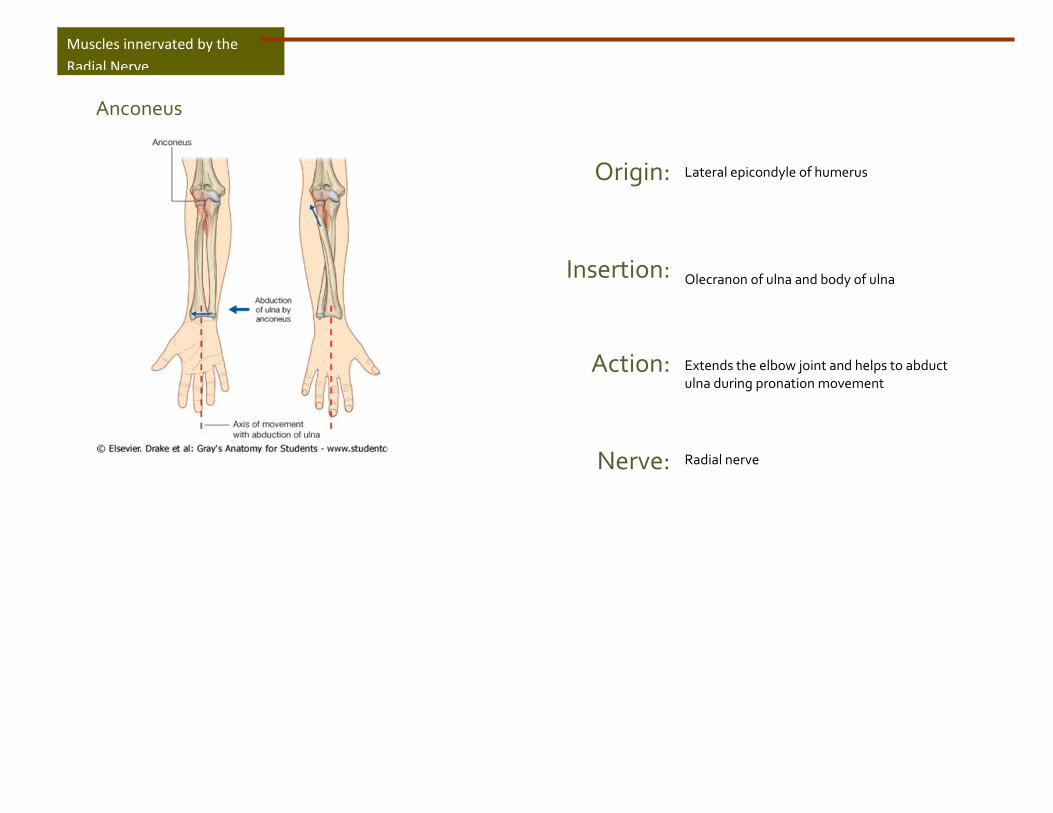
Anconeus
Origin:
Insertion:
Action:
Nerve:
Lateral epicondyle of humerus Olecranon of ulna and body of ulna
Extends the elbow joint and helps to abduct ulna during pronation movement
Radial nerve
Muscles innervated by the
Radial Nerve
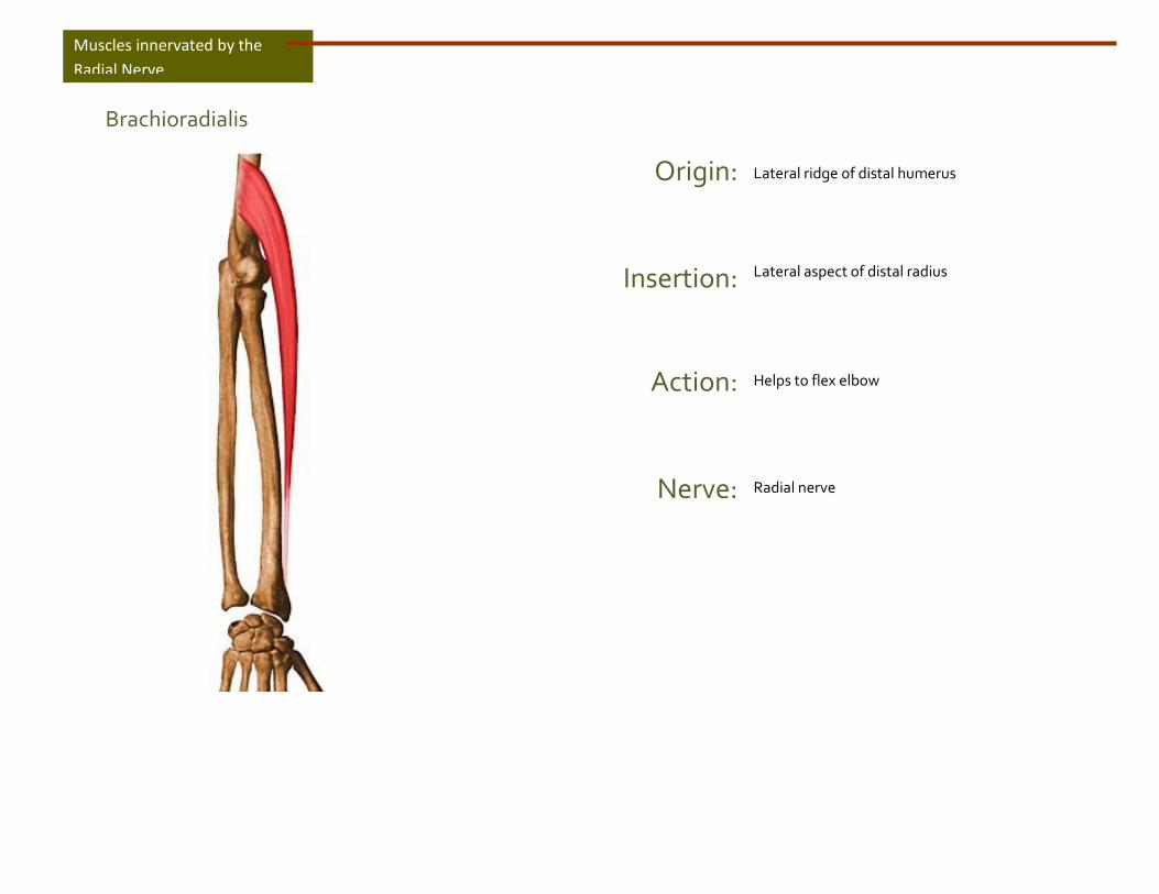
Brachioradialis
Origin:
Insertion:
Action:
Nerve:
Lateral ridge of distal humerus Lateral aspect of distal radius
Helps to flex elbow
Radial nerve
Muscles innervated by the
Radial Nerve

Extensor Carpi Radialis Longus
Origin:
Insertion:
Action:
Nerve:
Lateral ridge of humerus Base of 2nd metacarpal
Extends and abducts (radial deviation) hand at wrist
Radial nerve
Muscles innervated by the
Radial Nerve
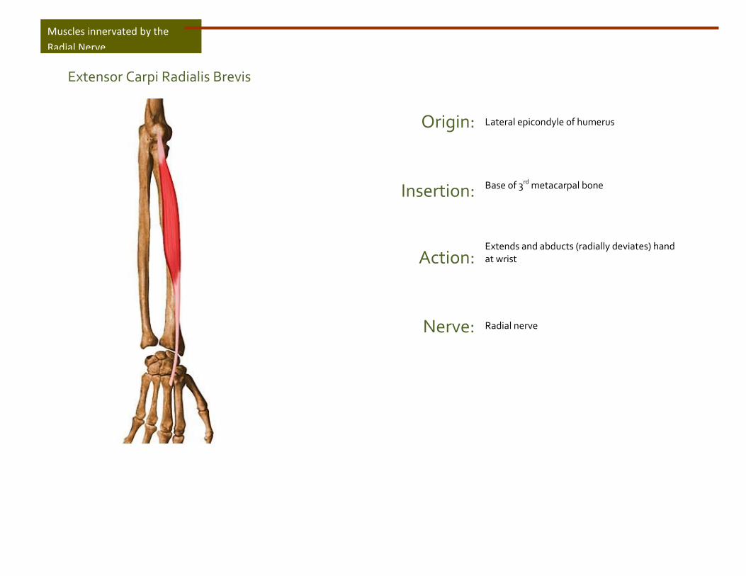
Extensor Carpi Radialis Brevis
Origin:
Insertion:
Action:
Nerve:
Lateral epicondyle of humerus Base of 3rd metacarpal bone
Extends and abducts (radially deviates) hand at wrist
Radial nerve
Muscles innervated by the
Radial Nerve
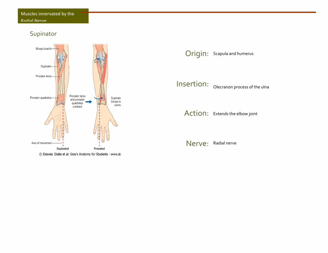
Supinator
Origin:
Insertion:
Action:
Nerve:
Scapula and humerus Olecranon process of the ulna
Extends the elbow joint
Radial nerve
Muscles innervated by the
Radial Nerve

Extensor Digitorum (Communis)
Origin:
Insertion:
Action:
Nerve:
Lateral epicondyle of humerus Extensor expansions of index to little fingers
Extends metacarpo-phalangeal and inter-phalangeal joints
Radial nerve (posterior interosseous branch)
Muscles innervated by the
Radial Nerve

Extensor Digiti Minimi
Origin:
Insertion:
Action:
Nerve:
Lateral epicondyle of humerus Extensor expansions of little finger
Extends metacarpo-phalangeal and inter-phalangeal joints of little finger
Radial nerve (posterior interosseous branch)
Muscles innervated by the
Radial Nerve

Extensor Carpi Ulnaris
Origin:
Insertion:
Action:
Nerve:
Lateral epicondyle of humerus and ulna Base of 5th metacarpal
Extends and adducts hand at wrist
Radial nerve (posterior interosseous branch)
Muscles innervated by the
Radial Nerve
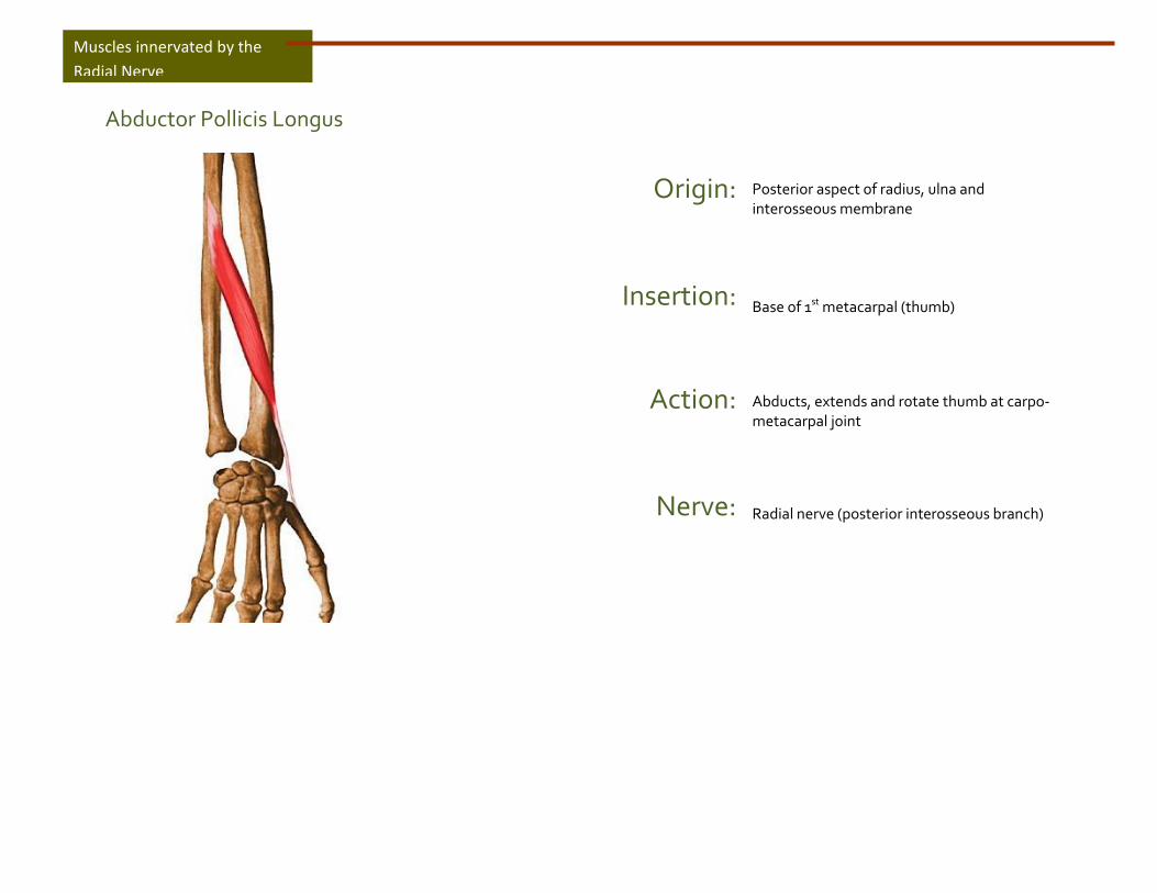
Abductor Pollicis Longus
Origin:
Insertion:
Action:
Nerve:
Posterior aspect of radius, ulna and interosseous membrane Base of 1st metacarpal (thumb)
Abducts, extends and rotate thumb at carpo-metacarpal joint
Radial nerve (posterior interosseous branch)
Muscles innervated by the
Radial Nerve

Extensor Pollicis Longus
Origin:
Insertion:
Action:
Nerve:
Posterior surface middle of ulna and interosseous membrane Base of distal phalanx of thumb
Extends distal phalanx of thumb
Radial nerve (posterior interosseous branch)
Muscles innervated by the
Radial Nerve

Extensor Pollicis Brevis
Origin:
Insertion:
Action:
Nerve:
Posterior surface radius and interosseous membrane Base of proximal phalanx of thumb
Extends proximal phalanx of thumb at metacarpo-phalangeal joint
Radial nerve (posterior interosseous branch)
Muscles innervated by the
Radial Nerve
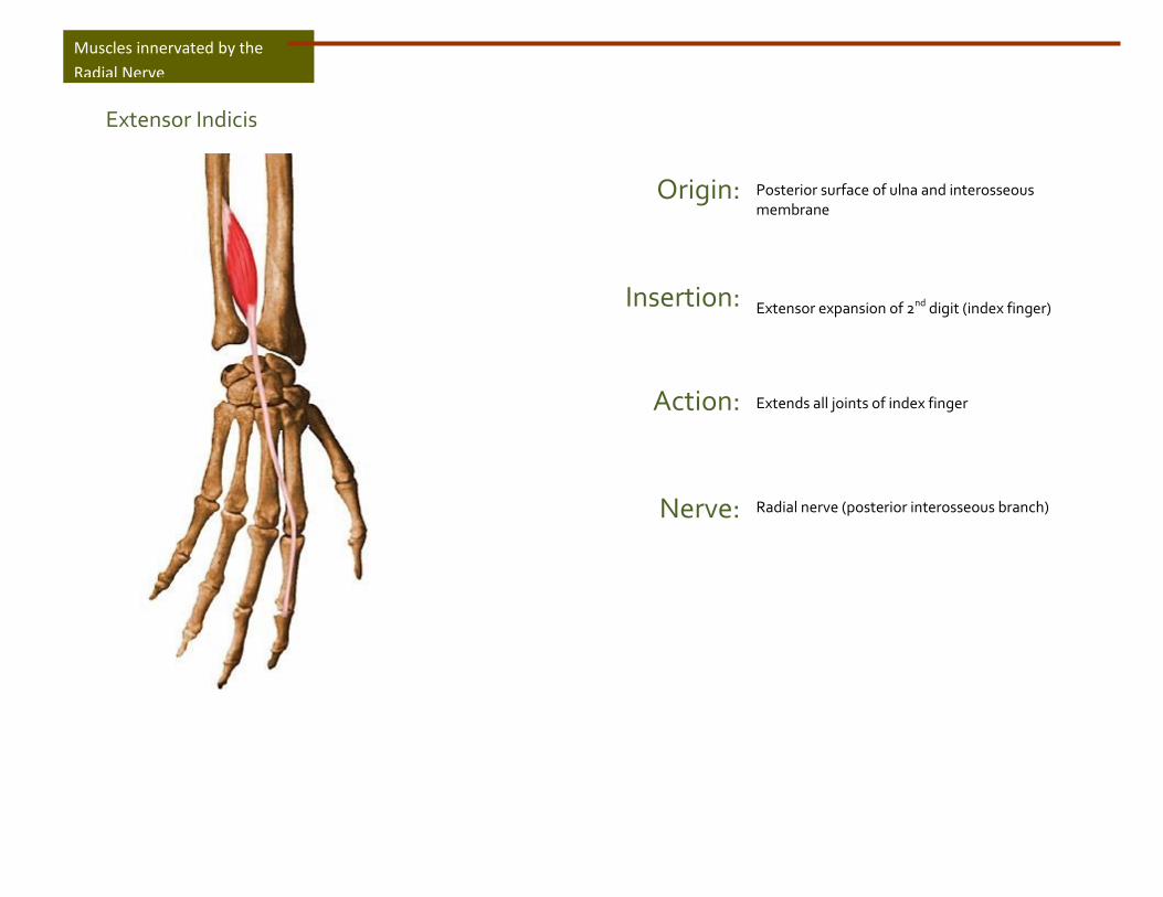
Extensor Indicis
Origin:
Insertion:
Action:
Nerve:
Posterior surface of ulna and interosseous membrane Extensor expansion of 2nd digit (index finger)
Extends all joints of index finger
Radial nerve (posterior interosseous branch)
Muscles innervated by the
Radial Nerve
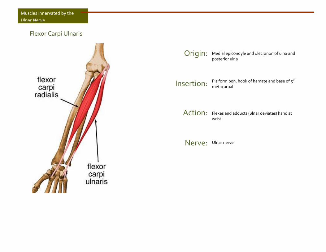
Flexor Carpi Ulnaris
Origin:
Insertion:
Action:
Nerve:
Medial epicondyle and olecranon of ulna and posterior ulna Pisiform bon, hook of hamate and base of 5th metacarpal
Flexes and adducts (ulnar deviates) hand at wrist
Ulnar nerve
Muscles innervated by the
Ulnar Nerve
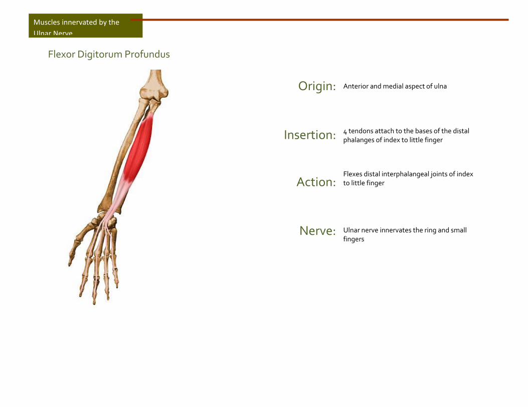
Flexor Digitorum Profundus
Origin:
Insertion:
Action:
Nerve:
Anterior and medial aspect of ulna 4 tendons attach to the bases of the distal phalanges of index to little finger
Flexes distal interphalangeal joints of index to little finger
Ulnar nerve innervates the ring and small fingers
Muscles innervated by the
Ulnar Nerve

Abductor Digiti Minimi
Origin:
Insertion:
Action:
Nerve:
Pisiform and tendon of flexor carpi ulnaris muscle Medial side of the base of the proximal phalanx of 5th digit
Adducts 5th digit
Deep branch of ulnar nerve
Muscles innervated by the
Ulnar Nerve
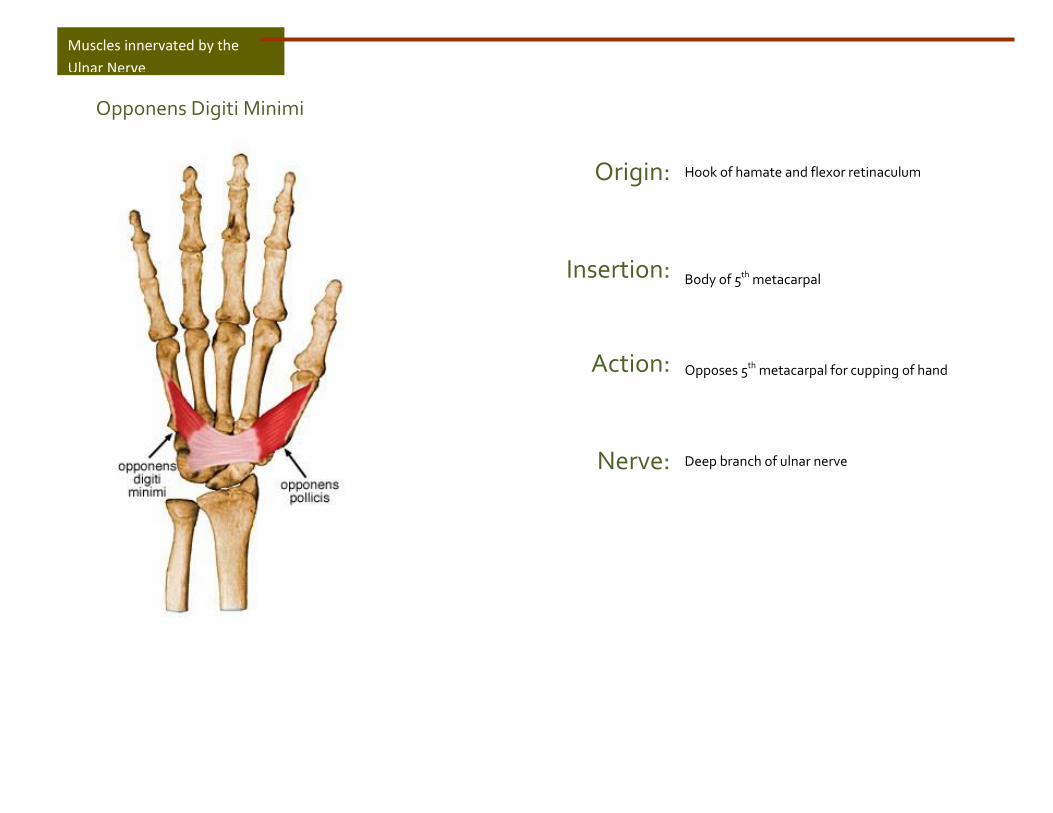
Opponens Digiti Minimi
Origin:
Insertion:
Action:
Nerve:
Hook of hamate and flexor retinaculum Body of 5th metacarpal
Opposes 5th metacarpal for cupping of hand
Deep branch of ulnar nerve
Muscles innervated by the
Ulnar Nerve
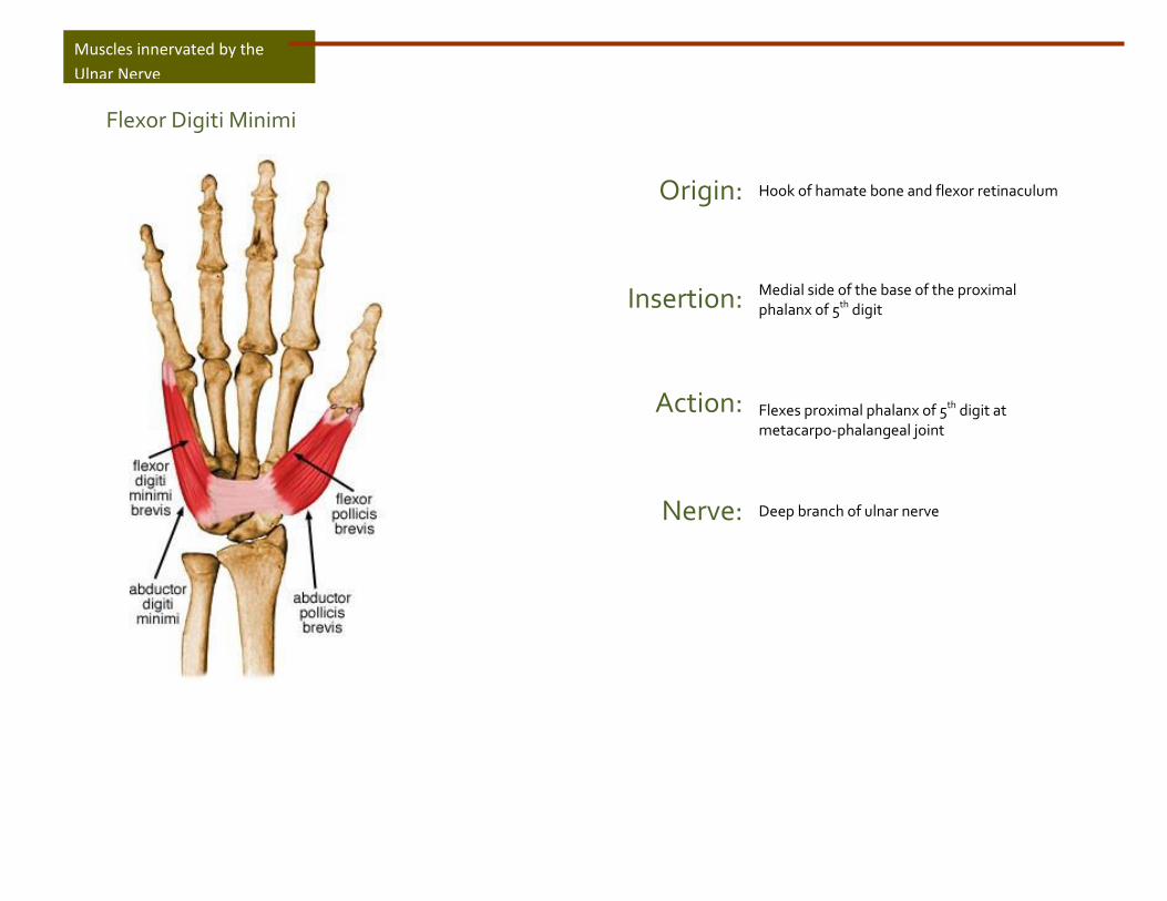
Flexor Digiti Minimi
Origin:
Insertion:
Action:
Nerve:
Hook of hamate bone and flexor retinaculum Medial side of the base of the proximal phalanx of 5th digit
Flexes proximal phalanx of 5th digit at metacarpo-phalangeal joint
Deep branch of ulnar nerve
Muscles innervated by the
Ulnar Nerve
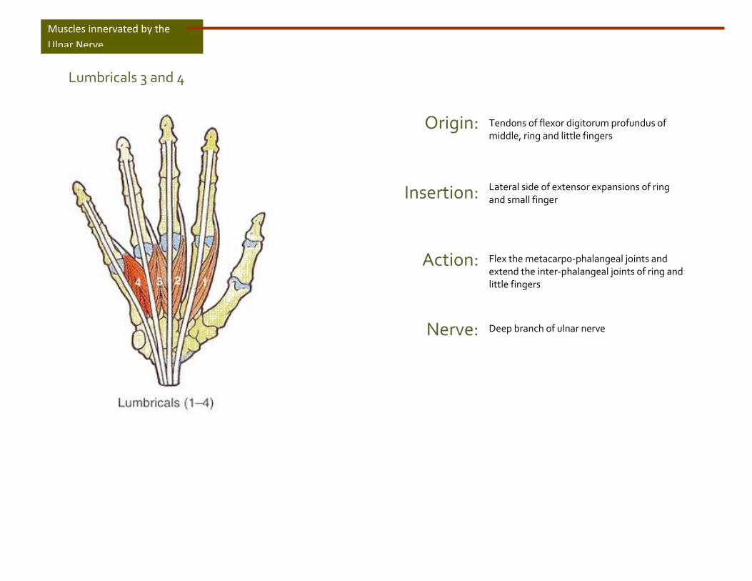
Lumbricals 3 and 4
Origin:
Insertion:
Action:
Nerve:
Tendons of flexor digitorum profundus of middle, ring and little fingers Lateral side of extensor expansions of ring and small finger
Flex the metacarpo-phalangeal joints and extend the inter-phalangeal joints of ring and little fingers
Deep branch of ulnar nerve
Muscles innervated by the
Ulnar Nerve

Dorsal Interossei (4)
Origin:
Insertion:
Action:
Nerve:
4 dorsal interossei originate at 2 heads from both sides of metacarpal bones Base of proximal phalanx and extensor expansions of index to ring fingers
Abducts fingers and also flex fingers at metacarpo-phalangeal joints and extends fingers at inter-phalangeal joints
Deep branch of ulnar nerve
Muscles innervated by the
Ulnar Nerve

Palmar Interossei (3)
Origin:
Insertion:
Action:
Nerve:
3 palmar interossei originate from metacarpal bones of index, ring and little fingers Extensor expansions of digits and bases of proximal phalanges of index, ring and little fingers
Adduct fingers at metacarpo-phalangeal joint
Deep branch of ulnar nerve
Muscles innervated by the
Ulnar Nerve

Flexor Pollicis Brevis
Origin:
Insertion:
Action:
Nerve:
2 heads: 1) Superficial: flexor retinaculum and trapezium and 2) Deep: floor of carpal bones Lateral side and base of proximal phalanx of thumb
Flexes proximal phalanx of thumb at metacarpo-phalangeal joint
Deep head is innervated by deep branch of ulnar nerve
Muscles innervated by the
Ulnar Nerve

Adductor Pollicis
Origin:
Insertion:
Action:
Nerve:
2 heads: 1) base of metacarpals 2 and 3 and capitate bone 2) body of 3rd metacarpal bone Base of proximal phalanx of thumb
Adduct proximal phalanx of thumb towards middle finger
Deep branch of ulnar nerve
Muscles innervated by the
Ulnar Nerve