Analysis of Trypanosoma brucei vsg expression site switching in vitro
13
Molecular and Biochemical Parasitology 84 (1997) 189 – 201 Analysis of Trypanosoma brucei 6sg expression site switching in vitro David Horn, George A.M. Cross* Laboratory of Molecular Parasitology, The Rockefeller Uni6ersity, 1230 York A6enue, New York, NY 10021 -6399, USA Received 19 August 1996; accepted 7 November 1996 Abstract Trypanosoma brucei can undergo antigenic variation by switching between distinct telomeric variant surface glycoprotein gene (6sg ) expression sites (ESs) or by replacing the active 6sg. DNA rearrangements have often been associated with ES switching, but it is unclear if such rearrangements are necessary or whether ES inactivation always accompanies ES activation. To explore these issues, we derived ten independent clones, from the same parent, that had undergone a similar 6sg activation event. This was achieved in the absence of an immune response, in vitro, using cells with selectable markers integrated into an ES. Nine of the ten clones had undergone ES switching. Such heritable changes in transcription state occurred at a frequency of approximately 6 ×10 -7 . Comparison of switched and un-switched clones highlighted the dynamic nature of T. brucei telomeres, but changes in telomere length were not specifically associated with ES switching. Mapping within and beyond the ESs revealed no detectable DNA rearrangements, indicating that rearrangements are not necessary for ES activation/inactivation. Examination of individual cells indicated that ES activation consistently accompanied inactivation of the previously active ES. In some cases, however, we found cells that appeared to have efficiently established the switched state but which subsequently, at a frequency of approximately 2 ×10 -3 , generated cells expressing both pre- and post-switch 6sgs. These results show that ES activation/inactivation is usually a coupled process but that cells can inherit a propensity to uncouple these events. © 1997 Elsevier Science B.V. Keywords: Antigenic variation; Expression site; Telomere; Trypanosoma brucei ; Variant surface glycoprotein 1. Introduction In the blood and tissue spaces of the mam- malian host, Trypanosoma brucei escapes elimina- tion by the host immune response by a process of antigenic variation (reviewed in [1,2]). Antigenic Abbre6iations: ES, expression site; TRF, telomere restriction fragment; Vsg (6sg ), variant surface glycoprotein (gene). * Corresponding author. Tel.: +1 212 3277571; fax: +1 212 3277845; e-mail: [email protected] 0166-6851/97/$17.00 © 1997 Elsevier Science B.V. All rights reserved PII S0166-6851(96)02794-6
-
Upload
david-horn -
Category
Documents
-
view
214 -
download
2
Transcript of Analysis of Trypanosoma brucei vsg expression site switching in vitro

Molecular and Biochemical Parasitology 84 (1997) 189–201
Analysis of Trypanosoma brucei 6sg expression site switchingin vitro
David Horn, George A.M. Cross*
Laboratory of Molecular Parasitology, The Rockefeller Uni6ersity, 1230 York A6enue, New York, NY 10021-6399, USA
Received 19 August 1996; accepted 7 November 1996
Abstract
Trypanosoma brucei can undergo antigenic variation by switching between distinct telomeric variant surfaceglycoprotein gene (6sg) expression sites (ESs) or by replacing the active 6sg. DNA rearrangements have often beenassociated with ES switching, but it is unclear if such rearrangements are necessary or whether ES inactivation alwaysaccompanies ES activation. To explore these issues, we derived ten independent clones, from the same parent, thathad undergone a similar 6sg activation event. This was achieved in the absence of an immune response, in vitro, usingcells with selectable markers integrated into an ES. Nine of the ten clones had undergone ES switching. Such heritablechanges in transcription state occurred at a frequency of approximately 6×10⁻⁷. Comparison of switched andun-switched clones highlighted the dynamic nature of T. brucei telomeres, but changes in telomere length were notspecifically associated with ES switching. Mapping within and beyond the ESs revealed no detectable DNArearrangements, indicating that rearrangements are not necessary for ES activation/inactivation. Examination ofindividual cells indicated that ES activation consistently accompanied inactivation of the previously active ES. Insome cases, however, we found cells that appeared to have efficiently established the switched state but whichsubsequently, at a frequency of approximately 2×10⁻³, generated cells expressing both pre- and post-switch 6sgs.These results show that ES activation/inactivation is usually a coupled process but that cells can inherit a propensityto uncouple these events. © 1997 Elsevier Science B.V.
Keywords: Antigenic variation; Expression site; Telomere; Trypanosoma brucei ; Variant surface glycoprotein
1. Introduction
In the blood and tissue spaces of the mam-malian host, Trypanosoma brucei escapes elimina-tion by the host immune response by a process ofantigenic variation (reviewed in [1,2]). Antigenic
Abbre6iations: ES, expression site; TRF, telomere restrictionfragment; Vsg (6sg), variant surface glycoprotein (gene).
* Corresponding author. Tel.: +1 212 3277571; fax: +1212 3277845; e-mail: [email protected]
0166-6851/97/$17.00 © 1997 Elsevier Science B.V. All rights reserved
PII S 0 1 66 -6851 (96 )02794 -6

D. Horn, G.A.M. Cross / Molecular and Biochemical Parasitology 84 (1997) 189–201190
variation involves the mutually exclusive and se-quential expression of individual variant surfaceglycoprotein (Vsg) genes (6sg) derived from arepertoire of approximately 1000 6sgs andpseudo-6sgs [3]. The expressed 6sg is invariablylocated at the telomere-proximal end of a poly-cistronic transcription unit known as an expres-sion site (ES). A number of ES-associated genesare also present in these polycistronic transcrip-tion units and the ES promoter is located approx-imately 50 kb upstream of the 6sg. Antigenicvariation is spontaneous and can occur by a num-ber of mechanisms, but most commonly involvesreplacement of the active 6sg, by gene conversionat the active ES, or by switching transcriptionfrom one ES to another (reviewed in [1,2]). The6sgs at silent telomeric ESs are more likely tobecome expressed and can be activated by eitherof these mechanisms [4].
We [5] and others [6] have recently shown thatposition effects repress transcription at a silent T.brucei ES. We wish to discover how ES domainsfaithfully and heritably maintain their transcrip-tional status over many generations, yet infre-quently allow alterations of state. DNArearrangements can alter gene expression in anumber of organisms [7] and rearrangementswithin an ES, or upstream of the promoter region,have been found after ES switching, (reviewed in[8]), but it is unclear if any of these changes arenecessary for ES switching. We wanted to deter-mine, using more stringent experimental protocolsthan hitherto possible, whether any DNA rear-rangements are necessary for ES switching, orwhether ES switching can be attributed solely toepigenetic phenomena. We also wanted to deter-mine if ES inactivation invariably accompaniesES activation. There are estimated to be approxi-mately 20 bloodstream-form ESs in the T. bruceigenome [9–12]. Usually, only one ES is active ata time in individual bloodstream-form cells andES activity is controlled at or near transcriptioninitiation [10], with notable exceptions. In aswitch associated with a large insertion upstreamof the silenced 6sg, the region upstream of theinsertion continued to be transcribed [13]. Fur-thermore, stable expression of two 6sgs from dif-ferent ESs in individual cells has been reported in
T. equiperdum [14], suggesting that ESs can switchon and off independently.
In T. brucei clone 118a, a single copy of 6sg221is present at a transcriptionally repressed ES. This6sg can be activated via duplication or ES switch-ing [15]. Activation of the 6sg221 ES occurredwith no detectable rearrangement up to 55 kbupstream of 6sg221 [15] and inactivation occurredwithout a single point mutation within 3.4 kbspanning the ES promoter [10]. The active ES inclone 118a can also undergo inactivation andsubsequent reactivation [16]. To capture andstudy multiple examples of activation of the6sg221 ES, we inserted genes encoding resistanceto phleomycin and G418 into the silent 6sg221 ESin 118a cells [5]. Acquisition of drug-resistancecan consequently be exploited to capture clones inwhich the 6sg221 ES has become transcriptionallyactive, or the marker cassette has been duplicatedto a transcriptionally active locus. Activationevents can be captured immediately while mini-mizing sub-culturing or other procedures thatcould allow unrelated DNA rearrangements toaccumulate. Also, because drug selection, as op-posed to immune selection, does not require inac-tivation of the previously active 6sg for survival,our approach allows the isolation of cells thatactivate a second ES without inactivating thepreviously active ES.
In this study, we show that detectable DNArearrangements are not necessary for ES switchingand that ES activation usually accompanies inac-tivation of the previously active ES. Our switchedclones, however, inherited different phenotypes.Some of these clones inherited a propensity touncouple ES activation and inactivation leadingto the generation of cells expressing two 6sgs.
2. Materials and methods
2.1. Cells
Cells were maintained and cloned as previouslydescribed [5]. T. brucei Mitat 1.5 clone 118a wasderived from stabilate 049 and Mitat 1.2 clone221a was derived in culture from stabilate 052[17,18]. G418 was purchased from Gibco andphleomycin from Cayla.

D. Horn, G.A.M. Cross / Molecular and Biochemical Parasitology 84 (1997) 189–201 191
2.2. DNA analysis
Genomic DNA was isolated and Southern blot-ting was carried out as previously described [5].Agarose blocks containing intact T. brucei chro-mosomes were prepared as described for mam-malian cells [19]. Restriction enzyme digestion ofDNA in agarose was carried out according to themanufacturer (New England Biolabs). DNA em-bedded in agarose was size-fractionated using aRotating Agarose Gel Electrophoresis apparatus(Stratagene). Gels were prepared from SeaKemLE agarose (FMC) in 0.5×TBE and the elec-trophoresis temperature was kept constant at12°C. Hansenula wingei, Saccharomyces cere6isiae
and Schizosaccharomyces pombe chromosomemarkers were from Bio-Rad and low-range pulsedfield gel electrophoresis markers were from NewEngland Biolabs. Large DNA fragments were vi-sualized and transferred to membrane as de-scribed [19]. All post-hybridization washes were at65°C in 0.2×SSC and 0.2% SDS.
2.3. Protein analysis
Western blotting and immunofluorescence anal-ysis were carried out as previously described [5].For detection of Neo or Ble, protein from 3×10⁵
cells was loaded per lane. For detection of Vsg,protein from 1.5×10⁵ cells was loaded per lane.Vsg, Neo or Ble proteins were separated in 10,12.5 and 17.5% polyacrylamide gels, respectively.The antiserum to Vsg221 was depleted of anti-body to the cross-reacting determinant [20].
Fig. 1. Selection of switched cells in vitro. (A) Physical mapsof the 6sg118 and 6sg221 ES loci in 118+/bRn cells, whichexpress 6sg118 [5]. The endogenous ES promoters and theinserted rRNA promoter are indicated as open and filledarrowheads respectively. The telomere is indicated down-stream of each 6sg.×70 represents imperfect ‘70 bp’ repeats,and some relevant restriction sites are shown. Abbreviationsused for restriction enzymes are E, EcoRI; N, NarI. Probesused for Southern analysis are indicated as bars below themaps; 6sg118 probe a contains 730 bp directly upstream of theEcoRI site; 6sg118 probe b contains 200 bp directly down-stream of the EcoRI site; the ble probe contains the entirecoding region from pUT-56 [26] and the 6sg221 probe is a770-bp fragment containing the first 520 bp of the codingregion. (B) Selection of drug-resistant cells from 118+/bRncultures. 106 118+/bRn cells were transferred to mediumcontaining 1 mg ml−1 phleomycin (O). As a control, approxi-mately 104 cells were grown in medium without phleomycin( ). Cell density was determined in both cultures after 6 h andevery day for ten days. (C) Western analysis of ten indepen-dent drug-resistant clones. 221+/bRn cells act as a positivecontrol for cells that have activated the linked ble, neo and6sg221 genes. This strain was generated by inserting selectablemarkers directly into an active 6sg221 ES [5]. Protein wasisolated from 221+/bRn cells (lane+ ), 118+/bRn parentalcells (lane P), four independent clones derived by phleomycinselection (selection for activation of ble, lanes B1–B4) and sixindependent clones derived by G418 selection (selection foractivation of neo, lanes N1–N6). Proteins were detected usingthe primary antibodies indicated to the left of each panel.

D. Horn, G.A.M. Cross / Molecular and Biochemical Parasitology 84 (1997) 189–201192
3. Results
3.1. Selection of cells in which 6sg221 wasacti6ated
We have previously reported insertion of acassette (pbRn), containing an rRNA promoterflanked by selectable markers encoding resistanceto phleomycin (ble) and G418 (neo), just up-stream of 6sg221 in cells expressing 6sg118 (118+/bRn cells, see Fig. 1A). Transcription from theinserted rRNA promoter is repressed at this locusand the upstream ES promoter is inactive, so therecombinant cells were resistant to only low levelsof G418 and remained completely sensitive tophleomycin [5]. 118+/bRn cells continue to ex-press Vsg118 and do not express detectableVsg221 (see Fig. 1C) because transcription fromthe inserted but repressed rRNA promoter is ap-proximately 80-fold lower at this locus relative toan active ES [5]. Because ble, neo and 6sg221 wereclosely linked in 118+/bRn cells, these markerswere expected to be activated concomitantly,whether by duplicative or non-duplicative mecha-nisms (Fig. 1A). The design of these experimentsallowed us to examine the activation and inactiva-tion of two well characterized ESs [10,11,21–24]in a panel of independent clones.
Indirect immunofluorescence analysis indicatedthat more than 99% of 118+/bRn cells were ex-pressing Vsg118 and that cells expressing Vsg221were undetectable (B0.1%). This showed that6sg221 remained stably repressed in 118+/bRncells [5]. In order to see if ble and neo could beexpressed at increased levels in 118+/bRn cells,they were grown in drug concentrations abovetheir previously determined IC50 values [5]. Usingphleomycin (IC50B0.2 mg ml−1) at 1 mg ml−1,we expected to select for cells in which the up-stream ES promoter was activated, or in whichthe cassette was duplicated and/or translocateddownstream of a fully active promoter. UsingG418 (IC50=13 mg ml−1) at 50 mg ml−1, weexpected to select for the same events and, possi-bly, for the independent activation of the insertedrRNA promoter (Fig. 1A). This type of eventcould conceivably lead to antigenic variation be-cause an rRNA promoter can replace the ES
promoter at the active ES [6]. T. brucei cellsappear to produce a growth inhibitor [25]. Toensure that this inhibitor did not prevent thegrowth of any drug-resistant cells, drugs wereapplied to cultures at a density of 105 cells ml−1
in 10 ml cultures. In this way we obtainedphleomycin-resistant cells, which grew at a similarrate to the parental cells (Fig. 1B). We did notobtain any G418 resistant cells in this particularexperiment (see below). To estimate the frequencyat which phleomycin-resistant cells arose, 24 inde-pendent sub-clones were derived from 118+/bRncells. 2×10⁵ cells of each clone were transferredto 2 ml cultures containing 1 mg ml−1 ofphleomycin. Three of the 24 sub-clones generatedresistant cells, giving an estimate of ble activationfrequency of 6.25×10⁻⁷. Phleomycin kills cellsby breaking DNA [26], so we were concerned thatthis might affect the 6sg switching mechanism orstimulate a switch. We therefore used G418 toselect additional switched clones. 106 cells fromten of the 118+/bRn sub-clones that had notgenerated phleomycin-resistant cells were placedin 50 mg ml−1 G418 in 10 ml cultures. Six of theten sub-clones generated G418-resistant cells, giv-ing an estimate of neo activation frequency of6×10−7. All ten independent drug-resistant cul-tures (B1–B4 and N1–N6) exhibited high-levelresistance to both drugs, indicating that selectionin G418 did not lead to independent activation ofthe rRNA promoter (see Fig. 1A) and that inter-mediate increases in drug-resistance probably didnot occur. The ten resistant cultures were immedi-ately cloned, to ensure that all resistant cultureswere derived from a single activation event, andexpanded in the absence of drug selection so thatwe could see how stable the resistant phenotypewas. DNA and protein samples were preparedfrom each clone as soon as possible, to minimizeany subsequent phenotypic or genotypic changes.Western blotting (Fig. 1C) with antibodies againstVsg118 (the antiserum to Vsg118 was not depletedof antibody to the cross reacting determinant [20],so some cross reaction with the faster migratingVsg221 is visible in this panel), Vsg221, Neo andBle, indicated that every drug-resistant clone wasexpressing Vsg221, in place of Vsg118 (residualVsg118 was detectable in clone N2), as well as

D. Horn, G.A.M. Cross / Molecular and Biochemical Parasitology 84 (1997) 189–201 193
Fig. 2. Chromosomal analysis. (A) Genomic DNA (3 mg) from wt221a (lane 1), wt118a (lane 2), 118+/bRn parental cells (lane P),and the ten 6sg221-expressing (switched) clones (B1–B4 and N1–N6) was digested with EcoRI and the resulting blot wassequentially hybridized with the 6sg118 (probe a) and ble probes (see Fig. 1A). The chromosome-internal and telomeric copies ofvsg118 are indicated. (B) Chromosomal DNA from the same strains listed in (A) was separated by Rotating Agarose GelElectrophoresis (see Section 2) in a 0.8% gel. The rotation angle was set at 106°. Pulse field conditions were a linear ramp, 100–300s for 10 h at 120 V, followed by a linear ramp, 1000–2500 s for 80 h at 50 V. H. wingei and S. pombe size markers can be seen inthe extreme left and right un-numbered lanes respectively. Molecular size markers are indicated in megabase pairs (Mbp). The toppanel shows the ethidium bromide stained gel. A blot of this gel was hybridized with 6sg118 (probe a, middle panel) and 6sg221(lower panel).
high levels of Neo and Ble. This indicated that theswitched phenotype was stable in the absence ofdrug selection, in most cases.
3.2. 6sg Switching occurred 6ia 6sg duplication orexpression site switching
A Southern blot hybridized with a 6sg118 probeindicated that nine of the ten clones generatedfrom 118+/bRn cells retained the telomeric copyof 6sg118 found in the parental cells (Fig. 2A).
These results suggested that 6sg221 was activatedin most of the clones by a non-duplicative mecha-nism. The telomeric copy of 6sg118 has beenreplaced by another 6sg in wt221a cells [4] (for adescription of the lineage of the clones used inthese experiments see Materials and methods and[17,18]). Similarly, disappearance of the telomericcopy of 6sg118 in one of the switched clones (B2)suggested that 6sg221 may have been duplicated,replacing 6sg118 in this clone. Rehybridizing thisblot with a ble probe showed that each clone was

D. Horn, G.A.M. Cross / Molecular and Biochemical Parasitology 84 (1997) 189–201194
indistinguishable from the parent in this digest.Any alterations in the length of the block of ‘70bp’ repeats at the activated 6sg221 locus [27]would have been detected in this fragment (seeFig. 1A). It has previously been shown that al-terations of the 70-bp repeat region are not nec-essary for ES switching to occur [28] andconsistent with these results, we detected no re-arrangements in this region following ES switch-ing.
To determine whether large-scale changes hadoccurred in chromosome size, and to determinewhich mechanisms were involved in 6sg switch-ing, we separated chromosomal DNA by pulsedfield gel electrophoresis (Fig. 2B). A chromo-some-internal copy of 6sg118 was present in allof the clones on a chromosome of approxi-mately 4 Mbp [29]. In 118a (lane 2) and 118+/bRn parental cells (lane P), the expressed copyof 6sg118 is on a chromosome of approximately2 Mbp. The chromosome blot hybridized with a6sg118 probe confirmed the absence of thetelomeric copy of 6sg118 in wt221a cells (lane 1)and one of the switched clones (B2). Hybridiza-tion of the same blot with a 6sg221 probe indi-cated that 6sg221 was duplicated, replacing6sg118 in clone B2. In all the other clones, thesingle copy of 6sg221 was on a chromosome ofapproximately 3 Mbp, indicating that the EScontaining 6sg221 was activated via ES switchingin nine out of the ten switched clones.
3.3. Telomere length measurements followingexpression site switching
Rearrangements at the telomere have been re-ported to be specifically associated with ESswitching [30]. We measured the telomere restric-tion fragment (TRF) length, at the activated(6sg221) and inactivated (6sg118) ES, in 118+/bRn cells and in all nine clones that had under-gone ES switching. Two other 6sg cDNA clones(6sgbR2 [4] and 6sgB [12]), which have copies atsilent telomeric loci in 118+/bRn cells, allowedus to also measure the TRF length at two non-switched loci. EcoRI digestion allowed us to de-termine TRF length at all four telomeres on thesame blot (Fig. 3). From initial TRF length
data, using clones that had undergone ESswitching, it was unclear if TRF length changeswere associated with cloning or switching. Wetherefore derived nine sub-clones from 118+/bRn parental cells (independent from the 24118+/bRn sub-clones described in Section 3.1)for comparison (Fig. 3). The panel of sub-clonesindicated that the silent telomeres (6sg221, 6s-gbR2, 6sgB) remained relatively stable. Phospho-rimager analysis of the distribution of TRFlengths at the active telomere (6sg118) in theparental 118+/bRn population indicated that aheterogeneous distribution had accumulated dur-ing clonal growth (Fig. 3). This suggested thatcloning involves selection of cells with differentTRF lengths rather than inducing shortening.Re-cloning after activation of the 6sg221 ES canalso account for the different 6sg221 TRFlengths seen in the switched clones. We saw noincreases in TRF length, in any of the clones,beyond what would be expected during normalmitotic growth [31,32].
The chromosome-internal 6sg118 can be seenat approximately 3 kbp and we assume that the6sgbR2 and 6sgB bands that do not displaylength heterogeneity also represent non-telomericfragments.
3.4. No rearrangements were detectable within orupstream of regulated expression sites
To determine if rearrangements at moretelomere-distal loci were associated with ESswitching, we mapped upstream of the regulatedESs in all of our switched clones, using PmeIand RsrII restriction enzymes (Fig. 4A). Weused a ble probe to detect 6sg221-associatedfragments, because there is an RsrII site directlydownstream of ble. The other bands representingESs appear at slightly different sizes becausethey are telomeric fragments, and this indicatesthat we can detect relatively small size differ-ences in these gels (compare Figs. 3 and 4B).Most of the RsrII digests were incomplete inthese experiments, as indicated by the bands atmore than 200 kb. Wt118a cells have previouslybeen reported to have a duplicated ES promoterlinked to 6sg118 [11,24]. We confirmed that
D. Horn, G.A.M. Cross / Molecular and Biochemical Parasitology 84 (1997) 189–201 195
Fig. 3. Determination of telomere restriction fragment length. Chromosomes from 118+/bRn parental cells (lane P), 9 118+/bRnsub-clones and all nine clones in which ES switching had occurred (B1, B3, B4 and N1-N6) were digested with EcoRI (see Fig. 1A)and separated by Rotating Agarose Gel Electrophoresis in a 1% gel. The rotation angle was set at 120° and a linear ramp, 1–2 spulses, was applied for 17 h at 150 V. The resulting blot was sequentially hybridized with the probes indicated in the upper righthand corner of each panel. The 6sg118, probe b (see Fig. 1A) was used with this blot. The 6sgbR2 [4] and 6sgB [12] probes contained330 and 340 bp from the 5%-end of their respective cDNA clones (obtained by reverse-transcriptase polymerase chain reaction(RT-PCR) using cells derived from other 6sg switching experiments [12], K.P. Davies, unpublished data). A phosphorimager scanof the 118+/bRn parental cell DNA hybridized with the 6sg118 probe is shown at the left hand side of this track.
118+/bRn cells retain this duplicated promoter(data not shown), but none of our nine clonesthat had undergone ES switching exhibited thepreviously reported [11] loss of 15 kb at theinactivated locus (Fig. 4B, PmeI digests). Noother rearrangements were detected up to 80 kb(6sg118, inactivated) or 180 kb (6sg221, activated)upstream of the 6sg. We also checked for rear-rangement in the sub-telomeric sequence justdownstream of 6sg221. We found no rearrange-ment down to the NarI site (Fig. 1A and data notshown) suggesting that the conserved sequence 3%of the 6sg [33] was unchanged.
3.5. Expression of two 6sgs by a sub-populationof switched cultures
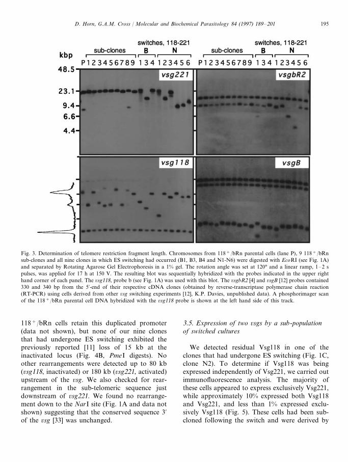
We detected residual Vsg118 in one of theclones that had undergone ES switching (Fig. 1C,clone N2). To determine if Vsg118 was beingexpressed independently of Vsg221, we carried outimmunofluorescence analysis. The majority ofthese cells appeared to express exclusively Vsg221,while approximately 10% expressed both Vsg118and Vsg221, and less than 1% expressed exclu-sively Vsg118 (Fig. 5). These cells had been sub-cloned following the switch and were derived by

D. Horn, G.A.M. Cross / Molecular and Biochemical Parasitology 84 (1997) 189–201196
selection in G418, indicating that a single G418-resistant cell could generate this heterogeneity.The accumulation of cells expressing exclusivelyVsg118 is possible because each resistant popula-
tion was sub-cloned and expanded in the absenceof drug (see Section 3.1). To determine the repro-ducibility of this phenomenon and to determine ifit is dependent upon selection in G418, we gener-ated further independent switches in each drug.Because we knew the switching frequency of 118+
/bRn cells was approximately 6×10⁻⁷, ratherthan sub-cloning parental cells, we placed 100cells (B0.1% expressing Vsg221 as determined byimmunofluorescence) in 20 10 ml flasks and addedphleomycin (1 mg ml−1) to ten and G418 (50 mgml−1) to the other ten when the density reachedapproximately 105 cells ml−1 (106 cells flask−1).We generated new switches so that we couldexamine cells as soon as possible after the switch(approximately 1 week or 20 generations, when106 drug-resistant cells were available: drug selec-tion was maintained throughout this period).
Immunofluorescence analysis indicated cells ex-pressing both Vsg118 and Vsg221 in one of threephleomycin and one of three G418-selected cul-tures, constituting approximately 1% of cells ineach culture. The appearance of cells expressingtwo Vsgs under phleomycin selection indicatesthat the ES promoter, rather than just the rRNApromoter, is active in the 6sg221 ES (see Fig. 1A).Cultures in which we did not detect cells express-ing both Vsgs were grown for a further ten gener-ations, but cells expressing both Vsgs remainedabsent or undetectable. It is possible that the‘founder’ cell for these heterogeneous cultures ex-pressed Vsg221 alone or both Vsg118 and Vsg221.Because cells expressing both Vsgs were moreabundant after longer periods of growth followingthe switch (1% after 20 generations and 10% afterapproximately 100 generations), we favor the hy-pothesis that the ‘founder’ cell expressed Vsg221alone. The accumulation of cells expressing bothVsgs is then easily explained by a modified versionof the Galton–Watson model (see [34], equation1), which was designed to measure mutation fre-quencies based on the accumulation of cells bear-ing a particular mutation. This equation is alsosuitable for measuring a phenotype switch notinvolving mutation, as many of the considerationsare the same for both situations. The model pre-dicts that the population of cells expressing twoVsgs should increase in a linear fashion with the
Fig. 4. Mapping upstream of regulated expression sites. (A)Physical maps spanning upstream of telomeric ESs containing6sg118 and 6sg221 as determined in 118+/bRn parental cells.ES and rRNA promoters are indicated as open and filledarrowheads respectively. Relevant restriction sites are shownand the abbreviations used for restriction enzymes are P, PmeIand R, RsrII. The scale indicates positions in kbp relative toeach 6sg start codon. (B) Chromosomal DNA from 118+/bRnparental cells (lane P) and all 9 clones in which ES switchinghad occurred (B1, B3, B4 and N1–N6) were digested withRsrII or PmeI and separated by Rotating Agarose Gel Elec-trophoresis in 1% gels. The rotation angle was set at 120°.Pulse field conditions for RsrII digested DNA were a linearramp, 2–15 s for 18 h at 150 V; and for PmeI digested DNAwas a linear ramp, 10–30 s for 20 h at 150 V. Both blots weresequentially hybridized with the probes indicated below thepanels. The 6sg118, probe a (see Fig. 1A) was used with theseblots.
D. Horn, G.A.M. Cross / Molecular and Biochemical Parasitology 84 (1997) 189–201 197
Fig. 5. Expression of two 6sgs by a sub-population of switched cultures. Fixed cells from the switched clone N2 were incubated withrat antiserum to Vsg221 and rabbit antiserum to Vsg118. Secondary antibodies were rhodamine-conjugated goat anti-rat andFITC-conjugated goat anti-rabbit. Pictures of the same field indicate cells expressing either Vsg221 (panel A) or Vsg118 (panel B,closed arrowheads) and a cell expressing both Vsgs (open arrowheads). A field containing an untypically high proportion ofdouble-expressing cells and a Vsg118 expressing cell was chosen for illustration.
number of generations, which is consistent withour observations. We used this model to estimatethe switch frequency (Vsg221 expression toVsg221/Vsg118 double-expression). Assumingthat cells do not switch back to expressing onlyVsg221, and that the growth rate of cells express-ing one or two Vsgs is not significantly different(see [35]), we derived a switch frequency of ap-proximately 2×10⁻³.
4. Discussion
Several DNA rearrangements have been foundin association with ES switching (reviewed in [8]).To determine if ES switching requires DNA rear-rangements or is controlled by an epigeneticmechanism, we generated a panel of clones inwhich the same ES switch has occurred. A searchfor rearrangements within and beyond the regu-
D. Horn, G.A.M. Cross / Molecular and Biochemical Parasitology 84 (1997) 189–201198
lated ESs, as well as an examination of changes intelomere length, indicated that antigenic variationcan occur without any of the previously reportedor any novel rearrangements. We also found thatES activation usually involves inactivation of thepreviously active ES, but that cells expressing two6sgs subsequently accumulate in some cultures.
We wanted to capture a panel of independentclones, all derived from the same clone, immedi-ately following a similar switching event (6sg118 to6sg221 expression). A selectable marker cassetteinserted into a repressed 6sg221 locus in 118+/bRn cells was exploited to generate clones inwhich selectable marker expression was activatedconcomitantly with 6sg221. This was achieved invitro and allowed accurate measurements ofswitching frequency. Antigenic variation in T. bru-cei, whether by recombinatorial mechanisms orotherwise, has been estimated to occur at a fre-quency ranging from 2.2×10⁻⁷ to 2.6×10⁻⁶ invitro [36]. These estimates were generated using aclone with the same active ES as that in 118+/bRncells. In our experiments, a specific switching eventoccurred at a frequency of 6×10⁻⁷, and weobtained a similar frequency using selection forneo or ble expression. From our panel of tenswitched clones, nine occurred via ES switching.We detected no examples of the independent acti-vation of the inserted rRNA promoter, suggestingthat the ES is regulated as a single unit and mayrepresent a chromatin domain, see [37]. Also, ifrepressed domains are assembled from the telom-ere, as in Saccharomyces cere6isiae, this result mayindicate continuous spreading of a repressed chro-matin structure along the ES (see [38]).
Changes in telomere length have been reportedto be associated with ES switching. ES inactiva-tion appeared to correlate with a reduction intelomere length while activation appeared to cor-relate with an increase in telomere length [30].Change in telomere length is probably due toaddition and deletion of T2AG3 repeats [39,40]. Inother organisms, telomeres appear to interact witheach other and with the nuclear envelope [41]. Anattractive model for ES switching is that the ESsinteract physically during the switch process. Amodel in which rearrangements between the twotelomeres leads to telomere end exchange is ap-
pealing. This does not appear to be the case,however, as we only see telomere loss and not asingle example of significant gain in telomerelength. Most if not all telomeres grow in T. brucei,with the active telomere growing faster and under-going progressive length reductions leading totelomere length heterogeneity within a clone[31,32]. Our panel of switched clones was derivedby sub-cloning 118+/bRn cells, from whichswitches were selected (switching may also beconsidered as sub-cloning) and subsequently sub-cloned again before generating DNA samples.Sub-cloning, and not ES switching, at points when6sg118 and 6sg221 were active, respectively, canexplain the TRF length heterogeneity found in theswitched clones. A cloning method involving heat-shock was reported to induce telomere shortening[31]. Our data show that changes in telomerelength may be accounted for by the dynamicnature of telomeres rather than necessarily beingassociated with ES switching or heat shock.
An ES with promoter repeats exhibited rear-rangement associated with inactivation [11,24] andactivation [12]. The rearrangement involved loss ofa tandemly repeated promoter via intra-chromoso-mal homologous recombination [11,42]. This rear-rangement, however, is not necessary forregulation of transcription as these promoters canexist in an active or silent ES with single ortandem copies [11,12,24]. Similar rearrangementsalso occur independently of ES switching [12]. Wedetected no rearrangement in the promoter regionof the same ES following inactivation of this ES innine independent examples. Also, no rearrange-ments were detected in related ESs following ESswitching [43]. Activation of an ES has beenassociated with another rearrangement upstreamof the promoter, within 50 bp repeat sequences[24]. The resolution of our gels allowed detectionof insertions or deletions of greater than 5 kb inthis region, and we detected no such rearrange-ments.
The relationship between some rearrangementsand 6sg switching, in cases in which a smallnumber of samples were examined, may be inci-dental. Other rearrangements, in the promoterregion [11,12,24,42], do appear to be associatedwith some ES switching events, however. These

D. Horn, G.A.M. Cross / Molecular and Biochemical Parasitology 84 (1997) 189–201 199
rearrangements may be sufficient but not obliga-tory for switching. Alternatively, these rearrange-ments may occur at a relatively high frequencyand subsequently influence the switching fre-quency of a particular ES. If, for example, telom-ere length influences the frequency of ESswitching, it is possible that telomere growth andbreakage could re-program the likely order of ESswitching.
Transient [44] and stable [45] transfection inbloodstream and procyclic-form T. brucei indi-cates the presence of factors necessary for tran-scription initiation at multiple ES promoters inboth life-cycle stages. Despite the presence ofmultiple ESs, 6sg expression is usually mutuallyexclusive in bloodstream-form cells. The discoveryof trypanosomes in which two 6sg ESs were stablytranscribed [13,14] suggested that ESs can switchon and off independently and that it was theimmune response that usually eliminates cells ex-pressing both pre- and post-switch Vsgs. Whenselecting for 6sg switching by drug selection ratherthan by immune selection, survival is not depen-dent upon inactivation of the previously expressed6sg. In our experiments, ES inactivation accompa-nied ES activation in nine independent examplesin the absence of immune selection. These resultssuggest that the activation/inactivation process isusually coupled and occurs as a coordinated eventor series of events. An examination of individualcells, however, did reveal the presence of cellsexpressing both Vsgs in some of our switchedcultures. Our results suggest that a mutually ex-clusive switch is efficiently established and rela-tively efficiently maintained, but that repression atthe inactivated locus is lost at a frequency of2×10−3 following some switch events. This pre-disposition toward generating cells expressing twoVsgs is a heritable phenotype, which is maintainedafter sub-cloning, as indicated by the switchedclone N2. In addition, some of these cells switchback to exclusive expression of the previouslyactive Vsg, at what appears to be a similarly highfrequency. Taken together, these observationsshow that ES activation/inactivation is clearly acoupled event but that the machinery maintainingall but one ES in a repressed state can be compro-mised in a heritable fashion (possibly during the
establishment step), which allows the activation/inactivation event to become uncoupled.
Isogenic cells can exist in mitotically stable yetphenotypically distinct epigenetic states (reviewedin [46]). Our results suggest that an epigeneticmechanism, and not DNA alteration, controls EStranscription in bloodstream-form T. brucei. Thishypothesis is supported by several lines of evidence.ES activation occurs in conjunction with ES inac-tivation and no detectable DNA rearrangementsare necessary for ES switching (this paper). Inacti-vation and reactivation of metacyclic [47] andbloodstream-form 6sgs [48], during the life cycle, isthought to occur in the absence of DNA rearrange-ments. Based on these results, we cannot rule outthat small undetected DNA rearrangements may bespecifically associated with ES switching. If sponta-neous DNA mutations were responsible for switch-ing, however, each phenotype would be expected tobe extremely stable. Mitotically metastable pheno-types have been demonstrated in early blood-stream-form infections, when metacyclic 6sgs (6sgsactivated in the insect salivary gland, see[49]) areexpressed [50] and, as demonstrated in this paper,in some clones expressing 6sgs from bloodstream-form ESs. These 6sg phenotypes switch at a signifi-cantly faster rate (2.7×10⁻² and 2×10⁻³,respectively) than would be expected by sponta-neous mutation. Repression is position-dependentand promoter-independent [5,6], suggesting that itis mediated by an altered chromatin structure (see[46]). 6sg ES switching appears to occur in astochastic manner, as does activation of a particu-lar ES from the metacyclic [49,51] and blood-stream-form [52] sub-set during the life cycle. Wetherefore suggest that an epigenetic mechanismcontrols 6sg activation and switching in metacyclicand bloodstream-form cells.
Acknowledgements
This work was supported by the National Insti-tutes of Health (grant AI 21729). We thankKelvin Davies (Rockefeller University) forproviding a 6sgbR2 cDNA clone, Miguel Navarro(Rockefeller University) for providing a 6sgBcDNA clone and Elizabeth Wirtz for critical read-ing of the manuscript.

D. Horn, G.A.M. Cross / Molecular and Biochemical Parasitology 84 (1997) 189–201200
References
[1] Vanhamme, L. and Pays, E. (1995) Control of geneexpression in trypanosomes. Microbiol. Rev. 59, 223–240.
[2] Cross, G.A.M. (1996) Antigenic variation in try-panosomes: secrets surface slowly. Bioessays 18, 283–291.
[3] Van der Ploeg, L.H.T., Valerio, D., De Lange, T.,Bernards, A., Borst, P. and Grosveld, F. G. (1982) Ananalysis of cosmid clones of nuclear DNA from Try-panosoma brucei shows that the genes for variant surfaceglycoproteins are clustered in the genome. Nucleic AcidsRes. 10, 5905–5923.
[4] Liu, A.Y., Michels, P.A., Bernards, A. and Borst, P.(1985) Trypanosome variant surface glycoprotein genesexpressed early in infection. J. Mol. Biol. 175, 383–386.
[5] Horn, D. and Cross, G.A.M. (1995) A developmentallyregulated position effect at a telomeric locus in Try-panosoma brucei. Cell 83, 555–561.
[6] Rudenko, G., Blundell, P.A., Dirks–Mulder, A., Kieft,R. and Borst, P. (1995) A ribosomal DNA promoterreplacing the promoter of a telomeric VSG gene expres-sion site can be efficiently switched on and off in T.brucei. Cell 83, 547–553.
[7] Borst, P. and Greaves, D.R. (1987) Programmed generearrangements altering gene expression. Science 235,658–667.
[8] Pays, E., Vanhamme, L. and Berberof, M. (1994) Geneticcontrols for the expression of surface antigens in Africantrypanosomes. Ann. Rev. Microbiol. 48, 25–52.
[9] Cully, D.F., Ip, H.S. and Cross, G.A. (1985) Coordinatetranscription of variant surface glycoprotein genes and anexpression site associated gene family in Trypanosomabrucei. Cell 42, 173–182.
[10] Zomerdijk, J.C.B.M., Ouellete, M., ten Asbroek,A.L.M.A., Kieft, R., Bommer, A.M.M., Clayton, C.E.and Borst, P. (1990) The promoter for a variant surfaceglycoprotein gene expression site in Trypanosoma brucei.EMBO J. 9, 2791–2801.
[11] Gottesdiener, K., Chung, H.-M., Brown, S.D., Lee, M.G.-S. and Van der Ploeg, L.H.T. (1991) Characterizationof VSG gene expression site promoters and promoter-as-sociated DNA rearrangement events. Mol. Cell. Biol. 11,2467–2480.
[12] Navarro, M. and Cross, G. A. M. (1996) DNA rearrange-ments associated with multiple consecutive directed anti-genic switches in Trypanosoma brucei. Mol. Cell. Biol. 16,3615–3625.
[13] Cornelissen, A.W., Kooter, P.J., Johnson, J.M., Van derPloeg, L.H.T. and Borst, P. (1985) Two simultaneouslyactive VSG gene transcription units in a single Try-panosoma brucei variant. Cell 41, 825–832.
[14] Baltz, T., Giroud, C., Baltz, D., Roth, C., Raibaud, A.and Eisen, H. (1986) Stable expression of two variablesurface glycoproteins by cloned Trypanosoma equiperdum.Nature 319, 602–604.
[15] Bernards, A., De Lange, T., Michels, P.A.M., Liu, A.
Y.C., Huisman, M.J. and Borst, P. (1984) Two modes ofactivation of a single surface antigen of Trypanosomabrucei. Cell 36, 163–170.
[16] Michels, P.A.M., Van der Ploeg, L.H.T., Liu, A.Y.C. andBorst, P. (1984) The inactivation and reactivation of anexpression-linked gene copy for a variant surface glyco-protein in Trypanosoma brucei. EMBO J. 3, 1345–1351.
[17] Cross, G.A.M. (1975) Identification, purification andproperties of variant-specific glycoprotein antigens consti-tuting the surface coat of Trypanosoma brucei. Parasitol-ogy 71, 393–417.
[18] Doyle, J.J., Hirumi, H., Hirumi, K., Lupton, E.N. andCross, G.A.M. (1980) Antigenic variation in clones ofanimal-infective Trypanosoma brucei derived and main-tained in vitro. Parasitology 80, 359–369.
[19] Sambrook, J., Fritsch, E.F. and Maniatis, T. (1989)Molecular Cloning: A Laboratory Manual, 2nd edn.,Cold Spring Harbor Laboratory, Cold Spring Harbor,New York.
[20] Zamze, S.E., Ferguson, M.A.J., Collins, R., Dwek, R.A.and Rademacher, T.W. (1988) Characterization of thecross-reacting determinant (CRD) of the glycosyl-phos-phatidylinositol membrane anchor of Trypanosoma bruceivariant surface glycoprotein. Eur. J. Biochem. 176, 527–534.
[21] Crozatier, M., Van der Ploeg, L.H.T., Johnson, P.J.,Gommers-Ampt, J. and Borst, P. (1990) Structure of atelomeric expression site for variant specific surface anti-gens in Trypanosoma brucei. Mol. Biochem. Parasitol. 42,1–12.
[22] Johnson, P.J., Kooter, J.M. and Borst, P. (1987) Inactiva-tion of transcription by UV irradiation of T. bruceiprovides evidence for a multicistronic transcription unitincluding a VSG gene. Cell 51, 273–281.
[23] Kooter, J.M., van der Spek, H.J., Wagter, R., d’Oliveira,C.E., van der Hoeven, F., Johnson, P.J. and Borst, P.(1987) The anatomy and transcription of a telomericexpression site for variant-specific surface antigens in T.brucei. Cell 51, 261–272.
[24] Zomerdijk, J.C.B.M., Kieft, R., Duyndam, M., Shiels,P.G. and Borst, P. (1991) Antigenic variation in Try-panosoma brucei : A telomeric expression site for variant-specific surface glycoprotein genes with novel features.Nucleic Acids Res. 19, 1359–1368.
[25] Hesse, F., Selzer, P.M., Muhlstadt, K. and Duszenko, M.(1995) A novel cultivation technique for long-termmaintenance of bloodstream form trypanosomes in vitro.Mol. Biochem. Parasitol. 70, 157–166.
[26] Drocourt, D., Calmels, T., Reynes, J., Baron, M. andTiraby, G. (1990) Cassettes of the Streptoalloteichus hin-dustanus ble gene for transformation of lower and highereukaryotes to phleomycin resistance. Nucleic Acids Res.18, 4009.
[27] Bernards, A., Kooter, J.M. and Borst, P. (1985) Structureand transcription of a telomeric surface antigen gene ofTrypanosoma brucei. Mol. Cell. Biol. 5, 545–553.
[28] Shah, J.S., Young, J.R., Kimmel, B.E., Iams, K.P. andWilliams, R.O. (1987) The 5% flanking sequence of a

D. Horn, G.A.M. Cross / Molecular and Biochemical Parasitology 84 (1997) 189–201 201
Trypanosoma brucei variable surface glycoprotein gene.Mol. Biochem. Parasitol. 24, 163–174.
[29] Van der Ploeg, L., Smith, C.L., Polvere, R.I. and Gottes-diener, K.M. (1989) Improved separation of chromo-some-sized DNA from Trypanosoma brucei, stock 427-60.Nucleic Acids Res. 17, 3217–3227.
[30] Myler, P.J., Aline, R.F.J., Scholler, J.K. and Stuart, K.D.(1988) Changes in telomere length associated with anti-genic variation in Trypanosoma brucei. Mol. Biochem.Parasitol. 29, 243–250.
[31] Bernards, A., Michels, P.A.M., Lincke, C.R. and Borst,P. (1983) Growth of chromosome ends in multiplyingtrypanosomes. Nature 303, 592–597.
[32] Pays, E., Laurent, M., Delinte, K., van Meirvenne, N.and Steinert, M. (1983) Differential size variations be-tween transcriptionally active and inactive telomeres ofTrypanosoma brucei. Nucleic Acids Res. 11, 8137–8147.
[33] Aline, R.F. and Stuart, K.D. (1989) Trypanosoma brucei :conserved sequence organisation 3% to telomeric variantsurface glycoprotein genes. Exp. Parasitol. 68, 57–66.
[34] Rossman, T.G., Goncharova, E.I. and Nadas, A. (1995)Modeling and measurement of the spontaneous mutationrate in mammalian cells. Mutat. Res. 328, 21–30.
[35] Munoz-Jordan, J.L., Davies, K.P. and Cross, G.A.M.(1996) Stable expression of mosaic coats of variant sur-face glycoproteins in Trypanosoma brucei. Science 272,1795–1797.
[36] Lamont, G.S., Tucker, R.S. and Cross, G.A.M. (1986)Analysis of antigen switching rates in Trypanosoma bru-cei. Parasitology 92, 355–367.
[37] Dillon, N. and Grosveld, F. (1994) Chromatin domains aspotential units of eukaryotic gene function. Curr. Opin.Genet. Dev. 4, 260–264.
[38] Renauld, H., Aparicio, O.M., Zierath, P.D., Billington,B.L., Chhablani, S.K. and Gottschling, D.E. (1993) Silentdomains are assembled continuously from the telomereand are defined by promoter distance and strength, andby SIR3 dosage. Genes Dev. 7, 1133–1145.
[39] Blackburn, E.H. and Challoner, P.B. (1984) Identificationof a telomeric DNA sequence in Trypanosoma brucei. Cell36, 447–457.
[40] Van der Ploeg, L.H.T., Liu, A.Y.C. and Borst, P.(1984)Structure of the growing telomeres of trypanosomes. Cell36, 459–468.
[41] Gilson, E., Laroche, T. and Gasser, S.M. (1993) Telom-eres and the functional architecture of the nucleus. TrendsCell Biol. 3, 128–134.
[42] Gottesdiener, K.M., Goriparthi, L., Masucci, J.P. andVan der Ploeg, L.H.T. (1992) A proposed mechanism for
promoter-associated DNA rearrangement events at avariant surface glycoprotein gene expression site. Mol.Cell. Biol. 12, 4784–4795.
[43] Lodes, M.J., Smiley, B.L., Stadnyk, A.W., Bennett, J.L.,Myler, P.J. and Stuart, K. (1993) Expression of a retropo-son-like sequence upstream of the putative Trypanosomabrucei variant surface glycoprotein gene expression sitepromoter. Mol. Cell. Biol. 13, 7036–7044.
[44] Jefferies, D., Tebabi, P. and Pays, E. (1991) Transientactivity assays of the Trypanosoma brucei variant surfaceglycoprotein gene promoter: Control of gene expressionat the post-transcriptional level. Mol. Cell. Biol. 11, 338–343.
[45] Biebinger, S., Rettenmaier, S., Flaspohler, J., Hartmann,C., Pena-Diaz, J., Wirtz, L.E., Hotz, H.R., Barry, J.D.and Clayton, C. (1996) The PARP promoter of Try-panosoma brucei is developmentally regulated in a chro-mosomal context. Nucleic Acids Res. 24, 1202–1211.
[46] Loo, S. and Rine, J. (1995) Silencing and heritable do-mains of gene expression. Ann. Rev. Cell Dev. Biol. 11,519–548.
[47] Graham, S.V. and Barry, J.D. (1995) Transcription regu-lation of metacyclic variant surface glycoprotein geneexpression during the life cycle of Trypanosoma brucei.Mol. Cell. Biol. 15, 5945–5956.
[48] Delauw, M.-F., Pays, E., Steinert, M., Aerts, D., VanMeirvenne, N. and LeRay, D. (1985) Inactivation andreactivation of a variant-specific antigen gene in cyclicallytransmitted Trypanosoma brucei. EMBO J. 4, 989–993.
[49] Tetley, L., Turner, C.M.R., Barry, J.D., Crowe, J.S. andVickerman, K. (1987) Onset of expression of the variantsurface glycoproteins of Trypanosoma brucei in the tsetsefly studied using immunoelectron microscopy. J. Cell Sci.87, 363–372.
[50] Esser, K.M. and Schoenbechler, M.J. (1985) Expressionof two variant surface glycoproteins on individual Africantrypanosomes during antigen switching. Science 229,190–193.
[51] Hajduk, S.L., Cameron, C.R., Barry, J.D. and Vicker-man, K. (1981) Antigenic variation in cyclically transmit-ted Trypanosoma brucei. Variable antigen typecomposition of metacyclic trypanosome populations fromthe salivary glands of Glossina morsitans. Parasitology 83,595–607.
[52] Hajduk, S.L. and Vickerman, K. (1981) Antigenic varia-tion in cyclically transmitted Trypanosoma brucei. Vari-able antigen type composition of the first parasitaemia inmice bitten by trypanosome-infected Glossina morsitans.Parasitology 83, 609–621.
.