ANALYSIS OF PULMONARY FIBROSIS IN MRI ... -...
31
ANALYSIS OF PULMONARY FIBROSIS IN MRI, USING AN ELASTIC REGISTRATION TECHNIQUE IN A MODEL OF FIBROSIS: Scleroderma ORAL DEFENSE 8 th of September 2017 Charlotte MARTIN Supervisor: Pr. MP REVEL M2 Bio Medical Imaging Subtract: Imaging from molecular to human 2016-2017 1
Transcript of ANALYSIS OF PULMONARY FIBROSIS IN MRI ... -...
ANALYSIS OF PULMONARY FIBROSIS IN MRI, USINGAN ELASTIC REGISTRATION TECHNIQUE IN A MODELOF FIBROSIS: Scleroderma
ORAL DEFENSE 8 th of September 2017
Charlotte MARTINSupervisor: Pr. MP REVEL
M2 Bio Medical ImagingSubtract: Imaging from molecular to human
2016-2017
1
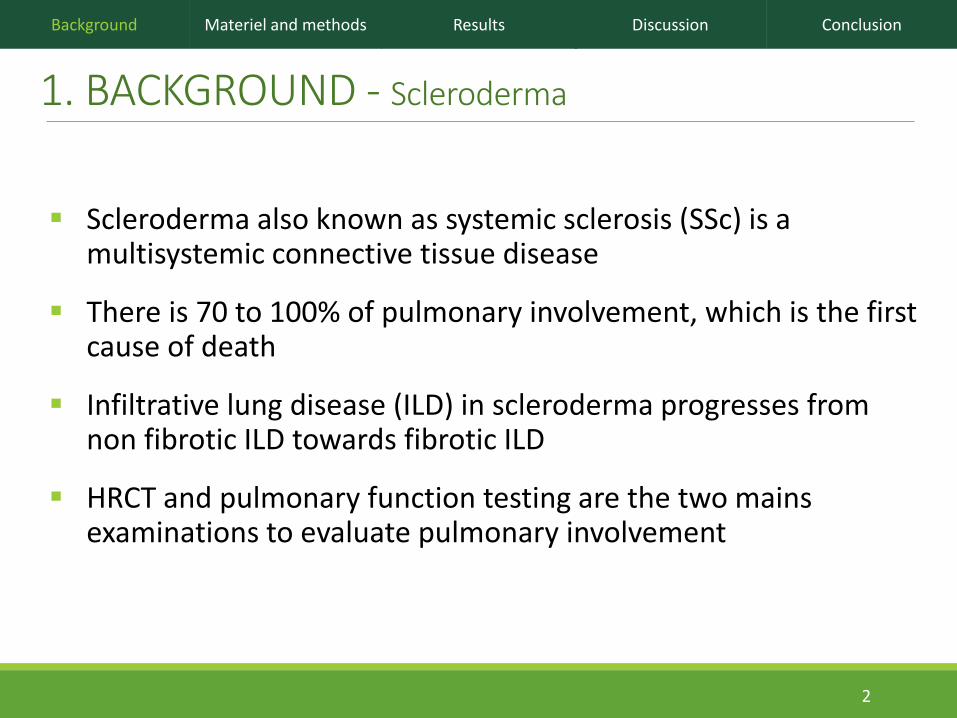
1. BACKGROUND - Scleroderma
Scleroderma also known as systemic sclerosis (SSc) is a multisystemic connective tissue disease
There is 70 to 100% of pulmonary involvement, which is the first cause of death
Infiltrative lung disease (ILD) in scleroderma progresses from non fibrotic ILD towards fibrotic ILD
HRCT and pulmonary function testing are the two mains examinations to evaluate pulmonary involvement
Background Materiel and methods Results Discussion Conclusion
2

1. BACKGROUND – Scleroderma evaluation
HRCT :• Has high spatial and contrast resolution
• Provides radiation exposure and no functional information
Background Materiel and methods Results Discussion Conclusion
3
No ILD ILD with FibrosisILD without fibrosis
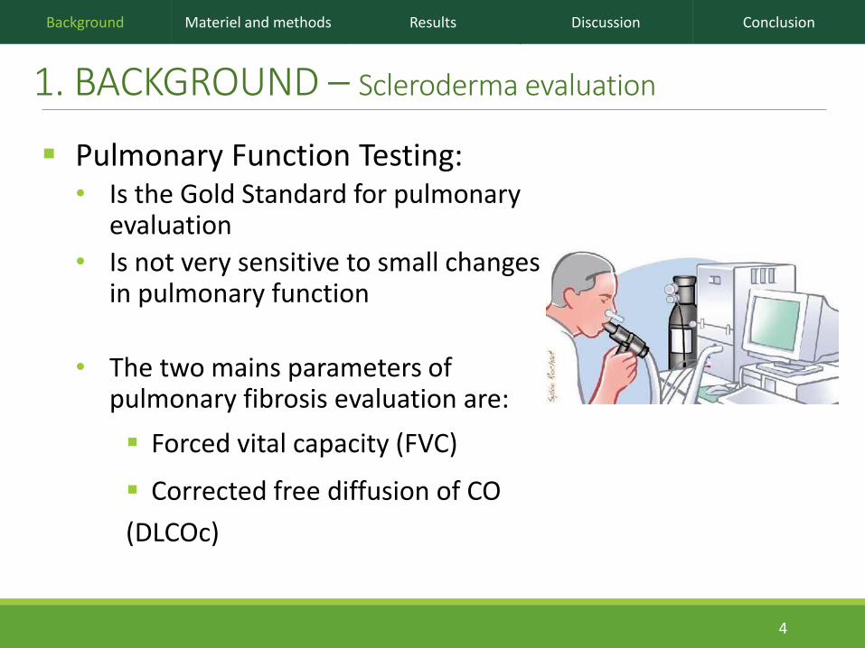
1. BACKGROUND – Scleroderma evaluation
Pulmonary Function Testing:• Is the Gold Standard for pulmonary
evaluation
• Is not very sensitive to small changes in pulmonary function
• The two mains parameters of pulmonary fibrosis evaluation are:
Forced vital capacity (FVC)
Corrected free diffusion of CO
(DLCOc)
Background Materiel and methods Results Discussion Conclusion
4

1. BACKGROUND - Lung MRI
MRI of lung parenchyma is not currently used in clinical practice, due to:• Lack of signal (low H+ proton density and short T2* relaxation time)
• Important artifacts (heterogeneity of the local magnetic field and respiratory movements)
New MR sequences named UTE (ultra-short time of echo) have been developed (1):• TE < 0.5 msec
• It allows minimizing the T2* signal decay and obtaining images from the lung parenchyma with sufficient signal intensity
Background Materiel and methods Results Discussion Conclusion
5(1) Holmes JE, Bydder GM. MR imaging with ultrashort TE (UTE) pulse sequences: Basic principles.
Radiography. 1 août 2005;11(3):163-74

OBJECTIVES
To evaluate the feasibility of an elastic registration method based on MR images for the detection of pulmonary fibrosis in patients with scleroderma
Background Materiel and methods Results Discussion Conclusion
6
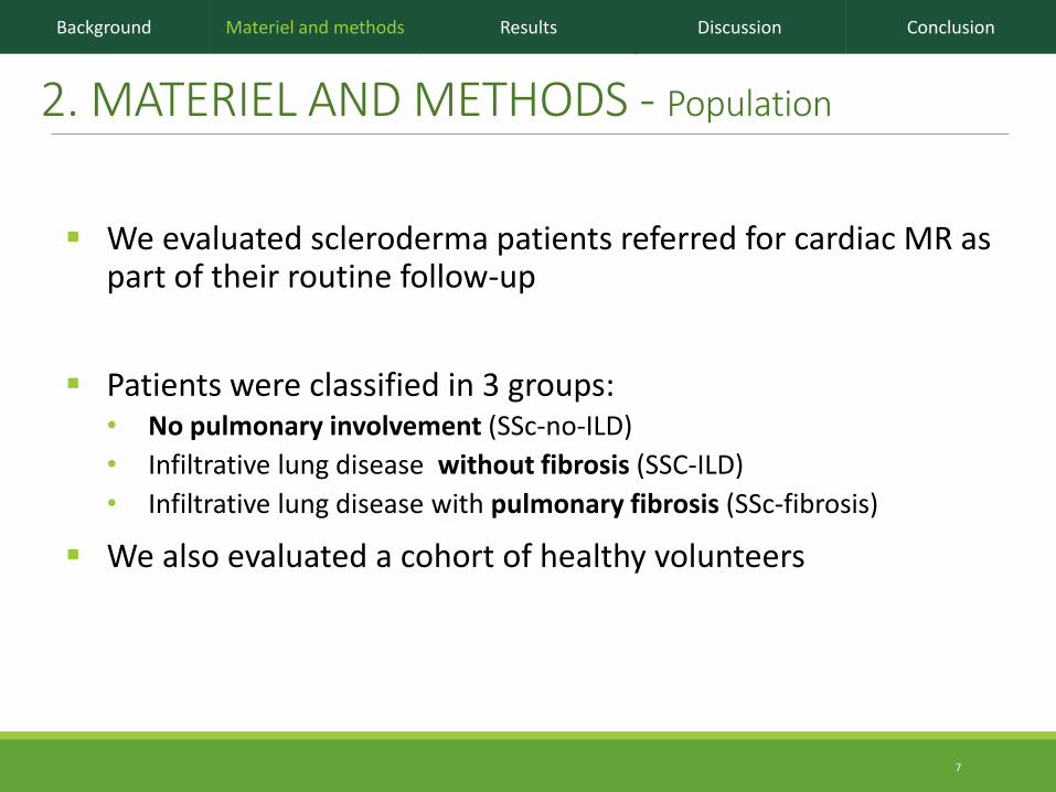
2. MATERIEL AND METHODS - Population
We evaluated scleroderma patients referred for cardiac MR as part of their routine follow-up
Patients were classified in 3 groups:• No pulmonary involvement (SSc-no-ILD)
• Infiltrative lung disease without fibrosis (SSC-ILD)
• Infiltrative lung disease with pulmonary fibrosis (SSc-fibrosis)
We also evaluated a cohort of healthy volunteers
Background Materiel and methods Results Discussion Conclusion
7

Two UTE sequences named spiral VIBE (volumetric Interpolated Breath-hold Examination) were performed, one at the end of full inspiration and another after full expiration on a 3T-unit
3D sequences, acquired on the coronal plane
Voxel size 2.14 x 2.14 x 2.5 mm
Echo time : 0.05ms
2. MATERIEL AND METHODS – MR examination
Background Materiel and methods Results Discussion Conclusion
8
Inspiration Expiration

2. MATERIEL AND METHODS – MR segmentation
Background Materiel and methods Results Discussion Conclusion
We created lung masks for inspiratory and expiratory volumes
9

We used an elastic registration algorithm (1)
We chose to make the registration from inspiratory to expiratory images• Because inspiratory images contain
more information
Registration was helped by the pre-processing masks
2. MATERIEL AND METHODS – Elastic registration
Background Materiel and methods Results Discussion Conclusion
(1) Glocker B, Sotiras A, Komodakis N, Paragios N. Deformable Medical Image Registration: Setting the State of the Art with Discrete Methods. Annu Rev Biomed Eng. 2011;13(1):219-44. 10
Inspiratory+Expiratory
Expiratory+Registered

The first step was to evaluate the validity of the elastic registration algorithm
11 anatomic landmarks were manually placed by one observer on inspiratory images
Then, the same landmarks were placed on expiratory images by:
• the same observer (manually)
• another independent observer (manually)
• the algorithm (registered)
2. MATERIEL AND METHODS – Evaluation of elastic registration
Background Materiel and methods Results Discussion Conclusion
11
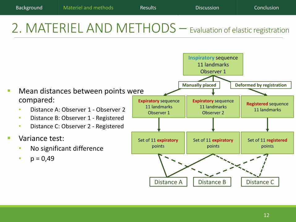
Mean distances between points were compared:• Distance A: Observer 1 - Observer 2
• Distance B: Observer 1 - Registered
• Distance C: Observer 2 - Registered
Variance test: • No significant difference
• p = 0,49
2. MATERIEL AND METHODS – Evaluation of elastic registration
Background Materiel and methods Results Discussion Conclusion
12
Inspiratory sequence11 landmarks
Observer 1
Expiratory sequence11 landmarks
Observer 1
Expiratory sequence11 landmarks
Observer 2
Registered sequence11 landmarks
Set of 11 expiratorypoints
Set of 11 expiratory points
Set of 11 registeredpoints
Manually placed Deformed by registration
Distance A Distance B Distance C

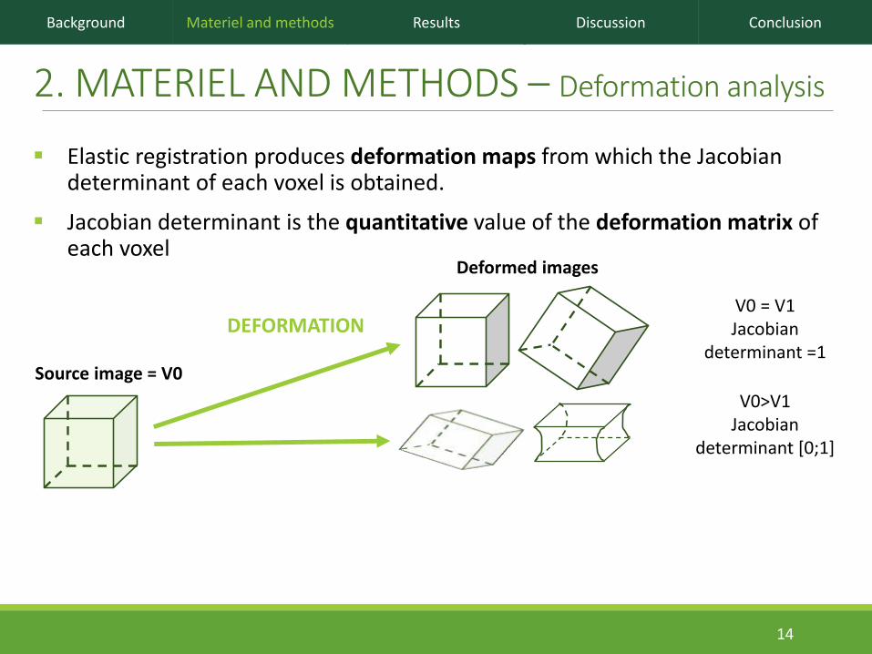
Elastic registration produces deformation maps from which the Jacobian determinant of each voxel is obtained.
Jacobian determinant is the quantitative value of the deformation matrix of each voxel
2. MATERIEL AND METHODS – Deformation analysis
Background Materiel and methods Results Discussion Conclusion
Deformed images
DEFORMATION
13
V0 = V1Jacobian
determinant =1Source image = V0
Elastic registration produces deformation maps from which the Jacobian determinant of each voxel is obtained.
Jacobian determinant is the quantitative value of the deformation matrix of each voxel
2. MATERIEL AND METHODS – Deformation analysis
Background Materiel and methods Results Discussion Conclusion
Deformed images
DEFORMATION
14
V0>V1Jacobian
determinant [0;1]
V0 = V1Jacobian
determinant =1Source image = V0

Elastic registration produces deformation maps from which the Jacobian determinant of each voxel is obtained.
Jacobian determinant is the quantitative value of the deformation matrix of each voxel
2. MATERIEL AND METHODS – Deformation analysis
Background Materiel and methods Results Discussion Conclusion
Deformed images
DEFORMATION
V0<V1Jacobian
determinant > 1
15
V0>V1Jacobian
determinant [0;1]
V0 = V1Jacobian
determinant =1Source image = V0
2. MATERIEL AND METHODS – Deformation analysis
Background Materiel and methods Results Discussion Conclusion
16
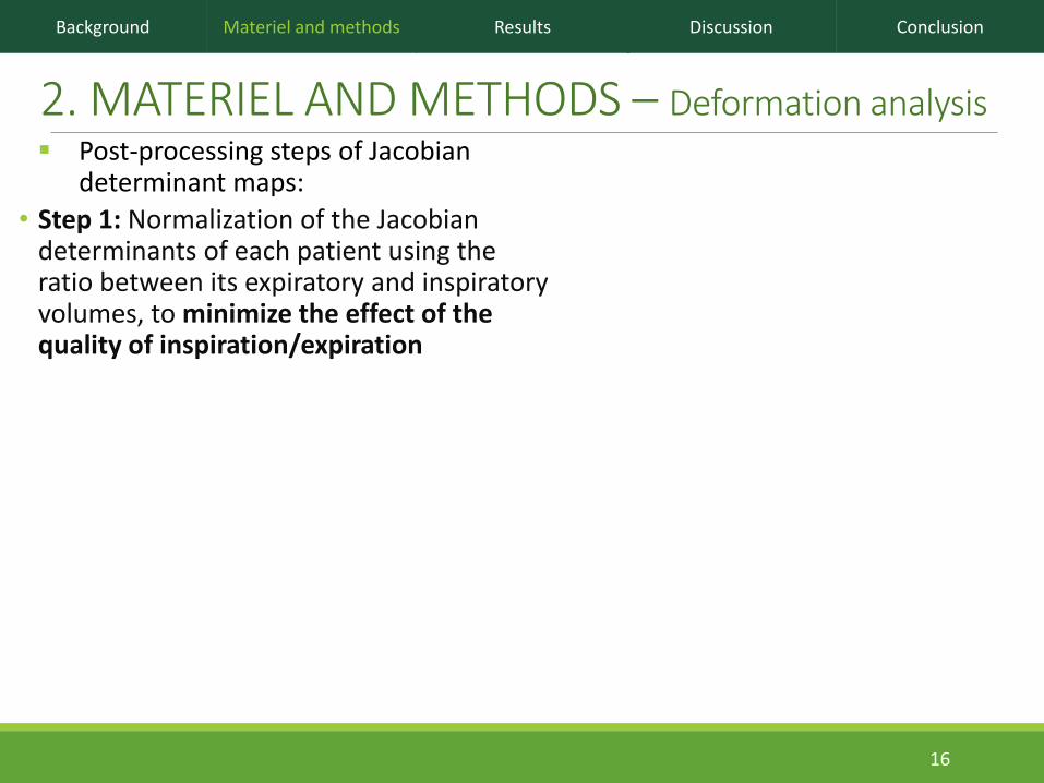
Post-processing steps of Jacobian determinant maps:
• Step 1: Normalization of the Jacobian determinants of each patient using the ratio between its expiratory and inspiratory volumes, to minimize the effect of the quality of inspiration/expiration

2. MATERIEL AND METHODS – Deformation analysis
Background Materiel and methods Results Discussion Conclusion
17
Post-processing steps of Jacobian determinant maps:
• Step 1: Normalization of the Jacobian determinants of each patient using the ratio between its expiratory and inspiratory volumes
• Step 2: Mapping to a common template (expiratory mask for a healthy volunteer) through elastic registration of the binary masks of the lung
Patient 1 Patient 4 Patient 29
Common mask

2. MATERIEL AND METHODS – Deformation analysis
Background Materiel and methods Results Discussion Conclusion
18
Patient 1 Patient 4 Patient 29
y axis
x axis
z axis
Post-processing steps of Jacobian determinant maps:
• Step 1: Normalization of the Jacobian determinants of each patient using the ratio between its expiratory and inspiratory volumes
• Step 2: Mapping to a common template (expiratory mask for a healthy volunteer) through elastic registration of the binary masks of the lung
• Step 3: For each patient, calculation of the mean of the Jacobian determinant along each axis (x; y; z)

2. MATERIEL AND METHODS – Deformation analysis
Background Materiel and methods Results Discussion Conclusion
SSc-Fibrosis=7
HealthyVolunteersn=11
SSc-ILDn=5
SSc-no-ILDn=9
19
Post-processing steps of Jacobian determinant maps:
• Step 1: Normalization of the Jacobian determinants of each patient using the ratio between its expiratory and inspiratory volumes
• Step 2: Mapping to a common template (expiratory mask for a healthy volunteer) through elastic registration of the binary masks of the lung
• Step 3: For each patient, calculation of the mean of the Jacobian determinant along each axis (x; y; z)
• Step 4: For each group of patients
Calculation of the mean of the Jacobian determinant for each voxel and representation with 3D color maps
Calculation of the mean of the Jacobian determinant along each axis.

2. MATERIEL AND METHODS – Quantitative analysis
Healthy volunteers group
Background Materiel and methods Results Discussion Conclusion
We isolated lung regions with the most important positive values of the Jacobian determinant logarithm (= areas with the most important positive deformation), using a threshold of 0.15 :
• In the healthy volunteers group
• And for each patient
• We calculated similarity index, Dice coefficient, between the healthy volunteers group and each patient
20

3. RESULTS - Population
33 subjects were included:• 11 healthy volunteers
• 22 patients
1 patient was excluded of the analysis because of poor quality of examination due to a misunderstanding of breathing instructions, so 32 subjects were evaluated
Total HealthyVolunteers SSc-no-ILD SSc-ILD SSc-Fibrosis pvalue
N=32 N=11 N=9 N=5 N=7
Gender 0,005
Female 19(59%) 2(18%) 7(78%) 5(100%) 5(71%)
Male 13(41%) 9(82%) 2(22%) 0(0%) 2(29%)
Age(years) 0,001
Mean(SD) 45,6(19,5) 29,1(9,3) 59(17,5) 43,6(14,7) 60(16,3)
BMI 0,79
Mean(SD) 21,9(2,5) 21,8(1,6) 22,6(2,3) 21,1(3,9) 21,8(2,9)
Smokers 7(21%) 3(27%) 4(44%) 0(0%) 3(42%) 0,042
N=21 N=9 N=5 N=7
FVC 0,012
Mean(SD) 2,76(1,47) 3,2(1,16) 2,83(0,24) 1,94(0,86)
DLCO 0,001
Mean(SD) 60(20) 67(14) 66(27) 38(6)
Background Materiel and methods Results Discussion Conclusion
21

Maps of the mean value logarithm of Jacobian determinant for each group of patients show:
• Differences in the repartition of the Jacobian determinant in each group
• In healthy volunteers, the most important stretching (red color) in the posterior part of lung bases
• In the corresponding areas, the lungs of SSc-fibrosis patients show only few changes, with Jacobian determinant near to 0 (green color)
SSc-Fibrosis=7
HealthyVolunteersn=11
SSc-ILDn=5
SSc-no-ILDn=9
3. RESULTS – Qualitative analysis
Background Materiel and methods Results Discussion Conclusion
22

3. RESULTS – Quantitative analysis
Disease + Disease -
Test + 11 5 16
Test - 1 15 16
12 20 32
Se = 91% Sp = 75%
A whole cohort analysis was performed with:• Disease+: SSc-fibrosis + SSc-ILD
• Disease-: SSc-no-ILD + Healthy Volunteers
0
0,1
0,2
0,3
0,4
0,5
0,6
0,7
0,8
0,9
1
0 0,2 0,4 0,6 0,8 1
Fra
cti
on
of
true
po
sit
ive
Fraction of false positive
ROCcurve/AUC=0.802
Background Materiel and methods Results Discussion Conclusion
23
Dice coefficient Best cut-off : 0.44

3. RESULTS – Quantitative analysis
A sub-group analysis was also performed:• Disease+: SSc-fibrosis
• Disease-: Healthy volunteers
Background Materiel and methods Results Discussion Conclusion
0
0,1
0,2
0,3
0,4
0,5
0,6
0,7
0,8
0,9
1
0 0,2 0,4 0,6 0,8 1
Fra
cti
on
of
true
po
sit
ive
Fraction of false positive
ROCcurve/AUC=0,929
Disease + Disease -
Test + 6 0 6
Test - 1 11 12
7 11 18
Se = 86% Sp = 100%
24
Dice coefficientBest cut-off : 0.44

3. RESULTS – Correlation with PFT DLCOc could not be evaluated for 2 patients due to severity of
disease
Analysis was performed :• FVC: n=21
• DLCOc: n=19
Background Materiel and methods Results Discussion Conclusion
0
0,1
0,2
0,3
0,4
0,5
0,6
0 1 2 3 4 5 6
Dic
e in
de
x
FCV
R= 0.12; p= 0.6
0
0,1
0,2
0,3
0,4
0,5
0,6
0 20 40 60 80 100 120 140
Dic
e c
oe
ffic
ien
t
DLCOc
R= 0.16; p= 0.5
25

4. DISCUSSION
TO SUMMARIZE OUR FINDINGS
Feasibility of an elastic registration algorithm applied on MR inspiratory and expiratory sequences
Visual differences in Jacobian maps between subjects with and without ILD
Distinction between healthy and fibrotic subject with acceptable sensitivity and perfect specificity
No correlation between PFT and our method of evaluation of fibrosis
26
Background Materiel and methods Results Discussion Conclusion

4. DISCUSSION
OUR LIMITATIONS
Small size of the cohort
No evaluation of repeatability
Subjective choice of threshold for Dice coefficient
27
Background Materiel and methods Results Discussion Conclusion

4. DISCUSSION
OUR PERSPECTIVES
Evaluation on a larger cohort
Quantitative analysis of Jacobian determinant repartition analysis with no subjective threshold:• Principal component analysis of the whole volume
• Comparison of joint entropy
Application to other types of fibrosis
28
Background Materiel and methods Results Discussion Conclusion

5. CONCLUSION
First study to assess ILD using an elastic registration between inspiratory and expiratory lung MR images
Lung deformation pattern was different between healthy subjects and scleroderma patients with pulmonary fibrosis
This functional approach of lung deformation based on elastic registration allows diagnosis of pulmonary fibrosis with an acceptable sensitivity and a perfect specificity, and no radiation exposure
Background Materiel and methods Results Discussion Conclusion
29

30
Thank you for your attention
Background Materiel and methods Results Discussion Conclusion

BIBLIOGRAPHIE• Giacomelli, R. et al. Interstitial lung disease in systemic sclerosis: current and future treatment.
Rheumatol. Int. 37, 853–863 (2017).
• Bastos, A. de L., Corrêa, R. de A. & Ferreira, G. A. Tomography patterns of lung disease in systemic sclerosis. Radiol. Bras. 49, 316–321 (2016).
• Behr, J. & Furst, D. E. Pulmonary function tests. Rheumatology 47, v65–v67 (2008).
• Qian, Y. & Boada, F. E. Acquisition-weighted stack of spirals for fast high-resolution three-dimensional ultra-short echo time MR imaging. Magn. Reson. Med. 60, 135–145 (2008).
• Glocker, B., Sotiras, A., Komodakis, N. & Paragios, N. Deformable Medical Image Registration: Setting the State of the Art with Discrete Methods. Annu. Rev. Biomed. Eng. 13, 219–244 (2011).
• Castillo, R. et al. A framework for evaluation of deformable image registration spatial accuracy using large landmark point sets. Phys. Med. Biol. 54, 1849–1870 (2009).
• Dice, L. R. Measures of the Amount of Ecologic Association Between Species. Ecology 26, 297–302 (1945).
31
Background Materiel and methods Results Discussion Conclusion