
AN ESTHETIC CASE SELECTION ON STRAUMANN® EMDOGAIN
58
GROWTH IN RECESSION AN ESTHETIC CASE SELECTION ON STRAUMANN ® EMDOGAIN
Transcript of AN ESTHETIC CASE SELECTION ON STRAUMANN® EMDOGAIN

GROWTH IN RECESSIONAN ESTHETIC CASE SELECTION ON STRAUMANN® EMDOGAIN


content
editorial 2
introduction 3
MeMbers of jury 6
case 1 10 Dr. Robert Carvalho da Silva, DDS, MS, PhD
Dr. Julio Cesar Joly, DDS, MS, PhD
Dr. Paulo Fernando Mesquita de Carvalho, DDS, MS
case 2 12 Dr. Robert Levine, DDS
case 3 14 Dr. Robert Levine, DDS
case 4 16 Ken Akimoto, DDS, MSD
case 5 18 Dr. Mark I. Gutt, DMD
case 6 20 Dr. Eunseok Eugene Oh, DDS
Dr. Vincent J. Iacono, DMD
case 7 22 Dr. med. dent. Andreas Hofmann, M.Sc.
case 8 24 Dr. Ion Zabalegui, M.D.
case 9 26 Paul G. Luepke, DDS, MS
case 10 28 Dr. Bjørn Greven
Dr. Bernd Heinz
case 11 30 Dr. Ira Paul Sy, DDS, MS, Dip. Periodontics

2
editorial
Dear Readers,It is with great pleasure that we present to you the result of the Esthetic Case Competition sum-marized in this Esthetic Case Book.
One of the most frequent reasons for recession treatment are esthetic considerations. The patient requires not only healthy teeth but an overall attractive look, along with a harmonious gingival line. Accordingly, the objective of this Esthetic Case Book is to show the amazing esthetic results that can be achieved when Straumann® Emdogain is used in combination with known surgical procedures for the treatment of gingival recession.
As members of the jury, we found it very enriching to evaluate the many cases received. At the same time, the high number and high quality of the cases submitted made the anonymous evaluation and selection process all the more challenging.
The overwhelming response to this case competition and its resulting Esthetic Case Book clearly show that the focus in periodontology, for both patient and professional, has shifted from the mere functional success of the surgical treatment to an increasing importance of a long-term and stable esthetic treatment outcome.
We would like to take this opportunity to sincerely thank all participants for their interest and effort in participating in the Esthetic Case Competition. Even though only a limited number of cases could be published in this Case Book, we hope that the participation in itself was a rewarding experience for all involved.
Sincerely,Members of the Jury

3
introduction
Gingival recession refers to the exposure of the roots of the teeth caused by a loss of gingival tissue and/or retraction of the gingival margin from the crown of the teeth. One of the dominating causative factors for the development of recession, especially in young individuals, is tissue trauma caused by vigorous toothbrushing.1 Among other factors that have been associated with recession are: alveolar bone dehiscence, inadequate gingival dimension, periodontitis and orthodontic treatment.1 In 1985 P. D. Miller proposed a classification of recession which allows for a relatively reliable prediction of the outcome of treatment regardless of the selected procedure.2
Miller class IThe recession does not reach the mucogingival border. There is no loss of interdental soft tissue or bone atrophy. Complete coverage of the recession is possible.
Miller class IIThe recession reaches or passes the mucogingival border. There is no loss of interdental soft tissue or bone atrophy. In these cases, too, complete coverage of the recession is possible.
Miller class IIIThe recession reaches or passes the mucogingival border. There is slight interdental soft tissue atrophy (partial loss of the interdental papillae) and bone atrophy. Only partial coverage of the recession is possible.
Miller class IVThere is loss of periodontal hard (bone) and soft tissue around the entire tooth or there is marked tooth deformity. Surgical treatment has low predictability.

4
In February 2008, the European Academy of Periodontology (EAP), a standing committee of the European Federation of Periodontology (EFP), invited seventy-three international research-ers and clinical experts for a 5-day consensus meeting on “Contemporary Periodontics”. The event entailed several workshops and the proceedings were summarized in a Consensus Paper published in the Journal of Clinical Periodontology.3
In the workshop, which focused on Periodontal Tissue Engineering and Regeneration, Straumann® Emdogain, the commercial formulation of enamel matrix derivative (EMD), was a major aspect in discussions. The workshop concluded that the application of Straumann® Emdogain or connec-tive tissue grafts (CTG) in conjunction with coronally advanced flap procedures (CAF) increases the probability of attaining complete root coverage in Miller Class I and II recessions. In terms of practical implications, these methods should be considered in conjunction with CAF to improve the probability of complete root coverage.4
Patients with gingival recession may suffer of root hypersensitivity, develop caries on the root surface and have esthetics impairing. Esthetic is not a medical indication, but it is nevertheless one of the most frequent reasons for recession treatment as patients want not only attractive and healthy teeth, but also a harmonious gingival line.
The 11 cases presented in this Case Book are the winners of the Growth Against Recession campaign, an international case competition evaluating the natural esthetic outcome of root coverage procedures using Straumann® Emdogain.
The submitted cases went through a stringent evaluation by an international jury of seven experts in the field of periodontal surgery. The jury included Prof. Giovanni Zucchelli, Prof. Stephan Hägewald, Prof. Alain Borghetti, Prof. Anton Sculean, Dr. Michael K. McGuire, Dr. Thomas G. Wilson and Prof. Véronique Benhamou (for more information on all members of the jury, see page 6).
The evaluation was supported by a scorecard developed and kindly provided by Prof. Giovanni Zucchelli based on his prior scientific work5 and professional expertise in the field of esthetic periodontology. The scorecard guaranteed that all experts were considering the same parameters during evaluation:
• Contiguity regarding the invisible confluence between the treated area and the adjacent soft tissues• Color match between the treated site and the adjacent gingiva• Correctly scalloped outline of the gingival margin in adjacent teeth (contour)

5
• Mucogingival junction alignment• Appropriate amount of keratinized tissue • Achievement of root coverage • Thickness of the soft tissue • Complexity of the case
In the next pages you will find clinical cases in which the clinicians have used Straumann® Emdogain in conjunction with coronal advancement flap (CAF), connective tissue graft (CTG) and tunneling for the treatment of single and multiple recession. For more information regarding Straumann® Emdogain, please do not hesitate to contact your local Straumann representatives.
Legal Notice Exclusion of liability for articles by external authors: The articles in this Esthetic Case Book are written by external authors. They have been systematically assessed and carefully selected by the publisher (Institut Straumann AG, Basel). These articles in every case reflect the opinion of the author(s) concerned and therefore do not necessarily coincide with the publisher’s opinion. Nor does the publisher guarantee the completeness or accuracy and correctness of articles by external authors published in this Esthetic Case Book. The information given in clinical case descriptions, in particular, cannot replace a dental assessment by an appropriately qualified dental specialist in an individual case. Any orientation to the articles in this Esthetic Case Book is therefore on the dentist’s responsibility. Articles published in this Esthetic Case Book are pro-tected by copyright and may not be reused, in full or in part, without the express consent of the publisher and the author(s) concerned.
Straumann® and all other trademarks and logos mentioned herein are registered trademarks of Straumann Holding AG
and/or of its affiliates. Third party corporate names and brand names that may be mentioned may be registered
or otherwise protected marks even if this is not specially indicated. The absence of such an indication shall not
therefore be interpreted as allowing such a name to be freely used.
6
MeMbers of jury
Véronique Benhamou, BSc, DDS, cert. Perio., FaDi, FPFaBSc/DDS, McGill University, Canada • Postgraduate training, Goldman School of Gradu-ate Dentistry, Boston, MA, USA • 1989 Specialty certificate in Periodontology and Implant Surgery • Recipient of the W.W. Wood Award for “Excellence in Dental Education” and the AAP award for “Outstanding Teaching and Mentoring in Periodontics” • Fellow of the International Team for Implantology (ITI) • Associate professor and director, Division of Periodontics, McGill University Dental School, Canada • Private practice, specialization in bone regeneration and esthetic periodontal surgery
alain Borghetti, DcD, DSo, Duo1971 Degree in Dental Surgery, Department of Odontology, University of Aix-Marseille, France • Author of numerous clinical and clinical research articles in national and international reviews • Author of a textbook entitled “Periodontal Plastic Surgery” • Scientific president of the French Society of Periodontology and Oral Implantology • Representative of the European Federation of Periodontology • Professor of Periodontology, Department of Odontology, Uni-versity of Aix-Marseille, France • Private clinical practice in Periodontology and Implantology
7
Dr. StePhan hägewalD, PDdegree in dentistry, charité berlin (freie universität berlin), berlin, Germany • 1991 Pd (title of doctoral thesis: “experimentelle untersuchung zum Verhalten sekretorischer antikörper nach oraler immunisierung”) • daad research fellowship, department of Microbial immu-nology, institut Pasteur, Paris, france • author of various scientific and clinical publications; researcher in the fields of periodontal treatment, surgery and regeneration; specialty in immunology and pathogenesis in periodontology • assistant professor at the department of Periodontology at charité berlin (freie universität berlin), berlin, Germany
michael K. mcguire, DDSbachelor’s degree in Psychology, baylor university, Waco, tX, usa • dds and certificate of Periodontics, emory university school of dentistry, atlanta, Ga, usa • author of over fifty scientific articles and textbook chapters on periodontology • Member of the editorial board of multiple publications, including the journal of Periodontology, the international journal of Periodontics and restorative dentistry • lecturer for both national and interna-tional audiences • recipient of the robinson regeneration award, the clinical research award and the Master clinician award by the american academy of Periodontology
• diplomate of the american board of Periodontology • President of numerous dental organiza-tions, including the american academy of Periodontology and the american academy of Periodontology foundation

8
ProFeSSor anton Sculean, DmD, mS, PhDStudy of dentistry, Semmelweis University, Budapest, Hungary • DMD Postgraduate training in Periodontology, Münster, Germany (Dr. med. dent.) and Royal Dental College • MS Aar-hus, Denmark; Specialist of the German Society of Periodontology (DGP) • PhD, University of Homburg, Germany • Over 150 publications in peer-reviewed journals • Professor and chairman of the department of Periodontology, University of Bern, Switzerland • Recipient of the Anthony Rizzo Award of the International Association for Dental Research • Honorary Doctorates (Dr. h.c.), Semmelweis University, Budapest, Hungary and Victor Babes University, Timisoara, Romania
ProFeSSor gioVanni Zucchelli, DDS, PhDDoctorate in Dentistry • PhD in Medical Biotechnology applied to Dentistry • Active member of the European Federation of Periodontology • Vice President of the Italian Society of Peri-odontology • Member of the editorial board of the European Journal of Aesthetic Dentistry
• Author of a text book on soft tissue plastic surgery • Professor of Periodontology, Bologna University, Bologna, Italy
thomaS g. wilSon, Jr., DDS1971 DDS, University of Tennessee School of Dentistry • 1974 Certificate in Periodontics from the University of Washington Graduate School of Dentistry • Diplomate of the American Board of Periodontology • Clinical Associate Professor, University of Texas at San Antonio Dental School/Baylor College of Dentistry, TX, USA • Author of award winning textbooks, including “Dental Maintenance for Patients with Periodontal Diseases”; “ITI Dental Implants: Planning, Placement, Restoration, and Maintenance”; and “Periodontal Regeneration Enhanced”, • Edi-tor in chief of Quintessence International; Associate Editor of the Journal of Periodontology

9

10
case 1 descriPtionIn June of 2008, a 26-year-old, healthy, non-smoking female (ASC) was
looking for treatment regarding several Miller Class I gingival recessions,
affecting especially the upper jaw and ranging from 1.5 to 6 mm in
depth. The main concern of the patient was to improve overall esthetics,
but she also complained about cervical root hypersensitivity.
After the education phase of the treatment, in which adequate plaque
control was established, clinical and radiographic exam did not shown
any proximal bone loss. The first procedure was performed on tooth # 12
to 24, and one month later the single defect at tooth 14 was treated.
The patient was observed over the following 21 months. As far as we
could observe, 100 % root coverage was obtained in all defects (gingi-
val margin is at the CEJ level or slightly coronal). The patient is happy
with the clinical esthetic outcome and does not have any sign of cervical
hypersensitivity.

Fig. 1
Fig. 9
Fig. 13
Fig. 2
Fig. 14
Fig. 3
Fig. 7 Fig. 8
Fig. 15
fiGuresFig. 1: unpleasant smile of the patient showing gingival disharmonies.
Fig. 2: Pre-op clinical aspect of the max-illa. optimal plaque control. all defects are Miller class i ranging from 1.5 to 6 mm and the papillae completely fills the proximal spaces.
Fig. 3: close-up of the anterior upper region (# 12–22). tooth # 11 is slightly tilted.
Fig. 4: close-up lateral view of the left side (# 23–25). the thickness and amount of the keratinized tissue is nar-rower than the adjacent second pre-molar.
Fig. 5: close-up view of tooth # 14 with one single defect. Fig. 6: flap elevation of anterior area. the epithelium of the papillae is removed to create a connective bed for coronal
flap advancement. apical sharp dissec-tion eliminates the tension of the flap.
Fig. 7: lateral view of the upper left side in which oblique incisions are used to cre-ate the flap design. note the distal par-amarginal incision around tooth # 25.
Fig. 8: a ct graft is positioned and su-tured over teeth # 23 and 24 due their defect extension and soft tissue profile.
Fig. 9: after mechanical debridement followed by root conditioning using Pref-Gel® (edta 24%), emdogain® is applied all over the roots and under the ct graft.

Fig. 5 Fig. 6
Fig. 10
Fig. 4
Fig. 11 Fig. 12
Fig. 17 Fig. 18 Fig. 16
Fig. 10: flaps are coronally advanced to the level of the cej and stabilized with vertical mattress sutures.
Fig. 11: lateral view of the sutured coro-nally flaps (23–25). Fig. 12: emdogain® is applied over the gingival margins for 5 minutes to foster soft tissue healing.
Fig. 13: immediate post-op of tooth # 14 treated with the coronally advanced flap associated with the ct graft and emdogain®. this site was treated one month after the first surgery.
Fig. 14: Harmonious patient smile 21 months after the surgery.
Fig. 15: clinical post-op aspect of the upper jaw 21 months after surgery.
Fig. 16: close-up view of the anterior re-gion (# 12–22), showing complete root coverage.
Fig. 17: close-up of the left lateral re-gion (# 23–25) depicting complete root coverage and increase in gingival di-mensions (width and thickness).
Fig. 18: close-up of tooth # 14 showing complete root coverage 20 months after surgery.

11 12
Dr. roBert carValho Da SilVa, DDS, mS, PhD1994 DDS, Uberlândia Federal University, Minas Gerais, Brazil • 1997 Certificate in Periodontics, Brazilian Dentistry Association/Brasília Section • 2002 Master of Sci-ence in Periodontics, Piracicaba dental school/campinas university, são Paulo, brazil • 2004 PhD in Periodontics, Piracicaba Dental School/Campinas University, São Paulo, Brazil • 2005 First recipient of the first edition of the E Bud Tarson Award, granted by the AAP • Author and co-author of several national and international papers • Author of the book “reconstrução tecidual estética: procedimentos plásticos e regenerativos periodon-tais e peri-implantares” • Professor at Implanteperio Institute – research and advanced training in dentistry, São Paulo, Brazil • Private practice at Implanteperio Institute, São Paulo, brazil
Dr. Julio ceSar Joly, DDS, mS, PhD1995 DDS, Piracicaba Dental School/Campinas University, São Paulo, Brazil • 1999 Master of science in Periodontics, Piracicaba dental school/campinas university, são Paulo, Brazil • 2002 PhD in Periodontics, Piracicaba Dental School/Campinas Univer-sity, São Paulo, Brazil • Author and co-author of several national and international pa-pers • Author of the book “Reconstrução tecidual estética: procedimentos plásticos e regenerativos periodontais e peri-implantares” • Professor at Implanteperio Institute – research and advanced training in dentistry, São Paulo, Brazil • Private practice at Im-planteperio institute, são Paulo, brazil
Dr. Paulo FernanDo meSquita De carValho, DDS, mS1995 DDS, Alfenas University, Alfenas, Brazil • 1997 Certificate in Maxillofacial Surgery, Alfenas University, Alfenas, Brazil • 1999 Certificate in Periodontics, São Paulo University/Ribeirão Preto Section • 2006 Master of Science in Periodontics, São Leopoldo Mandic/Campinas, São Paulo, Brazil • Author and co-author of several national and international papers • Author of the book “Reconstrução tecidual estética: procedimentos plásticos e re-generativos periodontais e peri-implantares” • Professor at Implanteperio Institute – research and advanced training in dentistry, São Paulo, Brazil • Private practice at Implanteperio institute, são Paulo, brazil
autHor & co-autHors

12
case 2 descriPtionA 50-year-old, non-smoking female presented with 8 mm of facial reces-
sion # 23. A Class II Miller Recession Defect was noted. The patient refused
orthodontic therapy to correct anterior crowding. The first phase of treatment
included non-surgical periodontal therapy.
Thorough root debridement and flattening of the root surface was com-
pleted followed by Straumann® Prefgel (2 minutes) to prepare the root for
Straumann® Emdogain. The root was thoroughly rinsed and air-dried prior
to the application of Emdogain®. Incisions were made at the level of the
CEJ to create a mesial and distal pedicle followed by vertical releasing
incisions and partial thickness dissection. The individual pedicles were
created and then sutured together as a double pedicle.
The maxillary left premolar palatal area was used for the donor tissue
for the subepithelial connective tissue graft. After harvesting, the CTG
was then sutured to the interproximal papillae and laterally to stabilize
the graft. Emdogain® was applied over the CTG and into the vestibule
prior to coronally position the double pedicle graft. A periosteal releasing
incision was made to coronally position the pedicle for tension-free sutur-
ing over the CT graft. The pedicle was intentionally positioned slightly
coronal to the CEJ.
At twelve days, healing was excellent. At 3 months, 100% root coverage
was achieved with 0.5 mm probing depth on the mid-buccal of # 23. An
increase in attached gingiva was achieved.

Fig. 1
Fig. 9
Fig. 13
Fig. 2
Fig. 14
Fig. 3
Fig. 7 Fig. 8
Fig. 1: Presentation of a 50-year-old, healthy, non-smoking female with # 23 recession. 0 mm of KG is measured as well as 8 mm of facial attachment loss. a class ii Miller recession defect is noted.
Fig. 2: close-up of # 23 area.
Fig. 3: after thorough root debride-ment, PrefGel® is applied for 2 minutes. emdogain® is added onto the root sur-
face after irrigation for 30 seconds and air-drying.
Fig. 4: incisions are made at the level of the cej to create a mesial and distal ped-icle with vertical releasing incisions. a partial thickness dissection is completed deep into the vestibule.
Fig. 5: the two individual pedicles have been formed and are lying passively in the vestibule.
Fig. 6: emdogain® is reapplied onto the root surface. the dP has been created by suturing of the pedicles together.
Fig. 7: the donor site.
Fig. 8: the final graft measuring 10 × 7 mm.
Fig. 9: the ctG is sutured to stabilize the graft. emdogain® is applied over the ctG prior to coronally positioning the dP.
f iGures

Fig. 5 Fig. 6
Fig. 10
Fig. 4
Fig. 11 Fig. 12
Fig. 10: a periosteal releasing incision is made to allow tension-free suturing.
Fig. 11: the dP is coronally positioned and sutured.
Fig. 12: the maxillary left palate at 12 days post-op.
Fig. 13: 12-day post-op of # 23.
Fig. 14: 3-month post-op. 100% root cov-erage has been achieved with 0.5 mm probing depth on the mid-buccal of # 23.

13 14
autHor
Dr. roBert leVine, DDS1977 B.S. University of Maryland, College Park • 1981 DDS Temple University School of Dentistry • 1984 Certificate in Periodontics, University of Pennsylvania School of Dental Medicine, PA, USA • Diplomate, American Board of Periodontology • Fellow, Interna-tional Team for Implantology (ITI) of Basel, Switzerland • Fellow, College of Physicians, Philadelphia, PA, USA • Chairman Emeritus of Periodontics at Albert Einstein Medical Center (1984–2003) • Clinical Professor in the Post-Graduate Department of Periodontology and Oral Implantology at Temple University Kornberg School of Dentistry • Clinical Asso-ciate Professor of Periodontics in the Post-Graduate department of Periodontics, Periodon-tal Prosthesis and implantology at the university of Pennsylvania school of dental Medicine • Member of the Editorial Boards of the Journal of Periodontology (1998–2007), Clinical Im-plant dentistry and related research, the compendium of continuing education in dentistry, and Inside Dentistry • Full-time private practice focusing on surgical implant placement, cosmetic oral plastic surgery procedures, regenerative therapy, adult orthodontics and oral medicine • Author and co-author of over 50 articles on periodontal related topics, dental implants, orthodontic-periodontal therapy and oral medicine; has also contributed to 6 textbooks

14
case 3 descriPtionA 58-year-old, non-smoking male presented with chief complaint of reces-
sion and root sensitivity of tooth # 6. At presentation, # 6 showed 4 mm
of facial attachment loss. The Double Pedicle Connective Tissue (DPCTG)
technique was used due to the wide interproximal papillae present.
Thorough root debridement and flattening of the root surface was com-
pleted and followed by Straumann® Prefgel (2 minutes) to prepare the
root for Straumann® Emdogain. The root was thoroughly rinsed and air-
dried prior to Straumann® Emdogain application. Incisions were made
at the level of the CEJ to create a mesial and distal pedicle followed by
vertical releasing incisions and partial thickness dissection. The individual
pedicles were created and then sutured together as a double pedicle.
Emdogain® was reapplied onto the root surface and into the vestibular
area prior to placement of the CT graft.
The CT graft was harvested from the UR palate (premolar area) and sutured
in place by 5-0 plain gut to the level of the CEJ. A periosteal releasing
incision was made to coronally position the pedicle for tension-free suturing
over the CT graft. The pedicle is intentionally positioned slightly coronal
to the CEJ.
At the 5-month visit, tooth # 6 showed excellent color blend and soft
tissue healing. Probing depths were < 1 mm on the labial aspect with
no bleeding upon probing and no sensitivity. 100 % root coverage was
achieved.

Fig. 1
Fig. 9
Fig. 2 Fig. 3
Fig. 7 Fig. 8
fiGuresFig. 1: Presentation of # 6 Miller class i, recession defect.
Fig. 2: Prepared root surface.
Fig. 3: emdogain® is immediately added onto the root surface judiciously.
Fig. 4: incision design for creating a dou-ble pedicle.
Fig. 5: the joining of the 2 papillae has been accomplished with sutures of 5-0 plain gut with a very fine P-2 needle.
Fig. 6: emdogain® being reapplied onto the root surface.
Fig. 7: the palatal ct graft has been harvested from the ur palate and su-tured in place by 5-0 plain gut to the
level of the cej. the sutured double pedicle is sitting passively apical to the ct graft.
Fig. 8: the double pedicle graft has been coronally positioned after a periosteal vestibular releasing incision.
Fig. 9: ur palatal donor site in the bi-cuspid region lingual to # 4 and 5.

Fig. 5 Fig. 6
Fig. 10
Fig. 4
Fig. 11 Fig. 12
Fig. 10: 2-week post-op of the ur palatal donor site.
Fig. 11: 2-week post-op of # 6.
Fig. 12: 5-month post-op visit; # 6 show-ing excellent color blend and soft tissue healing.
15 16
autHor
Dr. roBert leVine, DDS1977 B.S. University of Maryland, College Park • 1981 DDS Temple University School of Dentistry • 1984 Certificate in Periodontics, University of Pennsylvania School of Dental Medicine, PA, USA • Diplomate, American Board of Periodontology • Fellow, Interna-tional Team for Implantology (ITI) of Basel, Switzerland • Fellow, College of Physicians, Philadelphia, PA, USA • Chairman Emeritus of Periodontics at Albert Einstein Medical Center (1984–2003) • Clinical Professor in the Post-Graduate Department of Periodontology and Oral Implantology at Temple University Kornberg School of Dentistry • Clinical Asso-ciate Professor of Periodontics in the Post-Graduate department of Periodontics, Periodon-tal Prosthesis and implantology at the university of Pennsylvania school of dental Medicine • Member of the Editorial Boards of the Journal of Periodontology (1998–2007), Clinical Im-plant dentistry and related research, the compendium of continuing education in dentistry, and Inside Dentistry • Full-time private practice focusing on surgical implant placement, cosmetic oral plastic surgery procedures, regenerative therapy, adult orthodontics and oral medicine • Author and co-author of over 50 articles on periodontal related topics, dental implants, orthodontic-periodontal therapy and oral medicine; has also contributed to 6 textbooks

16
case 4 descriPtionA 39-year-old, non-smoking female with no systemic disease presented
with a chief complaint of “I want to improve my smile”. Upon clinical ex-
amination, probing depths ranged from 2–3 mm with minimal bleeding
found upon probing. 2–5 mm gingival recessions were found on most of
her maxillary and mandibular teeth. The patient admitted past aggressive
brushing and reported recessions having been present for over ten years.
She was using de-sensitizing toothpaste and only minor cold sensitivity
was reported. She learned to control her smile to hide gingival recessions
and exposed roots.
Diagnosis was made as Miller type I-II gingival recessions with lack of
keratinized tissue. Etiologies included thin periodontal biotype as well as
trauma from aggressive brushing. Oral hygiene instructions were given
and she was instructed to refrain from using hard bristle toothbrushes and
avoid excessive brushing pressure. The treatment plan included connec-
tive tissue graft with use of Straumann® Emdogain. Due to limitation of the
donor tissue, surgery was planned separately for maxilla and mandible.
The tunneling technique was used in the maxilla, whereas a combination
of tunneling and flap was used for the mandible to minimize trauma to
the thin tissue. The patient was instructed to use antibiotic mouthwash for
the first two weeks and all sutures were removed at two weeks. Healing
was uneventful. The patient was instructed to use extra soft toothbrush for
next two weeks and then to resume normal oral hygiene.

Fig. 1
Fig. 9
Fig. 13
Fig. 2
Fig. 14
Fig. 3
Fig. 7 Fig. 8
Fig. 15
fiGuresFig. 1: initial presentation (frontal view).
Fig. 2: initial presentation (upper left).
Fig. 3: initial presentation (upper right)
Fig. 4: initial presentation (lower left).
Fig. 5: initial presentation (lower right).
Fig. 6: situation immediately after max-illary surgery, in which tunneling was
used and part of graft was left exposed to increase keratinized tissue.
Fig. 7: situation immediately after max-illary surgery (upper left).
Fig. 8: situation immediately after max-illary surgery (upper right).
Fig. 9: eight weeks after maxillary surgery.
Fig. 10: Mandibular surgery. a combi-nation of tunneling and flap approach was used due to thin periodontal tissue.
Fig. 11: six months post-op in maxilla, three months post-op in mandible.
Fig. 12: six months post-op in maxilla (upper left).
Fig. 13: six months post-op in maxilla (upper right).

Fig. 5 Fig. 6
Fig. 10
Fig. 4
Fig. 11 Fig. 12
Fig. 16
Fig. 14: six months post-op in maxilla (anterior).
Fig. 15: six months post-op.
Fig. 16: final documentation. 18 months post-op in maxilla, 15 months post-op in mandible.

17 18
autHor
Ken aKimoto, DDS, mSD1989 Tokyo Medical and Dental University, School of Dentistry, Tokyo, Japan • 1989 Private practice in general dentistry, Tokyo, Japan • 1998 Graduate Periodontics, University of Washington, WA, USA • 2000 Diplomate, American board of Periodontology • 2003 Private practice limited to Periodontics and Implants, Bellevue, WA, USA • 2007 Affiliate Assistant Professor, University of Washington, WA, USA • 2009 Fellow, International team for implantology (iti)

18
case 5descriPtionThe 32-year-old female presented for treatment of recession and dishar-
mony of the gingival zeniths which she noted in her smile. The patient’s
general medical health was non-contributory to the case.
Sulcular incisions traversed the papillae 2–4 mm apical to the interproximal
heights of the papillae. Vertical releasing incisions were performed from
the distal aspect of the right central incisor tooth (# 8) through the distal
aspect of the left canine tooth (# 11). Once a full/partial thickness flap
was reflected, the remaining papillae were de-epithelialized to allow a
bleeding bed for the flap to be positioned at closure.
Enameloplasty was provided at the CEJ level for the left lateral incisor
tooth (# 10) with the aim of creating a more coronal CEJ to improve the
harmony of the uneven gingival zenith relationship. This would allow for
positioning of the gingival margin for the left lateral incisor at a more coro-
nal level than the adjacent central incisor and canine teeth. (The patient
declined similar treatment for the contra-lateral right lateral tooth (# 7).
The surgical site was isolated and treatment continued with an applica-
tion of Straumann® Prefgel (EDTA 24 %) for two minutes, followed by a
saline rinse, and then application of Straumann® Emdogain. A periosteal
release enabled the flap to be advanced coronally into position with su-
tures. Appropriate post-operative instructions were provided to the patient.
During the patient’s six-month post-operative visit, the blended gingival
appearance appeared stable and the patient was extremely satisfied
with the esthetic result.

Fig. 1
Fig. 9
Fig. 2 Fig. 3
Fig. 7 Fig. 8
fiGuresFig. 1: Maxillary anterior before surgery.
Fig. 2: left maxillary anterior before sur-gery.
Fig. 3: situation of patient smile before surgery.
Fig. 4: Maxillary anterior before surgery.
Fig. 5: Post-surgical view of maxillary anterior.
Fig. 6: Post-surgical view of left maxillary anterior.
Fig. 7: situation 2 weeks post-op.
Fig. 8: situation 2 weeks post-op (close- up).
Fig. 9: situation 2 weeks post-op.
Fig. 10: Patient smile 2 weeks post-op.
Fig. 11: situation 6 months post-op (up-per front).
Fig. 12: situation 6 months post-op (up-per right).
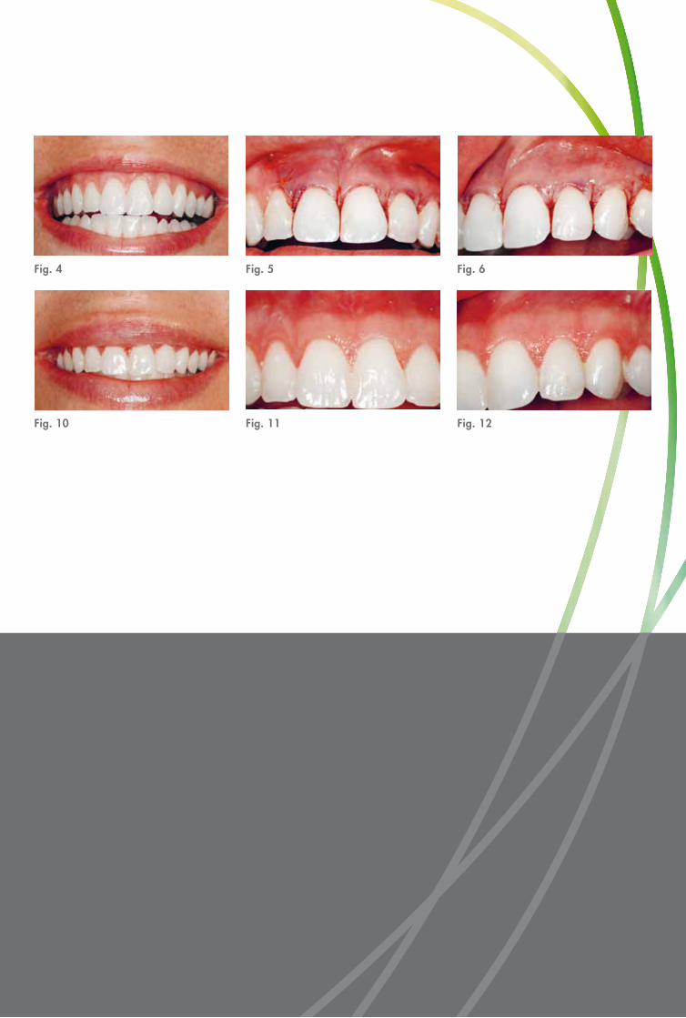
Fig. 5 Fig. 6
Fig. 10
Fig. 4
Fig. 11 Fig. 12

19 20
autHor
Dr. marK i. gutt, DmD1989 dMd from the university of Pennsylvania school of dental Medicine, Pa, usa • 1991 Specialty Degree in Periodontics from the University of Pennsylvania School of Dental Medicine, PA, USA • 1999 Certified Diplomate of the American Board of Periodon-tology • Active member of the American Academy of Periodontology and American Board of Periodontology • Practices Periodontics and Implantology in South Florida (since 1991) • Constantly raising the standard of periodontal and implant care

20
case 6 descriPtionA 24-year-old Caucasian female was referred for treatment of gingival
recession. Clinical evaluation revealed a medium lip line and facial gin-
gival recession of 3 mm, 2 mm, and 1 mm on teeth # 8, # 9, and # 10,
respectively (Fig. 1 and 2). The gingival biotype was thin and scalloped
with a triangular tooth presentation. There was a “black triangle” be-
tween # 9 and # 10 (Fig. 1). The etiology of the recession was difficult
to ascertain.
A treatment plan was developed that consisted of a coronally advanced
flap using a modified tunnel technique with an application of Straumann®
Emdogain. After buccal infiltration with 2 % lidocaine (1:100,000 epi),
a split thickness buccal mucoperiosteal flap was elevated following intra-
sulcular incisions from teeth # 7 through # 11 using a tunneling dissection
to elevate but not separate the papillae (Fig. 3 and 4). The teeth were
scaled and root planed followed by application of Straumann® Prefgel
(Fig. 5 and 6). The roots were lavaged with saline and Emdogain® ap-
plied to the root surfaces of teeth # 8, # 9 and # 10 (Fig. 7). The buccal
flap was then coronally advanced and adapted to the CEJs and sutured
with 6-0 Prolene® in an internal vertical mattress fashion (Fig. 8).
Post-op instruction was provided. The patient was seen after 2 weeks for
suture removal and has been evaluated for 7 months. The 7-month evalua-
tion revealed complete root coverage and elimination of the “black triangle”
with esthetically pleasing gingival and papillary appearance (Fig. 9).

Fig. 1
Fig. 9
Fig. 2 Fig. 3
Fig. 7 Fig. 8
fiGuresFig. 1: initial presentation indicating altered papillary architecture (# 9 and # 10) and gingival recession (# 8, # 9, # 10).
Fig. 2: initial presentation indicating de-gree of gingival recession.
Fig. 3: intrasulcular incision for flap el-evation.
Fig. 4: tunneling procedure performed to elevate but retain papillae.
Fig. 5: Mobility of flap after tunneling procedure.
Fig. 6: application of PrefGel® to the af-fected teeth.
Fig. 7: application of emdogain®.
Fig. 8: sutured coronally advanced flap.
Fig. 9: Healing at 7 months.

Fig. 5 Fig. 6Fig. 4

21 22
Dr. EunsEok EugEnE oh, DDsDDS, New York University, NY, USA • Post-graduate training in Periodontics, Stony Brook University School of Dental Medicine, NY, USA • Recipient of the Dr. Bernard E. Rudner Memorial Award for superior performance in comprehensive care and applied practice dentistry • Honors in Hospital Residency • Recipient of NYU Dental Scholarship from New York University • Author of numerous national and international publications • Private practitioner in Vienna, VA, USA
Dr. VInCEnT J. IACono, DMDDMD, Cum Laude, Harvard University, Cambridge, MA, USA • Certificate in Periodon-tology, Harvard University, Cambridge, MA, USA • BA New York University, NY, USA • Distinguished Service Professor and Chair of the Department of Periodontology and Implant Dentistry, Stony Brook University School of Dental Medicine, NY, USA • Director of the Advanced Education Program in Periodontics, and Associate Dean for Postgraduate Programs, Stony Brook University School of Dental Medicine, NY, USA • Past President of the American Academy of Periodontology (AAP) • Past President of the International Academy of Periodontology • President of the Academy of Osseointegration (AO) • Board member of the AAP Foundation • Chair of the Council of Hospitals and Advanced Education Programs (COHAEP) of ADEA
AUtHOR & CO-AUtHOR

22
case 7 descriPtionA 22-year-old, healthy, non-smoking female presented with 3 mm buccal
recessions on the upper canines. There was also a thin attached gingiva
with marked extension through the labial frenula. The periodontal tissue
was healthy and oral hygiene was well-controlled.
The left upper canine was treated with a coronally advanced flap with
Straumann® Emdogain plus subepithelial connective tissue. First, Emdogain®
was applied to the root surface followed by the connective tissue graft.
The last layer was the remaining Emdogain® to improve wound healing.
Successful root coverage was the result of the combination of the regen-
erative potential of Emdogain®, an improvement in wound healing and
the gain of attached gingiva with a subepithelial connective graft.

Fig. 1
Fig. 8
Fig. 2 Fig. 3
Fig. 7 Fig. 9
Fig. 14 Fig. 15 Fig. 13
fiGuresFig. 1: the upper left and right canine with 3 mm recessions.
Fig. 2: the left side. the canine with a 3 mm recession.
Fig. 3: the left side. the canine with a 3 mm recession (close-up).
Fig. 4: a split flap is raised after cleaning the tooth surfaces.
Fig. 5: entepithelisation of the first (12/11) papilla. the next follows.
Fig. 6: single incision technique on the palatal donor site.
Fig. 7: the connective tissue graft.
Fig. 8: the sbt is placed and fixed after conditioning and application of emdogain®.
Fig. 9: the remaining emdogain® is ap-plied.
Fig. 10: Wound closure utilizing 6.0 suture material.
Fig. 11: the post-operative situation after 7 days.
Fig. 12: the post-operative situation after 7 days.

Fig. 4
Fig. 11
Fig. 5 Fig. 6
Fig. 10 Fig. 12
Fig. 16
Fig. 13: the post-operative situation after 11 days. Fig. 14: the post-operative situation after 20 days.
Fig. 15: the post-operative situation after 2 months.
Fig. 16: the recession is completely covered and there is a wide zone of keratinizied gingiva.

23 24
autHor
Dr. meD. Dent. anDreaS hoFmann, m.Sc.1999 Degree in Dental Medicine, University of Leipzig, Germany • 2002 Doctorate • 2007 Master of Science in Periodontology following post-graduate studies at Donau university, Krems, Germany. Master thesis: “Möglichkeiten/Varianten der rezessions-deckungen und Regeneration des Zahnhalteapparates“ • 2008/2009 Master classes in Periodontology (IPI) • 2009 Master classes in Implantology (IPI)

24
case 8 descriPtionA 51-year-old female was referred for the placement of 3 implant fixtures
for a fixed partial denture on the third sextant. She complained about
general hypersensitivity and about her smile, particularly her long teeth.
The goal of the treatment was to reduce the length of the teeth by using
a tunnel connective tissue graft, including the application of Straumann®
Emdogain on the exposed root surfaces and the placement of three endo-
osseous root form dental implants on # 24, # 25 and # 26 in one sitting.
A tunnel connective tissue graft for the root coverage of multiple adjacent
recessions was performed. A partial thickness bilaminar bed was created
by intrasulcular incisions with a sclerotome micro-knife on teeth # 13,
# 12, # 11, # 21, # 22 and # 23. Enough relaxation of the tissue was
achieved that would allow enough space for both the introduction of the
harvested palatal tissue as well as the coronal advancement of the supra
laminar flap upon suturing. Emdogain® proteins were utilized to condition
the denuded roots as recommended by manufacturer.
Two pulling sutures were used from the mesial and distal aspects of the
created bilaminar tunnel in order to introduce the graft through the largest
recession. Conventional post-op care was prescribed. High patient sat-
isfaction has been reported so far, both from an esthetic and functional
(total reduction of hypersensitivity) point of view.
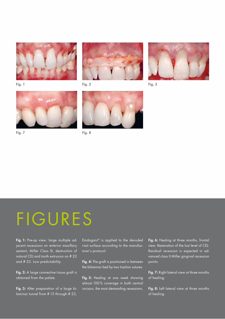
Fig. 1 Fig. 2 Fig. 3
Fig. 7 Fig. 8
fiGuresFig. 1: Pre-op view: large multiple ad-jacent recessions on anterior maxillary sextant, Miller class iii, destruction of natural cej and tooth extrusion on # 22 and # 23. low predictability.
Fig. 2: a large connective tissue graft is obtained from the palate.
Fig. 3: after preparation of a large bi-laminar tunnel from # 13 through # 23,
emdogain® is applied to the denuded root surface according to the manufac-turer’s protocol.
Fig. 4: the graft is positioned in between the bilaminar bed by two traction sutures.
Fig. 5: Healing at one week showing almost 100 % coverage in both central incisors, the most demanding recessions.
Fig. 6: Healing at three months, frontal view. restoration of the lost level of cej. residual recession is expected in ad-vanced class ii Miller gingival recession points.
Fig. 7: right lateral view at three months of healing.
Fig. 8: left lateral view at three months of healing.
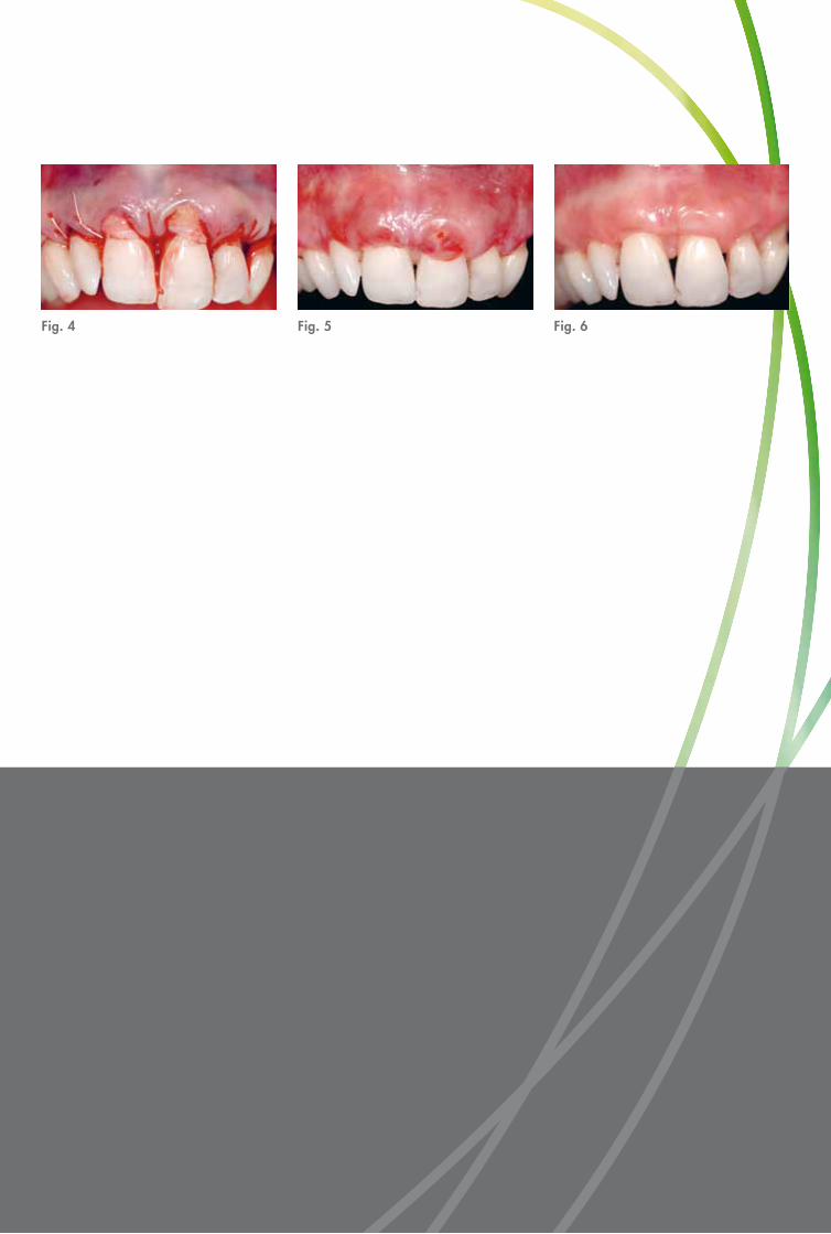
Fig. 5 Fig. 6Fig. 4

25 26
autHor
Dr. ion ZaBalegui, m.D.1982 M.D., Facultad de Medicina, Universidad del País Vasco, Spain • 1984 Speciality in Estomathology, Facultad de Medicina, Universidad del País Vasco, Spain • 1987 Certificate in Periodontology, University of Southern California, CA, USA • Former visiting professor, Odontología Integrada, Universidad del País Vasco, Spain • Member of the Sociedad española de Periodoncia, the american academy of Periodontology, the european academy of osseointegration, the sociedad española de Prótesis estomatológica, the academy of osseointegration, the Pierre fauchard academy and the international college of dentistry • Private practice in Bilbao, Spain • Visiting professor, Post-graduate Program in Peri-odontology, ucM, Madrid, spain

26
case 9 descriPtionA 27-year-old Caucasian female presented at the dental clinic for sensi-
tive mandibular incisors. She had been smoking for 7 years and had oral
piercings, a tongue ring and two lip rings (labrets), which had contributed
to the soft and hard tissue damage to teeth # 24 and # 25. The clinical
attachment loss of the mid-facial surfaces of # 24 and # 25 was 9 mm
and 11 mm, respectively, with recession of 6 mm and 8 mm, respectively,
and a class I mobility of both teeth. Non-surgical therapy had a minimal
affect on this area.
A surgical treatment plan was developed to include a connective tissue
graft with the addition of Straumann® Emdogain for a better potential
regenerative outcome on both # 24 and # 25. The initial healing at two
weeks was less than adequate with the suspicion of continued smok-
ing and less than adequate oral hygiene. However, the tissue started
showing continued improvement at the six-week follow-up, and at eleven
months the area had improved dramatically with minimal probing depths
(< 3 mm) and complete coverage of # 25 as well as a 2 mm residual
defect of # 24.
The impressive feature of this outcome was that regardless of the risk
factor of continued smoking, as confirmed by the patient, and less than
adequate hygiene, the tissues responded and improved to a healthy state
with much improved esthetics.

Fig. 1
Fig. 9
Fig. 2 Fig. 3
Fig. 7 Fig. 8
fiGuresFig. 1: depiction of initial situation.
Fig. 2: Pre-op radiograph of teeth # 24 and # 25.
Fig. 3: initial flap.
Fig. 4: Post-debridement.
Fig. 5: application of PrefGel® (edta).
Fig. 6: application of emdogain®.
Fig. 7: ct of graft suture.
Fig. 8: depiction of sutured flap.
Fig. 9: situation at two-week follow-up.
Fig. 10: situation at six-week follow-up. Fig. 11: final situation at eleven-month follow-up.
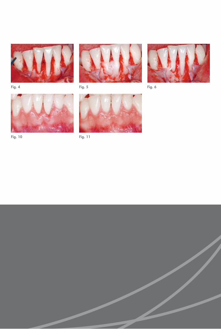
Fig. 5
Fig. 11
Fig. 6Fig. 4
Fig. 10

27 28
autHor
Paul g. luePKe, DDS, mS1996 Master’s degree in Periodontics, university of texas at san antonio Health science Center, TX, USA • 1997 Diplomate of the American Board of Periodontology • 2008 assistant Professor of surgical services Periodontics division, Marquette university school of Dentistry in Milwaukee, WI, USA • 2009 Interim Department Chair of Surgical Sciences at Marquette university school of dentistry in Milwaukee, Wi, usa

28
case 10 descriPtionThe referred 23-year-old female patient was presenting with multiple gingival
recessions (teeth # 11 to # 16). The prominent canine showed a Miller
Class III recession, the other teeth presented with Miller Class I recessions.
The treatment procedure began with a thorough cleaning and scaling of
the exposed root surfaces with hand and sonic instruments and was fol-
lowed by a split thickness flap preparation of a Zucchelli-Style flap (without
vertical releasing incisions). A dissection into the vestibular mucosa allowed
for further mobilisation.
Straumann® Prefgel (EDTA) was applied for two minutes on the root sur-
face. Subsequently, the surgical area was rinsed with sterile saline and
Straumann® Emdogain was applied to the root surfaces. Connective tissue
graft (CTG) was harvested from the palate with the single incision tech-
nique. The graft was then split. The CTG was fixed on the root surface
and the flap coronally positioned and fixed with sling sutures.
Mechanical tooth cleaning in the surgical area was avoided during the
first 4 weeks and a chlorhexidine solution was prescribed. Sutures were
removed 10 days after surgery.

Fig. 1
Fig. 9
Fig. 13
Fig. 2
Fig. 14
Fig. 3
Fig. 7 Fig. 8
Fig. 15
fiGuresFig. 1: Multiple recessions of the right upper jaw. the buccally positioned ca-nine shows a Miller class iii recession.
Fig. 2: outline of a Zucchelli-type flap. a split thickness preparation is chosen to facilitate graft survival.
Fig. 3: de-epithelisation of the papilla areas.
Fig. 4: finished flap preparation. no vertical releasing incisions are necces-sary for coronal positioning.
Fig. 5: application of PrefGel® for two minutes on the root surfaces.
Fig. 6: application of emdogain®.
Fig. 7: Preparation of the donor site with the single incision technique.
Fig. 8: Harvesting of the connective tissue graft.
Fig. 9: Harvesting of the connective tissue graft.
Fig. 10: the graft is bisected to allow covering of the complete area.
Fig. 11: the graft is bisected to allow covering of the complete area.
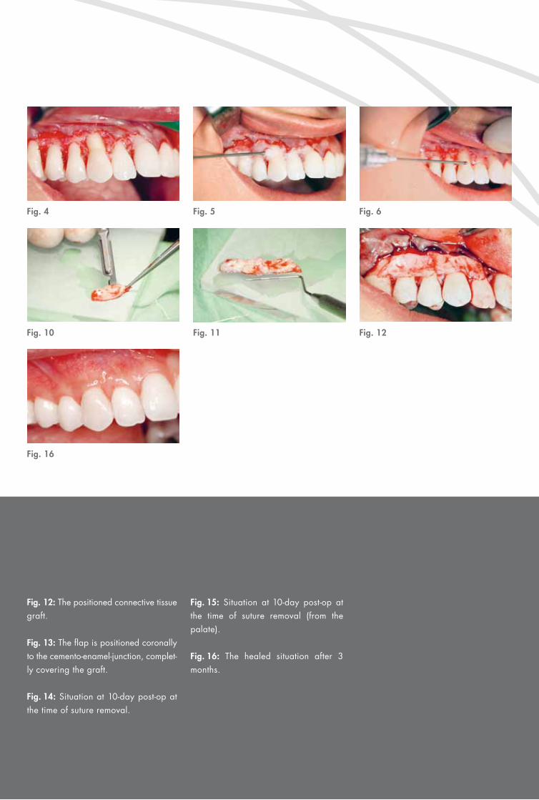
Fig. 5 Fig. 6
Fig. 10
Fig. 4
Fig. 11 Fig. 12
Fig. 16
Fig. 12: the positioned connective tissue graft.
Fig. 13: the flap is positioned coronally to the cemento-enamel-junction, complet-ly covering the graft.
Fig. 14: situation at 10-day post-op at the time of suture removal.
Fig. 15: situation at 10-day post-op at the time of suture removal (from the palate).
Fig. 16: the healed situation after 3 months.

29 30
Dr. BJørn greVen 1999 Graduation Düsseldorf University, Germany • General dentist in the German Navy • 2001–2006 Department for Periodontology, Forces Hospital Hamburg, Germany • 2007–2008 Forces Hospital Ulm, Germany • 2007 Best Practitioner Award, German Society of Periodontology (DGP) • 2009 Postgraduate Program M.Sc. in Oral Implantology • Several lectures and publications • Private practice in Hamburg, Germany
Dr. BernD heinZ1973 Private practice in Hamburg, Germany • Former member of the executive board of the German Society of Periodontology (DGP) • 1996 Award of the German Society of Periodontology (DGP) • 1997Specialist for periodontology (DGP/EFP) • 1999 Recipi-ent of the Corsodyl Innovation Award • 2005 Recipient of the Meridol Award • 2009 Honorary award of the German Society of Periodontology (DGP) • Lecturer at several postgraduate curricula and master study programs • Multiple scientific studies, publica-tions, lectures and seminars
autHor & co-autHor

30
case 11descriPtionThe 45-year-old female presented with localized recession areas in
the maxillary esthetic zone at teeth 1.1 and 2.1. These recession areas
ranged from 4–5 mm. The patient’s chief concern was that these areas
were sensitive and esthetically compromised due to the root exposure.
The treatment plan recommendation was to treat these areas for recession
coverage using a combination of a coronal rotational advanced flap
technique and with Straumann® Emdogain, which was applied to the root
surfaces of teeth 1.1 and 2.1.

Fig. 1 Fig. 2 Fig. 3
Fig. 7 Fig. 8
fiGuresFig. 1: Pre-surgical view of tooth 1.1 and 2.1.
Fig. 2: depiction of initial surgical design and incision.
Fig. 3: flap elevation and application of PrefGel® for root preparation.
Fig. 4: root debridement and irrigation.
Fig. 5: application of emdogain®.
Fig. 6: flap closure and stabilization.
Fig. 7: Post-surgical view at 3 weeks.
Fig. 8: Post-surgical view at 1 year.
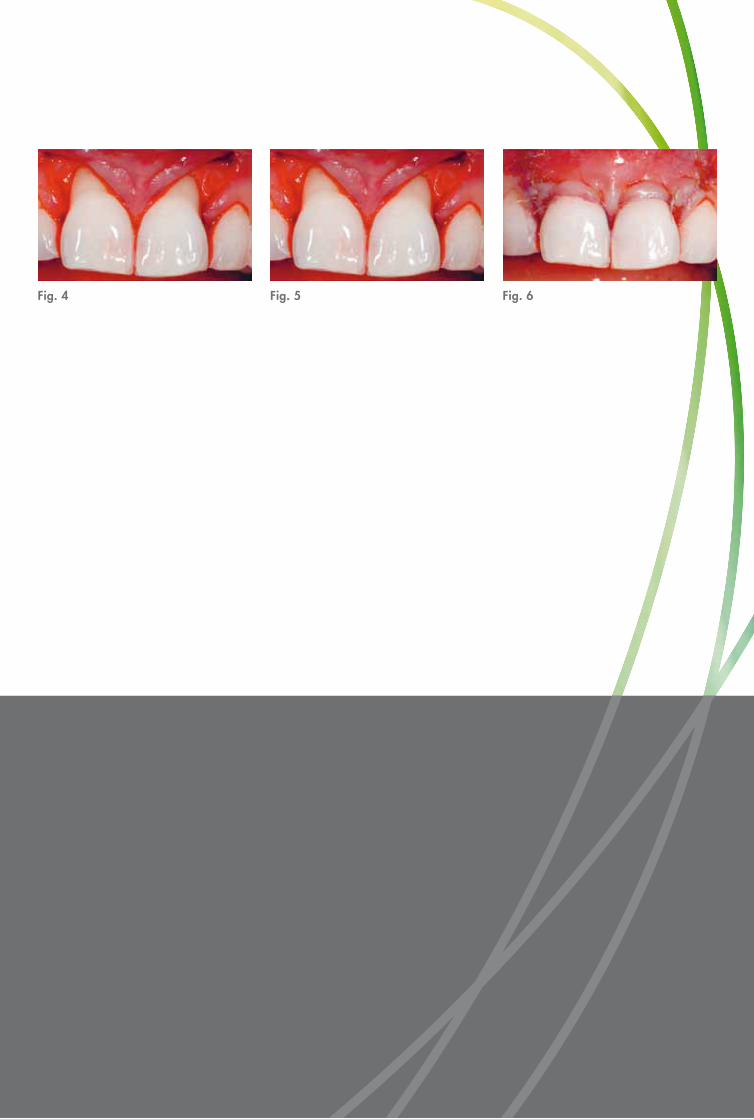
Fig. 5 Fig. 6Fig. 4

31
autHor
Dr. ira Paul Sy, DDS, mS, DiP. PerioDonticSDDS, Case Western Reserve University, Cleveland, OH, USA • MS in Oral Biology, specialty certification training in implants and Periodontics, university of north carolina at Chapel Hill, NC, USA • Fellowship in Implant Surgical and Prosthetic Therapy at the University of Berne, Switzerland • Diplomate of the American Board of Periodontol-ogy • Member of the Academy of Periodontology, Academy of Osseointegration, and International Congress of Oral Implantology • Faculty member of the University of North Carolina at Chapel Hill and the University of British Columbia • Clinical Assistant Professor in Periodontics, Case Western Reserve University, Cleveland, OH, USA • Visiting Professor of Periodontics, University of Witten, Germany • Former Chief of Periodontics at Perkins dental clinic and assistant mentor in Periodontics for the advanced General dentistry Program at Fort Hood, Texas • Former Director of the Graduate Periodontology and Implantology Residency Program at the University of British Columbia Canada • Faculty member of the University of British Columbia, Department of Orthodontics • Founder and Director of the Vancouver Institute of Osseointegration (VIOSS) • Consultant- Veteran’s Hospital in Northern California for Surgical & Prosthetic Implant Therapy • Member of the Peers north america (Platform for exchange, education, research, and science) • Private practice in Richmond and Vancouver, BC, Canada

International HeadquartersInstitut Straumann AG Peter Merian-Weg 12CH-4002 Basel, SwitzerlandPhone +41 (0)61 965 11 11Fax +41 (0)61 965 11 01
31
Stra
uman
n pr
oduc
ts ar
e C
E m
arke
d
10/
11
152.
262/
en
BA
2101
1
© Institut Straumann AG, 2011. All rights reserved. Straumann® and/or other trademarks and logos from Straumann® mentioned herein are the trademarks or registered trademarks of Straumann Holding AG and/or its affiliates. All rights reserved.
References1 Lindhe J, Karring T, Lang NP. Clinical periodontology and implant dentistry, 4th edition, 579 -586. 2 Miller
PD. A classification of marginal tissue recession. Int J Periodontol Rest Dent. 1985;5,8 -13. 3 Journal of Clinical
Periodontology, volume 35, supplement 8. 4 Cairo F, Pagliaro U, Nieri M. Treatment of gingival recession with
coronally advanced flap procedures: a systematic review. J Clin Periodontol. 2008;35 (Suppl. 8):136 -162. 5 Zucchelli G, Mele M, Mazzotti C, Marzadori M, Montebugnoli L, and De Sanctis M. Coronally advanced
flap with and without vertical releasing incisions for the treatment of multiple gingival recessions: A comparative
controlled randomized clinical trial. Journal of Periodontology. July 2009, Vol. 80, No. 7, 1083-1094.


![Enamel matrix derivative (Emdogain(R)) for periodontal ... · [Intervention Review] Enamel matrix derivative (Emdogain®) for periodontal tissue regeneration in intrabony defects](https://static.fdocuments.us/doc/165x107/5f552cf7423b6b423a7f833d/enamel-matrix-derivative-emdogainr-for-periodontal-intervention-review.jpg)


